BR112016024569B1 - EX VIVO METHOD FOR REMOVING BACTERIA FROM A SPECIMEN TAKEN FROM AN INDIVIDUAL WHO IS SUSPECTED TO BE INFECTED WITH THE SAME - Google Patents
EX VIVO METHOD FOR REMOVING BACTERIA FROM A SPECIMEN TAKEN FROM AN INDIVIDUAL WHO IS SUSPECTED TO BE INFECTED WITH THE SAME Download PDFInfo
- Publication number
- BR112016024569B1 BR112016024569B1 BR112016024569-5A BR112016024569A BR112016024569B1 BR 112016024569 B1 BR112016024569 B1 BR 112016024569B1 BR 112016024569 A BR112016024569 A BR 112016024569A BR 112016024569 B1 BR112016024569 B1 BR 112016024569B1
- Authority
- BR
- Brazil
- Prior art keywords
- bacteria
- blood
- sample
- adsorption
- heparin
- Prior art date
Links
Images

Classifications
-
- A—HUMAN NECESSITIES
- A61—MEDICAL OR VETERINARY SCIENCE; HYGIENE
- A61M—DEVICES FOR INTRODUCING MEDIA INTO, OR ONTO, THE BODY; DEVICES FOR TRANSDUCING BODY MEDIA OR FOR TAKING MEDIA FROM THE BODY; DEVICES FOR PRODUCING OR ENDING SLEEP OR STUPOR
- A61M1/00—Suction or pumping devices for medical purposes; Devices for carrying-off, for treatment of, or for carrying-over, body-liquids; Drainage systems
- A61M1/36—Other treatment of blood in a by-pass of the natural circulatory system, e.g. temperature adaptation, irradiation ; Extra-corporeal blood circuits
- A61M1/3679—Other treatment of blood in a by-pass of the natural circulatory system, e.g. temperature adaptation, irradiation ; Extra-corporeal blood circuits by absorption
-
- A—HUMAN NECESSITIES
- A61—MEDICAL OR VETERINARY SCIENCE; HYGIENE
- A61M—DEVICES FOR INTRODUCING MEDIA INTO, OR ONTO, THE BODY; DEVICES FOR TRANSDUCING BODY MEDIA OR FOR TAKING MEDIA FROM THE BODY; DEVICES FOR PRODUCING OR ENDING SLEEP OR STUPOR
- A61M1/00—Suction or pumping devices for medical purposes; Devices for carrying-off, for treatment of, or for carrying-over, body-liquids; Drainage systems
- A61M1/34—Filtering material out of the blood by passing it through a membrane, i.e. hemofiltration or diafiltration
- A61M1/3472—Filtering material out of the blood by passing it through a membrane, i.e. hemofiltration or diafiltration with treatment of the filtrate
- A61M1/3486—Biological, chemical treatment, e.g. chemical precipitation; treatment by absorbents
-
- A—HUMAN NECESSITIES
- A61—MEDICAL OR VETERINARY SCIENCE; HYGIENE
- A61M—DEVICES FOR INTRODUCING MEDIA INTO, OR ONTO, THE BODY; DEVICES FOR TRANSDUCING BODY MEDIA OR FOR TAKING MEDIA FROM THE BODY; DEVICES FOR PRODUCING OR ENDING SLEEP OR STUPOR
- A61M1/00—Suction or pumping devices for medical purposes; Devices for carrying-off, for treatment of, or for carrying-over, body-liquids; Drainage systems
- A61M1/36—Other treatment of blood in a by-pass of the natural circulatory system, e.g. temperature adaptation, irradiation ; Extra-corporeal blood circuits
- A61M1/3692—Washing or rinsing blood or blood constituents
-
- B—PERFORMING OPERATIONS; TRANSPORTING
- B01—PHYSICAL OR CHEMICAL PROCESSES OR APPARATUS IN GENERAL
- B01J—CHEMICAL OR PHYSICAL PROCESSES, e.g. CATALYSIS OR COLLOID CHEMISTRY; THEIR RELEVANT APPARATUS
- B01J20/00—Solid sorbent compositions or filter aid compositions; Sorbents for chromatography; Processes for preparing, regenerating or reactivating thereof
- B01J20/22—Solid sorbent compositions or filter aid compositions; Sorbents for chromatography; Processes for preparing, regenerating or reactivating thereof comprising organic material
- B01J20/26—Synthetic macromolecular compounds
-
- B—PERFORMING OPERATIONS; TRANSPORTING
- B01—PHYSICAL OR CHEMICAL PROCESSES OR APPARATUS IN GENERAL
- B01J—CHEMICAL OR PHYSICAL PROCESSES, e.g. CATALYSIS OR COLLOID CHEMISTRY; THEIR RELEVANT APPARATUS
- B01J20/00—Solid sorbent compositions or filter aid compositions; Sorbents for chromatography; Processes for preparing, regenerating or reactivating thereof
- B01J20/28—Solid sorbent compositions or filter aid compositions; Sorbents for chromatography; Processes for preparing, regenerating or reactivating thereof characterised by their form or physical properties
- B01J20/28002—Solid sorbent compositions or filter aid compositions; Sorbents for chromatography; Processes for preparing, regenerating or reactivating thereof characterised by their form or physical properties characterised by their physical properties
- B01J20/28004—Sorbent size or size distribution, e.g. particle size
-
- B—PERFORMING OPERATIONS; TRANSPORTING
- B01—PHYSICAL OR CHEMICAL PROCESSES OR APPARATUS IN GENERAL
- B01J—CHEMICAL OR PHYSICAL PROCESSES, e.g. CATALYSIS OR COLLOID CHEMISTRY; THEIR RELEVANT APPARATUS
- B01J20/00—Solid sorbent compositions or filter aid compositions; Sorbents for chromatography; Processes for preparing, regenerating or reactivating thereof
- B01J20/28—Solid sorbent compositions or filter aid compositions; Sorbents for chromatography; Processes for preparing, regenerating or reactivating thereof characterised by their form or physical properties
- B01J20/28014—Solid sorbent compositions or filter aid compositions; Sorbents for chromatography; Processes for preparing, regenerating or reactivating thereof characterised by their form or physical properties characterised by their form
- B01J20/28016—Particle form
-
- B—PERFORMING OPERATIONS; TRANSPORTING
- B01—PHYSICAL OR CHEMICAL PROCESSES OR APPARATUS IN GENERAL
- B01J—CHEMICAL OR PHYSICAL PROCESSES, e.g. CATALYSIS OR COLLOID CHEMISTRY; THEIR RELEVANT APPARATUS
- B01J20/00—Solid sorbent compositions or filter aid compositions; Sorbents for chromatography; Processes for preparing, regenerating or reactivating thereof
- B01J20/28—Solid sorbent compositions or filter aid compositions; Sorbents for chromatography; Processes for preparing, regenerating or reactivating thereof characterised by their form or physical properties
- B01J20/28014—Solid sorbent compositions or filter aid compositions; Sorbents for chromatography; Processes for preparing, regenerating or reactivating thereof characterised by their form or physical properties characterised by their form
- B01J20/28023—Fibres or filaments
-
- B—PERFORMING OPERATIONS; TRANSPORTING
- B01—PHYSICAL OR CHEMICAL PROCESSES OR APPARATUS IN GENERAL
- B01J—CHEMICAL OR PHYSICAL PROCESSES, e.g. CATALYSIS OR COLLOID CHEMISTRY; THEIR RELEVANT APPARATUS
- B01J20/00—Solid sorbent compositions or filter aid compositions; Sorbents for chromatography; Processes for preparing, regenerating or reactivating thereof
- B01J20/30—Processes for preparing, regenerating, or reactivating
- B01J20/32—Impregnating or coating ; Solid sorbent compositions obtained from processes involving impregnating or coating
- B01J20/3202—Impregnating or coating ; Solid sorbent compositions obtained from processes involving impregnating or coating characterised by the carrier, support or substrate used for impregnation or coating
- B01J20/3204—Inorganic carriers, supports or substrates
-
- B—PERFORMING OPERATIONS; TRANSPORTING
- B01—PHYSICAL OR CHEMICAL PROCESSES OR APPARATUS IN GENERAL
- B01J—CHEMICAL OR PHYSICAL PROCESSES, e.g. CATALYSIS OR COLLOID CHEMISTRY; THEIR RELEVANT APPARATUS
- B01J20/00—Solid sorbent compositions or filter aid compositions; Sorbents for chromatography; Processes for preparing, regenerating or reactivating thereof
- B01J20/30—Processes for preparing, regenerating, or reactivating
- B01J20/32—Impregnating or coating ; Solid sorbent compositions obtained from processes involving impregnating or coating
- B01J20/3202—Impregnating or coating ; Solid sorbent compositions obtained from processes involving impregnating or coating characterised by the carrier, support or substrate used for impregnation or coating
- B01J20/3206—Organic carriers, supports or substrates
- B01J20/3208—Polymeric carriers, supports or substrates
- B01J20/321—Polymeric carriers, supports or substrates consisting of a polymer obtained by reactions involving only carbon to carbon unsaturated bonds
-
- B—PERFORMING OPERATIONS; TRANSPORTING
- B01—PHYSICAL OR CHEMICAL PROCESSES OR APPARATUS IN GENERAL
- B01J—CHEMICAL OR PHYSICAL PROCESSES, e.g. CATALYSIS OR COLLOID CHEMISTRY; THEIR RELEVANT APPARATUS
- B01J20/00—Solid sorbent compositions or filter aid compositions; Sorbents for chromatography; Processes for preparing, regenerating or reactivating thereof
- B01J20/30—Processes for preparing, regenerating, or reactivating
- B01J20/32—Impregnating or coating ; Solid sorbent compositions obtained from processes involving impregnating or coating
- B01J20/3202—Impregnating or coating ; Solid sorbent compositions obtained from processes involving impregnating or coating characterised by the carrier, support or substrate used for impregnation or coating
- B01J20/3206—Organic carriers, supports or substrates
- B01J20/3208—Polymeric carriers, supports or substrates
- B01J20/3212—Polymeric carriers, supports or substrates consisting of a polymer obtained by reactions otherwise than involving only carbon to carbon unsaturated bonds
-
- B—PERFORMING OPERATIONS; TRANSPORTING
- B01—PHYSICAL OR CHEMICAL PROCESSES OR APPARATUS IN GENERAL
- B01J—CHEMICAL OR PHYSICAL PROCESSES, e.g. CATALYSIS OR COLLOID CHEMISTRY; THEIR RELEVANT APPARATUS
- B01J20/00—Solid sorbent compositions or filter aid compositions; Sorbents for chromatography; Processes for preparing, regenerating or reactivating thereof
- B01J20/30—Processes for preparing, regenerating, or reactivating
- B01J20/32—Impregnating or coating ; Solid sorbent compositions obtained from processes involving impregnating or coating
- B01J20/3231—Impregnating or coating ; Solid sorbent compositions obtained from processes involving impregnating or coating characterised by the coating or impregnating layer
- B01J20/3242—Layers with a functional group, e.g. an affinity material, a ligand, a reactant or a complexing group
- B01J20/3268—Macromolecular compounds
- B01J20/3272—Polymers obtained by reactions otherwise than involving only carbon to carbon unsaturated bonds
- B01J20/3274—Proteins, nucleic acids, polysaccharides, antibodies or antigens
-
- B—PERFORMING OPERATIONS; TRANSPORTING
- B01—PHYSICAL OR CHEMICAL PROCESSES OR APPARATUS IN GENERAL
- B01J—CHEMICAL OR PHYSICAL PROCESSES, e.g. CATALYSIS OR COLLOID CHEMISTRY; THEIR RELEVANT APPARATUS
- B01J20/00—Solid sorbent compositions or filter aid compositions; Sorbents for chromatography; Processes for preparing, regenerating or reactivating thereof
- B01J20/30—Processes for preparing, regenerating, or reactivating
- B01J20/32—Impregnating or coating ; Solid sorbent compositions obtained from processes involving impregnating or coating
- B01J20/3291—Characterised by the shape of the carrier, the coating or the obtained coated product
- B01J20/3293—Coatings on a core, the core being particle or fiber shaped, e.g. encapsulated particles, coated fibers
-
- A—HUMAN NECESSITIES
- A61—MEDICAL OR VETERINARY SCIENCE; HYGIENE
- A61M—DEVICES FOR INTRODUCING MEDIA INTO, OR ONTO, THE BODY; DEVICES FOR TRANSDUCING BODY MEDIA OR FOR TAKING MEDIA FROM THE BODY; DEVICES FOR PRODUCING OR ENDING SLEEP OR STUPOR
- A61M2202/00—Special media to be introduced, removed or treated
- A61M2202/0057—Special media to be introduced, removed or treated retained by adsorption
-
- A—HUMAN NECESSITIES
- A61—MEDICAL OR VETERINARY SCIENCE; HYGIENE
- A61M—DEVICES FOR INTRODUCING MEDIA INTO, OR ONTO, THE BODY; DEVICES FOR TRANSDUCING BODY MEDIA OR FOR TAKING MEDIA FROM THE BODY; DEVICES FOR PRODUCING OR ENDING SLEEP OR STUPOR
- A61M2202/00—Special media to be introduced, removed or treated
- A61M2202/04—Liquids
- A61M2202/0413—Blood
-
- A—HUMAN NECESSITIES
- A61—MEDICAL OR VETERINARY SCIENCE; HYGIENE
- A61M—DEVICES FOR INTRODUCING MEDIA INTO, OR ONTO, THE BODY; DEVICES FOR TRANSDUCING BODY MEDIA OR FOR TAKING MEDIA FROM THE BODY; DEVICES FOR PRODUCING OR ENDING SLEEP OR STUPOR
- A61M2202/00—Special media to be introduced, removed or treated
- A61M2202/20—Pathogenic agents
- A61M2202/203—Bacteria
-
- B—PERFORMING OPERATIONS; TRANSPORTING
- B01—PHYSICAL OR CHEMICAL PROCESSES OR APPARATUS IN GENERAL
- B01J—CHEMICAL OR PHYSICAL PROCESSES, e.g. CATALYSIS OR COLLOID CHEMISTRY; THEIR RELEVANT APPARATUS
- B01J2220/00—Aspects relating to sorbent materials
- B01J2220/50—Aspects relating to the use of sorbent or filter aid materials
- B01J2220/62—In a cartridge
Abstract
MÉTODO EX VIVO PARA REMOVER BACTÉRIAS DE UMA AMOSTRA RETIRADA DE UM INDIVÍDUO QUE É SUSPEITO DE ESTAR INFECTADO COM A MESMA. A presente invenção refere-se a métodos para a remoção de uma quantidade significativa de bactérias (por exemplo, bactérias gram-negativas e bactérias gram-positivas, incluindo as bactérias com baixa ou nenhuma afinidade com o sulfato de heparano) do sangue integral, soro ou plasma, utilizando um meio de adsorção. O método pode ser usado em tratamentos extracorpóreos envolvendo altas taxas de fluxo volumétrico e altas taxas de fluxo linear.EX VIVO METHOD FOR REMOVING BACTERIA FROM A SPECIMEN TAKEN FROM AN INDIVIDUAL WHO IS SUSPECTED TO BE INFECTED WITH THE SAME. The present invention relates to methods for removing a significant amount of bacteria (e.g., gram-negative bacteria and gram-positive bacteria, including bacteria with low or no affinity for heparan sulfate) from whole blood, serum or plasma, using an adsorption medium. The method can be used in extracorporeal treatments involving high volumetric flow rates and high linear flow rates.
Description
[001] O presente pedido reivindica prioridade apresentam a Pedido de Patente Provisório US No. 61/984.013, depositado em 24 de abril de 2014, cujos ensinamentos são aqui incorporados por referência na sua totalidade para todos os propósitos.[001] The present application claims priority feature the US Provisional Patent Application No. 61/984013, filed April 24, 2014, the teachings of which are hereby incorporated by reference in their entirety for all purposes.
[002] A infecção da corrente sanguínea, ou bacteremia, é um grande desafio na Unidade de Terapia Intensiva (UTI). A bacteremia pode levar rapidamente a choque séptico, meningite, endocardite, osteomielite e outras complicações metastáticas. Staphylococcus aureus, P. aeruginosa e Enterobacteriaceae são as bactérias mais comuns responsáveis por bacteremia e infecções nosocomiais. A gravidade dos resultados para os pacientes com bacteremia está correlacionada tanto com a carga bacteriana quanto com a duração da bacteremia. Por exemplo, um estudo quantitativo RT-PCR de pacientes com bacteremia por E. coli e S. aureus mostrou que quando o número de rDNA aumentou para mais de 1.238 cópias/mL, a mortalidade aumentou de 14,3% para 42,9% e o choque séptico aumentou de 31,4% para 85,7%. Constatou-se também que uma alta concentração no sangue de N. meningitides está correlacionada com a hospitalização prolongada, a integridade física ou perda de tecido, a necessidade de diálise e a mortalidade de pacientes. Outro estudo mostrou que a gravidade da pneumonia pneumocócica está correlacionada com a carga bacteriana no sangue: a mortalidade de pacientes com mais de 1.000 cópias do DNA de S. pneumoniae/mL de sangue foi 25,9% contra 6,1% para os pacientes que exibem menos do que 1000 cópias/mL. Em outro estudo, uma hemocultura positiva de acompanhamento entre 48 e 96 horas após o diagnóstico inicial revelou-se o mais forte preditor de bacteremia por S. aureus complicada. Para agravar a dificuldade de tratamento de bacteremia eficaz está a administração frequentemente adiada de terapia antibiótica adequada. Para cada hora de atraso no tratamento, o risco de mortalidade aumenta mais de 7%.[002] Bloodstream infection, or bacteremia, is a major challenge in the Intensive Care Unit (ICU). Bacteremia can rapidly lead to septic shock, meningitis, endocarditis, osteomyelitis, and other metastatic complications. Staphylococcus aureus, P. aeruginosa and Enterobacteriaceae are the most common bacteria responsible for bacteremia and nosocomial infections. The severity of outcomes for patients with bacteremia is correlated with both bacterial load and duration of bacteremia. For example, a quantitative RT-PCR study of patients with E. coli and S. aureus bacteremia showed that when the rDNA number increased to greater than 1238 copies/mL, mortality increased from 14.3% to 42.9% and septic shock increased from 31.4% to 85.7%. It was also found that a high blood concentration of N. meningitides is correlated with prolonged hospitalization, physical integrity or tissue loss, need for dialysis and patient mortality. Another study showed that the severity of pneumococcal pneumonia is correlated with the bacterial load in the blood: the mortality of patients with more than 1,000 copies of S. pneumoniae DNA/mL of blood was 25.9% against 6.1% for patients that exhibit less than 1000 copies/mL. In another study, a positive follow-up blood culture between 48 and 96 hours after initial diagnosis was found to be the strongest predictor of complicated S. aureus bacteremia. Compounding the difficulty of treating bacteremia effectively is the often delayed administration of appropriate antibiotic therapy. For every hour of delay in treatment, the risk of mortality increases by more than 7%.
[003] A estratégia convencional para combater as infecções bacterianas é administrar fármacos ativos que especificamente matam as bactérias, minimizando os danos ao tecido hospedeiro. Este é um grande desafio à medida que alguns dos antibióticos mais eficazes atualmente disponíveis são bastante tóxicos. Por exemplo, a vancomicina é nefrotóxica, e em breve poderá ser contraindicada para pacientes submetidos à oxigenação extracorpórea. Mesmo que novos antibióticos sejam desenvolvidos com sucesso para abordar a atual resistência a fármacos, novas superbactérias continuarão a surgir. Claramente, novas estratégias para combater a infecção são necessárias, em adição à descoberta de fármacos.[003] The conventional strategy to combat bacterial infections is to administer active drugs that specifically kill the bacteria, minimizing damage to the host tissue. This is a huge challenge as some of the most effective antibiotics currently available are quite toxic. For example, vancomycin is nephrotoxic, and may soon be contraindicated for patients undergoing extracorporeal oxygenation. Even if new antibiotics are successfully developed to address current drug resistance, new superbugs will continue to emerge. Clearly, new strategies to combat the infection are needed, in addition to drug discovery.
[004] Os patógenos resistentes a fármacos são uma ameaça crescente para o sistema de saúde. O CDC foi recentemente alertado do surgimento de Enterobacteriaceae resistente a carbapenem (CRE; “superbactérias”). A taxa de mortalidade para bacteremia por CRE pode ser de no máximo 50%. Resistência de CREs até mesmo aos antibióticos mais fortes disponíveis deixa os médicos com poucas opções de tratamento. A incidência de infecções por CRE adquiridas em hospitais aumentou 400% ao longo dos últimos 10 anos. Atualmente, as bacteremias por CRE são principalmente infecções nosocomiais, mas existe a preocupação de que a incidência de CRE adquirida em comunidade possa aumentar. Hoje, a única estratégia de reduzir as infecções por CRE é através da educação e prevenção.[004] Drug-resistant pathogens are a growing threat to the health care system. The CDC was recently alerted to the emergence of carbapenem-resistant Enterobacteriaceae (CRE; “superbugs”). The mortality rate for CRE bacteremia can be as high as 50%. Resistance of CREs to even the strongest antibiotics available leaves clinicians with few treatment options. The incidence of hospital-acquired SRC infections has increased by 400% over the last 10 years. Currently, SRC bacteremias are primarily nosocomial infections, but there is concern that the incidence of community-acquired SRC may increase. Today, the only strategy to reduce CRE infections is through education and prevention.
[005] Há a necessidade de uma tecnologia segura de amplo espectro que possa rapidamente reduzir a carga bacteriana, e diminuir a duração da bacteremia. A presente invenção satisfaz esta e outras necessidades fornecendo um meio de adsorção de afinidade extracorpórea com alta área de superfície que pode rapidamente e com segurança remover patógenos do sangue total ou soro total.[005] There is a need for a safe broad-spectrum technology that can rapidly reduce the bacterial burden, and shorten the duration of bacteremia. The present invention satisfies this and other needs by providing a high surface area extracorporeal affinity adsorption medium that can rapidly and safely remove pathogens from whole blood or whole serum.
[006] A presente invenção fornece métodos que podem rapidamente reduzir a carga bacteriana, e diminuir a duração da bacteremia mesmo sem primeiro identificar o tipo de bactérias presentes no sangue.[006] The present invention provides methods that can rapidly reduce the bacterial load, and shorten the duration of bacteremia even without first identifying the type of bacteria present in the blood.
[007] Em alguns aspectos, é aqui fornecido um método ex vivo para remover bactérias de uma amostra retirada de um indivíduo que é suspeito de estar infectado com a bactéria. O método compreende, consiste essencialmente de, ou consiste em: contatar uma amostra retirada do indivíduo com um meio de adsorção para permitir a formação de um complexo aderente, onde o complexo aderente compreende bactérias e o meio de adsorção; e separar a amostra do complexo aderente para produzir a amostra com uma quantidade reduzida de bactérias. Tipicamente, o meio de adsorção está contido dentro de uma coluna, um recipiente ou cartucho.[007] In some aspects, provided herein is an ex vivo method for removing bacteria from a sample taken from an individual who is suspected of being infected with the bacteria. The method comprises, essentially consists of, or consists of: contacting a sample taken from the individual with an adsorption medium to allow the formation of an adherent complex, where the adherent complex comprises bacteria and the adsorption medium; and separating the sample from the adherent complex to produce the sample with a reduced amount of bacteria. Typically, the adsorption medium is contained within a column, vessel or cartridge.
[008] Em algumas modalidades, a amostra é selecionada a partir do grupo que consiste em sangue total, soro e plasma. Em outras modalidades, a amostra é sangue total.[008] In some embodiments, the sample is selected from the pool consisting of whole blood, serum, and plasma. In other embodiments, the sample is whole blood.
[009] Em algumas modalidades, o meio de adsorção é um substrato sólido de alta área de superfície tendo uma superfície hidrofílica que é isenta de um adsorvente polissacarídeo. Em alguns casos, o substrato sólido compreende uma pluralidade de grânulos de polímero rígidos. Em algumas modalidades, o grânulo de polímero rígido é um membro selecionado a partir do grupo que consiste em poliuretano, polimetil metacrilato, polietileno ou copolímeros de etileno e outros monômeros, polietileno imina, polipropileno, e poli-isobutileno. Em outras modalidades, o substrato sólido compreende um ou uma pluralidade de fibras ou fios ocos.[009] In some embodiments, the adsorption medium is a high surface area solid substrate having a hydrophilic surface that is free of a polysaccharide adsorbent. In some cases, the solid substrate comprises a plurality of rigid polymer beads. In some embodiments, the rigid polymer bead is a member selected from the group consisting of polyurethane, polymethyl methacrylate, polyethylene or copolymers of ethylene and other monomers, polyethylene imine, polypropylene, and polyisobutylene. In other embodiments, the solid substrate comprises one or a plurality of hollow fibers or strands.
[010] Em algumas modalidades, a superfície hidrofílica é uma superfície catiônica. Em outras modalidades, a superfície hidrofílica é uma superfície de carga neutra.[010] In some embodiments, the hydrophilic surface is a cationic surface. In other embodiments, the hydrophilic surface is a charge neutral surface.
[011] Em algumas modalidades, as bactérias presentes na amostra são reduzidas em aproximadamente 20% a aproximadamente 99,9%. Em outras modalidades, as bactérias presentes na amostra são reduzidas em aproximadamente 20% a aproximadamente 40%.[011] In some embodiments, the bacteria present in the sample are reduced by approximately 20% to approximately 99.9%. In other embodiments, the bacteria present in the sample are reduced by approximately 20% to approximately 40%.
[012] Em algumas modalidades, a bactéria é uma bactéria gram- negativa. Em outras modalidades, a bactéria é uma bactéria gram- positiva. Em outras modalidades, a bactéria é selecionada a partir do grupo que consiste em Escherichia coli, Klebsiella pneumoniae, Escherichia coli resistente a carbapenem, Klebsiella pneumoniae resistente a carbapenem, e Klebsiella pneumoniae beta-lactamase de espectro estendido, Enterococcus faecium, Acinetobacter baumannii, e Staphylococcus aureus resistente à meticilina (MRSA). Em ainda outras modalidades, a bactéria é selecionada a partir do grupo que consiste em Staphylococcus aureus, Staphylococcus aureus resistente à meticilina (MRSA), e Escherichia coli.[012] In some embodiments, the bacterium is a gram-negative bacterium. In other embodiments, the bacterium is a gram-positive bacterium. In other embodiments, the bacterium is selected from the group consisting of Escherichia coli, Klebsiella pneumoniae, carbapenem-resistant Escherichia coli, carbapenem-resistant Klebsiella pneumoniae, and extended-spectrum beta-lactamase Klebsiella pneumoniae, Enterococcus faecium, Acinetobacter baumannii, and Methicillin-resistant Staphylococcus aureus (MRSA). In still other embodiments, the bacterium is selected from the group consisting of Staphylococcus aureus, methicillin-resistant Staphylococcus aureus (MRSA), and Escherichia coli.
[013] Em algumas modalidades, a superfície catiônica do meio de adsorção forma um complexo aderente com bactérias selecionadas a partir do grupo que consiste em Escherichia coli, Klebsiella pneumoniae, Escherichia coli resistente a carbapenem, Klebsiella pneumoniae resistente a carbapenem, e Klebsiella pneumoniae beta-lactamase de espectro estendido, Enterococcus faecium, Acinetobacter baumannii, e Staphylococcus aureus resistente à meticilina (MRSA). Em outras modalidades, a superfície com carga neutra forma um complexo aderente com bactérias selecionadas a partir do grupo que consiste em Staphylococcus aureus, Staphylococcus aureus resistente à meticilina (MRSA), e Escherichia coli.[013] In some embodiments, the cationic surface of the adsorption medium forms an adherent complex with bacteria selected from the group consisting of Escherichia coli, Klebsiella pneumoniae, carbapenem-resistant Escherichia coli, carbapenem-resistant Klebsiella pneumoniae, and beta Klebsiella pneumoniae extended-spectrum -lactamase, Enterococcus faecium, Acinetobacter baumannii, and methicillin-resistant Staphylococcus aureus (MRSA). In other embodiments, the neutrally charged surface forms an adherent complex with bacteria selected from the group consisting of Staphylococcus aureus, Methicillin-resistant Staphylococcus aureus (MRSA), and Escherichia coli.
[014] Em alguns aspectos, é fornecido aqui um método ex vivo para remover bactérias de uma amostra retirada de um indivíduo que é suspeito de estar infectado com bactérias, onde as bactérias são conhecidas por terem uma afinidade baixa ou nenhuma afinidade para o heparan sulfato. O método compreende, consiste essencialmente de, ou consiste em: contatar uma amostra retirada de um indivíduo com um meio de adsorção para permitir a formação de um complexo aderente, onde o meio de adsorção é um substrato sólido de alta área de superfície tendo ao menos um adsorvente polissacarídeo em sua superfície e separar a amostra do complexo aderente para produzir a amostra com uma quantidade reduzida de bactérias. O complexo aderente compreende bactérias e o meio de adsorção. Tipicamente, o meio de adsorção está contido dentro de uma coluna, um recipiente ou cartucho. Em certos aspectos, a amostra sai da coluna, do recipiente ou do cartucho, e o complexo aderente permanece atrás.[014] In some aspects, provided herein is an ex vivo method for removing bacteria from a sample taken from an individual who is suspected to be infected with bacteria, where the bacteria are known to have a low affinity or no affinity for heparan sulfate . The method comprises, essentially consists of, or consists of: contacting a sample taken from a subject with an adsorption medium to allow formation of an adherent complex, where the adsorption medium is a high surface area solid substrate having at least a polysaccharide adsorbent on its surface and separate the sample from the adherent complex to produce the sample with a reduced amount of bacteria. The adherent complex comprises bacteria and the adsorption medium. Typically, the adsorption medium is contained within a column, vessel or cartridge. In certain aspects, the sample exits the column, vessel, or cartridge, and the adherent complex remains behind.
[015] Em algumas modalidades, a amostra é selecionada a partir do grupo que consiste em sangue total, soro e plasma. Em outras modalidades, a amostra é sangue total.[015] In some embodiments, the sample is selected from the pool consisting of whole blood, serum, and plasma. In other embodiments, the sample is whole blood.
[016] Em algumas modalidades, o substrato sólido compreende uma pluralidade de grânulos de polímero rígidos. Em alguns casos, o grânulo de polímero rígido é um membro selecionado a partir do grupo que consiste em poliuretano, polimetil metacrilato, polietileno ou copolímeros de etileno e outros monômeros, polietileno imina, polipropileno, e poli-isobutileno. Em outras modalidades, o substrato sólido compreende uma ou uma pluralidade de fibras ocas.[016] In some embodiments, the solid substrate comprises a plurality of rigid polymer beads. In some cases, the rigid polymer bead is a member selected from the group consisting of polyurethane, polymethyl methacrylate, polyethylene or copolymers of ethylene and other monomers, polyethylene imine, polypropylene, and polyisobutylene. In other embodiments, the solid substrate comprises one or a plurality of hollow fibers.
[017] Em algumas modalidades, ao menos o adsorvente polissacarídeo é um membro selecionado a partir do grupo que consiste em heparina, heparan sulfato, ácido hialurônico, ácido siálico, carboidratos com sequências de manose, e quitosana. Em outras modalidades, ao menos o polissacarídeo é heparina ou heparan sulfato. Em alguns casos, ao menos o adsorvente é heparina.[017] In some embodiments, at least the polysaccharide adsorbent is a member selected from the group consisting of heparin, heparan sulfate, hyaluronic acid, sialic acid, carbohydrates with mannose sequences, and chitosan. In other embodiments, at least the polysaccharide is heparin or heparan sulfate. In some cases, at least the adsorbent is heparin.
[018] Em algumas modalidades, os grânulos são revestidos com aproximadamente 0,27 mg a aproximadamente 10 mg de heparina por grama de grânulo. Em outras modalidades, o grânulo é revestido com 2 ± 0,5 mg de heparina por grama de grânulo.[018] In some embodiments, the granules are coated with approximately 0.27 mg to approximately 10 mg of heparin per gram of granule. In other embodiments, the granule is coated with 2 ± 0.5 mg of heparin per gram of granule.
[019] Em algumas modalidades, as bactérias presentes na amostra são reduzidas em aproximadamente 20% a aproximadamente 99,9%. Em outras modalidades, as bactérias presentes na amostra são reduzidas em aproximadamente 20% a aproximadamente 40%.[019] In some embodiments, the bacteria present in the sample are reduced by approximately 20% to approximately 99.9%. In other embodiments, the bacteria present in the sample are reduced by approximately 20% to approximately 40%.
[020] Em algumas modalidades, as bactérias são bactérias gram- negativas. Em outras modalidades, as bactérias são as bactérias gram- positivas. Em ainda outras modalidades, as bactérias são selecionadas a partir do grupo que consiste em Escherichia coli, Klebsiella pneumoniae, Acinetobacter baumannii, Enterococcus faecium, Escherichia coli resistente a carbapenem, Klebsiella pneumoniae resistente a carbapenem, e Klebsiella pneumoniae beta-lactamase de espectro estendido.[020] In some embodiments, the bacteria are gram-negative bacteria. In other embodiments, the bacteria are gram-positive bacteria. In still other embodiments, the bacteria are selected from the group consisting of Escherichia coli, Klebsiella pneumoniae, Acinetobacter baumannii, Enterococcus faecium, carbapenem-resistant Escherichia coli, carbapenem-resistant Klebsiella pneumoniae, and extended-spectrum beta-lactamase Klebsiella pneumoniae.
[021] Em alguns aspectos, é fornecido aqui um método ex vivo para remover as bactérias de uma amostra retirada de um indivíduo submetido à diálise ou oxigenação extracorpórea. O método compreende, consiste essencialmente de, ou consiste em: contatar uma amostra retirada de um indivíduo com um cartucho de adsorção que compreende meio de adsorção, onde o cartucho de adsorção está em série com um cartucho de diálise ou oxigenador para permitir a formação de um complexo aderente e separar a amostra do complexo aderente para produzir a amostra com uma quantidade reduzida de bactérias. O complexo aderente compreende bactérias e meios de adsorção. Tipicamente, o meio de adsorção está contido dentro de uma coluna, um recipiente ou cartucho. Em certos aspectos, a amostra sai da coluna, do recipiente ou do cartucho, e o complexo aderente permanece atrás.[021] In some aspects, provided herein is an ex vivo method for removing bacteria from a sample taken from an individual undergoing dialysis or extracorporeal oxygenation. The method comprises, essentially consists of, or consists of: contacting a sample taken from a subject with an adsorption cartridge comprising adsorption medium, where the adsorption cartridge is in series with a dialysis cartridge or oxygenator to allow formation of an adherent complex and separate the sample from the adherent complex to produce the sample with a reduced amount of bacteria. The adherent complex comprises bacteria and adsorption media. Typically, the adsorption medium is contained within a column, vessel or cartridge. In certain aspects, the sample exits the column, vessel, or cartridge, and the adherent complex remains behind.
[022] Em algumas modalidades, a amostra tem um volume de sangue total de menos do que 200 mL.[022] In some embodiments, the sample has a total blood volume of less than 200 mL.
[023] Em algumas modalidades, o cartucho de adsorção tem uma altura da coluna entre 1 cm e 50 cm. Em algumas modalidades, o cartucho de adsorção tem um diâmetro da coluna entre 1 cm e 50 cm.[023] In some embodiments, the adsorption cartridge has a column height between 1 cm and 50 cm. In some embodiments, the adsorption cartridge has a column diameter between 1 cm and 50 cm.
[024] Em algumas modalidades, o cartucho de adsorção está próximo do indivíduo em comparação com o cartucho de diálise. Em outras modalidades, o cartucho de adsorção é distal ao indivíduo em comparação com o cartucho de diálise.[024] In some embodiments, the adsorption cartridge is close to the individual compared to the dialysis cartridge. In other embodiments, the adsorption cartridge is distal to the subject compared to the dialysis cartridge.
[025] Estes e outros aspectos, objetivos e vantagens serão mais evidentes quando lidos com as figuras e a descrição detalhada que segue.[025] These and other aspects, objectives and advantages will be more evident when read with the figures and the detailed description that follows.
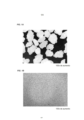
[026] As Figuras 1A-B mostram uma comparação do meio de adsorção e do sangue humano. A Figura 1A mostra o meio de adsorção e a Figura 1B mostra uma imagem de um esfregaço de sangue humano.[026] Figures 1A-B show a comparison of the adsorption medium and human blood. Figure 1A shows the adsorption medium and Figure 1B shows an image of a human blood smear.
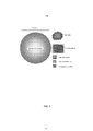
[027] A Figura 2 mostra uma comparação do tamanho das bactérias, por exemplo, Staphylococcus aureus e Chlamydia, e os vírus, por exemplo, vírus da varíola, vírus do herpes, vírus da influenza, e picornavírus (Polio).[027] Figure 2 shows a size comparison of bacteria, eg Staphylococcus aureus and Chlamydia, and viruses, eg smallpox virus, herpes virus, influenza virus, and picornavirus (Polio).
[028] A Figura 3 ilustra uma seção transversal do meio de adsorção contendo grânulos com um diâmetro (d) e uma célula com um diâmetro (a).[028] Figure 3 illustrates a cross section of the adsorption medium containing granules with a diameter (d) and a cell with a diameter (a).
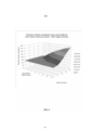
[029] A Figura 4 ilustra o tamanho mínimo de grânulo como uma função de fluxo linear e altura da coluna de cartucho de adsorção para um meio rígido submetido à convecção forçada.[029] Figure 4 illustrates the minimum granule size as a function of linear flow and height of the adsorption cartridge column for a rigid medium subjected to forced convection.
[030] A presente invenção é baseada, em parte, na descoberta de um meio de adsorção que é eficaz para a remoção de uma quantidade significativa de bactérias (por exemplo, bactérias gram-negativas e bactérias gram-positivas, incluindo as bactérias sem nenhuma afinidade conhecida ou baixa afinidade com o heparan sulfato) a partir do sangue (por exemplo, sangue total e soro sanguíneo). Em adição, o meio de adsorção pode ser utilizado em tratamentos extracorpóreos envolvendo altas taxas de fluxo volumétrico e altas taxas de fluxo linear. Tipicamente, o meio de adsorção está contido dentro de uma coluna, um recipiente ou cartucho. Em certos aspectos, a amostra sai da coluna, do recipiente ou do cartucho, e um complexo aderente permanece atrás.[030] The present invention is based, in part, on the discovery of an adsorption medium that is effective for removing a significant amount of bacteria (for example, gram-negative bacteria and gram-positive bacteria, including bacteria without any known affinity or low affinity for heparan sulfate) from blood (eg, whole blood and blood serum). In addition, the adsorption medium can be used in extracorporeal treatments involving high volumetric flow rates and high linear flow rates. Typically, the adsorption medium is contained within a column, vessel or cartridge. In certain aspects, the sample exits the column, vessel, or cartridge, and an adherent complex remains behind.
[031] Um primeiro aspecto da presente invenção fornece um método para remover as bactérias do sangue, tal como o sangue de mamíferos, através de contato do sangue com um substrato sólido. Os inventores descobriram que a arquitetura da superfície do substrato sólido é eficaz para remover patógenos tais como patógenos bacterianos ou vírus.[031] A first aspect of the present invention provides a method for removing bacteria from blood, such as mammalian blood, by contacting blood with a solid substrate. The inventors have found that the surface architecture of the solid substrate is effective in removing pathogens such as bacterial pathogens or viruses.
[032] O substrato da presente invenção possui dimensões intersticiais suficientemente grandes para permitir uma alta taxa de fluxo de sangue sobre o substrato sem uma grande queda de pressão. Por exemplo, à medida que o sangue é retirado de um paciente mamífero, ele passa sobre o substrato a uma taxa de fluxo pela qual a entrega de adsorbatos para a superfície do leito adsorvente é caracterizada principalmente por convecção forçada. Os substratos adequados para o transporte de convecção geralmente contam com “canais” macroscópicas ou interstícios visíveis entre o material não poroso essencial sólido, tal como partículas, grânulos, fibras, fios, espumas reticuladas, ou membranas densas opcionalmente enroladas em espiral.[032] The substrate of the present invention has sufficiently large interstitial dimensions to allow a high rate of blood flow over the substrate without a large pressure drop. For example, as blood is withdrawn from a mammalian patient, it passes over the substrate at a flow rate at which the delivery of adsorbates to the surface of the adsorbent bed is characterized primarily by forced convection. Substrates suitable for convection transport generally feature macroscopic "channels" or visible interstices between the solid essential non-porous material, such as particles, granules, fibers, threads, cross-linked foams, or optionally spirally wound dense membranes.
[033] Isto está em contraste com meios adsorventes altamente porosos (por exemplo, sílica porosa, Sephadex®, poliestireno reticulado e outros meios de exclusão por tamanho), e muitos outros meios microporosos que usam o processo muito mais lento de difusão molecular. Os substratos de adsorção que dependem do transporte de difusão são geralmente compostos por materiais porosos com poros microscópicos e uma área de superfície interna extremamente alta. I. Definições[033] This is in contrast to highly porous adsorbent media (eg, porous silica, Sephadex®, crosslinked polystyrene, and other size exclusion media), and many other microporous media that use the much slower process of molecular diffusion. Adsorption substrates that rely on diffusion transport are generally composed of porous materials with microscopic pores and an extremely high internal surface area. I. Definitions
[034] O termo “terapia extracorpórea” inclui um procedimento médico que é conduzido fora do corpo, ou seja, ex vivo. Em alguns casos, as terapias extracorpóreas incluem métodos em que um fluido corporal tal como o sangue é retirado do indivíduo e produtos desejados tais como, mas não limitados a oxigênio, anticoagulantes do sangue, anestésicos, e similares são adicionados ao fluido corporal antes de ser retornado para o indivíduo. Em outros casos, uma terapia extracorpórea inclui remover produtos indesejados como toxinas que ocorrem naturalmente, venenos ou vírus do corpo ou fluidos corporais. Exemplos não limitantes de terapias extracorpóreas incluem aférese, autotransfusão, hemodiálise, hemofiltração, plasmaférese, circulação extracorpórea (ECC), suporte de vida extracorpórea (ECLS), oxigenação de membrana extracorpórea (ECMO), e desvio cardiopulmonar.[034] The term “extracorporeal therapy” includes a medical procedure that is conducted outside the body, i.e. ex vivo. In some cases, extracorporeal therapies include methods in which a bodily fluid such as blood is withdrawn from the individual and desired products such as, but not limited to, oxygen, blood anticoagulants, anesthetics, and the like are added to the bodily fluid prior to being returned to the individual. In other cases, an out-of-body therapy includes removing unwanted products such as naturally occurring toxins, poisons or viruses from the body or body fluids. Non-limiting examples of extracorporeal therapies include apheresis, autotransfusion, hemodialysis, hemofiltration, plasmapheresis, cardiopulmonary bypass (ECC), extracorporeal life support (ECLS), extracorporeal membrane oxygenation (ECMO), and cardiopulmonary bypass.
[035] O termo “alta taxa de fluxo” ou “alta condição de fluxo” inclui uma taxa de fluxo ou velocidade do sangue que está acima do limite de difusão.[035] The term “high flow rate” or “high flow condition” includes a flow rate or blood velocity that is above the diffusion threshold.
[036] O termo “meio de adsorção” inclui um material ao qual uma célula, organismo, vírus, patógeno, polipeptídeo, polinucleotídeo, molécula química, molécula biológica pode aderir à superfície do mesmo e ser removida de uma amostra tal como sangue.[036] The term "adsorption medium" includes a material to which a cell, organism, virus, pathogen, polypeptide, polynucleotide, chemical molecule, biological molecule can adhere to the surface thereof and be removed from a sample such as blood.
[037] O termo “complexo aderente” inclui um complexo de ao menos duas moléculas onde a primeira molécula está acoplada (por exemplo, ligada, acoplada ou presa) a uma superfície tal como um substrato, e a segunda molécula está ligada à primeira molécula. Por exemplo, um patógeno ou vírus pode aderir à heparina para formar um complexo aderente. Tipicamente, nos métodos da presente invenção, o complexo aderente permanece atrás e a amostra é purificada do patógeno ou vírus.[037] The term "adherent complex" includes a complex of at least two molecules where the first molecule is coupled (e.g., bonded, coupled, or stuck) to a surface such as a substrate, and the second molecule is bonded to the first molecule . For example, a pathogen or virus can adhere to heparin to form an adherent complex. Typically, in the methods of the present invention, the adherent complex remains behind and the sample is purified from the pathogen or virus.
[038] O termo “alta área de superfície” inclui a propriedade de ter uma grande relação de área de superfície específica para volume.[038] The term "high surface area" includes the property of having a large specific surface area to volume ratio.
[039] O termo “adsorvente” inclui um substrato sólido com um composto químico, uma molécula biológica, ou um material que está anexado (por exemplo, ligado, acoplado ou preso) ao mesmo. Em certos casos, o adsorvente é o próprio substrato sólido. Em uma modalidade, um adsorvente é uma resina polimérica com um polissacarídeo, tal como heparina, ligada à mesma. O substrato pode ser um grânulo, fibras ou fios de polímero.[039] The term "adsorbent" includes a solid substrate with a chemical compound, a biological molecule, or a material that is attached (eg, bonded, coupled, or trapped) thereto. In certain cases, the adsorbent is the solid substrate itself. In one embodiment, an adsorbent is a polymeric resin with a polysaccharide, such as heparin, attached thereto. The substrate can be a granule, fibers or polymer threads.
[040] O termo “grânulo de polímero rígido” refere-se a um grânulo, pélete, esfera, partícula, microcápsula, microesfera, nanoesfera, micropartícula, nanopartícula, e similares, que é feito de uma resina polimérica. Um grânulo de polímero é útil como um substrato.[040] The term "rigid polymer granule" refers to a granule, pellet, sphere, particle, microcapsule, microsphere, nanosphere, microparticle, nanoparticle, and the like, which is made of a polymeric resin. A polymer bead is useful as a substrate.
[041] O termo “fibra” ou “fio” é útil como um substrato sólido. A fibra ou fio pode ser feito de um polímero sintético ou um polímero natural ou uma mistura desses. Em certos casos, uma fibra ou fio oco originalmente poroso é tornado denso ou não poroso, antes, durante ou após a ligação à heparina ou outros adsorventes às superfícies externas e/ou internas do mesmo.[041] The term “fiber” or “yarn” is useful as a solid substrate. The fiber or yarn can be made from a synthetic polymer or a natural polymer or a mixture thereof. In certain cases, an originally porous hollow fiber or yarn is made dense or non-porous before, during or after binding heparin or other adsorbents to the external and/or internal surfaces thereof.
[042] O termo “carboidrato” refere-se a uma molécula contendo átomos de carbono, hidrogênio e oxigênio, e, geralmente com a fórmula empírica C2(H2O)y, onde x e y são números diferentes. Exemplos de carboidratos incluem monossacarídeos, dissacarídeos, oligossacarídeos e polissacarídeos.[042] The term "carbohydrate" refers to a molecule containing carbon, hydrogen and oxygen atoms, and usually with the empirical formula C2(H2O)y, where x and y are different numbers. Examples of carbohydrates include monosaccharides, disaccharides, oligosaccharides and polysaccharides.
[043] O termo “polissacarídeo” refere-se a uma molécula de unidades de monossacarídeos ligados por ligações glicosídicas, e tendo uma fórmula empírica de C2(H2O)y, onde x é entre 200 a aproximadamente 3000.[043] The term "polysaccharide" refers to a molecule of monosaccharide units linked by glycosidic bonds, and having an empirical formula of C2(H2O)y, where x is between 200 to approximately 3000.
[044] O termo “superfície hidrofílica” inclui uma superfície com um ângulo de contato com a água inferior a 90°, quando a superfície é plana.[044] The term “hydrophilic surface” includes a surface with a contact angle with water of less than 90°, when the surface is flat.
[045] O termo “baixa afinidade com o heparan sulfato” no contexto de uma bactéria, refere-se à baixa afinidade de ligação das bactérias com o heparan sulfato. Em algumas modalidades, a afinidade de ligação é determinada utilizando ensaios convencionais, tais como um ensaio imunossorvente ligado à enzima (ELISA) para o heparan sulfato. Em outras modalidades, a afinidade de ligação é determinada com base em uma análise preditiva, tal como uma análise de proteínas de ligação ao heparan sulfato expressa pelo patógeno, por exemplo, bactérias. O termo “sem afinidade com o heparan sulfato” refere-se a uma bactéria que não tem afinidade de ligação com o heparan sulfato ou mais baixa do que a afinidade detectável com o heparan sulfato, ou ligação não conhecida ao heparan sulfato. Em alguns casos, não ter nenhuma afinidade com o heparan sulfato inclui ter nenhuma afinidade de ligação predita com o heparan sulfato.[045] The term "low affinity for heparan sulfate" in the context of a bacterium, refers to the low binding affinity of bacteria for heparan sulfate. In some embodiments, binding affinity is determined using conventional assays, such as an enzyme-linked immunosorbent assay (ELISA) for heparan sulfate. In other embodiments, binding affinity is determined based on a predictive analysis, such as an analysis of heparan sulfate binding proteins expressed by the pathogen, e.g., bacteria. The term "no heparan sulfate affinity" refers to a bacterium that has no binding affinity for heparan sulfate or lower than detectable affinity for heparan sulfate, or no known binding to heparan sulfate. In some cases, having no affinity for heparan sulfate includes having no predicted binding affinity for heparan sulfate.
[046] A ligação de patógenos bacterianos ao substrato de adsorção essencialmente não poroso da presente invenção durante o transporte de convecção é particularmente eficaz nas condições de fluxo relativamente de alto normalmente empregadas na operação segura dos circuitos extracorpóreos de sangue, por exemplo, quando medido pela velocidade de fluxo linear, > 8 cm/min, de preferência aproximadamente > 30 cm/min, e mais preferencialmente aproximadamente 30 a 1.000 cm/min.[046] The binding of bacterial pathogens to the essentially non-porous adsorption substrate of the present invention during convection transport is particularly effective in the relatively high flow conditions normally employed in the safe operation of extracorporeal blood circuits, for example, when measured by the linear flow rate, > 8 cm/min, preferably approximately > 30 cm/min, and most preferably approximately 30 to 1000 cm/min.
[047] Em algumas modalidades, o meio de adsorção remove patógenos do sangue total em circuitos extracorpóreos com uma taxa de fluxo linear de aproximadamente 8 cm/min a aproximadamente 1.000 cm/min, por exemplo, aproximadamente 8 cm/min a aproximadamente 30 cm/min, aproximadamente 25 cm/min a aproximadamente 100 cm/min, aproximadamente 50 cm/min a aproximadamente 200 cm/min, aproximadamente 100 cm/min a aproximadamente 1.000 cm/min, a aproximadamente 200 cm/min a aproximadamente 1.000 cm/min, a aproximadamente 400 cm/min a aproximadamente 1.000 cm/min, a aproximadamente 500 cm/min a aproximadamente 1.000 cm/min, a aproximadamente 600 cm/min a aproximadamente 1.000 cm/min, aproximadamente 100 cm/min a aproximadamente 500 cm/min ou aproximadamente 300 cm/min para aproximadamente 800 cm/min. Em certos casos, a taxa de fluxo é de aproximadamente 10, 15, 20, 25, 30, 35, 40, 45, 50, 55, 60, 65, 70, 75, 80, 85, 90, 95 100 cm/min ou aproximadamente 25 a 40 cm/min.[047] In some embodiments, the adsorption medium removes pathogens from whole blood in extracorporeal circuits with a linear flow rate of approximately 8 cm/min to approximately 1,000 cm/min, for example, approximately 8 cm/min to approximately 30 cm /min, approximately 25 cm/min to approximately 100 cm/min, approximately 50 cm/min to approximately 200 cm/min, approximately 100 cm/min to approximately 1000 cm/min, to approximately 200 cm/min to approximately 1000 cm/ min, at approximately 400 cm/min at approximately 1000 cm/min, at approximately 500 cm/min at approximately 1000 cm/min, at approximately 600 cm/min at approximately 1000 cm/min, approximately 100 cm/min at approximately 500 cm /min or approximately 300 cm/min to approximately 800 cm/min. In certain cases, the flow rate is approximately 10, 15, 20, 25, 30, 35, 40, 45, 50, 55, 60, 65, 70, 75, 80, 85, 90, 95 100 cm/min or approximately 25 to 40 cm/min.
[048] Em outras modalidades, o meio de adsorção remove patógenos do sangue total em circuitos extracorpóreos com uma taxa de fluxo volumétrico de aproximadamente 50 mL/minuto a aproximadamente 5 L/minuto, por exemplo, 50 mL/min, 100 mL/min, 150 mL/min, 200 mL/min, 250 mL/min, 300 mL/min, 350 mL/min, 400 mL/min, 500 mL/min, 550 mL/min, 600 mL/min, 650 mL/min, 700 mL/min, 750 mL/min, 800 mL/min, 850 mL/min, 900 mL/min, 950 mL/min, 1,0 L/min, 1,5 L/min, 2,0 L/min, 2,5 L/min, 3,0 L/min, 3,5 L/min, 4,0 L/min, 4,5 L/min e 5 L/min. Em algumas modalidades, a taxa de fluxo é de preferência > 150 mL/minuto.[048] In other embodiments, the adsorption medium removes pathogens from whole blood in extracorporeal circuits with a volumetric flow rate of approximately 50 mL/minute to approximately 5 L/minute, e.g., 50 mL/min, 100 mL/min , 150 mL/min, 200 mL/min, 250 mL/min, 300 mL/min, 350 mL/min, 400 mL/min, 500 mL/min, 550 mL/min, 600 mL/min, 650 mL/min , 700 mL/min, 750 mL/min, 800 mL/min, 850 mL/min, 900 mL/min, 950 mL/min, 1.0 L/min, 1.5 L/min, 2.0 L/ min, 2.5 L/min, 3.0 L/min, 3.5 L/min, 4.0 L/min, 4.5 L/min and 5 L/min. In some embodiments, the flow rate is preferably >150 mL/minute.
[049] O meio adsorvente altamente poroso, em contraste, exige taxas de fluxo muito menores de menos de 1 mL/minuto e aproximadamente menos de 50 mL/minuto. Além disso, o tempo de permanência no substrato de adsorção (por exemplo, a quantidade de tempo que o adsorbato (por exemplo, bactérias) está em contato com o meio adsorvente) precisa de ser muito maior para um meio exigindo transporte difusivo dos adsorbatos para o sítio adsorvente dentro do meio, em comparação com um meio utilizando a convecção forçada dos adsorbatos para os sítios de ligação, que não são compatíveis com sistemas de sangue extracorpóreos padrão.[049] The highly porous adsorbent medium, in contrast, requires much lower flow rates of less than 1 mL/minute and approximately less than 50 mL/minute. Furthermore, the residence time on the adsorption substrate (e.g., the amount of time the adsorbate (e.g., bacteria) is in contact with the adsorbent medium) needs to be much greater for a medium requiring diffusive transport of the adsorbates to the adsorbent site within the medium, compared to a medium using forced convection of the adsorbates to the binding sites, which are not compatible with standard extracorporeal blood systems.
[050] Tipicamente, é reconhecido que o “tempo de permanência” na coluna de adsorção precisa ser mais longo para um meio que exige o transporte difusivo do adsorbatos para o sítio adsorvente dentro do meio, quando comparado com o tempo de permanência inferior necessária para conduzir um adsorbato para o sítio de ligação (em um meio essencialmente não poroso) por convecção forçada. No entanto, existem limites práticos para as dimensões de um cartucho adsorvente, coluna, filtro, etc. seguro e eficaz, especialmente com relação ao volume de retenção máximo de sangue que ele pode conter, e a velocidade do fluxo de sangue ou de soro passado o meio de adsorção. Por esta razão, a taxa de fluxo média através do dispositivo de adsorção é considerada como uma variável de projeto.[050] Typically, it is recognized that the “dwell time” in the adsorption column needs to be longer for a medium that requires diffusive transport of adsorbates to the adsorbent site within the medium, when compared to the lower dwell time required for drive a adsorbate to the binding site (in an essentially non-porous medium) by forced convection. However, there are practical limits to the dimensions of an adsorbent cartridge, column, filter, etc. safe and effective, especially with regard to the maximum holding volume of blood it can hold, and the velocity of blood or serum flow past the adsorption medium. For this reason, the average flow rate through the adsorption device is considered as a design variable.
[051] Os substratos que dependem de transporte de convecção forçada são geralmente mais adequados para as altas taxas de fluxo, enquanto os substratos que dependem do transporte de difusão muito mais lento são muito menos eficazes quando são necessárias altas taxas de fluxo e tempos de permanência mais curtos. Por esta razão, em um dispositivo extracorpóreo de purificação de sangue, é preferencial que um adsorbato difunda-se rapidamente através dos poros dentro do meio adsorvente. Quando o sangue é bombeado através dos circuitos fabricados a partir de materiais sintéticos, é uma prática geral empregar taxas de fluxo de sangue relativamente altas de modo a impedir a estagnação e reduzir o risco de formação de coágulos. Por outro lado, as taxas de fluxo extremamente altas podem ser evitadas porque podem expor as células do sangue a altas taxas de cisalhamento e os danos do impacto que pode romper ou de outra forma danificar as células do sangue. A presente invenção, portanto, fornece um método e dispositivo para remover patógenos bacterianos do sangue usando as características preferenciais do transporte de convecção e sua cinética mais rápida desejável. Isto é conseguido através de passar/fluir o sangue sobre um substrato essencialmente não microporoso (por exemplo, um substrato sólido), que é capaz de se ligar à citocina desejada, patógeno ou bactérias para removê-los do sangue.[051] Substrates that rely on forced convection transport are generally better suited for high flow rates, while substrates that rely on much slower diffusion transport are much less effective when high flow rates and residence times are required. shorter. For this reason, in an extracorporeal blood purification device, it is preferred that an adsorbate rapidly diffuses through pores within the adsorbent medium. When blood is pumped through circuits made from synthetic materials, it is general practice to employ relatively high blood flow rates in order to prevent stagnation and reduce the risk of clot formation. On the other hand, extremely high flow rates can be avoided because they can expose the blood cells to high shear rates and impact damage that can rupture or otherwise damage the blood cells. The present invention, therefore, provides a method and device for removing bacterial pathogens from the blood using the preferred characteristics of convection transport and its desirable faster kinetics. This is accomplished by passing/flowing the blood over an essentially non-microporous substrate (eg, a solid substrate), which is capable of binding the desired cytokine, pathogen, or bacteria to remove them from the blood.
[052] O meio de adsorção fornecido aqui pode ser utilizado na circulação extracorpórea de sangue tradicional, com taxas de fluxo > 50 mL/min, e de preferência entre aproximadamente 150 mL/minuto a 5L/minuto. Se medido pela velocidade de fluxo linear, > 8 cm/min, de preferência aproximadamente > 24 cm/min e mais preferencialmente de aproximadamente 24 a 329 cm/min, ou mais. Por exemplo, a taxa de fluxo pode ser de 25, 50, 75, 100, 125, 150,175, 200, 225, 250, 275, 300, 325, 350, 375, 400, 425, 450, 475, 500, 525, 550, 575, 600, 625, 650, 675, 700, 725, 750, 775, 800 cm/min ou mais. Essas altas taxas de fluxo criam tempos de permanência curtos dentro da coluna de adsorção e o transporte de convecção domina sobre o transporte de difusão Browniano. Isto é particularmente importante para ligar partículas maiores, tais como vírus, bactérias e parasitas e outras proteínas e patógenos que se difundem lentamente.[052] The adsorption medium provided here can be used in traditional extracorporeal blood circulation, with flow rates > 50 mL/min, and preferably between approximately 150 mL/minute to 5L/minute. As measured by linear flow rate, > 8 cm/min, preferably approximately > 24 cm/min, and most preferably approximately 24 to 329 cm/min, or greater. For example, the flow rate can be 25, 50, 75, 100, 125, 150,175, 200, 225, 250, 275, 300, 325, 350, 375, 400, 425, 450, 475, 500, 525, 550, 575, 600, 625, 650, 675, 700, 725, 750, 775, 800 cm/min or more. These high flow rates create short residence times within the adsorption column and convection transport dominates over Brownian diffusion transport. This is particularly important for binding larger particles such as viruses, bacteria and parasites and other slowly diffusing proteins and pathogens.
[053] Os principais sítios de adsorção disponíveis para remover patógenos bacterianos se encontram nas superfícies dentro dos interstícios do leito de meio, recipiente ou cartucho através do qual o sangue flui ou é entregue por convecção forçada. Para tratar o sangue, os canais intersticiais precisam ser suficientemente grandes para permitir o transporte de glóbulos vermelhos, que têm um diâmetro médio de 6 mícrons. Para permitir que um cartucho de adsorção empacotado seja colocado em um circuito extracorpóreo com alta taxa de fluxo de sangue, os canais intersticiais podem ser várias vezes maiores do que o diâmetro dos glóbulos vermelhos. Isto pode evitar ou substancialmente eliminar as altas taxas de cisalhamento que levam à hemólise, minimizando simultaneamente a queda de pressão no sangue que flui através do leito ou cartucho empacotado. Além disso, meio é preferencialmente rígido para minimizar a deformação que pode entupir o cartucho do filtro de compactação. Com base nestas preferências, um meio rígido otimizado equilibra o tamanho do canal intersticial e a área de superfície total, por exemplo, para a remoção eficiente de patógenos e/ou citocinas em circuitos extracorpóreos de sangue de alto fluxo.[053] The main adsorption sites available to remove bacterial pathogens are found on surfaces within the interstices of the medium bed, vessel or cartridge through which blood flows or is delivered by forced convection. To treat blood, the interstitial ducts need to be large enough to allow the transport of red blood cells, which have an average diameter of 6 microns. To allow a packaged adsorption cartridge to be placed in an extracorporeal circuit with a high blood flow rate, the interstitial channels can be several times larger than the diameter of the red blood cells. This can prevent or substantially eliminate the high shear rates that lead to hemolysis, while minimizing the pressure drop in blood flowing through the packed bed or cartridge. Also, media is preferably rigid to minimize deformation that can clog the compacting filter cartridge. Based on these preferences, an optimized rigid medium balances interstitial canal size and total surface area, for example, for efficient removal of pathogens and/or cytokines in high-flow extracorporeal blood circuits.
[054] Os métodos reivindicados são destinados a serem aplicados principalmente nas terapias extracorpóreas ou procedimentos, e também dispositivos implantáveis.[054] The claimed methods are intended to be applied primarily in extracorporeal therapies or procedures, and also implantable devices.
[055] O sangue total e o soro sanguíneo de mamíferos podem ser utilizados na presente invenção. A quantidade de sangue ou de soro que pode ser utilizada nos métodos reivindicados não se destina a ser limitada. Ela pode variar de menos de 1 mL a acima de 1 L, até e incluindo o volume de sangue total de um paciente ou indivíduo, quando a recirculação contínua de volta para o paciente é empregada. Uma ou mais passagens através do leito de adsorção podem ser usadas se necessário. O sangue pode ser sangue humano ou de animal.[055] Whole blood and blood serum of mammals can be used in the present invention. The amount of blood or serum that can be used in the claimed methods is not intended to be limited. It can range from less than 1 mL to over 1 L, up to and including the total blood volume of a patient or individual, when continuous recirculation back to the patient is employed. One or more passes through the adsorption bed can be used if necessary. The blood may be human or animal blood.
[056] Em algumas modalidades, as bactérias ou patógenos presentes na amostra, por exemplo, sangue total ou soro do sangue, são reduzidas em aproximadamente 20% a aproximadamente 90%, por exemplo, aproximadamente 20%, 30%, 40%, 50%, 60%, 70%, 80%, 90% ou 99,9%. Em outras modalidades, as bactérias na amostra são reduzidas em aproximadamente 20% a aproximadamente 40%, por exemplo, aproximadamente 20%, 25%, 30%, 35%, ou 40% ou aproximadamente 5, 10, 15, 20, 25, 30, 35, 40, 45, 50, 55, 60, 65, 70, 75, 80, 85, 90, 95 ou 99,9% de redução das bactérias ou patógenos.[056] In some embodiments, bacteria or pathogens present in the sample, e.g., whole blood or blood serum, are reduced by approximately 20% to approximately 90%, e.g., approximately 20%, 30%, 40%, 50 %, 60%, 70%, 80%, 90% or 99.9%. In other embodiments, the bacteria in the sample are reduced by approximately 20% to approximately 40%, for example, approximately 20%, 25%, 30%, 35%, or 40% or approximately 5, 10, 15, 20, 25, 30, 35, 40, 45, 50, 55, 60, 65, 70, 75, 80, 85, 90, 95 or 99.9% reduction of bacteria or pathogens.
[057] Em algumas modalidades, as bactérias na amostra são bactérias gram-negativas, tal como quaisquer bactérias que não retêm corante violeta cristal. Exemplos não limitantes de uma bactéria gram- negativa são Klebsiella pneumoniae, Acinetobacter baumannii, Pseudomonas aeruginosa, Escherichia coli, Salmonella, Shigella, Stenotrophomonas maltophilia, Moraxella, Borrelia, Burkolderia, Campylobacter, Chlamydia, Haemophilus, Helicobacter, Stenotrophomonas, Vibrio, Leginella, outras Enterobacteriaceae, e cepas resistentes aos fármacos da mesma. Em outras modalidades, as bactérias na amostra são bactérias gram-positivas, tais como as bactérias que retêm corante violeta cristal. Exemplos não limitantes de uma bactéria gram-positiva são Actinomyces, Bacillus, Enterococcus, Lactobacillus, Listeria monocytogenes, Mycobacterium, Nocardia, Propionibacteriaum, Staphylococcus aureus, Staphylococcus epidermidis, Staphylococcus saprophyticus, Streptomyces, Streptococcus pneumoniae, Streptococcus pyrogenes, Streptococcus viridans, enterococos, Clostridium difficile, Enterococcus faecium, Enterococcus faecalis, e cepas resistentes aos fármacos da mesma.[057] In some embodiments, the bacteria in the sample are gram-negative bacteria, such as any bacteria that do not retain crystal violet dye. Non-limiting examples of a gram-negative bacteria are Klebsiella pneumoniae, Acinetobacter baumannii, Pseudomonas aeruginosa, Escherichia coli, Salmonella, Shigella, Stenotrophomonas maltophilia, Moraxella, Borrelia, Burkolderia, Campylobacter, Chlamydia, Haemophilus, Helicobacter, Stenotrophomonas, Vibrio, Leginella, others Enterobacteriaceae, and drug-resistant strains thereof. In other embodiments, the bacteria in the sample are gram-positive bacteria, such as crystal violet stain-retaining bacteria. Non-limiting examples of a gram-positive bacterium are Actinomyces, Bacillus, Enterococcus, Lactobacillus, Listeria monocytogenes, Mycobacterium, Nocardia, Propionibacteriaum, Staphylococcus aureus, Staphylococcus epidermidis, Staphylococcus saprophyticus, Streptomyces, Streptococcus pneumoniae, Streptococcus pyrogenes, Streptococcus viridans, Enterococci, Clostridium difficile, Enterococcus faecium, Enterococcus faecalis, and drug-resistant strains thereof.
[058] Em algumas modalidades, os métodos fornecidos aqui são usados para remover as bactérias gram-negativas de uma amostra de sangue total ou de soro sanguíneo. Em outras modalidades, os métodos são usados para remover as bactérias gram-positivas da amostra. Em ainda outras modalidades, o meio de adsorção descrito aqui tendo um adsorvente polissacarídeo em sua superfície é utilizado para remover bactérias tais como Escherichia coli, Klebsiella pneumoniae, Acinetobacter baumannii, Enterococcus faecium, Escherichia coli resistente a carbapenem, Klebsiella pneumoniae resistente a carbapenem, e/ou Klebsiella pneumonia beta-lactamase de espectro estendido a partir da amostra.[058] In some embodiments, the methods provided herein are used to remove gram-negative bacteria from a whole blood or blood serum sample. In other embodiments, the methods are used to remove gram-positive bacteria from the sample. In still other embodiments, the adsorption medium described herein having a polysaccharide adsorbent on its surface is used to remove bacteria such as Escherichia coli, Klebsiella pneumoniae, Acinetobacter baumannii, Enterococcus faecium, carbapenem-resistant Escherichia coli, carbapenem-resistant Klebsiella pneumoniae, and /or Klebsiella pneumonia extended spectrum beta-lactamase from the sample.
[059] Em algumas modalidades, o meio de absorção tendo uma superfície hidrofílica com carga neutra é usado para remover Staphylococcus aureus, Staphylococcus aureus resistente à meticilina (MRSA), e/ou Escherichia coli de uma amostra de sangue total ou de soro sanguíneo. Em outras modalidades, o meio de adsorção tendo uma superfície catiônica (superfície hidrofílica) é utilizado para remover Escherichia coli, Klebsiella pneumoniae, Escherichia coli resistente a carbapenem, Klebsiella pneumoniae resistente a carbapenem, e Klebsiella pneumoniae beta-lactamase de espectro estendido, Enterococcus faecium, Acinetobacter baumannii, e Staphylococcus aureus resistente à meticilina (MRSA) da amostra.[059] In some embodiments, the absorption medium having a neutrally charged hydrophilic surface is used to remove Staphylococcus aureus, Methicillin-resistant Staphylococcus aureus (MRSA), and/or Escherichia coli from a whole blood or blood serum sample. In other embodiments, the adsorption medium having a cationic surface (hydrophilic surface) is used to remove Escherichia coli, Klebsiella pneumoniae, carbapenem-resistant Escherichia coli, carbapenem-resistant Klebsiella pneumoniae, and extended-spectrum beta-lactamase Klebsiella pneumoniae, Enterococcus faecium , Acinetobacter baumannii, and methicillin-resistant Staphylococcus aureus (MRSA) from the sample.
[060] Vários materiais, em forma e composição, podem ser utilizados como um substrato na presente invenção. Todos os substratos adsorventes adequados fornecem uma alta área de superfície, enquanto promovendo o transporte de adsorbatos para os sítios adsorventes que se ligam a eles (principalmente) por transporte de convecção forçada. Os substratos úteis para criar o meio de adsorção incluem grânulos rígidos não porosos, partículas, ou espumas reticuladas, um leito monolítico rígido (por exemplo, formado a partir de grânulos ou de partículas sinterizadas), uma coluna empacotada com tecido ou não tecido, uma coluna empacotada com um fio ou fibras monofilamento não microporosa sólidas ou ocas, um cartucho enrolado em espiral formado a partir de membrana densa ou película plana, ou uma combinação de meios tais como um cartucho de grânulo/tecido misto. Em algumas modalidades, um substrato adequado para uso na presente invenção é um que é inicialmente microporoso, mas torna-se essencialmente não poroso quando a superfície é tratada antes, durante ou após a criação de sítios de adsorção.[060] Various materials, in shape and composition, can be used as a substrate in the present invention. All suitable adsorbent substrates provide a high surface area, while promoting the transport of adsorbates to the adsorbent sites that bind to them (mainly) by forced convection transport. Substrates useful for creating the adsorption medium include rigid nonporous granules, particles, or crosslinked foams, a rigid monolithic bed (e.g., formed from granules or sintered particles), a woven or nonwoven packed column, a column packed with a solid or hollow non-microporous monofilament yarn or fibers, a spiral wound cartridge formed from dense membrane or flat film, or a combination of media such as a mixed bead/fabric cartridge. In some embodiments, a substrate suitable for use in the present invention is one that is initially microporous, but becomes essentially non-porous when the surface is treated before, during or after the creation of adsorption sites.
[061] Um substrato útil está na forma de grânulos ou partículas sólidas. Os grânulos podem ser feitos de materiais que são suficientemente rígidos para resistir à deformação ou à compactação de acordo com as taxas de fluxo encontradas. Em algumas modalidades, a suficiente rigidez de substrato é a ausência de um aumento significativo na queda de pressão através do leito de adsorção durante aproximadamente uma hora de fluxo de água ou solução salina em típicas taxas de fluxo clínicas. Por exemplo, uma rigidez de substrato adequada é um aumento < 10-50% na queda de pressão em relação à queda de pressão inicial (por exemplo, medida dentro do primeiro minuto de fluxo), quando medida em uma taxa de fluxo similar, por exemplo, de solução salina.[061] A useful substrate is in the form of granules or solid particles. The granules can be made from materials that are rigid enough to resist deformation or compaction at the flow rates encountered. In some embodiments, sufficient substrate rigidity is the absence of a significant increase in pressure drop across the adsorption bed during approximately one hour of flowing water or saline at typical clinical flow rates. For example, adequate substrate stiffness is a < 10-50% increase in pressure drop over the initial pressure drop (e.g., measured within the first minute of flow), when measured at a similar flow rate, e.g. example, saline solution.
[062] Os grânulos de substrato adsorvente podem ser feitos a partir de um número de diferentes materiais biocompatíveis, tais como polímeros naturais ou sintéticos ou materiais não poliméricos incluindo vidros, cerâmicas e metais, que são essencialmente isentos de impurezas lixiviáveis. Alguns polímeros exemplificados incluem poliuretano, polimetil metacrilato, polietileno ou copolímeros de etileno e outros monômeros, polietileno imina, polipropileno, e poli-isobutileno. Exemplos de substratos úteis incluem Polietileno de Peso Molecular Ultra Alto não poroso (UHMWPE). Outros grânulos adequados são poliestireno, polietileno de baixa densidade e de alta densidade, sílica, poliuretano e quitosana.[062] Adsorbent substrate granules can be made from a number of different biocompatible materials, such as natural or synthetic polymers or non-polymeric materials including glasses, ceramics and metals, which are essentially free of leachable impurities. Some exemplified polymers include polyurethane, polymethyl methacrylate, polyethylene or copolymers of ethylene and other monomers, polyethylene imine, polypropylene, and polyisobutylene. Examples of useful substrates include non-porous Ultra High Molecular Weight Polyethylene (UHMWPE). Other suitable granules are polystyrene, low density and high density polyethylene, silica, polyurethane and chitosan.
[063] O substrato, tal como grânulos, fibras, fios e similares podem ser preparados com uma rugosidade de superfície ou topografia para aumentar a área da superfície de adsorção. Por exemplo, é possível aumentar a área de superfície, aumentando a relação de área de superfície para volume. Como é mostrado na Figura 1A, uma superfície irregular produz mais sítios de ligação para as bactérias e patógenos. Tipicamente, uma forma, formato ou geometria livre produz mais área de superfície e é vantajoso. A Figura 1A mostra grânulos UHMWPE como recebido fora de um reator.[063] The substrate such as granules, fibers, yarns and the like can be prepared with a surface roughness or topography to increase the adsorption surface area. For example, you can increase the surface area by increasing the surface area to volume ratio. As shown in Figure 1A, an uneven surface produces more binding sites for bacteria and pathogens. Typically, a free form, shape or geometry produces more surface area and is advantageous. Figure 1A shows UHMWPE beads as received out of a reactor.
[064] Os métodos para preparar os grânulos são conhecidos na técnica. Por exemplo, os grânulos de polietileno adequados e outros grânulos de poliolefina são produzidos diretamente durante o processo de síntese. Em alguns casos, os grânulos são processados para o tamanho e a forma desejada. Outros polímeros podem precisar ser moídos ou secos por pulverização e classificados, ou, de outro modo, processados para criar grânulos de distribuição de tamanho e de forma desejada.[064] Methods for preparing the granules are known in the art. For example, suitable polyethylene granules and other polyolefin granules are produced directly during the synthesis process. In some cases, the granules are processed to the desired size and shape. Other polymers may need to be milled or spray dried and graded or otherwise processed to create granules of the desired size and shape distribution.
[065] Em alguns aspectos, o meio de adsorção da presente invenção fornece uma superfície para anexar um adsorvente polissacarídeo que pode ligar-se um patógeno bacteriano. Em algumas modalidades, o meio de adsorção inclui um substrato sólido com uma alta área de superfície tendo ao menos um adsorvente polissacarídeo em sua superfície.[065] In some aspects, the adsorption medium of the present invention provides a surface to attach a polysaccharide adsorbent that can bind a bacterial pathogen. In some embodiments, the adsorption medium includes a solid substrate having a high surface area having at least one polysaccharide adsorbent on its surface.
[066] Em outros aspectos, um meio de adsorção da presente invenção fornece uma superfície hidrofílica sem um adsorvente polissacarídeo (“uma superfície nua”). Em algumas modalidades, o meio de adsorção inclui um substrato sólido com uma alta área de superfície e uma superfície hidrofílica catiônica. Em outras modalidades, o meio de adsorção inclui um substrato sólido com uma alta área de superfície e uma superfície neutra hidrofílica.[066] In other aspects, an adsorption medium of the present invention provides a hydrophilic surface without a polysaccharide adsorbent ("a bare surface"). In some embodiments, the adsorption medium includes a solid substrate having a high surface area and a cationic hydrophilic surface. In other embodiments, the adsorption medium includes a solid substrate having a high surface area and a hydrophilic neutral surface.
[067] O substrato sólido pode ser feito, por exemplo, mas não limitado a polietileno, poliestireno, polipropileno, polissulfona, poliacrilonitrilo, policarbonato, poliuretano, sílica, látex, vidro, celulose, agarose reticulada, quitina, quitosana, dextrano reticulado, alginato reticulado, silicone, fluoropolímero, e outros polímeros sintéticos. O substrato sólido com uma alta área de superfície pode ser uma pluralidade de monocamadas adsorventes, filtros, membranas, fibras sólidas, fibras ocas, partículas ou grânulos. Opcionalmente, o substrato sólido pode estar presente em outras formas ou formatos fornecendo uma grande área de superfície.[067] The solid substrate can be made, for example, but not limited to polyethylene, polystyrene, polypropylene, polysulfone, polyacrylonitrile, polycarbonate, polyurethane, silica, latex, glass, cellulose, crosslinked agarose, chitin, chitosan, crosslinked dextran, alginate cross-linked, silicone, fluoropolymer, and other synthetic polymers. The solid substrate having a high surface area can be a plurality of adsorbent monolayers, filters, membranes, solid fibers, hollow fibers, particles or granules. Optionally, the solid substrate can be present in other shapes or formats providing a large surface area.
[068] Em certos casos, o substrato sólido é uma pluralidade de grânulos de polímeros rígidos, tais como o polietileno, poliestireno, polipropileno, polissulfona, poliacrilonitrilo, policarbonato, poliuretano, sílica, látex, vidro, celulose, agarose reticulada, quitina, quitosana, dextrano reticulado, alginato reticulado, silicone, fluoropolímero, e grânulos de polímero sintético. De preferência, os grânulos de polímero rígido são grânulos de polietileno.[068] In certain cases, the solid substrate is a plurality of rigid polymer granules, such as polyethylene, polystyrene, polypropylene, polysulfone, polyacrylonitrile, polycarbonate, polyurethane, silica, latex, glass, cellulose, crosslinked agarose, chitin, chitosan , cross-linked dextran, cross-linked alginate, silicone, fluoropolymer, and synthetic polymer granules. Preferably, the rigid polymer granules are polyethylene granules.
[069] O tamanho do substrato sólido pode ser selecionado de acordo com o volume da amostra de teste usada no ensaio ou outros parâmetros. Em algumas modalidades, cada grânulo da pluralidade de grânulos de polímero rígido tem um diâmetro externo médio de aproximadamente 1 μm a aproximadamente 1 mm, por exemplo, 1 μm, 2 μm, 3 μm, 4 μm, 5 μm, 6 μm, 7 μm, 8 μm, 9 μm, 10 μm, 15 μm, 20 μm, 25 μm, 30 μm, 35 μm, 45 μm, 55 μm, 60 μm, 65 μm, 70 μm, 75 μm, 80 μm, 85 μm, 90 μm, 95 μm, 100 μm, 200 μm, 300 μm, 400 μm, 500 μm, 600 μm, 700 μm, 800 μm, 900 μm, ou 1 mm. Em outras modalidades, cada grânulo da pluralidade de grânulos de polímero rígido tem um diâmetro médio de aproximadamente 10 μm a aproximadamente 200 μm, por exemplo, 10 μm, 15 μm, 20 μm, 25 μm, 30 μm, 35 μm, 45 μm, 55 μm, 60 μm, 65 μm, 70 μm, 75 μm, 80 μm, 85 μm, 90 μm, 95 μm, 100 μm, 105 μm, 110 μm, 115 μm, 120 μm, 125 μm, 130 μm, 135 μm, 140 μm, 145 μm, 150 μm, 155 μm, 160 μm, 165 μm, 170 μm, 175 μm, 180 μm, 185 μm, 190 μm, 195 μm ou 200 μm.[069] The size of the solid substrate can be selected according to the volume of the test sample used in the test or other parameters. In some embodiments, each bead of the plurality of rigid polymer beads has an average outer diameter of about 1 µm to about 1 mm, for example, 1 µm, 2 µm, 3 µm, 4 µm, 5 µm, 6 µm, 7 µm , 8μm, 9μm, 10μm, 15μm, 20μm, 25μm, 30μm, 35μm, 45μm, 55μm, 60μm, 65μm, 70μm, 75μm, 80μm, 85μm, 90 μm, 95 μm, 100 μm, 200 μm, 300 μm, 400 μm, 500 μm, 600 μm, 700 μm, 800 μm, 900 μm, or 1 mm. In other embodiments, each bead of the plurality of rigid polymer beads has an average diameter of approximately 10 µm to approximately 200 µm, e.g., 10 µm, 15 µm, 20 µm, 25 µm, 30 µm, 35 µm, 45 µm, 55μm, 60μm, 65μm, 70μm, 75μm, 80μm, 85μm, 90μm, 95μm, 100μm, 105μm, 110μm, 115μm, 120μm, 125μm, 130μm, 135μm , 140μm, 145μm, 150μm, 155μm, 160μm, 165μm, 170μm, 175μm, 180μm, 185μm, 190μm, 195μm or 200μm.
[070] Em algumas modalidades, os grânulos úteis têm um tamanho que varia de aproximadamente 100 mícrons (um) a 500 μm, ou mais de diâmetro, por exemplo, 100 μm, 150 μm, 200 μm, 250 μm, 300 μm, 350 μm, 400 μm, 450 μm, 500 μm, ou mais, de diâmetro. O tamanho médio dos grânulos pode ser de aproximadamente 150 μm a aproximadamente 450 μm de diâmetro, por exemplo, 150 μm, 200 μm, 250 μm, 300 μm, 350 μm, 400 μm, ou 450 μm de diâmetro. Por exemplo, os grânulos de polietileno a partir de Polymer Technology Group (Berkeley, CA) tendo um diâmetro médio de 300 μm são adequados para a presente invenção.[070] In some embodiments, useful granules have a size ranging from approximately 100 microns (um) to 500 µm, or more in diameter, e.g., 100 µm, 150 µm, 200 µm, 250 µm, 300 µm, 350 μm, 400 μm, 450 μm, 500 μm or more in diameter. The average size of the granules can be from approximately 150 µm to approximately 450 µm in diameter, for example, 150 µm, 200 µm, 250 µm, 300 µm, 350 µm, 400 µm, or 450 µm in diameter. For example, polyethylene granules from Polymer Technology Group (Berkeley, CA) having an average diameter of 300 µm are suitable for the present invention.
[071] Em algumas modalidades, o substrato é uma membrana de barreira, por exemplo, uma película não porosa. Alternativamente, uma membrana microporosa pode ser tornada não porosa preenchendo os poros com material essencialmente não poroso, por exemplo, um polímero. A membrana na forma de uma folha ou de uma fibra oca ou sólida pode ser disposta dentro de um alojamento ou um recipiente.[071] In some embodiments, the substrate is a barrier membrane, for example, a non-porous film. Alternatively, a microporous membrane can be made non-porous by filling the pores with essentially non-porous material, for example a polymer. The membrane in the form of a sheet or a hollow or solid fiber can be disposed within a housing or container.
[072] O meio de adsorção pode estar em um recipiente tal como uma coluna, cartucho, tubo, tubo de centrífuga, leito, e similares, ou qualquer recipiente em que as células do sangue que não são capturadas no meio de adsorção ligado a polissacarídeo pode ser removido sem perturbar o patógeno bacteriano aderido ao meio.[072] The adsorption medium can be in a container such as a column, cartridge, tube, centrifuge tube, bed, and the like, or any container in which blood cells that are not captured in the polysaccharide-bound adsorption medium can be removed without disturbing the bacterial pathogen adhered to the medium.
[073] O substrato é tipicamente fornecido empacotado dentro de um alojamento ou recipiente, tal como uma coluna, que é concebida para manter o substrato dentro do recipiente e permitir que o sangue ou o soro flua sobre a superfície do substrato ou leito. O substrato pode ser disposto dentro do recipiente para maximizar a ligação dos adsorbatos aos lados adsorventes do substrato. O alojamento ou recipiente pode ter uma estrutura de superfície macroporosa que fornece uma grande área de superfície para o sangue ou soro.[073] The substrate is typically supplied packaged within a housing or container, such as a column, which is designed to hold the substrate within the container and allow blood or serum to flow over the surface of the substrate or bed. The substrate can be arranged within the vessel to maximize binding of the adsorbates to the adsorbent sides of the substrate. The housing or container can have a macroporous surface structure that provides a large surface area for the blood or serum.
[074] Uma coluna ou outra forma de alojamento pode ser empacotada com um tecido heparinizado ou não tecido ou a heparina, heparan sulfato ou sítios de adsorção não heparina opcionais podem ser anexados, por exemplo, por ligações covalentes, iônicas ou outras ligações físicas ou químicas, depois do alojamento ser preenchido com o meio de substrato. Ao controlar o denier ou densidade da fibra do tecido durante a tecelagem ou entrelaçamento ou durante a criação de uma tela não tecido, o tamanho do poro intersticial pode ser controlado. Os tecidos não tecidos úteis podem estar na forma de feltros, ou mantas eletrostaticamente fiadas ou fundido e soprado, tendo uma orientação aleatória mantida junta por entrelaçamento das fibras e/ou adesão ou coesão das fibras que se interceptam. Os tecidos úteis têm uma estrutura mais definida e não aleatória.[074] A column or other form of housing can be packed with a heparinized or non-woven fabric or the heparin, heparan sulfate or optional non-heparin adsorption sites can be attached, for example, by covalent, ionic or other physical bonds or chemicals after the housing is filled with the substrate medium. By controlling the denier or fiber density of the fabric during weaving or weaving or when creating a non-woven web, the interstitial pore size can be controlled. Useful nonwoven fabrics can be in the form of felts, or electrostatically spun or meltblown batts, having a random orientation held together by interweaving of the fibers and/or adhesion or cohesion of the intersecting fibers. Useful tissues have a more defined and non-random structure.
[075] Uma coluna ou alojamento pode ser empacotada com fibras ou fios feitos de fibras. O polietileno e outras fibras podem ser feitos em fibras monofilamento ocas ou sólidas ou fios multifilamentos, que pode ser empacotado em cartuchos do mesmo modo que as membranas de fibras ocas, são instaladas dentro de cartuchos de hemodiálise convencional ou oxigenadores de sangue. Na presente invenção, as fibras ocas originalmente porosas são feitas densas ou não porosas, antes, durante ou após ligar a heparina ou outros adsorventes às superfícies externas e/ou internas. Dyneema Purity® da Royal DSM é uma fibra sólida de alta resistência feita de UHMWPE. O polietileno de peso molecular ultra-alto (UHMWPE, UHMW) é um subconjunto do polietileno termoplástico. Dyneema pode ser heparinizada e empacotada em um cartucho para fornecer um suporte de alta área de superfície para a remoção de citocinas, bactérias e patógenos.[075] A column or housing can be packed with fibers or strands made from fibers. Polyethylene and other fibers can be made into solid or hollow monofilament fibers or multifilament yarns, which can be packaged into cartridges in the same way that hollow fiber membranes are installed inside conventional hemodialysis cartridges or blood oxygenators. In the present invention, the originally porous hollow fibers are made dense or non-porous before, during or after attaching heparin or other adsorbents to the external and/or internal surfaces. Royal DSM's Dyneema Purity® is a high strength solid fiber made from UHMWPE. Ultra-high molecular weight polyethylene (UHMWPE, UHMW) is a subset of thermoplastic polyethylene. Dyneema can be heparinized and packaged in a cartridge to provide high surface area support for the removal of cytokines, bacteria and pathogens.
[076] Um cartucho enrolado em espiral contém uma película ou membrana fina que é enrolada firmemente em conjunto com materiais de espaçamento opcionais para evitar o contato das superfícies adjacentes. A membrana pode ser feita a partir de polímeros tais como poliuretano, polipropileno, polietileno, polissulfona, policarbonato, PET, PBT e similares.[076] A spiral-wound cartridge contains a thin film or membrane that is tightly wound together with optional spacing materials to prevent contact of adjacent surfaces. The membrane can be made from polymers such as polyurethane, polypropylene, polyethylene, polysulfone, polycarbonate, PET, PBT and the like.
[077] Como mencionado acima, em certos casos, para utilização nos métodos da invenção, o tamanho dos canais ou espaço intersticial entre os grânulos individuais para filtração extracorpórea de sangue são otimizados para evitar uma alta queda de pressão entre a entrada e a saída do cartucho, para permitir a passagem segura das células do sangue entre os grânulos individuais em um ambiente de alto fluxo, e para fornecer área de superfície intersticial adequada para a ligação do adsorvente polissacarídeo às citocinas ou patógenos no sangue. Por exemplo, em um leito empacotado fechado de grânulos aproximadamente esféricos com 300 mícrons, um tamanho de poro intersticial adequado é de aproximadamente 68 mícrons de diâmetro.[077] As mentioned above, in certain cases, for use in the methods of the invention, the size of the channels or interstitial space between the individual granules for extracorporeal blood filtration are optimized to avoid a high pressure drop between the inlet and outlet of the cartridge, to allow safe passage of blood cells between individual granules in a high-flow environment, and to provide adequate interstitial surface area for binding of the polysaccharide adsorbent to cytokines or pathogens in the blood. For example, in a closed packed bed of approximately 300 micron spherical granules, a suitable interstitial pore size is approximately 68 microns in diameter.
[078] Em algumas modalidades, os grânulos rígidos do meio de adsorção têm um diâmetro médio como é apresentado na Tabela 5. Em algumas modalidades, os substratos não grânulo do meio de adsorção, tais como fios ou fibras tecidas, têm um tamanho de poro macroscópico quanto apresentado na Tabela 6.[078] In some embodiments, the rigid adsorption medium granules have an average diameter as shown in Table 5. In some embodiments, the non-granule substrates of the adsorption medium, such as yarns or woven fibers, have a pore size macroscopic as shown in Table 6.
[079] A superfície do substrato sólido aqui descrito pode ser funcionalizada para permitir a ligação covalente do adsorvente polissacarídeo aqui descrito. Em algumas modalidades, a superfície do substrato sólido tem ao menos um grupo químico, tal como um grupo amina.[079] The surface of the solid substrate described herein can be functionalized to allow the covalent binding of the polysaccharide adsorbent described herein. In some embodiments, the surface of the solid substrate has at least one chemical group, such as an amine group.
[080] Os polissacarídeos tais como a heparina ou o heparan sulfato ou outros polissacarídeos podem ser ligados à superfície do meio de adsorção por ligação covalente terminal (por exemplo, ligação covalente através do resíduo terminal da molécula de heparina). A ligação covalente em comparação com a ligação não covalente fornece vantajosamente um melhor controle da orientação das moléculas imobilizadas e a sua densidade de superfície. Em particular, a ligação terminal desses carboidratos de cadeia longa fornece uma função de espaçamento que conduz a uma concentração mais alta de oligômeros de carboidrato acessíveis disponíveis para a ligação aos patógenos. Na verdade, certos patógenos se ligam a superfícies revestidas com heparina de comprimento total (por exemplo, heparina com um peso molecular médio maior do que 10 kDa) muito mais eficientemente do que a superfícies convencionais revestidas com fragmentos de heparina, como é geralmente empregado na técnica.[080] Polysaccharides such as heparin or heparan sulfate or other polysaccharides can be attached to the surface of the adsorption medium by terminal covalent bonding (for example, covalent bonding through the terminal residue of the heparin molecule). Covalent binding compared to non-covalent binding advantageously provides better control of the orientation of the immobilized molecules and their surface density. In particular, the terminal linkage of these long-chain carbohydrates provides a spacing function that leads to a higher concentration of accessible carbohydrate oligomers available for binding to pathogens. Indeed, certain pathogens bind to surfaces coated with full-length heparin (e.g., heparin with an average molecular weight greater than 10 kDa) much more efficiently than to conventional surfaces coated with fragments of heparin, as is commonly employed in technique.
[081] Em algumas modalidades, as moléculas de heparina de comprimento total imobilizadas têm um peso molecular médio de mais de 10 kDa. Em outras modalidades, as moléculas de heparina imobilizada têm um peso molecular médio de mais de 15 kDa. Em outra modalidade, as moléculas de heparina imobilizada têm um peso molecular médio de mais de 21 kDa. Em ainda outra modalidade, as moléculas de heparina imobilizada têm um peso molecular médio de mais de 30 kDa. De preferência, as moléculas de heparina imobilizada têm um peso molecular médio dentro da faixa de 15 a 25 kDa. O peso molecular médio pode também ser mais alto, tal como na faixa de 25 a 35 kDa.[081] In some embodiments, the immobilized full-length heparin molecules have an average molecular weight of more than 10 kDa. In other embodiments, the immobilized heparin molecules have an average molecular weight of greater than 15 kDa. In another embodiment, the immobilized heparin molecules have an average molecular weight of greater than 21 kDa. In yet another embodiment, the immobilized heparin molecules have an average molecular weight of greater than 30 kDa. Preferably, the immobilized heparin molecules have an average molecular weight within the range of 15 to 25 kDa. The average molecular weight can also be higher, such as in the range of 25 to 35 kDa.
[082] Em algumas modalidades, a concentração na superfície do adsorvente heparina no substrato sólido está na faixa de 1 μg/cm2 a 20 μg/cm2, por exemplo, 1 μg/cm2, 2 μg/cm2, 3 μg/cm2, 4 μg/cm2, 5 μg/cm2, 6 μg/cm2, 7 μg/cm2, 8 μg/cm2, 9 μg/cm2, 10 μg/cm2, 11 μg/cm2, 12 μg/cm2, 13 μg/cm2, 14 μg/cm2, 15 μg/cm2, 16 μg/cm2, 17 μg/cm2, 18 μg/cm2, 19 μg/cm2, e 20 μg/cm2. Em outras modalidades, a concentração na superfície do adsorvente heparano no substrato sólido está na faixa de 5 μg/cm2 to 15 μg/cm2, por exemplo, 5 μg/cm2, 6 μg/cm2, 7 μg/cm2, 8 μg/cm2, 9 μg/cm2, 10 μg/cm2, 11 μg/cm2, 12 μg/cm2, 13 μg/cm2, 14 μg/cm2, e 15 μg/cm2.[082] In some embodiments, the surface concentration of the heparin adsorbent on the solid substrate is in the range of 1 μg/cm2 to 20 μg/cm2, for example, 1 μg/cm2, 2 μg/cm2, 3 μg/cm2, 4 μg/cm2, 5 μg/cm2, 6 μg/cm2, 7 μg/cm2, 8 μg/cm2, 9 μg/cm2, 10 μg/cm2, 11 μg/cm2, 12 μg/cm2, 13 μg/cm2, 14 μg/cm2, 15 μg/cm2, 16 μg/cm2, 17 μg/cm2, 18 μg/cm2, 19 μg/cm2, and 20 μg/cm2. In other embodiments, the surface concentration of the heparan adsorbent on the solid substrate is in the range of 5 µg/cm2 to 15 µg/cm2, e.g. 5 µg/cm2, 6 µg/cm2, 7 µg/cm2, 8 µg/cm2 , 9 µg/cm2, 10 µg/cm2, 11 µg/cm2, 12 µg/cm2, 13 µg/cm2, 14 µg/cm2, and 15 µg/cm2.
[083] A quantidade de adsorvente polissacarídeo por grama de substrato pode variar. Em uma modalidade particular, se grânulos são utilizados, a quantidade de polissacarídeo, tal como heparina por grama de grânulos, é determinada pelo número de camadas utilizadas e também pelo tamanho dos grânulos. Quanto maior o grânulo, menos polissacarídeo, tal como heparina por grama de grânulo, é conseguido. Uma quantidade preferencial é de 2,0 ± 0,5 mg de heparina/g de grânulo pelo método MBTH (Larm e outros, Biomater Med Devices Artif Organs, 1983, 11: 161 a 173 e Riesenfeld e Rosen, Anal Biochem, 1990, 188: 383 a 389).[083] The amount of polysaccharide adsorbent per gram of substrate may vary. In a particular embodiment, if granules are used, the amount of polysaccharide, such as heparin per gram of granules, is determined by the number of layers used and also the size of the granules. The larger the granule, the less polysaccharide, such as heparin per gram of granule, is achieved. A preferred amount is 2.0 ± 0.5 mg of heparin/g of granule by the MBTH method (Larm et al., Biomater Med Devices Artif Organs, 1983, 11: 161 to 173 and Riesenfeld and Rosen, Anal Biochem, 1990, 188: 383 to 389).
[084] A ligação covalente de moléculas de heparina de comprimento total a uma superfície pode ser conseguida pela reação de um grupo aldeído da molécula de heparina com um grupo amino primário presente na superfície do meio de adsorção. Uma propriedade inerente de todos os carboidratos é que eles têm um hemiacetal na sua extremidade redutora. Este acetal está em equilíbrio com a forma de aldeído e pode formar bases de Schiff com as aminas primárias. Estas bases de Schiff podem então ser reduzidas para aminas secundárias estáveis. Em algumas modalidades, a heparina de comprimento total é imobilizada na superfície no substrato sólido por conjugação covalente. Em outras modalidades, a heparina de comprimento total é ligada de forma covalente ao dito meio de adsorção via um grupo amino secundário estável.[084] Covalent attachment of full-length heparin molecules to a surface can be achieved by reacting an aldehyde group of the heparin molecule with a primary amino group present on the surface of the adsorption medium. An inherent property of all carbohydrates is that they have a hemiacetal at their reducing end. This acetal is in equilibrium with the aldehyde form and can form Schiff bases with primary amines. These Schiff bases can then be reduced to stable secondary amines. In some embodiments, the full-length heparin is surface-immobilized on the solid substrate by covalent conjugation. In other embodiments, the full-length heparin is covalently linked to said adsorption means via a stable secondary amino group.
[085] Em certos casos, vários métodos de preparação de adsorventes e os próprios adsorventes são descritos na Patente US. No. 8.663.148 e Publicações de Pedido de Patente Nos. US2009/0136586, US2010/0249689, US2011/0184377, e US2012/0305482, cujas descrições são aqui incorporadas por referência na sua totalidade para todos os propósitos.[085] In certain cases, various methods of preparing adsorbents and the adsorbents themselves are described in the US Patent. At the. 8,663,148 and Patent Application Publications Nos. US2009/0136586, US2010/0249689, US2011/0184377, and US2012/0305482, the descriptions of which are hereby incorporated by reference in their entirety for all purposes.
[086] Em algumas modalidades, o meio de adsorção é hidrofilizado antes da ligação do polissacarídeo, tal como a heparina, ou outros compostos. Os métodos para preparar a superfície hidrofílica do substrato incluem ataque ácido, tratamento de plasma, e exposição a oxidantes fortes. Por exemplo, uma superfície polimérica tal como um grânulo de polietileno (PE) pode ser gravada com um agente oxidante, tal como permanganato de potássio, peroxidissulfato de amónio e similares, para introduzir propriedades hidrofílicas juntamente com alguns grupos funcionais reativos (por exemplo, um grupo sulfonila, um grupo hidroxila, um grupo carboxila, um grupo carbonila, ou ligações duplas de carbono). A superfície pode ser gravada com plasma ou corona. Por exemplo, grânulos de PE podem ser gravados com um permanganato de potássio em ácido sulfúrico para produzir grânulos com uma superfície hidrofílica contendo grupos hidroxila e ligações duplas de carbono.[086] In some embodiments, the adsorption medium is hydrophilized prior to binding the polysaccharide, such as heparin, or other compounds. Methods for preparing the hydrophilic substrate surface include acid etching, plasma treatment, and exposure to strong oxidants. For example, a polymeric surface such as a polyethylene (PE) granule can be etched with an oxidizing agent such as potassium permanganate, ammonium peroxydisulfate and the like to introduce hydrophilic properties along with some reactive functional groups (e.g. a sulfonyl group, a hydroxyl group, a carboxyl group, a carbonyl group, or carbon double bonds). The surface can be etched with plasma or corona. For example, PE granules can be etched with a potassium permanganate in sulfuric acid to produce granules with a hydrophilic surface containing hydroxyl groups and carbon double bonds.
[087] Em certos casos, os métodos da invenção preparam o leito de adsorção a partir de uma mistura de meios heparinizados que é antitrombogênica e outro meio que é inerentemente trombogênico. Ao montar um cartucho de adsorção com ambas as superfícies heparinizadas e, por exemplo, superfícies hidrofílicas (superfícies catiônicas ou neutras), os patógenos bacterianos podem ser seguramente removidos do sangue ou de outro fluido biológico. Por exemplo, os meios heparinizados podem ser de 1% a 99% do leito de adsorção e o substrato inerentemente trombogênico pode ser de 99% a 1% do leito de adsorção.[087] In certain cases, the methods of the invention prepare the adsorption bed from a mixture of heparinized media that is antithrombogenic and another medium that is inherently thrombogenic. By assembling an adsorption cartridge with both heparinized surfaces and, for example, hydrophilic surfaces (cationic or neutral surfaces), bacterial pathogens can be safely removed from blood or other biological fluid. For example, heparinized media can be from 1% to 99% of the adsorption bed and the inherently thrombogenic substrate can be from 99% to 1% of the adsorption bed.
[088] Em algumas modalidades da presente invenção, o meio de adsorção fornece uma superfície antitrombogênica que está em contato íntimo com, ou em estreita proximidade com uma superfície trombogênica. Esse meio de adsorção pode impedir a formação de trombos clinicamente significativa que, de outra forma, ocorreria se a superfície inerentemente trombogênica fosse utilizada sozinha.[088] In some embodiments of the present invention, the adsorption medium provides an antithrombogenic surface that is in intimate contact with, or in close proximity to, a thrombogenic surface. This adsorption medium can prevent the clinically significant thrombus formation that would otherwise occur if the inherently thrombogenic surface were used alone.
[089] No caso do meio de adsorção na forma de grânulos ou partículas, uma aplicação preferencial da presente invenção é misturar os diferentes meios de adsorção em conjunto antes de empacotá-los em um cartucho ou outro alojamento. Isso fornece contato íntimo entre as várias substâncias químicas de superfície em grânulos adjacentes enquanto permitindo a fabricação eficiente de cartuchos ou filtros de adsorção. Uma abordagem relacionada é dispor em camadas os diferentes meios em um arranjo “do tipo parfait” dentro do alojamento de tal forma que o sangue contate diferentes meios em fluxo em série ou paralelo. Um arranjo dos diferentes meios dentro de um cartucho é posicionar meios antitrombogênicos não misturados na entrada e/ou na saída do cartucho, com uma região opcionalmente misturada contendo os meios mais trombogênicos interpostos entre as regiões de entrada e de saída.[089] In the case of adsorption media in the form of granules or particles, a preferred application of the present invention is to mix the different adsorption media together before packaging them in a cartridge or other housing. This provides intimate contact between the various surface chemicals on adjacent granules while allowing for the efficient manufacture of cartridges or adsorption filters. A related approach is to layer the different media in a "parfait-like" arrangement within the housing such that the blood contacts different media in series or parallel flow. One arrangement of the different media within a cartridge is to position unmixed antithrombogenic media at the inlet and/or outlet of the cartridge, with an optionally mixed region containing the more thrombogenic media interposed between the inlet and outlet regions.
[090] No caso de meios na forma de fibras, um tecido misto, malha, ou uma estrutura não tecida poderem ser preparados por métodos bem conhecidos na indústria têxtil para formar tecido a partir de fibra mista. Alternativamente, um fio pode ser preparado a partir de fios multifilamento ou monofilamento mais finos feitos de duas ou mais fibras com diferentes químicas de superfície, desde que um tipo de fibra contenha uma superfície que impede ativamente a coagulação do sangue em contato. O fio de fibra mista pode então ser utilizado para preparar tecido para contato com o sangue. Os meios de adsorção de fibra oca ou de fibra sólida podem ser misturados e utilizados para produzir cartuchos que se assemelham a dialisadores ou oxigenadores de fibras ocas. Para meios de adsorção do tipo membrana ou película que é usado em cartuchos de adsorção enrolados em espiral, dois ou mais produtos químicos de superfície podem ser usados em estreita proximidade um com o outro de tal modo que o sangue deve contatar ambas as químicas de superfície (quase) simultaneamente. Isto pode ser feito com um arranjo regular ou aleatório dos vários grupos de ligação dentro da camada de superfície da película de membrana, ou pela formação de um caminho de fluxo para o sangue entre duas películas de membrana estreitamente espaçadas, uma das quais é antitrombogênica.[090] In the case of media in the form of fibers, a mixed fabric, mesh, or a nonwoven structure can be prepared by methods well known in the textile industry to form fabric from mixed fiber. Alternatively, a yarn can be prepared from thinner multifilament or monofilament yarns made from two or more fibers with different surface chemistries, provided that one type of fiber contains a surface that actively prevents clotting of blood on contact. The mixed fiber yarn can then be used to prepare tissue for contact with blood. Hollow fiber or solid fiber adsorption media can be mixed and used to produce cartridges that resemble hollow fiber dialyzers or oxygenators. For membrane- or film-type adsorption media that is used in spiral-wound adsorption cartridges, two or more surface chemicals may be used in close proximity to each other such that the blood must contact both surface chemistries (almost) simultaneously. This can be done with a regular or random arrangement of the various binding groups within the surface layer of the membrane film, or by forming a blood flow path between two closely spaced membrane films, one of which is antithrombogenic.
[091] Em certos aspectos, os métodos fornecidos aqui podem ser utilizados em um dispositivo que compreende meios de adsorção para a remoção extracorpórea de patógenos a partir do sangue de mamíferos, por exemplo, sangue humano. Por exemplo, o dispositivo pode ser um dispositivo convencional para o tratamento extracorpóreo de sangue e soro de pacientes, por exemplo, um indivíduo que sofre de insuficiência renal.[091] In certain aspects, the methods provided herein can be used in a device comprising adsorption means for the extracorporeal removal of pathogens from mammalian blood, e.g., human blood. For example, the device may be a conventional device for the extracorporeal treatment of blood and serum from patients, for example an individual suffering from renal failure.
[092] Os padrões de fluxo sanguíneo local no sangue em contato com dispositivos médicos para circulação extracorpórea são conhecidos por influenciar a formação do coágulo através da ativação por cisalhamento e agregação de plaquetas em zonas estagnadas. O dispositivo contendo os meios de adsorção fornecidos aqui pode, por exemplo, ter uma ou mais das seguintes propriedades: a) um fluxo sanguíneo na faixa de 150 a 5.000 mL/min, ou se for medido pela velocidade de fluxo linear de > 8 cm/min; b) baixa resistência ao fluxo; c) grande área de superfície de substrato tendo carboidratos imobilizados para o efeito, por exemplo, aproximadamente 0,1 a 1 m2; d) um revestimento estável (por exemplo, sem vazamento clinicamente significativo de carboidrato para o sangue em contato com o mesmo); e) propriedades hemodinâmicas adequadas no dispositivo (por exemplo, sem zonas estagnadas); e f) biocompatibilidade ótima.[092] Local blood flow patterns in blood in contact with cardiopulmonary bypass medical devices are known to influence clot formation through shear activation and platelet aggregation in stagnant zones. The device containing the adsorption media provided here can, for example, have one or more of the following properties: a) a blood flow in the range of 150 to 5000 mL/min, or if measured by the linear flow velocity of > 8 cm /min; b) low flow resistance; c) large substrate surface area having immobilized carbohydrates therefor, for example approximately 0.1 to 1
[093] Exemplos não limitantes de um dispositivo para uso de acordo com os métodos da presente invenção incluem um dispositivo de oxigenação de membrana extracorpórea (ECMO), um dialisador de hemofluxo pediátrico que é um dispositivo extracorpóreo de filtração de sangue para a remoção de moléculas de citocina ou outro dispositivo extracorpóreo que pode acomodar altas taxas de fluxo.[093] Non-limiting examples of a device for use in accordance with the methods of the present invention include an extracorporeal membrane oxygenation (ECMO) device, a pediatric blood flow dialyzer which is an extracorporeal blood filtration device for removing molecules cytokine or other extracorporeal device that can accommodate high flow rates.
[094] Os métodos da presente invenção podem ser empregados antes ou após outros tratamentos convencionais, tal como a administração de antibióticos.[094] The methods of the present invention can be employed before or after other conventional treatments, such as the administration of antibiotics.
[095] Em algumas modalidades, os métodos são realizados em um ciclo contínuo de tal modo que, a amostra, por exemplo, sangue total, é extraída do corpo e processada de acordo com o método aqui fornecido, e, em seguida, a amostra resultante (por exemplo, amostra contendo uma quantidade reduzida de patógeno bacteriano) é reintroduzida no corpo, formando assim um ciclo que compreende parte da corrente sanguínea do paciente.[095] In some embodiments, the methods are performed in a continuous loop such that the sample, for example, whole blood, is extracted from the body and processed according to the method provided herein, and then the sample The resulting sample (eg, sample containing a reduced amount of bacterial pathogen) is reintroduced into the body, thus forming a cycle comprising part of the patient's bloodstream.
[096] Em outras modalidades, os métodos fornecidos aqui podem ser combinados com outras técnicas para filtrar ou tratar sangue de mamíferos. Por exemplo, um cartucho que é baseado em cinética de convecção pode, então, ser utilizado em série com circuitos extracorpóreos convencionais, tais como desvio cardiopulmonar (CPB), hemodiálise, oxigenação e ozonização extracorpórea do sangue (EBOO), e similares.[096] In other embodiments, the methods provided herein can be combined with other techniques for filtering or treating mammalian blood. For example, a cartridge that is based on convection kinetics can then be used in series with conventional extracorporeal circuits such as cardiopulmonary bypass (CPB), hemodialysis, extracorporeal blood oxygenation and ozonation (EBOO), and the like.
[097] Os vários aspectos da invenção são ainda descritos nos exemplos seguintes. Estes exemplos não se destinam a ser limitantes. Por exemplo, nos presentes exemplos, a heparina é usada. No entanto, outros carboidratos e adsorventes polissacarídeos podem ser utilizados sozinhos ou em adição aos substratos revestidos com heparina exemplificados abaixo.[097] The various aspects of the invention are further described in the following examples. These examples are not intended to be limiting. For example, in the present examples, heparin is used. However, other carbohydrate and polysaccharide adsorbents can be used alone or in addition to the heparin-coated substrates exemplified below.
[098] Os seguintes exemplos são oferecidos para ilustrar, mas não limitar, a invenção reivindicada.[098] The following examples are offered to illustrate, but not limit, the claimed invention.
[099] Exemplo 1. Remoção de bactérias com afinidade baixa ou indetectável com o heparan sulfato[099] Example 1. Removal of bacteria with low or undetectable affinity for heparan sulfate
[0100] Este exemplo ilustra a utilização de grânulos revestidos com heparina para remover patógenos bacterianos com afinidade baixa ou indetectável com o heparan sulfato a partir do sangue total.[0100] This example illustrates the use of heparin-coated beads to remove bacterial pathogens with low or undetectable affinity for heparan sulfate from whole blood.
[0101] Tem sido relatado na literatura que em mais de 50 patógenos alvo diferentes, os proteoglicanos de heparan sulfato foram encontrados em sindecanos como um sítio de ligação inicial durante a sua patogênese. Surpreendentemente, a heparina ligada à superfície pode funcionar como um substituto para os organismos de ligação ao heparan sulfato.[0101] It has been reported in the literature that in more than 50 different target pathogens, heparan sulfate proteoglycans have been found in syndecans as an early binding site during their pathogenesis. Surprisingly, surface-bound heparin can function as a surrogate for heparan sulfate-binding organisms.
[0102] Os estudos mostraram que os meios de adsorção heparinizados podem remover a alta concentração de S. aureus e MRSA a partir de sangue total. Além disso, o estudo demonstrou que as bactérias que aderem à superfície heparinizada não foram mortas, e assim não liberam potenciais toxinas inflamatórias e seus subprodutos para o sangue. Assim, os meios ligados à heparina podem ser utilizados em um dispositivo extracorpóreo para remover de forma eficaz e segura as bactérias circulantes incluindo cepas resistentes a fármacos a partir do sangue infectado.[0102] Studies have shown that heparinized adsorption media can remove high concentration of S. aureus and MRSA from whole blood. Furthermore, the study demonstrated that the bacteria adhering to the heparinized surface were not killed, and thus do not release potential inflammatory toxins and their by-products into the blood. Thus, heparin-bound media can be used in an extracorporeal device to effectively and safely remove circulating bacteria including drug-resistant strains from infected blood.
[0103] Este exemplo testa tanto patógenos de ligação ao heparan sulfato conhecidos quanto patógenos desconhecidos ou inesperados para se ligarem à heparina. Além disso, descobriu-se que os controles hidrofílicos, ou catiônicos ou com carga neutra, podem funcionar como uma superfície eficaz para se ligar a patógenos. As superfícies com carga neutra, em geral, não foram tão eficazes como as superfícies heparinizadas na remoção de patógenos, mas é possível que uma tecnologia de redução de patógenos possa ser desenvolvida usando superfícies hidrofílicas genéricas. As superfícies catiônicas hidrofílicas mostraram razoável capacidade de remover patógenos também.[0103] This example tests both known heparan sulfate binding pathogens and unknown or unexpected pathogens to bind heparin. Furthermore, it has been discovered that hydrophilic, or cationic, or neutrally charged controls can function as an effective surface for binding to pathogens. Neutrally charged surfaces, in general, were not as effective as heparinized surfaces in removing pathogens, but it is possible that a pathogen reduction technology could be developed using generic hydrophilic surfaces. The hydrophilic cationic surfaces showed reasonable ability to remove pathogens as well.
[0104] Este exemplo ilustra que um meio de adsorção compreendendo uma heparina ligada à superfície pode ser usado para remover os patógenos de ligação ao heparan sulfato tais como, S. aureus S. aureus resistente à meticilina (MRSA), E. faecalis, E. faecalis resistente à vancomicina, HSV-1 e HSV-2, e Candida albicans.[0104] This example illustrates that an adsorption medium comprising a surface-bound heparin can be used to remove heparan sulfate-binding pathogens such as, S. aureus, methicillin-resistant S. aureus (MRSA), E. faecalis, E. faecalis resistant to vancomycin, HSV-1 and HSV-2, and Candida albicans.
[0105] Este exemplo ilustra que um meio de adsorção compreendendo uma heparina ligada à superfície pode ser usado para remover patógenos de ligação de heparan sulfato com baixa afinidade (por exemplo, zero) tais como, E. coli, E. coli resistente a carbapenem, K. pneumoniae, K. pneumoniae resistente a carbapenem, K. pneumoniae beta-lactamase de espectro estendido, E. faecium, A. baumannii, e S. pneumonia, do sangue.[0105] This example illustrates that an adsorption medium comprising a surface-bound heparin can be used to remove heparan sulfate binding pathogens with low affinity (e.g., zero) such as E. coli, carbapenem-resistant E. coli , K. pneumoniae, carbapenem-resistant K. pneumoniae, extended-spectrum beta-lactamase K. pneumoniae, E. faecium, A. baumannii, and S. pneumoniae, from blood.
[0106] Em particular, um meio de adsorção compreendendo uma superfície hidrofílica neutra pode remover, por exemplo, S. aureus, S. aureus resistente à meticilina (MRSA), e E. coli. Além disso, um meio de adsorção compreendendo uma superfície hidrofílica catiônica pode remover, por exemplo, E. coli, K. pneumoniae, K. pneumoniae resistente a carbapenem, K. pneumoniae beta-lactamase de espectro estendido, E. faecium, A. baumannii, e S. aureus resistente à meticilina (MRSA).[0106] In particular, an adsorption medium comprising a neutral hydrophilic surface can remove, for example, S. aureus, methicillin-resistant S. aureus (MRSA), and E. coli. Furthermore, an adsorption medium comprising a cationic hydrophilic surface can remove, for example, E. coli, K. pneumoniae, carbapenem-resistant K. pneumoniae, extended-spectrum beta-lactamase K. pneumoniae, E. faecium, A. baumannii , and methicillin-resistant S. aureus (MRSA).
[0107] A bacteremia por S. aureus ou S. aureus resistente à meticilina (MRSA) exibe uma afinidade natural com a heparina e o sulfato de heparina (HS). Uma tecnologia de adsorção de afinidade tem sido desenvolvida que conta com este mecanismo natural para remover bactérias do sangue. O ligando primário é heparina ligada ao terminal, um análogo do heparan sulfato. A heparina não só fornece o mecanismo de ação para remover bactérias do sangue total, mas também fornece uma superfície antitrombogênica que aumenta a segurança do circuito extracorpóreo.[0107] S. aureus bacteremia or methicillin-resistant S. aureus (MRSA) exhibits a natural affinity for heparin and heparin sulfate (HS). An affinity adsorption technology has been developed that relies on this natural mechanism to remove bacteria from the blood. The primary ligand is terminal-linked heparin, an analogue of heparan sulfate. Heparin not only provides the mechanism of action to remove bacteria from whole blood, but also provides an antithrombogenic surface that enhances the safety of the extracorporeal circuit.
[0108] O alvo de carboidratos e proteoglicanos para a ligação inicial é um mecanismo comum da maioria dos patógenos. Por exemplo, o vírus da gripe se ligará ao ácido siálico, um carboidrato encontrado em muitas glicoproteínas. Muitas bactérias gram-negativas têm adesinas de ligação à manose localizadas nas pontas das fímbrias. Outros carboidratos que têm demonstrado ser alvo das bactérias incluem L- fucose, galactose, e várias glucosaminas ou galactoaminas. O tema comum dos patógenos que se ligam a carboidratos é a natureza ubíqua do glicocálice na superfície das células.[0108] Carbohydrate and proteoglycan targeting for initial binding is a common mechanism of most pathogens. For example, the flu virus will attach to sialic acid, a carbohydrate found in many glycoproteins. Many gram-negative bacteria have mannose-binding adhesins located at the tips of the fimbriae. Other carbohydrates that have been shown to be targets for bacteria include L-fucose, galactose, and various glucosamines or galactoamines. The common theme of carbohydrate-binding pathogens is the ubiquitous nature of the glycocalyx on the surface of cells.
[0109] As bactérias que têm sido alvo, neste exemplo, incluem E. coli, Klebsiella pneumoniae, e as suas cepas resistentes a carbapenem, e também P. aeruginosa. Existem muitas adesinas diferentes relatadas para as bactérias gram-negativas. As mais estudadas são fímbrias do Tipo 1, Tipo 3, tipo P e tipo S e também proteína da membrana externa A (OmpA). As fímbrias Tipo 1 e OmpA têm sido implicadas na ligação a células endoteliais. As fímbrias Tipo 1 mediam a ligação à manose (sensíveis à manose) e são expressas na maioria das Enterobacteriaceae. Outras fímbrias têm adesinas para diferentes carboiidratos e são consideradas como resistentes à manose. Tipicamente, vários tipos de fímbrias são expressos simultaneamente.[0109] Bacteria that have been targeted in this example include E. coli, Klebsiella pneumoniae, and their carbapenem-resistant strains, and also P. aeruginosa. There are many different adhesins reported for gram-negative bacteria. The most studied are
[0110] Em adição, tem sido mostrado que adesinas sensíveis à manose estão presentes na superfície de células bacterianas, mesmo quando as fímbrias não são expressas. As fímbrias Tipo 1 revelaram interagir com as células endoteliais humanas microvasculares do cérebro humano, sugerindo que as fímbrias podem ser expressas no sangue. As cepas resistentes a fármacos de Klebsiella pneumoniae expressar uma maior concentração de fímbrias Tipo 1 e Tipo 3.[0110] In addition, it has been shown that mannose-sensitive adhesins are present on the surface of bacterial cells, even when fimbriae are not expressed.
[0111] Uma superfície heparinizada para conseguir a remoção de S. aureus, MRSA, S. pneumoniae, E. faecalis, E. faecium, vírus herpes simplex, exotoxinas específicas, e outros patógenos direcionados a HS foi investigada. Os estudos in vitro confirmaram a afinidade de muitos destes patógenos e toxinas com meios heparinizados.[0111] A heparinized surface to achieve removal of S. aureus, MRSA, S. pneumoniae, E. faecalis, E. faecium, herpes simplex virus, specific exotoxins, and other pathogens targeting HS was investigated. In vitro studies have confirmed the affinity of many of these pathogens and toxins with heparinized media.
[0112] O segundo meio de adsorção desenvolvido foi uma superfície funcionalizada com manose para alvejar as bactérias gram- negativas, tais como E. coli, K. pneumoniae, e A. baumannii. Os estudos in vitro confirmaram que o meio de manose pode ligar esses patógenos. Demonstrou-se que o MRSA não teve nenhuma afinidade com o meio de manose. No entanto, o meio heparinizado foi também muito eficaz na remoção dessas bactérias gram-negativas que não se esperava ter uma alta afinidade com a heparina. Estes resultados foram inesperados e, portanto, não é possível prever com base na literatura somente que as bactérias podem ser removidas do sangue por uma superfície heparinizada.[0112] The second adsorption medium developed was a surface functionalized with mannose to target gram-negative bacteria such as E. coli, K. pneumoniae, and A. baumannii. In vitro studies confirmed that mannose medium can bind these pathogens. MRSA was shown to have no affinity for the mannose medium. However, the heparinized medium was also very effective in removing those gram-negative bacteria that were not expected to have a high affinity for heparin. These results were unexpected and therefore it is not possible to predict based on the literature alone that bacteria can be removed from the blood by a heparinized surface.
[0113] O primeiro relatório de remoção bem-sucedida de bactérias do sangue total foi publicado em 2011 (Mattsby-Baltzer e outros, J. Microbiol. Biotechnol., 2011, 21 (6), 659 a 664). Neste estudo, mostrou- se que uma alta concentração de S. aureus e MRSA foi removida do sangue total utilizando o meio heparinizado. Além disso, demonstrouse, utilizando PCR, que as bactérias não foram mortas quando se ligaram à superfície heparinizada e, portanto, não liberou potenciais toxinas inflamatórias/subprodutos na corrente sanguínea. O uso dos meios heparinizados cria um dispositivo de espectro muito amplo que pode remover com segurança as bactérias circulantes do sangue, independentemente da resistência aos fármacos.[0113] The first report of successful removal of bacteria from whole blood was published in 2011 (Mattsby-Baltzer et al., J. Microbiol. Biotechnol., 2011, 21(6), 659-664). In this study, it was shown that a high concentration of S. aureus and MRSA was removed from whole blood using the heparinized medium. Furthermore, it was demonstrated using PCR that bacteria were not killed when bound to the heparinized surface and therefore did not release potential inflammatory toxins/by-products into the blood stream. The use of heparinized media creates a very broad spectrum device that can safely remove circulating bacteria from the blood regardless of drug resistance.
[0114] O meio de adsorção heparina não funciona adicionando-se quaisquer substâncias químicas detectáveis ao sangue tratado ou hemoderivados. Em vez disso, ele usa heparina ligada ao terminal covalentemente ligada (sem lixiviação) como um ligante em um processo de adsorção rápida não limitado por difusão.[0114] The heparin adsorption medium does not work by adding any detectable chemicals to treated blood or blood products. Instead, it uses covalently attached terminal-bound heparin (no leaching) as a ligand in a non-diffusion limited fast adsorption process.
[0115] Como discutido aqui, S. aureus e MRSA podem ser removidos do sangue total utilizando o meio heparinizado. Várias cepas de S. aureus e MRSA foram testadas neste estudo. Os resultados são mostrados na Tabela 1. S. aureus e várias cepas de MRSA foram removidos com um alto rendimento do sangue total. Dependendo da cepa, até 85% das bactérias MRSA foram removidas pelo meio heparinizado.[0115] As discussed here, S. aureus and MRSA can be removed from whole blood using the heparinized medium. Several strains of S. aureus and MRSA were tested in this study. Results are shown in Table 1. S. aureus and several MRSA strains were removed in high yield from whole blood. Depending on the strain, up to 85% of MRSA bacteria were removed by the heparinized medium.
[0116] Tabela 1. Remoção de cepas de S. aureus e MRSA do sangue total 
[0117] Em um estudo em sangue in vitro, 85% de MRSA foram removidos por uma única passagem através do meio (Tabela 2).[0117] In an in vitro blood study, 85% of MRSA was removed by a single pass through the medium (Table 2).
[0118] Tabela 2. Remoção tanto de patógenos suscetíveis a fármacos quanto patógenos resistentes a fármacos 
[0119] A concentração inicial de bactérias foi de 5 x 10⁶ CFU/mL. Em adição à ligação a MRSA, a análise PCR indicou que a superfície heparinizada não era bactericida. Esta é uma conclusão importante que indica que os componentes celulares de bactérias (mortas), que podem ser inflamatórios e tóxicos para o destinatário, não são liberados no sangue quando as bactérias se ligam ao meio.[0119] The initial concentration of bacteria was 5 x 10⁶ CFU/mL. In addition to binding to MRSA, PCR analysis indicated that the heparinized surface was not bactericidal. This is an important finding that indicates that cellular components of (dead) bacteria, which may be inflammatory and toxic to the recipient, are not released into the blood when the bacteria bind to the medium.
[0120] Estudos adicionais foram realizados para testar a afinidade de vários patógenos com o meio heparinizado. Nestes estudos, seringas filtrantes de 2,5 mL foram preenchidas com meio heparinizado ou meio de controle para testar a remoção de várias bactérias gram-positivas e gram-negativas. As bactérias foram cultivadas utilizando métodos convencionais e diluídas em sangue de cavalo desfibrinado. O sangue foi então passado sobre o meio enxaguado com solução salina um total de 3 vezes, e depois colocado em placas para a contagem de CFU. A concentração alvo em CFU/mL foi típica para um ensaio antimicrobiano e variou entre 105 e 106 CFU/mL.[0120] Additional studies were performed to test the affinity of various pathogens with the heparinized medium. In these studies, 2.5 mL filter syringes were filled with heparinized medium or control medium to test the removal of various gram-positive and gram-negative bacteria. Bacteria were grown using conventional methods and diluted in defibrinated horse blood. The blood was then passed over the saline-rinsed medium a total of 3 times, and then plated for CFU counting. The target concentration in CFU/ml was typical for an antimicrobial assay and ranged between 105 and 106 CFU/ml.
[0121] Uma tabela resumo relatando a remoção de patógenos utilizando o meio heparinizado é mostrada na Tabela 2.[0121] A summary table reporting the removal of pathogens using the heparinized medium is shown in Table 2.
[0122] Vários patógenos descritos na literatura ou com pouca, nenhuma afinidade, ou afinidade desconhecida com a heparina ou sulfato de heparina foram testados usando os mesmos protocolos utilizados para os patógenos de ligação à heparina. A Tabela 3 lista essas bactérias e os resultados. Surpreendentemente, muitas bactérias gram-negativas e suas cepas resistentes a fármacos foram removidas em alta concentração do sangue.[0122] Several pathogens described in the literature or with little, no, or unknown affinity for heparin or heparin sulfate were tested using the same protocols used for heparin-binding pathogens. Table 3 lists these bacteria and the results. Surprisingly, many gram-negative bacteria and their drug-resistant strains were removed in high concentrations from the blood.
[0123] Tabela 3. Remoção inesperada de bactérias gram-negativas usando uma superfície heparinizada
[0124] Os resultados mostram que o meio heparinizado tem uma capacidade extremamente alta de remover um amplo espectro de bactérias do sangue. Inesperadamente, várias bactérias com nenhuma ou pouca afinidade conhecida com a heparina ou sulfato de heparina foram também removidas. Portanto, há pouca previsibilidade com relação à afinidade que muitos patógenos podem ou não ter em direção à química da superfície heparinizada. A adsorção de várias bactérias gram-positivas, incluindo patógenos de ligação à heparina relatados, sugere que estes patógenos se ligam especificamente à superfície heparinizada. Sem estar ligado a qualquer teoria em particular, acredita- se que as superfícies hidrofílicas, tais como as superfícies neutras ou catiônicas no meio de adsorção, podem ser utilizadas para remover as bactérias sem afinidade conhecida (ou baixa afinidade) com a heparina ou heparan sulfato. Alternativamente, a ligação das bactérias gram- negativas listadas acima pode ser através da interação de sítios específicos ou através de ligação não específica. A topografia da superfície do meio de adsorção pode ser importante para esta ligação.[0124] The results show that the heparinized medium has an extremely high ability to remove a broad spectrum of bacteria from the blood. Unexpectedly, several bacteria with no or little known affinity for heparin or heparin sulfate were also removed. Therefore, there is little predictability regarding the affinity that many pathogens may or may not have towards heparinized surface chemistry. Adsorption of several gram-positive bacteria, including reported heparin-binding pathogens, suggests that these pathogens bind specifically to the heparinized surface. Without being bound by any particular theory, it is believed that hydrophilic surfaces, such as neutral or cationic surfaces on the adsorption medium, can be used to remove bacteria with no known affinity (or low affinity) for heparin or heparan sulfate. . Alternatively, binding of the gram-negative bacteria listed above may be through site-specific interaction or through non-specific binding. The surface topography of the adsorption medium can be important for this binding.
[0125] Exemplo 2. Meio de adsorção com uma superfície hidrofílica[0125] Example 2. Adsorption medium with a hydrophilic surface
[0126] Este exemplo mostra o meio de adsorção compreendendo uma superfície hidrofílica que pode ser usada para remover bactérias do sangue total ou do soro.[0126] This example shows the adsorption medium comprising a hydrophilic surface that can be used to remove bacteria from whole blood or serum.
[0127] O meio de adsorção descrito aqui contém uma topografia de superfície que permite a sua ligação a patógenos, tais como aqueles com nenhuma ou baixa afinidade com a heparina (Figura 1A). Sem estar limitado por qualquer teoria em particular, acredita-se que uma superfície áspera e irregular pode contribuir para a afinidade das bactérias ao meio de adsorção.[0127] The adsorption medium described here contains a surface topography that allows its binding to pathogens, such as those with no or low affinity for heparin (Figure 1A). Without being bound by any particular theory, it is believed that a rough and uneven surface may contribute to the bacteria's affinity for the adsorption medium.
[0128] A Figura 1B mostra uma imagem de um esfregaço de sangue humano para comparação. A Figura 2 mostra uma comparação do tamanho de bactérias, por exemplo, Staphylococcus aureus e Chlamydia, e os vírus, por exemplo, vírus da varíola, vírus do herpes, vírus da influenza, e picornavírus (Polio).[0128] Figure 1B shows an image of a human blood smear for comparison. Figure 2 shows a size comparison of bacteria, eg Staphylococcus aureus and Chlamydia, and viruses, eg smallpox virus, herpes virus, influenza virus, and picornavirus (Polio).
[0129] Exemplo 3. Filtros de sangue para uso em terapias extracorpóreas com alta taxa de fluxo linear[0129] Example 3. Blood filters for use in extracorporeal therapies with high linear flow rate
[0130] Este exemplo fornece um modelo exemplificativo de um cartucho de filtro extracorpóreo que é usado para acomodar altas taxas de fluxo lineares.[0130] This example provides an exemplary model of an extracorporeal filter cartridge that is used to accommodate high linear flow rates.
[0131] Um filtro extracorpóreo de sangue pode ser projetado para operar com segurança em velocidades de fluxo específicas utilizadas com sistemas de bombas comuns. Se a queda de pressão através de um filtro de sangue é muito alta, pode ocorrer a hemólise. Tipicamente, sistemas de diálise operam com pressões abaixo de 34 kPa a fim de evitar o risco de hemólise.[0131] An extracorporeal blood filter can be designed to operate safely at specific flow rates used with common pump systems. If the pressure drop across a blood filter is too high, hemolysis can occur. Typically, dialysis systems operate at pressures below 34 kPa in order to avoid the risk of hemolysis.
[0132] Para um cartucho preenchido com meio adsorvente empacotado, a queda de pressão através do cartucho depende da taxa de fluxo, do tamanho das partículas, do módulo de partículas, da altura do meio empacotado, e da viscosidade do sangue. Se um meio filtrante não é suficientemente rígido, então a compressão do meio pode ocorrer com o aumento do fluxo sanguíneo, resultando em uma porosidade reduzida que pode conduzir a pressões não seguras.[0132] For a cartridge filled with packed adsorbent medium, the pressure drop across the cartridge depends on the flow rate, particle size, particle modulus, height of the packed medium, and blood viscosity. If a filter media is not rigid enough, then compression of the media can occur with increased blood flow, resulting in reduced porosity which can lead to unsafe pressures.
[0133] A primeira variável a determinar é o tamanho de partícula mínimo permitido para alturas de coluna específicas e taxas de fluxo lineares. As taxas de fluxo típicas de sistemas de diálise estão entre 100 e 400 mL/min, o que equivale a uma taxa de fluxo linear de aproximadamente 8 e 30 cm/min, dependendo do diâmetro do cartucho. As taxas de fluxo volumétrico típicas de desvio cardiopulmonar (CPB) e oxigenadores de membrana extracorpórea (ECMO) podem ser de até 5000 mL/min. Assim, dependendo da largura do cartucho, a taxa de fluxo linear poderia ser de no máximo 1000 cm/min. Se um cartucho é feito mais largo, a taxa de fluxo linear pode ser reduzida para reduzir a pressão.[0133] The first variable to determine is the minimum allowable particle size for specific column heights and linear flow rates. Typical flow rates for dialysis systems are between 100 and 400 mL/min, which equates to a linear flow rate of approximately 8 and 30 cm/min, depending on the cartridge diameter. Typical volumetric flow rates for cardiopulmonary bypass (CPB) and extracorporeal membrane oxygenators (ECMO) can be up to 5000 mL/min. Thus, depending on the width of the cartridge, the linear flow rate could be a maximum of 1000 cm/min. If a cartridge is made wider, the linear flow rate can be reduced to reduce pressure.
[0134] A fim de determinar o tamanho de partícula mínimo com base na taxa de fluxo linear e tamanho da partícula, é necessário não exceder pressões que podem causar hemólise. A equação Blake-Kozeny descreve a queda de pressão através do meio empacotado de sólidos rígidos.
[0135] onde μ é a viscosidade do sangue; Ko é uma constante; dp é o diâmetro da partícula; ε é a porosidade do leito intersticial ou volume de vazio; L é a altura do meio empacotado; e u é a taxa de fluxo linear.[0135] where μ is the blood viscosity; Ko is a constant; dp is the particle diameter; ε is the porosity of the interstitial bed or void volume; L is the height of the packed medium; and u is the linear flow rate.
[0136] A equação pode ser resolvida para dp 
[0137] Se 34 kPa é a pressão máxima permitida, então as seguintes variáveis são utilizadas para determinar o tamanho da partícula como uma função da taxa de fluxo e da altura da coluna.[0137] If 34 kPa is the maximum allowable pressure, then the following variables are used to determine particle size as a function of flow rate and column height.
[0138] μ = 4 cp viscosidade do sangue[0138] μ = 4 cp blood viscosity
[0139] Ko = 150 constante[0139] Ko = constant 150
[0140] ε = 0,36 (pode variar de 0,3 a 0,5 dependendo da eficiência do empacotamento)[0140] ε = 0.36 (can vary from 0.3 to 0.5 depending on packing efficiency)
[0141] ΔP = 34 kPa pressão máxima permitida[0141] ΔP = 34 kPa maximum allowable pressure
[0142] 255 mmHg[0142] 255 mmHg
[0143] 4,9 PSI
[0146] O diâmetro mínimo do grânulo para uma dada velocidade linear e a altura da coluna são dados na Tabela 4. Tabela 4. Baixas Taxas de Fluxo Volumétricas Diâmetro do grânulo (microns)

[0147] No entanto, o tamanho das células do sangue pode também ser levado em consideração, à medida que o tamanho efetivo dos poros não pode ser muito pequeno para bloquear a passagem de células do sangue. Os macrófagos são as maiores células no sangue e têm aproximadamente 21 mícrons, por isso, é importante que estas células sejam permitidas passar através do meio filtrante (Figura 3).[0147] However, the size of the blood cells can also be taken into account, as the effective pore size cannot be too small to block the passage of blood cells. Macrophages are the largest cells in the blood and are approximately 21 microns, so it is important that these cells are allowed to pass through the filter medium (Figure 3).
[0148] O tamanho da garganta representado por “a” na Figura 3, isto é, a menor abertura entre os grânulos em um meio empacotado, é descrito mais completamente a seguir. O tamanho do pescoço pode ser calculado pela seguinte equação. 
[0149] O tamanho mínimo do pescoço deve, então, ser de ao menos 21 mícrons. Portanto, o tamanho mínimo do grânulo é:
[0150] Onde dpmin = 136 mícrons[0150] Where dpmin = 136 microns
[0151] Assim, o tamanho mínimo permitido é de 136 mícrons. A Tabela 5 representa as taxas de fluxo lineares úteis e as alturas de coluna para grânulos iguais ou maiores do que 136 μm de diâmetro.[0151] Thus, the minimum size allowed is 136 microns. Table 5 represents useful linear flow rates and column heights for beads equal to or greater than 136 µm in diameter.
[0152] Tabela 5. Tamanho do grânulo em relação ao fluxo linear e à altura da coluna
[0153] A Figura 4 representa um gráfico da Tabela 5. O gráfico mostra o tamanho mínimo do grânulo no eixo y, a taxa de fluxo linear no eixo x e a altura da coluna no eixo z. A Figura 4 tem 6 diferentes tons de cinza à medida que o corte de tamanho do grânulo é de 136 mícrons. Portanto, os tons que representam os grânulos menores do que esse tamanho não são representados. (Por exemplo, 0-50 e 50-100).[0153] Figure 4 represents a graph of Table 5. The graph shows the minimum granule size on the y-axis, the linear flow rate on the x-axis, and the column height on the z-axis. Figure 4 has 6 different shades of gray as the bead size cutoff is 136 microns. Therefore, tones representing granules smaller than this size are not represented. (For example, 0-50 and 50-100).
[0154] Os dados foram utilizados para determinar o tamanho de abertura de poro mínimo de material não grânulo, tais como fios ou fibras. A tabela a seguir (Tabela 6) fornece o tamanho mínimo correspondente da abertura de poro em relação à altura da coluna e à taxa de fluxo linear.[0154] The data were used to determine the minimum pore opening size of non-granule material, such as yarn or fiber. The following table (Table 6) gives the corresponding minimum pore opening size in relation to column height and linear flow rate.
[0155] Tabela 6. Tamanhos de poros macroscópicos para materiais não grânulo
[0156] Se um meio de adsorção é compressível, o tamanho de poro macroscópico diminuirá como uma função da taxa de fluxo, devido à tensão de cisalhamento da corrente sanguínea. Um meio compressível pode ser “pré-comprimido” para alcançar o tamanho mínimo de poro calculado na Tabela 6 para uma taxa de fluxo desejada. Para um meio compressível ligeiramente empacotado, o tamanho de poro macroscópico não deve diminuir abaixo dos valores na Tabela 6 sob condições de fluxo, caso contrário, a pressão do sistema aumentará, o que poderia conduzir à hemólise e macrófagos também seriam filtrados.[0156] If an adsorption medium is compressible, the macroscopic pore size will decrease as a function of the flow rate, due to the shear stress of the blood stream. A compressible medium can be “pre-compressed” to achieve the minimum pore size calculated in Table 6 for a desired flow rate. For a slightly packed compressible medium, the macroscopic pore size must not decrease below the values in Table 6 under flowing conditions, otherwise the system pressure will increase, which could lead to hemolysis and macrophages would also be filtered out.
[0157] Em adição à determinação do tamanho de partículas e/ou do tamanho de poro macroscópico, o diâmetro (por exemplo, diâmetro, interno) do cartucho de filtro extracorpóreo pode ser determinado. A Tabela 7 fornece diâmetros de cartucho úteis necessários para atingir a taxa de fluxo linear necessária em uma taxa de fluxo volumétrico específico. Tabela 7. Diâmetros de Cartuchos
[0158] Outro fator a considerar é o volume total de sangue usado com um dispositivo extracorpóreo. Por exemplo, o volume total removido do corpo durante um tratamento de circulação extracorpórea é tipicamente não mais do que 8 a 10% de sangue do paciente. Para um adulto médio, isto equivale a 500 mL de sangue. Um típico volume de sangue de cartucho ou tubo de diálise pode variar de 250 a 300 mL. Se um cartucho de diálise é utilizado em série com um cartucho de adsorção, então o volume de sangue do cartucho de adsorção deveria ser não mais do que 200 mL. As dimensões práticas para um cartucho de adsorção da presente invenção são fornecidas na Tabela 8.[0158] Another factor to consider is the total volume of blood used with an extracorporeal device. For example, the total volume removed from the body during cardiopulmonary bypass treatment is typically no more than 8 to 10% of the patient's blood. For an average adult, this is equivalent to 500 mL of blood. A typical dialysis cartridge or tube blood volume can range from 250 to 300 mL. If a dialysis cartridge is used in series with an adsorption cartridge, then the blood volume of the adsorption cartridge should be no more than 200 mL. Practical dimensions for an adsorption cartridge of the present invention are provided in Table 8.
[0159] Tabela 8. Volume de sangue de cartucho empacotado (mL) - 0,36 de razão de volume de vazio Tabela 8. Volume de sangue de cartucho empacotado (mL) - 0,36 de razão de volume de vazio
[0160] Este exemplo fornece modalidades exemplificadas do meio de adsorção e do cartucho de adsorção descritos acima. O meio de adsorção pode ser utilizado em terapias extracorpóreas com taxa de fluxo volumétrica de até 5000 mL/min e taxas de fluxo linear até 1000 cm/min.[0160] This example provides exemplary embodiments of the adsorption medium and adsorption cartridge described above. The adsorption medium can be used in extracorporeal therapies with volumetric flow rates up to 5000 mL/min and linear flow rates up to 1000 cm/min.
[0161] Exemplo 4. Filtros de sangue para a remoção de vírus da hepatite C e vírus da hepatite B[0161] Example 4. Blood filters for removing hepatitis C virus and hepatitis B virus
[0162] Este exemplo fornece um cartucho de filtro extracorpóreo que é usado para remover vírus da hepatite C e vírus da hepatite B. Neste exemplo, o meio de adsorção é misto. O meio misto compreende uma relação de 70:30 de grânulos de polietileno heparinizado: proteína A fixada em um gel de celulose.[0162] This example provides an extracorporeal filter cartridge that is used to remove hepatitis C virus and hepatitis B virus. In this example, the adsorption medium is mixed. The mixed medium comprises a 70:30 ratio of heparinized polyethylene granules: protein A fixed on a cellulose gel.
[0163] Os grânulos de PE heparinizado têm ligação terminal covalente de heparina degradada por ácido nitroso em grânulos de PE aminado. Os grânulos de PE heparinizado contêm 2,6 mg de heparina/g de grânulos.[0163] Heparinized PE granules have terminal covalent linkage of heparin degraded by nitrous acid in aminated PE granules. Heparinized PE granules contain 2.6 mg of heparin/g of granules.
[0164] A ligação terminal covalente de heparina degradada por ácido nitroso em grânulos de PE aminado é preparada utilizando tampão de acetato a 0,1 M de pH 4,0 (100 mL) e heparina degradada por ácido nitroso (1,6 g). Após agitação durante 15 min, NaBH3CN (100 mg) dissolvido em tampão de acetato a 0,1 M pH 4,0 (10 mL) é adicionado. A mistura reacional é agitada durante 24 h em temperatura ambiente e NaBH3CN adicional (100 mg) dissolvido em de tampão de acetato a 0,1 M pH 4,0 (10 mL) é adicionado, e a agitação é continuada durante mais 24 h em temperatura ambiente para produzir a ligação terminal covalente de heparina.[0164] The terminal covalent linkage of nitrous acid-degraded heparin to aminated PE granules is prepared using 0.1 M acetate buffer pH 4.0 (100 mL) and nitrous acid-degraded heparin (1.6 g) . After stirring for 15 min, NaBH3CN (100 mg) dissolved in 0.1 M acetate buffer pH 4.0 (10 mL) is added. The reaction mixture is stirred for 24 h at room temperature and additional NaBH3CN (100 mg) dissolved in 0.1 M acetate buffer pH 4.0 (10 mL) is added, and stirring is continued for a further 24 h in room temperature to produce the covalent terminal linkage of heparin.
[0165] Em 0,5 mL de tampão borato a 0,05 M (pH 10,0) são dissolvidos 4 mg de proteína A (Sigma), e 0,01 N de NaOH/água é adicionado de modo a trazer o pH para 10 e produzir um volume total de 1,0 mL (solução de proteína A). Esta solução de proteína (quantidade total) é adicionada a 1 mL de um gel de celulose ativado com epóxi e a mistura é agitada a 37°C durante 16 horas e lavada com uma quantidade suficiente de PBS (tampão de fosfato 10 a mM suplementado com cloreto de sódio a 150 mM) para fornecer GCL 2000m-Proteína A.[0165] In 0.5 mL of 0.05 M borate buffer (pH 10.0) 4 mg of protein A (Sigma) is dissolved, and 0.01 N of NaOH/water is added in order to bring the pH to 10 and produce a total volume of 1.0 mL (protein A solution). This protein solution (total amount) is added to 1 ml of an epoxy-activated cellulose gel and the mixture is stirred at 37°C for 16 hours and washed with a sufficient amount of PBS (10 mM phosphate buffer supplemented with 150 mM sodium chloride) to provide GCL 2000m-Protein A.
[0166] O meio de adsorção misto é usado para remover vírus da hepatite C e vírus da hepatite B do sangue.[0166] The mixed adsorption medium is used to remove hepatitis C virus and hepatitis B virus from the blood.
[0167] Entende-se que os exemplos e modalidades aqui descritos são apenas para fins ilustrativos e que várias modificações ou alterações serão sugeridas pelos versados na técnica e são incluídas dentro do espírito e âmbito deste pedido e escopo das reivindicações em anexo. Todas as publicações, patentes e pedidos de patente aqui citados são aqui incorporados por referência na sua totalidade para todos os propósitos.[0167] It is understood that the examples and embodiments described herein are for illustrative purposes only and that various modifications or changes will be suggested by those skilled in the art and are included within the spirit and scope of this application and scope of the appended claims. All publications, patents and patent applications cited herein are incorporated by reference in their entirety for all purposes.
Claims (14)
Applications Claiming Priority (3)
| Application Number | Priority Date | Filing Date | Title |
|---|---|---|---|
| US201461984013P | 2014-04-24 | 2014-04-24 | |
| US61/984,013 | 2014-04-24 | ||
| PCT/US2015/026340 WO2015164198A1 (en) | 2014-04-24 | 2015-04-17 | Method for removing bacteria from blood using high flow rate |
Publications (3)
| Publication Number | Publication Date |
|---|---|
| BR112016024569A2 BR112016024569A2 (en) | 2017-08-15 |
| BR112016024569A8 BR112016024569A8 (en) | 2021-06-01 |
| BR112016024569B1 true BR112016024569B1 (en) | 2023-01-03 |
Family
ID=54333028
Family Applications (1)
| Application Number | Title | Priority Date | Filing Date |
|---|---|---|---|
| BR112016024569-5A BR112016024569B1 (en) | 2014-04-24 | 2015-04-17 | EX VIVO METHOD FOR REMOVING BACTERIA FROM A SPECIMEN TAKEN FROM AN INDIVIDUAL WHO IS SUSPECTED TO BE INFECTED WITH THE SAME |
Country Status (12)
| Country | Link |
|---|---|
| US (2) | US20170035956A1 (en) |
| EP (1) | EP3134146A4 (en) |
| JP (3) | JP2017513636A (en) |
| KR (1) | KR20170020749A (en) |
| CN (1) | CN106255520B (en) |
| AU (2) | AU2015250107A1 (en) |
| BR (1) | BR112016024569B1 (en) |
| CA (1) | CA2946294C (en) |
| DE (1) | DE15782250T1 (en) |
| ES (1) | ES2625351T1 (en) |
| MX (1) | MX2016013873A (en) |
| WO (1) | WO2015164198A1 (en) |
Families Citing this family (17)
| Publication number | Priority date | Publication date | Assignee | Title |
|---|---|---|---|---|
| KR20140021725A (en) | 2005-12-13 | 2014-02-20 | 엑스테라 메디컬 코퍼레이션 | Method for extracorporeal removal of a pathogenic microbe, an inflammatory cell or an inflammatory protein from blood |
| CN102740859B (en) | 2009-12-01 | 2016-08-10 | 艾克塞拉医疗公司 | Use and be fixed on the method that the polysaccharide on surface removes cytokine from blood |
| WO2012112724A1 (en) | 2011-02-15 | 2012-08-23 | Exthera Medical, Llc | Device and method for removal of blood-borne pathogens, toxins and inflammatory cytokines |
| DK2861273T3 (en) | 2012-06-13 | 2017-11-27 | Exthera Medical Corp | Use of heparin and carbohydrates for the treatment of cancer. |
| EP3620218A1 (en) | 2013-06-24 | 2020-03-11 | ExThera Medical Corporation | Blood filtration system containing mannose coated substrate |
| WO2015069942A1 (en) | 2013-11-08 | 2015-05-14 | Exthera Medical Corporation | Methods for diagnosing infectious diseases using adsorption media |
| AU2015250107A1 (en) | 2014-04-24 | 2016-11-17 | Exthera Medical Corporation | Method for removing bacteria from blood using high flow rate |
| JP7100454B2 (en) | 2014-09-22 | 2022-07-13 | エクスセラ メディカル コーポレイション | Wearable blood perfusion device |
| EP3422943A4 (en) | 2016-03-02 | 2019-10-16 | ExThera Medical Corporation | Method for treating drug intoxication |
| US11911551B2 (en) | 2016-03-02 | 2024-02-27 | Exthera Medical Corporation | Method for treating drug intoxication |
| EP3463646A4 (en) | 2016-05-26 | 2020-01-08 | Cytosorbents Corporation | The use of a hemocompatible porous polymer bead sorbent for removal of endotoxemia-inducing molecules |
| EP3548164A1 (en) * | 2016-11-29 | 2019-10-09 | Gambro Lundia AB | Membrane for the adsorption of bacteria |
| EP3388140A1 (en) * | 2017-04-10 | 2018-10-17 | Gambro Lundia AB | Extracorporeal blood circuit |
| WO2019071056A1 (en) * | 2017-10-05 | 2019-04-11 | Minnetronix, Inc. | Systems, catheters, and methods for treating along the central nervous system |
| KR102419508B1 (en) * | 2020-10-14 | 2022-07-11 | 대전보건대학교 산학협력단 | Method for Detecting Methicillin-Resistant Staphylococcus aureus and Uses Thereof |
| US20240001017A1 (en) * | 2020-11-30 | 2024-01-04 | Alteco Medical Ab | Device for binding and separation of at least one component from a body fluid |
| CN114011389B (en) * | 2021-12-08 | 2023-10-31 | 广州康盛生物科技股份有限公司 | Blood purification material for sepsis and preparation method and application thereof |
Family Cites Families (166)
| Publication number | Priority date | Publication date | Assignee | Title |
|---|---|---|---|---|
| US3782382A (en) | 1972-02-03 | 1974-01-01 | K N Enterprises Inc | Means for blood administration and the like |
| US4103685A (en) | 1976-01-05 | 1978-08-01 | Lupien Paul J | Method and apparatus for extravascular treatment of blood |
| JPS54127493U (en) | 1978-02-25 | 1979-09-05 | ||
| US4430496A (en) | 1980-01-17 | 1984-02-07 | Varian Associates, Inc. | Strong anion exchange composition and methods |
| US4415665A (en) | 1980-12-12 | 1983-11-15 | Pharmacia Fine Chemicals Ab | Method of covalently binding biologically active organic substances to polymeric substances |
| JPS5853757A (en) | 1981-09-25 | 1983-03-30 | Denki Kagaku Kogyo Kk | Adsorbent for immuno-affinity chromatography and manufacture of same |
| SE8200751L (en) | 1982-02-09 | 1983-08-10 | Olle Larm | PROCEDURE FOR COVALENT COUPLING FOR MANUFACTURE OF CONJUGATE AND REQUIRED PRODUCTS |
| JPS58146354A (en) | 1982-02-26 | 1983-08-31 | 日機装株式会社 | Production of body liquid washer |
| US4820302A (en) | 1982-04-22 | 1989-04-11 | Sterling Drug Inc. | Bio compatible and blood compatible materials and methods |
| CA1221307A (en) | 1982-12-02 | 1987-05-05 | Nobutaka Tani | Adsorbent and process for preparing the same |
| JPS59227827A (en) | 1983-06-10 | 1984-12-21 | Nippon Koutai Kenkyusho:Kk | Chain-relating antigen originated from plasmodium, and its preparation |
| FR2558063B1 (en) | 1984-01-18 | 1987-02-20 | Hospal Ind | ARTIFICIAL KIDNEY WITH SINGLE NEEDLE |
| DE3609021A1 (en) | 1985-03-25 | 1986-09-25 | Bio-Rad Laboratories, Inc., Richmond, Calif. | CHROMATOGRAPHIC SEPARATION MEDIUM FOR FAST ANALYSIS OF SMALL SAMPLES |
| DE3519011A1 (en) | 1985-05-25 | 1986-11-27 | Behringwerke Ag, 3550 Marburg | METHOD FOR PRODUCING A MATERIAL FOR AFFINITY CHROMATOGRAPHY |
| CA1327963C (en) | 1987-09-08 | 1994-03-22 | Ryuichi Yokohari | Autoantibody adsorbent and apparatus for removing autoantibodies using the same |
| DE3853219T2 (en) | 1987-11-20 | 1995-06-29 | Kanegafuchi Chemical Ind | Method of removing serum amyloid protein. |
| US4955870A (en) | 1988-08-23 | 1990-09-11 | Ridderheim Kristen A | Hypodermic syringe with retractable needle |
| JPH0489500A (en) | 1990-07-31 | 1992-03-23 | Kirin Brewery Co Ltd | Method for purifying substance by affinity chromatography and apparatus for purification |
| US5227049A (en) | 1990-08-20 | 1993-07-13 | Hospal Industrie | Single-needle circuit for circulating blood outside the body in blood treatment apparatus |
| FR2672219B1 (en) | 1991-02-06 | 1998-09-11 | Hospal Ind | METHOD FOR CONTROLLING THE BLOOD CIRCULATION IN A SINGLE NEEDLE CIRCUIT. |
| SE469256B (en) | 1991-02-18 | 1993-06-14 | Medscand Ab | RENGOERINGSPRODUKT |
| CA2083168C (en) | 1991-03-26 | 1999-03-23 | Kaneji Asakawa | Adsorbent for cellular fibronectin and a method for fractional purification of fibronectin |
| US5211850A (en) | 1991-07-26 | 1993-05-18 | Research Medical, Inc. | Plasma filter sorbent system for removal of components from blood |
| DE4217917A1 (en) | 1992-05-30 | 1993-12-02 | Job Prof Dr Med Harenberg | Heparin-polymer conjugate used to inhibit coagulation in analytical tests - comprises heparin fragments bonded to amino-phenyl substd. polymer |
| DE4331358A1 (en) | 1992-10-12 | 1994-04-14 | Braun Melsungen Ag | Process for the quantitative selective removal or preparation of tumor necrosis factor (TNF) and / or lipopolysaccharides (LPS) from aqueous liquids |
| US5344455A (en) * | 1992-10-30 | 1994-09-06 | Medtronic, Inc. | Graft polymer articles having bioactive surfaces |
| US5437861A (en) | 1993-03-16 | 1995-08-01 | Applied Immune Sciences, Inc. | Removal of selected factors from whole blood or its components; and prevention and treatment of septic shock syndrome |
| JP2643823B2 (en) | 1993-03-25 | 1997-08-20 | 有限会社 エンゼル総合研究所 | Adsorption material and method for producing the same |
| NO931809L (en) | 1993-05-19 | 1994-11-21 | Norsk Hydro As | hemophilia |
| US5447859A (en) | 1993-07-16 | 1995-09-05 | Viagene | Method for the purification or removal of retroviruses using sulfated cellulose |
| US5753227A (en) | 1993-07-23 | 1998-05-19 | Strahilevitz; Meir | Extracorporeal affinity adsorption methods for the treatment of atherosclerosis, cancer, degenerative and autoimmune diseases |
| JPH09501719A (en) | 1993-08-19 | 1997-02-18 | ミネソタ マイニング アンド マニュファクチャリング カンパニー | Heparin functional affinity support |
| JPH07178161A (en) | 1993-12-24 | 1995-07-18 | Terumo Corp | Medical instrument |
| CA2213079C (en) | 1995-02-16 | 2004-12-14 | Kanegafuchi Kagaku Kogyo Kabushiki Kaisha | Adsorbent for tumor necrosis factor .alpha., method of removal by adsorption, and adsorption device using said adsorbent |
| SE9501056D0 (en) | 1995-03-23 | 1995-03-23 | Probi Ab | Epithelial adherent lactobacilli |
| US6312907B1 (en) | 1995-04-24 | 2001-11-06 | The Texas A & M University System | DbpA compositions and methods of use |
| DE19515554C2 (en) | 1995-04-27 | 1999-06-17 | Braun Melsungen Ag | Use of an agent and device for the simultaneous extracorporeal elimination of tumor necrosis factor alpha and bacterial lipopolysaccharides from whole blood and / or blood plasma |
| US6544727B1 (en) | 1995-06-07 | 2003-04-08 | Cerus Corporation | Methods and devices for the removal of psoralens from blood products |
| ES2337852T3 (en) | 1995-06-07 | 2010-04-29 | Cerus Corporation | METHODS AND DEVICES FOR THE ELIMINATION OF PSORALENES FROM BLOOD PRODUCTS. |
| US5935846A (en) | 1995-06-20 | 1999-08-10 | Schumacher; Johannes | Device for determining the activity of enzymes in liquids |
| CA2200119C (en) | 1996-03-18 | 2005-08-16 | Masaru Nakatani | Adsorbent for removing chemokine, method for removing the same, and adsorber for the same |
| RU2089283C1 (en) | 1996-03-23 | 1997-09-10 | Научно-исследовательская фирма "Ультрасан" | Bio- and heme-compatible sorbents based on super-cross-linked styrene polymers with modified surface, method of preparation thereof (versions), and method of preparing sorbent matrix |
| SE507028C2 (en) | 1996-08-06 | 1998-03-16 | Medicarb Ab | New medical use |
| WO1998029727A2 (en) | 1996-12-31 | 1998-07-09 | Stephon, Robert, L. | Microsphere-enhanced immunoassay for the detection of veterinary diseases |
| US20010009756A1 (en) | 1998-01-06 | 2001-07-26 | Derek Hei | Flow devices for the reduction of compounds from biological compositions and methods of use |
| US20010018179A1 (en) | 1998-01-06 | 2001-08-30 | Derek J. Hei | Batch devices for the reduction of compounds from biological compositions containing cells and methods of use |
| CA2284619A1 (en) | 1997-03-25 | 1998-10-01 | Kaneka Corporation | Absorbent for eliminating hepatitis c virus, absorber, and absorption method |
| ES2251090T3 (en) | 1997-05-27 | 2006-04-16 | Sanofi-Aventis | DEVICE AND METHOD FOR APPLYING FLAT ITEMS. |
| US6465170B2 (en) | 1997-06-13 | 2002-10-15 | Baxter Aktiengesellschaft | Method for depleting viral and molecular pathogens in a biological material |
| US6197568B1 (en) | 1997-07-29 | 2001-03-06 | The Regents Of The University Of Michigan | Method and compositions for isolation, diagnosis and treatment of polyanion-binding microorganisms |
| US20020197249A1 (en) | 2001-04-10 | 2002-12-26 | Renal Tech International | Devices, systems, and methods for reducing levels of pro-inflammatory or anti-inflammatory stimulators or mediators in blood products |
| US20020159995A1 (en) | 1997-07-30 | 2002-10-31 | Renal Tech International | Devices, systems, and methods for reducing levels of pro-inflammatory or anti-inflammatory stimulators or mediators in the blood, generated as a result of extracorporeal blood processing |
| US20020197250A1 (en) | 2001-04-10 | 2002-12-26 | Renal Tech International | Biocompatible devices, systems, and methods for reducing levels of pro-inflammatory or anti-inflammatory stimulators or mediators in the blood |
| US6053894A (en) | 1997-08-04 | 2000-04-25 | Shadd, Jr.; Daniel L. | Hypodermic syringe |
| US7611831B2 (en) | 1998-01-06 | 2009-11-03 | Cerus Corporation | Adsorbing pathogen-inactivating compounds with porous particles immobilized in a matrix |
| US6136424A (en) | 1998-02-06 | 2000-10-24 | Renal Tech International, Llc | Method of and material for purification of physiological liquids of organism, and method of producing the material |
| GB9804632D0 (en) | 1998-03-05 | 1998-04-29 | Cantab Pharma Res | Virus preparations and methods |
| US8197430B1 (en) | 1998-05-22 | 2012-06-12 | Biopheresis Technologies, Inc. | Method and system to remove cytokine inhibitor in patients |
| US6248127B1 (en) | 1998-08-21 | 2001-06-19 | Medtronic Ave, Inc. | Thromboresistant coated medical device |
| JP2000086688A (en) | 1998-09-04 | 2000-03-28 | Ngk Insulators Ltd | Parbovirus-absorbing sugar-chain ligand, and prabovirus- removing device by using the same |
| NO984143L (en) | 1998-09-09 | 2000-03-10 | Norsk Hydro As | New process for producing surface modifying substances |
| AU5670299A (en) | 1998-10-19 | 2000-05-08 | Cbd Technologies Ltd. | Methods of concentrating microorganisms using affinity separation |
| US6461517B1 (en) | 1998-12-22 | 2002-10-08 | Toray Industries, Inc. | Bacterial-derived component removal material |
| US6659289B1 (en) | 1998-12-24 | 2003-12-09 | Toray Industries, Inc. | Hemocathartic column for purifying blood |
| JP2000217575A (en) | 1999-02-01 | 2000-08-08 | Masashi Funayama | Adsorbent of physiologically active substance, human blood cell differentiation cell and human cell |
| DE19916352A1 (en) | 1999-04-12 | 2000-10-19 | Braun Melsungen Ag | Removal of enveloped viruses from blood, plasma or serum |
| JP3478268B2 (en) | 1999-04-28 | 2003-12-15 | 東レ株式会社 | Cannabinoid removal material, cannabinoid removal column and cannabinoid removal method using the same |
| EP1219639A4 (en) | 1999-09-08 | 2009-03-25 | Toray Industries | Materials for extracorporeal circulation, adsorbents for diabetic complication factors, containers for eliminating diabetic complication factors and method of eliminating diabetic complication factors |
| SE515295C2 (en) | 1999-11-23 | 2001-07-09 | Medicarb Ab | Process for the preparation of conjugates of a biologically active polysaccharide and a solid substrate, and such conjugates |
| JP2001178817A (en) | 1999-12-24 | 2001-07-03 | Terumo Corp | Device for artificial kidney, quality evaluating device using the same and fluid circuit |
| US7067060B2 (en) * | 2000-01-11 | 2006-06-27 | Nephros, Inc. | Ionic enhanced dialysis/diafiltration system |
| JP2001190273A (en) | 2000-01-14 | 2001-07-17 | Jsr Corp | Particle for concentrating virus, method for concentrating and detecting the virus by using the particle |
| GB0001450D0 (en) | 2000-01-21 | 2000-03-08 | Genpoint As | Cell isolation method |
| EP1264181B1 (en) | 2000-03-06 | 2007-06-06 | Dade Behring Marburg GmbH | Carriers coated with polysaccharides, their preparation and use |
| JP4945876B2 (en) | 2000-04-05 | 2012-06-06 | 東レ株式会社 | High mobility group protein adsorbent and body fluid purification column |
| US7181261B2 (en) | 2000-05-15 | 2007-02-20 | Silver James H | Implantable, retrievable, thrombus minimizing sensors |
| DE10045434B4 (en) | 2000-09-14 | 2005-07-14 | Fresenius Hemocare Gmbh | Adsorbent with differently modified surface areas, process for its preparation and use thereof |
| JP4283430B2 (en) | 2000-09-26 | 2009-06-24 | 株式会社カネカ | Enterotoxin adsorbent, adsorption removal method and adsorber |
| US8178304B2 (en) | 2000-10-06 | 2012-05-15 | Smith Terry J | Diagnostic methods relating to Graves' disease and other autoimmune disorders |
| US8153121B2 (en) | 2000-10-06 | 2012-04-10 | Los Angeles Biomedical Research Institute at Harbor—UCLA Medical Center | Diagnosis and therapy of antibody-mediated inflammatory autoimmune disorders |
| US6509104B2 (en) | 2000-12-05 | 2003-01-21 | Michigan Biotechnology Institute | Antithrombogenic polymer coating |
| US20040140265A1 (en) | 2000-12-29 | 2004-07-22 | Lihme Allan Otto Fog | Extracorporeal capturing of specific bio-macromolecular entities from extracellular body fluids |
| EP1356832B1 (en) | 2001-01-30 | 2012-07-11 | Kaneka Corporation | Body fluid processor enabling direct hemoperfusion |
| DE10117043A1 (en) | 2001-04-05 | 2002-11-07 | Gerhard Puetz | Process for the elimination of potentially toxic and / or harmful substances |
| US20020197252A1 (en) | 2001-04-10 | 2002-12-26 | Renal Tech International | Selective adsorption devices and systems |
| US6773412B2 (en) | 2001-04-13 | 2004-08-10 | Chf Solutions, Inc. | User interface for blood treatment device |
| US20040248075A1 (en) | 2001-10-10 | 2004-12-09 | Koushi Yamaguchi | Photocatalytic material that selectively inactivating biologically harmful substance and utilization thereof |
| JP2003128502A (en) | 2001-10-18 | 2003-05-08 | Noritake Co Ltd | Treating material for harmful substance and method for producing the same |
| US8980568B2 (en) | 2001-10-11 | 2015-03-17 | Aviva Biosciences Corporation | Methods and compositions for detecting non-hematopoietic cells from a blood sample |
| US20030148017A1 (en) | 2001-12-07 | 2003-08-07 | Olli Tuominen | Copolymer coating for a hydrophobic membrane |
| JP2005519744A (en) | 2002-03-14 | 2005-07-07 | バクスター・インターナショナル・インコーポレイテッド | Compound remover |
| EP1507557A4 (en) | 2002-04-23 | 2006-06-14 | Meir Strahilevitz | Methods and devices for targeting a site in a mammal and for removing species from a mammal |
| US7488309B2 (en) | 2002-07-03 | 2009-02-10 | Bioanalytical Systems, Inc. | Device and method for drug delivery to animals |
| US20050249724A1 (en) | 2002-07-11 | 2005-11-10 | Upfront Chromatography A/S | Extracorporeal stablised expanded bed adsorption method for the treatment of sepsis |
| DE60326864D1 (en) | 2002-07-24 | 2009-05-07 | Bio Rad Laboratories | PROTEIN INTERACTION DIFFERENCE PICTURE |
| US20040182783A1 (en) | 2003-03-17 | 2004-09-23 | Walker Kimberly A. | Filter and concentrator device for treatment of blood |
| US7207964B2 (en) * | 2003-03-17 | 2007-04-24 | Hemavation, Llc | Apparatus and method for down-regulating immune system mediators in blood |
| PL2103694T3 (en) | 2003-08-15 | 2011-10-31 | Univ South Florida | Materials and methods for capture of pathogens and removal of aurintricarboxylic acid from a sample |
| US7399400B2 (en) | 2003-09-30 | 2008-07-15 | Nano-Proprietary, Inc. | Nanobiosensor and carbon nanotube thin film transistors |
| CA2546222A1 (en) | 2003-11-19 | 2005-06-09 | Chiron Corporation | Methods and reagents for treating, preventing and diagnosing bunyavirus infection |
| US7540958B2 (en) | 2004-03-08 | 2009-06-02 | Gambro Lundia Ab | Device for controlling blood circulation for a single needle circuit |
| WO2006093524A2 (en) | 2004-07-16 | 2006-09-08 | The General Hospital Corporation | Antigen-carbohydrate conjugates |
| CN1988926B (en) | 2004-07-23 | 2010-05-05 | 株式会社钟化 | Direct hemoperfusion adsorber packed with adsorbent having water insoluble microparticle removed therefrom, and method of obtaining direct hemoperfusion adsorbent having water insoluble microparticle |
| DE102004037475A1 (en) | 2004-07-30 | 2006-03-23 | Heinrich, Hans-Werner, Prof. Dr. | Filter system for the membrane-separated, adsorptive treatment of particle-containing liquids |
| WO2006017763A2 (en) | 2004-08-04 | 2006-02-16 | Aspira Biosystems, Inc. | Capture and removal of biomolecules from body fluids using partial molecular imprints |
| WO2006025371A1 (en) | 2004-08-30 | 2006-03-09 | Kaneka Corporation | Granulocyte adsorbent |
| US8445293B2 (en) | 2005-02-09 | 2013-05-21 | Rapid Pathogen Screening, Inc. | Method to increase specificity and/or accuracy of lateral flow immunoassays |
| GB0509433D0 (en) | 2005-05-09 | 2005-06-15 | Uni For Milj Og Biovitenskap | Method |
| CA2613078A1 (en) | 2005-06-24 | 2007-01-04 | Board Of Regents, The University Of Texas System | Systems and methods including self-contained cartridges with detection systems and fluid delivery systems |
| WO2008045021A2 (en) | 2005-08-01 | 2008-04-17 | Rensselaer Polytechnic Institute | Blood compatible nanomaterials and methods of making and using the same |
| AU2006316039B2 (en) | 2005-11-21 | 2011-08-18 | Ge Healthcare Bio-Sciences Ab | A method of chromatography using semi-synthetic heparin ligands |
| KR20140021725A (en) | 2005-12-13 | 2014-02-20 | 엑스테라 메디컬 코퍼레이션 | Method for extracorporeal removal of a pathogenic microbe, an inflammatory cell or an inflammatory protein from blood |
| CN101529246B (en) | 2005-12-21 | 2013-05-15 | 梅索斯卡莱科技公司 | Assay apparatuses, methods and reagents |
| US8211048B2 (en) | 2006-02-22 | 2012-07-03 | Henry Ford Health System | System and method for delivery of regional citrate anticoagulation to extracorporeal blood circuits |
| WO2007146162A2 (en) | 2006-06-08 | 2007-12-21 | University Of Pittsburgh - Of The Commonwealth System Of Higher Education | Devices, systems and methods for reducing the concentration of a chemical entity in fluids |
| EP2055329B1 (en) | 2006-06-16 | 2016-03-09 | Terumo Kabushiki Kaisha | Blood treatment filter and blood treatment circuit |
| FR2902670B1 (en) | 2006-06-22 | 2009-04-24 | Gambro Lundia Ab | USE OF A SUSPENSION FOR TREATING MEDICAL MEDIA, MEDICAL MEDIA, EXCHANGER, AND ADSORPTION DEVICE COMPRISING THE MEDIUM |
| WO2008147382A1 (en) | 2006-09-27 | 2008-12-04 | Micronics, Inc. | Integrated microfluidic assay devices and methods |
| CA2667012A1 (en) | 2006-10-26 | 2008-05-02 | Abbott Laboratories | Immunoassay of analytes in samples containing endogenous anti-analyte antibodies |
| GB0702183D0 (en) | 2007-02-05 | 2007-03-14 | Iti Scotland Ltd | Pathogen binding |
| WO2008128251A1 (en) | 2007-04-17 | 2008-10-23 | The Children's Hospital Of Philadelphia | Humanized viral vectors and methods of use thereof |
| CN101715317A (en) | 2007-04-19 | 2010-05-26 | C.G.M.3有限公司 | Device system and method for monitoring and controlling blood analyte levels |
| WO2008155683A1 (en) | 2007-06-18 | 2008-12-24 | Firmenich Sa | Malodor counteracting compositions and method for their use |
| AU2008265838A1 (en) | 2007-06-18 | 2008-12-24 | Exthera Medical Llc | Device and method for restoration of the condition of blood |
| JP4226050B1 (en) | 2007-09-12 | 2009-02-18 | 株式会社Reiメディカル | Absorption column for body fluid purification treatment |
| CN101952727A (en) | 2007-12-31 | 2011-01-19 | 3M创新有限公司 | Microorganism catches with composition and method |
| US11051733B2 (en) | 2008-01-18 | 2021-07-06 | Wake Forest University Health Sciences | Isolating and purifying cells for therapy |
| EP2087916A1 (en) | 2008-02-11 | 2009-08-12 | ICinnovation BV | Electrosorption device for the purification of blood and other fluids |
| CN101632686A (en) | 2008-04-04 | 2010-01-27 | 马斯特里赫特大学 | Use of glycosaminoglycan for restoring glycocalyx |
| NZ590471A (en) | 2008-06-23 | 2013-04-26 | Temasek Polytechnic | A FLOW SYSTEM OF A PORTABLE DIALYSIS DEVICE with a peritoneal cavity dialysate conduit, pump for both inflow and outflow, valves, sorbent and storage chamber. |
| US20100021622A1 (en) | 2008-07-24 | 2010-01-28 | National Chiao Tung University | Apparatus and method for forming multilayer polymer thin film |
| US20100040546A1 (en) | 2008-08-13 | 2010-02-18 | Searete Llc, A Limited Liability Corporation Of The State Of Delaware | Biological targeting compositions and methods of using the same |
| CN106823033A (en) | 2008-09-10 | 2017-06-13 | Tla靶向免疫疗法公司 | The treatment of inflammation |
| US8310415B2 (en) | 2008-09-30 | 2012-11-13 | Animas Corporation | Medical devices using bistable displays |
| US8454547B2 (en) | 2009-02-25 | 2013-06-04 | The Invention Science Fund I, Llc | Device, system, and method for controllably reducing inflammatory mediators in a subject |
| US8801922B2 (en) | 2009-06-24 | 2014-08-12 | State Of Oregon Acting By And Through The State Board Of Higher Education On Behalf Of Oregon State University | Dialysis system |
| DE112010003137T5 (en) | 2009-07-31 | 2012-06-06 | Board Of Regents, The University Of Texas System | PROPHYLAXIS AGAINST CANCER TREATMENT |
| CN102740859B (en) | 2009-12-01 | 2016-08-10 | 艾克塞拉医疗公司 | Use and be fixed on the method that the polysaccharide on surface removes cytokine from blood |
| US8366649B2 (en) | 2009-12-09 | 2013-02-05 | Araz Ibragimov | Manually operated disposable single-needle circuit for extracorporeal treatment of blood |
| WO2011100354A1 (en) | 2010-02-09 | 2011-08-18 | Exthera Medical, Llc | Removal of virulence factors through extracorporeal therapy |
| US20130102948A1 (en) | 2010-04-19 | 2013-04-25 | Ilan K. Reich | Portable blood filtration devices, systems, and methods |
| AU2011315839B2 (en) | 2010-10-15 | 2016-02-18 | Seastar Medical Inc. | Cytopheresic cartridge and use thereof |
| WO2012112724A1 (en) * | 2011-02-15 | 2012-08-23 | Exthera Medical, Llc | Device and method for removal of blood-borne pathogens, toxins and inflammatory cytokines |
| US20130131423A1 (en) * | 2011-04-12 | 2013-05-23 | Tianxin Wang | Methods to detect and treat diseases |
| CN103857421B (en) | 2011-06-13 | 2017-09-22 | Tla指标免疫疗法公司 | The method for the treatment of cancer and the binding reagents used wherein |
| SG10201608671SA (en) | 2011-07-18 | 2016-12-29 | Harvard College | Engineered Microbe-Targeting Molecules and Uses Thereof |
| EP2556849A1 (en) | 2011-08-08 | 2013-02-13 | Gambro Lundia AB | Separation material |
| DK2861273T3 (en) | 2012-06-13 | 2017-11-27 | Exthera Medical Corp | Use of heparin and carbohydrates for the treatment of cancer. |
| US9717841B2 (en) | 2012-09-11 | 2017-08-01 | Gary L. McNeil | Closed-circuit device and methods for isolation, modification, and re-administration of specific constituents from a biological fluid source |
| WO2014085713A1 (en) | 2012-11-30 | 2014-06-05 | The Methodist Hospital Research Institute | Methods and compositions for detecting tuberculosis |
| EP3620218A1 (en) | 2013-06-24 | 2020-03-11 | ExThera Medical Corporation | Blood filtration system containing mannose coated substrate |
| JP2016531874A (en) | 2013-09-26 | 2016-10-13 | ファーマケア,インク. | Autotaxin inhibitor compounds |
| WO2015069942A1 (en) | 2013-11-08 | 2015-05-14 | Exthera Medical Corporation | Methods for diagnosing infectious diseases using adsorption media |
| JP6040926B2 (en) | 2013-12-16 | 2016-12-07 | 株式会社オートネットワーク技術研究所 | Board built-in connector |
| AU2015250107A1 (en) | 2014-04-24 | 2016-11-17 | Exthera Medical Corporation | Method for removing bacteria from blood using high flow rate |
| JP7100454B2 (en) | 2014-09-22 | 2022-07-13 | エクスセラ メディカル コーポレイション | Wearable blood perfusion device |
| EP4029539A1 (en) | 2015-04-07 | 2022-07-20 | NxStage Medical Inc. | Blood treatment device priming devices, methods, and systems |
| WO2017001357A1 (en) | 2015-06-29 | 2017-01-05 | Gambro Lundia Ab | Extracorporeal blood treatment apparatus for single-needle treatments |
| EP3422943A4 (en) | 2016-03-02 | 2019-10-16 | ExThera Medical Corporation | Method for treating drug intoxication |
| US9694344B2 (en) | 2016-05-02 | 2017-07-04 | LiSo Plastics, L.L.C. | Multilayer polymeric membrane and process |
| CN206345699U (en) | 2016-09-13 | 2017-07-21 | 南京佳乐净膜科技有限公司 | A kind of multistage composite supermicro filtration membrane filter core with long-acting backwashing function |
| US20200023001A1 (en) | 2018-07-20 | 2020-01-23 | Northeastern University | GlycoFix (Structurally And Functionally Repaired Endothelial Glycocalyx) |
| WO2020231830A1 (en) | 2019-05-16 | 2020-11-19 | Exthera Medical Corporation | Method for modulating endothelial glycocalyx structure |
| EP4072613A1 (en) | 2019-12-12 | 2022-10-19 | Stavro Medical, Inc. | Syringe-based manual extracorporeal blood treatment systems and methods employing batch processing |
| EP4073254A4 (en) | 2019-12-12 | 2024-01-10 | Stavro Medical Inc | Extracorporeal blood treatment systems and methods employing batch processing |
| BR112023018481A2 (en) | 2021-04-06 | 2023-10-10 | Exthera Medical Corp | FILTRATION MEDIUM |
-
2015
- 2015-04-17 AU AU2015250107A patent/AU2015250107A1/en not_active Abandoned
- 2015-04-17 DE DE15782250.3T patent/DE15782250T1/en active Pending
- 2015-04-17 ES ES15782250.3T patent/ES2625351T1/en active Pending
- 2015-04-17 KR KR1020167032452A patent/KR20170020749A/en not_active IP Right Cessation
- 2015-04-17 CA CA2946294A patent/CA2946294C/en active Active
- 2015-04-17 EP EP15782250.3A patent/EP3134146A4/en active Pending
- 2015-04-17 MX MX2016013873A patent/MX2016013873A/en unknown
- 2015-04-17 BR BR112016024569-5A patent/BR112016024569B1/en active IP Right Grant
- 2015-04-17 JP JP2016564198A patent/JP2017513636A/en not_active Withdrawn
- 2015-04-17 CN CN201580020663.2A patent/CN106255520B/en active Active
- 2015-04-17 WO PCT/US2015/026340 patent/WO2015164198A1/en active Application Filing
-
2016
- 2016-10-24 US US15/332,801 patent/US20170035956A1/en not_active Abandoned
-
2019
- 2019-12-19 US US16/720,720 patent/US11844895B2/en active Active
-
2020
- 2020-01-03 AU AU2020200029A patent/AU2020200029B2/en active Active
- 2020-12-01 JP JP2020199557A patent/JP7184864B2/en active Active
-
2022
- 2022-06-07 JP JP2022092455A patent/JP2022125040A/en active Pending
Also Published As
| Publication number | Publication date |
|---|---|
| CA2946294C (en) | 2023-03-21 |
| US20170035956A1 (en) | 2017-02-09 |
| JP2021045573A (en) | 2021-03-25 |
| KR20170020749A (en) | 2017-02-24 |
| AU2015250107A1 (en) | 2016-11-17 |
| JP2017513636A (en) | 2017-06-01 |
| JP2022125040A (en) | 2022-08-26 |
| EP3134146A1 (en) | 2017-03-01 |
| AU2020200029B2 (en) | 2022-07-28 |
| BR112016024569A8 (en) | 2021-06-01 |
| CN106255520A (en) | 2016-12-21 |
| DE15782250T1 (en) | 2017-08-10 |
| EP3134146A4 (en) | 2018-01-10 |
| US11844895B2 (en) | 2023-12-19 |
| AU2020200029A1 (en) | 2020-01-30 |
| US20200171233A1 (en) | 2020-06-04 |
| ES2625351T1 (en) | 2017-07-19 |
| MX2016013873A (en) | 2017-03-09 |
| BR112016024569A2 (en) | 2017-08-15 |
| CN106255520B (en) | 2022-08-12 |
| CA2946294A1 (en) | 2015-10-29 |
| JP7184864B2 (en) | 2022-12-06 |
| WO2015164198A1 (en) | 2015-10-29 |
Similar Documents
| Publication | Publication Date | Title |
|---|---|---|
| US11844895B2 (en) | Method for removing bacteria from blood using high flow rate | |
| US11123466B2 (en) | Methods for removing cytokines from blood with surface immobilized polysaccharides | |
| EP2861273B1 (en) | Use of heparin and carbohydrates to treat cancer | |
| BR112015032134B1 (en) | Method of end-linking a non-reducing mannose to a substrate and bead having an end-linked mannose | |
| KR101256363B1 (en) | Method for the elimination of leukocytes from blood | |
| WO2012112724A1 (en) | Device and method for removal of blood-borne pathogens, toxins and inflammatory cytokines | |
| CN106714669A (en) | Wearable hemoperfusion device | |
| JP2022534191A (en) | Methods for modulating endothelial glycocalyx | |
| AU2014277852B2 (en) | Method for removing cytokines from blood with surface immobilized polysaccharides | |
| ES2369630T3 (en) | PROCEDURE TO SEPARATE LEUKOCYTES FROM THE BLOOD. |
Legal Events
| Date | Code | Title | Description |
|---|---|---|---|
| B06U | Preliminary requirement: requests with searches performed by other patent offices: procedure suspended [chapter 6.21 patent gazette] | ||
| B350 | Update of information on the portal [chapter 15.35 patent gazette] | ||
| B06A | Patent application procedure suspended [chapter 6.1 patent gazette] | ||
| B09A | Decision: intention to grant [chapter 9.1 patent gazette] | ||
| B16A | Patent or certificate of addition of invention granted [chapter 16.1 patent gazette] |
Free format text: PRAZO DE VALIDADE: 20 (VINTE) ANOS CONTADOS A PARTIR DE 17/04/2015, OBSERVADAS AS CONDICOES LEGAIS |