JP2010506657A - Gel with a certain conductivity used for irreversible electroporation of tissue - Google Patents
Gel with a certain conductivity used for irreversible electroporation of tissue Download PDFInfo
- Publication number
- JP2010506657A JP2010506657A JP2009533353A JP2009533353A JP2010506657A JP 2010506657 A JP2010506657 A JP 2010506657A JP 2009533353 A JP2009533353 A JP 2009533353A JP 2009533353 A JP2009533353 A JP 2009533353A JP 2010506657 A JP2010506657 A JP 2010506657A
- Authority
- JP
- Japan
- Prior art keywords
- gel
- tissue
- electroporation
- conductivity
- electric field
- Prior art date
- Legal status (The legal status is an assumption and is not a legal conclusion. Google has not performed a legal analysis and makes no representation as to the accuracy of the status listed.)
- Withdrawn
Links
Images


Classifications
-
- A—HUMAN NECESSITIES
- A61—MEDICAL OR VETERINARY SCIENCE; HYGIENE
- A61K—PREPARATIONS FOR MEDICAL, DENTAL OR TOILETRY PURPOSES
- A61K9/00—Medicinal preparations characterised by special physical form
- A61K9/0002—Galenical forms characterised by the drug release technique; Application systems commanded by energy
- A61K9/0009—Galenical forms characterised by the drug release technique; Application systems commanded by energy involving or responsive to electricity, magnetism or acoustic waves; Galenical aspects of sonophoresis, iontophoresis, electroporation or electroosmosis
-
- A—HUMAN NECESSITIES
- A61—MEDICAL OR VETERINARY SCIENCE; HYGIENE
- A61K—PREPARATIONS FOR MEDICAL, DENTAL OR TOILETRY PURPOSES
- A61K9/00—Medicinal preparations characterised by special physical form
- A61K9/0012—Galenical forms characterised by the site of application
- A61K9/0019—Injectable compositions; Intramuscular, intravenous, arterial, subcutaneous administration; Compositions to be administered through the skin in an invasive manner
- A61K9/0024—Solid, semi-solid or solidifying implants, which are implanted or injected in body tissue
-
- A—HUMAN NECESSITIES
- A61—MEDICAL OR VETERINARY SCIENCE; HYGIENE
- A61N—ELECTROTHERAPY; MAGNETOTHERAPY; RADIATION THERAPY; ULTRASOUND THERAPY
- A61N1/00—Electrotherapy; Circuits therefor
- A61N1/02—Details
- A61N1/04—Electrodes
- A61N1/0404—Electrodes for external use
- A61N1/0408—Use-related aspects
- A61N1/0412—Specially adapted for transcutaneous electroporation, e.g. including drug reservoirs
- A61N1/0416—Anode and cathode
- A61N1/042—Material of the electrode
-
- A—HUMAN NECESSITIES
- A61—MEDICAL OR VETERINARY SCIENCE; HYGIENE
- A61M—DEVICES FOR INTRODUCING MEDIA INTO, OR ONTO, THE BODY; DEVICES FOR TRANSDUCING BODY MEDIA OR FOR TAKING MEDIA FROM THE BODY; DEVICES FOR PRODUCING OR ENDING SLEEP OR STUPOR
- A61M5/00—Devices for bringing media into the body in a subcutaneous, intra-vascular or intramuscular way; Accessories therefor, e.g. filling or cleaning devices, arm-rests
- A61M5/178—Syringes
- A61M5/31—Details
- A61M5/32—Needles; Details of needles pertaining to their connection with syringe or hub; Accessories for bringing the needle into, or holding the needle on, the body; Devices for protection of needles
- A61M5/329—Needles; Details of needles pertaining to their connection with syringe or hub; Accessories for bringing the needle into, or holding the needle on, the body; Devices for protection of needles characterised by features of the needle shaft
-
- A—HUMAN NECESSITIES
- A61—MEDICAL OR VETERINARY SCIENCE; HYGIENE
- A61N—ELECTROTHERAPY; MAGNETOTHERAPY; RADIATION THERAPY; ULTRASOUND THERAPY
- A61N1/00—Electrotherapy; Circuits therefor
- A61N1/18—Applying electric currents by contact electrodes
- A61N1/32—Applying electric currents by contact electrodes alternating or intermittent currents
- A61N1/327—Applying electric currents by contact electrodes alternating or intermittent currents for enhancing the absorption properties of tissue, e.g. by electroporation
Abstract
細胞および組織の可逆的エレクトロポレーションおよび不可逆的エレクトロポレーションを導くために使用される、導電率が調整されたゲル組成物を開示する。前記ゲル組成物は、細胞および組織に温熱療法を行う目的でも同様の様式で使用される。

Description
発明の分野
本発明は、概してゲルおよびゲルを用いて実施される手法に関し、より具体的には、組織の不可逆的エレクトロポレーションおよび温熱療法の実施に使用される、導電率が特異的に調整されたゲルに関する。
FIELD OF THE INVENTION The present invention relates generally to gels and procedures performed with gels, and more specifically, the conductivity is specifically regulated for use in performing irreversible electroporation and thermotherapy of tissue. Relates to the prepared gel.
発明の背景
エレクトロポレーションまたは電気透過化処理(electropermeabilization)とは、細胞を短時間(マイクロ秒〜ミリ秒)かつ高電圧の電場パルスに曝露することによってイオンおよび高分子に対する細胞膜の透過性が高まる現象である(E. Neumann, M. Schaeffer-Ridder, Y. Wang, P.H. Hofschneider, Gene transfer into mouse lymphoma cells by electroporation in high electric fields, EMBO J 1 (1982) 841-845(非特許文献1))。実験では、電気パルスの印加により、パルスの振幅、長さ、波形、反復回数、およびパルス間隔など種々のパルスパラメータに応じて、細胞膜に対する複数の異なる作用が生じうることが示されている。これらパラメータに応じて、電気パルスの印加は何も作用を生じないか、可逆的エレクトロポレーションとして知られる一時的な透過化作用を生じるか、または不可逆的エレクトロポレーションとして知られる永久的な透過化をもたらす可能性がある。可逆的および不可逆的なエレクトロポレーションのいずれも生物工学および医学において重要な用途を持つ。
BACKGROUND OF THE INVENTION Electroporation or electropermeabilization increases cell membrane permeability to ions and macromolecules by exposing cells to high-voltage electric field pulses for a short time (microseconds to milliseconds). It is a phenomenon (E. Neumann, M. Schaeffer-Ridder, Y. Wang, PH Hofschneider, Gene transfer into mouse lymphoma cells by electroporation in high electric fields, EMBO J 1 (1982) 841-845 (Non-patent Document 1)) . Experiments have shown that the application of electrical pulses can cause several different effects on the cell membrane depending on various pulse parameters such as pulse amplitude, length, waveform, number of repetitions, and pulse interval. Depending on these parameters, the application of an electrical pulse has no effect, causes a temporary permeabilization effect known as reversible electroporation, or permanent permeation known as irreversible electroporation. There is a possibility of bringing Both reversible and irreversible electroporation have important applications in biotechnology and medicine.
現在、可逆的エレクトロポレーションは、微生物および培養中の細胞に対してトランスフェクションおよび高分子の導入を行うかまたは各細胞から高分子を除去するため、広く使用されている。不可逆的エレクトロポレーションは微生物由来の液体培地を滅菌するために使用されている。可逆的エレクトロポレーションは過去10年の間に、インビボ遺伝子治療のため(電気遺伝子治療(electrogenetherapy))(M.J. Jaroszeski, R. Heller, R. Gilbert, Electrochemotherapy, electrogenetherapy, and transdermal drug delivery: electrically mediated delivery of mollecules to cells, Humana Press, Totowa, New Jersey, 2000(非特許文献2); D.A. Dean, Nonviral gene transfer to skeletal, smooth, and cardiac muscle in living animals, Am J Physiol Cell Physiol 289 (2005) C233-245(非特許文献3); L.M. Mir, P.H. Moller, F. Andre, J. Gehl, in Advances in Genetics, Academic Press, 2005, pp. 83-114(非特許文献4))、および望ましくない細胞内への抗癌薬の浸透を高めるため(電気化学療法)(A. Gothelf, L.M. Mir, J. Gehl, Electrochemotherapy: results of cancer treatment using enhanced delivery of bleomycin by electroporation, Cancer Treat. Rev. 29 (2003) 371-387(非特許文献5))、生きている組織に使われはじめた。最近では、不可逆的エレクトロポレーションも、アジュバント薬の使用を伴わずに望ましくない組織を除去するための低侵襲手術手法として組織での使用が見出されている(R.V. Davalos, L.M. Mir, B. Rubinsky, Tissue Ablation with Irreversible Electroporation, Ann. Biomed. Eng. 33 (2005) 223(非特許文献6); L. Miller, J. Leor, B. Rubinsky, Cancer cells ablation with irreversible electroporation, Technology in Cancer Research and Treatment 4 (2005) 699-706(非特許文献7); J. Edd, L. Horowitz, R.V. Davalos, L.M. Mir, B. Rubinsky, In-Vivo Results of a New Focal Tissue Ablation Technique: Irreversible Electroporation, IEEE Trans. Biomed. Eng. 53 (2006) 1409-1415(非特許文献8))。 Currently, reversible electroporation is widely used for transfection and introduction of macromolecules into or removal of macromolecules and cells in culture. Irreversible electroporation has been used to sterilize microorganism-derived liquid media. Reversible electroporation has been used in the last decade for in vivo gene therapy (electrogene therapy) (MJ Jaroszeski, R. Heller, R. Gilbert, Electrochemotherapy, electrogenetherapy, and transdermal drug delivery: electrically mediated delivery) of mollecules to cells, Humana Press, Totowa, New Jersey, 2000 (non-patent document 2); DA Dean, Nonviral gene transfer to skeletal, smooth, and cardiac muscle in living animals, Am J Physiol Cell Physiol 289 (2005) C233- 245 (non-patent document 3); LM Mir, PH Moller, F. Andre, J. Gehl, in Advances in Genetics, Academic Press, 2005, pp. 83-114 (non-patent document 4)), and undesired intracellular (A. Gothelf, LM Mir, J. Gehl, Electrochemotherapy: results of cancer treatment using enhanced delivery of bleomycin by electroporation, Cancer Treat. Rev. 29 (2003) 371-387 (Non-Patent Document 5)), living group It began to be used for weaving. Recently, irreversible electroporation has also been found in tissue use as a minimally invasive surgical technique to remove unwanted tissue without the use of adjuvant drugs (RV Davalos, LM Mir, B. Rubinsky, Tissue Ablation with Irreversible Electroporation, Ann. Biomed. Eng. 33 (2005) 223 (L. Miller, J. Leor, B. Rubinsky, Cancer cells ablation with irreversible electroporation, Technology in Cancer Research and Treatment 4 (2005) 699-706 (7); J. Edd, L. Horowitz, RV Davalos, LM Mir, B. Rubinsky, In-Vivo Results of a New Focal Tissue Ablation Technique: Irreversible Electroporation, IEEE Trans Biomed. Eng. 53 (2006) 1409-1415 (Non-patent Document 8)).
エレクトロポレーションは、細胞膜の各点における局所膜間電位に依存する動的な現象である。与えられたパルス幅および波形に対して、エレクトロポレーション現象が発現するための特異的な膜間電位閾値が存在する(0.5 V〜1 V)ことが、一般に認められている。このことから、エレクトロポレーションに関する電場強度閾値(Eth)の定義が導かれる。すなわち、E≧Ethの領域内にある細胞だけがエレクトロポレーションされる。第二の閾値(Eth_irr)に達するかまたはこれを超えると、エレクトロポレーションにより細胞の生存度が損なわれうる、すなわち不可逆的エレクトロポレーションが生じる。 Electroporation is a dynamic phenomenon that depends on the local transmembrane potential at each point of the cell membrane. It is generally accepted that there is a specific transmembrane potential threshold (0.5 V to 1 V) for the electroporation phenomenon to occur for a given pulse width and waveform. This leads to the definition of the electric field strength threshold (E th ) for electroporation. That is, only cells in the region where E ≧ E th are electroporated. When the second threshold (E th_irr ) is reached or exceeded, cell viability can be compromised by electroporation, ie irreversible electroporation occurs.
組織内に生じる電場を正確に制御することはエレクトロポレーション療法にとって重要である(J. Gehl, T.H. Sorensen, K. Nielsen, P. Raskmark, S.L. Nielsen, T. Skovsgaard, L.M. Mir, In vivo electroporation of skeletal muscle: threshold, efficacy and relation to electric field distribution, Biochimica et Biophysica Acta 1428 (1999) 233-240(非特許文献9); D. Miklavcic, D. Semrov, H. Mekid, L.M. Mir, A validated model of in vivo electric field distribution in tissues for electrochemotherapy and for DNA electrotransfer for gene therapy, Biochimica et Biophysica Acta 1523 (2000) 73-83(非特許文献10); D. Miklavcic, K. Beravs, D. Semrov, M. Cemazar, F. Demsar, G. Sersa, The Importance of Electric Field Distribution for Effective in vivo Electroporation of Tissues, Biophys. J. 74 (1998) 2152-2158(非特許文献11))。例えば、可逆的エレクトロポレーションでは、関心対象の領域において均一な電場(Eth≦E<Eth_irr)を発生させ、かつ、処置対象でない領域においてはゼロ電場を発生させることが望ましい。現在、エレクトロポレーション中の電場分布の最適化は、最適な電極構成を設計することによって行われている(G.A. Hofmann, in M.J. Jaroszeski, R. Heller, R.A. Gilbert (Editors), Electrochemotherapy, electrogenetherapy and transdermal drug delivery: electrically mediated delivery of molecules to cells, Humana Press, Totowa, New Jersey, 2000, pp. 37-61(非特許文献12))。しかし、特に形状が不規則な組織をエレクトロポレーションする場合または特定の組織領域の保護が必要なときなど、最適な電場を得るために電極構成だけでは十分でない場合がある。 Accurate control of the electric field generated in the tissue is important for electroporation therapy (J. Gehl, TH Sorensen, K. Nielsen, P. Raskmark, SL Nielsen, T. Skovsgaard, LM Mir, In vivo electroporation of skeletal muscle: threshold, efficacy and relation to electric field distribution, Biochimica et Biophysica Acta 1428 (1999) 233-240 (9); D. Miklavcic, D. Semrov, H. Mekid, LM Mir, A validated model of in vivo electric field distribution in tissues for electrochemotherapy and for DNA electrotransfer for gene therapy, Biochimica et Biophysica Acta 1523 (2000) 73-83 (non-patent document 10); D. Miklavcic, K. Beravs, D. Semrov, M. Cemazar F. Demsar, G. Sersa, The Importance of Electric Field Distribution for Effective in vivo Electroporation of Tissues, Biophys. J. 74 (1998) 2152-2158 (Non-patent Document 11)). For example, in reversible electroporation, it is desirable to generate a uniform electric field (E th ≦ E <E th — irr ) in the region of interest, and to generate a zero electric field in the region not to be treated. Currently, optimization of the electric field distribution during electroporation is performed by designing an optimal electrode configuration (GA Hofmann, in MJ Jaroszeski, R. Heller, RA Gilbert (Editors), Electrochemotherapy, electrogenetherapy and transdermal. drug delivery: electrically mediated delivery of molecules to cells, Humana Press, Totowa, New Jersey, 2000, pp. 37-61 (Non-patent Document 12)). However, the electrode configuration alone may not be sufficient to obtain an optimal electric field, especially when electroporating tissue with irregular shapes or when protection of specific tissue regions is required.
周囲組織に対して所定の導電率を持つゲルを患者に挿入する段階を含む、組織のエレクトロポレーションを行う方法を開示する。組織中の細胞が不可逆的エレクトロポレーションに供されるような量の電流が組織に印加される。ゲルが絶縁体として作用し、標的組織において不可逆的エレクトロポレーションを受ける特定の領域だけを流れるように電流を導くよう、ゲルの導電率を周囲組織の導電率より十分低く調整してもよい。ゲルの導電率を、周囲組織の導電率と実質的に同じであり、電流が印加されたときに均一な電場が印加されて標的組織の特定領域が不可逆的エレクトロポレーションを受けるように調整してもよい。ゲルは水の液相およびコラーゲンなどのポリマーの固相からなってもよく、かつ、治療活性のある薬物をさらに含んでいてもよい。ゲルの導電率は所定の濃度のイオンを組み入れることによって調整してもよく、イオンは塩化ナトリウム由来であってもよい。 Disclosed is a method for performing electroporation of a tissue, including inserting a gel having a predetermined conductivity into the patient with respect to surrounding tissue. An amount of current is applied to the tissue such that the cells in the tissue are subjected to irreversible electroporation. The conductivity of the gel may be adjusted to be sufficiently lower than that of the surrounding tissue so that the gel acts as an insulator and conducts current so that it flows only through certain areas subject to irreversible electroporation in the target tissue. Adjust the conductivity of the gel so that it is substantially the same as the conductivity of the surrounding tissue, and when a current is applied, a uniform electric field is applied and a specific area of the target tissue undergoes irreversible electroporation. May be. The gel may consist of a liquid phase of water and a solid phase of a polymer such as collagen and may further comprise a therapeutically active drug. The conductivity of the gel may be adjusted by incorporating a predetermined concentration of ions, and the ions may be derived from sodium chloride.
エレクトロポレーション、すなわち短い電場パルスによる細胞膜の透過化は、インビボ遺伝子治療、薬物療法、および低侵襲組織切除を目的として組織に使用される。エレクトロポレーションを成功させるにはパルス印加中に生じる電場分布を正確に制御する必要がある。 Electroporation, or permeabilization of cell membranes with short electric field pulses, is used in tissues for the purposes of in vivo gene therapy, drug therapy, and minimally invasive tissue excision. For successful electroporation, it is necessary to accurately control the electric field distribution generated during pulse application.
制御されたインビボエレクトロポレーションに必要である正確な電場を発生させるため、電解性および非電解性の添加剤、例えばゲルが使用される。本発明は、固体電極に基づく現行のエレクトロポレーション法の制限のいくつかを克服する、このアプローチに基づく一連の技法を含む。 Electrolytic and non-electrolytic additives, such as gels, are used to generate the precise electric field necessary for controlled in vivo electroporation. The present invention includes a series of techniques based on this approach that overcome some of the limitations of current electroporation methods based on solid electrodes.
本明細書では、有限要素コンピュータシミュレーションを使用して、形状の不規則な臓器および内腔の処置など種々の用途を示す。この概念の実施可能性は、不可逆的エレクトロポレーションを施したラット肝臓によりインビボで実験的に示された。 Herein, finite element computer simulation is used to illustrate various applications such as the treatment of irregularly shaped organs and lumens. The feasibility of this concept has been demonstrated experimentally in vivo by a rat liver subjected to irreversible electroporation.
本発明は、処置組織の電気特性を調節するため、または組織エレクトロポレーション中に電場を最適化する手段として組織もしくは電極の幾何学形状を修正するために、添加剤を使用する。添加剤には、さまざまな導電率の流体の使用が含まれ、さらにより具体的にはさまざまなイオン含量のゲルの使用が含まれる。ゲルは、固体としての挙動が可能でありかつシリンジで容易に注入することが可能であるため、特に興味深い。 The present invention uses additives to adjust the electrical properties of the treated tissue or to modify the tissue or electrode geometry as a means of optimizing the electric field during tissue electroporation. Additives include the use of fluids of different conductivity, and even more specifically the use of gels of different ionic content. Gels are of particular interest because they can behave as solids and can be easily injected with a syringe.
この概念の用途は多数あり、そのうちいくつかを以下に列挙する。本発明は、この概念のさらなる可能な用途を、幅広く提供する。 There are many uses for this concept, some of which are listed below. The present invention offers a wide range of further possible applications of this concept.
本発明のひとつの局面は、可逆的エレクトロポレーションまたは不可逆的エレクトロポレーションを含みうるエレクトロポレーションに関連して有用であるよう、導電率の点において調整されたゲルを含む。 One aspect of the invention includes gels that are tuned in terms of conductivity to be useful in connection with electroporation, which can include reversible or irreversible electroporation.
本発明の別の局面は、可逆的エレクトロポレーション、不可逆的エレクトロポレーション、および温熱療法などのいずれかを実施するのに使用される構成要素からなるキットであって、導電率が調整されたゲルと、方法を実施するための説明書と、ゲル内に含まれていてもよい添加剤とを含んでいてもよいキットである。 Another aspect of the present invention is a kit comprising components used to perform any of reversible electroporation, irreversible electroporation, hyperthermia, and the like, with conductivity adjusted A kit that may include a gel, instructions for performing the method, and additives that may be included in the gel.
本発明のまた別の局面は、組織の細胞がエレクトロポレーションを受けるような量の電流をゲルと接触している組織に印加する組織エレクトロポレーション処置のための、周囲組織に対して所定の導電率を有するゲル組成物の製造におけるゲルの使用である。 Yet another aspect of the present invention provides a method for applying a predetermined amount of current to a surrounding tissue for a tissue electroporation procedure in which an amount of current is applied to the tissue in contact with the gel such that the cells of the tissue undergo electroporation. Use of a gel in the manufacture of a gel composition having electrical conductivity.
別の局面は、電流が印加されたときにゲルを流れる電流が実質的になくかつエレクトロポレーションを受ける組織の特定標的領域に電流が導かれるように周囲組織の導電率より十分に低い導電率を有するゲルの使用である。 Another aspect is a conductivity that is substantially lower than the conductivity of the surrounding tissue so that when the current is applied, there is substantially no current flowing through the gel and the current is directed to a specific target region of the tissue undergoing electroporation. The use of a gel with
本発明のさらに別の局面は、周囲組織の導電率と実質的に同じ導電率を有し、かつ、電流が印加されたときに均一な電場が印加されて標的組織の特定領域の細胞がエレクトロポレーションを受けるゲルの使用である。 Yet another aspect of the present invention is that a cell having a specific region of a target tissue is electrolyzed by applying a uniform electric field when electric current is applied, and having substantially the same conductivity as that of surrounding tissue. The use of gels that undergo poration.
本発明の別の局面は、正常な細胞透過性を壊すのに十分な温度変化を組織の細胞が受けるような量の電流をゲルと接触している組織に印加する組織エレクトロポレーション処置のための、周囲組織に対して所定の導電率を有するゲル組成物の製造における、ゲルの使用である。 Another aspect of the present invention is for tissue electroporation treatment in which an amount of current is applied to the tissue in contact with the gel such that the tissue cells undergo a temperature change sufficient to disrupt normal cell permeability. The use of a gel in the manufacture of a gel composition having a predetermined conductivity relative to surrounding tissue.
本発明のこれらおよび他の目的、利点、および特徴は、以下にさらに詳しく説明する具体的な態様の詳細を読むことによって当業者に明らかになるであろう。 These and other objects, advantages, and features of the present invention will become apparent to those of ordinary skill in the art upon reading the details of specific embodiments that are described in further detail below.
本発明は、以下の詳細な説明を添付の図面とともに読むことによって最もよく理解される。強調される点として、慣例に従い、図面の種々の特徴は縮尺通りではない。むしろ、種々の特徴の寸法は、明白性のため任意に拡大または縮小されている。添付の図面には以下の図が含まれる。
発明の詳細な説明
本発明のゲル、キット、および方法を説明する前に、本発明は、説明される特定の態様に限定されるわけではなくしたがって当然ながら変動しうることが、理解されるべきである。本発明の範囲は添付の特許請求の範囲によってのみ限定されるのであるから、本明細書で使用する用語は特定の態様を説明することのみを目的とし限定的な意図はないこともまた理解されるべきである。
DETAILED DESCRIPTION OF THE INVENTION Before describing the gels, kits, and methods of the present invention, it is to be understood that the present invention is not limited to the specific embodiments described and, therefore, can naturally vary. It is. It is also understood that the terminology used herein is for the purpose of describing particular embodiments only and is not intended to be limiting, since the scope of the present invention is limited only by the appended claims. Should be.
ある範囲の値が示される場合、特記されない限り下限の単位の10分の1まで、その範囲の上限と下限との間にある各中間値も具体的に開示されるものと理解される。言明された範囲内の言明値または中間値と、その言明された範囲内の任意の別の言明値または中間値との間のより小さな範囲それぞれも、本発明に含まれる。これら小範囲の上限および下限は、独立に、その範囲に含まれても含まれなくてもよく、上限および下限のいずれかもしくは両方が小範囲に含まれるかまたはいずれも含まれない各範囲も本発明に含まれ、言明された範囲内に特定の除外限界値がある場合はその対象となる。言明された範囲が一方または両方の限界値を含む場合、これらの含まれる限界値のいずれか一方または両方を除外した範囲もまた本発明に含まれる。 Where a range of values is indicated, it is understood that each intermediate value between the upper and lower limits of the range is specifically disclosed, unless otherwise specified, to one tenth of the lower limit unit. Each smaller range between a stated value or intermediate value within the stated range and any other stated value or intermediate value within that stated range is also included in the invention. The upper and lower limits of these subranges may be independently included or not included in the range, and each range in which either or both of the upper and lower limits are included in the subrange or neither is included. If there is a specific exclusion limit within the stated and included range of the present invention, that is the subject. Where the stated range includes one or both of the limit values, ranges excluding either or both of those included limit values are also included in the invention.
特に断りがない限り、本明細書で使用されるすべての技術用語および科学用語は、本発明が属する分野の当業者に一般的に理解されているのと同じ意味を持つ。本発明の実施または検証においては本明細書に記載のものと類似または同等の任意の方法および物質が使用可能であるが、可能性がありかつ好ましいいくつかの方法および物質を以下に記載する。本明細書中で言及するすべての刊行物は、それと組み合わせて刊行物が引用されている方法および/または物質を開示および記載するため、参照により本明細書に組み入れられる。本開示は、矛盾が存在する範囲において、組み入れられる刊行物のいかなる開示にも優先することが理解される。 Unless defined otherwise, all technical and scientific terms used herein have the same meaning as commonly understood by one of ordinary skill in the art to which this invention belongs. Although any methods and materials similar or equivalent to those described herein can be used in the practice or testing of the present invention, some possible and preferred methods and materials are described below. All publications mentioned in this specification are herein incorporated by reference to disclose and describe the methods and / or materials in which the publications are cited. It is understood that this disclosure supersedes any disclosure of the incorporated publications to the extent that there is a conflict.
本明細書および添付の特許請求の範囲で使用される単数形「1つの(a)」、「1つの(an)」および「その(the)」は、特に断りがない限り複数形も含むことを留意されたい。したがって例えば、「1つのゲル(a gel)」という言及には複数のゲルが含まれ、「そのイオン(the ion)」という言及には1つまたは複数の異種類のイオンおよび当業者に公知のその同等物が含まれる。他の表現も同様である。 As used herein and in the appended claims, the singular forms “a”, “an” and “the” include plural referents unless the context clearly dictates otherwise. Please note. Thus, for example, reference to “a gel” includes a plurality of gels, and reference to “the ion” includes one or more different types of ions and known to those skilled in the art. Its equivalent is included. The same applies to other expressions.
本明細書で言及する刊行物は、本出願の提出日より先に開示されたという理由でのみ提供される。本明細書におけるいかなる記載も、先行発明によるそのような発表に先行する権利を本発明が有さないことの承認として解釈されるべきでない。さらに、提供される発表日は実際の発表日と異なる可能性があり、独自に確認を要する場合がある。 Publications mentioned herein are provided solely for their disclosure prior to the filing date of the present application. Nothing herein is to be construed as an admission that the invention is not entitled to antedate such publication by the prior invention. Furthermore, the announcement date provided may be different from the actual announcement date and may require confirmation on its own.
定義
本明細書において「ゲル(gel)」という用語は、コロイド溶液から作製される、見かけ上は固体のゼリー状物質を指すために使用される。重量では、大多数のゲルは液体であるが、固体のような挙動を示す。例としては、ゼラチン、および手術との関連で使用される市販のゲル材料などが挙げられる。ただし本発明との関連においては、ゲルの導電率を上昇または低下させるために塩化ナトリウムの濃度など異なるイオン濃度を使用して、ゲルの導電率が調整される。典型的なゲルは、液相として水、および固相として薬学等級のコラーゲンなどのポリマーを含む。ゲルは、容易に溶解しないコロイド状態の物質で構成された、いくらか弾性の半固体物質であると考えられる。本発明との関連において、ゲルを、電流の流れおよび/または熱伝導に対する遮断を提供するよう調整することができる。ナノサイズ粒子(直径1ミクロン未満、0.1ミクロン未満、または直径0.01ミクロン未満)を含む金属粒子を少量含めることによって、ならびに、液相に溶解されてゲルのコロイド溶液の一部となった溶解金属およびイオンを含めることによって、ゲルを変化させることができる。
Definitions As used herein, the term “gel” is used to refer to an apparently solid jelly-like material made from a colloidal solution. By weight, the majority of gels are liquid but behave like solids. Examples include gelatin and commercially available gel materials used in the context of surgery. However, in the context of the present invention, the conductivity of the gel is adjusted using different ion concentrations, such as the concentration of sodium chloride, to increase or decrease the conductivity of the gel. A typical gel includes water as the liquid phase and a polymer such as pharmaceutical grade collagen as the solid phase. Gels are considered to be somewhat elastic semi-solid materials composed of colloidal materials that do not dissolve easily. In the context of the present invention, the gel can be tailored to provide a block against current flow and / or heat conduction. By including a small amount of metal particles containing nano-sized particles (less than 1 micron in diameter, less than 0.1 micron, or less than 0.01 micron in diameter), By including ions, the gel can be altered.
組織の電気特性または電極-組織境界面の調整(1.1)
エレクトロポレーションにおいて異なる組織層を横切って電流が流されると、より高い抵抗率を有する組織はより強い電場に供される。したがって、一部の組織層は他の組織層よりエレクトロポレーションが起きやすくなる。このことは必ずしも不都合ではないが、ジュール効果があるため、一部の組織を可逆的にエレクトロポレーションするには他の組織を焼くとは言わないものの不可逆的にエレクトロポレーションする必要があることを示唆しうる。さらに、抵抗率の高い層における電圧低下は大きく、多くの場合は制御不能である。したがってこれらの場合は、関心対象の領域で十分な電場が発生するのに必要な外部電圧を評価することが困難になる。
Adjustment of tissue electrical properties or electrode-tissue interface (1.1)
When current is passed across different tissue layers in electroporation, tissues with higher resistivity are subjected to a stronger electric field. Therefore, some tissue layers are more susceptible to electroporation than other tissue layers. This is not necessarily inconvenient, but due to the Joule effect, reversible electroporation of some tissues does not say that other tissues are burned, but irreversible electroporation is required. Can be suggested. In addition, the voltage drop in the high resistivity layer is large and often uncontrollable. Therefore, in these cases, it becomes difficult to evaluate the external voltage necessary to generate a sufficient electric field in the region of interest.
さらに、電極-組織境界面のインピーダンスも同じ意味で不都合である。電極表面では、電子輸送(電極金属)をイオン輸送(組織)に変換する電子交換反応が生じる。このような変換もまた、イオンの利用可能性およびその移動度など種々の要因に依存しうる、抵抗とその結果生じる電圧低下とを示唆している。 Furthermore, the electrode-tissue interface impedance is also inconvenient in the same sense. On the electrode surface, an electron exchange reaction that converts electron transport (electrode metal) into ion transport (tissue) occurs. Such a conversion also suggests a resistance and resulting voltage drop that may depend on various factors such as the availability of ions and their mobility.
この両方の現象が組み合わさった事例の1つは、皮膚を折り畳んで、その折り畳みの向かい合う側面上の平行板を用いてエレクトロポレーションを行う、スキンフォールドエレクトロポレーション(skin-fold electroporation)技法である(U. Pliquett, R. Elez, A. Piiper, E. Neumann, Electroporation of subcutaneous mouse tumors by trapezium high voltage pulses, Bioelectrochemistry 62 (2004) 83-93)。多くの場合、皮膚の生存している組織層が処置の対象となるが、一方、最上層である角質層は抵抗率が高いため処置の障害となる。さらに、この場合の電極-組織境界面はどちらかといえば乾燥しているため、イオンの利用可能性および移動度が低く、かつ、関連する境界面の抵抗がかなり高い。事実このことは、エレクトロポレーションに関してのみならず、体外除細動など電極を伴う種々の生体電気用途に関しても問題となる。これらの他の事例では電解性のゲルおよびペーストが何十年も使用されている(L.A. Geddes, Electrodes and the measurement of bioelectric events, Wiley-Interscience, New York, 1972)。したがって、エレクトロポレーションの分野においても電極と組織との間の「接触」を改善するために研究者らがゲルを採用していたのは驚くべきことではない(J. Gehl, T.H. Sorensen, K. Nielsen, P. Raskmark, S.L. Nielsen, T. Skovsgaard, L.M. Mir, In vivo electroporation of skeletal muscle: threshold, efficacy and relation to electric field distribution, Biochimica et Biophysica Acta 1428 (1999) 233-240; D. Miklavcic, K. Beravs, D. Semrov, M. Cemazar, F. Demsar, G. Sersa, The Importance of Electric Field Distribution for Effective in Vivo Electroporation of Tissues, Biophys. J. 74 (1998) 2152-2158)。これらの構成物は、水およびイオンを供給することによって電極-組織境界面のインピーダンスおよび皮膚最上層の導電率を両方とも改善し、一部の事例では角質層の抵抗を減らすのを助けるため研磨剤を含むことすらある。 One example of a combination of both phenomena is the skin-fold electroporation technique where the skin is folded and electroporation is performed using parallel plates on opposite sides of the fold. (U. Pliquett, R. Elez, A. Piiper, E. Neumann, Electroporation of subcutaneous mouse tumors by trapezium high voltage pulses, Bioelectrochemistry 62 (2004) 83-93). In many cases, the living tissue layer of the skin is the target of treatment, while the stratum corneum, which is the uppermost layer, is a treatment obstacle due to its high resistivity. Furthermore, since the electrode-tissue interface in this case is rather dry, the availability and mobility of ions is low, and the associated interface resistance is quite high. In fact, this is a problem not only for electroporation but also for various bioelectric applications involving electrodes such as extracorporeal defibrillation. In these other cases, electrolytic gels and pastes have been used for decades (L.A. Geddes, Electrodes and the measurement of bioelectric events, Wiley-Interscience, New York, 1972). Thus, it is not surprising that in the field of electroporation researchers have also adopted gels to improve “contact” between electrodes and tissue (J. Gehl, TH Sorensen, K. Nielsen, P. Raskmark, SL Nielsen, T. Skovsgaard, LM Mir, In vivo electroporation of skeletal muscle: threshold, efficacy and relation to electric field distribution, Biochimica et Biophysica Acta 1428 (1999) 233-240; D. Miklavcic, K. Beravs, D. Semrov, M. Cemazar, F. Demsar, G. Sersa, The Importance of Electric Field Distribution for Effective in Vivo Electroporation of Tissues, Biophys. J. 74 (1998) 2152-2158). These components are polished to help improve both electrode-tissue interface impedance and skin top layer conductivity by supplying water and ions, and in some cases help reduce stratum corneum resistance. It may even contain agents.
したがって導電性のゲルはエレクトロポレーションの分野において公知である。しかし本発明では、「接触」インピーダンスの改善に加えて、エレクトロポレーションにおいて電解性および非電解性の添加剤およびゲルの価値ある用途がさらに多数あることを示す。 Conductive gels are therefore known in the field of electroporation. However, in addition to improving “contact” impedance, the present invention shows that there are many more valuable applications of electrolytic and non-electrolytic additives and gels in electroporation.
組織領域の絶縁(1.2)
エレクトロポレーション中は、印加される電場による影響を組織の特定領域が受けないよう確実を期すことが重要であることが多い。このことを実現するための可能な方法の1つは、非導電性の、すなわち自由イオン非含有のゲルによって、処置領域を保護対象領域から隔離することである。ただし、以下のことを考慮に入れなければならない:(1)いったんゲルが組織と接触すると生体イオンが内部へと拡散を始め、その結果、しばらくするとゲルの導電率が上昇しその絶縁体としての挙動が損なわれると考えられる;ならびに、(2)ゲルは連続層として完全に沈積していなければならず、さもなければどのような裂け目または穴も導電経路をもたらすと考えられる。したがって非導電性ゲルの使用は、絶縁フィルムとしてではなくむしろ「注入可能なスペーサー」とみなすほうがよい。すなわち、エレクトロポレーション対象領域を保護対象領域から物理的に分離するために非導電性ゲルを使用する。
Tissue insulation (1.2)
During electroporation it is often important to ensure that certain areas of the tissue are not affected by the applied electric field. One possible way to accomplish this is to isolate the treatment area from the area to be protected by a non-conductive, ie free ion-free gel. However, the following must be taken into account: (1) Once the gel comes into contact with the tissue, the biological ions begin to diffuse into the interior, and as a result, after a while the conductivity of the gel increases and acts as its insulator. It is believed that behavior is impaired; and (2) the gel must be completely deposited as a continuous layer, otherwise any tear or hole will provide a conductive path. Thus, the use of non-conductive gel should be considered as an “injectable spacer” rather than as an insulating film. That is, a non-conductive gel is used to physically separate the electroporation target area from the protection target area.
図1にそのような戦略の1つの可能な適用を示す:上層1は、電極3および4による不可逆的エレクトロポレーションによって処置される領域2を含む。領域2は皮膚黒色腫を表し、下部の領域5は筋肉などの保護対象となる任意の皮下組織を表す。この事例において、ゲル6は、針電極(E1およびE2)を適用する前にシリンジを介して皮下に注入される。この事例と似た構造のシミュレーション結果をセクション3.1.2に示す。
FIG. 1 shows one possible application of such a strategy: the top layer 1 comprises a
不規則な形状の組織における電場の均一化(1.3)
スキンフォールド法の場合のように、その間に均一な組織の平板(slab)が置かれているとき、平行な2枚の板電極はほぼ均一な電場分布を発生させる。しかしセクション3.1.2に示すように(図4C)、処置対象の組織部分が不規則な形状をしているとき、板電極は均一な電場を発生させない。組織の電気特性を調節するために添加剤を使用するという趣旨において、この問題に対する1つの解決法とは、エレクトロポレーション対象の組織の導電率と等しいかまたは同程度である導電率を有するゲル(「導電率一致ゲル」)で板電極間の空間を満たすことである。これを行うことにより、板間の物質は電気的な観点において均一となり、発生する電場分布もまた均一となる。非常に大きな改善を得るためにゲルと組織との間で導電率を完全に一致させる必要はないことを、コンピュータシミュレーションは示している。
Electric field uniformity in irregularly shaped tissue (1.3)
When a uniform textured slab is placed between them, as in the case of the skin fold method, the two parallel plate electrodes generate a substantially uniform electric field distribution. However, as shown in section 3.1.2 (FIG. 4C), the plate electrode does not generate a uniform electric field when the tissue part to be treated has an irregular shape. In the sense of using additives to adjust the electrical properties of the tissue, one solution to this problem is a gel with a conductivity that is equal to or similar to the conductivity of the tissue to be electroporated. ("Conductivity matching gel") to fill the space between the plate electrodes. By doing this, the material between the plates is uniform from an electrical point of view, and the generated electric field distribution is also uniform. Computer simulations show that it is not necessary to have an exact match between the gel and the tissue to obtain a very large improvement.
上記に関連する仮説的事例のシミュレーション結果をセクション3.1.2に示す。この実施例は、外部板電極により可逆的にエレクトロポレーションする必要がある、不規則な形状の硬い腫瘍を表しうるものである。 The simulation results of hypothetical cases related to the above are shown in Section 3.1.2. This example can represent an irregularly shaped hard tumor that needs to be reversibly electroporated by an external plate electrode.
上記概念のインビボ実験による検証をセクション3.2に示す。ラット肝葉の端部を2枚の板電極間で不可逆的にエレクトロポレーションした。電場を均一化するため、「導電率一致ゲル」を用いて電極と肝臓表面との間の空間を満たした。 Verification of the above concept by in vivo experiments is shown in section 3.2. The end of rat liver lobe was irreversibly electroporated between two plate electrodes. In order to homogenize the electric field, a “conductivity matching gel” was used to fill the space between the electrode and the liver surface.
組織表面の幾何学的な不規則性のほか、大血管も電場分布に大きな影響を与える可能性がある。大血管は導電率が高いため、重大な電場不均一性をもたらしうることが予想されうる。事実、本発明者らは、針電極によりエレクトロポレーションを行った際(本明細書では報告していない)に処置の不均一性を観察しており、本発明者らはこれをそのような現象に起因するものと考えている。1つの可能な解決法は、処置対象の実質組織の導電率と同程度の導電率をもつ流体で血管を灌流することである。近傍に血管を含む領域の可逆的エレクトロポレーションを行った仮説的事例のシミュレーション結果をセクション3.1.3に示す。この事例では、結果として生じる電場分布不均一のため、薄く細長い組織のみエレクトロポレーションを受けていない。しかし、電気化学療法による腫瘍切除の場合、特に生存腫瘍細胞が血管に近いであろうことを考慮に入れると、これは劇的な結果をもたらす可能性がある。セクション3.1.3における別のシミュレーション結果は、導電率を組織に一致させた流体で血液を置換することの効果が良い影響を有することを示している。 In addition to geometric irregularities on the tissue surface, large blood vessels can also have a significant effect on the electric field distribution. It can be expected that large blood vessels have high electrical conductivity and can lead to significant electric field inhomogeneities. In fact, we have observed treatment heterogeneity when electroporated with a needle electrode (not reported herein) and we have identified this as such. I think it is due to the phenomenon. One possible solution is to perfuse the blood vessel with a fluid having a conductivity comparable to that of the parenchymal tissue to be treated. Section 3.1.3 shows the simulation results of a hypothetical example of reversible electroporation of a region containing a blood vessel in the vicinity. In this case, only the thin and thin tissue has not been electroporated due to the resulting non-uniform electric field distribution. However, in the case of tumor excision with electrochemotherapy, this can have dramatic consequences, especially considering that viable tumor cells will be close to blood vessels. Another simulation result in Section 3.1.3 shows that the effect of substituting blood with a fluid whose conductivity is matched to tissue has a positive impact.
注入可能な電極の実現(1.4)
高導電率のゲルは良好な導体であり、したがって電極としても機能できる。このことは、他の可能性のある興味深い特徴の中でも、軟らかく、注入可能で、成形可能で、かつ生分解性である電極を実現できることを示唆している。そのような注入可能な電極の可能な用途を本明細書において2つ説明する。
Realization of injectable electrodes (1.4)
High conductivity gels are good conductors and can therefore also function as electrodes. This suggests that, among other possible interesting features, an electrode can be realized that is soft, injectable, moldable, and biodegradable. Two possible uses of such injectable electrodes are described herein.
組織領域の遮蔽(1.4.1)
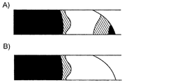
2つの電極間に電圧が印加されると抵抗が最も小さい経路を通って電流が流れ、ほとんどの場合その経路は最短経路と一致する。したがって、特定の組織領域がエレクトロポレーションされないことを保証するための可能な1つの方法は、その領域を電極の後ろ、すなわち電極対で挟まれる領域の外に置くことである。一部の場合においては、実際に組織の位置をずらすかまたは分離用のスペーサーを使うことが可能である(セクション1.2)。他の事例において、図2Aに示す戦略を用いてもよい。すなわち、保護対象の組織領域(図2Aの下層)が両電極間の領域の外になるように埋め込み電極を構築する。
Shielding the tissue area (1.4.1)
When a voltage is applied between the two electrodes, current flows through the path with the smallest resistance, and in most cases the path matches the shortest path. Thus, one possible way to ensure that a particular tissue region is not electroporated is to place that region behind the electrode, ie, outside the region sandwiched by the electrode pair. In some cases, it is possible to actually shift the position of the tissue or use a separation spacer (section 1.2). In other cases, the strategy shown in FIG. 2A may be used. That is, the embedded electrode is constructed so that the tissue region to be protected (the lower layer in FIG. 2A) is outside the region between both electrodes.
図2Aに示す例は、筋肉および他のより深部の構造5を保護する必要があるる、皮膚1のエレクトロポレーションの事例を示している。この過程はシリンジによってゲル6を皮下に注入することから始まる。次に、軸上に電気絶縁体8を有する同じ注射針7または皮膚とゲル6とに通したワイヤを、ゲル領域(注入された電極)とパルス発生器端子との間の電気接続用に使用する。この方法により、概ね、ゲル6と上部電極9との間の皮膚2だけがエレクトロポレーションされる。提唱されるこの方法の1つの興味深い特徴は、注入可能な電極10を処置対象の領域2の形態に適合させてもよいことである。
The example shown in FIG. 2A shows an example of electroporation of the skin 1 that needs to protect muscles and other deeper structures 5. This process begins with the
特定の組織領域5を保護するための注入可能な電極10の使用を図2Aに示す。ゲル6および電極10は上層の下に注入される。電圧パルスが印加されると、ゲル6と上部電極9との間の領域だけがエレクトロポレーションされる。
The use of an
血管11など中空構造のエレクトロポレーションを図2Bに示す。ゲル電極12をパルス発生器に接続するためにも使用されるカテーテル13を介して、ゲル電極12を注入する。反対側の電極14は身体表面に置いてもよい。
Electroporation of a hollow structure such as the
セクション3.1.4は、注入された電極が特定の組織領域を保護するためにどのように用いられうるかを示す事例のシミュレーション結果を含む。ただし、この事例では、外側の層を損傷することなく内部組織を可逆的にエレクトロポレーションすることが目的である。 Section 3.1.4 contains example simulation results that show how an implanted electrode can be used to protect a particular tissue region. However, in this case, the goal is to reversibly electroporate the internal tissue without damaging the outer layers.
中空構造をエレクトロポレーションする方法(1.4.2)
血管組織のエレクトロポレーションは、血液により提供される導電率を利用する管腔内カテーテルによって可能である(N.B. Dev, T.J. Preminger, G.A. Hofmann, S.B. Dev, Sustained local delivery of heparin to the rabbit arterial wall with an electroporation catheter, Catheterization and Cardiovascular Diagnosis 45 (1998) 337-345)。しかし、胃腸管または尿路など他の場合においては、管腔内電極と臓器壁との間に天然の電気接続媒体がない。これらの場合については、処置対象の壁と直接接続するよう設計された可撓性の電極が提唱されている(D.M. Soden, J.O. Larkin, C.G. Collins, M. Tangney, S. Aarons, J. Piggott, A. Morrissey, C. Dunne, G.C. O'Sullivan, Successful application of targeted electrochemotherapy using novel flexible electrodes and low dose bleomycin to solid tumours, Cancer Letters 232 (2006) 300-310)。本明細書において本発明者らは、導電性のゲルまたはペーストなどの本研究で研究した添加剤がはるかに簡便でなおかつ有効な解決法となりうることを提唱する。例えば、図2Bに示すように、電源14にさらに接続されたワイヤと接触状態になったときにエレクトロポレーション用電極として機能しうる導電性のゲル15を、カテーテル13を使用して脈管11に注入してもよい。この戦略の1つの利点とは、ゲル15の調製物が治療用薬剤も含むことができ、エレクトロポレーション用の管腔内カテーテルの場合のようにこれをバルーンまたは他の手段により処置対象の領域内に閉じ込める必要がないことである(N.B. Dev, T.J. Preminger, G.A. Hofmann, S.B. Dev, Sustained local delivery of heparin to the rabbit arterial wall with an electroporation catheter, Catheterization and Cardiovascular Diagnosis 45 (1998) 337-345)。
Electroporation of hollow structure (1.4.2)
Electroporation of vascular tissue is possible with an intraluminal catheter that utilizes the conductivity provided by blood (NB Dev, TJ Preminger, GA Hofmann, SB Dev, Sustained local delivery of heparin to the rabbit arterial wall with an electroporation catheter, Catheterization and Cardiovascular Diagnosis 45 (1998) 337-345). However, in other cases such as the gastrointestinal tract or urinary tract, there is no natural electrical connection medium between the intraluminal electrode and the organ wall. For these cases, flexible electrodes designed to connect directly to the wall to be treated have been proposed (DM Soden, JO Larkin, CG Collins, M. Tangney, S. Aarons, J. Piggott, A. Morrissey, C. Dunne, GC O'Sullivan, Successful application of targeted electrochemotherapy using novel flexible electrodes and low dose bleomycin to solid tumours, Cancer Letters 232 (2006) 300-310). Here we propose that the additives studied in this study, such as conductive gels or pastes, can be a much simpler and more effective solution. For example, as shown in FIG. 2B, a
図2Bの例において、一方の電極12は注入されたゲルであり、他方の電極14は身体表面上の大きな電極である。特に脈管壁の導電率が周囲の実質組織より低い場合、この方法によりゲル15の周囲に最も強い電場が発生すると考えられ、これはほとんどの場合起こりうる。したがって、ゲル周囲の輪状領域だけがエレクトロポレーションされる。そのような構造をセクション3.1.5でシミュレートする。
In the example of FIG. 2B, one
ただし、この場合において、ゲル領域と外部電極との間のインピーダンスは十分正確にはわからないと考えられるため、必要なエレクトロポレーション電圧を演繹的に計算することは些末ではないことに留意されたい。この不都合を克服するため、本発明ではエレクトロポレーション前に内部電極と外部電極との間のインピーダンスを測定する。この測定結果から、エレクトロポレーションの際の系の抵抗の近似値(R)が得られる。すなわち以下
![]()
![]()
![]()
![]()
脈管壁厚が脈管の直径よりはるかに小さければ、脈管壁を横切る電場は非常に均一になると考えられる。 If the vessel wall thickness is much smaller than the vessel diameter, the electric field across the vessel wall will be very uniform.
イオン性ゲル
ゲルはコロイド分散であり、ここで、分散媒は液体であり、連続媒質は固体であり一般にはポリマー鎖のネットワークである。水が液体媒質である特定の場合において、ゲルはヒドロゲルとも呼ばれる。ほとんどのゲルの興味深い特性はチキソトロピーであり、すなわちこれらは、力学的に乱されるとより流動的になる。したがって、安定した状態においてゲルは軟らかい固体または粘度の高い流体としての挙動を示すことができる一方、剪断力の作用のおかげで小ゲージの針を通して容易に注入することもできる。ヒドロゲルは、乳房インプラント、創傷被覆材、薬物送達系、電極、およびコンタクトレンズなど、種々の医学用途に広範に使用されている。このタイプのヒドロゲルを、生体適合性、生分解性、温度感受性、および化学物質感受性などの特徴が実現される本発明との関連において使用することができる。
Ionic gels are colloidal dispersions, where the dispersion medium is a liquid and the continuous medium is a solid, generally a network of polymer chains. In the specific case where water is the liquid medium, the gel is also referred to as a hydrogel. An interesting property of most gels is thixotropy, i.e. they become more fluid when mechanically disturbed. Thus, in a stable state, the gel can behave as a soft solid or a viscous fluid, but can also be easily injected through a small gauge needle thanks to the action of shear forces. Hydrogels are widely used in various medical applications such as breast implants, wound dressings, drug delivery systems, electrodes, and contact lenses. This type of hydrogel can be used in the context of the present invention where features such as biocompatibility, biodegradability, temperature sensitivity, and chemical sensitivity are realized.
所望の導電率を有する液体またはゲルを生成するための直接的な1つの方法は、自由イオンの含有量を制御することである。おそらく、低イオン性(hypoionic)溶液は、これらが短時間適用されかつ等張性となるよう非イオン種でバランスが取られているのであれば、生体組織に対して有意な影響を及ぼさないだろう。一方、導電率の高いゲルはほぼ確実に高張性を示唆する。したがって、浸透圧不均衡によるいくらかの組織損傷(細胞の脱水により引き起こされる細胞傷害性)が予測されうる。事実、高張のゲルは切除方法として提唱されている(J. Rehman, J. Landman, D. Lee, R. Venkatesh, D.G. Bostwick, C. Sundaram, R.V. Clayman, Needle-Based Ablation of Renal Parenchyma Using Microwave, Cryoablation, Impedance- and Temperature-Based Monopolar and Bipolar Radiofrequency, and Liquid and Gel Chemoablation: Laboratory Studies and Review of the Literature, Journal of Endourology 18 (2004) 83-104)。にもかかわらず、非常に高張のゲル(23.4% NaCl)が試されたが、観察される損傷は小さいままであった。非常に幸運なことに、本明細書に提示する理論的な結果によれば、15% NaCl(σ ≒ 240 mS/cm)を上回る濃度を用いる必要はないと考えられる。したがって、高張ゲルの存在が必要になるのは短時間だけであろうことを考慮に入れると、材料の生体適合性に関する問題は軽減または解消される。 One direct way to produce a liquid or gel with the desired conductivity is to control the free ion content. Perhaps hypoionic solutions do not have a significant effect on living tissue if they are applied for a short time and are balanced with nonionic species so that they are isotonic. Let's go. On the other hand, high conductivity gels almost certainly suggest hypertonicity. Thus, some tissue damage due to osmotic imbalance (cytotoxicity caused by cell dehydration) can be predicted. In fact, hypertonic gels have been proposed as excision methods (J. Rehman, J. Landman, D. Lee, R. Venkatesh, DG Bostwick, C. Sundaram, RV Clayman, Needle-Based Ablation of Renal Parenchyma Using Microwave, Cryoablation, Impedance- and Temperature-Based Monopolar and Bipolar Radiofrequency, and Liquid and Gel Chemoablation: Laboratory Studies and Review of the Literature, Journal of Endourology 18 (2004) 83-104). Nevertheless, a very hypertonic gel (23.4% NaCl) was tried, but the observed damage remained small. Fortunately, according to the theoretical results presented herein, it may not be necessary to use concentrations above 15% NaCl (σ≈240 mS / cm). Thus, taking into account that the presence of a hypertonic gel will only be required for a short period of time, the issues regarding material biocompatibility are reduced or eliminated.
流体の導電率を制御するためにイオンを使用することの興味深い代替として、ナノテクノロジーで得られるものを含む微視的粒子の使用がありうる。例えば、導電率特性と、磁場を介して操作される磁気特性とを組み合わせた粒子である。 An interesting alternative to using ions to control fluid conductivity can be the use of microscopic particles, including those obtained with nanotechnology. For example, particles that combine conductivity properties and magnetic properties that are manipulated via a magnetic field.
方法(2)
有限要素法により計算される電場分布(2.1)
本発明では、エレクトロポレーションを制御するための電解性および非電解性のゲルの種々の適用を探究および説明するため数学的解析を使用する。この目的のため、本発明者らは、導電率が一定かつ電流および電場が静的であるという仮定のもと、有限要素法(FEM)を用いて電場分布を計算する。この方法論は本分野において以前の研究者らに使用されており(D. Sel, S. Mazeres, J. Teissie, D. Miklavcic, Finite-element modeling of needle electrodes in tissue from the perspective of frequent model computation, IEEE Trans. Biomed. Eng. 50 (2003) 1221; S.B. Dev, D. Dhar, W. Krassowska, Electric field of a six-needle array electrode used in drug and DNA delivery in vivo: analytical versus numerical solution, IEEE Trans. Biomed. Eng. 50 (2003) 1296; K. Sugibayashi, M. Yoshida, K. Mori, T. Watanabe, T. Hasegawa, Electric field analysis on the improved skin concentration of benzoate by electroporation, International Journal of Pharmaceutics 219 (2001) 107-112)、その妥当性も証明されている(J. Edd, L. Horowitz, R.V. Davalos, L.M. Mir, B. Rubinsky, In-Vivo Results of a New Focal Tissue Ablation Technique: Irreversible Electroporation, IEEE Trans. Biomed. Eng. 53 (2006) 1409-1415; D. Miklavcic, D. Semrov, H. Mekid, L.M. Mir, A validated model of in vivo electric field distribution in tissues for electrochemotherapy and for DNA electrotransfer for gene therapy, Biochimica et Biophysica Acta 1523 (2000) 73-83)。
Method (2)
Electric field distribution calculated by the finite element method (2.1)
In the present invention, mathematical analysis is used to explore and account for various applications of electrolytic and non-electrolytic gels to control electroporation. For this purpose, we calculate the electric field distribution using the finite element method (FEM) under the assumption that the conductivity is constant and the current and electric field are static. This methodology has been used by previous researchers in the field (D. Sel, S. Mazeres, J. Teissie, D. Miklavcic, Finite-element modeling of needle electrodes in tissue from the perspective of frequent model computation, IEEE Trans. Biomed. Eng. 50 (2003) 1221; SB Dev, D. Dhar, W. Krassowska, Electric field of a six-needle array electrode used in drug and DNA delivery in vivo: analytical versus numerical solution, IEEE Trans. Biomed. Eng. 50 (2003) 1296; K. Sugibayashi, M. Yoshida, K. Mori, T. Watanabe, T. Hasegawa, Electric field analysis on the improved skin concentration of benzoate by electroporation, International Journal of Pharmaceutics 219 (2001 107-112) and its validity has been proven (J. Edd, L. Horowitz, RV Davalos, LM Mir, B. Rubinsky, In-Vivo Results of a New Focal Tissue Ablation Technique: Irreversible Electroporation, IEEE Trans Biomed. Eng. 53 (2006) 1409-1415; D. Miklavcic, D. Semrov, H. Mekid, LM Mir, A validated model of in vivo ele ctric field distribution in tissues for electrochemotherapy and for DNA electrotransfer for gene therapy, Biochimica et Biophysica Acta 1523 (2000) 73-83).
FEMの重要な概念とは、任意の幾何学形状を、研究対象の現象に関連する微分方程式を解くことが可能となるような小さく単純な要素へと分解することである。適切な境界条件が与えられると、次に解の集合が得られ、完全な幾何学形状に対する近似解が提供される。本発明では、各要素について解かれる方程式はポアソン方程式である:
![]()
![]()
ここで使用した具体的なFEMツールはCOMSOL Multiphysics 3.2(www.comsol.com)であり、シミュレーション用に選択したモードは「3D conductive media DC」である。境界条件は、外面がすべて絶縁されていることとした。FEMツールにより四面体要素の非構造メッシュが自動的に生成された。 The specific FEM tool used here is COMSOL Multiphysics 3.2 (www.comsol.com), and the mode selected for simulation is “3D conductive media DC”. The boundary condition was that all outer surfaces were insulated. An unstructured mesh of tetrahedral elements was automatically generated by the FEM tool.
解析した事例の幾何学形状およびシミュレーションに関連するその他の詳細は次のセクションで述べる。特に断りがない限り、Eth = 500 V/cm(可逆的エレクトロポレーション閾値)、Eth_irr = 1000 V/cm(不可逆的エレクトロポレーション閾値)、組織の導電率(σ)は1 mS/cmと仮定してシミュレーションを行った。得られたグラフにおいて、黒色はE<500 V/cm(作用なし)、灰色は500 V/cm≦E<1000 V/cm(可逆的エレクトロポレーション)、白色はE≧1000 V/cm(不可逆的エレクトロポレーション)を示す。 The analyzed geometry and other details related to the simulation are described in the next section. E th = 500 V / cm (reversible electroporation threshold), E th_irr = 1000 V / cm (irreversible electroporation threshold), tissue conductivity (σ) is 1 mS / cm unless otherwise noted Simulation was performed assuming that. In the obtained graph, black is E <500 V / cm (no action), gray is 500 V / cm ≦ E <1000 V / cm (reversible electroporation), white is E ≧ 1000 V / cm (irreversible) Electroporation).
実施例
以下の実施例は、本発明をどのように作製および使用するかについて完全な開示および説明を当業者に提供するために提示するものであり、本発明者らが自らの発明であると考える範囲を限定する意図はなく、以下の実験が実施されたすべてまたは唯一の実験であることを表す意図もない。用いた数字(例えば量、温度など)に関して正確性を確保するよう努力が払われているが、いくらかの実験誤差および偏りが計上されるべきである。特に指示がない限り、割合は重量あたりの割合であり、分子量は重量平均分子量であり、温度は摂氏温度であり、圧力は大気圧または大気圧付近である。
EXAMPLES The following examples are presented in order to provide those skilled in the art with a complete disclosure and description of how to make and use the present invention and that they are their invention. There is no intention to limit the scope considered, nor is it intended to represent that the following experiment is all or the only experiment performed. Efforts have been made to ensure accuracy with respect to numbers used (eg amounts, temperature, etc.) but some experimental errors and deviations should be accounted for. Unless otherwise indicated, percentages are percentages by weight, molecular weight is weight average molecular weight, temperature is in degrees Centigrade, and pressure is at or near atmospheric.
概念のインビボ立証(2.2)
エレクトロポレーションにゲルを使用することの実現可能性を実証するため、本発明者らは、数学的解析に加えて、ラット肝葉の端部を2枚の板電極で不可逆的にエレクトロポレーションする実験を実施した。
In vivo proof of concept (2.2)
In order to demonstrate the feasibility of using gels for electroporation, in addition to mathematical analysis, we irreversibly electroporated the ends of rat liver lobes with two plate electrodes. An experiment was conducted.
本発明者らは、以前の実験的研究(J. Edd, L. Horowitz, R.V. Davalos, L.M. Mir, B. Rubinsky, In-Vivo Results of a New Focal Tissue Ablation Technique: Irreversible Electroporation, IEEE Trans. Biomed. Eng. 53 (2006) 1409-1415)から、ラット肝臓において不可逆的にエレクトロポレーションされた領域は、肉眼的(充血による暗色化)にも顕微鏡的にも観察可能な赤血球の捕捉を30分以内に示すことを認識している。したがって、この現象を用いて、可逆的なエレクトロポレーションを受けた領域と不可逆的なエレクトロポレーションを受けた領域との境界面を推定でき、これによりエレクトロポレーションの際の電場の分布を推定することができる。 The inventors have previously conducted experimental studies (J. Edd, L. Horowitz, RV Davalos, LM Mir, B. Rubinsky, In-Vivo Results of a New Focal Tissue Ablation Technique: Irreversible Electroporation, IEEE Trans. Biomed. From Eng. 53 (2006) 1409-1415), irreversibly electroporated regions in the rat liver capture red blood cells within 30 minutes, which can be observed both macroscopically (darkened by hyperemia) and microscopically. Recognize that. Therefore, using this phenomenon, it is possible to estimate the boundary surface between a region that has undergone reversible electroporation and a region that has undergone irreversible electroporation, thereby estimating the electric field distribution during electroporation. can do.
実験手順(2.2.1)
California大学(Berkeley)のOffice of Laboratory Animal Careを通じて、Charles River Labsより雄性Sprague-Dawleyラット1匹(350 g)を入手した。ラットは、Institute of Laboratory Animal Resourcesが作成および策定しU.S. National Institutes of Health(NIH)が発表したPrincipals of Laboratory Animal CareおよびGuide for the Care and Use of Laboratory Animalsに準拠し、適切な訓練を受けた専門家による人道的な配慮を受けた。
Experimental procedure (2.2.1)
One male Sprague-Dawley rat (350 g) was obtained from Charles River Labs through the Office of Laboratory Animal Care at the University of California (Berkeley). Rats are professionally trained in accordance with the Principles of Laboratory Animal Care and Guide for the Care and Use of Laboratory Animals, created and formulated by the Institute of Laboratory Animal Resources and published by the US National Institutes of Health (NIH). Received humanitarian consideration by the house.
実験は、ラット体重1 kgあたりペントバルビタールナトリウムが総量100 mg となるよう、Nembutal溶液(50 mg/ml ペントバルビタールナトリウム, Abbott Labs, イリノイ州North Chicago)を腹腔内注射して動物を麻酔することから開始した。30分後、正中切開により肝臓を露出させた。 The experiment involves anesthetizing animals by intraperitoneal injection of Nembutal solution (50 mg / ml sodium pentobarbital, Abbott Labs, North Chicago, Ill.) So that the total amount of sodium pentobarbital is 100 mg per kg body weight of the rat. Started. After 30 minutes, the liver was exposed through a midline incision.
ラット肝臓の露出後、5 mm間隔で離した2つの平らな円形電極の間に肝葉を置いた。幾何学的な不規則性をエミュレートするため、肝葉端部を2つの電極間に部分的に挿入した。肝臓に対する電極の配置は、同時にこの事例のFEMシミュレーションのモデルでもある、図8に示したものと類似していた。次に、空隙を導電率一致ゲルで満たし、市販のパルス発生器(ECM 830, Harvard Apparatus, マサチューセッツ州Holliston)によりエレクトロポレーションパルスシーケンス(750 V、持続時間100 μs、周期100 msを8パルス)を印加した。 After exposure of the rat liver, the liver lobe was placed between two flat circular electrodes spaced 5 mm apart. To emulate geometric irregularities, the liver lobe edge was partially inserted between the two electrodes. The placement of the electrodes relative to the liver was similar to that shown in FIG. 8, which is also the model for the FEM simulation in this case. The gap is then filled with a conductivity matching gel and electroporated pulse sequence (750 V, duration 100 μs, period 100 ms, 8 pulses) with a commercial pulse generator (ECM 830, Harvard Apparatus, Holliston, Mass.) Was applied.
エレクトロポレーションシーケンスから2時間半後、動物を安楽死させ、肝臓標本を組織学的分析用に調製した。 Two and a half hours after the electroporation sequence, the animals were euthanized and liver specimens were prepared for histological analysis.
組織学(2.2.2)
顕微鏡観察用に肝臓を現状のまま固定するため、本発明者らは、高く持ち上げた静脈内滴注による静水圧80 mmHgの生理食塩水で脈管構造を10分間洗い流した。このことは、流体を左心室に注入し、右心房に作った切開部から流出させることによって実現した。生理食塩水灌流の直後に、5%ホルムアルデヒドの固定剤を同じ方法で10分間灌流させた。次に、処置した肝葉を取り出し、同じホルムアルデヒド溶液中で保存した。次に、エレクトロポレーションの影響を調べるため、処置領域の中心を通る断面についてヘマトキシリン-エオシン染色を行った。
Histology (2.2.2)
In order to fix the liver as it is for microscopic observation, the present inventors washed the vasculature for 10 minutes with physiological saline with a hydrostatic pressure of 80 mmHg by intravenous drip that was lifted high. This was achieved by injecting fluid into the left ventricle and out of the incision made in the right atrium. Immediately following saline perfusion, 5% formaldehyde fixative was perfused in the same manner for 10 minutes. The treated liver lobe was then removed and stored in the same formaldehyde solution. Next, in order to examine the influence of electroporation, hematoxylin-eosin staining was performed on a cross section passing through the center of the treatment area.
ゲルの調製(2.2.3)
本発明者らは、標準的な生理溶液(0.9% NaCl)より濃度が20倍低い0.045% NaCl溶液から生理食塩水ゲルを調製した。このような電解質含有量は約0.7 mS/cmの導電率をもたらすと考えられる。報告されている肝臓の導電率はこれより少し高い(約1 mS/cm)可能性があるが、シミュレーションが示すように、この違いが重大な影響をもたらすことはないと考えられる。ゲル作製の段階は次のとおりである:(1) 0.045% NaCl溶液100 mlに生の寒天0.8 gを添加する;(2) 沸点において生理食塩水中で寒天を溶かす;(3) 凝固するまで溶液を冷却する;(4) ゲルが形成されるまで撹拌する。
Gel preparation (2.2.3)
We prepared a saline gel from a 0.045% NaCl solution that was 20 times lower in concentration than a standard physiological solution (0.9% NaCl). Such electrolyte content is believed to result in a conductivity of about 0.7 mS / cm. The reported liver conductivity may be slightly higher (about 1 mS / cm), but as the simulation shows, this difference is not expected to have a significant effect. The steps for gel preparation are as follows: (1) Add 0.8 g of raw agar to 100 ml of 0.045% NaCl solution; (2) Dissolve the agar in physiological saline at the boiling point; (3) Solution until solidified (4) Stir until a gel is formed.
結果および考察(3)
典型的な用途のシミュレーション(3.1)
本セクションの目的は、導入部で提起した概念を典型的な例により説明することである。
Results and discussion (3)
Typical application simulation (3.1)
The purpose of this section is to illustrate the concepts raised in the introductory part with typical examples.
組織の絶縁(3.1.1)
本セクションで本発明者らは図1で述べた事例(セクション1.2)をシミュレートする。すなわち、組織の上層を、保護する必要がある下部組織から物理的に分離するため、上層の下に非導電性のゲルを注入する。この特定の事例は不可逆的エレクトロポレーションにより除去される必要がある皮膚黒色腫を表す。
Tissue insulation (3.1.1)
In this section we simulate the case described in Figure 1 (section 1.2). That is, a non-conductive gel is injected under the upper layer to physically separate the upper layer of tissue from the lower tissue that needs to be protected. This particular case represents cutaneous melanoma that needs to be removed by irreversible electroporation.
シミュレーションに使用したモデルは5つの構成要素からなる:(1) 下部組織(組織2)のモデルとなる導電率 = 1 mS/cmの正四角柱(20 mm×20 mm×5 mm);(2) 該角柱の上の、絶縁ゲル用の導電率 0.1 mS/cmの半楕円領域(half ellipsoid volume)(10 mm×10 mm×0.5 mm);(3) 上部組織(組織1)のモデルとなる、両構成要素の上の導電率 = 1 mS/cmの被膜(厚さ = 1 mm);ならびに、(4) および (5) 上部被膜を貫通する電極を表す、高導電率(1000 S/cm)の2つの円柱(直径 = 0.1 mm、長さ = 5 mm)。電極間の分離距離は2 mmであり、印加される差分電圧は1000 Vである。メッシュの要素数は102,614である。 The model used for the simulation consists of five components: (1) Regular square column (20 mm x 20 mm x 5 mm) with conductivity = 1 mS / cm, which is a model of the substructure (tissue 2); (2) On the prism, a half ellipsoid volume (10 mm × 10 mm × 0.5 mm) with a conductivity of 0.1 mS / cm for insulating gel; (3) a model of the superstructure (tissue 1); Conductivity on both components = 1 mS / cm coating (thickness = 1 mm); and (4) and (5) High conductivity (1000 S / cm) representing the electrode through the top coating Two cylinders (diameter = 0.1 mm, length = 5 mm). The separation distance between the electrodes is 2 mm and the applied differential voltage is 1000 V. The number of mesh elements is 102,614.
ゲル中の不純物および埋植後の組織からのイオン拡散によってゲルが完全な絶縁体ではなくなるという事実を考慮に入れるため、本発明者らが絶縁用ゲルの導電率として、理想的な0 mS/cmではなく0.1 mS/cmを選択したことに注意されたい。 In order to take into account the impurities in the gel and the fact that the gel is no longer a perfect insulator due to ion diffusion from the tissue after implantation, we have determined that the conductivity of the insulating gel is an ideal 0 mS / Note that we chose 0.1 mS / cm instead of cm.
シミュレーションの結果(図3)は、上部組織2は電極3および4を囲む領域全体にわたって不可逆的にエレクトロポレーションされたが、下層に対する損傷はごくわずかであり、かつ2つの単一スポットが可逆的エレクトロポレーションを受けたのみであることを示す。シミュレーションしたゲル6は完全な非導電性ではなく、実際のところ、その導電率は組織の導電率より1桁低いだけであることは、興味深いことである。
The simulation results (Figure 3) show that the
図3は、スペーサーとして使用した非導電性ゲル6による組織領域保護のシミュレーション結果を示している(組織1の導電率 = 組織5の導電率 = 1 mS/cm、ゲルの導電率 = 0.1 mS/cm;ゲル領域の直径 = 10 mm、ゲル領域の高さ = 0.5 mm;組織1の厚さ = 1 mm;電極の直径 = 0.1 mm、電極の分離間隔 = 2 mm)。図3Aは両電極軸を含む垂直面を示している。図3Bは左側の図に灰色の線で示した高さ(関心対象の領域における組織1の厚さの約1/4)における水平面のシミュレーション像を示している。黒色はE<500 V/cm(作用なし)、灰色は500 V/cm≦E<1000 V/cm(可逆的エレクトロポレーション)、白色はE≧1000 V/cm(不可逆的エレクトロポレーション)を示す。
FIG. 3 shows a simulation result of tissue region protection with
不規則な形状の組織における電場の均一化(3.1.2)
本セクションに提示する実施例はセクション1.3で述べた用途に関連する。すなわち、導電率を一致させた添加剤による不規則な形状の組織における電場の均一化である。
Electric field uniformity in irregularly shaped tissue (3.1.2)
The examples presented in this section relate to the applications described in section 1.3. That is, the electric field is uniformized in an irregularly shaped tissue by an additive having the same conductivity.
図4に、半球形の腫瘍16(直径 = 1 cm)を550 Vでエレクトロポレーションするシミュレーションを示す。図4Aはシミュレーションで用いたモデルを示す。図4Bはゲル19が存在する場合のシミュレーションから得られた電場強度を示す(電極の中心を通る垂直面)。図4Cは導電率一致ゲルを用いない場合のシミュレーションされた電場強度を示す(電極17および18の中心を通る垂直面)。図4Dは、図4Cと同じであるがここでは電極間の電圧が1100 Vである。
FIG. 4 shows a simulation of electroporation of a hemispherical tumor 16 (diameter = 1 cm) at 550 V. FIG. 4A shows the model used in the simulation. FIG. 4B shows the electric field strength obtained from the simulation in the presence of gel 19 (vertical plane through the center of the electrode). FIG. 4C shows the simulated electric field strength without a conductivity matching gel (vertical plane through the centers of
図4Aに示す構造は、不規則な形状の硬い腫瘍を板電極を通事知恵エレクトロポレーションする事例に対応しうる。このモデルは、組織(導電率 = 1 mS/cm)を表す正四角柱(50 mm×50 mm×20 mm)の上にある半球(直径 = 10 mm)、およびこの半球の2つの向かい合う側面にある2枚の板電極(20 mm×10 mm×1 mm;導電率 =1000 S/cm)からなる。組織部分の最上部には、半球形のくぼみ(直径 = 2 mm)という、形状のさらなる不規則性を含んでいる。メッシュの要素数は21,888である。電極間に印加される電圧は550 Vである。 The structure shown in FIG. 4A can correspond to a case where an irregularly shaped hard tumor is electroporated through a plate electrode. This model is on a hemisphere (diameter = 10 mm) on a regular square column (50 mm x 50 mm x 20 mm) representing the tissue (conductivity = 1 mS / cm), and on two opposite sides of this hemisphere It consists of two plate electrodes (20 mm x 10 mm x 1 mm; conductivity = 1000 S / cm). The top of the tissue part contains a further irregularity of shape, a hemispherical depression (diameter = 2 mm). The number of elements in the mesh is 21,888. The voltage applied between the electrodes is 550 V.
ゲルがない場合のエレクトロポレーションのシミュレーション結果を図4Cに示す。電場分布はきわめて不均一である。さらに、非常に高い電圧を用いた事例でも(図4D)、エレクトロポレーションされていない領域がある。無論、これは癌治療では許容不可能なものであり、もしかするとその理由から、この種の腫瘍については板電極より針アレイ電極のほうが好まれる。一方、導電率一致ゲルの追加をシミュレートすると(図4B)、その結果は、組織の導電率とゲルの導電率との整合性が完全ではない(整合性誤差 = 30%)この事例においてさえ、電場はさらにもっと均一である。 The simulation result of electroporation in the absence of gel is shown in FIG. 4C. The electric field distribution is very uneven. Furthermore, even in the case of using very high voltages (Fig. 4D), there are areas that are not electroporated. Of course, this is unacceptable for cancer treatment, and for that reason needle array electrodes are preferred over plate electrodes for this type of tumor. On the other hand, simulating the addition of a conductivity matching gel (Figure 4B) shows that the result is not a perfect match between tissue conductivity and gel conductivity (consistency error = 30%) even in this case The electric field is even more uniform.
血管を含む組織における電場の均一化(3.1.3)
本セクションに提示するシミュレーションもセクション1.3に関連する。この事例は、近傍に血管を含む領域の可逆的エレクトロポレーションを表す(図5A)。アレイ間の領域に非常に均一な電場を発生させると考えられる、針電極の平行なアレイ2つを用いてエレクトロポレーションを実施する。
Electric field homogenization in tissues including blood vessels (3.1.3)
The simulation presented in this section is also relevant to section 1.3. This case represents reversible electroporation of a region that contains nearby blood vessels (FIG. 5A). Electroporation is performed using two parallel arrays of needle electrodes that are believed to generate a very uniform electric field in the area between the arrays.
図5は、下部の境界に血管(直径 = 3 mm)を含む正方形領域の可逆的エレクトロポレーションのシミュレーションを示す。図5Aはシミュレーションに用いたモデルを示しており、各電極(E+およびE-)は3本の針(直径 = 1 mm、分離間隔 = 5 mm、挿入深度 = 5mm)のアレイで構成されている;両アレイ間の分離間隔は10 mmである;血管の導電率 = 10 mS/cm、組織の導電率 = 1 mS/cmであり;印加電圧 = 1000 Vである。図5Bに、血管と実質組織との上部境界面における、シミュレートされた電場強度の水平断面を示し;白色の矢印は血管の上の全くエレクトロポレーションされない領域を示している。図5Cに、アレイ中心を通るシミュレーション結果の垂直断面を示し;同じく、白色の矢印は血管の上の全くエレクトロポレーションされない領域を示している。図5Dに、血管の導電率を10 mS/cmから1.5 mS/cmに変更した場合の同じ結果を示す。 FIG. 5 shows a simulation of reversible electroporation in a square area with blood vessels (diameter = 3 mm) at the lower boundary. Figure 5A shows the model used for the simulation, where each electrode (E + and E-) consists of an array of three needles (diameter = 1 mm, separation interval = 5 mm, insertion depth = 5 mm) Separation distance between both arrays is 10 mm; blood vessel conductivity = 10 mS / cm, tissue conductivity = 1 mS / cm; applied voltage = 1000 V. FIG. 5B shows a simulated horizontal cross-section of the electric field strength at the upper boundary between the blood vessel and the parenchyma; the white arrow indicates the area on the blood vessel that is not electroporated at all. FIG. 5C shows a vertical cross section of the simulation results through the center of the array; similarly, the white arrows indicate the areas on the vessel that are not electroporated at all. FIG. 5D shows the same result when the blood vessel conductivity is changed from 10 mS / cm to 1.5 mS / cm.
シミュレーションに使用したモデルは以下からなる:(1) 導電率が1 mS/cmである組織を表す長方角柱(50 mm×50 mm×20 mm);(2) 5 mmの深さで角柱の1つの側面から他の側面へと走行する血管を表す円柱(導電率 =10 mS/cm;直径 = 3 mm;長さ = 50 mm);および、(3) 互いに10 mm間隔で平行に置かれた2つの電極アレイ。両アレイの各々は3本の円柱形の棒(導電率 = 1000 S/cm;直径 = 1 mm、長さ = 15 mm、分離間隔 = 5 mm)からなる。メッシュの要素数は68,571である。両アレイ間に印加される電圧は1000 Vである。 The model used for the simulation consists of the following: (1) a rectangular prism (50 mm x 50 mm x 20 mm) representing a structure with a conductivity of 1 mS / cm; (2) a 5 mm deep prism A cylinder representing a blood vessel running from one side to the other (conductivity = 10 mS / cm; diameter = 3 mm; length = 50 mm); and (3) placed parallel to each other at 10 mm intervals Two electrode arrays. Each of both arrays consists of three cylindrical rods (conductivity = 1000 S / cm; diameter = 1 mm, length = 15 mm, separation distance = 5 mm). The number of mesh elements is 68,571. The voltage applied between both arrays is 1000 V.
シミュレーション結果が示すところによれば、結果として生じた電場分布不均一性のため、エレクトロポレーションされない薄く細長い組織があった(図5Bおよび図5C)。一方、導電率を一致させた添加剤で血液を置換(血管の導電率を10 mS/cmから1.5 mS/cmに変更)する場合、シミュレートされた電場分布(図5D)はさらにより均一であり、電極間の領域を完全にエレクトロポレーションするという目的が達成される。 Simulation results show that there was a thin and elongated tissue that was not electroporated due to the resulting electric field distribution inhomogeneity (FIGS. 5B and 5C). On the other hand, the simulated electric field distribution (Figure 5D) is even more uniform when the blood is replaced with an additive with matched conductivity (vessel conductivity changed from 10 mS / cm to 1.5 mS / cm). Yes, the goal of complete electroporation of the area between the electrodes is achieved.
皮下電極(3.1.4)
本セクションに提示する実施例はセクション1.4.1で述べた用途に関連する。すなわち、特定の組織領域を保護するために注入可能な電極を使用する。
Subcutaneous electrode (3.1.4)
The examples presented in this section relate to the applications described in section 1.4.1. That is, an injectable electrode is used to protect a particular tissue region.
図6に、軸上に絶縁体を有する金属針(E+およびE-)に接続された半楕円形の注入ゲル領域を介して700 Vで筋肉(厚さ = 5 mm)をエレクトロポレーションするシミュレーションを示す。(a)シミュレーションに用いたモデル;ゲルの導電率 = 200 mS/cm、筋肉の導電率 = 1 mS/cm、皮膚の導電率 = 0.1 mS/cm。図6Bにシミュレーションから得られた電場強度を示す。メッシュの要素数は196,534である。 Figure 6 shows a simulation of electroporating a muscle (thickness = 5 mm) at 700 V through a semi-elliptical injection gel region connected to a metal needle (E + and E-) with an insulator on its axis. Indicates. (A) Model used for simulation: gel conductivity = 200 mS / cm, muscle conductivity = 1 mS / cm, skin conductivity = 0.1 mS / cm. FIG. 6B shows the electric field strength obtained from the simulation. The number of mesh elements is 196,534.
図6Aに示す事例において、目的は、外側の層(厚さ 1 mmの被膜;導電率 = 0.1 mS/cm)を損傷することなく内部組織(長方角柱(20 mm×20 mm×5 mm);導電率 = 1 mS/cm)を可逆的にエレクトロポレーションすることである。これは、図2Aに提示したものと相補的な事例と見なしてもよい。このように選択的なエレクトロポレーションを行うため、本発明では、軸上に絶縁体を有する針を介してエレクトロポレーション対象領域の両側に導電性のゲルを注入することを使用してもよい。この方法により、両方の領域が平行な板電極として挙動する(ここでゲル領域は導電率200 mS/cmの半楕円領域(10 mm×10 mm×0.5 mm)としてモデル化される)。事実、シミュレーション結果(図6B)によれば、そのような挙動が得られる。ただし、ゲル領域端部で電場の増強が起こり、これが上部組織(皮膚)にいくらかの損傷を引き起こすことに注意されたい。 In the example shown in Fig. 6A, the objective is to create an internal structure (rectangular prism (20 mm x 20 mm x 5 mm) without damaging the outer layer (1 mm thick coating; conductivity = 0.1 mS / cm)) Electroconductivity reversibly; conductivity = 1 mS / cm). This may be considered a complementary case to that presented in FIG. 2A. In order to perform selective electroporation in this way, in the present invention, it may be used to inject a conductive gel on both sides of a region to be electroporated through a needle having an insulator on an axis. . By this method, both regions behave as parallel plate electrodes (where the gel region is modeled as a semi-elliptical region (10 mm × 10 mm × 0.5 mm) with a conductivity of 200 mS / cm). In fact, according to the simulation result (FIG. 6B), such behavior is obtained. Note, however, that an electric field enhancement occurs at the end of the gel region, which causes some damage to the upper tissue (skin).
中空構造のエレクトロポレーション(3.1.5)
この実施例は図2Bで提示した事例のシミュレーションを示す。空の脈管の中央領域を高導電率のゲルで満たす。次に、このゲルと大きな外部電極との間にエレクトロポレーション電圧を印加する(図7A)。電圧を適切に選択すると、エレクトロポレーションの選択性の点で重大な結果を達成できる。このことはシミュレーション結果により示されている(図7B)。同図は、ゲルと接触している脈管壁だけがエレクトロポレーションされることを示している。
Hollow structure electroporation (3.1.5)
This example shows a simulation of the case presented in FIG. 2B. Fill the central region of the empty vessel with a high conductivity gel. Next, an electroporation voltage is applied between the gel and the large external electrode (FIG. 7A). Proper selection of voltages can achieve critical results in terms of electroporation selectivity. This is shown by the simulation results (Figure 7B). The figure shows that only the vessel wall in contact with the gel is electroporated.
このモデル(図7A)は以下からなる:腔部と外部電極との間の組織を表す大きな円柱(直径 = 100 mm、長さ = 80 mm)(導電率 = 1000 S/cm);抵抗率無限の円柱形の腔部(直径 = 9 mm);腔部と組織との間の、導電率 = 0.25 mS/cmの薄い壁(厚さ = 0.5 mm);および、内部ゲル電極を表し導電率が200 mS/cmである、幾何学形状の中央部の円柱(直径 = 9 mm、長さ = 20 mm)(図7Aでは見えない)。内部電極(ゲルの内側)と外部電極との間の印加電圧は200 Vである。メッシュの要素数は56,364である。 This model (Figure 7A) consists of: a large cylinder representing the tissue between the cavity and the external electrode (diameter = 100 mm, length = 80 mm) (conductivity = 1000 S / cm); resistivity infinite Cylindrical cavity (diameter = 9 mm); conductivity = thin wall of 0.25 mS / cm (thickness = 0.5 mm) between cavity and tissue; and the internal gel electrode represents conductivity A geometrical central cylinder (diameter = 9 mm, length = 20 mm), 200 mS / cm (not visible in Figure 7A). The applied voltage between the internal electrode (inside the gel) and the external electrode is 200 V. The number of mesh elements is 56,364.
図7に、注入された円柱ゲル領域および外部の金属電極により空の血管(外径= 10 mm、内径 = 9 mm)を200 Vでエレクトロポレーションしたシミュレーション結果を示す。図7Aはシミュレーションで用いたモデルを示している;ゲル円柱は血管内の幾何学形状の中央にある;ゲルの導電率 = 200 mS/cm、組織の導電率 = 1 mS/cm、血管壁の導電率 = 0.25 mS/cmである。図7Bにシミュレーションから得られた電場強度を示し;幾何学形状の中央の2つの横断面図を示している。識別が困難ではあるものの、ゲル円柱の位置にある血管壁だけが可逆的にエレクトロポレーションされているのが観察できる。 FIG. 7 shows a simulation result of electroporating an empty blood vessel (outer diameter = 10 mm, inner diameter = 9 mm) at 200 V with the injected cylindrical gel region and an external metal electrode. Figure 7A shows the model used in the simulation; the gel cylinder is in the middle of the geometry within the vessel; gel conductivity = 200 mS / cm, tissue conductivity = 1 mS / cm, vessel wall Conductivity = 0.25 mS / cm. FIG. 7B shows the electric field strength obtained from the simulation; two cross-sectional views of the center of the geometry are shown. Although it is difficult to identify, it can be observed that only the blood vessel wall at the position of the gel cylinder is reversibly electroporated.
概念のインビボ立証(3.2)
概念モデルのインビボ立証(図8)に類似した構造のシミュレーション結果が示すところによれば、ゲルを適用しない場合、肝葉のごく先端は全くエレクトロポレーションされなかった(図9A)。一方、ゲルを適用した場合、導電率一致ゲル(整合性誤差 = 20%)の効果とは、先端も含めて肝葉全体の完全な不可逆的エレクトロポレーションをもたらすことである(図9B)。ホルムアルデヒド固定前のエレクトロポレーション領域の肉眼観察(図10)も、シミュレーション結果と一致していた。シミュレーションで予測された凹型形状の不可逆的エレクトロポレーション領域さえ識別できることに注意されたい。顕微鏡観察(図11A)では、肝葉の先端全体で赤血球の捕捉が起こった一方で内部領域ではそれが存在しなかった(図11B)ことが確認された。
In vivo proof of concept (3.2)
Simulation results of a structure similar to the in vivo proof of concept model (FIG. 8) showed that the very tip of the liver lobe was not electroporated without applying the gel (FIG. 9A). On the other hand, when the gel is applied, the effect of the conductivity matching gel (consistency error = 20%) is to provide complete irreversible electroporation of the entire liver lobe including the tip (FIG. 9B). Visual observation of the electroporation region before formaldehyde fixation (Fig. 10) was also consistent with the simulation results. Note that even concave shaped irreversible electroporation regions predicted by simulation can be identified. Microscopic observation (FIG. 11A) confirmed that red blood cell capture occurred throughout the tip of the liver lobe but was absent in the internal region (FIG. 11B).
図8に、肝葉先端のエレクトロポレーションをシミュレートするために用いたモデルの図を示す(ゲルは示していない)。5 mm間隔で離した円盤電極E+およびE-(直径 = 10 mm、導電率 = 1000 S/cm)の間で肝葉先端をエレクトロポレーションする。印加電圧は750 V、肝臓組織の導電率は1 mS/cmである。メッシュの要素数は61,346である。 FIG. 8 shows a diagram of the model used to simulate hepatic lobe tip electroporation (gel not shown). Electroporate the liver lobe tip between disc electrodes E + and E- (diameter = 10 mm, conductivity = 1000 S / cm) separated by 5 mm. The applied voltage is 750 V, and the liver tissue conductivity is 1 mS / cm. The number of mesh elements is 61,346.
図9に肝葉のエレクトロポレーションのシミュレーション結果を示す(x-y面、z = 0)。図9Aにゲルを用いないシミュレーション結果を示す;不規則なエレクトロポレーションパターンが得られ、先端のかなりの部分は不可逆的エレクトロポレーションを受けていない。図9Bに導電率 = 0.8 mS/cmの充填ゲルが存在する場合のシミュレーション結果を示す;電極間の先端領域が不可逆的にエレクトロポレーションされている。 Fig. 9 shows the simulation result of the electroporation of the liver lobe (x-y plane, z = 0). FIG. 9A shows the simulation results without the gel; an irregular electroporation pattern was obtained and a significant portion of the tip did not undergo irreversible electroporation. FIG. 9B shows the simulation results in the presence of a filled gel with conductivity = 0.8 mS / cm; the tip region between the electrodes is irreversibly electroporated.
図10にエレクトロポレーションされたラット肝葉の図を示す。これはエレクトロポレーション領域を中央で切開したものである。処置された組織と処置されていない組織との境界に白色の点を配置する。 FIG. 10 shows a diagram of the rat liver lobe electroporated. This is an incision in the center of the electroporation region. A white dot is placed at the boundary between the treated and untreated tissue.
図11にエレクトロポレーションされた肝葉の顕微鏡写真を示す。図11Aに肝葉の先端を示す;赤血球の有意な捕捉(矢印で示す)は不可逆的エレクトロポレーションがもたらされたことを示している。図11Bに内部域の中心静脈領域を示す;変質は全く観察されない。バーは100 μmを表す。写真はいずれも同じ倍率である。 FIG. 11 shows a micrograph of the electroporated liver lobe. FIG. 11A shows the tip of the hepatic lobe; significant capture of red blood cells (indicated by arrows) indicates that irreversible electroporation resulted. FIG. 11B shows the central venous region of the inner zone; no alteration is observed. Bar represents 100 μm. All photographs are at the same magnification.
導電性のゲルに基づく腔部および脈管の温熱療法
異なる用途の中でも、人為的に誘発した高体温は化学療法および放射線療法の局所強化過程として特に有用である。
Cavity and vascular thermotherapy based on conductive gels Among other applications, artificially induced hyperthermia is particularly useful as a local enhancement process for chemotherapy and radiation therapy.
現在、制御された様式で体内の脈管および腔部から熱を発生させるために用いることができる臨床的に利用可能な器具がいくつか存在している。しかし、これらの機器はすべてある種の面倒なカテーテルを用いており、そのため用途が特定の状況に限られている。 Currently, there are several clinically available devices that can be used to generate heat from the vessels and cavities in the body in a controlled manner. However, all of these devices use some sort of cumbersome catheter, which limits their use to certain situations.
本発明ではそのような目的のために導電性のゲルを使用する。その方法は腔部のエレクトロポレーションについて提唱したものと同等である。すなわち、腔部を導電性のゲルで満たし、そのゲルに接続した電極と身体表面上の外部電極との間に電流を印加することである。しかしこの場合は、両電極間に印加される電気刺激は、細胞膜のエレクトロポレーションを行うことではなく、ジュール効果により組織および/またはゲルを加熱することを意図したものである。したがって、前記刺激は、振幅がより小さいが持続時間がより長い、AC信号である。実際のところ、処置の持続期間中ずっと連続的に信号を注入してもよく、何らかの種類の温度読み取り値(例えばカテーテル先端の温度センサ)に応じて、制御されたモードにおいて振幅を調節してもよい。 In the present invention, a conductive gel is used for such a purpose. The method is equivalent to that proposed for cavity electroporation. That is, filling the cavity with a conductive gel and applying an electric current between an electrode connected to the gel and an external electrode on the body surface. In this case, however, the electrical stimulation applied between the two electrodes is not intended to electroporate the cell membrane, but to heat the tissue and / or gel by the Joule effect. Thus, the stimulus is an AC signal with a smaller amplitude but a longer duration. In fact, the signal may be injected continuously throughout the duration of the procedure, and the amplitude adjusted in a controlled mode, depending on some kind of temperature reading (eg temperature sensor at the catheter tip). Good.
以上に説明したエレクトロポレーションと同様に、化学療法薬などの治療剤をゲル組成物の一部としてもよい。 Similar to the electroporation described above, a therapeutic agent such as a chemotherapeutic agent may be part of the gel composition.
ゲル組成物は異なる目的に応じて区別されてもよい。 Gel compositions may be distinguished for different purposes.
(1)導電性が高い(例えば電解質の含有量が高い)組成物の場合、ジュール効果による熱損失はゲル内にほとんど生じない。ほとんどのエネルギーはゲル周囲の輪状領域で散逸するが、それは電流密度および抵抗率がより大きいからである。 (1) In the case of a composition having high conductivity (for example, a high electrolyte content), heat loss due to the Joule effect hardly occurs in the gel. Most of the energy is dissipated in the annular region around the gel because of the higher current density and resistivity.
(2)導電性が中程度の組成物の場合、ゲル内でかなりのエネルギーが散逸し、周囲組織は、組織内部の直接的なジュール散逸によってではなく加熱されたゲルとの直接接触によって加熱される。この現象は、ゲルの熱伝導率を高める金属ナノ粒子などの添加剤を使用することによって増強することができる。 (2) For moderately conductive compositions, significant energy is dissipated in the gel and the surrounding tissue is heated by direct contact with the heated gel, not by direct Joule dissipation inside the tissue. The This phenomenon can be enhanced by using additives such as metal nanoparticles that increase the thermal conductivity of the gel.
温熱療法またはエレクトロポレーションのいずれについても、薬物のイオン導入を誘発するために処置前、処置後、または処置中(すなわち重ねて)に電極間にDC電圧を印加してもよい。このことは、ゲル周囲の細胞および組織へのこれら薬物の浸透を高めるために行ってもよい。 For either hyperthermia or electroporation, a DC voltage may be applied between the electrodes before, after, or during (ie, overlapping) to induce iontophoresis of the drug. This may be done to increase penetration of these drugs into the cells and tissues surrounding the gel.
上記は本発明の原理を示したにすぎない。当業者であれば、本明細書に明示的に説明しまたは示してはいないものの本発明の原理を具現化しかつ本発明の趣旨および範囲に含まれる、種々の組合せを考案できると考えられ、このことは理解されるべきである。さらに、本明細書に列挙するすべての実施例および条件的文言は、本発明の原理および本発明者らが本技術を促進するために提供する概念を読者が理解することを助けることをもっぱら意図したものであり、具体的に列挙されたそのような実施例および条件に限定されるわけではないものとして解釈されるべきである。さらに、本発明の原理、局面、および態様、ならびに本発明の具体的な実施例を列挙した、本明細書におけるすべての言明は、それらの構造的および機能的な同等物の両方を包含することを意図している。さらに、そのような同等物は、現在公知の同等物および今後開発される同等物の両方、すなわち、構造にかかわらず同じ機能を実施する、開発されたあらゆる要素を含むことが意図される。したがって、本発明の範囲は、本明細書に示しかつ説明した例示的な態様に限定されるとは意図されない。むしろ、本発明の範囲および趣旨は、添付の特許請求の範囲により具体化される。 The foregoing merely illustrates the principles of the invention. Those skilled in the art will be able to devise various combinations that embody the principles of the invention but are not explicitly described or shown herein, and that fall within the spirit and scope of the invention. That should be understood. Moreover, all examples and conditional language listed herein are intended solely to assist the reader in understanding the principles of the invention and the concepts we provide to facilitate the technology. And should not be construed as being limited to such specifically recited examples and conditions. Furthermore, all statements herein reciting principles, aspects, and embodiments of the invention, as well as specific examples of the invention, include both their structural and functional equivalents. Is intended. In addition, such equivalents are intended to include both currently known equivalents and future developed equivalents, ie, any element developed that performs the same function regardless of structure. Accordingly, the scope of the invention is not intended to be limited to the exemplary embodiments shown and described herein. Rather, the scope and spirit of the invention is embodied by the appended claims.
Claims (17)
該ゲルを注入するためのシリンジ;
患者に挿入されたときに電極間に電流をもたらすための第一および第二の電極;ならびに
エレクトロポレーションを行うための説明書
を含む、エレクトロポレーションを行うためのキット。 A gel with a predetermined conductivity relative to the tissue surrounding the area to be electroporated;
A syringe for injecting the gel;
A kit for performing electroporation, comprising first and second electrodes for providing an electrical current between the electrodes when inserted into a patient; and instructions for performing electroporation.
Applications Claiming Priority (2)
| Application Number | Priority Date | Filing Date | Title |
|---|---|---|---|
| US82958706P | 2006-10-16 | 2006-10-16 | |
| PCT/US2007/022110 WO2008048620A2 (en) | 2006-10-16 | 2007-10-16 | Gels with predetermined conductivity used in irreversible electroporation of tissue |
Publications (2)
| Publication Number | Publication Date |
|---|---|
| JP2010506657A true JP2010506657A (en) | 2010-03-04 |
| JP2010506657A5 JP2010506657A5 (en) | 2010-12-02 |
Family
ID=39314648
Family Applications (1)
| Application Number | Title | Priority Date | Filing Date |
|---|---|---|---|
| JP2009533353A Withdrawn JP2010506657A (en) | 2006-10-16 | 2007-10-16 | Gel with a certain conductivity used for irreversible electroporation of tissue |
Country Status (4)
| Country | Link |
|---|---|
| US (3) | US7674249B2 (en) |
| EP (1) | EP2076313A4 (en) |
| JP (1) | JP2010506657A (en) |
| WO (1) | WO2008048620A2 (en) |
Families Citing this family (43)
| Publication number | Priority date | Publication date | Assignee | Title |
|---|---|---|---|---|
| EP2076313A4 (en) * | 2006-10-16 | 2012-07-25 | Univ California | Gels with predetermined conductivity used in irreversible electroporation of tissue |
| WO2009047774A2 (en) * | 2007-10-09 | 2009-04-16 | Transpharma Ltd. | Magnetic patch coupling |
| CN101969873B (en) | 2007-12-05 | 2015-05-13 | 赛诺龙医疗公司 | A disposable electromagnetic energy applicator and method of using it |
| AU2009205297A1 (en) | 2008-01-17 | 2009-07-23 | Syneron Medical Ltd. | A hair removal apparatus for personal use and the method of using same |
| EP2237732A4 (en) | 2008-01-24 | 2011-06-01 | Syneron Medical Ltd | A device, apparatus, and method of adipose tissue treatment |
| US9867652B2 (en) | 2008-04-29 | 2018-01-16 | Virginia Tech Intellectual Properties, Inc. | Irreversible electroporation using tissue vasculature to treat aberrant cell masses or create tissue scaffolds |
| US11272979B2 (en) | 2008-04-29 | 2022-03-15 | Virginia Tech Intellectual Properties, Inc. | System and method for estimating tissue heating of a target ablation zone for electrical-energy based therapies |
| US9283051B2 (en) | 2008-04-29 | 2016-03-15 | Virginia Tech Intellectual Properties, Inc. | System and method for estimating a treatment volume for administering electrical-energy based therapies |
| US10245098B2 (en) | 2008-04-29 | 2019-04-02 | Virginia Tech Intellectual Properties, Inc. | Acute blood-brain barrier disruption using electrical energy based therapy |
| US10238447B2 (en) | 2008-04-29 | 2019-03-26 | Virginia Tech Intellectual Properties, Inc. | System and method for ablating a tissue site by electroporation with real-time monitoring of treatment progress |
| CA2722296A1 (en) | 2008-04-29 | 2009-11-05 | Virginia Tech Intellectual Properties, Inc. | Irreversible electroporation to create tissue scaffolds |
| US8992517B2 (en) | 2008-04-29 | 2015-03-31 | Virginia Tech Intellectual Properties Inc. | Irreversible electroporation to treat aberrant cell masses |
| US8926606B2 (en) | 2009-04-09 | 2015-01-06 | Virginia Tech Intellectual Properties, Inc. | Integration of very short electric pulses for minimally to noninvasive electroporation |
| US10702326B2 (en) | 2011-07-15 | 2020-07-07 | Virginia Tech Intellectual Properties, Inc. | Device and method for electroporation based treatment of stenosis of a tubular body part |
| US10272178B2 (en) | 2008-04-29 | 2019-04-30 | Virginia Tech Intellectual Properties Inc. | Methods for blood-brain barrier disruption using electrical energy |
| US9198733B2 (en) | 2008-04-29 | 2015-12-01 | Virginia Tech Intellectual Properties, Inc. | Treatment planning for electroporation-based therapies |
| US11254926B2 (en) | 2008-04-29 | 2022-02-22 | Virginia Tech Intellectual Properties, Inc. | Devices and methods for high frequency electroporation |
| US10117707B2 (en) | 2008-04-29 | 2018-11-06 | Virginia Tech Intellectual Properties, Inc. | System and method for estimating tissue heating of a target ablation zone for electrical-energy based therapies |
| ES2412783T3 (en) | 2008-09-21 | 2013-07-12 | Syneron Medical Ltd. | A method and apparatus for personal skin treatment |
| US20100211055A1 (en) * | 2009-02-18 | 2010-08-19 | Shimon Eckhouse | Method for body toning and an integrated data management system for the same |
| US11638603B2 (en) | 2009-04-09 | 2023-05-02 | Virginia Tech Intellectual Properties, Inc. | Selective modulation of intracellular effects of cells using pulsed electric fields |
| US11382681B2 (en) | 2009-04-09 | 2022-07-12 | Virginia Tech Intellectual Properties, Inc. | Device and methods for delivery of high frequency electrical pulses for non-thermal ablation |
| WO2010138919A2 (en) | 2009-05-28 | 2010-12-02 | Angiodynamics, Inc. | System and method for synchronizing energy delivery to the cardiac rhythm |
| US9895189B2 (en) | 2009-06-19 | 2018-02-20 | Angiodynamics, Inc. | Methods of sterilization and treating infection using irreversible electroporation |
| US20120191032A1 (en) | 2009-07-15 | 2012-07-26 | Newsouth Innovations Pty. Limited | Method of providing agents to the cochlea |
| CN102985132A (en) * | 2010-06-03 | 2013-03-20 | 加利福尼亚大学董事会 | Electroporation electrode configuration and methods |
| US9700368B2 (en) | 2010-10-13 | 2017-07-11 | Angiodynamics, Inc. | System and method for electrically ablating tissue of a patient |
| WO2012088149A2 (en) * | 2010-12-20 | 2012-06-28 | Virginia Tech Intellectual Properties, Inc. | High-frequency electroporation for cancer therapy |
| JP6104899B2 (en) * | 2011-06-07 | 2017-03-29 | スイストム・アーゲー | Electrode sensor kit, electrode sensor assembly, and topical formulation for establishing electrical contact with skin |
| US9382510B2 (en) | 2011-08-25 | 2016-07-05 | Jian Chen | Methods and devices for electroporation |
| US9078665B2 (en) | 2011-09-28 | 2015-07-14 | Angiodynamics, Inc. | Multiple treatment zone ablation probe |
| US20140081356A1 (en) * | 2012-09-20 | 2014-03-20 | Georgia Tech Research Corporation | Thermal capacitors for minimizing complications and side effects from thermal medicine |
| US9072885B2 (en) * | 2012-09-27 | 2015-07-07 | Covidien Lp | Systems for hydrating defibrillation electrodes |
| WO2014201511A1 (en) | 2013-06-21 | 2014-12-24 | Gary David Housley | Method and apparatus for close-field electroporation |
| EP3143124A4 (en) | 2014-05-12 | 2018-01-17 | Virginia Tech Intellectual Properties, Inc. | Selective modulation of intracellular effects of cells using pulsed electric fields |
| US10368938B2 (en) * | 2014-10-07 | 2019-08-06 | City Of Hope | Margin extension device and method |
| WO2016100325A1 (en) | 2014-12-15 | 2016-06-23 | Virginia Tech Intellectual Properties, Inc. | Devices, systems, and methods for real-time monitoring of electrophysical effects during tissue treatment |
| EP3254725B8 (en) * | 2015-02-03 | 2020-03-25 | VIOL Co. Ltd. | Apparatus for treating blood vessels in skin |
| US10905492B2 (en) | 2016-11-17 | 2021-02-02 | Angiodynamics, Inc. | Techniques for irreversible electroporation using a single-pole tine-style internal device communicating with an external surface electrode |
| US11607537B2 (en) | 2017-12-05 | 2023-03-21 | Virginia Tech Intellectual Properties, Inc. | Method for treating neurological disorders, including tumors, with electroporation |
| US11311329B2 (en) | 2018-03-13 | 2022-04-26 | Virginia Tech Intellectual Properties, Inc. | Treatment planning for immunotherapy based treatments using non-thermal ablation techniques |
| US11925405B2 (en) | 2018-03-13 | 2024-03-12 | Virginia Tech Intellectual Properties, Inc. | Treatment planning system for immunotherapy enhancement via non-thermal ablation |
| US11950835B2 (en) | 2019-06-28 | 2024-04-09 | Virginia Tech Intellectual Properties, Inc. | Cycled pulsing to mitigate thermal damage for multi-electrode irreversible electroporation therapy |
Family Cites Families (143)
| Publication number | Priority date | Publication date | Assignee | Title |
|---|---|---|---|---|
| US1653819A (en) | 1926-08-07 | 1927-12-27 | Northcott Ephraim | Electrotherapeutical apparatus |
| DE863111C (en) | 1951-07-03 | 1953-01-15 | Walter Hallegger | Instrument for transcutaneous and subcutaneous heating and iontophoresis and method of its use |
| US2800378A (en) * | 1955-01-25 | 1957-07-23 | Hastings Mfg Co | Piston ring assembly and element thereof |
| US3746004A (en) * | 1971-06-14 | 1973-07-17 | B Jankelson | Disposable electrodes for electrical stimulation of muscles and nerves of the head |
| US4016886A (en) * | 1974-11-26 | 1977-04-12 | The United States Of America As Represented By The United States Energy Research And Development Administration | Method for localizing heating in tumor tissue |
| US4407943A (en) * | 1976-12-16 | 1983-10-04 | Millipore Corporation | Immobilized antibody or antigen for immunoassay |
| CH617590A5 (en) * | 1977-05-27 | 1980-06-13 | Carba Ag | |
| DE2800039C2 (en) * | 1978-01-02 | 1984-06-20 | Horst Dr.Med. 6700 Ludwigshafen Kief | Acupuncture device |
| US4406827A (en) * | 1979-09-04 | 1983-09-27 | Minnesota Mining And Manufacturing Company | Cohesive nonsticky electrically conductive gel composition |
| US5370675A (en) * | 1992-08-12 | 1994-12-06 | Vidamed, Inc. | Medical probe device and method |
| US5385544A (en) | 1992-08-12 | 1995-01-31 | Vidamed, Inc. | BPH ablation method and apparatus |
| US4580572A (en) * | 1983-06-01 | 1986-04-08 | Bio-Stimu Trend Corp. | Garment apparatus for delivering or receiving electric impulses |
| GB8408529D0 (en) * | 1984-04-03 | 1984-05-16 | Health Lab Service Board | Concentration of biological particles |
| US4946793A (en) * | 1986-05-09 | 1990-08-07 | Electropore, Inc. | Impedance matching for instrumentation which electrically alters vesicle membranes |
| US5098843A (en) * | 1987-06-04 | 1992-03-24 | Calvin Noel M | Apparatus for the high efficiency transformation of living cells |
| US5389069A (en) * | 1988-01-21 | 1995-02-14 | Massachusetts Institute Of Technology | Method and apparatus for in vivo electroporation of remote cells and tissue |
| ATE131081T1 (en) * | 1988-01-21 | 1995-12-15 | Massachusetts Inst Technology | MOLECULAR TRANSPORT THROUGH TISSUES USING ELECTROPORATION. |
| US4919148A (en) * | 1988-06-13 | 1990-04-24 | Muccio Philip E | Apparatus and method for transcutaneous electrical stimulation |
| EP0346513A1 (en) * | 1988-06-15 | 1989-12-20 | Etama Ag | Assembly for electrotherapy |
| ES2012944A6 (en) | 1989-01-09 | 1990-04-16 | Tomas Justribo Jose Ramon | A device for the administration of medication by iontopheresis for local - regional treatment. |
| CS275292B2 (en) * | 1989-02-22 | 1992-02-19 | Cvut Fakulta Elektrotechnick | Private rehabilitation apparatus with ion transcutaneous acceleration |
| DE4000893A1 (en) | 1990-01-15 | 1991-07-18 | Bosch Gmbh Robert | Multichannel appts. for electro-simulation - provides several current circuits for patient with electrodes applying pulse signals |
| US5134070A (en) * | 1990-06-04 | 1992-07-28 | Casnig Dael R | Method and device for cell cultivation on electrodes |
| US5193537A (en) * | 1990-06-12 | 1993-03-16 | Zmd Corporation | Method and apparatus for transcutaneous electrical cardiac pacing |
| US5052391A (en) * | 1990-10-22 | 1991-10-01 | R.F.P., Inc. | High frequency high intensity transcutaneous electrical nerve stimulator and method of treatment |
| US5990095A (en) | 1991-07-03 | 1999-11-23 | Hyal Pharmaceutical Corporation | Use of hyaluronic acid and forms to prevent arterial restenosis |
| US5173158A (en) * | 1991-07-22 | 1992-12-22 | Schmukler Robert E | Apparatus and methods for electroporation and electrofusion |
| US5328451A (en) * | 1991-08-15 | 1994-07-12 | Board Of Regents, The University Of Texas System | Iontophoretic device and method for killing bacteria and other microbes |
| US5425752A (en) * | 1991-11-25 | 1995-06-20 | Vu'nguyen; Dung D. | Method of direct electrical myostimulation using acupuncture needles |
| US6210402B1 (en) * | 1995-11-22 | 2001-04-03 | Arthrocare Corporation | Methods for electrosurgical dermatological treatment |
| US6500173B2 (en) | 1992-01-07 | 2002-12-31 | Ronald A. Underwood | Methods for electrosurgical spine surgery |
| US6132419A (en) * | 1992-05-22 | 2000-10-17 | Genetronics, Inc. | Electroporetic gene and drug therapy |
| US5318563A (en) * | 1992-06-04 | 1994-06-07 | Valley Forge Scientific Corporation | Bipolar RF generator |
| US5273525A (en) | 1992-08-13 | 1993-12-28 | Btx Inc. | Injection and electroporation apparatus for drug and gene delivery |
| US5634899A (en) * | 1993-08-20 | 1997-06-03 | Cortrak Medical, Inc. | Simultaneous cardiac pacing and local drug delivery method |
| US5807306A (en) | 1992-11-09 | 1998-09-15 | Cortrak Medical, Inc. | Polymer matrix drug delivery apparatus |
| GB9226376D0 (en) * | 1992-12-18 | 1993-02-10 | British Tech Group | Tomography |
| JPH08506259A (en) * | 1993-02-02 | 1996-07-09 | ヴィーダメッド インコーポレイテッド | Transurethral needle excision device and method |
| US5792187A (en) * | 1993-02-22 | 1998-08-11 | Angeion Corporation | Neuro-stimulation to control pain during cardioversion defibrillation |
| US5403311A (en) * | 1993-03-29 | 1995-04-04 | Boston Scientific Corporation | Electro-coagulation and ablation and other electrotherapeutic treatments of body tissue |
| FR2703253B1 (en) * | 1993-03-30 | 1995-06-23 | Centre Nat Rech Scient | APPLICATOR OF ELECTRIC PULSES FOR TREATING BIOLOGICAL TISSUES. |
| US5702359A (en) | 1995-06-06 | 1997-12-30 | Genetronics, Inc. | Needle electrodes for mediated delivery of drugs and genes |
| US5439440A (en) * | 1993-04-01 | 1995-08-08 | Genetronics, Inc. | Electroporation system with voltage control feedback for clinical applications |
| US6832996B2 (en) * | 1995-06-07 | 2004-12-21 | Arthrocare Corporation | Electrosurgical systems and methods for treating tissue |
| US5464437A (en) | 1993-07-08 | 1995-11-07 | Urologix, Inc. | Benign prostatic hyperplasia treatment catheter with urethral cooling |
| US5533999A (en) * | 1993-08-23 | 1996-07-09 | Refractec, Inc. | Method and apparatus for modifications of visual acuity by thermal means |
| US5458625A (en) * | 1994-05-04 | 1995-10-17 | Kendall; Donald E. | Transcutaneous nerve stimulation device and method for using same |
| US5836905A (en) | 1994-06-20 | 1998-11-17 | Lemelson; Jerome H. | Apparatus and methods for gene therapy |
| JPH11505435A (en) * | 1995-01-17 | 1999-05-21 | ヘールライン・クリストフ | Balloon catheter for preventing restenosis of blood vessels after angioplasty and method for constructing the balloon catheter |
| US5720921A (en) * | 1995-03-10 | 1998-02-24 | Entremed, Inc. | Flow electroporation chamber and method |
| US5810762A (en) * | 1995-04-10 | 1998-09-22 | Genetronics, Inc. | Electroporation system with voltage control feedback for clinical applications |
| US6041252A (en) * | 1995-06-07 | 2000-03-21 | Ichor Medical Systems Inc. | Drug delivery system and method |
| US6607529B1 (en) * | 1995-06-19 | 2003-08-19 | Medtronic Vidamed, Inc. | Electrosurgical device |
| US5919142A (en) * | 1995-06-22 | 1999-07-06 | Btg International Limited | Electrical impedance tomography method and apparatus |
| US5983131A (en) | 1995-08-11 | 1999-11-09 | Massachusetts Institute Of Technology | Apparatus and method for electroporation of tissue |
| EP0776678A1 (en) * | 1995-11-30 | 1997-06-04 | Hewlett-Packard Company | System for administering transcutaneous cardiac pacing with transcutaneous electrical nerve stimuli |
| US6010613A (en) * | 1995-12-08 | 2000-01-04 | Cyto Pulse Sciences, Inc. | Method of treating materials with pulsed electrical fields |
| US5645855A (en) * | 1996-03-13 | 1997-07-08 | Ridge Scientific Enterprises, Inc. | Adhesive compositions including polyvinylpyrrolidone acrylic acid polymers, and polyamines |
| US6016452A (en) * | 1996-03-19 | 2000-01-18 | Kasevich; Raymond S. | Dynamic heating method and radio frequency thermal treatment |
| US5778894A (en) * | 1996-04-18 | 1998-07-14 | Elizabeth Arden Co. | Method for reducing human body cellulite by treatment with pulsed electromagnetic energy |
| US5944710A (en) * | 1996-06-24 | 1999-08-31 | Genetronics, Inc. | Electroporation-mediated intravascular delivery |
| SE509241C2 (en) | 1996-07-18 | 1998-12-21 | Radinvent Ab | Devices for electrodynamic radiation therapy of tumor diseases |
| US6102885A (en) * | 1996-08-08 | 2000-08-15 | Bass; Lawrence S. | Device for suction-assisted lipectomy and method of using same |
| US6106521A (en) * | 1996-08-16 | 2000-08-22 | United States Surgical Corporation | Apparatus for thermal treatment of tissue |
| US5991697A (en) | 1996-12-31 | 1999-11-23 | The Regents Of The University Of California | Method and apparatus for optical Doppler tomographic imaging of fluid flow velocity in highly scattering media |
| WO1998033451A1 (en) * | 1997-02-04 | 1998-08-06 | National Aeronautics And Space Administration | Multimodality instrument for tissue characterization |
| US5873849A (en) * | 1997-04-24 | 1999-02-23 | Ichor Medical Systems, Inc. | Electrodes and electrode arrays for generating electroporation inducing electrical fields |
| US6085115A (en) * | 1997-05-22 | 2000-07-04 | Massachusetts Institite Of Technology | Biopotential measurement including electroporation of tissue surface |
| US5978704A (en) * | 1997-06-03 | 1999-11-02 | Uab Research Foundation | Method and apparatus for treating cardiac arrhythmia |
| US6055453A (en) * | 1997-08-01 | 2000-04-25 | Genetronics, Inc. | Apparatus for addressing needle array electrodes for electroporation therapy |
| US6216034B1 (en) * | 1997-08-01 | 2001-04-10 | Genetronics, Inc. | Method of programming an array of needle electrodes for electroporation therapy of tissue |
| US6348277B1 (en) * | 1997-10-06 | 2002-02-19 | Reveo, Inc. | Method of and system for producing and supplying electrical power to an electrical power consuming device using a metal-air fuel cell battery (FCB) module and a supply of metal-fuel cards |
| US5999847A (en) | 1997-10-21 | 1999-12-07 | Elstrom; John A. | Apparatus and method for delivery of surgical and therapeutic agents |
| US20020055731A1 (en) * | 1997-10-24 | 2002-05-09 | Anthony Atala | Methods for promoting cell transfection in vivo |
| US6208893B1 (en) * | 1998-01-27 | 2001-03-27 | Genetronics, Inc. | Electroporation apparatus with connective electrode template |
| US6009347A (en) | 1998-01-27 | 1999-12-28 | Genetronics, Inc. | Electroporation apparatus with connective electrode template |
| US6440127B2 (en) * | 1998-02-11 | 2002-08-27 | Cosman Company, Inc. | Method for performing intraurethral radio-frequency urethral enlargement |
| CA2229528A1 (en) * | 1998-02-13 | 1999-08-13 | Shailesh Mehta | Apparatus and method for analyzing particles |
| SE513814C2 (en) * | 1998-03-31 | 2000-11-06 | Aditus Medical Ab | Device for the treatment of diseases with electric fields |
| US6219577B1 (en) | 1998-04-14 | 2001-04-17 | Global Vascular Concepts, Inc. | Iontophoresis, electroporation and combination catheters for local drug delivery to arteries and other body tissues |
| US6159163A (en) | 1998-05-07 | 2000-12-12 | Cedars-Sinai Medical Center | System for attenuating pain during bone marrow aspiration and method |
| CA2331532A1 (en) * | 1998-05-08 | 1999-11-18 | Genetronics, Inc. | Electrically induced vessel vasodilation |
| EP2428250A1 (en) * | 1998-07-13 | 2012-03-14 | Genetronics, Inc. | Skin and muscle-targeted gene therapy by pulsed electrical field |
| US6212433B1 (en) * | 1998-07-28 | 2001-04-03 | Radiotherapeutics Corporation | Method for treating tumors near the surface of an organ |
| EP1102559B1 (en) | 1998-09-30 | 2003-06-04 | Cygnus, Inc. | Method and device for predicting physiological values |
| US6611706B2 (en) * | 1998-11-09 | 2003-08-26 | Transpharma Ltd. | Monopolar and bipolar current application for transdermal drug delivery and analyte extraction |
| AU1727400A (en) * | 1998-11-16 | 2000-06-05 | United States Surgical Corporation | Apparatus for thermal treatment of tissue |
| US6090016A (en) * | 1998-11-18 | 2000-07-18 | Kuo; Hai Pin | Collapsible treader with enhanced stability |
| US6351674B2 (en) * | 1998-11-23 | 2002-02-26 | Synaptic Corporation | Method for inducing electroanesthesia using high frequency, high intensity transcutaneous electrical nerve stimulation |
| IT1307909B1 (en) * | 1999-01-21 | 2001-11-29 | Medicalplastic S R L | EXTERNAL FIXER FOR ORTHOPEDICS AND TRAUMATOLOGY. |
| US6261831B1 (en) * | 1999-03-26 | 2001-07-17 | The United States Of America As Represented By The Secretary Of The Air Force | Ultra-wide band RF-enhanced chemotherapy for cancer treatmeat |
| US6738663B2 (en) | 1999-04-09 | 2004-05-18 | Oncostim, A Minnesota Corporation | Implantable device and method for the electrical treatment of cancer |
| US6627421B1 (en) * | 1999-04-13 | 2003-09-30 | Imarx Therapeutics, Inc. | Methods and systems for applying multi-mode energy to biological samples |
| US6927049B2 (en) * | 1999-07-21 | 2005-08-09 | The Regents Of The University Of California | Cell viability detection using electrical measurements |
| US7053063B2 (en) | 1999-07-21 | 2006-05-30 | The Regents Of The University Of California | Controlled electroporation and mass transfer across cell membranes in tissue |
| US6387671B1 (en) | 1999-07-21 | 2002-05-14 | The Regents Of The University Of California | Electrical impedance tomography to control electroporation |
| US6403348B1 (en) | 1999-07-21 | 2002-06-11 | The Regents Of The University Of California | Controlled electroporation and mass transfer across cell membranes |
| US6300108B1 (en) | 1999-07-21 | 2001-10-09 | The Regents Of The University Of California | Controlled electroporation and mass transfer across cell membranes |
| US20020010491A1 (en) * | 1999-08-04 | 2002-01-24 | Schoenbach Karl H. | Method and apparatus for intracellular electro-manipulation |
| US6326177B1 (en) | 1999-08-04 | 2001-12-04 | Eastern Virginia Medical School Of The Medical College Of Hampton Roads | Method and apparatus for intracellular electro-manipulation |
| US20030078499A1 (en) * | 1999-08-12 | 2003-04-24 | Eppstein Jonathan A. | Microporation of tissue for delivery of bioactive agents |
| US6613211B1 (en) * | 1999-08-27 | 2003-09-02 | Aclara Biosciences, Inc. | Capillary electrokinesis based cellular assays |
| JP4676042B2 (en) * | 1999-10-01 | 2011-04-27 | 帝國製薬株式会社 | Topical analgesic / anti-inflammatory patch containing felbinac |
| US6493592B1 (en) | 1999-12-01 | 2002-12-10 | Vertis Neuroscience, Inc. | Percutaneous electrical therapy system with electrode position maintenance |
| US20010044596A1 (en) | 2000-05-10 | 2001-11-22 | Ali Jaafar | Apparatus and method for treatment of vascular restenosis by electroporation |
| KR100375657B1 (en) * | 2000-06-21 | 2003-03-15 | 주식회사 몸앤맘 | Apparatus and method for eliminating a fat mass in human body |
| US6669691B1 (en) | 2000-07-18 | 2003-12-30 | Scimed Life Systems, Inc. | Epicardial myocardial revascularization and denervation methods and apparatus |
| CA2416581A1 (en) | 2000-07-25 | 2002-04-25 | Rita Medical Systems, Inc. | Apparatus for detecting and treating tumors using localized impedance measurement |
| US6697670B2 (en) * | 2001-08-17 | 2004-02-24 | Minnesota Medical Physics, Llc | Apparatus and method for reducing subcutaneous fat deposits by electroporation with improved comfort of patients |
| US6795728B2 (en) * | 2001-08-17 | 2004-09-21 | Minnesota Medical Physics, Llc | Apparatus and method for reducing subcutaneous fat deposits by electroporation |
| US6892099B2 (en) * | 2001-02-08 | 2005-05-10 | Minnesota Medical Physics, Llc | Apparatus and method for reducing subcutaneous fat deposits, virtual face lift and body sculpturing by electroporation |
| US6702808B1 (en) * | 2000-09-28 | 2004-03-09 | Syneron Medical Ltd. | Device and method for treating skin |
| US20050043726A1 (en) * | 2001-03-07 | 2005-02-24 | Mchale Anthony Patrick | Device II |
| EP1377680B1 (en) * | 2001-04-12 | 2011-10-05 | Imperial Innovations Limited | Diagnosis and treatment of breast cancer based upon scn5a |
| WO2002087692A1 (en) | 2001-04-26 | 2002-11-07 | The Procter & Gamble Company | A method and apparatus for the treatment of cosmetic skin conditioins |
| US20020188242A1 (en) * | 2001-06-12 | 2002-12-12 | Allan Wu | Method and invention for the treatment of diseases and disorders of the cervix |
| US6832111B2 (en) * | 2001-07-06 | 2004-12-14 | Hosheng Tu | Device for tumor diagnosis and methods thereof |
| US7130697B2 (en) * | 2002-08-13 | 2006-10-31 | Minnesota Medical Physics Llc | Apparatus and method for the treatment of benign prostatic hyperplasia |
| US6994706B2 (en) * | 2001-08-13 | 2006-02-07 | Minnesota Medical Physics, Llc | Apparatus and method for treatment of benign prostatic hyperplasia |
| EP1429678B1 (en) | 2001-09-28 | 2006-03-22 | Rita Medical Systems, Inc. | Impedance controlled tissue ablation apparatus |
| AU2002362441A1 (en) | 2001-10-01 | 2003-04-14 | Am Discovery, Incorporated | Devices for treating atrial fibrilation |
| FR2830767B1 (en) * | 2001-10-12 | 2004-03-12 | Optis France Sa | DEVICE FOR DELIVERING DRUGS BY IONTOPHORESIS OR INTROCULAR ELECTROPORATION |
| RU2323017C2 (en) * | 2001-10-24 | 2008-04-27 | Пауэр Пэйпер Лтд. | Method and device for carrying out controlled delivery of active substances into skin |
| WO2003047684A2 (en) * | 2001-12-04 | 2003-06-12 | University Of Southern California | Method for intracellular modifications within living cells using pulsed electric fields |
| AU2003220143A1 (en) | 2002-03-11 | 2003-09-29 | Altea Therapeutics Corporation | Transdermal integrated actuator device, methods of making and using same |
| US6912417B1 (en) * | 2002-04-05 | 2005-06-28 | Ichor Medical Systmes, Inc. | Method and apparatus for delivery of therapeutic agents |
| US7653438B2 (en) * | 2002-04-08 | 2010-01-26 | Ardian, Inc. | Methods and apparatus for renal neuromodulation |
| US8175711B2 (en) | 2002-04-08 | 2012-05-08 | Ardian, Inc. | Methods for treating a condition or disease associated with cardio-renal function |
| US6801804B2 (en) | 2002-05-03 | 2004-10-05 | Aciont, Inc. | Device and method for monitoring and controlling electrical resistance at a tissue site undergoing iontophoresis |
| US6780178B2 (en) | 2002-05-03 | 2004-08-24 | The Board Of Trustees Of The Leland Stanford Junior University | Method and apparatus for plasma-mediated thermo-electrical ablation |
| ES2289307T3 (en) * | 2002-05-06 | 2008-02-01 | Covidien Ag | BLOOD DETECTOR TO CONTROL AN ELECTROCHIRURGICAL UNIT. |
| EP1515775A4 (en) | 2002-05-07 | 2010-03-03 | Oncostim Inc | Method and device for treating concer with electrical therapy in conjunction with chemotherapeutic agents and radiation therapy |
| US7063698B2 (en) * | 2002-06-14 | 2006-06-20 | Ncontact Surgical, Inc. | Vacuum coagulation probes |
| US6972014B2 (en) | 2003-01-04 | 2005-12-06 | Endocare, Inc. | Open system heat exchange catheters and methods of use |
| US7211083B2 (en) * | 2003-03-17 | 2007-05-01 | Minnesota Medical Physics, Llc | Apparatus and method for hair removal by electroporation |
| AU2004311842C1 (en) | 2003-12-24 | 2011-01-06 | The Regents Of The University Of California | Tissue ablation with irreversible electroporation |
| US7647112B2 (en) * | 2004-02-11 | 2010-01-12 | Ethicon, Inc. | System and method for selectively stimulating different body parts |
| US20050261672A1 (en) | 2004-05-18 | 2005-11-24 | Mark Deem | Systems and methods for selective denervation of heart dysrhythmias |
| US7261710B2 (en) * | 2004-10-13 | 2007-08-28 | Medtronic, Inc. | Transurethral needle ablation system |
| US20060264752A1 (en) | 2005-04-27 | 2006-11-23 | The Regents Of The University Of California | Electroporation controlled with real time imaging |
| US20060293730A1 (en) | 2005-06-24 | 2006-12-28 | Boris Rubinsky | Methods and systems for treating restenosis sites using electroporation |
| US20080052786A1 (en) * | 2006-08-24 | 2008-02-28 | Pei-Cheng Lin | Animal Model of Prostate Cancer and Use Thereof |
| EP2076313A4 (en) * | 2006-10-16 | 2012-07-25 | Univ California | Gels with predetermined conductivity used in irreversible electroporation of tissue |
-
2007
- 2007-10-16 EP EP07852804A patent/EP2076313A4/en not_active Withdrawn
- 2007-10-16 US US11/872,985 patent/US7674249B2/en active Active
- 2007-10-16 JP JP2009533353A patent/JP2010506657A/en not_active Withdrawn
- 2007-10-16 WO PCT/US2007/022110 patent/WO2008048620A2/en active Application Filing
-
2010
- 2010-03-03 US US12/716,967 patent/US8162918B2/en active Active
-
2012
- 2012-03-22 US US13/427,027 patent/US8348921B2/en active Active
Also Published As
| Publication number | Publication date |
|---|---|
| US20120179091A1 (en) | 2012-07-12 |
| EP2076313A4 (en) | 2012-07-25 |
| US8348921B2 (en) | 2013-01-08 |
| WO2008048620A2 (en) | 2008-04-24 |
| EP2076313A2 (en) | 2009-07-08 |
| US8162918B2 (en) | 2012-04-24 |
| US20080214986A1 (en) | 2008-09-04 |
| US7674249B2 (en) | 2010-03-09 |
| US20100160850A1 (en) | 2010-06-24 |
| WO2008048620A3 (en) | 2008-08-28 |
Similar Documents
| Publication | Publication Date | Title |
|---|---|---|
| JP2010506657A (en) | Gel with a certain conductivity used for irreversible electroporation of tissue | |
| US20190099214A1 (en) | Controlled irreversible electroporation | |
| JP5059786B2 (en) | Apparatus and method for single needle in vivo electroporation | |
| Lacković et al. | Three-dimensional finite-element analysis of joule heating in electrochemotherapy and in vivo gene electrotransfer | |
| Ivorra | Tissue electroporation as a bioelectric phenomenon: Basic concepts | |
| Mercadal et al. | Avoiding nerve stimulation in irreversible electroporation: a numerical modeling study | |
| Yarmush et al. | Electroporation-based technologies for medicine: principles, applications, and challenges | |
| ES2729378T3 (en) | Tissue ablation with irreversible electroporation | |
| US20130197425A1 (en) | Current cage for reduction of a non-target tissue exposure to electric fields in electroporation based treatment | |
| US20130345779A1 (en) | Two dimensional and one dimensional field electroporation | |
| US11213671B2 (en) | Electroporation system for controlled localized therapeutics delivery | |
| Mir | Electroporation-based gene therapy: recent evolution in the mechanism description and technology developments | |
| Pirc et al. | Dosimetry in electroporation-based technologies and treatments | |
| Castiello et al. | A new grid electrode for electrochemotherapy treatment of large skin tumors | |
| Ivorra et al. | Historical review of irreversible electroporation in medicine | |
| Mossop et al. | Electric fields in tumors exposed to external voltage sources: implication for electric field-mediated drug and gene delivery | |
| Campana et al. | Electrical resistance in inhomogeneous samples during electroporation | |
| Ivorra et al. | Electric field modulation in tissue electroporation with electrolytic and non-electrolytic additives | |
| Tzoneva | Influence of electric field on cell behavior. Electrotreatment of cells for biomedical applications | |
| Malyško-Ptašinskė et al. | Invasive and non-invasive electrodes for successful drug and gene delivery in electroporation-based treatments | |
| Pavselj et al. | A numerical model of permeabilized skin with local transport regions | |
| Campana et al. | Prototype of a flexible grid electrode to treat large surfaces by means of electrochemotherapy | |
| AU2015201643B2 (en) | Tissue ablation with irreversible electroporation | |
| Iusan et al. | Aplication of Blade Electrodes in Electroporation and his Importance in Electrical Properties of Skin | |
| Ivorra et al. | Optimum conductivity of gels for electric field homogenization in tissue electroporation therapies |
Legal Events
| Date | Code | Title | Description |
|---|---|---|---|
| A521 | Written amendment |
Free format text: JAPANESE INTERMEDIATE CODE: A523 Effective date: 20101015 |
|
| A621 | Written request for application examination |
Free format text: JAPANESE INTERMEDIATE CODE: A621 Effective date: 20101015 |
|
| A072 | Dismissal of procedure [no reply to invitation to correct request for examination] |
Free format text: JAPANESE INTERMEDIATE CODE: A073 Effective date: 20120216 |
|
| A300 | Application deemed to be withdrawn because no request for examination was validly filed |
Free format text: JAPANESE INTERMEDIATE CODE: A300 Effective date: 20120306 |