EP1377680B1 - Diagnosis and treatment of breast cancer based upon scn5a - Google Patents
Diagnosis and treatment of breast cancer based upon scn5a Download PDFInfo
- Publication number
- EP1377680B1 EP1377680B1 EP20020713072 EP02713072A EP1377680B1 EP 1377680 B1 EP1377680 B1 EP 1377680B1 EP 20020713072 EP20020713072 EP 20020713072 EP 02713072 A EP02713072 A EP 02713072A EP 1377680 B1 EP1377680 B1 EP 1377680B1
- Authority
- EP
- European Patent Office
- Prior art keywords
- nucleic acid
- scn5a
- vgsc
- cells
- moiety
- Prior art date
- Legal status (The legal status is an assumption and is not a legal conclusion. Google has not performed a legal analysis and makes no representation as to the accuracy of the status listed.)
- Expired - Lifetime
Links
Images


Classifications
-
- C—CHEMISTRY; METALLURGY
- C07—ORGANIC CHEMISTRY
- C07K—PEPTIDES
- C07K14/00—Peptides having more than 20 amino acids; Gastrins; Somatostatins; Melanotropins; Derivatives thereof
- C07K14/435—Peptides having more than 20 amino acids; Gastrins; Somatostatins; Melanotropins; Derivatives thereof from animals; from humans
- C07K14/705—Receptors; Cell surface antigens; Cell surface determinants
-
- A—HUMAN NECESSITIES
- A61—MEDICAL OR VETERINARY SCIENCE; HYGIENE
- A61P—SPECIFIC THERAPEUTIC ACTIVITY OF CHEMICAL COMPOUNDS OR MEDICINAL PREPARATIONS
- A61P35/00—Antineoplastic agents
-
- A—HUMAN NECESSITIES
- A61—MEDICAL OR VETERINARY SCIENCE; HYGIENE
- A61P—SPECIFIC THERAPEUTIC ACTIVITY OF CHEMICAL COMPOUNDS OR MEDICINAL PREPARATIONS
- A61P35/00—Antineoplastic agents
- A61P35/04—Antineoplastic agents specific for metastasis
-
- A—HUMAN NECESSITIES
- A61—MEDICAL OR VETERINARY SCIENCE; HYGIENE
- A61P—SPECIFIC THERAPEUTIC ACTIVITY OF CHEMICAL COMPOUNDS OR MEDICINAL PREPARATIONS
- A61P43/00—Drugs for specific purposes, not provided for in groups A61P1/00-A61P41/00
-
- C—CHEMISTRY; METALLURGY
- C12—BIOCHEMISTRY; BEER; SPIRITS; WINE; VINEGAR; MICROBIOLOGY; ENZYMOLOGY; MUTATION OR GENETIC ENGINEERING
- C12Q—MEASURING OR TESTING PROCESSES INVOLVING ENZYMES, NUCLEIC ACIDS OR MICROORGANISMS; COMPOSITIONS OR TEST PAPERS THEREFOR; PROCESSES OF PREPARING SUCH COMPOSITIONS; CONDITION-RESPONSIVE CONTROL IN MICROBIOLOGICAL OR ENZYMOLOGICAL PROCESSES
- C12Q1/00—Measuring or testing processes involving enzymes, nucleic acids or microorganisms; Compositions therefor; Processes of preparing such compositions
- C12Q1/68—Measuring or testing processes involving enzymes, nucleic acids or microorganisms; Compositions therefor; Processes of preparing such compositions involving nucleic acids
- C12Q1/6876—Nucleic acid products used in the analysis of nucleic acids, e.g. primers or probes
- C12Q1/6883—Nucleic acid products used in the analysis of nucleic acids, e.g. primers or probes for diseases caused by alterations of genetic material
- C12Q1/6886—Nucleic acid products used in the analysis of nucleic acids, e.g. primers or probes for diseases caused by alterations of genetic material for cancer
-
- A—HUMAN NECESSITIES
- A61—MEDICAL OR VETERINARY SCIENCE; HYGIENE
- A61K—PREPARATIONS FOR MEDICAL, DENTAL OR TOILETRY PURPOSES
- A61K38/00—Medicinal preparations containing peptides
-
- A—HUMAN NECESSITIES
- A61—MEDICAL OR VETERINARY SCIENCE; HYGIENE
- A61K—PREPARATIONS FOR MEDICAL, DENTAL OR TOILETRY PURPOSES
- A61K48/00—Medicinal preparations containing genetic material which is inserted into cells of the living body to treat genetic diseases; Gene therapy
-
- C—CHEMISTRY; METALLURGY
- C12—BIOCHEMISTRY; BEER; SPIRITS; WINE; VINEGAR; MICROBIOLOGY; ENZYMOLOGY; MUTATION OR GENETIC ENGINEERING
- C12Q—MEASURING OR TESTING PROCESSES INVOLVING ENZYMES, NUCLEIC ACIDS OR MICROORGANISMS; COMPOSITIONS OR TEST PAPERS THEREFOR; PROCESSES OF PREPARING SUCH COMPOSITIONS; CONDITION-RESPONSIVE CONTROL IN MICROBIOLOGICAL OR ENZYMOLOGICAL PROCESSES
- C12Q2600/00—Oligonucleotides characterized by their use
- C12Q2600/118—Prognosis of disease development
-
- C—CHEMISTRY; METALLURGY
- C12—BIOCHEMISTRY; BEER; SPIRITS; WINE; VINEGAR; MICROBIOLOGY; ENZYMOLOGY; MUTATION OR GENETIC ENGINEERING
- C12Q—MEASURING OR TESTING PROCESSES INVOLVING ENZYMES, NUCLEIC ACIDS OR MICROORGANISMS; COMPOSITIONS OR TEST PAPERS THEREFOR; PROCESSES OF PREPARING SUCH COMPOSITIONS; CONDITION-RESPONSIVE CONTROL IN MICROBIOLOGICAL OR ENZYMOLOGICAL PROCESSES
- C12Q2600/00—Oligonucleotides characterized by their use
- C12Q2600/156—Polymorphic or mutational markers
-
- C—CHEMISTRY; METALLURGY
- C12—BIOCHEMISTRY; BEER; SPIRITS; WINE; VINEGAR; MICROBIOLOGY; ENZYMOLOGY; MUTATION OR GENETIC ENGINEERING
- C12Q—MEASURING OR TESTING PROCESSES INVOLVING ENZYMES, NUCLEIC ACIDS OR MICROORGANISMS; COMPOSITIONS OR TEST PAPERS THEREFOR; PROCESSES OF PREPARING SUCH COMPOSITIONS; CONDITION-RESPONSIVE CONTROL IN MICROBIOLOGICAL OR ENZYMOLOGICAL PROCESSES
- C12Q2600/00—Oligonucleotides characterized by their use
- C12Q2600/158—Expression markers
Definitions
- the present invention relates to methods of determining whether a patient has cancer and whether the cancer is likely to metastasise; and it relates to compounds for use in treating breast cancer.
- Cancer is a serious disease and a major killer. Although there have been advances in the diagnosis and treatment of certain cancers in recent years, there is still a need for improvements in diagnosis and treatment.
- Cancer is a genetic disease and in most cases involves mutations in one or more genes. There are believed to be around 40,000 genes in the human genome but only a handful of these genes have been shown to be involved in cancer. Although it is surmised that many more genes than have been presently identified will be found to be involved in cancer, progress in this area has remained slow despite the availability of molecular analytical techniques. This may be due to the varied structure and function of genes which have been identified to date which suggests that cancer genes can take many forms, occur in different combinations and have many different functions.
- Breast cancer is one of the most significant diseases that affects women. At the current rate, American women have a 1 in 8 risk of developing cancer by the age of 95 (American Cancer Society, Cancer Facts and Figures, 1992, American Cancer Society, Atlanta, Georgia, USA). Genetic factors contribute to an ill-defined proportion of breast cancer cases, estimated to be about 5% of all cases but approximately 25% of cases diagnosed before the age of 40 ( Claus et al (1991) Am J. Hum. Genet. 48, 232-242 ). Breast cancer has been divided into two types, early-age onset and late stage onset, based on an inflection in the age-specific incidence curve at around the age of 50.
- BRCA1 Mutation of one gene, BRCA1, is thought to account for approximately 45 % of familial breast cancer, but at least 80 % of families with both breast and ovarian cancer ( Easton et al (1993) Am. J. Hum. Genet. 52, 678-701 ).
- Breast carcinoma is potentially curable only when truly localised. The most common problem is either late presentation with overt metastases or, more frequently, the development of systemic metastases after apparent local cure. Metastatic breast carcinoma is highly chemosensitive and effective chemotherapy routinely induces disease remission, allowing delay in the onset of secondary disease or amelioration of the symptoms of extensive disease.
- tumour-associated antigens which are involved in melanoma and certain other cancers, such as breast cancer.
- Therapeutic and diagnostic approaches making use of the MAGE antigens are described in Gattoni-Celli & Cole (1996) Seminars in Oncology 23, 754-758 , Itoh et al (1996) J. Biochem. 119, 385-390 , WO 92/20356 , WO 94/23031 , WO 94/05304 , WO 95/20974 and WO 95/23874 .
- other tumour-associated antigen have also been implicated in breast cancer.
- breast cancer cells studies concerning the antigens expressed by breast cancer cells, and in particular how these relate to the antigenic profile of the normal mammary epithelial cell, have been and continue to be a major activity in breast cancer research.
- Other breast cancer associated antigens include MAGE-1 and CEA.
- BRCA1 and BRCA2 Despite the recent interest in the breast cancer predisposing genes, BRCA1 and BRCA2, there remains the need for further information on breast cancer, and the need for further diagnostic markers and targets for therapeutic intervention.
- VGSC voltage-gated Na + current expressed in the highly metastatic Mat-LyLu cell line of rat prostate cancer.
- the underlying VGSC is identified as belonging to the "tetrodotoxin-sensitive" class.
- UK Patent application No 0021617.6 entitled “Diagnosis and treatment of cancer” filed on 2 September 2000 relates to methods of treatment and diagnosis of cancer, particularly prostate cancer concerning expression of VGSCs.
- VGSC expression correlates with pathological progression and a VGSC which is associated with human cancer, particularly prostate cancer and its metastases, is hNe-Na (SCN9A).
- SCN9A hNe-Na
- the amino acid sequence of the protein, and cDNA of the mRNA encoding it is known ( Klugbauer et al (1995) EMBO J. 14, 1084-1090 ).
- US 5,599,673 identified a role for the SCN5A gene and mutants thereof in Long QT syndrome (cardiac arrhythmia).
- VGSCs The involvement of VGSCs in breast cancer has not been demonstrated, and the particular VGSC(s) involved in human breast cancer have not been identified.
- VGSC expression correlates with pathological progression and that VGSCs which are associated with human cancer, particularly breast cancer and its metastases, are SCN5A, SCN8A and SCN9A, particularly SCN5A (also termed h1, SkM2 and Na v 1.5).
- SCN5A also termed h1, SkM2 and Na v 1.5.
- SCN9A Klugbauer et al (1995) EMBO J. 14, 1084-1090 , GenBank Accession No.
- SCN5A Gellens et al (1992) Proc. Natl. Acad. Sci. U.S.A. 89 (2), 554-558 , GenBank Accession No. M77235; SCN8A: GenBank Accession No. AB027567).
- Splice variants for example neonatal splice variants; discussed further below
- other variants of the reported SCN5A, SCN8A and SCN9A are included by the terms SCN5A, SCN8A and SCN9A.
- sequences determined in the present work are included and particularly preferred, and are discussed in Example 1.
- SCN9A also termed hNe-Na (human) and PN1 (rat), and recently Na v 1.7
- the mouse equivalent has been located to the voltage-gated Na + channel cluster on mouse chromosome 2 ( Beckers et al (1997) Genomics 36, 202-205 ).
- This cluster is also present in human chromosome 2 where SCN9A may similarly be present ( Malo et al (1994) Cytogen. Cell. Genet. 67, 178-186 ; Malo et al (1994) Proc. Natl. Acad. Sci. USA 91, 2975-2979 ; George et al (1994) Genomics 19, 395-397 ).
- hNe-Na gene human SCN9A intron/exon organisation has not yet been determined but could be inferred from other known, conserved VGSC intron positions, as reported in gene structure studies on SCN4A ( George et al (1993) Genomics 15, 598-606 ), SCN5A ( Wang et al (1996) Genomics 34, 9-16 ), SCN10A ( Sonslova et al (1997) Genomics 41, 201-209 ) and the Drosophila para VGSC gene ( Loughey et al (1989) Cell 58, 1143-1154 ).
- the brain-type voltage-gated Na + channels (rat brain I-III ( Noda et al (1986) Nature 322, 826-828 ; Kayano et al (1988) FEBS Lett. 228, 187-194 ) that are most similar to hNe-Na are 20 % different over the whole sequence (human skeletal, 30%; heart 34% different).
- Subtype-specific antibodies for SCN5A are described in, for example, Cohen & Levitt (1993) Circ Res 73, 735-742 .
- SCN8A is most similar to the brain-type VGSCs, sharing 70% amino acid similarity (and approximately 60% similarity with other VGSCs). SCN5A shares 60% similarity with most VGSCs, including the brain types.
- an object of the invention to provide methods useful in providing diagnoses and prognoses breast cancer, and for aiding the clinician in the management of breast cancer.
- an object of the invention is to provide a method of assessing the metastatic potential of breast cancer.
- Further objects of the invention include the provision of methods of identifying compounds which selectively inhibit the VGSCs associated with human breast cancer, since these may be useful in treating cancer, and the use of said compounds in the manufacture of medicaments for treating breast cancer.
- a first aspect of the invention provides a method of diagnosing breast cancer in a human patient comprising determining whether a sample containing nucleic acid and/or protein from the patient contains a level of SCN5A voltage-gated Na + channel nucleic acid or protein associated with cancer.
- determining whether the sample contains a level of SCN5A VGSC nucleic acid or protein associated with cancer may in itself be diagnostic of cancer or it may be used by the clinician as an aid in reaching a diagnosis.
- the method comprises the step of determining whether the sample contains a level of SCN5A voltage-gated Na + channel nucleic acid or protein associated with breast cancer.
- the method may further comprise the step of determining whether the sample contains a level of SCN9A voltage-gated Na + channel nucleic acid or protein associated with cancer.
- determination of the level of a said VGSC (including determination of the level of more than one, for example two said VGSCs) in the sample will be useful to the clinician in determining how to manage the cancer in the patient.
- the clinician may use the information concerning the levels of the said VGSC(s) to facilitate decision making regarding treatment of the patient.
- the level of said VGSC (preferably SCN5A) is indicative of a low metastatic potential of the cancer, preferably a breast cancer, unnecessary radical surgery may be avoided.
- the level of said VGSC is indicative of a high metastatic potential of said cancer, preferably breast cancer, radical surgery (ie mastectomy) may be the preferred treatment. Even if it is not appropriate to alter the type of surgery carried out, determining whether the level of said VGSC is indicative of a high metastatic potential may help the clinician decide whether the patient needs adjuvant systemic treatment or not.
- a major aim in oncology is to be able to distinguish those breast cancers with a high metastatic potential from those with a low metastatic potential, because those with a low metastatic potential should not need to be put through six months of very toxic chemotherapy treatment.
- the method of the invention is employed to predict whether a given breast cancer would metastasise.
- the level of said VGSC which is indicative of cancer or metastatic potential may be defined as the increased level present in known cancerous or metastatic breast cells (preferably epithelial cells but possibly also or alternatively other cell types such as neuroendocrine or myoepithelial cells) over known non-cancerous or non-metastatic breast cells.
- the level of said VGSC protein may be, for example, at least 11 ⁇ 2 fold higher in cancerous cells or metastatic cells, or it may be at least 2-fold or 3-fold higher. Quantitative analysis by micro-densitometry of immunohistochemically processed tissue sections may be used. An antibody that is believed to react with all VGSCs may be used, possibly in combination with PCR analysis, which may be capable of distinguishing between VGSC types.
- the level of mRNA encoding said VGSC may be, for example, at least 11 ⁇ 2 fold higher in cancerous cells or metastatic cells, or it may be at least 2-fold or 3-fold higher, or at least 10, 50, 100, 500, 1000, 1200, 1500 or 1800-fold higher. Measurements by semi-quantitative PCR indicates that the level of SCNSA mRNA is about 1800-fold higher in the highly metastatic cell lines than in the lowly-metastatic cell lines, as described in the Examples.
- the level of SCN5A nucleic acid in particular mRNA, is a level associated with cancer or metastatic potential.
- the sample contains nucleic acid, such as mRNA, and the level of SCN5A is measured by contacting said nucleic acid with a nucleic acid which hybridises selectively to SCN5A nucleic acid.
- nucleic acid has sufficient nucleotide sequence similarity with the said human nucleic acid that it can hybridise under moderately or highly stringent conditions.
- stringency of nucleic acid hybridization depends on factors such as length of nucleic acid over which hybridisation occurs, degree of identity of the hybridizing sequences and on factors such as temperature, ionic strength and CG or AT content of the sequence.
- any nucleic acid which is capable of selectively hybridising as said is useful in the practice of the invention.
- Nucleic acids which can selectively hybridise to the said human nucleic acid include nucleic acids which have >95% sequence identity, preferably those with > 98 % , more preferably those with > 99 % sequence identity, over at least a portion of the nucleic acid with the said human nucleic acid.
- human genes usually contain introns such that, for example, a mRNA or cDNA derived from a gene would not match perfectly along its entire length with the said human genomic DNA but would nevertheless be a nucleic acid capable of selectively hybridising to the said human DNA.
- the invention specifically includes nucleic acids which selectively hybridise to said VGSC mRNA or cDNA but may not hybridise to a said VGSC gene.
- nucleic acids which span the intron-exon boundaries of the said VGSC gene may not be able to selectively hybridise to the said VGSC mRNA or cDNA.
- Typical moderately or highly stringent hybridisation conditions which lead to selective hybridisation are known in the art, for example those described in Molecular Cloning, a laboratory manual, 2nd edition, Sambrook et al (eds), Cold Spring Harbor Laboratory Press, Cold Spring Harbor, NY, USA .
- the hybridisation is performed at 68°C.
- the nylon membrane, with the nucleic acid immobilised may be washed at 68°C in 1 x SSC or, for high stringency, 0.1 x SSC.
- 20 x SSC may be prepared in the following way. Dissolve 175.3 g of NaCl and 88.2 g of Na + citrate in 800 ml of H 2 O. Adjust the pH to 7.0 with a few drops of a 10 N solution of NaOH. Adjust the volume to 1 litre with H 2 O. Dispense into aliquots. Sterilize by autoclaving.
- An example of a typical hybridisation solution when a nucleic acid is immobilised on a nylon membrane and the probe is an oligonucleotide of between 15 and 50 bases is:
- the optimal temperature for hybridization is usually chosen to be 5°C below the T i for the given chain length.
- T i is the irreversible melting temperature of the hybrid formed between the probe and its target sequence. Jacobs et al (1988) Nucl. Acids Res. 16, 4637 discusses the determination of T i s.
- the recommended hybridization temperature for 17-mers in 3 M TMACl is 48-50°C; for 19-mers, it is 55-57 °C; and for 20-mers, it is 58-66°C.
- nucleic acid which selectively hybridises is also included nucleic acids which will amplify DNA from the said VGSC mRNA by any of the well known amplification systems such as those described in more detail below, in particular the polymerase chain reaction (PCR).
- PCR polymerase chain reaction
- Suitable conditions for PCR amplification include amplification in a suitable 1 x amplification buffer:
- a suitable denaturing agent or procedure (such as heating to 95°C) is used in order to separate the strands of double-stranded DNA.
- the annealing part of the amplification is between 37°C and 60°C, preferably 50°C.
- nucleic acid which is useful in the methods of the invention may be RNA or DNA
- DNA is preferred.
- nucleic acid which is useful in the methods of the invention may be double-stranded or single-stranded, single-stranded nucleic acid is preferred under some circumstances such as in nucleic acid amplification reactions.
- the nucleic acid which is useful in the methods of the invention may be any suitable size. However, for certain diagnostic, probing or amplifying purposes, it is preferred if the nucleic acid has fewer than 10 000, more preferably fewer than 1000, more preferably still from 10 to 100, and in further preference from 15 to 30 base pairs (if the nucleic acid is double-stranded) or bases (if the nucleic acid is single stranded). As is described more fully below, single-stranded DNA primers, suitable for use in a polymerase chain reaction, are particularly preferred.
- the nucleic acid for use in the methods of the invention is a nucleic acid capable of hybridising to the said VGSC mRNA or mRNAs. Fragments of the said VGSC genes and cDNAs derivable from the mRNA encoded by the said VGSC genes are also preferred nucleic acids for use in the methods of the invention.
- the nucleic acid for use in the methods of the invention is an oligonucleotide primer which can be used to amplify a portion of the said VGSC nucleic acid, particularly VGSC mRNA.
- Nucleic acids for use in the invention may hybridise to more than one, for example all, substantially all or a particular subset of VGSC mRNAs.
- the SCN5A, SCN8A and SCN9A mRNAs are similar to, but distinct from other VGSC mRNAs. This is discussed further in Examples 1 and 2.
- the nucleic acid for use in the invention may hybridise to a part of VGSC mRNAs that encodes a region of the VGSC polypeptide that is conserved between VGSCs, for example has the same amino acid sequence in all, substantially all or a particular subset of VGSCs.
- Preferred nucleic acids for use in the invention are those that selectively hybridise to the SCN5A, SCN8A or SCN9A mRNA and do not hybridise to other VGSC mRNAs.
- Such selectively hybridising nucleic acids can be readily obtained, for example, by reference to whether or not they hybridise to the said VGSC mRNA and not to other VGSC mRNAs.
- SCN5A may be distinguished from other VGSC ⁇ s by possession of C-terminal PDZ domains, as discussed in Example 1; a nucleic acid hybridising to a nucleic acid encoding at least part of this C-terminal region in combination with a nucleic acid hybridising to a nucleic acid encoding another (non-PDZ domain) portion of SCN5A may be specific for SCN5A.
- the nucleic acids may be part of the same nucleic acid molecule or may be separate nucleic acid molecules.
- nucleic acids as described, for example, in Example 1 may be used.
- a semi-quantitative PCR technique for example as described in Example 1, may be .used.
- Examples of selectively hybridising nucleic acids for SCN5A, SCN8A and SCN9A are shown in Table 1.
- the methods are suitable in respect of any cancer but the invention relates to breast cancer. It will be appreciated that the methods of the invention include methods of prognosis and methods which aid diagnosis. It will also be appreciated that the methods of the invention are useful to the physician or surgeon in determining a course of management or treatment of the patient.
- the diagnostic and prognostic methods of the invention are particularly suited to female patients.
- the nucleic acid is derived from a sample of the tissue in which cancer is suspected or in which cancer may be or has been found.
- tissue in which cancer is suspected or in which cancer may be or has been found is breast
- sample containing nucleic acid is derived from the breast (including armpit tissue, for example lymph node tissue) of the patient.
- Samples of breast may be obtained by surgical excision, "true cut” biopsies, needle biopsy, nipple aspirate, aspiration of a lump or image-guided biopsy.
- the image may be generated by X-ray, ultrasound or (less preferably) technetium-99-labelled antibodies or antibody fragments which bind or locate selectively at the breast.
- Magnetic resonance imaging (MRI) may be used to distinguish fibrosis from breast cancer.
- the sample may be directly derived from the patient, for example, by biopsy of the tissue, or it may be derived from the patient from a site remote from the tissue, for example because cells from the tissue have migrated from the tissue to other parts of the body.
- the sample may be indirectly derived from the patient in the sense that, for example, the tissue or cells therefrom may be cultivated in vitro, or cultivated in a xenograft model; or the nucleic acid sample may be one which has been replicated (whether in vitro or in vivo ) from nucleic acid from the original source from the patient.
- the nucleic acid derived from the patient may have been physically within the patient, it may alternatively have been copied from nucleic acid which was physically within the patient.
- the tumour tissue may be taken from the primary tumour or from metastases.
- the sample may be lymph nodes, lymph or blood and the spread of disease detected.
- the nucleic acid capable of hybridising to the said VGSC mRNA and which is used in the methods of the invention further comprises a detectable label.
- detecttable label any convenient radioactive label such as 32 P, 33 P or 35 S which can readily be incorporated into a nucleic acid molecule using well known methods; any convenient fluorescent or chemiluminescent label which can readily be incorporated into a nucleic acid is also included.
- detecttable label also includes a moiety which can be detected by virtue of binding to another moiety (such as biotin which can be detected by binding to streptavidin); and a moiety, such as an enzyme, which can be detected by virtue of its ability to convert a colourless compound into a coloured compound, or vice versa (for example, alkaline phosphatase can convert colourless O-nitrophenylphosphate into coloured o-nitrophenol).
- the nucleic acid probe may occupy a certain position in a fixed assay and whether the nucleic acid hybridises to the said VGSC nucleic acid can be determined by reference to the position of hybridisation in the fixed assay.
- the detectable label may also be a fluorophore-quencher pair as described in Tyagi & Kramer (1996) Nature Biotechnology 14, 303-308 .
- the nucleic acid may be branched nucleic acid (see Urdea et al (1991) Nucl. Acids Symposium Series 24, 197-200 ).
- the aforementioned methods may be used for presymptomatic screening of a patient who is in a risk group for cancer.
- High risk patients for screening include patients over 50 years of age or patients who carry a gene resulting in increased susceptibility (eg predisposing versions of BRCA1, BRCA2 or p53); patients with a family history of breast/ovarian cancer; patients with affected siblings; nulliparous women; and women who have a long interval between menarche and menopause.
- the methods may be used for the pathological classification of tumours such as breast tumours.
- nucleic acid which is capable of the said selective hybridisation is contacted with nucleic acid (eg mRNA) derived from the patient under hybridising conditions.
- nucleic acid eg mRNA
- Suitable hybridising conditions include those described above.
- the presence of a complex which is selectively formed by the nucleic acid hybridising to the VGSC mRNA may be detected, for example the complex may be a DNA:RNA hybrid which can be detected using antibodies.
- the complex formed upon hybridisation may be a substrate for an enzymatic reaction the product of which may be detected (suitable enzymes include polymerases, ligases and endonucleases).
- the sample containing nucleic acid (eg mRNA) derived from the patient is not a substantially pure sample of the tissue or cell type in question that the sample is enriched for the said tissue or cells.
- enrichment for breast cells in a sample such as a blood sample may be achieved using, for example, cell sorting methods such as fluorescent activated cell sorting (FACS) using a breast cell-selective antibody, or at least an antibody which is selective for an epithelial cell.
- FACS fluorescent activated cell sorting
- anti-MUC1 antibodies such as HMFG-1 and HMFG-2 may be used ( Taylor-Papadimitriou et al (1986) J. Exp. Pathol.
- the source of the said sample also includes biopsy material as discussed above and tumour samples, also including fixed paraffin mounted specimens as well as fresh or frozen tissue.
- the nucleic acid sample from the patient may be processed prior to contact with the nucleic acid which selectively hybridises to the VGSC mRNA.
- the nucleic acid sample from the patient may be treated by selective amplification, reverse transcription, immobilisation (such as sequence specific immobilisation), or incorporation of a detectable marker.
- Cells may be analysed individually, for example using single-cell immobilisation techniques. Methods by which single cells may be analysed include methods in which the technique of Laser Capture Microdissection (LCM) is used. This technique may be used to collect single cells or homogeneous cell populations for molecular analysis and is described in, for example, Jin et al (1999) Lab Invest 79(4), 511-512 ; Simone et al (1998) Trends Genet 14(7), 272-276 ; Luo et al (1999) Nature Med 5(1), 117-122 ; Arcuturs Updates, for example June 1999 and February 1999; US 5,859,699 .
- LCM Laser Capture Microdissection
- the cells of interest are visualised, for example by immunohistochemical techniques, and transferred to a polymer film that is activated by laser pulses.
- the technique may also be used for isolation of cells which are negative for a particular component.
- Microscopes useful in performing LCM are manufactured by Arcturus Engineering, Inc., 1220 Terra Bella Avenue, Mountain View, CA 94042, USA.
- LCM may be used with other isolation or enrichment methods.
- LCM may be used following a method which enriches the sample for the target cell type.
- VGSC mRNA may be identified by reverse-transcriptase polymerase chain reaction (RT-PCR) using methods well known in the art.
- RT-PCR reverse-transcriptase polymerase chain reaction
- PCR polymerase chain reaction
- PCR primers do not contain any complementary structures with each other longer than 2 bases, especially at their 3' ends, as this feature may promote the formation of an artifactual product called "primer dimer”.
- primer dimer When the 3' ends of the two primers hybridize, they form a “primed template” complex, and primer extension results in a short duplex product called “primer dimer”.
- Optimum annealing temperatures may be determined empirically and may be higher than predicted.
- Taq DNA polymerase does have activity in the 37-55°C region, so primer extension will occur during the annealing step and the hybrid will be stabilized.
- concentrations of the primers are equal in conventional (symmetric) PCR and, typically, within 0.1- to 1- ⁇ M range.
- nucleic acid amplification protocols can be used in the method of the invention including the polymerase chain reaction, QB replicase and ligase chain reaction.
- NASBA nucleic acid sequence based amplification
- 3SR can be used as described in Compton (1991) Nature 350, 91-92 and AIDS (1993)
- Vol 7 (Suppl 2)
- S108 or SDA strand displacement amplification
- the polymerase chain reaction is particularly preferred because of its simplicity.
- a pair of suitable nucleic acids of the invention are used in a PCR it is convenient to detect the product by gel electrophoresis and ethidium bromide staining.
- a labelled oligonucleotide capable of hybridising to the amplified DNA as a probe.
- the oligonucleotide probe hybridises to the interprimer sequence as defined by the two primers.
- the oligonucleotide probe is preferably between 10 and 50 nucleotides long, more preferably between 15 and 30 nucleotides long
- the probe may be labelled with a radionuclide such as 32 P, 33 P and 35 S using standard techniques, or may be labelled with a fluorescent dye.
- the amplified DNA product may be detected in solution (see for example Balaguer et al (1991) "Quantification of DNA sequences obtained by polymerase chain reaction using a bioluminescence adsorbent" Anal. Biochem. 195, 105-110 and Dilesare et al (1993) "A high-sensitivity electrochemiluminescence-based detection system for automated PCR product quantitation" BioTechniques 15, 152-157 .
- PCR products can also be detected using a probe which may have a fluorophore-quencher pair or may be attached to a solid support or may have a biotin tag or they may be detected using a combination of a capture probe and a detector probe.
- Fluorophore-quencher pairs are particularly suited to quantitative measurements of PCR reactions (eg RT-PCR). Fluorescence polarisation using a suitable probe may also be used to detect PCR products.
- Oligonucleotide primers can be synthesised using methods well known in the art, for example using solid-phase phosphoramidite chemistry.
- the present invention provides the use of a nucleic acid which selectively hybridises to SCN5A nucleic acid (eg mRNA) in a method of diagnosing breast cancer ; or in the manufacture of a reagent for carrying out these methods.
- the present invention further provides the use of a nucleic acid which selectively hybridises to SCN5A nucleic acid (eg mRNA) in a method of diagnosing breast cancer ; or in the manufacture of a reagent for carrying out these methods.
- Methods for determining the relative amount of the said VGSC mRNA include: in situ hybridisation (In Situ Hybridization Protocols. Methods in Molecular Biology Volume 33. Edited by K H A Choo. 1994, Humana Press Inc (Totowa, NJ, USA) pp 480p and In Situ Hybridization:
- the mRNA may be amplified prior to or during detection and quantitation.
- 'Real time' amplification methods wherein the product is measured for each amplification cycle may be particularly useful (eg Real time PCR Hid et al (1996) Genome Research 6, 986-994 , Gibson et al (1996) Genome Research 6, 995-1001 ; Real time NASBA Oehlenschlager et al (1996 Nov 12) PNAS (USA) 93(23), 12811-6 .
- Primers should be designed to preferentially amplify from an mRNA template rather than from the DNA, or be designed to create a product where the mRNA or DNA template origin can be distinguished by size or by probing.
- NASBA may be particularly useful as the process can be arranged such that only RNA is recognised as an initial substrate.
- Detecting mRNA includes detecting mRNA in any context, or detecting that there are cells present which contain mRNA (for example, by in situ hybridisation, or in samples obtained from lysed cells). It is useful to detect the presence of mRNA or that certain cells are present (either generally or in a specific location) which can be detected by virtue of their expression of the said VGSC mRNA. As noted, the presence versus absence of the said VGSC mRNA may be a useful marker, or low levels versus high levels of the said VGSC mRNA may be a useful marker, or specific quantified levels may be associated with a specific disease state. It will be appreciated that similar possibilities exist in relation to using the said VGSC polypeptide as a marker.
- the level of said VGSC protein is measured.
- the level of said protein is measured by contacting the protein with a molecule which selectively binds to said VGSC polypeptide.
- the sample containing protein derived from the patient is conveniently a sample tissue. It may be useful to measure the presence (tumour) versus absence (normal) of the said VGSC polypeptide in some circumstances, such as when assessing breast tissue.
- the methods of the invention also include the measurement and detection of the said VGSC polypeptide in test samples and their comparison in a control sample.
- the sample containing protein derived from the patient is conveniently a sample of the tissue in which cancer is suspected or in which cancer may be or has been found. These methods are particularly suitable in respect of breast cancer. Methods of obtaining suitable samples are described in relation to earlier methods.
- the sample may also be blood, serum or lymph node-derived material which may be particularly useful in determining whether a cancer has spread. Single cells may be analysed, as noted above.
- the methods of the invention involving detection of the said VGSC proteins are particularly useful in relation to historical samples such as those containing paraffin-embedded sections of tumour samples.
- the level of said VGSC protein may be determined in a sample in any suitable way.
- the molecule which selectively binds to SCN5A is an antibody.
- Antibodies which can selectively bind to VGSCs or a particular form or forms of VGSC are described above and can be made, for example, by using peptides which are respectively conserved in all or in particular VGSCs, or which encompass the differences between one form of VGSC and the other forms.
- SCN5A may be distinguished from other VGSC ⁇ s by possession of C-terminal PDZ domains, as discussed in Example 1; an antibody binding to part of this C-terminal region may be useful in distinguishing SCN5A from other VGSCs (for example in conjunction with an antibody binding to another portion of SCN5A (and other VGSCs)).
- the antibodies may be monoclonal or polyclonal. Suitable monoclonal antibodies may be prepared by known techniques, for example those disclosed in “ Monoclonal Antibodies: A manual of techniques", H Zola (CRC Press, 1988 ) and in “ Monoclonal Hybridoma Antibodies: Techniques and applications", J G R Hurrell (CRC Press, 1982 ).
- the level of the said VGSC which is indicative of cancer or metastatic potential may be defined as the increased level present in known cancerous or metastatic cells, preferably cancerous or metastatic breast cells over known non-cancerous or non-metastatic breast cells.
- the level may be, for example, at least 11 ⁇ 2 fold higher in cancerous or metastatic cells, or it may be at least 2-fold or 3-fold higher.
- the relative amount of said VGSC protein is meant the amount of said VGSC protein per unit mass of sample tissue or per unit number of sample cells compared to the amount of said VGSC protein per unit mass of known normal tissue or per unit number of normal cells.
- the relative amount may be determined using any suitable protein quantitation method.
- an increased level of the said VGSC, for example SCN5A in a sample compared with a known normal tissue sample is suggestive of a tumorigenic sample, with high metastatic potential.
- the presence of the said VGSC (SCN5A and/or SCN9A), compared to its absence, is suggestive of carcinogenesis.
- antibodies will immunoprecipitate said VGSC proteins from solution as well as react with said VGSC protein on western or immunoblots of polyacrylamide gels.
- antibodies will detect said VGSC proteins in paraffin or frozen tissue sections, using immunocytochemical techniques.
- Preferred embodiments relating to methods for detecting said VGSC protein include enzyme linked immunosorbent assays (ELISA), radioimmunoassay (RIA), immunoradiometric assays (IRMA) and immunoenzymatic assays (IEMA), including sandwich assays using monoclonal and/or polyclonal antibodies.
- ELISA enzyme linked immunosorbent assays
- RIA radioimmunoassay
- IRMA immunoradiometric assays
- IEMA immunoenzymatic assays
- sandwich assays are described by David et al in US Patent Nos. 4,376,110 and 4,486,530 .
- antibody-like molecules may be used in the method of the inventions including, for example, antibody fragments which retain their antigen-binding sites, synthetic antibody-like molecules such as single-chain Fv fragments (ScFv) and domain antibodies (dAbs), and other molecules with antibody-like antigen binding motifs.
- synthetic antibody-like molecules such as single-chain Fv fragments (ScFv) and domain antibodies (dAbs)
- dAbs domain antibodies
- a further aspect of the invention provides the use of a molecule which selectively binds to SCN5A VGSC polypeptide (including a natural fragment or variant thereof) in a method of diagnosing breast cancer; or in the manufacture of a reagent for diagnosing breast cancer.
- the level of the SCN5A VGSC is measured by selectively assaying its activity in the sample.
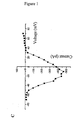
- the activity of SCN5A VGSC in a sample may be assayed by dissociating a biopsy into single cells and in culture assaying (i) the effect of voltage-gated Na + channel blockers on their motility and (ii) detecting voltage-gated Na + channel activity by electrophysiological recording. Suitable methods and voltage-gated Na + channel blockers are described in Example 1.
- Preferred diagnostic methods of the invention include what can broadly be described as “invasive” methods and "non-invasive” methods.
- Invasive methods include, for example, the taking of a biopsy for detection of voltage-gated Na + channel expression by, for example, (a) immunohistochemical application of a sequence-specific antibody, (b) in situ PCR on tissue sections, or (c) reverse transcription (RT)-PCR of cells, for example epithelial cells (and/or other cell types, for example neuroendocrine or myoepithelial cells) after separating them from the biopsy.
- Non-invasive methods include obtaining breast-derived cells from blood, which may be separated by affinity and assayed for voltage-gated Na + channel expression by PCR.
- a further aspect of the invention provides a kit of parts useful for diagnosing breast cancer comprising an agent which is capable of use in determining the level of SCN5A VGSC protein or nucleic acid in a sample.
- the agent may be a nucleic acid which selectively hybridises to the said VGSC nucleic acid or the agent may be a molecule which selectively binds to the said VGSC protein.
- the kit further comprises a control sample containing the said VGSC nucleic acid or protein wherein the control sample may be a negative control (which contains a level of the said VGSC protein or nucleic acid which is not associated with cancer or a high metastatic potential for cancer) or it may be a positive control (which contains a level of the said VGSC protein or nucleic acid which is associated with cancer or a high metastatic potential for cancer).
- the kit may contain both negative and positive controls.
- the kit may usefully contain controls of the said VGSC protein or nucleic acid which correspond to different amounts such that a calibration curve may be made.
- the kit further comprises means for separating breast cells (for example epithelial cells, neuroendocrine or myoepithelial cells) from a sample in order to carry out said VGSC assay.
- the means for separating breast cells includes antibody-coated micro-beads or columns. These are coated with antibodies to cell membrane proteins.
- anti-MUC1 antibodies such as HMFG-1 and HMFG-2 may be used ( Taylor-Papadimitriou et al (1986) J. Exp. Pathol. 2, 247-260 ); other anti-MUC1 antibodies which may be useful are described in Cao et al (1998) Tumour Biol. 19, (Suppl 1), 88-99 .
- anti-MUC1 antibodies may bind to normal bone marrow cells. It is preferred to use an anti epithelial cell adhesion molecule antibody, preferably coated on magnetic beads. A preferred antibody is termed BER-EP-4.
- a further aspect of the invention provides a kit of parts useful for diagnosing breast cancer, comprising (1) an agent which is capable of use in determining the level of SCN5A protein or nucleic acid in a sample, and (2) means for separating breast cells (for example epithelial cells, neuroendocrine or myoepithelial cells) from a sample in order to carry out said VGSC assay.
- an agent which is capable of use in determining the level of SCN5A protein or nucleic acid in a sample
- means for separating breast cells for example epithelial cells, neuroendocrine or myoepithelial cells
- kits may usefully further comprise a component for testing for a further cancer-related polypeptide such as antibodies which are reactive with one or more of the following cancer-related polypeptides, all of which are well known in the art: MAGE-1, MAGE-3, BAGE, GAGE-1, CAG-3, CEA, p53, oestrogen receptor (ER), progesterone receptor (PR), MUC1, p52 trefoil peptide, Her2, PCNA, Ki67, cyclin D, p90 rak3 , p170 glycoprotein (mdr-1) CA-15-3, c-erbB1, cathepsin D, PSA, CA125, CA19-9, PAP, myc, cytokeratins, bcl-2, telomerase, glutathione S transferases, rand51, VEGF, thymidine phosphorylase, Flk1 or Flk2.
- a further cancer-related polypeptide such as antibodies which are reactive with one or more of the following cancer
- the kit may usefully still further or alternatively comprise a nucleic acid which selectively hybridises to a further cancer-related nucleic acid such as a gene or mRNA which encodes any of the cancer-related polypeptides as described above.
- useful nucleic acids which may be included in the kit are those which selectively hybridise with the genes or 15-05-200 mRNAs: ras, APC, BRCA1, BRCA2, ataxia telangiectasia (ATM), hMSH2, hMCH1, hPMS2 or hPMS1. It is preferred if the further nucleic acid is one which selectively hybridises to the gene or mRNA of any of erbB2, p53, BRCA1, BRCA2 or ATM.
- kits usefully may contain controls and detection material, (for example, for immunohistochemistry, secondary antibodies labelled fluorophores, or enzymes, or biotin, or digoxygenin or the like).
- additional components to the kit may include a second antibody to a different epitope on the VGSC (optionally labelled or attached to a support), secondary antibodies (optionally labelled or attached to a support), and dilution and reaction buffers. Similar additional components may usefully be included in all of the kits of the invention.
- an agent which selectively prevents the function of a voltage-gated Na + channel we include agents that (a) inhibit the expression of a said VGSC or (b) inhibit the activity of a said VGSC.
- Agents that prevent the expression of said VGSC include antisense agents and ribozymes.
- Antisense oligonucleotides are single-stranded nucleic acid, which can specifically bind to a complementary nucleic acid sequence. By binding to the appropriate target sequence, an RNA-RNA, a DNA-DNA, or RNA-DNA duplex is formed. These nucleic acids are often termed "antisense” because they are complementary to the sense or coding strand of the gene. Recently, formation of a triple helix has proven possible where the oligonucleotide is bound to a DNA duplex. It was found that oligonucleotides could recognise sequences in the major groove of the DNA double helix. A triple helix was formed thereby. This suggests that it is possible to synthesise sequence-specific molecules which specifically bind double-stranded DNA via recognition of major groove hydrogen binding sites.
- the above oligonucleotides can inhibit the function of the target nucleic acid. This could, for example, be a result of blocking the transcription, processing, poly(A)addition, replication, translation, or promoting inhibitory mechanisms of the cells, such as promoting RNA degradations.
- Antisense oligonucleotides are prepared in the laboratory and then introduced into cells, for example by microinjection or uptake from the cell culture medium into the cells, or they are expressed in cells after transfection with plasmids or retroviruses or other vectors carrying an antisense gene.
- Antisense oligonucleotides were first discovered to inhibit viral replication or expression in cell culture for Rous sarcoma virus, vesicular stomatitis virus, herpes simplex virus type 1, simian virus and influenza virus. Since then, inhibition of mRNA translation by antisense oligonucleotides has been studied extensively in cell-free systems including rabbit reticulocyte lysates and wheat germ extracts.
- Oligonucleotides are subject to being degraded or inactivated by cellular endogenous nucleases.
- modified oligonucleotides eg having altered internucleotide linkages, in which the naturally occurring phosphodiester linkages have been replaced with another linkage.
- Agrawal et al (1988) Proc. Natl. Acad. Sci. USA 85, 7079-7083 showed increased inhibition in tissue culture of HIV-1 using oligonucleotide phosphoramidates and phosphorothioates.
- Oligonucleotides having artificial linkages have been shown to be resistant to degradation in vivo.
- Shaw et al (1991) in Nucleic Acids Res. 19, 747-750 report that otherwise unmodified oligonucleotides become more resistant to nucleases in vivo when they are blocked at the 3' end by certain capping structures and that uncapped oligonucleotide phosphorothioates are not degraded in vivo.
- oligonucleotide is a deoxyribonucleic acid (DNA), although ribonucleic acid (RNA) sequences may also be synthesized and applied.
- DNA deoxyribonucleic acid
- RNA ribonucleic acid
- the oligonucleotides useful in the invention preferably are designed to resist degradation by endogenous nucleolytic enzymes. In vivo degradation of oligonucleotides produces oligonucleotide breakdown products of reduced length. Such breakdown products are more likely to engage in non-specific hybridization and are less likely to be effective, relative to their full-length counterparts. Thus, it is desirable to use oligonucleotides that are resistant to degradation in the body and which are able to reach the targeted cells.
- the present oligonucleotides can be rendered more resistant to degradation in vivo by substituting one or more internal artificial internucleotide linkages for the native phosphodiester linkages, for example, by replacing phosphate with sulphur in the linkage.
- linkages examples include phosphorothioates, methylphosphonates, sulphone, sulphate, ketyl, phosphorodithioates, various phosphoramidates, phosphate esters, bridged phosphorothioates and bridged phosphoramidates.
- Such examples are illustrative, rather than limiting, since other internucleotide linkages are known in the art. See, for example, Cohen, (1990) Trends in Biotechnology .
- oligonucleotides having one or more of these linkages substituted for the phosphodiester internucleotide linkages is well known in the art, including synthetic pathways for producing oligonucleotides having fixed internucleotide linkages.
- Oligonucleotides can be made resistant to extension by endogenous enzymes by "capping" or incorporating similar groups on the 5' or 3' terminal nucleotides.
- a reagent for capping is commercially available as AminoLink II TM from Applied BioSystems Inc, Foster City, CA. Methods for capping are described, for example, by Shaw et al (1991) Nucleic Acids Res. 19, 747-750 and Agrawal et al (1991) Proc. Natl. Acad. Sci. USA 88(17), 7595-7599 .
- oligonucleotides resistant to nuclease attack are for them to be "self-stabilized” as described by Tang et al (1993) Nucl. Acids Res. 21, 2729-2735 .
- Self stabilized oligonucleotides have hairpin loop structures at their 3' ends, and show increased resistance to degradation by snake venom phosphodiesterase, DNA polymerase I and fetal bovine serum.
- the self-stabilized region of the oligonucleotide does not interfere in hybridization with complementary nucleic acids, and pharmacokinetic and stability studies in mice have shown increased in vivo persistence of self-stabilized oligonucleotides with respect to their linear counterparts.
- the inherent binding specificity of antisense oligonucleotides characteristic of base pairing is enhanced by limiting the availability of the antisense compound to its intend locus in vivo, permitting lower dosages to be used and mimimizing systemic effects.
- oligonucleotides are applied locally to achieve the desired effect.
- the concentration of the oligonucleotides at the desired locus is much higher than if the oligonucleotides were administered systemically, and the therapeutic effect can be achieved using a significantly lower total amount.
- the local high concentration of oligonucleotides enhances penetration of the targeted cells and effectively blocks translation of the target nucleic acid sequences.
- the oligonucleotides can be delivered to the locus by any means appropriate for localized administration of a drug.
- a solution of the oligonucleotides can be injected directly to the site or can be delivered by infusion using an infusion pump.
- the oligonucleotides also can be incorporated into an implantable device which when placed at the desired site, permits the oligonucleotides to be released into the surrounding locus.
- the oligonucleotides may be administered via a hydrogel material.
- the hydrogel is noninflammatory and biodegradable. Many such materials now are known, including those made from natural and synthetic polymers.
- the method exploits a hydrogel which is liquid below body temperature but gels to form a shape-retaining semisolid hydrogel at or near body temperature.
- Preferred hydrogel are polymers of ethylene oxide-propylene oxide repeating units. The properties of the polymer are dependent on the molecular weight of the polymer and the relative percentage of polyethylene oxide and polypropylene oxide in the polymer.
- Preferred hydrogels contain from about 10 to about 80 % by weight ethylene oxide and from about 20 to about 90 % by weight propylene oxide.
- a particularly preferred hydrogel contains about 70% polyethylene oxide and 30 % polypropylene oxide. Hydrogels which can be used are available, for example, from BASF Corp., Parsippany, NJ, under the tradename Pluronic R .
- the hydrogel is cooled to a liquid state and the oligonucleotides are admixed into the liquid to a concentration of about 1 mg oligonucleotide per gram of hydrogel.
- the resulting mixture then is applied onto the surface to be treated, for example by spraying or painting during surgery or using a catheter or endoscopic procedures.
- the polymer warms, it solidifies to form a gel, and the oligonucleotides diffuse out of the gel into the surrounding cells over a period of time defined by the exact composition of the gel.
- the oligonucleotides can be administered by means of other implants that are commercially available or described in the scientific literature, including liposomes, microcapsules and implantable devices.
- implants made of biodegradable materials such as polyanhydrides, polyorthoesters, polylactic acid and polyglycolic acid and copolymers thereof, collagen, and protein polymers, or non-biodegradable materials such as ethylenevinyl acetate (EVAc), polyvinyl acetate, ethylene vinyl alcohol, and derivatives thereof can be used to locally deliver the oligonucleotides.
- biodegradable materials such as polyanhydrides, polyorthoesters, polylactic acid and polyglycolic acid and copolymers thereof, collagen, and protein polymers
- non-biodegradable materials such as ethylenevinyl acetate (EVAc), polyvinyl acetate, ethylene vinyl alcohol, and derivatives thereof can be used to locally deliver the oligonucleotides.
- EVAc
- the oligonucleotides can be incorporated into the material as it is polymerized or solidified, using melt or solvent evaporation techniques, or mechanically mixed with the material.
- the oligonucleotides are mixed into or applied onto coatings for implantable devices such as dextran coated silica beads, stents, or catheters.
- Polymeric nanoparticles/biodegradable drug carriers may also be used ( Mader (1998) Radiol. Oncol. 32, 89-94 ).
- the dose of oligonucleotides is dependent on the size of the oligonucleotides and the purpose for which it is administered. In general, the range is calculated based on the surface area of tissue to be treated.
- the effective dose of oligonucleotide is somewhat dependent on the length and chemical composition of the oligonucleotide but is generally in the range of about 30 to 3000 ⁇ g per square centimetre of tissue surface area.
- the oligonucleotides may be administered to the patient systemically for both therapeutic and prophylactic purposes.
- the oligonucleotides may be administered by any effective method, for example, parenterally (eg intravenously, subcutaneously, intramuscularly) or by oral, nasal or other means which permit the oligonucleotides to access and circulate in the patient's bloodstream.
- Oligonucleotides administered systemically preferably are given in addition to locally administered oligonucleotides, but also have utility in the absence of local administration.
- a dosage in the range of from about 0.1 to about 10 grams per administration to an adult human generally will be effective for this purpose.
- the antisense oligonucleotides may be desirable to target the antisense oligonucleotides to the cancerous tissue, for example to the breast. This may be achieved by administering the antisense oligonucleotides to the cancer location (for example the breast), or it may be achieved by using antisense oligonucleotides which are in association with a molecule which selectively directs the antisense oligonucleotide to the cancer location.
- the antisense oligonucleotide may be associated with an antibody or antibody like molecule which selectively binds a breast-related antigen such as MUC-1.
- the antisense oligonucleotide and the cancer-directing entity are so associated that the cancer-directing entity is able to direct the antisense oligonucleotide to the location of the cancer cells, for example breast cells.
- antisense agents also include larger molecules, for example of around one hundred to several hundred bases which bind to said VGSC mRNA or genes and substantially prevent expression of said VGSC mRNA or genes and substantially prevent expression of said VGSC protein.
- expression of an antisense molecule which is substantially complementary to said VGSC mRNA is envisaged as part of the invention.
- synthetic oligonucleotides with antisense sequence to specific regions of (i) SCN5A (and optionally also SCN9A) channels or (ii) (for patients with or at risk of breast cancer) SCN5A or SCN9A channels or VGSCs are administered (preferably to patients with or at risk of breast cancer) to block channel activity. Details of particular synthetic oligonucleotides are given in Example 2.
- antisense oligonucleotide technology has already been used to manipulate potassium channels in vitro ( Roy et al (1996) Glia 18, 174-188 ) and VGSCs in vitro ( Biochem Biophys Res Comm (1997) 234, 235-241 ) and in blocking neuropathic pain ( Lai et al (1999) "Blockade of neuropathic pain by antisense targeting of tetrodotoxin-resistant sodium channels in sensory neurons" Methods in Enzymol 314, 201-213 ).
- a further method for blocking said VGSC activity includes dominant negative suppression.
- a mutated VGSC gene product suppresses or eliminates the activity of the corresponding normal gene product when the two are co-expressed.
- VGPCs voltage-gated potassium channels
- such an approach making use of a highly truncated gene product has been used successfully to suppress functional expression of VGPCs in vitro ( Tu et al (1995) Biophys. J. 68, 147-156 ) and in vivo ( London et al (1998) Proc. Natl. Acad. Sci. USA 95, 2926-2931 ).
- the truncated subunit is capable of binding to other VGPC subunits but does not contain the residues required for channel functioning. Thus, the activity of the "combined" VGPC is blocked.
- a number of naturally occurring alternatively spliced channel subunits have been detected which may function to suppress VGPC activity by a similar mechanism in vivo ( Baumann et al (1987) EMBO J. 6, 3419-3429 ; Kamb et al (1988) Neuron 1, 421-430 ; and Pongs et al (1988) EMBO J. 7, 1087-1096 ).
- Baumann et al (1987) EMBO J. 6, 3419-3429 a similar mechanism in vivo
- Kamb et al (1988) Neuron 1, 421-430 a similar mechanism in vivo
- Pongs et al (1988) EMBO J. 7, 1087-1096 we believe that VGSC may similarly be suppressed by interfering with functional channel formation.
- VGSCs are formed from a single alpha subunit (comprising four functional domains)
- recent studies have demonstrated the specific expression (during development in human, mouse and fish) of truncated VGSC proteins possessing only two domains which might function in a dominant negative manner to control VGSC activity ( Plummer et al (1997) J. Biol. Chem. 272, 24008-24015 ; and Oh & Waxman (1998) NeuroReport 9, 1267-1271 ).
- the neonatal VGSCs may act as inhibitors of VGSC activity, but this inhibition is most probably specific to the related adult VGSC. It is much less likely that, for example, neonatal SCN8A could inhibit the activity of VGSC proteins derived from VGSC genes other than SCN8A.
- the larger molecules may be expressed from any suitable genetic construct as is described below and delivered to the patient.
- the genetic construct which expresses the antisense molecule comprises at least a portion of the said VGSC cDNA or gene operatively linked to a promoter which can express the antisense molecule in the cell, preferably breast cell, which is or may become cancerous.
- the genetic construct can be DNA or RNA it is preferred if it is DNA.
- the genetic construct is adapted for delivery to a human cell.
- the constructs of the invention may be introduced into the tumour cells by any convenient method, for example methods involving retroviruses, so that the construct is inserted into the genome of the tumour cell.
- retroviruses provide a potential means of selectively infecting cancer cells because they can only integrate into the genome of dividing cells; most normal cells surrounding cancers are in a quiescent, non-receptive stage of cell growth or, at least, are dividing much less rapidly than the tumour cells.
- Retroviral DNA constructs which encode said antisense agents may be made using methods well known in the art.
- To produce active retrovirus from such a construct it is usual to use an ecotropic psi2 packaging cell line grown in Dulbecco's modified Eagle's medium (DMEM) containing 10% foetal calf serum (FCS).
- DMEM Dulbecco's modified Eagle's medium
- FCS foetal calf serum
- Transfection of the cell line is conveniently by calcium phosphate co-precipitation, and stable transformants are selected by addition of G418 to a final concentration of 1 mg/ml (assuming the retroviral construct contains a neo R gene).
- Independent colonies are isolated and expanded and the culture supernatant removed, filtered through a 0.45 ⁇ m pore-size filter and stored at -70°.
- retroviral supernatant For the introduction of the retrovirus into the tumour cells, it is convenient to inject directly retroviral supernatant to which 10 ⁇ g/ml Polybrene has been added. For tumours exceeding 10 mm in diameter it is appropriate to inject between 0.1 ml and 1 ml of retroviral supernatant; preferably 0.5 ml.
- cells which produce retroviruses are injected into the tumour.
- the retrovirus-producing cells so introduced are engineered to actively produce retroviral vector particles so that continuous productions of the vector occurred within the tumour mass in situ.
- proliferating tumour cells can be successfully transduced in vivo if mixed with retroviral vector-producing cells.
- Targeted retroviruses are also available for use in the invention; for example, sequences conferring specific binding affinities may be engineered into preexisting viral env genes (see Miller & Vile (1995) Faseb J. 9, 190-199 for a review of this and other targeted vectors for gene therapy).
- Immunoliposomes are especially useful in targeting to cancer cell types which over-express a cell surface protein for which antibodies are available.
- the immunoliposomes may be targeted by means of antibodies binding to a breast cancer cell antigen such as MUC-1, or the said VGSC (preferably in combination with other targeting means or methods), as discussed further below.
- MPB-PE N-[4-(p-maleimidophenyl)butyryl]-phosphatidylethanolamine
- MPB-PE N-[4-(p-maleimidophenyl)butyryl]-phosphatidylethanolamine
- MPB-PE is incorporated into the liposomal bilayers to allow a covalent coupling of the antibody, or fragment thereof, to the liposomal surface.
- the liposome is conveniently loaded with the DNA or other genetic construct of the invention for delivery to the target cells, for example, by forming the said liposomes in a solution of the DNA or other genetic construct, followed by sequential extrusion through polycarbonate membrane filters with 0.6 ⁇ m and 0.2 ⁇ m pore size under nitrogen pressures up to 0.8 MPa. After extrusion, entrapped DNA construct is separated from free DNA construct by ultracentrifugation at 80 000 x g for 45 min.
- Freshly prepared MPB-PE-liposomes in deoxygenated buffer are mixed with freshly prepared antibody (or fragment thereof) and the coupling reactions are carried out in a nitrogen atmosphere at 4°C under constant end over end rotation overnight.
- the immunoliposomes are separated from unconjugated antibodies by ultracentrifugation at 80 000 x g, for 45 min.
- Immunoliposomes may be injected intraperitoneally or directly into the tumour.
- adenoviruses carrying external DNA via an antibody-polylysine bridge see Curiel Prog. Med. Virol. 40, 1-18
- transferrin-polycation conjugates as carriers
- a polycation-antibody complex is formed with the DNA construct or other genetic construct of the invention, wherein the antibody is specific for either wild-type adenovirus or a variant adenovirus in which a new epitope has been introduced which binds the antibody.
- the polycation moiety binds the DNA via electrostatic interactions with the phosphate backbone.
- the adenovirus because it contains unaltered fibre and penton proteins, is internalized into the cell and carries into the cell with it the DNA construct of the invention. It is preferred if the polycation is polylysine.
- the DNA may also be delivered by adenovirus wherein it is present within the adenovirus particle, for example, as described below.
- a high-efficiency nucleic acid delivery system that uses receptor-mediated endocytosis to carry DNA macromolecules into cells is employed. This is accomplished by conjugating the iron-transport protein transferrin to polycations that bind nucleic acids.
- Human transferrin, or the chicken homologue conalbumin, or combinations thereof is covalently linked to the small DNA-binding protein protamine or to polylysines of various sizes through a disulfide linkage. These modified transferrin molecules maintain their ability to bind their cognate receptor and to mediate efficient iron transport into the cell.
- the transferrin-polycation molecules form electrophoretically stable complexes with DNA constructs or other genetic constructs of the invention independent of nucleic acid size (from short oligonucleotides to DNA of 21 kilobase pairs).
- complexes of transferrin-polycation and the DNA constructs or other genetic constructs of the invention are supplied to the tumour cells, a high level of expression from the construct in the cells is expected.
- High-efficiency receptor-mediated delivery of the DNA constructs or other genetic constructs of the invention using the endosome-disruption activity of defective or chemically inactivated adenovirus particles produced by the methods of Cotten et al (1992) Proc. Natl. Acad. Sci. USA 89, 6094-6098 may also be used.
- This approach appears to rely on the fact that adenoviruses are adapted to allow release of their DNA from an endosome without passage through the lysosome, and in the presence of, for example transferrin linked to the DNA construct or other genetic construct of the invention, the construct is taken up by the cell by the same route as the adenovirus particle.
- This approach has the advantages that there is no need to use complex retroviral constructs; there is no permanent modification of the genome as occurs with retroviral infection; and the targeted expression system is coupled with a targeted delivery system, thus reducing toxicity to other cell types.
- tumours may be desirable to locally perfuse a tumour with the suitable delivery vehicle comprising the genetic construct for a period of time; additionally or alternatively the delivery vehicle or genetic construct can be injected directly into accessible tumours.
- naked DNA and DNA complexed with cationic and neutral lipids may also be useful in introducing the DNA of the invention into cells of the patient to be treated.
- Non-viral approaches to gene therapy are described in Ledley (1995) Human Gene Therapy 6, 1129-1144 .
- Alternative targeted delivery systems are also known such as the modified adenovirus system described in WO 94/10323 wherein, typically, the DNA is carried within the adenovirus, or adenovirus-like, particle.
- Michael et al (1995) Gene Therapy 2, 660-668 describes modification of adenovirus to add a cell-selective moiety into a fibre protein.
- Mutant adenoviruses which replicate selectively in p53-deficient human tumour cells such as those described in Bischoff et al (1996) Science 274, 373-376 are also useful for delivering a genetic construct to a cell.
- Other suitable viruses or virus-like particles include HSV, AAV, vaccinia and parvovirus.
- the agent which selectively prevents the function of the said VGSC is a ribozyme capable of cleaving targeted VGSC RNA or DNA.
- a gene expressing said ribozyme may be administered in substantially the same way and using substantially the same vehicles as for the antisense molecules.
- Ribozymes which may be encoded in the genomes of the viruses or virus-like particles herein disclosed are described in Cech and Herschlag "Site-specific cleavage of single stranded DNA” US 5,180,818 ; Altman et al "Cleavage of targeted RNA by RNAse P" US 5,168,053 , Cantin et al "Ribozyme cleavage of HIV-1 RNA” US 5,149,796 ; Cech et al “RNA ribozyme restriction endoribonucleases and methods", US 5,116,742 ; Been et al "RNA ribozyme polymerases, dephosphorylases, restriction endonucleases and methods", US 5,093,246 ; and Been et al "RNA ribozyme polymerases, dephosphorylases, restriction endoribonucleases and methods; cleaves single-stranded RNA at specific site by transesterification", US 4,987,07
- the antisense molecule or ribozyme is expressed from a breast cell-specific promoter element.
- breast cell-specific promoters include the promoter element for c-erbB2 or the oestrogen receptor.
- a variety of methods have been developed to operably link DNA to vectors via complementary cohesive termini. For instance, complementary homopolymer tracts can be added to the DNA segment to be inserted to the vector DNA. The vector and DNA segment are then joined by hydrogen bonding between the complementary homopolymeric tails to form recombinant DNA molecules.
- Synthetic linkers containing one or more restriction sites provide an alternative method of joining the DNA segment to vectors.
- the DNA segment generated by endonuclease restriction digestion as described earlier, is treated with bacteriophage T4 DNA polymerase or E. coli DNA polymerase I, enzymes that remove protruding, 3'-single-stranded termini with their 3'-5'-exonucleolytic activities, and fill in recessed 3'-ends with their polymerizing activities.
- the combination of these activities therefore generates blunt-ended DNA segments.
- the blunt-ended segments are then incubated with a large molar excess of linker molecules in the presence of an enzyme that is able to catalyze the ligation of blunt-ended DNA molecules, such as bacteriophage T4 DNA ligase.
- an enzyme that is able to catalyze the ligation of blunt-ended DNA molecules, such as bacteriophage T4 DNA ligase.
- the products of the reaction are DNA segments carrying polymeric linker sequences at their ends.
- These DNA segments are then cleaved with the appropriate restriction enzyme and ligated to an expression vector that has been cleaved with an enzyme that produces termini compatible with those of the DNA segment.
- Synthetic linkers containing a variety of restriction endonuclease sites are commercially available from a number of sources including International Biotechnologies Inc, New Haven, CN, USA.
- a desirable way to modify the DNA encoding the polypeptide of the invention is to use the polymerase chain reaction as disclosed by Saiki et al (1988) Science 239, 487-491 .
- the DNA to be enzymatically amplified is flanked by two specific oligonucleotide primers which themselves become incorporated into the amplified DNA.
- the said specific primers may contain restriction endonuclease recognition sites which can be used for cloning into expression vectors using methods known in the art.
- a further aspect of the invention provides use of an agent which selectively prevents (including inhibits) the function of SCN5A voltage-gated Na + channel in the manufacture of a medicament for treating breast cancer, wherein the agent prevents the expression of the said voltage-gated Na + channel.
- the molecule capable of preventing the function of the said VGSC is an antisense molecule or a ribozyme as disclosed above.
- the present invention also provides uses in which treatment is targeted to breast cancer cells by means of targeting to cells expressing SCN5A.
- targeting to cells expressing a said VGSC may preferably be performed in conjunction with another targeting means or method, for example local administration, in order to minimise adverse effects on any normal tissues that express the said VGSC.
- cardiac tissue expresses SCN5A at high levels.
- anti-said VGSC antibodies (VGSC-Abs) conjugated with a dye substance may be applied to the cancerous tissue in vivo (eg Yasmuch et al (1993) "Antibody targeted photolysis” Critical Review Revue Ther. Drug Carrier System 10, 197-252 ; Pogrebniak et al (1993) "Targetted phototherapy with sensitizer-monoclonal antibody conjugate and light” Surgical Onoclogy 2, 31-42 ).
- the tissue is then irradiated locally with a wavelength of light/laser matching the absorption peak of the 'attached' dye. Absorption of the light energy by the dye leads to local heating and cell death.
- VGSC-Abs labelled with the following dyes may be used: fluorescein ( Pelegrin et al (1991) "Antibody fluorescein conjugates for photoimmunodiagnosis of human colon-carcinoma in nude-mice” Cancer 67, 2529-2537 ); rhodamine ( Haghighat et al (1992) "Laser-dyes for experimental phototherapy of human cancer - comparison of 3 rhodamines" Laryngoscope 102, 81-87 ); cyanins ( Folli et al (1994) "Antibody-indocyanin conjugates for immunophotodetection of human squamous-cell carcinoma in nude-mice” Cancer Research 54, 2643-2649 ; Lipshutz et al (1994) "Evaluation of 4 new carbocyanine dyes for photodynamic therapy with lasers" Laryngoscope 104, 9
- a further aspect of the invention provides for the use of a compound comprising a moiety which selectively binds SCN5A voltage-gated Na + channel protein, wherein said moiety is an antibody, antibody fragment, or a single-chain Fv fragment (ScFv) or a domain antibody (dAb); and a further moiety, in the manufacture of a medicament for the treatment and/or diagnosis of human breast cancer.
- a moiety which selectively binds SCN5A voltage-gated Na + channel protein we mean any suitable such moiety which binds the said VGSC but does not substantially bind other molecules, for example other VGSCs, for example SCN9A.
- the compound comprising the binding moiety is one which preferably, in use, is able to localise to areas of cancerous cells (preferably breast cancerous cells), particularly metastatic cancer cells, but not localise substantially to other areas where there are no cancerous cells.
- the binding moiety is able to bind to the said VGSC with high affinity.
- the binding constant for the binding of the binding moiety to the said VGSC is preferably between 10 -7 and 10 -10 M.
- the binding moiety is an anti-SNC5A antibody.
- anti-SNC5A antibody Such antibodies and methods of preparing suitable antibodies are discussed above.
- the further moiety may be any further moiety which confers on the compound a useful property with respect to the treatment or imaging or diagnosis of cancer.
- the further moiety is one which is useful in killing or imaging cancer cells, particularly metastatic cancer cells.
- the further moiety is one which is able to kill the cancer cells to which the compound is targeted.
- the further moiety is directly or indirectly cytotoxic.
- the further moiety is preferably directly or indirectly toxic to cancer cells, particularly metastatic cancer cells.
- directly cytotoxic we include the meaning that the moiety is one which on its own is cytotoxic.
- directly cytotoxic we include the meaning that the moiety is one which, although is not itself cytotoxic, can induce cytotoxicity, for example by its action on a further molecule or by further action on it.
- the cytotoxic moiety is a cytotoxic chemotherapeutic agent.
- Cytotoxic chemotherapeutic agents are well known in the art.
- Cytotoxic chemotherapeutic agents include: alkylating agents including nitrogen mustards such as mechlorethamine (HN 2 ), cyclophosphamide, ifosfamide, melphalan (L-sarcolysin) and chlorambucil; ethylenimines and methylmelamines such as hexamethylmelamine, thiotepa; alkyl sulphonates such as busulfan; nitrosoureas such as carmustine (BCNU), lomustine (CCNU), semustine (methyl-CCNU) and streptozocin (streptozotocin); and triazenes such as decarbazine (DTIC; dimethyltriazenoimidazole-carboxamide); Antimetabolites including folic acid analogues such as methotrexate (amethopterin); pyrimidine analogues such as fluorouracil (5-fluorouracil; 5-FU),
- Natural Products including vinca alkaloids such as vinblastine (VLB) and vincristine; epipodophyllotoxins such as etoposide and teniposide; antibiotics such as dactinomycin (actinomycin D), daunorubicin (daunomycin; rubidomycin), doxorubicin, bleomycin, plicamycin (mithramycin) and mitomycin (mitomycin C); enzymes such as L-asparaginase; and biological response modifiers such as interferon alphenomes.
- VLB vinblastine
- epipodophyllotoxins such as etoposide and teniposide
- antibiotics such as dactinomycin (actinomycin D), daunorubicin (daunomycin; rubidomycin), doxorubicin, bleomycin, plicamycin (mithramycin) and mitomycin (mitomycin C)
- enzymes such as L-asparaginas
- Miscellaneous agents including platinum coordination complexes such as cisplatin ( cis- DDP) and carboplatin; anthracenedione such as mitoxantrone and anthracycline; substituted urea such as hydroxyurea; methyl hydrazine derivative such as procarbazine (N-methylhydrazine, MIH); and adrenocortical suppressant such as mitotane ( o,p -DDD) and aminoglutethimide; taxol and analogues/derivatives; and hormone agonists/antagonists such as flutamide and tamoxifen.
- platinum coordination complexes such as cisplatin ( cis- DDP) and carboplatin
- anthracenedione such as mitoxantrone and anthracycline
- substituted urea such as hydroxyurea
- methyl hydrazine derivative such as procarbazine (N-methylhydrazine,
- the preparative procedure is simple, relatively fast, and is carried out under mild conditions.
- Carbodiimide compounds attack carboxylic groups to change them into reactive sites for free amino groups.
- the water soluble carbodiimide, 1-ethyl-3-(3-dimethylaminopropyl) carbodiimide is particularly useful for conjugating a functional moiety to a binding moiety and may be used to conjugate doxorubicin to tumor homing peptides.
- the conjugation of doxorubicin and a binding moiety requires the presence of an amino group, which is provided by doxorubicin, and a carboxyl group, which is provided by the binding moiety such as an antibody or peptide.
- EDC also can be used to prepare active esters such as N-hydroxysuccinimide (NHS) ester.
- NHS N-hydroxysuccinimide
- the NHS ester which binds only to amino groups, then can be used to induce the formation of an amide bond with the single amino group of the doxorubicin.
- EDC and NHS in combination is commonly used for conjugation in order to increase yield of conjugate formation (Bauminger & Wilchek, supra, 1980).
- the cytotoxic moiety is a cytotoxic peptide or polypeptide moiety by which we include any moiety which leads to cell death.
- Cytotoxic peptide and polypeptide moieties are well known in the art and include, for example, ricin, abrin, Pseudomonas exotoxin, tissue factor and the like. Methods for linking them to targeting moieties such as antibodies are also known in the art. The use of ricin as a cytotoxic agent is described in Burrows & Thorpe (1993) Proc. Natl. Acad. Sci.
- cytokines such as TNF ⁇ and IL-2, may also be useful as cytotoxic agents.
- radioactive atoms may also be cytotoxic if delivered in sufficient doses.
- the cytotoxic moiety may comprise a radioactive atom which, in use, delivers a sufficient quantity of radioactivity to the target site so as to be cytotoxic.
- Suitable radioactive atoms include phosphorus-32, iodine-125, iodine-131, indium-111, rhenium-186, rhenium-188 or yttrium-90, or any other isotope which emits enough energy to destroy neighbouring cells, organelles or nucleic acid.
- the isotopes and density of radioactive atoms in the compound of the invention are such that a dose of more than 4000 cGy (preferably at least 6000, 8000 or 10000 cGy) is delivered to the target site and, preferably, to the cells at the target site and their organelles, particularly the nucleus.
- the radioactive atom may be attached to the binding moiety in known ways.
- EDTA or another chelating agent may be attached to the binding moiety and used to attach 111 In or 90 Y.
- Tyrosine residues may be labelled with 125 I or 131 I.
- the cytotoxic moiety may be a suitable indirectly cytotoxic polypeptide.
- the indirectly cytotoxic polypeptide is a polypeptide which has enzymatic activity and can convert a relatively non-toxic prodrug into a cytotoxic drug.
- ADEPT Antibody-Directed Enzyme Prodrug Therapy
- the system requires that the targeting moiety locates the enzymatic portion to the desired site in the body of the patient (ie the site expressing the said VGSC, such as metastatic cancer cells) and after allowing time for the enzyme to localise at the site, administering a prodrug which is a substrate for the enzyme, the end product of the catalysis being a cytotoxic compound.
- the object of the approach is to maximise the concentration of drug at the desired site and to minimise the concentration of drug in normal tissues (see Senter, P.D. et al (1988) "Anti-tumor effects of antibody-alkaline phosphatase conjugates in combination with etoposide phosphate" Proc. Natl. Acad. Sci. USA 85, 4842-4846 ; Bagshawe (1987) Br. J. Cancer 56, 531-2 ; and
- any said VGSC-binding moiety may be used in place of an anti-said VGSC antibody in this type of directed enzyme prodrug therapy system.
- the enzyme and prodrug of the system using a said VGSC-targeted enzyme as described herein may be any of those previously proposed.
- the cytotoxic substance may be any existing anti-cancer drug such as an alkylating agent; an agent which intercalates in DNA; an agent which inhibits any key enzymes such as dihydrofolate reductase, thymidine synthetase, ribonucleotide reductase, nucleoside kinases or topoisomerase; or an agent which effects cell death by interacting with any other cellular constituent.
- Etoposide is an example of a topoisomerase inhibitor.
- Reported prodrug systems include: a phenol mustard prodrug activated by an E. coli ⁇ -glucuronidase (Wang et al, 1992 and Roffler et al, 1991); a doxorubicin prodrug activated by a human ⁇ -glucuronidase (Bosslet et al, 1994); further doxorubicin prodrugs activated by coffee bean ⁇ -galactosidase (Azoulay et al, 1995); daunorubicin prodrugs, activated by coffee bean ⁇ -D-galactosidase (Gesson et al, 1994); a 5-fluorouridine prodrug activated by an E.
- methotrexate prodrugs eg methotrexate-alanine activated by carboxypeptidase A (Kuefner et al, 1990, Vitols et al, 1992 and Vitols et al, 1995). These and others are included in the following table.
- Enzyme Prodrug Carboxypeptidase G2 Derivatives of L-glutamic acid and benzoic acid mustards, aniline mustards, phenol mustards and phenylenediamine mustards; fluorinated derivatives of these Alkaline phosphatase Etoposide phosphate Mitomycin phosphate Beta-glucuronidase p- Hydroxyaniline mustard-glucuronide Epirubicin-glucuronide Penicillin-V-amidase Adriamycin-N phenoxyacetyl Penicillin-G-amidase N-(4'-hydroxyphenyl acetyl) palytoxin Doxorubicin and melphalan Beta-lactamase Nitrogen mustard-cephalosporin p- phenylenediamine; doxorubicin derivatives; vinblastine derivative-cephalosporin, cephalosporin mustard; a taxol derivative Beta-glucosidase Cyanophenylmethyl-beta-D-glu
- Suitable enzymes for forming part of the enzymatic portion of the invention include: exopeptidases, such as carboxypeptidases G, G1 and G2 (for glutamylated mustard prodrugs), carboxypeptidases A and B (for MTX-based prodrugs) and aminopeptidases (for 2- ⁇ -aminocyl MTC prodrugs); endopeptidases, such as eg thrombolysin (for thrombin prodrugs); hydrolases, such as phosphatases (eg alkaline phosphatase) or sulphatases (eg aryl sulphatases) (for phosphylated or sulphated prodrugs); amidases, such as penicillin amidases and arylacyl amidase; lactamases, such as ⁇ -lactamases; glycosidases, such as ⁇ -glucuronidase (for ⁇ -glucuronomide anthracyclines), ⁇ -galactosi
- the prodrug is relatively non-toxic compared to the cytotoxic drug. Typically, it has less than 10 % of the toxicity, preferably less than 1% of the toxicity as measured in a suitable in vitro cytotoxicity test.
- each moiety of the compound is a polypeptide
- the two portions may be linked together by any of the conventional ways of cross-linking polypeptides, such as those generally described in O'Sullivan et al (1979) Anal. Biochem. 100, 100-108 .
- the said VGSC binding moiety may be enriched with thiol groups and the further moiety reacted with a bifunctional agent capable of reacting with those thiol groups, for example the N-hydroxysuccinimide ester of iodoacetic acid (NHIA) or N-succinimidyl-3-(2-pyridyldithio)propionate (SPDP).
- Amide and thioether bonds for example achieved with m-maleimidobenzoyl-N-hydroxysuccinimide ester, are generally more stable in vivo than disulphide bonds.
- the compound may be produced as a fusion compound by recombinant DNA techniques whereby a length of DNA comprises respective regions encoding the two moieties of the compound of the invention either adjacent one another or separated by a region encoding a linker peptide which does not destroy the desired properties of the compound.
- a length of DNA comprises respective regions encoding the two moieties of the compound of the invention either adjacent one another or separated by a region encoding a linker peptide which does not destroy the desired properties of the compound.
- the two portions of the compound may overlap wholly or partly.
- the DNA is then expressed in a suitable host to produce a polypeptide comprising the compound of the invention.
- the cytotoxic moiety may be a radiosensitizer.
- Radiosensitizers include fluoropyrimidines, thymidine analogues, hydroxyurea, gemcitabine, fludarabine, nicotinamide, halogenated pyrimidines, 3-aminobenzamide, 3-aminobenzodiamide, etanixadole, pimonidazole and misonidazole (see, for example, McGinn et al (1996) J. Natl. Cancer Inst. 88, 1193-11203 ; Shewach & Lawrence (1996) Invest. New Drugs 14, 257-263 ; Horsman (1995) Acta Oncol.
- genes into cells can radiosensitise them, for example delivery of the p53 gene or cyclin D ( Lang et al (1998) J. Neurosurg. 89, 125-132 ; Coco Martin et al (1999) Cancer Res. 59, 1134-1140 ).
- the further moiety may be one which becomes cytotoxic, or releases a cytotoxic moiety, upon irradiation.
- the boron-10 isotope when appropriately irradiated, releases ⁇ particles which are cytotoxic (see for example, US 4, 348, 376 to Goldenberg ; Primus et al (1996) Bioconjug. Chem. 7, 532-535 ).
- the cytotoxic moiety may be one which is useful in photodynamic therapy such as photofrin (see, for example, Dougherty et al (1998) J. Natl. Cancer Inst. 90, 889-905 ).
- the further moiety may comprise a nucleic acid molecule which is directly or indirectly cytotoxic.
- the nucleic acid molecule may be an antisense oligonucleotide which, upon localisation at the target site is able to enter cells and lead to their death.
- the oligonucleotide therefore, may be one which prevents expression of an essential gene, or one which leads to a change in gene expression which causes apoptosis.
- Suitable oligonucleotides include those directed at bcl-2 ( Ziegler et al (1997) J. Natl. Cancer Inst. 89, 1027-1036 ), and DNA polymerase ⁇ and topoisomerase II ⁇ ( Lee et al (1996) Anticancer Res. 16, 1805-1811 .
- Peptide nucleic acids may be useful in place of conventional nucleic acids (see Knudsen & Nielsen (1997) Anticancer Drugs 8, 113-118 ).
- the binding moiety may be comprised in a delivery vehicle for delivering nucleic acid to the target, for example a nucleic acid as discussed in relation to earlier aspects of the invention.
- the delivery vehicle may be any suitable delivery vehicle. It may, for example, be a liposome containing nucleic acid, or it may be a virus or virus-like particle which is able to deliver nucleic acid.
- the moiety which selectively binds to the said VGSC is typically present on the surface of the delivery vehicle.
- the moiety which selectively binds to the said VGSC such as a suitable antibody fragment, may be present in the outer surface of a liposome and the nucleic acid to be delivered may be present in the interior of the liposome.
- a viral vector such as a retroviral or adenoviral vector
- a retroviral or adenoviral vector is engineered so that the moiety which selectively binds to the said VGSC is attached to or located in the surface of the viral particle thus enabling the viral particle to be targeted to the desired site.
- Targeted delivery systems are also known, such as the modified adenovirus system discussed above.
- Immunoliposomes (antibody-directed liposomes) may be used in which the moiety which selectively binds to the said VGSC is an antibody.
- the preparation of immunoliposomes is described above.
- the nucleic acid delivered to the target site may be any suitable DNA which leads, directly or indirectly, to cytotoxicity.
- the nucleic acid may encode a ribozyme which is cytotoxic to the cell, or it may encode an enzyme which is able to convert a substantially non-toxic prodrug into a cytotoxic drug (this latter system is sometime called GDEPT: Gene Directed Enzyme Prodrug Therapy).
- Ribozymes which may be encoded in the nucleic acid to be delivered to the target are described in references cited above. Suitable targets for ribozymes include transcription factors such as c-fos and c-myc, and bcl-2. Durai et al (1997) Anticancer Res. 17, 3307-3312 describes a hammerhead ribozyme against bcl-2.
- EP 0 415 731 describes the GDEPT system. Similar considerations concerning the choice of enzyme and prodrug apply to the GDEPT system as to the ADEPT system described above.
- the nucleic acid delivered to the target site may encode a directly cytotoxic polypeptide.
- the further moiety comprised in the compound of the invention is a readily detectable moiety.
- a "readily detectable moiety” we include the meaning that the moiety is one which, when located at the target site following administration of the compound of the invention into a patient, may be detected, typically non-invasively from outside the body and the site of the target located.
- the compounds of this embodiment of the invention are useful in imaging and diagnosis.
- the readily detectable moiety is or comprises a radioactive atom which is useful in imaging.
- Suitable radioactive atoms include technetium-99m or iodine-123 for scinitgraphic studies.
- Other readily detectable moieties include, for example, spin labels for magnetic resonance imaging (MRI) such as iodine-123 again, iodine-131, indium-111, fluorine-19, carbon-13, nitrogen-15, oxygen-17, gadolinium, manganese or iron.
- MRI magnetic resonance imaging
- the compound of the invention must have sufficient of the appropriate atomic isotopes in order for the molecule to be readily detectable.
- the radio- or other labels may be incorporated in the compound of the invention in known ways.
- the binding moiety is a polypeptide it may be biosynthesized or may be synthesized by chemical amino acid synthesis using suitable amino acid precursors involving, for example, fluorine-19 in place of hydrogen.
- Labels such as 99m Tc, 123 I, 186 Rh, 188 Rh and 111 In can, for example, be attached via cysteine residues in the binding moiety.
- Yttrium-90 can be attached via a lysine residue.
- the IODOGEN method Fraker er al (1978) Biochem. Biophys. Res. Comm. 80, 49-57
- Reference Monoclonal Antibodies in Immunoscintigraphy", J-F Chatal, CRC Press, 1989 ) describes other methods in detail.
- the further moiety is able to bind selectively to a directly or indirectly cytotoxic moiety or to a readily detectable moiety.
- the further moiety may be any moiety which binds to a further compound or component which is cytotoxic or readily detectable.
- the further moiety may, therefore be an antibody which selectively binds to the further compound or component, or it may be some other binding moiety such as streptavidin or biotin or the like.
- the following examples illustrate the types of molecules that are included in the invention; other such molecules are readily apparent from the teachings herein.
- the further moiety may be or comprise a bispecific antibody wherein one binding site comprises the moiety which selectively binds to the said VGSC and the second binding site comprises a moiety which binds to, for example, an enzyme which is able to convert a substantially non-toxic prodrug to a cytotoxic drug.
- the compound may be an antibody which selectively binds to the said VGSC, to which is bound biotin.
- Avidin or streptavidin which has been labelled with a readily detectable label may be used in conjunction with the biotin labelled antibody in a two-phase imaging system wherein the biotin labelled antibody is first localised to the target site in the patient, and then the labelled avidin or streptavidin is administered to the patient.
- Bispecific antibodies and biotin/streptavidin (avidin) systems are reviewed by Rosebrough (1996) Q J Nucl. Med. 40, 234-251 .
- the moiety which selectively binds to the said VGSC and the further moiety are polypeptides which are fused.
- the further moiety of the compound is one which either directly or indirectly is of therapeutic benefit to the patient.
- a still further aspect of the invention comprises the use of an effective amount of a compound of the preceding aspect wherein the further moiety of the compound is one which comprises a readily detectable moiety, in the manufacture of a medicament for imaging breast cancer.
- a still further aspect of the invention provides a compound comprising a moiety which selectively binds SCN5A, and a further moiety (as defined above) for use in the treatment and/or diagnosis of human breast cancer.
- a still further aspect of the invention comprises a compound comprising a moiety which selectively binds SCN5A, and a further moiety (as defined above) wherein the further moiety of the compound is one which comprises a readily detectable moiety for use in imaging breast cancer in a human patient.
- the compound for example compound of the invention
- comprises a readily detectable moiety or a directly cytotoxic moiety it may be that only the compound, in a suitable formulation, is administered to the patient.
- agents such as immunosuppressive agents and the like may be administered.
- imaging takes place once the compound has localised at the target site.
- the compound of the invention may be administered and allowed to localise at the target site, and then the further component administered at a suitable time thereafter.
- the binding moiety-enzyme moiety compound is administered and localises to the target site. Once this is done, the prodrug is administered.
- the compound may be administered first and allowed to localise at the target site, and subsequently the further component is administered.
- a biotin-labelled anti-SNC5A antibody (for example) is administered to the patient and, after a suitable period of time, detectably labelled streptavidin is administered. Once the streptavidin has localised to the sites where the antibody has localised (ie the target sites) imaging takes place.
- the compounds of the invention wherein the further moiety is a readily detectable moiety may be useful in determining the metastatic state of cancer cells. This may be an important factor influencing the nature and outcome of future therapy.
- Example 1 Upregulation of voltage-gated Na + channel expression and metastatic potential in human breast cancer: Correlative studies on cell lines and biopsy tissues
- VGSC Voltage-gated Na + channel
- scn5a > > scn9a > scn8a The predominant increase in the expression of scn5a ( ⁇ 1800-fold), also termed h1 or SkM2, which indeed yields TTX-resistant VGSCs, was largely responsible for the greater level of VGSC ⁇ expression in the strongly metastatic cells.
- RT-PCRs performed on breast cancer tissues in a double-blind test showed a strong correlation between the detection of SCNSA gene products and clinically assessed lymph node metastasis.
- breast cancer is the third most common cancer world wide and the most common cancer of women, affecting 1 in 8 in the western world (1,2). In the USA, breast cancer is the second leading cause of female cancer mortality accounting for about 10% of all cancer deaths (3). In breast cancer, as in other cancers, metastasis is the main cause of death in most patients. To date, several breast cancer metastasis-associated genes have been identified (for review, see reference 2). However, indirect measures of metastatic progression, including assessment of intratumoral vascular invasion, presence or absence of lymph node involvement and size of the primary carcinoma remain the most, widely used methods for the assessment of breast cancer progression. Electro-diagnosis has also been practised, although its cellular/molecular basis remains unknown (4).
- VGSC voltage-gated Na + channel
- Voltage-gated Na + channels (VGSCs) 1 are composed of a large ( ⁇ 240 kD), four-transmembrane domain ⁇ -subunit (VGSC ⁇ ) and several different auxillary ⁇ -subunits (VGSC ⁇ s) ( Catterall, W.A. (1986) Ann. Rev. Biochem. 55, 953-985 ). Expression of the VGSC ⁇ alone is sufficient for functional channel formation ( Goldin, A. L et al (1986). Proc. Natl. Acad. Sci USA 83, 7503-7507 ).
- the VGSC ⁇ (s) serve a number of supporting roles such as facilitating functional channel availability ( Isom, L.L., et al (1995) Cell 83, 433-442 ), modulating channel kinetics ( Isom, L.L., et al (1992) Science 256, 839-842 , Cannon, S.C., et al (1993) Pflugers Arch. 423, 155-157 ) and perhaps even altering pharmacological characteristics ( Bonhaus, D.W., et al (1996) Neuropharmacol. 35, 605-613 ) (see also (15)).
- VGSC ⁇ s constitute a family of at least twelve different genes in higher vertebrates ( Plummer, N.W. and Meisler, M.H. (1998) Genomics 57, 323-331 ; 17), denoted SCN1A to SCN11A; their products have been cloned from a variety of excitable cell types. Their specific expressions are under dynamic, spatio-temporal control. At least two subfamilies of VGSC ⁇ genes have been described based on sequence data: Na v 1 and Na v 2 ( George, A.L., et al (1992) Proc. Natl. Acad. Sci. USA 89, 4893-4897 ).
- NaN/SNS2 a third subfamily, Na v 3
- DRG dorsal root ganglion
- VGSC ⁇ s have been found to be expressed at different levels, with expression under dynamic control (e.g. during development or injury).
- mRNAs for at least eight different VGSC ⁇ s were found in adult rat DRG cells, with a wide range of expression levels: RB1, Na6, NaN/SNS2 and SCL-11 mRNAs were expressed at very high levels, PN1 and SNS/PN3 at intermediate levels, and RB2 and RB3 at very low levels ( Dib-Hajj, S.D, et al (1998) Proc. Natl. Acad. Sci. USA 95, 8963-8968 ; Black, J.A., et al (1996) Molec. Brain Res. 43, 117-131 ; Sangameswaren, L., et al (1997) J. Biol. Chem. 272, 14805-14809 ).
- VGSC ⁇ genes can occur as a number of alternatively spliced isoforms, expression of which is also under dynamic control.
- Alternative splicing of exons coding for the third segment (S3) of the first transmembrane domain (D1) has been found to be developmentally regulated for SCN2A and SCN3A (19, 20), yielding "neonatal” and “adult” forms.
- S3 third segment of the first transmembrane domain
- VGSCs The functional roles of VGSCs are best understood in the central nervous system where VGSC activity controls not only basic impulse generation and conduction but also directional and patterned growth, including target-specific axonal migration and regional synaptic connectivity ( Catalano, S.M. and Shatz, C.J. (1998) Science 281, 559-562 ; Penn, A.A., et al (1998) Science 279, 2108-2112 ; Shatz, C.J. (1990) Neuron 5, 745-756 ). VGSCs have also been implicated in several hereditary diseases of excitable tissues ( Plummer, N.W. and Meisler, M.H. (1998) Genomics 57, 323-331 ; Zhou, J. and Hoffman, E.P.
- VGSC expression is also associated with strong metastatic potential in rat (MAT-LyLu) and human (PC-3) models of prostate cancer ( Grimes, J.A., et al (1995) FEBS Letts. 369, 290-294 ; Laniado, M., et al (1997) Am. J. Pathol. 150, 1213-1221 ; Smith, P., et al (1998) FEBS Letts 423, 19-24 ). Indeed, expression of functional VGSCs may have a direct, positive influence upon the metastatic process.
- VGS currents in the rat MAT-LyLu cells were not consistent with those reported for this VGSC ⁇ . This could result from (1) the numerous differences determined in the MAT-LyLu/AT-2 rSkM1 primary sequence; (2) differences in post-translational mechanisms (eg association with auxiliary subunits, level of glycosylation/ phosphorylation of the channel) in these cells; or (3) the presence of other VGSC ⁇ s in the MAT-LyLu cells that produce the recorded VGS currents.
- the present study aimed to determine (i) whether mRNA and functional protein expression of VGSCs differed between strongly and weakly metastatic breast cancer cells; (ii) which member(s) of the VGSC ⁇ family was responsible for the voltage-gated Na + (VGS) currents detected; and (iii) whether the VGSC ⁇ expression pattern found in in vitro models would also be reflected in human breast cancer biopsy tissues.
- VCS voltage-gated Na +
- VGSC mRNA expression was also investigated in frozen biopsy tissues of different clinical grade to test whether VGSC ⁇ occurrence could also be correlated with cancer progression in vivo.
- MDA-MB-231 and MCF-7 cells were grown and maintained in Dulbecco's modified Eagle's medium (Life Technologies Ltd, Paisley, UK) supplemented with 4 mM L-glutamine and 10% foetal bovine serum. Cells were seeded into 100 mm Falcon tissue culture dishes (Becton Dickinson Ltd, Madison, UK) and grown in an incubator at 37 °C, 100 % humidity and 5% CO 2 .
- Electrophysiology Patch pipettes (of normal resistances between 5-15 M ⁇ ) were filled with a solution containing (in mM) NaCl 5, KCl 145, MgCl 2 2, CaCl 2 1, HEPES 10 and EGTA 11, adjusted to pH 7.4 with 1 M KOH.
- Whole-cell membrane currents were recorded from cells that appeared 'isolated' in culture using an Axopatch 200B (Axon Instruments) amplifier. Analogue signals were filtered at 5 KHz using a low-pass Bessel filter. Signals were sampled at 5 KHz and digitised using a Digidata (1200) interface. Data acquisition and analysis of whole-cell currents were performed using pClamp (Axon Instruments) software.
- Holding potentials of -90 mV or -100 mV were used to study K + and Na + currents, respectively, unless stated otherwise. Resting potentials were measured immediately following attainment of the 'whole-cell' recording configuration. Experiments on both MDA-MB-231 and MCF-7 cells were performed on three separate dishes which had been plated for between 1-3 days.
- Tetrodotoxin purchased from Alomone Labs Ltd (Jerusalem, Israel), was made as a stock solution (x1000) in the external bath solution, frozen at -20 °C, defrosted and diluted as required. Briefly, TTX was back-loaded into a glass capillary (with a tip size of ⁇ 5 ⁇ m). The glass capillary was then connected to a pneumatic picopump (PV 800, WP Instruments), mounted on a microdrive (Lang-Electronik, Huttenberg, Germany) and manoeuvred to within ⁇ 10 ⁇ m of the cell under investigation.
- PV 800 pneumatic picopump
- VGSC ⁇ degenerate primer screens Total cellular RNA. was isolated from two batches of each of the cell lines by the acid guanidium thiocyanate-phenol-chloroform method (20) or as described below. Briefly, cells were homogenized in a solution ("A"), using an IKA homogeniser, such that 1 ml of solution was used per 0.1 g of tissue. Solution A contained 4 M guanidinium thiocyanate, 25 mM Na + citrate (pH 7.0), 0.5% sarcosyl and 0.72% (v /v) ⁇ -mercaptoethanol.
- RNA extracts Twenty five clones with "inserts" were selected by gel electrophoresis for each of the RNA extracts. A subset of the twenty five clones with inserts, derived from each cell line, were then sequenced using the Amersham Thermo Sequenase fluorescent cycle sequencing kit and the Vistra DNA 725 automated sequencer. Sequences were identified by searching the GenBank DNA database using BLAST 2.0.8 (22). Oligonucleotide primers specific for scn5a and scn9a VGSC ⁇ s, identified by the sequencing, were subsequently designed (Table 1A). These worked in conjunction with the Universal vector primers and permitted rapid PCR screening of all other clones without the need for sequencing.
- PCRs using these primers were initially tested on sequenced clones to confirm that they yielded only specific products. Rapid screening PCR reactions were then performed as in (21) and GB 0021617.6 , supra. Products were analysed by gel electrophoresis on 0.8 % agarose gels. Minipreps that did not test positive for these VGSC ⁇ types were sequenced to determine identity.
- YJ1 5' GCGAAGCTT(C/T)TGG(C/T)TIATITT(C/T)I(A/C/G/T)IAT(A/T/C)AT GGG 3' YJ2C:- 5' ATAGGATCCAICCI(A/C/G/T)I(A/G)AAIGC(A/C/G/T)AC(C/T)TG 3' (40 °C)
- Scn5a-P1 5' TACAATTCTCCGGTCAAGTT 3' (4312-4331; 56 °C) Scn9a-P1 : 5' ATGTTAGTCAAAATGTGCGA 3' (4139-4158; 54 °C)
- Scn5a-P2 - 5' CATCCTCACCAACTGCGTGT 3' (570-589) Scn5a-P3: - 5' CACTGAGGTAAAGGTCCAGG 3' (1059-1078; 58 °C) Scn8a-P1: - 5' AGACCATCCGCACCATCCTG 3' (3855-3874) Scn8a-P2: - 5' TGTCAAAGTTGATCTTCACG 3' (4351-4370; 60 °C) Scn9a-P2: - 5' TATGACCATGAATAACCCGC 3' (474-493) Scn9a-P3: - 5' TCAGGTTTCCCATGAACAGC 3' (843-862; 59 °C) hCytb5-P1: - 5' TATACACCCATCTCCAGCGA 3' (299-318) hCytb5R-P2: - 5' CATCTCCTCATTCACGAAGC 3' (771-7
- Scn5a-P4 - 5' CTGCTGGTCTTCTTGCTTGT 3' (2896-2915)
- Scna-P5 - 5' GCTGTTCTCCTCATCCTCTT 3' (3329-3348; 60 ° C)
- Scn8a-P3 - 5' AACCCTATTCCGAGTCATCC 3' (3827-3846)
- Scn8a-P4 - 5' TGCACTTTCCTCTGTGGCTA 3' (4325-4344; 60 ° C)
- Scn9a-P4 - 5' AAGGAAGACAAAGGGAAAGA 3' (5941-5960)
- Scn9a-P5 - 5' TCCTGTGAAAAGATGACAAG 3' (6289-6308; 56 ° C)
- VGSC ⁇ -specific PCR tests were performed, as in (21) and GB 0021617.6 in order to ensure that the GSC ⁇ s found in the degenerate primer screens were truly expressed in .the respective cell lines (and not produced from contaminating genomic DNA).
- RNA was used as the template for single-stranded cDNA (sscDNA) synthesis (Superscript II, GIBCO BRL).
- sscDNA synthesis was primed with a random hexamer mix (R6) in a final volume of 20 ⁇ 1.
- VGSC ⁇ cDNA was then amplified from the R6-sscDNA pool by PCR ( Taq DNA polymerase, Amersham Pharmacia) using degenerate PCR primers (YJ1 and YJ2C) used previously to amplify both Na v 1 and Na v 2 VGSC ⁇ s from adult rat retinal pigment epithelial cells ( Dawes, H., et al (1995) Vis. Neurosci. 12, 1001-1005 ), and novel VGSC ⁇ s from a protochordate ascidian ( Okamura, Y., et al (1994) Neuron 13, 937-948 ).
- PCR reactions were performed on 4 ⁇ 1 of the R6-sscDNA template, using 200 ⁇ M of each dNTP, 1 unit of Taq, 1x Taq buffer and 1 ⁇ M of each primer, in a final volume of 20 ⁇ 1.
- Amplification was via: (i) initial denaturation at 94 °C for 5 min; (ii) addition of 1 U enzyme; (iii) 33-35 cycles of denaturation at 94 °C for 1 min, annealing at 40 °C for 1 min, and elongation at 72 °C for 1 min; and (iv) elongation at 72 °C for 10 min.
- reactions with no sscDNA added were also carried out to control for cross-contamination from other DNA sources.
- PCR products were analysed by electrophoresis and gel purified prior to ligation into the pGEM-T vector (pGEM-T Easy Vector System, Promega). These were then used to transform E . coli (pMosBlue, Amersham). Plasmid DNA was recovered from bacterial cultures using a modified version of the Vistra Labstation 625 miniprep procedure (Vistra DNA Systems, Amersham).
- SQT-PCR Semi-quantitative PCR
- DNased RNA extracts were used to produce sets of R6-sscDNAs for each extract.
- 2.4 ⁇ l of these R6-sscDNAs was used as the template for VGSC ⁇ -specific PCRs (performed as above), in a final volume of 60 ⁇ l.
- all comparable R6-sscDNA and PCR reactions were performed simultaneously.
- 'Blanks', with no template added, were used as controls.
- PCRs were performed using different 20-mer primers for each of the three VGSC ⁇ s which did not amplify multiple VGSC ⁇ products derived from different splice variants (unlike the specific PCRs above).
- scn8a and scn9a VGSC ⁇ products did not span conserved intron sites so control PCR reactions were performed for these SQT-PCRs in which the sscDNA template was replaced by an aliquot from a reverse transcription reaction which had no reverse transcriptase added. All products were cloned and sequenced, as above, to ensure that only VGSC ⁇ -specific products were amplified.
- Mean CNt values were calculated for each of the VGSC ⁇ s present in MDA-MB-231 and MCF-7 cell extracts using the results of SQT-PCRs on both cell batches (except for scn9a, amplified from only one of the two MCF-7 cell line batches, and scn5a, which was apparently expressed too lowly in one MCF-7 batch for CNe to be calculated). Assuming that the PCR reactions performed on strongly and weakly metastatic cell RNA extracts had similar efficiencies, differences in the calculated CNt and CNe values would reflect real differences in expression levels.
- NADH-cytochrome b5 reductase (Cytb 5 R), which is expressed at very similar levels in normal, cancerous and strongly metastatic cells derived from numerous tissue types (20; Fitzsimmons, S.A., et al (1996) J. Natl. Cancer Inst. 88, 259-269 ; Marin, A., et al (1997) Br. J. Cancer 76, 923-929 ), was present in both rat and human degenerate primer screens as a major constituent of the non-specific products found (the "non-VGSC ⁇ " clones).
- RNA quality was preliminarily assessed by gel electrophoresis and quantity determined by spectrophotometric analysis.
- RNA extracts were then used as the template for sscDNA synthesis, performed as above.
- the possible expression of scn5a, scn8a and scn9a RNAs in the biopsy samples was tested by PCR, using the same primers as for the specific PCRs (Table 1B).
- hCytb5R specific PCR tests were also carried out to further control for the quality of the extracted RNA; samples which did not yield evident hCytb5R products were rejected.
- PCRs were performed, using 2.5 ⁇ l of the synthesised sscDNA, 0.2 millimolar dNTPs, 1 micomolar of each specific primer and 1 unit of Taq, under the following conditions: 94 °C for 5 min; 1U enzyme added; 94 °C for 1 min; 59-62 °C for 1 min (depending on the primer pair); 72 °C 1 min; final incubation at 72 °C for 10 min with the main section repeated 30-60 times (depending on the primer pair). PCR reactions with no template added were also performed to control for cross-contamination from other DNA sources. 5 ⁇ l aliquots of the final reaction were analysed by gel electrophoresis on 0.8 % agarose gels.
- PCR tests were carried out on each of at least two cDNA templates (except for sample 1, from which only 5 ⁇ g of RNA was obtained), manufactured independently from the same RNA extract, thus controlling for possible variability in cDNA manufacture and PCR efficiency.
- GenBank sequence nucleotide numbers were according to accession numbers M77235, AB027567, X82835, Y09501 for scn5a, scn8a, scn9a and hCytb5R, respectively.
- none of the MCF-7 cells tested showed an inward current ( Figure 1B ).
- the VGS current was suppressed partially by micromolar TTX ( Figure 2A ).
- VGSC ⁇ mRNA expression in the cell lines The results of the degenerate-primer screens for the different cell line RNA extracts are shown in Table 2. Two VGSC ⁇ s were identified in the screens on the strongly metastatic cell line: products of SCN5A and SCN9A VGSC ⁇ genes. In contrast, scn8a was the only VGSC ⁇ found in the degenerate screens of the weakly metastatic MCF-7 cells. It has previously been shown that for Nav1 VGSC ⁇ s, the proportion of clones in degenerate primer screens representing each VGSC ⁇ type reflects the actual proportion of that subunit within the cellular VGSC ⁇ mRNA pool (21).
- hCytb5R was the major constituent of the 'non-VGSC ⁇ 'clones found in the degenerate screens, representing almost all of the non-VGSC ⁇ clones (equivalent to 28.0 ⁇ 0 % of all clones) in the MDA-MB-231 cells and 54.0 ⁇ 6.0 % of all the clones in the MCF-7 cell line screen.
- the increased incidence of this non-VGSC ⁇ , clone in the degenerate screens of the MCF-7 cells is consistent with a lower VGSC ⁇ target to noise ratio in these cells compared to their strongly metastatic counterpart, also evident from the SQT-PCR data.
- VGSC ⁇ mRNA expression in breast biopsy tissue. RNA was extracted successfully, with positive hCytb5R tests obtained, from 12 samples. Generally, PCR results of the VGSC ⁇ and hCytb5R control tests were readily repeatable across different synthesised cDNA batches. The results obtained are summarised in Table 3. All three VGSC ⁇ genes found to be expressed in the cell lines were detected in the biopsy samples, confirming the conservation in vivo of the VGSC ⁇ expression profile of the in vitro models.
- SCN5A Three nucleotide differences from the published sequence (GenBank M77235) in the sequence obtained outside the D1 neonatal exon: 689/690 (CT to GC) differences would substitute an alanine for a glycineresidue at amino acid position 180 (all numbering according to M77235). All other voltage-gated sodium channel alpha subunit genes have a glycine at this residue, and thus it is most probable that the published sequence (M77235) contains sequence errors at this location. 992 (T to C) difference would not change the amino acid sequence and may represent a natural, silent-polymorphism in the SCN5A gene.
- the SCN5A gene structure has been investigated previously (Wang et al., 1996), using scn5a cDNA sequences to probe a human genomic library but alternative exons for D1S3 were not found, presumably because the hybridizing cDNAs were of the known adult rather than the neonatal form.
- the scn5a neonatal form differs from the previously published adult form (Gellens et al., 1992; GenBank M77235) at 31 of the 92 . nucleotides in this conserved exon. These 31 nucleotide differences in the neonatal SCN5A form result in 7 amino acid substitutions, many more than observed for the other VGSC alpha subunit genes studied thus far.
- (+) indicates that a specific product was obtained; (-) indicates that no specific product was amplified; NT that the test was not performed; ND that the grade of the tumour was not determined.
- PCR tests were performed for up to 55, 50 and 50 cycles for scn5a, scn8a and scn9a, respectively.
- hCytb5R tests were performed for 30 cycles except for sample 6, for which 40 cycles were used (denoted by * ).
- Clinically assessment lymph node metastasis (LNM) and tumour grade are also shown for each case. For LNM (+), the values in parentheses indicate the number of lymph nodes which were determined as positive / number of nodes examined.
- the present study shows (i) that strongly, but not weakly metastatic breast cancer cells displayed VGS currents, almost entirely composed of a TTX-resistant (TTX-R) component; (ii) that a particular TTX-R VGSC ⁇ gene, SCN5A, was predominantly expressed in strongly metastatic cells, but expressed at only very low levels in weakly metastatic cells; and (iii) that scn5a expression in biopsy samples correlated strongly with clinically assessed lymph node metastasis. Furthermore, the high-level VGSC expression was accompanied inversely by much reduced outward currents in the cell lines, and a relatively depolarised resting potential. Taken together, these characteristics would render metastatic cell membranes potentially 'excitable', consistent with their hyperactive behaviour.
- TTX-R TTX-resistant
- Scn5a expression is associated with breast cancer metastasis.
- SCN5A gene products also termed h1 or SkM2
- VGSC activity contributes to cellular behaviours integral to metastasis, including cellular process extension (7), lateral migration (8), transverse invasion (5,6,9) and secretory membrane activity (10).
- scn9a was identified as the 'culprit' VGSC ⁇ (21).
- the correlation of scn5a expression with increased cellular metastatic potential in vitro, and lymph node metastasis in vivo would strongly indicate a significant role for sen5a activity in the metastatic behaviour of breast cancer cells.
- scn5a Such possible, characteristic features of scn5a include the following: (i) Possession of C-terminal PDZ domains (28,29), potentially enabling particular interaction with the cytoskeleton; (ii) extremely low level of protein glycosylation (5 % of the total protein mass, compared to up to 40% protein mass in other VGSC ⁇ s; (30)); (iii) highly promiscuous ion selectivity in given conditions, allowing Ca 2+ entry (31); (iv) very slow activation and inactivation kinetics (28,32); and (v) regulation of expression by steroid hormones (33).
- SCN5A gene products are classically expressed at very high levels in cardiac and neonatal/denervated skeletal muscle (29,34).
- Scn5a mRNA has also been detected in non-excitable, cultured spinal cord astrocytes (35) but not in a variety of cell types which express almost all other VGSC ⁇ s, like dorsal root ganglion neurones (26,36,37).
- skeletal muscle particularly, significant changes in expression levels can occur over a period of only days after birth (34) and in response to denervation (38).
- VGSC ⁇ expression in biopsy tissue The profile of VGSC ⁇ expression in weakly and strongly metastatic breast cancer cells that we have obtained from the two cell lines ( Figure 5 ) is consistent with the results of the PCRs performed on the biopsy tissues. All three VGSC ⁇ s found in the cell lines were found to be expressed in the tissue samples and the expression of the predominant scn5a type was correlated strongly with the surgically characterised metastases.
- scn5a, scn8a and scn9a cannot directly be determined from the PCR tests, the apparent ease of amplification of the different VGSC ⁇ s from the biopsy tissues is consistent with the biopsy samples consisting almost entirely of a mass of essentially non-metastatic primary tumour cells with only a very small number of strongly metastatic cancer cells present in malignant tumours (e.g. 39,40).
- scn8a and scn9a (which are expressed at greater levels than Scn5a in weakly metastatic cells) could be detected in biopsy tissue using a lower number of PCR cycles, compared with scn5a, even from samples displaying evident lymph node metastasis.
- PCRs performed on the biopsy tissues did not yield reliable quantitative information concerning expression levels of the various VGSC ⁇ s, mainly due to the large variability in the quality of extracted RNA from one sample to another, as monitored by the control RNA.
- Scn5a expression was apparently so low in weakly metastatic cells that it could not be detected in non-malignant biopsy tissues.
- the expression being greatly upregulated in the strongly metastatic cells within the biopsies, became readily detectable by PCR.
- Multiplicity of VGSC ⁇ expression in breast cancer cell lines The expression of multiple VGSC ⁇ genes was determined in both breast cancer cell lines and is consistent with the relative VGSC ⁇ expression profiles illustrated in Figure 5 . In brief, the level of scn8a was similar for both cell lines but very low, whilst expression of scn5a and scn9a were significantly greater for the MDA-MB-231 cell line. In particular, scn5a expression accounted for > 80% of the VGSC ⁇ s in these cells. A D1 neonatal splice form of SCN5A may be of clinical importance, as discussed above. Multiplicity of VGSC ⁇ expression has also been found in rat and human prostate cancer cell lines of differing metastatic potential (21).
- TTX blockage The pharmacological data (TTX blockage) indicated that the VGS currents detected in the MDA-MB-231 cells were mainly TTX-R (IC 50 > 1 ⁇ M). This is consistent with the determined mRNA expression profile of these cells in which the TTX-R scn5a VGSC ⁇ is the predominant channel. The scn9a VGSC ⁇ expressed, but at much lower levels ( Figure 5 ), would yield TTX-S currents which could contribute to the TTX sensitivity observed at lower (100 nM) concentrations. Possible consequences of multiple VGSC ⁇ expression have been discussed previously (21).
- scn8a the full-length scn8a products detected in both strongly and weakly metastatic breast cancer cells were the neonatal splice form as determined by the product size (Diss, J.K.J., unpublished observation).
- This form of scn8a codes for a highly truncated VGSC ⁇ protein, and has been found to be preferentially expressed in neonatal and non-excitable adult tissues (41).
- Neonatal scn8a is thought not to be capable of creating functional VGSC S , instead acting as a "fail-safe" mechanism, preventing the functional expression of leakily expressed, non-truncated scn8a VGSC ⁇ s.
- the detection of neonatal scn8a mRNA in biopsy samples indicates that this mechanism is also present in vivo.
- VGSC expression in breast and prostate cancer Comparative aspects. Many aspects of the findings of this study are similar to those determined using similar techniques in rat and human prostate cancer cell lines of differing metastatic potential (5,6,21): (i) the strongly metastatic cells had relatively depolarised resting potentials (6).
- VGS currents were detected in a sub-population of strongly metastatic cells (54% MAT-LyLu, 10% PC-3, 29% MDA-MB-231) and never detected in corresponding weakly metastatic cells (AT-2, LNCaP, MCF-7);
- VGSC ⁇ mRNA was detected in cells of both strong and weak metastatic potential, but with greater expression in strongly metastatic cells;
- multiple VGSC ⁇ expression was determined in all cells;
- the predominant VGSC ⁇ scn9a in prostate cancer cells; scn5a in breast cancer cells was expressed more than 1000-fold more in strongly vs. weakly metastatic cells.
- VGSC ⁇ expression has been determined in developing small cell carcinoma of the lung (42) and gliomas (43,44). Thus, functional VGSC expression may be part of a general mechanism for cancer progression and metastasis.
- VGSC ⁇ should be associated with metastasis in breast and prostate cancers. Whilst not intending to be bound by theory, it is possible that all VGSC ⁇ s may have the capability of potentiating the metastatic cascade, or only specific types (including sen5a and scn9a). All VGSC ⁇ s that are capable of potentiating metastasis may affect the same basic cellular process(es) within the cascade, for example cellular process extension, lateral migration, secretion or transverse invasion.
- the specific association of a particular VGSC ⁇ with metastasis in a given cancer type may result from tissue- (or cancer-) specific transcriptional regulation/control mechanisms, for example androgens in prostate cancer or oestrogen in breast cancer.
- this specific association may result from different VGSC ⁇ (s) affecting different cellular processes which may be more or less important for successful metastasis from different primary tumour sites.
- scn5a VGSC ⁇ is already the specific target of numerous anti-arrhythmic and anti-convulsant drugs, since dysfunction of scn5a in cardiac tissue is intricately linked to several forms of heart disease and arrhythmia (46).
- the breast cancer drug tamoxifen has been found also to protect the heart (47) although it is not known if this involves VGSC modulation (48).
- the present work which identifies scn5a as the potential 'culprit' VGSC ⁇ in breast cancer metastasis, therefore, indicates that scn5a-specific drugs may be inhibitors of the metastatic cascade.
- Sequence information The question of whether there is any difference in the sequences of the 'wild type' and the 'breast cancer culprit' SCN5A gene is an important one. In general, there are two major reasons that suggest that differences in sequence are less important than differences in level of expression: (a) The sequence data that we have obtained so far shows identity (note however that our data represent at the most only some 17% and often less than 10% of the whole sequence) and (b) the expression levels are > 1000-fold different between the strongly vs the weakly metastatic cells. Taken together, we think that it is the level of expression (and whatever is responsible for it) rather than sequence difference(s) that is important. Of course, there may be some sequence differences that are important for cancer.
- Example 2 Design of antisense oligonucleotides for suppressing VGSC expression in human prostate cancer
- HNeNa is derived from SCN9A and Na6 from SCN8A.
- the human VGSC equivalent has been used where possible.
- the alignment has been optimised by the introduction of sequence gaps indicated by a dash although gaps are not actually present in the real sequence or any oligonucleotide design.
- the most commonly occurring nucleotides are indicated in the consensus line (Cons).
- Potential sites for the design of 20mer antisense oligonucleotides are in bold case and underlined in four human VGSC types. The most unconserved region of the fragment produced by the degenerate screen has been used to produce this line-up.
Abstract
Description
- The present invention relates to methods of determining whether a patient has cancer and whether the cancer is likely to metastasise; and it relates to compounds for use in treating breast cancer.
- Cancer is a serious disease and a major killer. Although there have been advances in the diagnosis and treatment of certain cancers in recent years, there is still a need for improvements in diagnosis and treatment.
- Cancer is a genetic disease and in most cases involves mutations in one or more genes. There are believed to be around 40,000 genes in the human genome but only a handful of these genes have been shown to be involved in cancer. Although it is surmised that many more genes than have been presently identified will be found to be involved in cancer, progress in this area has remained slow despite the availability of molecular analytical techniques. This may be due to the varied structure and function of genes which have been identified to date which suggests that cancer genes can take many forms, occur in different combinations and have many different functions.
- Breast cancer is one of the most significant diseases that affects women. At the current rate, American women have a 1 in 8 risk of developing cancer by the age of 95 (American Cancer Society, Cancer Facts and Figures, 1992, American Cancer Society, Atlanta, Georgia, USA). Genetic factors contribute to an ill-defined proportion of breast cancer cases, estimated to be about 5% of all cases but approximately 25% of cases diagnosed before the age of 40 (Claus et al (1991) Am J. Hum. Genet. 48, 232-242). Breast cancer has been divided into two types, early-age onset and late stage onset, based on an inflection in the age-specific incidence curve at around the age of 50. Mutation of one gene, BRCA1, is thought to account for approximately 45 % of familial breast cancer, but at least 80 % of families with both breast and ovarian cancer (Easton et al (1993) Am. J. Hum. Genet. 52, 678-701).
- Breast carcinoma is potentially curable only when truly localised. The most common problem is either late presentation with overt metastases or, more frequently, the development of systemic metastases after apparent local cure. Metastatic breast carcinoma is highly chemosensitive and effective chemotherapy routinely induces disease remission, allowing delay in the onset of secondary disease or amelioration of the symptoms of extensive disease.
- Recently, the role of tumour-associated antigens in the biology of cancer has begun to be investigated. Probably the best studied example of tumour-associated antigens are the MAGE antigens which are involved in melanoma and certain other cancers, such as breast cancer. Therapeutic and diagnostic approaches making use of the MAGE antigens are described in Gattoni-Celli & Cole (1996) Seminars in , Itoh et al (1996) J. Biochem. 119, 385-390,
WO 92/20356 WO 94/23031 WO 94/05304 WO 95/20974 WO 95/23874 - Immunotherapeutic strategies and vaccines involving the MUC1 gene or PEM are described in Burchell et al (1996), pp 309-313, In Breast Cancer, Advances in Biology and Therapeutics, Calvo et al (eds), John Libbey Eurotext; Graham et al (1996) Int. J. Cancer 65, 664-670; Graham et al (1995) Tumor Targeting 1, 211-221; Finn et al (1995) Immunol. Rev. 145, 61-89; Burchell et al (1993) Cancer Surveys 18, 135-148; Scholl & Pouillart (1997) Bull. Cancer 84, 61-64; and Zhang et al (1996) Cancer Res. 56, 3315-3319.
- Despite the recent interest in the breast cancer predisposing genes, BRCA1 and BRCA2, there remains the need for further information on breast cancer, and the need for further diagnostic markers and targets for therapeutic intervention.
- For cancers such as breast cancer, present screening methods are therefore unsatisfactory; there is no reliable method for diagnosing the cancer, or predicting or preventing its possible metastatic spread, which is the main cause of death for most patients.
- Grimes et al (1995) FEBS Lett. 369, 290-294 describes the differential expression of voltage-gated Na+ currents in two prostatic tumour cell lines and discusses their contribution to invasiveness in vitro. The cell lines studied were rat cell lines and there is no indication of which particular voltage-gated Na+ channels may be involved.
- Laniado et al (1997) Am J. Pathol. 150, 1213-1221 describes the expression and functional analysis of voltage-gated Na+ channels in human prostate cancer cell lines and discusses their contribution to invasion in vitro. There is no indication of which particular voltage-gated Na+ channels may be involved.
- Smith et al (1998) FEBS Lett. 423, 19-24 suggests that Na+ channel protein expression enhances the invasiveness of rat and human prostate cancer cell lines.
- Grimes & Djamgoz (1998) J. Cell. Physiol. 175, 50-58 describes the electrophysiological and pharmacological characterisation of voltage-gated Na+ current expressed in the highly metastatic Mat-LyLu cell line of rat prostate cancer. The underlying VGSC is identified as belonging to the "tetrodotoxin-sensitive" class.
- Dawes et al (1995) Visual Neuroscience 12, 1001-1005 describes the identification of voltage-gated Na+ channel subtypes induced in cultured retinal pigment epithelium cells.
-
UK Patent application No 0021617.6 - Lanlado et al. (1997) American Journal of Pathology 150, 1213-1221 identified that voltage activated Na+ channels are present in a proportion of PC-3 cells (a human prostatic cell line).
-
US 5,599,673 identified a role for the SCN5A gene and mutants thereof in Long QT syndrome (cardiac arrhythmia). - Reviews of voltage-gated Na+ channels may be found in, for example, Black & Waxman (1996) Develop. Neurosci. 18, 139-152; Fozzard & Hanck (1996) Physiol. Rev. 76, 887-926; Bullman (1997) Hum. Mol. Genet. 6, 1679-1685; Cannon (1999); Marban et al (1998) J. Physiol. 508, 647-657; Catterall (2000) Neuron 26, 13-25; Plummer & Meisler (1999) Genomics 57, 323-331, and Goldin (2001) Ann Rev Physiol 63, 871-894. Some Na+ and other ion channels are well known to underly certain genetic defects as is described in Bullman (1997) Hum. Mol. Genet. 6, 1679-1685; Burgess et al (1995) Nature Genet. 10, 461-465; and Cannon (1998) Mol Neurology (JB Martin, Ed) Scientific American Inc., NY.
- The involvement of VGSCs in breast cancer has not been demonstrated, and the particular VGSC(s) involved in human breast cancer have not been identified.
- We have now found, surprisingly, that VGSC expression correlates with pathological progression and that VGSCs which are associated with human cancer, particularly breast cancer and its metastases, are SCN5A, SCN8A and SCN9A, particularly SCN5A (also termed h1, SkM2 and Nav1.5). These are known VGSCs (although for SCN5A and SCN8A not previously known to be associated with human cancer, in particular human breast cancer) and amino acid sequences of the proteins, and cDNA of the mRNA encoding them have been reported (SCN9A: Klugbauer et al (1995) EMBO J. 14, 1084-1090, GenBank Accession No. X82835; SCN5A: Gellens et al (1992) Proc. Natl. Acad. Sci. U.S.A. 89 (2), 554-558, GenBank Accession No. M77235; SCN8A: GenBank Accession No. AB027567). Splice variants (for example neonatal splice variants; discussed further below) and other variants of the reported SCN5A, SCN8A and SCN9A are included by the terms SCN5A, SCN8A and SCN9A. For example, sequences determined in the present work are included and particularly preferred, and are discussed in Example 1.
- The chromosomal location of SCN9A (also termed hNe-Na (human) and PN1 (rat), and recently Nav1.7) has not yet been determined. However, the mouse equivalent has been located to the voltage-gated Na+ channel cluster on mouse chromosome 2 (Beckers et al (1997) Genomics 36, 202-205). This cluster is also present in
human chromosome 2 where SCN9A may similarly be present (Malo et al (1994) Cytogen. Cell. Genet. 67, 178-186; Malo et al (1994) Proc. Natl. Acad. Sci. USA 91, 2975-2979; George et al (1994) Genomics 19, 395-397). The hNe-Na gene (human SCN9A) intron/exon organisation has not yet been determined but could be inferred from other known, conserved VGSC intron positions, as reported in gene structure studies on SCN4A (George et al (1993) Genomics 15, 598-606), SCN5A (Wang et al (1996) Genomics 34, 9-16), SCN10A (Sonslova et al (1997) Genomics 41, 201-209) and the Drosophila para VGSC gene (Loughey et al (1989) Cell 58, 1143-1154). - The brain-type voltage-gated Na+ channels (rat brain I-III (Noda et al (1986) Nature 322, 826-828; Kayano et al (1988) FEBS Lett. 228, 187-194) that are most similar to hNe-Na are 20 % different over the whole sequence (human skeletal, 30%; heart 34% different). However, (i) if sequence comparison is made within specific structural/functional domains this homology is much reduced (eg first one-third of DII-DIII cytoplasmic linker region is only 45 % homologous to the most similar channel (RBII/HBII); (ii) hNe-Na has sufficiently different regions (eg residues 446-460: EYTSIRRSRIMGLSE) to make specific antibodies (see, for example, Toledo-Aral et al (1997) Proc. Natl. Acad. Sci. USA 94, 1527-1532).
- Subtype-specific antibodies for SCN5A are described in, for example, Cohen & Levitt (1993) Circ Res 73, 735-742.
- SCN8A is most similar to the brain-type VGSCs, sharing 70% amino acid similarity (and approximately 60% similarity with other VGSCs). SCN5A shares 60% similarity with most VGSCs, including the brain types.
- It is an object of the invention to provide methods useful in providing diagnoses and prognoses breast cancer, and for aiding the clinician in the management of breast cancer. In particular, an object of the invention is to provide a method of assessing the metastatic potential of breast cancer.
- Further objects of the invention include the provision of methods of identifying compounds which selectively inhibit the VGSCs associated with human breast cancer, since these may be useful in treating cancer, and the use of said compounds in the manufacture of medicaments for treating breast cancer.
- A first aspect of the invention provides a method of diagnosing breast cancer in a human patient comprising determining whether a sample containing nucleic acid and/or protein from the patient contains a level of SCN5A voltage-gated Na+ channel nucleic acid or protein associated with cancer.
- It will be appreciated that determining whether the sample contains a level of SCN5A VGSC nucleic acid or protein associated with cancer may in itself be diagnostic of cancer or it may be used by the clinician as an aid in reaching a diagnosis.
- For example, in relation to breast cancer, it is useful if the clinician undertakes a histopathological examination of biopsy tissue or carries out external digital examination or carries out imaging. Clinical examination of breast cancer is done currently through morphological assessment of cells removed in a needle aspirate and also by mammography. Mammography is also dependent on morphological changes on the mammogram. There is currently no biochemical assessment which is used routinely to distinguish between cancer and non cancer in relation to breast cancer. Screening tests mentioned above relating to BRCA1 and BRCA2 may be used. It will be appreciated that the clinician will wish to take in to account these or other factors, as well as consider the level of a said VGSC, before making a diagnosis.
- It is preferred that the method comprises the step of determining whether the sample contains a level of SCN5A voltage-gated Na+ channel nucleic acid or protein associated with breast cancer. The method may further comprise the step of determining whether the sample contains a level of SCN9A voltage-gated Na+ channel nucleic acid or protein associated with cancer.
- It will be appreciated that determination of the level of a said VGSC (including determination of the level of more than one, for example two said VGSCs) in the sample will be useful to the clinician in determining how to manage the cancer in the patient. For example, since elevated levels of a said VGSC, particularly SCN5A, are associated with metastatic potential, particularly in a breast cancer, the clinician may use the information concerning the levels of the said VGSC(s) to facilitate decision making regarding treatment of the patient. Thus, if the level of said VGSC (preferably SCN5A) is indicative of a low metastatic potential of the cancer, preferably a breast cancer, unnecessary radical surgery may be avoided. Similarly, if the level of said VGSC is indicative of a high metastatic potential of said cancer, preferably breast cancer, radical surgery (ie mastectomy) may be the preferred treatment. Even if it is not appropriate to alter the type of surgery carried out, determining whether the level of said VGSC is indicative of a high metastatic potential may help the clinician decide whether the patient needs adjuvant systemic treatment or not. At present, a major aim in oncology is to be able to distinguish those breast cancers with a high metastatic potential from those with a low metastatic potential, because those with a low metastatic potential should not need to be put through six months of very toxic chemotherapy treatment.
- It will be appreciated from the foregoing, and from the Examples below, that the determination of the levels of SCN5A may be exploited diagnostically to predict whether breast cancer, would metastasise since expression of SCN5A is believed to correspond to possible future spread of a tumour.
- It is particularly preferred if the method of the invention is employed to predict whether a given breast cancer would metastasise.
- The level of said VGSC which is indicative of cancer or metastatic potential may be defined as the increased level present in known cancerous or metastatic breast cells (preferably epithelial cells but possibly also or alternatively other cell types such as neuroendocrine or myoepithelial cells) over known non-cancerous or non-metastatic breast cells. The level of said VGSC protein may be, for example, at least 1½ fold higher in cancerous cells or metastatic cells, or it may be at least 2-fold or 3-fold higher. Quantitative analysis by micro-densitometry of immunohistochemically processed tissue sections may be used. An antibody that is believed to react with all VGSCs may be used, possibly in combination with PCR analysis, which may be capable of distinguishing between VGSC types. The level of mRNA encoding said VGSC may be, for example, at least 1½ fold higher in cancerous cells or metastatic cells, or it may be at least 2-fold or 3-fold higher, or at least 10, 50, 100, 500, 1000, 1200, 1500 or 1800-fold higher. Measurements by semi-quantitative PCR indicates that the level of SCNSA mRNA is about 1800-fold higher in the highly metastatic cell lines than in the lowly-metastatic cell lines, as described in the Examples.
- In one preferred embodiment of the invention it is determined whether the level of SCN5A nucleic acid, in particular mRNA, is a level associated with cancer or metastatic potential. Preferably, the sample contains nucleic acid, such as mRNA, and the level of SCN5A is measured by contacting said nucleic acid with a nucleic acid which hybridises selectively to SCN5A nucleic acid.
- By "selectively hybridising" is meant that the nucleic acid has sufficient nucleotide sequence similarity with the said human nucleic acid that it can hybridise under moderately or highly stringent conditions. As is well known in the art, the stringency of nucleic acid hybridization depends on factors such as length of nucleic acid over which hybridisation occurs, degree of identity of the hybridizing sequences and on factors such as temperature, ionic strength and CG or AT content of the sequence. Thus, any nucleic acid which is capable of selectively hybridising as said is useful in the practice of the invention.
- Nucleic acids which can selectively hybridise to the said human nucleic acid include nucleic acids which have >95% sequence identity, preferably those with > 98 % , more preferably those with > 99 % sequence identity, over at least a portion of the nucleic acid with the said human nucleic acid. As is well known, human genes usually contain introns such that, for example, a mRNA or cDNA derived from a gene would not match perfectly along its entire length with the said human genomic DNA but would nevertheless be a nucleic acid capable of selectively hybridising to the said human DNA. Thus, the invention specifically includes nucleic acids which selectively hybridise to said VGSC mRNA or cDNA but may not hybridise to a said VGSC gene. For example, nucleic acids which span the intron-exon boundaries of the said VGSC gene may not be able to selectively hybridise to the said VGSC mRNA or cDNA.
- Typical moderately or highly stringent hybridisation conditions which lead to selective hybridisation are known in the art, for example those described in Molecular Cloning, a laboratory manual, 2nd edition, Sambrook et al (eds), Cold Spring Harbor Laboratory Press, Cold Spring Harbor, NY, USA.
- An example of a typical hybridisation solution when a nucleic acid is immobilised on a nylon membrane and the probe nucleic acid is ≥ 500 bases or base pairs is:
- 6 x SSC (saline Na+ citrate)
- 0.5% Na+ dodecyl sulphate (SDS)
- 100 µg/ml denatured, fragmented salmon sperm DNA
- The hybridisation is performed at 68°C. The nylon membrane, with the nucleic acid immobilised, may be washed at 68°C in 1 x SSC or, for high stringency, 0.1 x SSC.
- 20 x SSC may be prepared in the following way. Dissolve 175.3 g of NaCl and 88.2 g of Na+ citrate in 800 ml of H2O. Adjust the pH to 7.0 with a few drops of a 10 N solution of NaOH. Adjust the volume to 1 litre with H2O. Dispense into aliquots. Sterilize by autoclaving.
- An example of a typical hybridisation solution when a nucleic acid is immobilised on a nylon membrane and the probe is an oligonucleotide of between 15 and 50 bases is:
- 3.0 M trimethylammonium chloride (TMACl)
- 0.01 M Na+ phosphate (pH 6.8)
- 1 mm EDTA (pH 7.6)
- 0.5% SDS
- 100 µg/ml denatured, fragmented salmon sperm DNA
- 0.1 % nonfat dried milk
- The optimal temperature for hybridization is usually chosen to be 5°C below the Ti for the given chain length. Ti is the irreversible melting temperature of the hybrid formed between the probe and its target sequence. Jacobs et al (1988) Nucl. Acids Res. 16, 4637 discusses the determination of Tis. The recommended hybridization temperature for 17-mers in 3 M TMACl is 48-50°C; for 19-mers, it is 55-57 °C; and for 20-mers, it is 58-66°C.
- By "nucleic acid which selectively hybridises" is also included nucleic acids which will amplify DNA from the said VGSC mRNA by any of the well known amplification systems such as those described in more detail below, in particular the polymerase chain reaction (PCR). Suitable conditions for PCR amplification include amplification in a suitable 1 x amplification buffer:
- 10 x amplification buffer is 500 mM KCl; 100 mM Tris.Cl (pH 8.3 at room temperature); 15 mM MgCl2; 0.1 % gelatin.
- A suitable denaturing agent or procedure (such as heating to 95°C) is used in order to separate the strands of double-stranded DNA.
- Suitably, the annealing part of the amplification is between 37°C and 60°C, preferably 50°C.
- Although the nucleic acid which is useful in the methods of the invention may be RNA or DNA, DNA is preferred. Although the nucleic acid which is useful in the methods of the invention may be double-stranded or single-stranded, single-stranded nucleic acid is preferred under some circumstances such as in nucleic acid amplification reactions.
- The nucleic acid which is useful in the methods of the invention may be any suitable size. However, for certain diagnostic, probing or amplifying purposes, it is preferred if the nucleic acid has fewer than 10 000, more preferably fewer than 1000, more preferably still from 10 to 100, and in further preference from 15 to 30 base pairs (if the nucleic acid is double-stranded) or bases (if the nucleic acid is single stranded). As is described more fully below, single-stranded DNA primers, suitable for use in a polymerase chain reaction, are particularly preferred.
- The nucleic acid for use in the methods of the invention is a nucleic acid capable of hybridising to the said VGSC mRNA or mRNAs. Fragments of the said VGSC genes and cDNAs derivable from the mRNA encoded by the said VGSC genes are also preferred nucleic acids for use in the methods of the invention.
- It is particularly preferred if the nucleic acid for use in the methods of the invention is an oligonucleotide primer which can be used to amplify a portion of the said VGSC nucleic acid, particularly VGSC mRNA.
- Nucleic acids for use in the invention may hybridise to more than one, for example all, substantially all or a particular subset of VGSC mRNAs. The SCN5A, SCN8A and SCN9A mRNAs are similar to, but distinct from other VGSC mRNAs. This is discussed further in Examples 1 and 2. Thus the nucleic acid for use in the invention may hybridise to a part of VGSC mRNAs that encodes a region of the VGSC polypeptide that is conserved between VGSCs, for example has the same amino acid sequence in all, substantially all or a particular subset of VGSCs. Preferred nucleic acids for use in the invention are those that selectively hybridise to the SCN5A, SCN8A or SCN9A mRNA and do not hybridise to other VGSC mRNAs. Such selectively hybridising nucleic acids can be readily obtained, for example, by reference to whether or not they hybridise to the said VGSC mRNA and not to other VGSC mRNAs.
- For example, SCN5A may be distinguished from other VGSCαs by possession of C-terminal PDZ domains, as discussed in Example 1; a nucleic acid hybridising to a nucleic acid encoding at least part of this C-terminal region in combination with a nucleic acid hybridising to a nucleic acid encoding another (non-PDZ domain) portion of SCN5A may be specific for SCN5A. The nucleic acids may be part of the same nucleic acid molecule or may be separate nucleic acid molecules.
- Methods and nucleic acids as described, for example, in Example 1, may be used. In particular, a semi-quantitative PCR technique, for example as described in Example 1, may be .used. Examples of selectively hybridising nucleic acids for SCN5A, SCN8A and SCN9A are shown in Table 1.
- The methods are suitable in respect of any cancer but the invention relates to breast cancer. It will be appreciated that the methods of the invention include methods of prognosis and methods which aid diagnosis. It will also be appreciated that the methods of the invention are useful to the physician or surgeon in determining a course of management or treatment of the patient.
- The diagnostic and prognostic methods of the invention are particularly suited to female patients.
- It is preferred if the nucleic acid is derived from a sample of the tissue in which cancer is suspected or in which cancer may be or has been found. For example, if the tissue in which cancer is suspected or in which cancer may be or has been found is breast, it is preferred if the sample containing nucleic acid is derived from the breast (including armpit tissue, for example lymph node tissue) of the patient. Samples of breast may be obtained by surgical excision, "true cut" biopsies, needle biopsy, nipple aspirate, aspiration of a lump or image-guided biopsy. The image may be generated by X-ray, ultrasound or (less preferably) technetium-99-labelled antibodies or antibody fragments which bind or locate selectively at the breast. Magnetic resonance imaging (MRI) may be used to distinguish fibrosis from breast cancer.
- The sample may be directly derived from the patient, for example, by biopsy of the tissue, or it may be derived from the patient from a site remote from the tissue, for example because cells from the tissue have migrated from the tissue to other parts of the body. Alternatively, the sample may be indirectly derived from the patient in the sense that, for example, the tissue or cells therefrom may be cultivated in vitro, or cultivated in a xenograft model; or the nucleic acid sample may be one which has been replicated (whether in vitro or in vivo) from nucleic acid from the original source from the patient. Thus, although the nucleic acid derived from the patient may have been physically within the patient, it may alternatively have been copied from nucleic acid which was physically within the patient. The tumour tissue may be taken from the primary tumour or from metastases. The sample may be lymph nodes, lymph or blood and the spread of disease detected.
- Conveniently, the nucleic acid capable of hybridising to the said VGSC mRNA and which is used in the methods of the invention further comprises a detectable label.
- By "detectable label" is included any convenient radioactive label such as 32P, 33P or 35S which can readily be incorporated into a nucleic acid molecule using well known methods; any convenient fluorescent or chemiluminescent label which can readily be incorporated into a nucleic acid is also included. In addition the term "detectable label" also includes a moiety which can be detected by virtue of binding to another moiety (such as biotin which can be detected by binding to streptavidin); and a moiety, such as an enzyme, which can be detected by virtue of its ability to convert a colourless compound into a coloured compound, or vice versa (for example, alkaline phosphatase can convert colourless O-nitrophenylphosphate into coloured o-nitrophenol). Conveniently, the nucleic acid probe may occupy a certain position in a fixed assay and whether the nucleic acid hybridises to the said VGSC nucleic acid can be determined by reference to the position of hybridisation in the fixed assay. The detectable label may also be a fluorophore-quencher pair as described in Tyagi & Kramer (1996) Nature Biotechnology 14, 303-308.
- Other types of labels and tags are disclosed above. The nucleic acid may be branched nucleic acid (see Urdea et al (1991) Nucl. Acids Symposium Series 24, 197-200).
- It will be appreciated that the aforementioned methods may be used for presymptomatic screening of a patient who is in a risk group for cancer. High risk patients for screening include patients over 50 years of age or patients who carry a gene resulting in increased susceptibility (eg predisposing versions of BRCA1, BRCA2 or p53); patients with a family history of breast/ovarian cancer; patients with affected siblings; nulliparous women; and women who have a long interval between menarche and menopause. Similarly, the methods may be used for the pathological classification of tumours such as breast tumours. Conveniently, in the methods of the invention the nucleic acid which is capable of the said selective hybridisation (whether labelled with a detectable label or not) is contacted with nucleic acid (eg mRNA) derived from the patient under hybridising conditions. Suitable hybridising conditions include those described above.
- The presence of a complex which is selectively formed by the nucleic acid hybridising to the VGSC mRNA may be detected, for example the complex may be a DNA:RNA hybrid which can be detected using antibodies. Alternatively, the complex formed upon hybridisation may be a substrate for an enzymatic reaction the product of which may be detected (suitable enzymes include polymerases, ligases and endonucleases).
- It is preferred that if the sample containing nucleic acid (eg mRNA) derived from the patient is not a substantially pure sample of the tissue or cell type in question that the sample is enriched for the said tissue or cells. For example, enrichment for breast cells in a sample such as a blood sample may be achieved using, for example, cell sorting methods such as fluorescent activated cell sorting (FACS) using a breast cell-selective antibody, or at least an antibody which is selective for an epithelial cell. For example, anti-MUC1 antibodies such as HMFG-1 and HMFG-2 may be used (Taylor-Papadimitriou et al (1986) J. Exp. Pathol. 2, 247-260); other anti-MUC1 antibodies which may be useful are described in Cao et al (1998) Tumour Biol. 19, (Suppl 1), 88-99. The source of the said sample also includes biopsy material as discussed above and tumour samples, also including fixed paraffin mounted specimens as well as fresh or frozen tissue. The nucleic acid sample from the patient may be processed prior to contact with the nucleic acid which selectively hybridises to the VGSC mRNA. For example, the nucleic acid sample from the patient may be treated by selective amplification, reverse transcription, immobilisation (such as sequence specific immobilisation), or incorporation of a detectable marker.
- Cells may be analysed individually, for example using single-cell immobilisation techniques. Methods by which single cells may be analysed include methods in which the technique of Laser Capture Microdissection (LCM) is used. This technique may be used to collect single cells or homogeneous cell populations for molecular analysis and is described in, for example, Jin et al (1999) Lab Invest 79(4), 511-512; Simone et al (1998) Trends Genet 14(7), 272-276; Luo et al (1999) Nature Med 5(1), 117-122; Arcuturs Updates, for example June 1999 and February 1999;
US 5,859,699 . The cells of interest are visualised, for example by immunohistochemical techniques, and transferred to a polymer film that is activated by laser pulses. The technique may also be used for isolation of cells which are negative for a particular component. Microscopes useful in performing LCM are manufactured by Arcturus Engineering, Inc., 1220 Terra Bella Avenue, Mountain View, CA 94042, USA. - LCM may be used with other isolation or enrichment methods. For example, LCM may be used following a method which enriches the sample for the target cell type.
- It will be appreciated that the VGSC mRNA may be identified by reverse-transcriptase polymerase chain reaction (RT-PCR) using methods well known in the art.
- Primers which are suitable for use in a polymerase chain reaction (PCR; Saiki et al (1988) Science, 239, 487-491) are preferred. Suitable PCR primers may have the following properties:
- It is well known that the sequence at the 5' end of the oligonucleotide need not match the target sequence to be amplified.
- It is usual that the PCR primers do not contain any complementary structures with each other longer than 2 bases, especially at their 3' ends, as this feature may promote the formation of an artifactual product called "primer dimer". When the 3' ends of the two primers hybridize, they form a "primed template" complex, and primer extension results in a short duplex product called "primer dimer".
- Internal secondary structure should be avoided in primers. For symmetric PCR, a 40-60% G+C content is often recommended for both primers, with no long stretches of any one base. The classical melting temperature calculations used in conjunction with DNA probe hybridization studies often predict that a given primer should anneal at a specific temperature or that the 72°C extension temperature will dissociate the primer/template hybrid prematurely. In practice, the hybrids are more effective in the PCR process than generally predicted by simple Tm calculations.
- Optimum annealing temperatures may be determined empirically and may be higher than predicted. Taq DNA polymerase does have activity in the 37-55°C region, so primer extension will occur during the annealing step and the hybrid will be stabilized. The concentrations of the primers are equal in conventional (symmetric) PCR and, typically, within 0.1- to 1-µM range.
- Any of the nucleic acid amplification protocols can be used in the method of the invention including the polymerase chain reaction, QB replicase and ligase chain reaction. Also, NASBA (nucleic acid sequence based amplification), also called 3SR, can be used as described in Compton (1991) Nature 350, 91-92 and AIDS (1993), Vol 7 (Suppl 2), S108 or SDA (strand displacement amplification) can be used as described in Walker et al (1992) Nucl. Acids Res. 20, 1691-1696. The polymerase chain reaction is particularly preferred because of its simplicity.
- When a pair of suitable nucleic acids of the invention are used in a PCR it is convenient to detect the product by gel electrophoresis and ethidium bromide staining. As an alternative to detecting the product of DNA amplification using agarose gel electrophoresis and ethidium bromide staining of the DNA, it is convenient to use a labelled oligonucleotide capable of hybridising to the amplified DNA as a probe. When the amplification is by a PCR the oligonucleotide probe hybridises to the interprimer sequence as defined by the two primers. The oligonucleotide probe is preferably between 10 and 50 nucleotides long, more preferably between 15 and 30 nucleotides long The probe may be labelled with a radionuclide such as 32P, 33P and 35S using standard techniques, or may be labelled with a fluorescent dye. When the oligonucleotide probe is fluorescently labelled, the amplified DNA product may be detected in solution (see for example Balaguer et al (1991) "Quantification of DNA sequences obtained by polymerase chain reaction using a bioluminescence adsorbent" Anal. Biochem. 195, 105-110 and Dilesare et al (1993) "A high-sensitivity electrochemiluminescence-based detection system for automated PCR product quantitation" BioTechniques 15, 152-157.
- PCR products can also be detected using a probe which may have a fluorophore-quencher pair or may be attached to a solid support or may have a biotin tag or they may be detected using a combination of a capture probe and a detector probe.
- Fluorophore-quencher pairs are particularly suited to quantitative measurements of PCR reactions (eg RT-PCR). Fluorescence polarisation using a suitable probe may also be used to detect PCR products.
- Oligonucleotide primers can be synthesised using methods well known in the art, for example using solid-phase phosphoramidite chemistry.
- The present invention provides the use of a nucleic acid which selectively hybridises to SCN5A nucleic acid (eg mRNA) in a method of diagnosing breast cancer ; or in the manufacture of a reagent for carrying out these methods. The present invention further provides the use of a nucleic acid which selectively hybridises to SCN5A nucleic acid (eg mRNA) in a method of diagnosing breast cancer ; or in the manufacture of a reagent for carrying out these methods.
- Other methods of detecting mRNA levels are included.
- Methods for determining the relative amount of the said VGSC mRNA include: in situ hybridisation (In Situ Hybridization Protocols. Methods in and In Situ Hybridization:
- A Practical Approach. Edited by D G Wilkinson. 1992, Oxford University Press, Oxford, pp 163), in situ amplification, northerns, nuclease protection, probe arrays, and amplification based systems;
- The mRNA may be amplified prior to or during detection and quantitation. 'Real time' amplification methods wherein the product is measured for each amplification cycle may be particularly useful (eg Real time PCR Hid et al (1996) , Gibson et al (1996) ; Real time NASBA Oehlenschlager et al (1996 Nov 12) PNAS (USA) 93(23), 12811-6. Primers should be designed to preferentially amplify from an mRNA template rather than from the DNA, or be designed to create a product where the mRNA or DNA template origin can be distinguished by size or by probing. NASBA may be particularly useful as the process can be arranged such that only RNA is recognised as an initial substrate.
- Detecting mRNA includes detecting mRNA in any context, or detecting that there are cells present which contain mRNA (for example, by in situ hybridisation, or in samples obtained from lysed cells). It is useful to detect the presence of mRNA or that certain cells are present (either generally or in a specific location) which can be detected by virtue of their expression of the said VGSC mRNA. As noted, the presence versus absence of the said VGSC mRNA may be a useful marker, or low levels versus high levels of the said VGSC mRNA may be a useful marker, or specific quantified levels may be associated with a specific disease state. It will be appreciated that similar possibilities exist in relation to using the said VGSC polypeptide as a marker.
- In a further preferred embodiment, the level of said VGSC protein is measured. Preferably, the level of said protein is measured by contacting the protein with a molecule which selectively binds to said VGSC polypeptide.
- The sample containing protein derived from the patient is conveniently a sample tissue. It may be useful to measure the presence (tumour) versus absence (normal) of the said VGSC polypeptide in some circumstances, such as when assessing breast tissue. The methods of the invention also include the measurement and detection of the said VGSC polypeptide in test samples and their comparison in a control sample.
- The sample containing protein derived from the patient is conveniently a sample of the tissue in which cancer is suspected or in which cancer may be or has been found. These methods are particularly suitable in respect of breast cancer. Methods of obtaining suitable samples are described in relation to earlier methods. The sample may also be blood, serum or lymph node-derived material which may be particularly useful in determining whether a cancer has spread. Single cells may be analysed, as noted above.
- The methods of the invention involving detection of the said VGSC proteins are particularly useful in relation to historical samples such as those containing paraffin-embedded sections of tumour samples.
- The level of said VGSC protein may be determined in a sample in any suitable way.
- It is particularly preferred if the molecule which selectively binds to SCN5A is an antibody.
- Antibodies which can selectively bind to VGSCs or a particular form or forms of VGSC are described above and can be made, for example, by using peptides which are respectively conserved in all or in particular VGSCs, or which encompass the differences between one form of VGSC and the other forms. For example, SCN5A may be distinguished from other VGSCαs by possession of C-terminal PDZ domains, as discussed in Example 1; an antibody binding to part of this C-terminal region may be useful in distinguishing SCN5A from other VGSCs (for example in conjunction with an antibody binding to another portion of SCN5A (and other VGSCs)).
- The antibodies may be monoclonal or polyclonal. Suitable monoclonal antibodies may be prepared by known techniques, for example those disclosed in "Monoclonal Antibodies: A manual of techniques", H Zola (CRC Press, 1988) and in "Monoclonal Hybridoma Antibodies: Techniques and applications", J G R Hurrell (CRC Press, 1982).
- The level of the said VGSC which is indicative of cancer or metastatic potential may be defined as the increased level present in known cancerous or metastatic cells, preferably cancerous or metastatic breast cells over known non-cancerous or non-metastatic breast cells. The level may be, for example, at least 1½ fold higher in cancerous or metastatic cells, or it may be at least 2-fold or 3-fold higher.
- By "the relative amount of said VGSC protein" is meant the amount of said VGSC protein per unit mass of sample tissue or per unit number of sample cells compared to the amount of said VGSC protein per unit mass of known normal tissue or per unit number of normal cells. The relative amount may be determined using any suitable protein quantitation method. In particular, it is preferred if antibodies are used and that the amount of said VGSC protein is determined using methods which include quantitative western blotting, enzyme-linked immunosorbent assays (ELISA) or quantitative immunohistochemistry.
- As noted above, an increased level of the said VGSC, for example SCN5A in a sample compared with a known normal tissue sample is suggestive of a tumorigenic sample, with high metastatic potential. In relation to breast tissue, the presence of the said VGSC (SCN5A and/or SCN9A), compared to its absence, is suggestive of carcinogenesis.
- Other techniques for raising and purifying antibodies are well known in the art and any such techniques may be chosen to achieve the preparations useful in the methods claimed in this invention. In a preferred embodiment of the invention, antibodies will immunoprecipitate said VGSC proteins from solution as well as react with said VGSC protein on western or immunoblots of polyacrylamide gels. In another preferred embodiment, antibodies will detect said VGSC proteins in paraffin or frozen tissue sections, using immunocytochemical techniques.
- Preferred embodiments relating to methods for detecting said VGSC protein include enzyme linked immunosorbent assays (ELISA), radioimmunoassay (RIA), immunoradiometric assays (IRMA) and immunoenzymatic assays (IEMA), including sandwich assays using monoclonal and/or polyclonal antibodies. Exemplary sandwich assays are described by
David et al in US Patent Nos. 4,376,110 and4,486,530 . - It will be appreciated that other antibody-like molecules may be used in the method of the inventions including, for example, antibody fragments which retain their antigen-binding sites, synthetic antibody-like molecules such as single-chain Fv fragments (ScFv) and domain antibodies (dAbs), and other molecules with antibody-like antigen binding motifs.
- A further aspect of the invention provides the use of a molecule which selectively binds to SCN5A VGSC polypeptide (including a natural fragment or variant thereof) in a method of diagnosing breast cancer; or in the manufacture of a reagent for diagnosing breast cancer. In a further embodiment the level of the SCN5A VGSC is measured by selectively assaying its activity in the sample. The activity of SCN5A VGSC in a sample may be assayed by dissociating a biopsy into single cells and in culture assaying (i) the effect of voltage-gated Na+ channel blockers on their motility and (ii) detecting voltage-gated Na+ channel activity by electrophysiological recording. Suitable methods and voltage-gated Na+ channel blockers are described in Example 1.
- Preferred diagnostic methods of the invention include what can broadly be described as "invasive" methods and "non-invasive" methods. Invasive methods include, for example, the taking of a biopsy for detection of voltage-gated Na+ channel expression by, for example, (a) immunohistochemical application of a sequence-specific antibody, (b) in situ PCR on tissue sections, or (c) reverse transcription (RT)-PCR of cells, for example epithelial cells (and/or other cell types, for example neuroendocrine or myoepithelial cells) after separating them from the biopsy. Non-invasive methods include obtaining breast-derived cells from blood, which may be separated by affinity and assayed for voltage-gated Na+ channel expression by PCR.
- A further aspect of the invention provides a kit of parts useful for diagnosing breast cancer comprising an agent which is capable of use in determining the level of SCN5A VGSC protein or nucleic acid in a sample. The agent may be a nucleic acid which selectively hybridises to the said VGSC nucleic acid or the agent may be a molecule which selectively binds to the said VGSC protein.
- Preferably, the kit further comprises a control sample containing the said VGSC nucleic acid or protein wherein the control sample may be a negative control (which contains a level of the said VGSC protein or nucleic acid which is not associated with cancer or a high metastatic potential for cancer) or it may be a positive control (which contains a level of the said VGSC protein or nucleic acid which is associated with cancer or a high metastatic potential for cancer). The kit may contain both negative and positive controls. The kit may usefully contain controls of the said VGSC protein or nucleic acid which correspond to different amounts such that a calibration curve may be made.
- Suitably, the kit further comprises means for separating breast cells (for example epithelial cells, neuroendocrine or myoepithelial cells) from a sample in order to carry out said VGSC assay. Preferably, the means for separating breast cells includes antibody-coated micro-beads or columns. These are coated with antibodies to cell membrane proteins. For example, as noted above, anti-MUC1 antibodies such as HMFG-1 and HMFG-2 may be used (Taylor-Papadimitriou et al (1986) J. Exp. Pathol. 2, 247-260); other anti-MUC1 antibodies which may be useful are described in Cao et al (1998) Tumour Biol. 19, (Suppl 1), 88-99. However, anti-MUC1 antibodies may bind to normal bone marrow cells. It is preferred to use an anti epithelial cell adhesion molecule antibody, preferably coated on magnetic beads. A preferred antibody is termed BER-EP-4.
- A further aspect of the invention provides a kit of parts useful for diagnosing breast cancer, comprising (1) an agent which is capable of use in determining the level of SCN5A protein or nucleic acid in a sample, and (2) means for separating breast cells (for example epithelial cells, neuroendocrine or myoepithelial cells) from a sample in order to carry out said VGSC assay.
- The kits may usefully further comprise a component for testing for a further cancer-related polypeptide such as antibodies which are reactive with one or more of the following cancer-related polypeptides, all of which are well known in the art: MAGE-1, MAGE-3, BAGE, GAGE-1, CAG-3, CEA, p53, oestrogen receptor (ER), progesterone receptor (PR), MUC1, p52 trefoil peptide, Her2, PCNA, Ki67, cyclin D, p90rak3, p170 glycoprotein (mdr-1) CA-15-3, c-erbB1, cathepsin D, PSA, CA125, CA19-9, PAP, myc, cytokeratins, bcl-2, telomerase, glutathione S transferases, rand51, VEGF, thymidine phosphorylase, Flk1 or Flk2.
- The kit may usefully still further or alternatively comprise a nucleic acid which selectively hybridises to a further cancer-related nucleic acid such as a gene or mRNA which encodes any of the cancer-related polypeptides as described above. In addition, useful nucleic acids which may be included in the kit are those which selectively hybridise with the genes or 15-05-200 mRNAs: ras, APC, BRCA1, BRCA2, ataxia telangiectasia (ATM), hMSH2, hMCH1, hPMS2 or hPMS1. It is preferred if the further nucleic acid is one which selectively hybridises to the gene or mRNA of any of erbB2, p53, BRCA1, BRCA2 or ATM.
- The kits usefully may contain controls and detection material, (for example, for immunohistochemistry, secondary antibodies labelled fluorophores, or enzymes, or biotin, or digoxygenin or the like). For immunoassays, additional components to the kit may include a second antibody to a different epitope on the VGSC (optionally labelled or attached to a support), secondary antibodies (optionally labelled or attached to a support), and dilution and reaction buffers. Similar additional components may usefully be included in all of the kits of the invention.
- By "an agent which selectively prevents the function of a voltage-gated Na+ channel" we include agents that (a) inhibit the expression of a said VGSC or (b) inhibit the activity of a said VGSC.
- Agents that prevent the expression of said VGSC include antisense agents and ribozymes.
- Antisense oligonucleotides are single-stranded nucleic acid, which can specifically bind to a complementary nucleic acid sequence. By binding to the appropriate target sequence, an RNA-RNA, a DNA-DNA, or RNA-DNA duplex is formed. These nucleic acids are often termed "antisense" because they are complementary to the sense or coding strand of the gene. Recently, formation of a triple helix has proven possible where the oligonucleotide is bound to a DNA duplex. It was found that oligonucleotides could recognise sequences in the major groove of the DNA double helix. A triple helix was formed thereby. This suggests that it is possible to synthesise sequence-specific molecules which specifically bind double-stranded DNA via recognition of major groove hydrogen binding sites.
- By binding to the target nucleic acid, the above oligonucleotides can inhibit the function of the target nucleic acid. This could, for example, be a result of blocking the transcription, processing, poly(A)addition, replication, translation, or promoting inhibitory mechanisms of the cells, such as promoting RNA degradations.
- Antisense oligonucleotides are prepared in the laboratory and then introduced into cells, for example by microinjection or uptake from the cell culture medium into the cells, or they are expressed in cells after transfection with plasmids or retroviruses or other vectors carrying an antisense gene. Antisense oligonucleotides were first discovered to inhibit viral replication or expression in cell culture for Rous sarcoma virus, vesicular stomatitis virus, herpes
simplex virus type 1, simian virus and influenza virus. Since then, inhibition of mRNA translation by antisense oligonucleotides has been studied extensively in cell-free systems including rabbit reticulocyte lysates and wheat germ extracts. Inhibition of viral function by antisense oligonucleotides has been demonstrated in vitro using oligonucleotides which were complementary to the AIDS HIV retrovirus RNA (Goodchild, J. 1988 "Inhibition of Human Immunodeficiency Virus Replication by Antisense Oligodeoxynucleotides", Proc. Natl. Acad. Sci. (USA) 85(15), 5507-11). The Goodchild study showed that oligonucleotides that were most effective were complementary to the poly(A) signal; also effective were those targeted at the 5' end of the RNA, particularly the cap and 5' untranslated region, next to the primer binding site and at the primer binding site. The cap, 5' untranslated region, and poly(A) signal lie within the sequence repeated at the ends of retrovirus RNA (R region) and the oligonucleotides complementary to these may bind twice to the RNA. - Oligonucleotides are subject to being degraded or inactivated by cellular endogenous nucleases. To counter this problem, it is possible to use modified oligonucleotides, eg having altered internucleotide linkages, in which the naturally occurring phosphodiester linkages have been replaced with another linkage. For example, Agrawal et al (1988) Proc. Natl. Acad. Sci. USA 85, 7079-7083 showed increased inhibition in tissue culture of HIV-1 using oligonucleotide phosphoramidates and phosphorothioates. Sarin et al (1988) Proc. Natl. Acad. Sci. USA 85, 7448-7451 demonstrated increased inhibition of HIV-1 using oligonucleotide methylphosphonates. Agrawal et al (1989) Proc. Natl. Acad. Sci. USA 86, 7790-7794 showed inhibition of HIV-1 replication in both early-infected and chronically infected cell cultures, using nucleotide sequence-specific oligonucleotide phosphorothioates. Leither et al (1990) Proc. Natl. Acad. Sci. USA 87, 3430-3434 report inhibition in tissue culture of influenza virus replication by oligonucleotide phosphorothioates.
- Oligonucleotides having artificial linkages have been shown to be resistant to degradation in vivo. For example, Shaw et al (1991) in Nucleic Acids Res. 19, 747-750, report that otherwise unmodified oligonucleotides become more resistant to nucleases in vivo when they are blocked at the 3' end by certain capping structures and that uncapped oligonucleotide phosphorothioates are not degraded in vivo.
- A detailed description of the H-phosphonate approach to synthesizing oligonucleoside phosphorothioates is provided in Agrawal and Tang (1990) . Syntheses of oligonucleoside methylphosphonates, phosphorodithioates, phosphoramidates, phosphate esters, bridged phosphoramidates and bridge phosphorothioates are known in the art. See, for example, Agrawal and Goodchild. (1987) ; Nielsen et al (1988) ; Jager et al (1988) Biochemistry 27, 7237; Uznanski et al (1987) ; Bannwarth (1988) Helv. Chim. Acta. 71, 1517; Crosstick and Vyle (1989) ; Agrawal et al (1990) Proc. Natl. Acad. Sci. USA 87, 1401-1405. Other methods for synthesis or production also are possible. In a preferred embodiment the oligonucleotide is a deoxyribonucleic acid (DNA), although ribonucleic acid (RNA) sequences may also be synthesized and applied.
- The oligonucleotides useful in the invention preferably are designed to resist degradation by endogenous nucleolytic enzymes. In vivo degradation of oligonucleotides produces oligonucleotide breakdown products of reduced length. Such breakdown products are more likely to engage in non-specific hybridization and are less likely to be effective, relative to their full-length counterparts. Thus, it is desirable to use oligonucleotides that are resistant to degradation in the body and which are able to reach the targeted cells. The present oligonucleotides can be rendered more resistant to degradation in vivo by substituting one or more internal artificial internucleotide linkages for the native phosphodiester linkages, for example, by replacing phosphate with sulphur in the linkage. Examples of linkages that may be used include phosphorothioates, methylphosphonates, sulphone, sulphate, ketyl, phosphorodithioates, various phosphoramidates, phosphate esters, bridged phosphorothioates and bridged phosphoramidates. Such examples are illustrative, rather than limiting, since other internucleotide linkages are known in the art. See, for example, Cohen, (1990) Trends in Biotechnology . The synthesis of oligonucleotides having one or more of these linkages substituted for the phosphodiester internucleotide linkages is well known in the art, including synthetic pathways for producing oligonucleotides having fixed internucleotide linkages.
- Oligonucleotides can be made resistant to extension by endogenous enzymes by "capping" or incorporating similar groups on the 5' or 3' terminal nucleotides. A reagent for capping is commercially available as AminoLink II™ from Applied BioSystems Inc, Foster City, CA. Methods for capping are described, for example, by Shaw et al (1991) Nucleic Acids Res. 19, 747-750 and Agrawal et al (1991) Proc. Natl. Acad. Sci. USA 88(17), 7595-7599.
- A further method of making oligonucleotides resistant to nuclease attack is for them to be "self-stabilized" as described by Tang et al (1993) Nucl. Acids Res. 21, 2729-2735. Self stabilized oligonucleotides have hairpin loop structures at their 3' ends, and show increased resistance to degradation by snake venom phosphodiesterase, DNA polymerase I and fetal bovine serum. The self-stabilized region of the oligonucleotide does not interfere in hybridization with complementary nucleic acids, and pharmacokinetic and stability studies in mice have shown increased in vivo persistence of self-stabilized oligonucleotides with respect to their linear counterparts.
- In accordance with the invention, the inherent binding specificity of antisense oligonucleotides characteristic of base pairing is enhanced by limiting the availability of the antisense compound to its intend locus in vivo, permitting lower dosages to be used and mimimizing systemic effects. Thus, oligonucleotides are applied locally to achieve the desired effect. The concentration of the oligonucleotides at the desired locus is much higher than if the oligonucleotides were administered systemically, and the therapeutic effect can be achieved using a significantly lower total amount. The local high concentration of oligonucleotides enhances penetration of the targeted cells and effectively blocks translation of the target nucleic acid sequences.
- The oligonucleotides can be delivered to the locus by any means appropriate for localized administration of a drug. For example, a solution of the oligonucleotides can be injected directly to the site or can be delivered by infusion using an infusion pump. The oligonucleotides also can be incorporated into an implantable device which when placed at the desired site, permits the oligonucleotides to be released into the surrounding locus. The oligonucleotides may be administered via a hydrogel material. The hydrogel is noninflammatory and biodegradable. Many such materials now are known, including those made from natural and synthetic polymers. In a preferred embodiment, the method exploits a hydrogel which is liquid below body temperature but gels to form a shape-retaining semisolid hydrogel at or near body temperature. Preferred hydrogel are polymers of ethylene oxide-propylene oxide repeating units. The properties of the polymer are dependent on the molecular weight of the polymer and the relative percentage of polyethylene oxide and polypropylene oxide in the polymer. Preferred hydrogels contain from about 10 to about 80 % by weight ethylene oxide and from about 20 to about 90 % by weight propylene oxide. A particularly preferred hydrogel contains about 70% polyethylene oxide and 30 % polypropylene oxide. Hydrogels which can be used are available, for example, from BASF Corp., Parsippany, NJ, under the tradename PluronicR.
- In this embodiment, the hydrogel is cooled to a liquid state and the oligonucleotides are admixed into the liquid to a concentration of about 1 mg oligonucleotide per gram of hydrogel. The resulting mixture then is applied onto the surface to be treated, for example by spraying or painting during surgery or using a catheter or endoscopic procedures. As the polymer warms, it solidifies to form a gel, and the oligonucleotides diffuse out of the gel into the surrounding cells over a period of time defined by the exact composition of the gel.
- The oligonucleotides can be administered by means of other implants that are commercially available or described in the scientific literature, including liposomes, microcapsules and implantable devices. For example, implants made of biodegradable materials such as polyanhydrides, polyorthoesters, polylactic acid and polyglycolic acid and copolymers thereof, collagen, and protein polymers, or non-biodegradable materials such as ethylenevinyl acetate (EVAc), polyvinyl acetate, ethylene vinyl alcohol, and derivatives thereof can be used to locally deliver the oligonucleotides. The oligonucleotides can be incorporated into the material as it is polymerized or solidified, using melt or solvent evaporation techniques, or mechanically mixed with the material. In one embodiment, the oligonucleotides are mixed into or applied onto coatings for implantable devices such as dextran coated silica beads, stents, or catheters. Polymeric nanoparticles/biodegradable drug carriers may also be used (Mader (1998) Radiol. Oncol. 32, 89-94).
- The dose of oligonucleotides is dependent on the size of the oligonucleotides and the purpose for which it is administered. In general, the range is calculated based on the surface area of tissue to be treated. The effective dose of oligonucleotide is somewhat dependent on the length and chemical composition of the oligonucleotide but is generally in the range of about 30 to 3000 µg per square centimetre of tissue surface area.
- The oligonucleotides may be administered to the patient systemically for both therapeutic and prophylactic purposes. The oligonucleotides may be administered by any effective method, for example, parenterally (eg intravenously, subcutaneously, intramuscularly) or by oral, nasal or other means which permit the oligonucleotides to access and circulate in the patient's bloodstream. Oligonucleotides administered systemically preferably are given in addition to locally administered oligonucleotides, but also have utility in the absence of local administration. A dosage in the range of from about 0.1 to about 10 grams per administration to an adult human generally will be effective for this purpose.
- It will be appreciated that it may be desirable to target the antisense oligonucleotides to the cancerous tissue, for example to the breast. This may be achieved by administering the antisense oligonucleotides to the cancer location (for example the breast), or it may be achieved by using antisense oligonucleotides which are in association with a molecule which selectively directs the antisense oligonucleotide to the cancer location. For example, the antisense oligonucleotide may be associated with an antibody or antibody like molecule which selectively binds a breast-related antigen such as MUC-1. By "associated with" we mean that the antisense oligonucleotide and the cancer-directing entity are so associated that the cancer-directing entity is able to direct the antisense oligonucleotide to the location of the cancer cells, for example breast cells.
- It will be appreciated that antisense agents also include larger molecules, for example of around one hundred to several hundred bases which bind to said VGSC mRNA or genes and substantially prevent expression of said VGSC mRNA or genes and substantially prevent expression of said VGSC protein. Thus, expression of an antisense molecule which is substantially complementary to said VGSC mRNA is envisaged as part of the invention.
- Thus, in this approach, synthetic oligonucleotides with antisense sequence to specific regions of (i) SCN5A (and optionally also SCN9A) channels or (ii) (for patients with or at risk of breast cancer) SCN5A or SCN9A channels or VGSCs generally, are administered (preferably to patients with or at risk of breast cancer) to block channel activity. Details of particular synthetic oligonucleotides are given in Example 2. It is noteworthy that antisense oligonucleotide technology has already been used to manipulate potassium channels in vitro (Roy et al (1996) Glia 18, 174-188) and VGSCs in vitro (Biochem Biophys Res Comm (1997) 234, 235-241) and in blocking neuropathic pain (Lai et al (1999) "Blockade of neuropathic pain by antisense targeting of tetrodotoxin-resistant sodium channels in sensory neurons" Methods in Enzymol 314, 201-213).
- A further method for blocking said VGSC activity includes dominant negative suppression. In this technique, a mutated VGSC gene product suppresses or eliminates the activity of the corresponding normal gene product when the two are co-expressed. In the case of voltage-gated potassium channels (VGPCs) which comprise 4 alpha subunits, such an approach making use of a highly truncated gene product, has been used successfully to suppress functional expression of VGPCs in vitro (Tu et al (1995) Biophys. J. 68, 147-156) and in vivo (London et al (1998) Proc. Natl. Acad. Sci. USA 95, 2926-2931). The truncated subunit is capable of binding to other VGPC subunits but does not contain the residues required for channel functioning. Thus, the activity of the "combined" VGPC is blocked. A number of naturally occurring alternatively spliced channel subunits have been detected which may function to suppress VGPC activity by a similar mechanism in vivo (Baumann et al (1987) EMBO J. 6, 3419-3429; Kamb et al (1988) ; and Pongs et al (1988) EMBO J. 7, 1087-1096). We believe that VGSC may similarly be suppressed by interfering with functional channel formation. Although VGSCs are formed from a single alpha subunit (comprising four functional domains), recent studies have demonstrated the specific expression (during development in human, mouse and fish) of truncated VGSC proteins possessing only two domains which might function in a dominant negative manner to control VGSC activity (Plummer et al (1997) J. Biol. Chem. 272, 24008-24015; and Oh & Waxman (1998) ). The neonatal VGSCs may act as inhibitors of VGSC activity, but this inhibition is most probably specific to the related adult VGSC. It is much less likely that, for example, neonatal SCN8A could inhibit the activity of VGSC proteins derived from VGSC genes other than SCN8A.
- The larger molecules may be expressed from any suitable genetic construct as is described below and delivered to the patient. Typically, the genetic construct which expresses the antisense molecule comprises at least a portion of the said VGSC cDNA or gene operatively linked to a promoter which can express the antisense molecule in the cell, preferably breast cell, which is or may become cancerous.
- Although the genetic construct can be DNA or RNA it is preferred if it is DNA.
- Preferably, the genetic construct is adapted for delivery to a human cell.
- Means and methods of introducing a genetic construct into a cell in an animal body are known in the art. For example, the constructs of the invention may be introduced into the tumour cells by any convenient method, for example methods involving retroviruses, so that the construct is inserted into the genome of the tumour cell. For example, in Kuriyama et al (1991) Cell Struc. and Func. 16, 503-510 purified retroviruses are administered. Retroviruses provide a potential means of selectively infecting cancer cells because they can only integrate into the genome of dividing cells; most normal cells surrounding cancers are in a quiescent, non-receptive stage of cell growth or, at least, are dividing much less rapidly than the tumour cells. Retroviral DNA constructs which encode said antisense agents may be made using methods well known in the art. To produce active retrovirus from such a construct it is usual to use an ecotropic psi2 packaging cell line grown in Dulbecco's modified Eagle's medium (DMEM) containing 10% foetal calf serum (FCS). Transfection of the cell line is conveniently by calcium phosphate co-precipitation, and stable transformants are selected by addition of G418 to a final concentration of 1 mg/ml (assuming the retroviral construct contains a neoR gene). Independent colonies are isolated and expanded and the culture supernatant removed, filtered through a 0.45 µm pore-size filter and stored at -70°. For the introduction of the retrovirus into the tumour cells, it is convenient to inject directly retroviral supernatant to which 10 µg/ml Polybrene has been added. For tumours exceeding 10 mm in diameter it is appropriate to inject between 0.1 ml and 1 ml of retroviral supernatant; preferably 0.5 ml.
- Alternatively, as described in Culver et al (1992) Science 256, 1550-1552, cells which produce retroviruses are injected into the tumour. The retrovirus-producing cells so introduced are engineered to actively produce retroviral vector particles so that continuous productions of the vector occurred within the tumour mass in situ. Thus, proliferating tumour cells can be successfully transduced in vivo if mixed with retroviral vector-producing cells.
- Targeted retroviruses are also available for use in the invention; for example, sequences conferring specific binding affinities may be engineered into preexisting viral env genes (see Miller & Vile (1995) Faseb J. 9, 190-199 for a review of this and other targeted vectors for gene therapy).
- Other methods involve simple delivery of the construct into the cell for expression therein either for a limited time or, following integration into the genome, for a longer time. An example of the latter approach includes (preferably tumour-cell-targeted) liposomes (Nässander et al (1992) Cancer Res. 52, 646-653).
- Immunoliposomes (antibody-directed liposomes) are especially useful in targeting to cancer cell types which over-express a cell surface protein for which antibodies are available. For example, the immunoliposomes may be targeted by means of antibodies binding to a breast cancer cell antigen such as MUC-1, or the said VGSC (preferably in combination with other targeting means or methods), as discussed further below. For the preparation of immuno-liposomes MPB-PE (N-[4-(p-maleimidophenyl)butyryl]-phosphatidylethanolamine) is synthesised according to the method of Martin & Papahadjopoulos (1982) J. Biol. Chem. 257, 286-288. MPB-PE is incorporated into the liposomal bilayers to allow a covalent coupling of the antibody, or fragment thereof, to the liposomal surface. The liposome is conveniently loaded with the DNA or other genetic construct of the invention for delivery to the target cells, for example, by forming the said liposomes in a solution of the DNA or other genetic construct, followed by sequential extrusion through polycarbonate membrane filters with 0.6 µm and 0.2 µm pore size under nitrogen pressures up to 0.8 MPa. After extrusion, entrapped DNA construct is separated from free DNA construct by ultracentrifugation at 80 000 x g for 45 min. Freshly prepared MPB-PE-liposomes in deoxygenated buffer are mixed with freshly prepared antibody (or fragment thereof) and the coupling reactions are carried out in a nitrogen atmosphere at 4°C under constant end over end rotation overnight. The immunoliposomes are separated from unconjugated antibodies by ultracentrifugation at 80 000 x g, for 45 min. Immunoliposomes may be injected intraperitoneally or directly into the tumour.
- Other methods of delivery include adenoviruses carrying external DNA via an antibody-polylysine bridge (see Curiel Prog. Med. Virol. 40, 1-18) and transferrin-polycation conjugates as carriers (Wagner et al (1990) Proc. Natl. Acad. Sci. USA 87, 3410-3414). In the first of these methods a polycation-antibody complex is formed with the DNA construct or other genetic construct of the invention, wherein the antibody is specific for either wild-type adenovirus or a variant adenovirus in which a new epitope has been introduced which binds the antibody. The polycation moiety binds the DNA via electrostatic interactions with the phosphate backbone. The adenovirus, because it contains unaltered fibre and penton proteins, is internalized into the cell and carries into the cell with it the DNA construct of the invention. It is preferred if the polycation is polylysine.
- The DNA may also be delivered by adenovirus wherein it is present within the adenovirus particle, for example, as described below.
- In the second of these methods, a high-efficiency nucleic acid delivery system that uses receptor-mediated endocytosis to carry DNA macromolecules into cells is employed. This is accomplished by conjugating the iron-transport protein transferrin to polycations that bind nucleic acids. Human transferrin, or the chicken homologue conalbumin, or combinations thereof is covalently linked to the small DNA-binding protein protamine or to polylysines of various sizes through a disulfide linkage. These modified transferrin molecules maintain their ability to bind their cognate receptor and to mediate efficient iron transport into the cell. The transferrin-polycation molecules form electrophoretically stable complexes with DNA constructs or other genetic constructs of the invention independent of nucleic acid size (from short oligonucleotides to DNA of 21 kilobase pairs). When complexes of transferrin-polycation and the DNA constructs or other genetic constructs of the invention are supplied to the tumour cells, a high level of expression from the construct in the cells is expected.
- High-efficiency receptor-mediated delivery of the DNA constructs or other genetic constructs of the invention using the endosome-disruption activity of defective or chemically inactivated adenovirus particles produced by the methods of Cotten et al (1992) Proc. Natl. Acad. Sci. USA 89, 6094-6098 may also be used. This approach appears to rely on the fact that adenoviruses are adapted to allow release of their DNA from an endosome without passage through the lysosome, and in the presence of, for example transferrin linked to the DNA construct or other genetic construct of the invention, the construct is taken up by the cell by the same route as the adenovirus particle.
- This approach has the advantages that there is no need to use complex retroviral constructs; there is no permanent modification of the genome as occurs with retroviral infection; and the targeted expression system is coupled with a targeted delivery system, thus reducing toxicity to other cell types.
- It may be desirable to locally perfuse a tumour with the suitable delivery vehicle comprising the genetic construct for a period of time; additionally or alternatively the delivery vehicle or genetic construct can be injected directly into accessible tumours.
- It will be appreciated that "naked DNA" and DNA complexed with cationic and neutral lipids may also be useful in introducing the DNA of the invention into cells of the patient to be treated. Non-viral approaches to gene therapy are described in Ledley (1995) .
- Alternative targeted delivery systems are also known such as the modified adenovirus system described in
WO 94/10323 - In a further embodiment the agent which selectively prevents the function of the said VGSC is a ribozyme capable of cleaving targeted VGSC RNA or DNA. A gene expressing said ribozyme may be administered in substantially the same way and using substantially the same vehicles as for the antisense molecules.
- Ribozymes which may be encoded in the genomes of the viruses or virus-like particles herein disclosed are described in Cech and Herschlag "Site-specific cleavage of single stranded DNA"
US 5,180,818 ; Altman et al "Cleavage of targeted RNA by RNAse P"US 5,168,053 , Cantin et al "Ribozyme cleavage of HIV-1 RNA"US 5,149,796 ; Cech et al "RNA ribozyme restriction endoribonucleases and methods",US 5,116,742 ; Been et al "RNA ribozyme polymerases, dephosphorylases, restriction endonucleases and methods",US 5,093,246 ; and Been et al "RNA ribozyme polymerases, dephosphorylases, restriction endoribonucleases and methods; cleaves single-stranded RNA at specific site by transesterification",US 4,987,071 . - It will be appreciated that it may be desirable that the antisense molecule or ribozyme is expressed from a breast cell-specific promoter element. Examples of breast cell-specific promoters include the promoter element for c-erbB2 or the oestrogen receptor.
- Genetic constructs can be prepared using methods well known in the art.
- A variety of methods have been developed to operably link DNA to vectors via complementary cohesive termini. For instance, complementary homopolymer tracts can be added to the DNA segment to be inserted to the vector DNA. The vector and DNA segment are then joined by hydrogen bonding between the complementary homopolymeric tails to form recombinant DNA molecules.
- Synthetic linkers containing one or more restriction sites provide an alternative method of joining the DNA segment to vectors. The DNA segment, generated by endonuclease restriction digestion as described earlier, is treated with bacteriophage T4 DNA polymerase or E. coli DNA polymerase I, enzymes that remove protruding, 3'-single-stranded termini with their 3'-5'-exonucleolytic activities, and fill in recessed 3'-ends with their polymerizing activities.
- The combination of these activities therefore generates blunt-ended DNA segments. The blunt-ended segments are then incubated with a large molar excess of linker molecules in the presence of an enzyme that is able to catalyze the ligation of blunt-ended DNA molecules, such as bacteriophage T4 DNA ligase. Thus, the products of the reaction are DNA segments carrying polymeric linker sequences at their ends. These DNA segments are then cleaved with the appropriate restriction enzyme and ligated to an expression vector that has been cleaved with an enzyme that produces termini compatible with those of the DNA segment.
- Synthetic linkers containing a variety of restriction endonuclease sites are commercially available from a number of sources including International Biotechnologies Inc, New Haven, CN, USA.
- A desirable way to modify the DNA encoding the polypeptide of the invention is to use the polymerase chain reaction as disclosed by Saiki et al (1988) Science 239, 487-491.
- In this method the DNA to be enzymatically amplified is flanked by two specific oligonucleotide primers which themselves become incorporated into the amplified DNA. The said specific primers may contain restriction endonuclease recognition sites which can be used for cloning into expression vectors using methods known in the art.
- A further aspect of the invention provides use of an agent which selectively prevents (including inhibits) the function of SCN5A voltage-gated Na+ channel in the manufacture of a medicament for treating breast cancer, wherein the agent prevents the expression of the said voltage-gated Na+ channel.
- The molecule capable of preventing the function of the said VGSC is an antisense molecule or a ribozyme as disclosed above.
- The present invention also provides uses in which treatment is targeted to breast cancer cells by means of targeting to cells expressing SCN5A. It will be appreciated that targeting to cells expressing a said VGSC may preferably be performed in conjunction with another targeting means or method, for example local administration, in order to minimise adverse effects on any normal tissues that express the said VGSC. For example, cardiac tissue expresses SCN5A at high levels.
- For example, anti-said VGSC antibodies (VGSC-Abs) conjugated with a dye substance may be applied to the cancerous tissue in vivo (eg Yasmuch et al (1993) "Antibody targeted photolysis" Critical Review Revue Ther. ; Pogrebniak et al (1993) "Targetted phototherapy with sensitizer-monoclonal antibody conjugate and light" ). The tissue is then irradiated locally with a wavelength of light/laser matching the absorption peak of the 'attached' dye. Absorption of the light energy by the dye leads to local heating and cell death. In this way, only the labelled (ie metastatic) cells will be ablated. VGSC-Abs labelled with the following dyes may be used: fluorescein (Pelegrin et al (1991) "Antibody fluorescein conjugates for photoimmunodiagnosis of human colon-carcinoma in nude-mice" Cancer 67, 2529-2537); rhodamine (Haghighat et al (1992) "Laser-dyes for experimental phototherapy of human cancer - comparison of 3 rhodamines" Laryngoscope 102, 81-87); cyanins (Folli et al (1994) "Antibody-indocyanin conjugates for immunophotodetection of human squamous-cell carcinoma in nude-mice" Cancer Research 54, 2643-2649; Lipshutz et al (1994) "Evaluation of 4 new carbocyanine dyes for photodynamic therapy with lasers" Laryngoscope 104, 996-1002; Haddad et al (1998) "In vitro and in vivo effects of photodynamic therapy on murine malignant melanoma" Annals of ).
- A further aspect of the invention provides for the use of a compound comprising a moiety which selectively binds SCN5A voltage-gated Na+ channel protein, wherein said moiety is an antibody, antibody fragment, or a single-chain Fv fragment (ScFv) or a domain antibody (dAb); and a further moiety, in the manufacture of a medicament for the treatment and/or diagnosis of human breast cancer. By "a moiety which selectively binds SCN5A voltage-gated Na+channel protein" we mean any suitable such moiety which binds the said VGSC but does not substantially bind other molecules, for example other VGSCs, for example SCN9A. The compound comprising the binding moiety is one which preferably, in use, is able to localise to areas of cancerous cells (preferably breast cancerous cells), particularly metastatic cancer cells, but not localise substantially to other areas where there are no cancerous cells.
- Preferably the binding moiety is able to bind to the said VGSC with high affinity. For example, the binding constant for the binding of the binding moiety to the said VGSC is preferably between 10-7 and 10-10 M.
- Typically the binding moiety is an anti-SNC5A antibody. Such antibodies and methods of preparing suitable antibodies are discussed above.
- The further moiety may be any further moiety which confers on the compound a useful property with respect to the treatment or imaging or diagnosis of cancer. In particular, the further moiety is one which is useful in killing or imaging cancer cells, particularly metastatic cancer cells. Preferably, the further moiety is one which is able to kill the cancer cells to which the compound is targeted.
- In a preferred embodiment of the invention the further moiety is directly or indirectly cytotoxic. In particular the further moiety is preferably directly or indirectly toxic to cancer cells, particularly metastatic cancer cells.
- By "directly cytotoxic" we include the meaning that the moiety is one which on its own is cytotoxic. By "indirectly cytotoxic" we include the meaning that the moiety is one which, although is not itself cytotoxic, can induce cytotoxicity, for example by its action on a further molecule or by further action on it.
- In one embodiment the cytotoxic moiety is a cytotoxic chemotherapeutic agent. Cytotoxic chemotherapeutic agents are well known in the art.
- Cytotoxic chemotherapeutic agents, such as anticancer agents, include: alkylating agents including nitrogen mustards such as mechlorethamine (HN2), cyclophosphamide, ifosfamide, melphalan (L-sarcolysin) and chlorambucil; ethylenimines and methylmelamines such as hexamethylmelamine, thiotepa; alkyl sulphonates such as busulfan; nitrosoureas such as carmustine (BCNU), lomustine (CCNU), semustine (methyl-CCNU) and streptozocin (streptozotocin); and triazenes such as decarbazine (DTIC; dimethyltriazenoimidazole-carboxamide); Antimetabolites including folic acid analogues such as methotrexate (amethopterin); pyrimidine analogues such as fluorouracil (5-fluorouracil; 5-FU), floxuridine (fluorodeoxyuridine; FUdR) and cytarabine (cytosine arabinoside); and purine analogues and related inhibitors such as mercaptopurine (6-mercaptopurine; 6-MP), thioguanine (6-thioguanine; TG) and pentostatin (2-deoxycoformycin). Natural Products including vinca alkaloids such as vinblastine (VLB) and vincristine; epipodophyllotoxins such as etoposide and teniposide; antibiotics such as dactinomycin (actinomycin D), daunorubicin (daunomycin; rubidomycin), doxorubicin, bleomycin, plicamycin (mithramycin) and mitomycin (mitomycin C); enzymes such as L-asparaginase; and biological response modifiers such as interferon alphenomes. Miscellaneous agents including platinum coordination complexes such as cisplatin (cis-DDP) and carboplatin; anthracenedione such as mitoxantrone and anthracycline; substituted urea such as hydroxyurea; methyl hydrazine derivative such as procarbazine (N-methylhydrazine, MIH); and adrenocortical suppressant such as mitotane (o,p-DDD) and aminoglutethimide; taxol and analogues/derivatives; and hormone agonists/antagonists such as flutamide and tamoxifen.
- Various of these agents have previously been attached to antibodies and other target site-delivery agents, and so compounds of the invention comprising these agents may readily be made by the person skilled in the art. For example, carbodiimide conjugation (Bauminger & Wilchek (1980) Methods Enzymol. 70, 151-159) may be used to conjugate a variety of agents, including doxorubicin, to antibodies or peptides.
- Carbodiimides comprise a group of compounds that have the general formula R-N=C=N-R', where R and R' can be aliphatic or aromatic, and are used for synthesis of peptide bonds. The preparative procedure is simple, relatively fast, and is carried out under mild conditions. Carbodiimide compounds attack carboxylic groups to change them into reactive sites for free amino groups.
- The water soluble carbodiimide, 1-ethyl-3-(3-dimethylaminopropyl) carbodiimide (EDC) is particularly useful for conjugating a functional moiety to a binding moiety and may be used to conjugate doxorubicin to tumor homing peptides. The conjugation of doxorubicin and a binding moiety requires the presence of an amino group, which is provided by doxorubicin, and a carboxyl group, which is provided by the binding moiety such as an antibody or peptide.
- In addition to using carbodiimides for the direct formation of peptide bonds, EDC also can be used to prepare active esters such as N-hydroxysuccinimide (NHS) ester. The NHS ester, which binds only to amino groups, then can be used to induce the formation of an amide bond with the single amino group of the doxorubicin. The use of EDC and NHS in combination is commonly used for conjugation in order to increase yield of conjugate formation (Bauminger & Wilchek, supra, 1980).
- Other methods for conjugating a functional moiety to a binding moiety also can be used. For example, sodium periodate oxidation followed by reductive alkylation of appropriate reactants can be used, as can glutaraldehyde crosslinking. However, it is recognised that, regardless of which method of producing a conjugate of the invention is selected, a determination must be made that the binding moiety maintains its targeting ability and that the functional moiety maintains its relevant function.
- In a further embodiment of the invention, the cytotoxic moiety is a cytotoxic peptide or polypeptide moiety by which we include any moiety which leads to cell death. Cytotoxic peptide and polypeptide moieties are well known in the art and include, for example, ricin, abrin, Pseudomonas exotoxin, tissue factor and the like. Methods for linking them to targeting moieties such as antibodies are also known in the art. The use of ricin as a cytotoxic agent is described in Burrows & Thorpe (1993) Proc. Natl. Acad. Sci. USA 90, 8996-9000, and the use of tissue factor, which leads to localised blood clotting and infarction of a tumour, has been described by Ran et al (1998) Cancer Res. 58, 4646-4653 and Huang et al (1997) Science 275, 547-550. Tsai et al (1995) Dis. describes the abrin A chain conjugated to a monoclonal antibody. Other ribosome inactivating proteins are described as cytotoxic agents in
WO 96/06641 - Certain cytokines, such as TNFα and IL-2, may also be useful as cytotoxic agents.
- Certain radioactive atoms may also be cytotoxic if delivered in sufficient doses. Thus, the cytotoxic moiety may comprise a radioactive atom which, in use, delivers a sufficient quantity of radioactivity to the target site so as to be cytotoxic. Suitable radioactive atoms include phosphorus-32, iodine-125, iodine-131, indium-111, rhenium-186, rhenium-188 or yttrium-90, or any other isotope which emits enough energy to destroy neighbouring cells, organelles or nucleic acid. Preferably, the isotopes and density of radioactive atoms in the compound of the invention are such that a dose of more than 4000 cGy (preferably at least 6000, 8000 or 10000 cGy) is delivered to the target site and, preferably, to the cells at the target site and their organelles, particularly the nucleus.
- The radioactive atom may be attached to the binding moiety in known ways. For example EDTA or another chelating agent may be attached to the binding moiety and used to attach 111In or 90Y. Tyrosine residues may be labelled with 125I or 131I.
- The cytotoxic moiety may be a suitable indirectly cytotoxic polypeptide. In a particularly preferred embodiment, the indirectly cytotoxic polypeptide is a polypeptide which has enzymatic activity and can convert a relatively non-toxic prodrug into a cytotoxic drug. When the targeting moiety is an antibody this type of system is often referred to as ADEPT (Antibody-Directed Enzyme Prodrug Therapy). The system requires that the targeting moiety locates the enzymatic portion to the desired site in the body of the patient (ie the site expressing the said VGSC, such as metastatic cancer cells) and after allowing time for the enzyme to localise at the site, administering a prodrug which is a substrate for the enzyme, the end product of the catalysis being a cytotoxic compound. The object of the approach is to maximise the concentration of drug at the desired site and to minimise the concentration of drug in normal tissues (see Senter, P.D. et al (1988) "Anti-tumor effects of antibody-alkaline phosphatase conjugates in combination with etoposide phosphate" Proc. Natl. Acad. Sci. USA 85, 4842-4846; Bagshawe (1987) Br. J. Cancer 56, 531-2; and
- Bagshawe, K.D. et al (1988) "A cytotoxic agent can be generated selectively at cancer sites" Br. J. Cancer. 58, 700-703.)
- Clearly, any said VGSC-binding moiety may be used in place of an anti-said VGSC antibody in this type of directed enzyme prodrug therapy system.
- The enzyme and prodrug of the system using a said VGSC-targeted enzyme as described herein may be any of those previously proposed. The cytotoxic substance may be any existing anti-cancer drug such as an alkylating agent; an agent which intercalates in DNA; an agent which inhibits any key enzymes such as dihydrofolate reductase, thymidine synthetase, ribonucleotide reductase, nucleoside kinases or topoisomerase; or an agent which effects cell death by interacting with any other cellular constituent. Etoposide is an example of a topoisomerase inhibitor.
- Reported prodrug systems include: a phenol mustard prodrug activated by an E. coli β-glucuronidase (Wang et al, 1992 and Roffler et al, 1991); a doxorubicin prodrug activated by a human β-glucuronidase (Bosslet et al, 1994); further doxorubicin prodrugs activated by coffee bean α-galactosidase (Azoulay et al, 1995); daunorubicin prodrugs, activated by coffee bean α-D-galactosidase (Gesson et al, 1994); a 5-fluorouridine prodrug activated by an E. coli β-D-galactosidase (Abraham et al, 1994); and methotrexate prodrugs (eg methotrexate-alanine) activated by carboxypeptidase A (Kuefner et al, 1990, Vitols et al, 1992 and Vitols et al, 1995). These and others are included in the following table.
Enzyme Prodrug Carboxypeptidase G2 Derivatives of L-glutamic acid and benzoic acid mustards, aniline mustards, phenol mustards and phenylenediamine mustards; fluorinated derivatives of these Alkaline phosphatase Etoposide phosphate Mitomycin phosphate Beta-glucuronidase p-Hydroxyaniline mustard-glucuronide Epirubicin-glucuronide Penicillin-V-amidase Adriamycin-N phenoxyacetyl Penicillin-G-amidase N-(4'-hydroxyphenyl acetyl) palytoxin Doxorubicin and melphalan Beta-lactamase Nitrogen mustard-cephalosporin p-phenylenediamine; doxorubicin derivatives; vinblastine derivative-cephalosporin, cephalosporin mustard; a taxol derivative Beta-glucosidase Cyanophenylmethyl-beta-D-gluco-pyranosiduronic acid Nitroreductase 5-(Azaridin-1-yl-)-2,4-dinitrobenzamide Cytosine deaminase 5-Fluorocytosine Carboxypeptidase A Methotrexate-alanine - (This table is adapted from Bagshawe (1995) Drug Dev. Res. 34, 220-230, from which full references for these various systems may be obtained; the taxol derivative is described in Rodrigues, M.L. et al (1995) Chemistry & ).
- Suitable enzymes for forming part of the enzymatic portion of the invention include: exopeptidases, such as carboxypeptidases G, G1 and G2 (for glutamylated mustard prodrugs), carboxypeptidases A and B (for MTX-based prodrugs) and aminopeptidases (for 2-α-aminocyl MTC prodrugs); endopeptidases, such as eg thrombolysin (for thrombin prodrugs); hydrolases, such as phosphatases (eg alkaline phosphatase) or sulphatases (eg aryl sulphatases) (for phosphylated or sulphated prodrugs); amidases, such as penicillin amidases and arylacyl amidase; lactamases, such as β-lactamases; glycosidases, such as β-glucuronidase (for β-glucuronomide anthracyclines), α-galactosidase (for amygdalin) and β-galactosidase (for β-galactose anthracycline); deaminases, such as cytosine deaminase (for 5FC); kinases, such as urokinase and thymidine kinase (for gancyclovir); reductases, such as nitroreductase (for CB1954 and analogues), azoreductase (for azobenzene mustards) and DT-diaphorase (for CB1954); oxidases, such as glucose oxidase (for glucose), xanthine oxidase (for xanthine) and lactoperoxidase; DL-racemases, catalytic antibodies and cyclodextrins.
- The prodrug is relatively non-toxic compared to the cytotoxic drug. Typically, it has less than 10 % of the toxicity, preferably less than 1% of the toxicity as measured in a suitable in vitro cytotoxicity test.
- It is likely that the moiety which is able to convert a prodrug to a cytotoxic drug will be active in isolation from the rest of the compound but it is necessary only for it to be active when (a) it is in combination with the rest of the compound and (b) the compound is attached to, adjacent to or internalised in target cells.
- When each moiety of the compound is a polypeptide, the two portions may be linked together by any of the conventional ways of cross-linking polypeptides, such as those generally described in O'Sullivan et al (1979) Anal. Biochem. 100, 100-108. For example, the said VGSC binding moiety may be enriched with thiol groups and the further moiety reacted with a bifunctional agent capable of reacting with those thiol groups, for example the N-hydroxysuccinimide ester of iodoacetic acid (NHIA) or N-succinimidyl-3-(2-pyridyldithio)propionate (SPDP). Amide and thioether bonds, for example achieved with m-maleimidobenzoyl-N-hydroxysuccinimide ester, are generally more stable in vivo than disulphide bonds.
- Alternatively, the compound may be produced as a fusion compound by recombinant DNA techniques whereby a length of DNA comprises respective regions encoding the two moieties of the compound of the invention either adjacent one another or separated by a region encoding a linker peptide which does not destroy the desired properties of the compound. Conceivably, the two portions of the compound may overlap wholly or partly.
- The DNA is then expressed in a suitable host to produce a polypeptide comprising the compound of the invention.
- The cytotoxic moiety may be a radiosensitizer. Radiosensitizers include fluoropyrimidines, thymidine analogues, hydroxyurea, gemcitabine, fludarabine, nicotinamide, halogenated pyrimidines, 3-aminobenzamide, 3-aminobenzodiamide, etanixadole, pimonidazole and misonidazole (see, for example, McGinn et al (1996) J. Natl. Cancer Inst. 88, 1193-11203; Shewach & Lawrence (1996) Invest. New Drugs 14, 257-263; Horsman (1995) Acta Oncol. 34, 571-587; Shenoy & Singh (1992) Clin. Invest. 10, 533-551; Mitchell et al (1989) Int. J. Radiat. Biol. 56, 827-836; Iliakis & Kurtzman (1989) Int. J. Radiat. Oncol. Biol. Phys. 16, 1235-1241; Brown (1989) Int. J. Radiat. Oncol. Biol. Phys. 16, 987-993; Brown (1985) Cancer 55, 2222-2228).
- Also, delivery of genes into cells can radiosensitise them, for example delivery of the p53 gene or cyclin D (Lang et al (1998) J. Neurosurg. 89, 125-132; Coco Martin et al (1999) Cancer Res. 59, 1134-1140).
- The further moiety may be one which becomes cytotoxic, or releases a cytotoxic moiety, upon irradiation. For example, the boron-10 isotope, when appropriately irradiated, releases α particles which are cytotoxic (see for example,
US 4, 348, 376 to Goldenberg - Similarly, the cytotoxic moiety may be one which is useful in photodynamic therapy such as photofrin (see, for example, Dougherty et al (1998) J. Natl. Cancer Inst. 90, 889-905).
- The further moiety may comprise a nucleic acid molecule which is directly or indirectly cytotoxic. For example, the nucleic acid molecule may be an antisense oligonucleotide which, upon localisation at the target site is able to enter cells and lead to their death. The oligonucleotide, therefore, may be one which prevents expression of an essential gene, or one which leads to a change in gene expression which causes apoptosis.
- Examples of suitable oligonucleotides include those directed at bcl-2 (Ziegler et al (1997) J. Natl. Cancer Inst. 89, 1027-1036), and DNA polymerase α and topoisomerase IIα (Lee et al (1996) Anticancer Res. 16, 1805-1811.
- Peptide nucleic acids may be useful in place of conventional nucleic acids (see Knudsen & Nielsen (1997) ).
- In a further embodiment, the binding moiety may be comprised in a delivery vehicle for delivering nucleic acid to the target, for example a nucleic acid as discussed in relation to earlier aspects of the invention. The delivery vehicle may be any suitable delivery vehicle. It may, for example, be a liposome containing nucleic acid, or it may be a virus or virus-like particle which is able to deliver nucleic acid. In these cases, the moiety which selectively binds to the said VGSC is typically present on the surface of the delivery vehicle. For example, the moiety which selectively binds to the said VGSC, such as a suitable antibody fragment, may be present in the outer surface of a liposome and the nucleic acid to be delivered may be present in the interior of the liposome. As another example, a viral vector, such as a retroviral or adenoviral vector, is engineered so that the moiety which selectively binds to the said VGSC is attached to or located in the surface of the viral particle thus enabling the viral particle to be targeted to the desired site. Targeted delivery systems are also known, such as the modified adenovirus system discussed above.
- Immunoliposomes (antibody-directed liposomes) may be used in which the moiety which selectively binds to the said VGSC is an antibody. The preparation of immunoliposomes is described above.
- The nucleic acid delivered to the target site may be any suitable DNA which leads, directly or indirectly, to cytotoxicity. For example, the nucleic acid may encode a ribozyme which is cytotoxic to the cell, or it may encode an enzyme which is able to convert a substantially non-toxic prodrug into a cytotoxic drug (this latter system is sometime called GDEPT: Gene Directed Enzyme Prodrug Therapy).
- Ribozymes which may be encoded in the nucleic acid to be delivered to the target are described in references cited above. Suitable targets for ribozymes include transcription factors such as c-fos and c-myc, and bcl-2. Durai et al (1997) Anticancer Res. 17, 3307-3312 describes a hammerhead ribozyme against bcl-2.
-
EP 0 415 731 - The nucleic acid delivered to the target site may encode a directly cytotoxic polypeptide.
- In a further embodiment of the invention, the further moiety comprised in the compound of the invention is a readily detectable moiety.
- By a "readily detectable moiety" we include the meaning that the moiety is one which, when located at the target site following administration of the compound of the invention into a patient, may be detected, typically non-invasively from outside the body and the site of the target located. Thus, the compounds of this embodiment of the invention are useful in imaging and diagnosis.
- Typically, the readily detectable moiety is or comprises a radioactive atom which is useful in imaging. Suitable radioactive atoms include technetium-99m or iodine-123 for scinitgraphic studies. Other readily detectable moieties include, for example, spin labels for magnetic resonance imaging (MRI) such as iodine-123 again, iodine-131, indium-111, fluorine-19, carbon-13, nitrogen-15, oxygen-17, gadolinium, manganese or iron. Clearly, the compound of the invention must have sufficient of the appropriate atomic isotopes in order for the molecule to be readily detectable.
- The radio- or other labels may be incorporated in the compound of the invention in known ways. For example, if the binding moiety is a polypeptide it may be biosynthesized or may be synthesized by chemical amino acid synthesis using suitable amino acid precursors involving, for example, fluorine-19 in place of hydrogen. Labels such as 99mTc, 123I, 186Rh, 188Rh and 111In can, for example, be attached via cysteine residues in the binding moiety. Yttrium-90 can be attached via a lysine residue. The IODOGEN method (Fraker er al (1978) Biochem. Biophys. Res. Comm. 80, 49-57) can be used to incorporate iodine-123. Reference ("Monoclonal Antibodies in Immunoscintigraphy", J-F Chatal, CRC Press, 1989) describes other methods in detail.
- In a further preferred embodiment of the invention the further moiety is able to bind selectively to a directly or indirectly cytotoxic moiety or to a readily detectable moiety. Thus, in this embodiment, the further moiety may be any moiety which binds to a further compound or component which is cytotoxic or readily detectable.
- The further moiety may, therefore be an antibody which selectively binds to the further compound or component, or it may be some other binding moiety such as streptavidin or biotin or the like. The following examples illustrate the types of molecules that are included in the invention; other such molecules are readily apparent from the teachings herein.
- The further moiety may be or comprise a bispecific antibody wherein one binding site comprises the moiety which selectively binds to the said VGSC and the second binding site comprises a moiety which binds to, for example, an enzyme which is able to convert a substantially non-toxic prodrug to a cytotoxic drug.
- The compound may be an antibody which selectively binds to the said VGSC, to which is bound biotin. Avidin or streptavidin which has been labelled with a readily detectable label may be used in conjunction with the biotin labelled antibody in a two-phase imaging system wherein the biotin labelled antibody is first localised to the target site in the patient, and then the labelled avidin or streptavidin is administered to the patient. Bispecific antibodies and biotin/streptavidin (avidin) systems are reviewed by Rosebrough (1996) Q J Nucl. Med. 40, 234-251.
- In a preferred embodiment of the invention, the moiety which selectively binds to the said VGSC and the further moiety are polypeptides which are fused.
- In an embodiment of the preceding aspect, the further moiety of the compound is one which either directly or indirectly is of therapeutic benefit to the patient.
- A still further aspect of the invention comprises the use of an effective amount of a compound of the preceding aspect wherein the further moiety of the compound is one which comprises a readily detectable moiety, in the manufacture of a medicament for imaging breast cancer.
- in a human patient.
- A still further aspect of the invention provides a compound comprising a moiety which selectively binds SCN5A, and a further moiety (as defined above) for use in the treatment and/or diagnosis of human breast cancer.
- A still further aspect of the invention comprises a compound comprising a moiety which selectively binds SCN5A, and a further moiety (as defined above) wherein the further moiety of the compound is one which comprises a readily detectable moiety for use in imaging breast cancer in a human patient.
- It will readily be appreciated that, depending on the particular compound used in treatment, imaging or diagnosis, the timing of administration may vary and the number of other components used in therapeutic systems disclosed herein may vary.
- For example, in the case where the compound (for example compound of the invention) comprises a readily detectable moiety or a directly cytotoxic moiety, it may be that only the compound, in a suitable formulation, is administered to the patient. Of course, other agents such as immunosuppressive agents and the like may be administered.
- In respect of compounds which are detectably labelled, imaging takes place once the compound has localised at the target site.
- However, if the compound is one which requires a further component in order to be useful for treatment, imaging or diagnosis, the compound of the invention may be administered and allowed to localise at the target site, and then the further component administered at a suitable time thereafter.
- For example, in respect of the ADEPT and ADEPT-like systems above, the binding moiety-enzyme moiety compound is administered and localises to the target site. Once this is done, the prodrug is administered.
- Similarly, for example, in respect of the compounds wherein the further moiety comprised in the compound is one which binds a further component, the compound may be administered first and allowed to localise at the target site, and subsequently the further component is administered.
- Thus, in one embodiment a biotin-labelled anti-SNC5A antibody (for example) is administered to the patient and, after a suitable period of time, detectably labelled streptavidin is administered. Once the streptavidin has localised to the sites where the antibody has localised (ie the target sites) imaging takes place.
- It is believed that the compounds of the invention wherein the further moiety is a readily detectable moiety may be useful in determining the metastatic state of cancer cells. This may be an important factor influencing the nature and outcome of future therapy.
- The invention will now be described by reference to the following, nonlimiting Example and Figures.
-
Figure 1 . Voltage-gated membrane currents recorded in (A) MDA-MB-231 cells and (B) MCF-7 cells (B). The currents were generated by pulsing the membrane potential from a holding voltage of -100 mV, in 10 mV steps, to +60 mV for 40 ms (A) and 200 ms (B), respectively. Voltage pulses (indicated by the arrow-heads) were applied with a repeat interval of 20 s. Only every second current trace generated is displayed. (C) A typical current-voltage (I-V) relationship generated in MDA-MB-231 cells by pulsing the membrane potential from a holding voltage of - 100 mV to test potentials between -70 to +70 mV in 5 mV increments. Voltage pulses were applied with a repeat interval of 20 s. -
Figure 2 . Suppression of the inward current in MDA-MB-231 cells by tetrodotoxin (TTX). (A) A typical recording showing the effect of 1 µM TTX; the suppression effect, which was partial, was fully reversible. The currents were generated by pulsing the membrane potential from a holding voltage of -mV to -10 mV for 40 ms. Voltage pulses (indicated by the arrow-head) were applied with a repeat interval of 20 s. The effect of TTX shown resulted from the fourth pulse following drug application. (B) TTX dose-response curve for MDA-MB-231 cells. Cells were depolarised from -100 mV to -10 mV for 40 ms with a repeat interval of 20 s. The percentage reduction of the peak current at the fourth pulse following TTX application, was plotted as a function of drug concentration. Each point represents the mean of >5 different cells; error bars denote standard errors. Dotted line denotes 0 % reduction. -
Figure 3 . SQT-PCR electrophoresis results for scn5a (A), scn8a (B), scn9a (C) and hCytb5R (D). Representative PCR cycle numbers for given bands are indicated above the gels. In each panel, the top image was derived from MDA-MB-231 cell extracts; the bottom image, from MCF-7 extracts, as indicated in (A). -
Figure 4 . Electrophoresis results of scn5a (C), scn8a (D), scn9a (E) and hCytb5R (F) RT-PCRs performed on breast cancer tissue samples. Sample numbers and associated evident lymph node metastasis (LNM) are indicated above the gel images (A and B). Multiple bands corresponding to the evident splice form products (previously described in reference [21]) are shown to the left. PCRs were performed for 55, 40, 40 and 30 cycles for scn5a, scn8a, scn9a and hCytb5R tests, respectively, except for samples 5 (scn8a and scn9a, 50 cycles each) and 6 (hCytb5R, 40 cycles). (+) indicates evident LNM, (-) indicates that LNM was not clinically evident. NT = not tested. -
Figure 5 . Proposed relative (%) expression levels of the three VGSCαs found to occur in the strongly (white bars) and weakly (black bars) metastatic cell lines. In each case, the vertical axis denotes the approximate level of expression with respect to total levels of expression of these three VGSCαs in the strongly metastatic MDA-MB-231 cells. Relative expression levels were estimated from degenerate screens and SQT-PCR data, taken together. - Voltage-gated Na+ channel (VGSC) expression in human breast cancer cell lines and breast cancer tissues was studied by electrophysiological and reverse-transcription polymerase chain reaction (RT-PCR) methods in a correlative approach. Whole-cell patch-clamp recordings revealed depolarisation-activated Na+ currents in 29% of the strongly metastatic MDA-MB-231 cell line, but never in the weakly metastatic MCF-7 cells. These currents were largely tetrodotoxin (TTX)-resistant. The expression of three VGSC α subunit (VGSCα) genes, SCN5A, SCN8A and SCN9A was determined in both cell lines. Two of these genes (SCN5A and SCN9A) were found to be more highly expressed in the MDA-MB-231 cells, with semi-quantitative RT-PCRs indicating the relative levels of expression as: scn5a > > scn9a > scn8a. The predominant increase in the expression of scn5a (∼1800-fold), also termed h1 or SkM2, which indeed yields TTX-resistant VGSCs, was largely responsible for the greater level of VGSCα expression in the strongly metastatic cells. RT-PCRs performed on breast cancer tissues in a double-blind test showed a strong correlation between the detection of SCNSA gene products and clinically assessed lymph node metastasis. Thus, all biopsies with evident lymph node metastases expressed scn5a; the reverse situation was also mainly true. We conclude that VGSC upregulation occurs as an integral part of the metastatic process in breast cancer, as in prostate cancer, and could serve as a novel marker of the metastatic phenotype.
- Breast cancer is the third most common cancer world wide and the most common cancer of women, affecting 1 in 8 in the western world (1,2). In the USA, breast cancer is the second leading cause of female cancer mortality accounting for about 10% of all cancer deaths (3). In breast cancer, as in other cancers, metastasis is the main cause of death in most patients. To date, several breast cancer metastasis-associated genes have been identified (for review, see reference 2). However, indirect measures of metastatic progression, including assessment of intratumoral vascular invasion, presence or absence of lymph node involvement and size of the primary carcinoma remain the most, widely used methods for the assessment of breast cancer progression. Electro-diagnosis has also been practised, although its cellular/molecular basis remains unknown (4).
- We have shown previously that the functional expression of a voltage-gated Na+ channel (VGSC) can distinguish strongly and weakly metastatic human and rat prostatic cancer cells (5,6) and that VGSC activity contributes to cellular behaviours integral to metastasis, including cellular process extension (7), lateral motility (8), transverse invasion (5,6,9) and secretory membrane activity (10). Carcinomas of the breast and prostate have some similar features, including hormone-sensitivity, a pronounced tropism for metastasis to bone and tendency for co-occurrence in families (11).
- Voltage-gated Na+ channels (VGSCs)1 are composed of a large (≅ 240 kD), four-transmembrane domain α-subunit (VGSCα) and several different auxillary β-subunits (VGSCβs) (Catterall, W.A. (1986) Ann. Rev. Biochem. 55, 953-985). Expression of the VGSCα alone is sufficient for functional channel formation (Goldin, A. L et al (1986). Proc. Natl. Acad. Sci USA 83, 7503-7507). The VGSCβ(s) serve a number of supporting roles such as facilitating functional channel availability (Isom, L.L., et al (1995) Cell 83, 433-442), modulating channel kinetics (Isom, L.L., et al (1992) Science 256, 839-842, Cannon, S.C., et al (1993) Pflugers Arch. 423, 155-157) and perhaps even altering pharmacological characteristics (Bonhaus, D.W., et al (1996) Neuropharmacol. 35, 605-613) (see also (15)).
- VGSCαs constitute a family of at least twelve different genes in higher vertebrates (Plummer, N.W. and Meisler, M.H. (1998) Genomics 57, 323-331; 17), denoted SCN1A to SCN11A; their products have been cloned from a variety of excitable cell types. Their specific expressions are under dynamic, spatio-temporal control. At least two subfamilies of VGSCα genes have been described based on sequence data:
Na v1 and Nav2 (George, A.L., et al (1992) Proc. Natl. Acad. Sci. USA 89, 4893-4897). Although not yet experimentally determined, it is generally held that these subfamilies represent VGSCαs with markedly different electro-physiological properties (Akopian, A.N., et al (1997) FEBS Letts. 400, 183-187). In fact the lack of conservation of landmark VGSCα sequences inNa v2 VGSCαs implies that they may not even be voltage-gated or Na+ selective (Akopian, A.N., et al (1997) FEBS Letts. 400, 183-187, Schlief, T., et al (1996) Eur. Biophys. J. 25, 75-91). The existence of a third subfamily,Na v3, has recently been proposed with the cloning of a cDNA (NaN/SNS2) from rat dorsal root ganglion (DRG) cells. Although NaN/SNS2 shares less than 50% sequence homology with other VGSCαs, its deduced amino acid sequence possesses all the characteristic sequences ofNa v1 VGSCαs (Dib-Hajj, S.D, et al (1998) Proc. Natl. Acad. Sci. USA 95, 8963-8968). - By utilizing RT-PCR and in situ hybridization methods, several studies have documented the simultaneous expression of multiple VGSCαs within diverse cell types (Black, J.A., et al (1994) Mol. Brain Res. 23, 235-245; Dib-Hajj, S.D., et al (1996) FEBS Letts. 384, 78-82; Fjell, J., et al (1999) Mol. Brain Res. 67, 267-282). Particular VGSCαs have been found to be expressed at different levels, with expression under dynamic control (e.g. during development or injury). For example, mRNAs for at least eight different VGSCαs were found in adult rat DRG cells, with a wide range of expression levels: RB1, Na6, NaN/SNS2 and SCL-11 mRNAs were expressed at very high levels, PN1 and SNS/PN3 at intermediate levels, and RB2 and RB3 at very low levels (Dib-Hajj, S.D, et al (1998) Proc. Natl. Acad. Sci. USA 95, 8963-8968; Black, J.A., et al (1996) Molec. Brain Res. 43, 117-131; Sangameswaren, L., et al (1997) J. Biol. Chem. 272, 14805-14809). Following axonal injury SNS and NaN/SNS2 mRNAs were dramatically down-regulated, whilst expression of RB1, RB2 and RB3 was up-regulated (Dib-Hajj, S., et al (1996) Proc. Natl. Acad. Sci. USA 93, 14950-14954; Dib-Hajj, S.D., et al (1998) J. Neurophysiology 79, 2668-2676).
- VGSCα genes can occur as a number of alternatively spliced isoforms, expression of which is also under dynamic control. Alternative splicing of exons coding for the third segment (S3) of the first transmembrane domain (D1) has been found to be developmentally regulated for SCN2A and SCN3A (19, 20), yielding "neonatal" and "adult" forms. These code for proteins which differ by only one amino acid, positioned at the extreme extracellular end of S3. The effect of this change on VGSCα function is presently unclear. Similar alternatively spliced exons exist at the corresponding position in SCN8A and SCN9A (Belcher, S.M et al (1995) Proc. Natl. Acad. Sci. USA 92, 11034-11038; Plummer, N.W., et al (1998) Genomics 54, 287-296) but not in SCN4A, SCN5A, SCN10A and SCN11A (George, A.L., et al (1993) Genomics 15, 598-606; Wang, D.W., et al (1996) Biophys. J. 70, 238-245; Souslova, V.A., et al (1997) Genomics 41, 201-209; Dib-Hajj, S.D., et al (1999) Genomics 59, 309-318). To date, no evidence of such alternative splicing has been found for SCN1A or SCN7A. Alternative splicing also occurs in other regions of the VGSCα, particularly inter-domain (ID) 1-2 and D3.
- The strict regulation of multiple VGSCα gene and splice product expression within the available VGSCα mRNA pool, among different tissue types and during development or following injury (e.g. Dib-Hajj, S., et al (1996) Proc. Natl. Acad. Sci. USA 93, 14950-14954; Dib-Hajj, S.D., et al (1998) J. Neurophysiology 79, 2668-2676; Kallen, R.G., et al (1990) ) would suggest that different VGSCα gene products and their isoforms are likely to have significantly different functional roles, which, at present, are largely unknown.
- The functional roles of VGSCs are best understood in the central nervous system where VGSC activity controls not only basic impulse generation and conduction but also directional and patterned growth, including target-specific axonal migration and regional synaptic connectivity (Catalano, S.M. and Shatz, C.J. (1998) Science 281, 559-562; Penn, A.A., et al (1998) Science 279, 2108-2112; Shatz, C.J. (1990) ). VGSCs have also been implicated in several hereditary diseases of excitable tissues (Plummer, N.W. and Meisler, M.H. (1998) Genomics 57, 323-331; Zhou, J. and Hoffman, E.P. (1994) J. Biol. Chem. 269, 18563-18571), and in more complicated pathological disorders, including chronic pain syndromes (Tanaka, M., et al (1998) ), epilepsy (Bartolomei, F., et al (1997) J.Neurocytol. 26, 667-678), ischaemic stroke (Skaper, S.D., et al (1998) FASEB J. 12, 725-731) and Alzheimer's disease (Kanazirska, M., et al (1997) Biochem. Biophys. Res. Comm. 232, 84-87). There is increasing evidence that VGSC expression is also associated with strong metastatic potential in rat (MAT-LyLu) and human (PC-3) models of prostate cancer (Grimes, J.A., et al (1995) FEBS Letts. 369, 290-294; Laniado, M., et al (1997) Am. J. Pathol. 150, 1213-1221; Smith, P., et al (1998) FEBS Letts 423, 19-24). Indeed, expression of functional VGSCs may have a direct, positive influence upon the metastatic process. Accordingly, blockage of VGS currents in these strongly metastatic cell lines, by application of tetrodotoxin (TTX), significantly (∼30 %) reduced the cells' invasive potential. Electro-physiological and pharmacological properties of the current in the rat were consistent with the channels being neuronal, TTX-sensitive (Nav1) type (Grimes, J.A. and Djamgoz, M.B.A. (1998) J. Cell. Physiol. 175, 50-58). SCN4A gene expression was found in both strongly and weakly metastatic cell lines of human and rat (Diss, J.K.J., et al (1998) FEBS Letts 427, 5-10). However, the pharmacological properties of the VGS currents in the rat MAT-LyLu cells were not consistent with those reported for this VGSCα. This could result from (1) the numerous differences determined in the MAT-LyLu/AT-2 rSkM1 primary sequence; (2) differences in post-translational mechanisms (eg association with auxiliary subunits, level of glycosylation/ phosphorylation of the channel) in these cells; or (3) the presence of other VGSCαs in the MAT-LyLu cells that produce the recorded VGS currents.
- The present study aimed to determine (i) whether mRNA and functional protein expression of VGSCs differed between strongly and weakly metastatic breast cancer cells; (ii) which member(s) of the VGSCα family was responsible for the voltage-gated Na+ (VGS) currents detected; and (iii) whether the VGSCα expression pattern found in in vitro models would also be reflected in human breast cancer biopsy tissues. These aspects were studied using electrophysiological and reverse-transcription polymerase chain reaction (RT-PCR) based techniques. Initially, two robust breast cancer cell lines of contrasting metastatic aggressiveness were adopted: the strongly metastatic MDA-MB-231 cells and the weakly metastatic MCF-7 cells (18,19). The mRNA(s) responsible for the functional VGSC α-subunit expression was determined. Finally, VGSC mRNA expression was also investigated in frozen biopsy tissues of different clinical grade to test whether VGSCα occurrence could also be correlated with cancer progression in vivo.
- Cell culture. MDA-MB-231 and MCF-7 cells were grown and maintained in Dulbecco's modified Eagle's medium (Life Technologies Ltd, Paisley, UK) supplemented with 4 mM L-glutamine and 10% foetal bovine serum. Cells were seeded into 100 mm Falcon tissue culture dishes (Becton Dickinson Ltd, Plymouth, UK) and grown in an incubator at 37 °C, 100 % humidity and 5% CO2.
- Electrophysiology. Patch pipettes (of normal resistances between 5-15 MΩ) were filled with a solution containing (in mM)
NaCl 5, KCl 145,MgCl 2 2,CaCl 2 1,HEPES 10 andEGTA 11, adjusted to pH 7.4 with 1 M KOH. Whole-cell membrane currents were recorded from cells that appeared 'isolated' in culture using an Axopatch 200B (Axon Instruments) amplifier. Analogue signals were filtered at 5 KHz using a low-pass Bessel filter. Signals were sampled at 5 KHz and digitised using a Digidata (1200) interface. Data acquisition and analysis of whole-cell currents were performed using pClamp (Axon Instruments) software. Holding potentials of -90 mV or -100 mV were used to study K+ and Na+ currents, respectively, unless stated otherwise. Resting potentials were measured immediately following attainment of the 'whole-cell' recording configuration. Experiments on both MDA-MB-231 and MCF-7 cells were performed on three separate dishes which had been plated for between 1-3 days. - Two basic command voltage protocols were used to study the electrophysiological and pharmacological properties of the Na+ and K+ currents, as follows:
- 1. Current-voltage (I-V) protocol. This protocol was used to study the voltage-dependence of Na+ and K+ channel activation. Cells were pulsed to depolarising test potentials between -70 and +60 mV, in 5 mV steps. The test pulse duration was 40 ms (Na+ currents) or 200 ms (K+ currents); the interpulse period was 20 s.
- 2. Repeat single-pulse protocol. This was used to monitor the effects of drugs on current amplitude. Test pulses were to -10 mV (Na+ currents) or +60 mV (K+ currents). The test pulse duration was 40 ms (Na+ currents) or 200 ms (K+ currents); the interpulse duration was 20 s and there were 5 repeat pulses.
- Pharmacology. Tetrodotoxin (TTX), purchased from Alomone Labs Ltd (Jerusalem, Israel), was made as a stock solution (x1000) in the external bath solution, frozen at -20 °C, defrosted and diluted as required. Briefly, TTX was back-loaded into a glass capillary (with a tip size of ∼5 µm). The glass capillary was then connected to a pneumatic picopump (PV 800, WP Instruments), mounted on a microdrive (Lang-Electronik, Huttenberg, Germany) and manoeuvred to within ∼10 µm of the cell under investigation.
- The effect of TTX on the inward current (I) has been presented as the percentage block of current (B) in comparison to the control values, calculated as follows:

- VGSCα degenerate primer screens. Total cellular RNA. was isolated from two batches of each of the cell lines by the acid guanidium thiocyanate-phenol-chloroform method (20) or as described below. Briefly, cells were homogenized in a solution ("A"), using an IKA homogeniser, such that 1 ml of solution was used per 0.1 g of tissue. Solution A contained 4 M guanidinium thiocyanate, 25 mM Na+ citrate (pH 7.0), 0.5% sarcosyl and 0.72% (v /v) β-mercaptoethanol. The following were then added and shaken vigorously for 10 seconds: 2 M Na+ acetate, pH 4.0 (10 % volume of solution A), phenol (equal volume of solution A) and chloroform (20% volume of solution A). Centrifugation was performed at 10,000 x g for 20 mins at 4°C. The supernatant was taken and precipitated with isopropanol. Then, the samples were centrifuged as before and the pellet was resuspended in about 30% of the initial volume of solution A. A second isopropanol precipitate was performed, the pellet was washed with 75% ethanol, and resuspended in sterile distilled water.
- Screens were then performed on each of the four extracts, as described previously (21 and
GB 0021617.6 - Twenty five clones with "inserts" were selected by gel electrophoresis for each of the RNA extracts. A subset of the twenty five clones with inserts, derived from each cell line, were then sequenced using the Amersham Thermo Sequenase fluorescent cycle sequencing kit and the Vistra DNA 725 automated sequencer. Sequences were identified by searching the GenBank DNA database using BLAST 2.0.8 (22). Oligonucleotide primers specific for scn5a and scn9a VGSCαs, identified by the sequencing, were subsequently designed (Table 1A). These worked in conjunction with the Universal vector primers and permitted rapid PCR screening of all other clones without the need for sequencing. PCRs using these primers were initially tested on sequenced clones to confirm that they yielded only specific products. Rapid screening PCR reactions were then performed as in (21) and
GB 0021617.6 - Table 1. PCR primers used in (A) degenerate VGSCα primer screening, (B) specific PCRs and (C) SQT-PCRs (numbering according to GenBank). Primer annealing positions are indicated in parentheses.
-
YJ1: 5' GCGAAGCTT(C/T)TGG(C/T)TIATITT(C/T)I(A/C/G/T)IAT(A/T/C)AT GGG 3' YJ2C:- 5' ATAGGATCCAICCI(A/C/G/T)I(A/G)AAIGC(A/C/G/T)AC(C/T)TG 3' (40 °C) Scn5a-P1: 5' TACAATTCTCCGGTCAAGTT 3' (4312-4331; 56 °C) Scn9a-P1: 5' ATGTTAGTCAAAATGTGCGA 3' (4139-4158; 54 °C) -
Scn5a-P2: - 5' CATCCTCACCAACTGCGTGT 3' (570-589) Scn5a-P3: - 5' CACTGAGGTAAAGGTCCAGG 3' (1059-1078; 58 °C) Scn8a-P1: - 5' AGACCATCCGCACCATCCTG 3' (3855-3874) Scn8a-P2: - 5' TGTCAAAGTTGATCTTCACG 3' (4351-4370; 60 °C) Scn9a-P2: - 5' TATGACCATGAATAACCCGC 3' (474-493) Scn9a-P3: - 5' TCAGGTTTCCCATGAACAGC 3' (843-862; 59 °C) hCytb5-P1: - 5' TATACACCCATCTCCAGCGA 3' (299-318) hCytb5R-P2: - 5' CATCTCCTCATTCACGAAGC 3' (771-790; 60 °C) -
Scn5a-P4: - 5' CTGCTGGTCTTCTTGCTTGT 3' (2896-2915) Scna-P5: - 5' GCTGTTCTCCTCATCCTCTT 3' (3329-3348; 60 ° C) Scn8a-P3: - 5' AACCCTATTCCGAGTCATCC 3' (3827-3846) Scn8a-P4: - 5' TGCACTTTCCTCTGTGGCTA 3' (4325-4344; 60 ° C) Scn9a-P4: - 5' AAGGAAGACAAAGGGAAAGA 3' (5941-5960) Scn9a-P5: - 5' TCCTGTGAAAAGATGACAAG 3' (6289-6308; 56 ° C) - VGSCα-specific PCR tests. These were performed, as in (21) and
GB 0021617.6 - Briefly, DNA was removed from the extracts by digestion with DNase I and 5 µgof the total RNA was used as the template for single-stranded cDNA (sscDNA) synthesis (Superscript II, GIBCO BRL). sscDNA synthesis was primed with a random hexamer mix (R6) in a final volume of 20 µ1. VGSCα cDNA was then amplified from the R6-sscDNA pool by PCR (Taq DNA polymerase, Amersham Pharmacia) using degenerate PCR primers (YJ1 and YJ2C) used previously to amplify both
Na v1 andNa v2 VGSCαs from adult rat retinal pigment epithelial cells (Dawes, H., et al (1995) Vis. Neurosci. 12, 1001-1005), and novel VGSCαs from a protochordate ascidian (Okamura, Y., et al (1994) Neuron 13, 937-948). PCR reactions were performed on 4 µ1 of the R6-sscDNA template, using 200 µM of each dNTP, 1 unit of Taq, 1x Taq buffer and 1 µM of each primer, in a final volume of 20 µ1. Amplification was via: (i) initial denaturation at 94 °C for 5 min; (ii) addition of 1 U enzyme; (iii) 33-35 cycles of denaturation at 94 °C for 1 min, annealing at 40 °C for 1 min, and elongation at 72 °C for 1 min; and (iv) elongation at 72 °C for 10 min. For this and all PCRs performed, reactions with no sscDNA added were also carried out to control for cross-contamination from other DNA sources. - PCR products were analysed by electrophoresis and gel purified prior to ligation into the pGEM-T vector (pGEM-T Easy Vector System, Promega). These were then used to transform E. coli (pMosBlue, Amersham). Plasmid DNA was recovered from bacterial cultures using a modified version of the Vistra Labstation 625 miniprep procedure (Vistra DNA Systems, Amersham).
- Reactions designed to amplify specific VGSCαs were performed on both strongly and weakly metastatic cell line extracts, irrespective of whether these subunits had previously been found in degenerate screens. The primer sequences and reaction annealing temperatures used are shown in Table 1B. Evident products were cloned and sequenced, and a consensus sequence for each VGSCα in each cell line then produced (using at least three clones).
- Semi-quantitative PCR (SQT-PCR). SQT-PCRs based on kinetic observation of reactions were performed similarly to (21) and
GB 0021617.6 - DNased RNA extracts were used to produce sets of R6-sscDNAs for each extract. 2.4 µl of these R6-sscDNAs was used as the template for VGSCα-specific PCRs (performed as above), in a final volume of 60 µl. To allow direct comparison of results obtained from strongly and weakly metastatic cell lines, all comparable R6-sscDNA and PCR reactions were performed simultaneously. 'Blanks', with no template added, were used as controls. PCRs were performed using different 20-mer primers for each of the three VGSCαs which did not amplify multiple VGSCα products derived from different splice variants (unlike the specific PCRs above). The primers and annealing temperatures of the PCRs used are shown in Table 1C. scn8a and scn9a VGSCα products did not span conserved intron sites so control PCR reactions were performed for these SQT-PCRs in which the sscDNA template was replaced by an aliquot from a reverse transcription reaction which had no reverse transcriptase added. All products were cloned and sequenced, as above, to ensure that only VGSCα-specific products were amplified.
- A kinetic observation approach (45; Hoof et al (1991) Anal. Biochem. 196, 161-169; Wiesner et al (1992) Biochem. Biophys. Res. Comm. 183, 553-559) was adopted such that an aliquot of 5 µl from the 60 µl reaction was taken at the end of each amplification cycle, for eleven cycles, while reactions were held at 72 °C. The amplification cycle at which aliquots were first taken differed depending on the VGSCα studied. These aliquots were then electrophoresed (0.8 % agarose gels) with DNA markers of known concentration. Gels were post-stained for 15 minutes (TBE buffer containing 0.8 µg/ml ethidium bromide), and digitally imaged (GDS 7500 Advanced Gel Documentation System, Ultra-Violet Products). Total product mass (nanograms) in each aliquot was determined by image analysis (1D Image Analysis Software, Kodak Digital Science). Two characteristic stages in each PCR reaction were quantified:
- (1) Threshold PCR cycle number (CNt)at which a given PCR product could just be detected by the image analysis software (default settings).
- (2) PCR cycle number at which the exponential phase of the reaction finished (CNe).
- Accumulation of reaction product with increasing PCR cycle number follows a sigmoid curve (Kohler, T. (1995).Quantitation of mRNA by Polymerase Chain Reaction, pp 3-14, eds. Kohler, T., Lassner, D., Rost, A.-K., Thamm, B., Pustowoit, B. and Remke, H. (Springer, Heidelberg)). However, the two extremes of this curve were unknown or undetermined for the SQT-PCR data (i.e. the initial mass of cDNA at zero cycles was unknown, and the final product mass at the end of the PCR undetermined). Thus, a sigmoid curve could not be fitted to the data. Instead a third-order polynomial equation, which also has only one possible point of inflexion (here corresponding to the end of the exponential phase of the PCR), was used to approximate a sigmoid curve. Curve-fitting was performed using STATISTICA (SoftStat Inc.), and the second derivative then calculated, to give CNe. This procedure could be performed successfully, with the calculated values of CNe falling within the data points obtained experimentally (
Fig.1 ). Data are presented as means and standard errors for each cell line (three repeats on two extracts for each VGSCα). The values of CNt and CNe were used directly to compare the levels of expression of each VGSCα in the strongly and weakly metastatic cell lines. - Mean CNt values were calculated for each of the VGSCαs present in MDA-MB-231 and MCF-7 cell extracts using the results of SQT-PCRs on both cell batches (except for scn9a, amplified from only one of the two MCF-7 cell line batches, and scn5a, which was apparently expressed too lowly in one MCF-7 batch for CNe to be calculated). Assuming that the PCR reactions performed on strongly and weakly metastatic cell RNA extracts had similar efficiencies, differences in the calculated CNt and CNe values would reflect real differences in expression levels.
- NADH-cytochrome b5 reductase (Cytb5R), which is expressed at very similar levels in normal, cancerous and strongly metastatic cells derived from numerous tissue types (20; Fitzsimmons, S.A., et al (1996) J. Natl. Cancer Inst. 88, 259-269; Marin, A., et al (1997) Br. J. Cancer 76, 923-929), was present in both rat and human degenerate primer screens as a major constituent of the non-specific products found (the "non-VGSCα" clones). Consequently, this was used as a control amplicon in SQT-PCRs, ie to control for the effects of variations in quality and quantity of the initial RNA, efficiency of the reverse-transcription and amplification between samples (primers are shown in Table 1B). Cytb5R 20-mer primers amplified nucleotides 385-809 and 299-790 of rat and human homologues, respectively (annealing temperature, 60 °C for both).
- PCR tests on breast biopsy tissue. 0.1 - 0.5 g pieces of frozen tissue were chopped into small pieces using a sterile scalpel and forceps and placed in a cold, glass homogenizer. Total cellular RNA was then isolated as described above. RNA quality was preliminarily assessed by gel electrophoresis and quantity determined by spectrophotometric analysis.
- RNA extracts were then used as the template for sscDNA synthesis, performed as above. The possible expression of scn5a, scn8a and scn9a RNAs in the biopsy samples was tested by PCR, using the same primers as for the specific PCRs (Table 1B). hCytb5R specific PCR tests were also carried out to further control for the quality of the extracted RNA; samples which did not yield evident hCytb5R products were rejected. PCRs were performed, using 2.5 µl of the synthesised sscDNA, 0.2 millimolar dNTPs, 1 micomolar of each specific primer and 1 unit of Taq, under the following conditions: 94 °C for 5 min; 1U enzyme added; 94 °C for 1 min; 59-62 °C for 1 min (depending on the primer pair); 72 °
C 1 min; final incubation at 72 °C for 10 min with the main section repeated 30-60 times (depending on the primer pair). PCR reactions with no template added were also performed to control for cross-contamination from other DNA sources. 5 µl aliquots of the final reaction were analysed by gel electrophoresis on 0.8 % agarose gels. - PCR tests were carried out on each of at least two cDNA templates (except for
sample 1, from which only 5 µg of RNA was obtained), manufactured independently from the same RNA extract, thus controlling for possible variability in cDNA manufacture and PCR efficiency. - Data analysis. All quantitative data were determined to be normally distributed and are presented in the text as means ± standard errors. Statistical significance was determined with Student's t test or χ2 test, as appropriate.
- GenBank sequence nucleotide numbers. Nucleotide numbering was according to accession numbers M77235, AB027567, X82835, Y09501 for scn5a, scn8a, scn9a and hCytb5R, respectively.
- Electrophysiological studies. The average resting potential of MDA-MB-231 cells was -18.9 ± 2.1 mV (n=27; range -12 to -61 mV) which was significantly more depolarised than the value of -38.9 ± 2.5 mV (n=26; range -8 to -51 mV) for the MCF-7 cells (p < 0.001). The membrane capacitance of the MDA-MB-231 cells was 28.5 ± 2.7 pF (n=35; range 14.7 to 76.6 pF) which was significantly smaller than the value of 36.9 ± 2.8 pF (n=38; range 13.5 to 90.0 pF) for the MCF-7 cells (p < 0.05).
- 29 % of the MDA-MB-231 cells tested (n=16/56) expressed an inward current of up to 600 pA in amplitude (
Figure 1A ), which corresponded to a current density of 5.6 ± 0.5 pA/pF (n=16). The inward currents activated at -41.3 2.4 mV (n=4), peaked at -6.3 2.4 mV (n=4;Figure 1C ) and were abolished in Na ± -free medium (not shown; n=2), consistent with them being VGS currents. In contrast, none of the MCF-7 cells tested (n=72) showed an inward current (Figure 1B ). - The VGS current was suppressed partially by micromolar TTX (
Figure 2A ). The effect of the toxin was concentration dependent in therange 100 nM - 6 µM (Figure 2B ). However, even at the highest concentration used (6 µM), only 64.7 ± 6.1 % of the current was blocked by TTX (n=5). There was a small (9 ± 3 %) reduction in peak current with 100 nM TTX, which was significant (p < 0.05), indicating that a minor, TTX-sensitive (TTX-S) component was also present (Figure 2B ). - Voltage-gated outward currents were also recorded. 100 % of the .MCF-7 cells tested (n=72) expressed large outward currents of up to 7 nA in amplitude (
Figure 1B ), which corresponded to a current density of 27.4 ± 4.9 pA/pF (n=33). These outward currents activated at -9.2 ± 1.9 mV (n= 12) and showed a peak amplitude of 1081.1 ± 264.7 pA at +90 mV (n=12). The current was reduced to 34.3 + 5.4 pA (n=15; p < 0.01), i.e. by 97 % , by substituting Cs+ for K+ in the internal pipette solution. In comparison, MDA-MB-231 cells showed much smaller outward currents of up to 150 pA (n=35;Figure 1B ), which corresponded to a current density of only 2.6 ± 0.4 pA/pF (n=13; p < 0.01 cf. comparable currents recorded in the MCF-7 cells). - VGSCα mRNA expression in the cell lines. The results of the degenerate-primer screens for the different cell line RNA extracts are shown in Table 2. Two VGSCαs were identified in the screens on the strongly metastatic cell line: products of SCN5A and SCN9A VGSCα genes. In contrast, scn8a was the only VGSCα found in the degenerate screens of the weakly metastatic MCF-7 cells. It has previously been shown that for Nav1 VGSCαs, the proportion of clones in degenerate primer screens representing each VGSCα type reflects the actual proportion of that subunit within the cellular VGSCα mRNA pool (21). Thus, in the strongly metastatic cells, screen results indicated that scn5a (56.0 + 4.0 %) was expressed at a much greater level than scn9a (12.0 + 4.0 %) and scn8a (0 %) (Table 2).
Table 2. Summary of the VGCSα degenerate primer screen results. Results are shown as percentage of clones tested (n=25 in each case). Each screen is the result of two extracts from each cell line. Errors indicate standard errors. VGSCα MDA-MB-231 MCF-7 Scn5a 56.0 ± 4.0 0 Scn8a 0 2.0 ± 2.0 Scn9a 12.0 ± 4.0 0 Non - VGSCα 32.0 ± 0 98.0 ± 2.0 - Primer-specific PCRs yielded products for scn5a, scn8a and scn9a (as well as for hCytb5R) in both cell lines, indicating that all of these mRNAs were expressed in both MDA-MB-231 and MCF-7 cells. However, scn5a and scn9a required markedly less amplification (CNt) to yield detectable products and reach CNe in SQT-PCRs on MDA-MB-231 vs. MCF-7 cell extracts, indicating an overall greater level of expression in the strongly metastatic cells (
Figure 3A and 3C ). Importantly, the most striking, consistent difference was seen for SCNSA: CNt = 24.75 ± 0.48 vs. 37.50 + 1.56; CNe = 28.36 ± 0.46 vs. 38.54 ± 0.14, for MDA-MB-231s vs. MCF-7 cells, respectively (Figure 3A ). Assuming an 80% PCR efficiency (21), this would indicate ∼1800-fold difference in expression levels between the two cell lines. - Scn9a was more readily amplified in the strongly metastatic (CNt = 30.75 ± 0.63; CNe = 34.44 ± 0.65) than the weakly metastatic cells (CNt = 42.5 ± 4.5; CNe = 46.0 ± 3.2), but this TTX-S VGSCαwas not as prominent as scn5a in degenerate screens, indicating a lower level of expression. In contrast, hCytb5R 'control' and scn8a SQT-PCRs showed very similar levels of expression in both MDA-MB-231 and MCF-7 cells (
Figure 3B and 3D ): CNt = 20.25 ± 0.25 vs. 22.0 + 0.56, CNe = 23.96 ± 1.00 vs. 25.16 ± 0.34, for hCytb5R; CNt = 33.25 ± 0.25 vs. 32.75 ± 0.63, CNe = 36.85 ± 0.32 vs. 35.61 ± 0.49, for scn8a. Importantly, hCytb5R was the major constituent of the 'non-VGSCα'clones found in the degenerate screens, representing almost all of the non-VGSCα clones (equivalent to 28.0 ± 0 % of all clones) in the MDA-MB-231 cells and 54.0 ± 6.0 % of all the clones in the MCF-7 cell line screen. The increased incidence of this non-VGSCα, clone in the degenerate screens of the MCF-7 cells is consistent with a lower VGSCα target to noise ratio in these cells compared to their strongly metastatic counterpart, also evident from the SQT-PCR data. - The MDA-MB-231/MCF-7 VGSCαsequences obtained have been submitted to GenBank.
- VGSCα. mRNA expression in breast biopsy tissue. RNA was extracted successfully, with positive hCytb5R tests obtained, from 12 samples. Generally, PCR results of the VGSCα and hCytb5R control tests were readily repeatable across different synthesised cDNA batches. The results obtained are summarised in Table 3. All three VGSCα genes found to be expressed in the cell lines were detected in the biopsy samples, confirming the conservation in vivo of the VGSCα expression profile of the in vitro models. Several SCN8A and SCN9A products (corresponding to different splice forms of these genes; (21)) were amplified from all samples (
Figure 4 D and E ), as was the hCytb5R control (Figure 4F ), except insample 6. It is likely that the RNA extracted from this sample was significantly more degraded than the other samples, as evidenced by the greater number of PCR cycles required to amplify the hCytb5R control product (40 not 30 cycles). There was, however, no evident correlation between scn8a or scn9a expression and lymph node metastasis (LNM). In contrast, expression of scn5a was strictly sample-dependent (Figure 4A and B ). All evident products of these tests were cloned and sequenced, and it was verified that these products were truly derived from SCN5A. Scn5a VGSCαsequences obtained from these samples have been submitted to GenBank. - Accession numbers of submitted sequences are as follows:
- Accession#: AJ310882
Description: Homo sapiens partial mRNA for voltage-gated sodium channel
Nav1.7(SCN9A gene) cell line MDA-MB-231 - Accession#: AJ310883
Description: Homo sapiens partial mRNA for Nav1.7 voltage-gated-sodium
channel(SCN9A gene) cell line MCF-7 - Accession#: AJ310884
Description: Homo sapiens mRNA for Nav1.6 voltage-gated-sodium (SCN8A
gene)Nav1.6, D3 neonatal splice variant, cell lines MDA-MB-231 - Accession#: AJ310885
Description: Homo sapiens partial mRNA for voltage-gated sodium channel
Nav1.6(SCNBA gene), D3 neonatal splice variant, cell line MCF-7 - Accession#: AJ310886
Description: Homo sapiens partial mRNA for voltage gated sodium channel
Nav1.5(SCN5A gene), D1 neonatal splice variant, cell line MDA-MB-231 1 - Accession#: AJ310887
Description: Homo sapiens partial mRNA for voltage-gated sodium channel
Nav1.5(SCN5A gene) D1 neonatal splice variant, cell line MCF-7 - Accession#: AJ310888
Description: Homo sapiens partial mRNA for voltage gated sodium channel
Nav1.5(SCN5A gene), D1 neonatal splice variant,biopsy sample 2 - Accession#: AJ310889
Description: Homo sapiens partial mRNA for voltage-gated sodium channel
Nav1.5(SCN5A gene), D1 neonatal splice variant,biopsy sample 3 - Accession#: AJ310890
Description: Homo sapiens partial mRNA for voltage-gated sodium channel
Nav1.5(SCN5A gene) D1 adult splice variant,biopsy sample 1 - Accession#: AJ310891
Description: Homo sapiens partial mRNA for voltage gated sodium channel
Nav1.5(SCN5A gene), D1 adult splice variant,biopsy sample 7 - Accession#: AJ310892
Description: Homo sapiens partial mRNA for voltage-gated sodium channel
Nav1.5(SCN5A gene),biopsy sample 6 - Accession#: AJ310893
Description: Homo sapiens partial mRNA for voltage gated sodium channel
Nav1.5(SCN5A gene), D1:S3 exon-skipped splice variant,biopsy sample 8 - Accession#: AJ310894
Description: Homo sapiens partial mRNA for voltage-gated sodium channel
Nav1.5(SCN5A gene), D1 neonatal splice variant,biopsy sample 4 - Accession#: AJ310895
Description: Homo sapiens partial mRNA for Nave1.5 (scn5a/h1) voltage-gated
sodiumchannel (SCN5A gene), D1 neonatal splice variant,biopsy sample 5 - Accession#: AJ310896
Description: Homo sapiens partial mRNA for voltage-gated sodium Nave1.5
(SCN5A gene)(SCN5A gene), cell line MDA-MB-231 - Accession#: AJ310897
Description: Homo sapiens partial mRNA for voltage-gated sodium Nav1.7
(SCN9A gene)(SCN9A gene), cell line MDA-MB-231 - Accession#: AJ310898
Description: Homo sapiens partial mRNA for voltage-gated sodium channel
Nav1.6(SCN8A gene), cell line MCF-7 - Accession#: AJ310899
Description: Homo sapiens partial mRNA for NADH-cytochrome b5 reductase
(B5R gene)cell line MDA-MB-231 - Accession#: AJ310900
Description: Homo sapiens partial mRNA for NADH-cytochrome b5 reductase
(B5R gene)cell line MCF-7 - Sequences have also been submitted for the following:
- scn5a MDA-MB-231 SQT-PCR sequence
- scn5a MCF-7 SQT-PCR sequence
- scn9a MDA-MB-231 SQT-PCR sequence (3UTR)
- scn9a MCF-7 SQT-PCR sequence (3UTR)
- Some specific points regarding the SEQUENCE data:
- SCN5A - Three nucleotide differences from the published sequence (GenBank M77235) in the sequence obtained outside the D1 neonatal exon: 689/690 (CT to GC) differences would substitute an alanine for a glycineresidue at amino acid position 180 (all numbering according to M77235). All other voltage-gated sodium channel alpha subunit genes have a glycine at this residue, and thus it is most probable that the published sequence (M77235) contains sequence errors at this location. 992 (T to C) difference would not change the amino acid sequence and may represent a natural, silent-polymorphism in the SCN5A gene.
- SCN8A - No nucleotide differences from our previously published sequences [SCN8A in prostate cancer cell lines] (GenBank AJ276141 and AJ276142).
- SCN9A - No nucleotide differences from the published sequence (GenBank X82835).
- The SCN5A D1 neonatal exon - this could be clinically important. This is the first report of the existence of an apparent alternative splice form of scn5a at this location. The SCN5A gene structure has been investigated previously (Wang et al., 1996), using scn5a cDNA sequences to probe a human genomic library but alternative exons for D1S3 were not found, presumably because the hybridizing cDNAs were of the known adult rather than the neonatal form. The scn5a neonatal form differs from the previously published adult form (Gellens et al., 1992; GenBank M77235) at 31 of the 92.nucleotides in this conserved exon. These 31 nucleotide differences in the neonatal SCN5A form result in 7 amino acid substitutions, many more than observed for the other VGSC alpha subunit genes studied thus far.
- To date, alternative splicing of neonatal and adult exons has been found in D1S3 in four other VGSC alpha genes: SCN2A, SCN3A, SCN8A and SCN9A. In each of these instances the alternative exons have 19-21 nucleotide differences, which result in 1-2 amino acid substitutions. One amino acid substitution at residue seven of this exon is consistent across all of these genes: the substitution of an aspartate residue in the adult form to a neutral amino acid in the neonatal form. Alternative splicing in scn5a was not completely consistent with D1S3 splicing previously described for other VGSC alphas in two main ways:
(1) In the scn5a neonatal form, the aspartate residue in the adult form was not substituted for a non-charged amino acid, but a positively charged lysine residue.
(2) The 31 nucleotide differences in the neonatal scn5a result in 7 amino acid substitutions, many more than the 1-2 amino acid substitutions observed for the other VGSC alpha genes with alternative splicing at D1S3, previously studied.Table 3. Summary of results of specific RT-PCR tests on breast cancer biopsy samples. (+) indicates that a specific product was obtained; (-) indicates that no specific product was amplified; NT that the test was not performed; ND that the grade of the tumour was not determined. PCR tests were performed for up to 55, 50 and 50 cycles for scn5a, scn8a and scn9a, respectively. hCytb5R tests were performed for 30 cycles except for sample 6, for which 40 cycles were used (denoted by *). Clinically assessment lymph node metastasis (LNM) and tumour grade are also shown for each case. For LNM (+), the values in parentheses indicate the number of lymph nodes which were determined as positive / number of nodes examined.Sample Clinical Grade hCytb5R scn8a scn9a Scn5a LNM 1 2 + NT + + + (4/4) 2 3 + + + + + (3/7) 3 2 + + + + + (3/7) 4 2 + + + + + (8/12) 5 ND + + + + + (8/13) 6 2 +(*) - - + + (9/14) 7 1 + + + + - (0/22) 8 2 + + + + - (0/13) 9 2 + + + - - 0/15) 10 3 + + + - - (0/10) 11 1 + + + - - (0/15) 12 3 + + + - - (0/9) - A 'double-blind' test associating scn5a expression with LNM revealed that these two characteristics were directly correlated in 10 out of the 12 (83 %) cases examined, giving combinations of scn5a+/LNM+ (n=6) and scn5a-/LNM- (n=4) (χ2 = 6.0; df = 1; 0.02 > p > 0.01; Table 3). One of the two exceptions where the sample was scn5a+ but apparently LNM-(sample 7) was interesting in that the patient subsequently relapsed, developing distant metastases within three years of the preliminary diagnosis. Whether relapse also occurred for the patient who provided the other exceptional case (sample 8) could not be determined.
- The present study shows (i) that strongly, but not weakly metastatic breast cancer cells displayed VGS currents, almost entirely composed of a TTX-resistant (TTX-R) component; (ii) that a particular TTX-R VGSCα gene, SCN5A, was predominantly expressed in strongly metastatic cells, but expressed at only very low levels in weakly metastatic cells; and (iii) that scn5a expression in biopsy samples correlated strongly with clinically assessed lymph node metastasis. Furthermore, the high-level VGSC expression was accompanied inversely by much reduced outward currents in the cell lines, and a relatively depolarised resting potential. Taken together, these characteristics would render metastatic cell membranes potentially 'excitable', consistent with their hyperactive behaviour.
- Scn5a expression is associated with breast cancer metastasis. The electrophysiological and RT-PCR results demonstrated consistently that SCN5A gene products (also termed h1 or SkM2) were predominantly expressed in the strongly metastatic cell line (
Figure 5 ) and were associated with breast cancer metastasis in vivo. We have shown previously for human and/or rat prostate cancer cells that VGSC activity contributes to cellular behaviours integral to metastasis, including cellular process extension (7), lateral migration (8), transverse invasion (5,6,9) and secretory membrane activity (10). Subsequently, scn9a was identified as the 'culprit' VGSCα (21). In the present study, the correlation of scn5a expression with increased cellular metastatic potential in vitro, and lymph node metastasis in vivo, would strongly indicate a significant role for sen5a activity in the metastatic behaviour of breast cancer cells. - Although the present study is the first to associate scn5a with cellular metastatic potential, others have previously reported expression of this VGSCα in cancer cell lines. Scn5a mRNA and functional protein expression have been shown to occur in B104 neuroblastoma cells (25) and RT4 peripheral neurotumour cancer cell lines (26,27). At present, it is not clear why strongly metastatic cells from carcinomas derived from different tissues should specifically upregulate the expression of different VGSCαs. Also, it is not known if, amongst the various VGSCαs, only scn5a would be capable of potentiating metastasis in breast carcinoma. If so, then it may be that this ability results from characteristics peculiar only to this VGSCα. Such possible, characteristic features of scn5a include the following: (i) Possession of C-terminal PDZ domains (28,29), potentially enabling particular interaction with the cytoskeleton; (ii) extremely low level of protein glycosylation (5 % of the total protein mass, compared to up to 40% protein mass in other VGSCαs; (30)); (iii) highly promiscuous ion selectivity in given conditions, allowing Ca2+ entry (31); (iv) very slow activation and inactivation kinetics (28,32); and (v) regulation of expression by steroid hormones (33).
- Another notable characteristic of scn5a is that its expression appears to be under very tight spatio-temporal control and highly dynamic regulation. SCN5A gene products are classically expressed at very high levels in cardiac and neonatal/denervated skeletal muscle (29,34). However, Scn5a mRNA has also been detected in non-excitable, cultured spinal cord astrocytes (35) but not in a variety of cell types which express almost all other VGSCαs, like dorsal root ganglion neurones (26,36,37). Furthermore, in skeletal muscle particularly, significant changes in expression levels can occur over a period of only days after birth (34) and in response to denervation (38).
- Conservation of breast cancer VGSCα expression in biopsy tissue. The profile of VGSCα expression in weakly and strongly metastatic breast cancer cells that we have obtained from the two cell lines (
Figure 5 ) is consistent with the results of the PCRs performed on the biopsy tissues. All three VGSCαs found in the cell lines were found to be expressed in the tissue samples and the expression of the predominant scn5a type was correlated strongly with the surgically characterised metastases. - Although relative expression levels of scn5a, scn8a and scn9a cannot directly be determined from the PCR tests, the apparent ease of amplification of the different VGSCαs from the biopsy tissues is consistent with the biopsy samples consisting almost entirely of a mass of essentially non-metastatic primary tumour cells with only a very small number of strongly metastatic cancer cells present in malignant tumours (e.g. 39,40). Thus, scn8a and scn9a (which are expressed at greater levels than Scn5a in weakly metastatic cells) could be detected in biopsy tissue using a lower number of PCR cycles, compared with scn5a, even from samples displaying evident lymph node metastasis.
- The PCRs performed on the biopsy tissues did not yield reliable quantitative information concerning expression levels of the various VGSCαs, mainly due to the large variability in the quality of extracted RNA from one sample to another, as monitored by the control RNA. Scn5a expression was apparently so low in weakly metastatic cells that it could not be detected in non-malignant biopsy tissues. However, the expression, being greatly upregulated in the strongly metastatic cells within the biopsies, became readily detectable by PCR.
- Multiplicity of VGSCα expression in breast cancer cell lines. The expression of multiple VGSCα genes was determined in both breast cancer cell lines and is consistent with the relative VGSCα expression profiles illustrated in
Figure 5 . In brief, the level of scn8a was similar for both cell lines but very low, whilst expression of scn5a and scn9a were significantly greater for the MDA-MB-231 cell line. In particular, scn5a expression accounted for > 80% of the VGSCαs in these cells. A D1 neonatal splice form of SCN5A may be of clinical importance, as discussed above. Multiplicity of VGSCα expression has also been found in rat and human prostate cancer cell lines of differing metastatic potential (21). The pharmacological data (TTX blockage) indicated that the VGS currents detected in the MDA-MB-231 cells were mainly TTX-R (IC50 > 1 µM). This is consistent with the determined mRNA expression profile of these cells in which the TTX-R scn5a VGSCα is the predominant channel. The scn9a VGSCα expressed, but at much lower levels (Figure 5 ), would yield TTX-S currents which could contribute to the TTX sensitivity observed at lower (100 nM) concentrations. Possible consequences of multiple VGSCα expression have been discussed previously (21). Interestingly, the full-length scn8a products detected in both strongly and weakly metastatic breast cancer cells were the neonatal splice form as determined by the product size (Diss, J.K.J., unpublished observation). This form of scn8a codes for a highly truncated VGSCα protein, and has been found to be preferentially expressed in neonatal and non-excitable adult tissues (41). Neonatal scn8a is thought not to be capable of creating functional VGSCS, instead acting as a "fail-safe" mechanism, preventing the functional expression of leakily expressed, non-truncated scn8a VGSCαs. The detection of neonatal scn8a mRNA in biopsy samples (as determined by the product size;Figure 4D ) indicates that this mechanism is also present in vivo. - VGSC expression in breast and prostate cancer: Comparative aspects. Many aspects of the findings of this study are similar to those determined using similar techniques in rat and human prostate cancer cell lines of differing metastatic potential (5,6,21): (i) the strongly metastatic cells had relatively depolarised resting potentials (6). (ii) VGS currents were detected in a sub-population of strongly metastatic cells (54% MAT-LyLu, 10% PC-3, 29% MDA-MB-231) and never detected in corresponding weakly metastatic cells (AT-2, LNCaP, MCF-7); (iii) VGSCα mRNA was detected in cells of both strong and weak metastatic potential, but with greater expression in strongly metastatic cells; (iv) multiple VGSCα expression was determined in all cells; (v) all cells expressed scn8a, mainly in the non-functional, neonatal form (21); and (vi) the predominant VGSCα (scn9a in prostate cancer cells; scn5a in breast cancer cells) was expressed more than 1000-fold more in strongly vs. weakly metastatic cells.
- That we should find a similar mechanism potentially involved in metastasis of both breast and prostate cancer is not completely surprising in view of their similarities in tumour biology (e.g. hormone-responsiveness and propensity for bone metastasis) but does strongly encourage future work to investigate VGSC activity and metastasis in other cancer types. VGSCα expression has been determined in developing small cell carcinoma of the lung (42) and gliomas (43,44). Thus, functional VGSC expression may be part of a general mechanism for cancer progression and metastasis.
- On the other hand, it is unclear why a specific, but different VGSCα should be associated with metastasis in breast and prostate cancers. Whilst not intending to be bound by theory, it is possible that all VGSCαs may have the capability of potentiating the metastatic cascade, or only specific types (including sen5a and scn9a). All VGSCαs that are capable of potentiating metastasis may affect the same basic cellular process(es) within the cascade, for example cellular process extension, lateral migration, secretion or transverse invasion. The specific association of a particular VGSCα with metastasis in a given cancer type may result from tissue- (or cancer-) specific transcriptional regulation/control mechanisms, for example androgens in prostate cancer or oestrogen in breast cancer. Alternatively, this specific association may result from different VGSCα(s) affecting different cellular processes which may be more or less important for successful metastasis from different primary tumour sites.
- Clinical implications. Prior to the present invention, only indirect indicators as to the likelihood of metastatic potential were available, since, although it is possible to detect micrometastases in a proportion of patients with breast cancer, many patients who do not have micrometastases at presentation eventually develop overt metastatic disease during follow-up (45). Consequently, clinicians, therefore, require a more accurate method for predicting the likelihood of development of metastatic disease, and the presence of VGSCα could act as an independent prognostic parameter in a multivariant approach to this problem. Of perhaps greater significance in the future is the potential implications of inhibiting VGSC activity. The scn5a VGSCα is already the specific target of numerous anti-arrhythmic and anti-convulsant drugs, since dysfunction of scn5a in cardiac tissue is intricately linked to several forms of heart disease and arrhythmia (46). Interestingly, the breast cancer drug tamoxifen has been found also to protect the heart (47) although it is not known if this involves VGSC modulation (48). The present work, which identifies scn5a as the potential 'culprit' VGSCα in breast cancer metastasis, therefore, indicates that scn5a-specific drugs may be inhibitors of the metastatic cascade.
- Sequence information. The question of whether there is any difference in the sequences of the 'wild type' and the 'breast cancer culprit' SCN5A gene is an important one. In general, there are two major reasons that suggest that differences in sequence are less important than differences in level of expression: (a) The sequence data that we have obtained so far shows identity (note however that our data represent at the most only some 17% and often less than 10% of the whole sequence) and (b) the expression levels are > 1000-fold different between the strongly vs the weakly metastatic cells. Taken together, we think that it is the level of expression (and whatever is responsible for it) rather than sequence difference(s) that is important. Of course, there may be some sequence differences that are important for cancer. To test that would require complete sequencing of the gene which is not a trivial exercise. There are examples of quite subtle nucleotide changes in VGSC genes giving rise to profound changes in function, leading to a pathological condition [see J. Physiol.(2000) 529:533-539, for a recent example].
-
- 1. Parkin, D.M., Pisani, P. and Ferlay, J. Estimates of the worldwide incidence of 25 major cancers in 1990. Int. J. Cancer, 80: 827-841, 1999.
- 2. Schwirzke, M., Schiemann, S., Gnirke, A. and Weidle, U. New genes potentially involved in breast cancer metastasis. Anticancer Res., 19: 1801-1814, 1999.
- 3. Wingo, P.A., Ries, L.A., Rosenberg, H.M., Miller, D.S. and Edwards, B.K. Cancer incidence and mortality, 1973-1995 - A report card for the US. Cancer, 82: 1197-1207, 1998.
- 4. Cuzick, J., Holland, R., Barth, V., Davies, R., Faupel, M., Fentiman, I., Frischbier, H.J., LaMarque, J.L., Merson, M., Sacchini, V., Vanel, D. and Veronesi, U. Electropotential measurements as a new diagnostic modality for breast cancer. The Lancet, 352: 359-363, 1998.
- 5. Grimes, J.A., Fraser, S.P., Stephens, G.J., Downing, J.E.G., Laniado, M.E., Foster, C.S., Abel, P.D. and Djamgoz, M.B.A. Differential expression of voltage-gated Na+ currents in two prostatic tumour cell lines: contribution to invasiveness in vitro FEBS Letts., 369: 290-294, 1995.
- 6. Laniado, M., Lalani, E.N., Fraser, S.P., Grimes, J.A., Bhangal, G., Djamgoz, M.B.A. and Abel, P.D. Expression and functional analysis of voltage-activated Na+ channels in human prostate cancer cell lines and their contribution to invasiveness in vitro. Am. J. Pathol., 150: 1213-1221, 1997.
- 7. Fraser, S.P., Ding, Y., Liu, A., Foster, C.S. and Djamgoz, M.B.A. Tetrodotoxin suppresses morphological enhancement of the metastatic MAT-LyLu rat prostate cancer cell line. Cell Tissue Res., 295: 505-512, 1999.
- 8. Fraser, S.P., Salvador, V. and Djamgoz, M.B.A. Voltage-gated Na+ channel activity contributes to rodent prostate cancer migration in vitro. J. Physiol. , 513P: 131P, 1998.
- 9. Smith, P., Rhodes, N.P., Shortland, A.P., Fraser, S.P., Djamgoz, M.B.A., Ke, Y. and Foster, C.S. Sodium channel protein expression enhances the invasiveness of rat and human prostate cancer cells. FEBS Letts., 423: 19-24, 1998.
- 10. Mycielska, M., Fraser, S.P., Szatkowski, M. and Djamgoz, M.B.A. Endocytic membrane activity in rat prostate cancer cell lines: potentiation by functional voltage-gated sodium channels. J. Physiol., (In the press).
- 11. Rodriguez, C., Calle, E.E., Tatham, L.M., Wingo, P.A., Miracle-McMahill, H.L., Thun, M.J. and Heath, C.W. Family history of breast cancer as a predictor for fatal prostate cancer. Epidemiology, 9: 525-529, 1998.
- 12. Catterall, W.A. From ionic currents to molecular mechanisms: the structure and function of voltage-gated sodium channels. Neuron, 26: 13-25, 2000.
- 13. Goldin, A. L., Snutch, T., Lubbert, H., Dowsett, A., Marshall, J., Auld, V., Downey, W., Fritz, L.C., Lester, H. A., Dunn, R., Catterall, W. A. and Davidson, N. Messenger-RNA coding for only the alpha-subunit of the rat-brain Na-channel is sufficient for expression of functional channels in Xenopus-oocytes. Proc. Natl. Acad. Sci. USA, 83: 7503-7507, 1986.
- 14. Isom, L.L., De Jongh, K.S., Patton, D.E., Reber, B.F.X., Offord,J., Charbonneau, H., Walsh, K., Goldin, A.L. and Catterall, W.A. Primary structure and functional expression of the beta-1-subunit of the rat-brain sodium-channel. Science, 256: 839-842, 1992.
- 15. Morgan, K., Stevens, E.B., Shah, B., Cox, P.J., Dixon, A.K., Lee, K., Pinnock, R.D., Hughes, J., Richardson, P.J., Mizuguchi, K. and Jackson, A.P. 3: An additional auxiliary subunit of the voltage-sensitive sodium channel that modulates channel gating with distinct kinetics. Proc. Natl. Acad. Sci. USA., 97: 2308-2313, 2000.
- 16. Plummer, N.W. and Meisler, M.H. Exon organization, coding sequence, physical mapping, and polymorphic intragenic markers for the human neuronal sodium channel gene SCN8A. Genomics, 57: 323-331, 1998.
- 17. Jeong, S.Y., Goto, J., Hashida, H., Suzuki, T., Ogata, K., Masuda, N., Hirai, M., Isahara, K., Uchiyama, Y. and Kanazawa, I. Identification of a novel human voltage-gated sodium channel alpha subunit gene, SCN12A. Biochem. Biophys. Res. Commun., 267: 262-270, 2000.
- 18. Soule, H.D., Vasguez, J., Long, A., Albert, S. and Brennan, M. A human cell line from a pleural effusion derived from a breast carcinoma. J. Natl. Cancer Inst., 51: 1409-1416, 1973.
- 19. Zhang, R.D., Fidler, I.J. and Price, J.E. Relative malignant potential of human breast-carcinoma cell-lines established from pleural effusions and a brain metastasis. Invasion Metastasis, 11: 204-215, 1991.
- 20. Chomczynski, P. and Sacchi, N. Single-step method of RNA isolation by acid guanidinium thiocyanate-phenol-chloroform extraction. Ann. Biochem., 162: 156-159, 1987.
- 21. Diss, J.K.J., Archer, S.N., Hirano, J., Fraser, S.P. and Djamgoz, M.B.A. Predominant expression of the SCN9A voltage-gated Na+ channel a-subunit gene in strongly metastatic cell lines of both rat and human prostate cancer: Semi-quantitative PCR analyses. Prostate (In press).
- 22. Altschul, S.F., Madden, T.L., Schaffer, A.A., Zhang, J., Zhang, Z., Miller, W. and Lipman, D.J. Gapped BLAST and PSI-BLAST: a new generation of protein database search programs. Nucleic Acids Res., 25: 3389-3402, 1997.
- 23. Fitzsimmons, S.A., Workman, P., Grever, M., Paull, K., Camalier, R. and Lewis, A.D. Reductase enzyme expression across the National Cancer Institute Tumor cell line panel: correlation with sensitivity to mitomycin C and E09. J. Natl. Cancer Inst., 88: 259-269, 1996.
- 24. Marin, A., Lopez de Cerain, A., Hamilton, E., Lewis, A.D., Martinez-Penuela, J.M., Idoate, M.A. and Bello, J. DT-diaphorase and cytochrome B5 reductase in human lung and breast tumours. Br. J. Cancer, 76: 923-929, 1997.
- 25. Gu, X.Q., Dib-Hajj, S.D., Rizzo, M. and Waxman, S.G. TTX-sensitive and -resistant Na+ currents, and mRNA for the TTX-resistant rH1 channel, are expressed in B104 neuroblastoma cells. J. Neurophysiol., 77: 236-246, 1997.
- 26. Donahue, L.M., Schaller, K. and Sueoka, N. Segregation of Na+-channel gene-expression during neuronal glial branching of a rat PNS-derived stem-cell line, RT4-AC. Devel. Biol., 147: 415-424, 1991.
- 27. Zeng, D.W., Kyle, J.W., Martin, R.L., Ambler, K.S. and Hanck, D.A. Cardiac sodium channels expressed in a peripheral neurotumor-derived cell line, RT4-B8. Am. J. Physiol., 39: C1522-C1531, 1996.
- 28. Gellens, M.E., George, A.L., Chen, L.Q., Chahine, M., Horn, R., Barchi, R.C. and Kallen, R.G. Primary structure and functional expression of the human cardiac tetrodotoxin-insensitive voltage-dependent sodium-channel. Proc. Natl. Acad. Sci. USA., 89: 554-558, 1992.
- 29. Rogart, R.B., Cribbs, L.L., Muglia, L.K., Kephart, D.D. and Kaiser, M.W. Molecular-cloning of a putative tetrodotoxin-resistant rat-heart Na+ channel isoform. Proc. Natl. Acad. Sci. USA., 86: 8170-8174, 1989.
- 30. Cohen, S.A. and Levitt, L.K. Partial characterization of the rH1 sodium-channel protein from rat-heart using subtype-specific antibodies. Circulation Res., 73: 735-742, 1993.
- 31. Santana, L.F., Gomez, A.M. and Lederer, W.J. Ca2+ flux through promiscuous cardiac Na+ channels: Slip-mode conductance. Science, 279: 1027-1033, 1998.
- 32. White, M.M., Chen, L.Q., Kleinfield, R., Kallen, R.G. and Barchi, R.L. Skm2, a Na+ channel cDNA clone from denervated skeletal-muscle, encodes a tetrodotoxin-insensitive Na+ channel. Mol. Pharmacol., 39: 604-608, 1991.
- 33. Rich, M.M., Kraner, S.D. and Barchi, R.L. Altered gene expression in steroid-treated denervated muscle. Neurobiol. Dis., 6 : 515-522, 1999.
- 34. Kallen R.G., Sheng Z.H., Yang J., Chen L.Q., Rogart R.B. and Barchi R.L. Primary structure and expression of a sodium-channel characteristic of denervated and immature rat skeletal-muscle. Neuron 4: 233-242, 1990.
- 35. Black, J.A., Dib-Hajj, S.D., Cohen, S., Hinson, A.W. and Waxman, S.G. Glial cells have heart: rH1 Na+ channel mRNA and protein in spinal cord astrocytes. Glia 23: 200-208, 1998.
- 36. Black, J.A., Dib-Hajj, S.D., McNabola, K., Jeste, S., Rizzo, M.A., Kocsis, J.D. and Waxman; S.G. Spinal sensory neurons express multiple sodium channel alpha-subunit mRNAs. Mol. Brain Res., 43: 117-131, 1996.
- 37. Dib-Hajj, S.D., Tyrell, L., Black, J.A. and Waxman, S.G. NaN, a novel voltage-gated Na channel, is expressed preferentially in peripheral sensory neurons and down-regulated after axotomy. Proc. Natl. Acad. Sci. USA., 95: 8963-8968, 1998.
- 38. Yang, J.S.J., Sladky, J.T., Kallen, R.G. and Barchi, R.L. TTX-sensitive and TTX -insensitive sodium-channel messenger-RNA transcripts are independently regulated in adult skeletal-muscle after denervation. Neuron, 7: 421-427, 1991.
- 39. Fidler, I.J. Tumor heterogeneity and the biology of cancer invasio n and metastasis. Cancer Res., 38: 2651-2660, 1978.
- 40. Fidler, I.J. and Hart, I.R. Biological diversity in metastatic neoplasms: origins ands implications. Science, 217: 998-1003, 1982.
- 41. Plummer, N.W., McBurney, M.W. and Meisler, M.H. Alternative splicing of the sodium channel SCN8A predicts a truncated two-domain protein in fetal brain and non-neuronal cells. J. Biol. Chem., 272: 24008-24015, 1997.
- 42. Blandino, J.K.W., Viglione, M.P., Bradley, W.A., Oie, H.K., Kim, Y.I. Voltage-dependent sodium-channels in human small-cell lung-cancer cells - Role in action-potentials and inhibition by Lambert-Eaton syndrome IgG. J. Membr. Biol., 143: 153-163, 1995.
- 43. Patt, S., Wlasak, R. and Kraft, R. Influence of voltage-activated sodium channels on growth and motility of human neuroblastoma cells in vitro. Brain Pathol., 10: 738, 2000.
- 44. Patt, S., Labrakakis, C., Bernstein, M., Weydt, P., CervosNavarro, J., Nisch, G., Kettenmann, H. Neuron-like physiological properties of cells from human oligodendroglial tumors. Neurosci., 71: 601-611, 1996.
- 45. Mansi, J.L., Gogas, H., Bliss, J.M., Gazet, J.C., Berger, U. and Coombes, R.C. Outcome of primary-breast-cancer patients with micrometastases: a long-term follow-up study. The Lancet, 354: 197-202, 1999.
- 46. Jongsma, H.J. Sudden cardiac death: A matter of faulty ion channels? Curr. Biol. , 8: R568-R571, 1998.
- 47. Hardy, S.P., deFelipe, C., Valverde, M.A. Inhibition of voltage-gated cationic channels in rat embryonic hypothalamic neurones and C1300 neuroblastoma cells by triphenylethylene antooestrogens. FEBS Lett 434: 236-240, 1998.
- 48. Cushman, M., Costantino, J.P., Tracy, R.P., Song, K., Buckley, L., Roberts, J.D., D.N. Tamoxifen and cardiac risk factors in healthy women: suggestion of an anti-inflammatory effect. Thromb Vasc Biol 21: 255-261, 2001.
-
Cons agtgagtgtgaaagtcttatggagagcaacaaaactg---tccgatggaaa hNav2.1 agtcggtgtgaaagccttctgt---ttaacgaatcca---tgctatgggaa hNe-Na tccgaatgttttgcccttatgaATGTTAGTCAAAATG--- TGCGA tggaaa Human brain 1 actgattgcctaaaactaatagaaagaaatgagactg--- ctcgatggaaa Human brain 2 agtgagtgcaAAGCTCTCATTGAGAGCAATcaaactg--- ccaggtggaaa Human brain 3 agtgactgtc--aggctcttggcaagcaa-------g---ctcggtggaaa Na6 (human) actgaatgtgaaaagcttatggaggggAACAATACAGAGATCAGATGgaag hSkM1 aacaagtctgagtgcgagagCCTCATGCACACAGGCCAGGtccgctggctc Human heart 1 aacaagagccagtgtgagtccttgaacttgaccggagaattgtactggacc PN3/SNS (rat) aacaagtccgagtgtcacaatcaaaacagcaccggccacttcttctgggtc - HNeNa is derived from SCN9A and Na6 from SCN8A.
- In the above alignment the human VGSC equivalent has been used where possible. The alignment has been optimised by the introduction of sequence gaps indicated by a dash although gaps are not actually present in the real sequence or any oligonucleotide design. The most commonly occurring nucleotides are indicated in the consensus line (Cons). Potential sites for the design of 20mer antisense oligonucleotides are in bold case and underlined in four human VGSC types. The most unconserved region of the fragment produced by the degenerate screen has been used to produce this line-up.
- It would also be possible to design 20mer antisense oligos in the 3/4 cytoplasmic linker (where VGSC sequence is highly conserved across all types) that are individually capable of 'silencing' simultaneously a number of VGSC types. For example, below the same 20 nucleotide sections of the 3/4 linker from three VGSC types are shown aligned. In this section, the hNe-Na and the
human brain 2 sequences are identical and the hSkM1 sequence differs at only two nucleotide positions. Therefore, in this region it is possible to design two antisense oligonucleotides that will knock-out at least three of the channels (possibly four when the Na6 (human) sequence has been confirmed for this region).hNe-Na TTATGACAGAAGAACAGAAG Human brain 2 TTATGACAGAAGAACAGAAG hSkM1 TTATGACgGAgGAACAGAAG
Claims (25)
- A method of aiding in diagnosing breast cancer in a human patient comprising determining whether a sample containing nucleic acid and/or protein from the patient contains a level of SCN5A voltage-gated Na+ channel (SCN5A VGSC) nucleic acid or protein associated with breast cancer.
- A method according to claim 1 wherein the cancer is metastatic.
- A method according to any one of claims 1 or 2 wherein the sample contains nucleic acid and the level of said voltage-gated Na+ channel nucleic acid is measured by contacting the said nucleic acid with a nucleic acid which hybridises selectively to said voltage-gated Na+ channel nucleic acid.
- A method according to Claim 3 wherein the sample contains mRNA and the nucleic acid as said selectively hybridises to the said voltage-gated Na+ channel mRNA.
- A method according to Claim 3 or 4 wherein the nucleic acid which hybridises as said is detectably labelled.
- A method according to any one of Claims 3 to 5 wherein the nucleic acid which selectively hybridises as said is single stranded.
- A method according to any one of Claims 3 to 6 wherein the nucleic acid which selectively hybridises as said is suitable for use in a nucleic acid amplification reaction.
- A method according to any one of Claims 1 to 2 wherein the sample contains protein and the level of said voltage-gated Na+ channel protein is measured.
- A method according to Claim 8 wherein the level of said protein is measured by contacting the protein with a molecule which selectively binds to the said voltage-gated Na+ channel protein wherein the selective binding molecule is an antibody or antibody fragment, a single-chain Fv fragment (ScFv) or domain antibody (dAb).
- A method according to Claim 9 wherein the selective binding molecule comprises a detectable label.
- Use of an agent which selectively hybridises to SCN5A VGSC nucleic acid, in the manufacture of a reagent for diagnosing breast cancer, wherein the agent is a nucleic acid.
- Use of an agent which selectively binds to SCN5A VGSC protein, in the manufacture of a reagent for diagnosing breast cancer, wherein the agent is an antibody, an antibody fragment, or a single-chain Fv fragment (ScFv) or a domain antibody (dAb).
- A kit of parts useful for diagnosing breast cancer comprising: an agent according to Claim 11 which selectively hybridises to SCN5A VGSC nucleic acid, or an agent according to Claim 12 which selectively binds to SCN5A VGSC protein; and wherein the agent is capable of use in determining the level of SCN5A VGSC protein or nucleic acid in a sample.
- A kit of parts according to Claim 13 further comprising a negative control and/or a positive control.
- A kit of parts according to any one of Claims 13 or 14 further comprising means for separating breast cells from a sample.
- Use of an agent which selectively prevents the function of SCN5A voltage-gated Na+ channel in the manufacture of a medicament for treating breast cancer, wherein the agent prevents the expression of the said voltage-gated Na+ channel, and wherein said agent is an antisense molecule, or a ribozyme.
- Use of a compound comprising: a moiety which selectively binds SCN5A voltage-gated Na+ channel protein, wherein said moiety is an antibody, antibody fragment, or a single-chain Fv fragment (ScFv) or a domain antibody (dAb); and a further moiety, in the manufacture of a medicament for the treatment and/or diagnosis of human breast cancer.
- The use according to Claim 17, wherein the further moiety is a directly or indirectly cytotoxic moiety.
- The use according to Claim 17, wherein the further moiety is a readily detectable moiety.
- The use according to Claim 17, wherein the moiety which selectively binds the said voltage-gated Na+ channel protein and the further moiety are polypeptides which are fused.
- The use according to any one of Claims 17, 18 or 20 wherein the further moiety of the compound is one which either directly or indirectly is of therapeutic benefit to the patient.
- Use of an effective amount of a compound as defined in Claim 19 in the manufacture of a medicament for imaging breast cancer in a human patient.
- A compound as defined in any one of Claims 17 to 20 for use in the treatment and/or diagnosis of human breast cancer.
- A compound as defined in Claim 19 for use in imaging breast cancer in a human patient.
- The use according to Claim 18, wherein the further moiety is a cytotoxic moiety which is able to convert a relatively non-toxic prodrug into a cytotoxic drug.
Priority Applications (1)
| Application Number | Priority Date | Filing Date | Title |
|---|---|---|---|
| EP20100179809 EP2341148A3 (en) | 2001-04-12 | 2002-04-11 | Diagnosis and treatment of cancer |
Applications Claiming Priority (3)
| Application Number | Priority Date | Filing Date | Title |
|---|---|---|---|
| US28329501P | 2001-04-12 | 2001-04-12 | |
| US283295P | 2001-04-12 | ||
| PCT/GB2002/001692 WO2002083945A2 (en) | 2001-04-12 | 2002-04-11 | Diagnosis and treatment of cancer: i |
Related Child Applications (1)
| Application Number | Title | Priority Date | Filing Date |
|---|---|---|---|
| EP10179809.8 Division-Into | 2010-09-26 |
Publications (2)
| Publication Number | Publication Date |
|---|---|
| EP1377680A2 EP1377680A2 (en) | 2004-01-07 |
| EP1377680B1 true EP1377680B1 (en) | 2011-10-05 |
Family
ID=23085371
Family Applications (2)
| Application Number | Title | Priority Date | Filing Date |
|---|---|---|---|
| EP20100179809 Withdrawn EP2341148A3 (en) | 2001-04-12 | 2002-04-11 | Diagnosis and treatment of cancer |
| EP20020713072 Expired - Lifetime EP1377680B1 (en) | 2001-04-12 | 2002-04-11 | Diagnosis and treatment of breast cancer based upon scn5a |
Family Applications Before (1)
| Application Number | Title | Priority Date | Filing Date |
|---|---|---|---|
| EP20100179809 Withdrawn EP2341148A3 (en) | 2001-04-12 | 2002-04-11 | Diagnosis and treatment of cancer |
Country Status (7)
| Country | Link |
|---|---|
| US (3) | US7393657B2 (en) |
| EP (2) | EP2341148A3 (en) |
| JP (2) | JP4541645B2 (en) |
| AT (1) | ATE527375T1 (en) |
| AU (3) | AU2002244862B2 (en) |
| CA (1) | CA2443968A1 (en) |
| WO (1) | WO2002083945A2 (en) |
Families Citing this family (75)
| Publication number | Priority date | Publication date | Assignee | Title |
|---|---|---|---|---|
| US6300108B1 (en) * | 1999-07-21 | 2001-10-09 | The Regents Of The University Of California | Controlled electroporation and mass transfer across cell membranes |
| US8251986B2 (en) * | 2000-08-17 | 2012-08-28 | Angiodynamics, Inc. | Method of destroying tissue cells by eletroporation |
| US6697670B2 (en) * | 2001-08-17 | 2004-02-24 | Minnesota Medical Physics, Llc | Apparatus and method for reducing subcutaneous fat deposits by electroporation with improved comfort of patients |
| US6795728B2 (en) | 2001-08-17 | 2004-09-21 | Minnesota Medical Physics, Llc | Apparatus and method for reducing subcutaneous fat deposits by electroporation |
| US6892099B2 (en) | 2001-02-08 | 2005-05-10 | Minnesota Medical Physics, Llc | Apparatus and method for reducing subcutaneous fat deposits, virtual face lift and body sculpturing by electroporation |
| US6994706B2 (en) | 2001-08-13 | 2006-02-07 | Minnesota Medical Physics, Llc | Apparatus and method for treatment of benign prostatic hyperplasia |
| US7130697B2 (en) * | 2002-08-13 | 2006-10-31 | Minnesota Medical Physics Llc | Apparatus and method for the treatment of benign prostatic hyperplasia |
| USRE42016E1 (en) | 2001-08-13 | 2010-12-28 | Angiodynamics, Inc. | Apparatus and method for the treatment of benign prostatic hyperplasia |
| US7659082B2 (en) * | 2002-02-19 | 2010-02-09 | Xenon Pharmaceuticals Inc. | Methods for identifying analgesic agents |
| US9278088B2 (en) | 2002-02-19 | 2016-03-08 | Xenon Pharmaceuticals Inc. | Methods for identifying analgesic agents |
| US8048067B2 (en) * | 2003-12-24 | 2011-11-01 | The Regents Of The University Of California | Tissue ablation with irreversible electroporation |
| US8298222B2 (en) | 2003-12-24 | 2012-10-30 | The Regents Of The University Of California | Electroporation to deliver chemotherapeutics and enhance tumor regression |
| EP1711517A4 (en) * | 2004-01-21 | 2008-02-13 | Univ Utah Res Found | MUTANT SODIUM CHANNEL NAv1.7 AND METHODS RELATED THERETO |
| JP4443278B2 (en) * | 2004-03-26 | 2010-03-31 | テルモ株式会社 | Catheter with expansion body |
| US20060264752A1 (en) * | 2005-04-27 | 2006-11-23 | The Regents Of The University Of California | Electroporation controlled with real time imaging |
| US20060293730A1 (en) | 2005-06-24 | 2006-12-28 | Boris Rubinsky | Methods and systems for treating restenosis sites using electroporation |
| US8114070B2 (en) * | 2005-06-24 | 2012-02-14 | Angiodynamics, Inc. | Methods and systems for treating BPH using electroporation |
| US20060293725A1 (en) * | 2005-06-24 | 2006-12-28 | Boris Rubinsky | Methods and systems for treating fatty tissue sites using electroporation |
| US20060293731A1 (en) * | 2005-06-24 | 2006-12-28 | Boris Rubinsky | Methods and systems for treating tumors using electroporation |
| US20070156135A1 (en) * | 2006-01-03 | 2007-07-05 | Boris Rubinsky | System and methods for treating atrial fibrillation using electroporation |
| JP2010506657A (en) * | 2006-10-16 | 2010-03-04 | ザ リージェンツ オブ ザ ユニバーシティ オブ カリフォルニア | Gel with a certain conductivity used for irreversible electroporation of tissue |
| US20080132885A1 (en) * | 2006-12-01 | 2008-06-05 | Boris Rubinsky | Methods for treating tissue sites using electroporation |
| US20080132884A1 (en) * | 2006-12-01 | 2008-06-05 | Boris Rubinsky | Systems for treating tissue sites using electroporation |
| NZ584848A (en) | 2007-09-28 | 2012-09-28 | Intrexon Corp | Therapeutic gene-switch constructs and bioreactors for the expression of biotherapeutic molecules, and uses thereof |
| CA2702462A1 (en) * | 2007-11-19 | 2009-05-28 | Genentech, Inc. | Compositions and methods for inhibiting tumor progression |
| EP2093567A1 (en) * | 2008-02-21 | 2009-08-26 | Pangaea Biotech, S.A. | Brca1 mRNA expression levels predict survival in breast cancer patients treated with neoadjuvant chemotherapy |
| WO2009121017A1 (en) * | 2008-03-27 | 2009-10-01 | The Regents Of The University Of California | Balloon catheter for reducing restenosis via irreversible electroporation |
| US20100004623A1 (en) * | 2008-03-27 | 2010-01-07 | Angiodynamics, Inc. | Method for Treatment of Complications Associated with Arteriovenous Grafts and Fistulas Using Electroporation |
| US8926606B2 (en) * | 2009-04-09 | 2015-01-06 | Virginia Tech Intellectual Properties, Inc. | Integration of very short electric pulses for minimally to noninvasive electroporation |
| US10117707B2 (en) | 2008-04-29 | 2018-11-06 | Virginia Tech Intellectual Properties, Inc. | System and method for estimating tissue heating of a target ablation zone for electrical-energy based therapies |
| US9867652B2 (en) | 2008-04-29 | 2018-01-16 | Virginia Tech Intellectual Properties, Inc. | Irreversible electroporation using tissue vasculature to treat aberrant cell masses or create tissue scaffolds |
| US8992517B2 (en) * | 2008-04-29 | 2015-03-31 | Virginia Tech Intellectual Properties Inc. | Irreversible electroporation to treat aberrant cell masses |
| US11254926B2 (en) | 2008-04-29 | 2022-02-22 | Virginia Tech Intellectual Properties, Inc. | Devices and methods for high frequency electroporation |
| US9283051B2 (en) | 2008-04-29 | 2016-03-15 | Virginia Tech Intellectual Properties, Inc. | System and method for estimating a treatment volume for administering electrical-energy based therapies |
| US10272178B2 (en) | 2008-04-29 | 2019-04-30 | Virginia Tech Intellectual Properties Inc. | Methods for blood-brain barrier disruption using electrical energy |
| WO2009134876A1 (en) | 2008-04-29 | 2009-11-05 | Virginia Tech Intellectual Properties, Inc. | Irreversible electroporation to create tissue scaffolds |
| US10245098B2 (en) | 2008-04-29 | 2019-04-02 | Virginia Tech Intellectual Properties, Inc. | Acute blood-brain barrier disruption using electrical energy based therapy |
| US10702326B2 (en) | 2011-07-15 | 2020-07-07 | Virginia Tech Intellectual Properties, Inc. | Device and method for electroporation based treatment of stenosis of a tubular body part |
| US11272979B2 (en) | 2008-04-29 | 2022-03-15 | Virginia Tech Intellectual Properties, Inc. | System and method for estimating tissue heating of a target ablation zone for electrical-energy based therapies |
| US9198733B2 (en) | 2008-04-29 | 2015-12-01 | Virginia Tech Intellectual Properties, Inc. | Treatment planning for electroporation-based therapies |
| US10238447B2 (en) | 2008-04-29 | 2019-03-26 | Virginia Tech Intellectual Properties, Inc. | System and method for ablating a tissue site by electroporation with real-time monitoring of treatment progress |
| US10448989B2 (en) | 2009-04-09 | 2019-10-22 | Virginia Tech Intellectual Properties, Inc. | High-frequency electroporation for cancer therapy |
| US20090281477A1 (en) | 2008-05-09 | 2009-11-12 | Angiodynamics, Inc. | Electroporation device and method |
| US9173704B2 (en) * | 2008-06-20 | 2015-11-03 | Angiodynamics, Inc. | Device and method for the ablation of fibrin sheath formation on a venous catheter |
| WO2010008834A2 (en) * | 2008-06-23 | 2010-01-21 | Angiodynamics, Inc. | Treatment devices and methods |
| US20100152725A1 (en) * | 2008-12-12 | 2010-06-17 | Angiodynamics, Inc. | Method and system for tissue treatment utilizing irreversible electroporation and thermal track coagulation |
| WO2010085765A2 (en) * | 2009-01-23 | 2010-07-29 | Moshe Meir H | Therapeutic energy delivery device with rotational mechanism |
| US8231603B2 (en) * | 2009-02-10 | 2012-07-31 | Angiodynamics, Inc. | Irreversible electroporation and tissue regeneration |
| US11638603B2 (en) | 2009-04-09 | 2023-05-02 | Virginia Tech Intellectual Properties, Inc. | Selective modulation of intracellular effects of cells using pulsed electric fields |
| US11382681B2 (en) | 2009-04-09 | 2022-07-12 | Virginia Tech Intellectual Properties, Inc. | Device and methods for delivery of high frequency electrical pulses for non-thermal ablation |
| USD630321S1 (en) | 2009-05-08 | 2011-01-04 | Angio Dynamics, Inc. | Probe handle |
| US8903488B2 (en) | 2009-05-28 | 2014-12-02 | Angiodynamics, Inc. | System and method for synchronizing energy delivery to the cardiac rhythm |
| US9895189B2 (en) | 2009-06-19 | 2018-02-20 | Angiodynamics, Inc. | Methods of sterilization and treating infection using irreversible electroporation |
| US20120283116A1 (en) | 2009-09-11 | 2012-11-08 | Leppert Mark F | Mutant Sodium Channel Nav1.7 and Methods Related Thereto |
| US20110118732A1 (en) | 2009-11-19 | 2011-05-19 | The Regents Of The University Of California | Controlled irreversible electroporation |
| ES2863526T3 (en) * | 2010-06-23 | 2021-10-11 | Curna Inc | Treatment of voltage-controlled sodium channel alpha subunit (SCNA) -related diseases by inhibiting natural antisense transcription to SCNA |
| WO2012051433A2 (en) | 2010-10-13 | 2012-04-19 | Angiodynamics, Inc. | System and method for electrically ablating tissue of a patient |
| TW201300407A (en) | 2011-03-16 | 2013-01-01 | Amgen Inc | Potent and selective inhibitors of Nav1.3 and Nav1.7 |
| CA2877994C (en) | 2011-07-21 | 2019-09-24 | Board Of Supervisors Of Louisiana State University And Agricultural And Mechanical College | Targeted osmotic lysis of cancer cells |
| US9078665B2 (en) | 2011-09-28 | 2015-07-14 | Angiodynamics, Inc. | Multiple treatment zone ablation probe |
| ITMO20120015A1 (en) * | 2012-01-26 | 2013-07-27 | Guescini Michele Proprieta Al 25 | PROCESSING METHOD FOR THE QUANTITATIVE DETERMINATION OF NUCLEIC ACIDS |
| US9414881B2 (en) | 2012-02-08 | 2016-08-16 | Angiodynamics, Inc. | System and method for increasing a target zone for electrical ablation |
| US9888956B2 (en) | 2013-01-22 | 2018-02-13 | Angiodynamics, Inc. | Integrated pump and generator device and method of use |
| AR096927A1 (en) | 2013-03-12 | 2016-02-10 | Amgen Inc | POWERFUL AND SELECTIVE INHIBITORS OF NaV1.7 |
| TW201446792A (en) | 2013-03-12 | 2014-12-16 | Amgen Inc | Potent and selective inhibitors of Nav1.7 |
| US10166321B2 (en) | 2014-01-09 | 2019-01-01 | Angiodynamics, Inc. | High-flow port and infusion needle systems |
| AU2015259303B2 (en) | 2014-05-12 | 2021-10-28 | Arena, Christopher B. | Selective modulation of intracellular effects of cells using pulsed electric fields |
| WO2016100325A1 (en) | 2014-12-15 | 2016-06-23 | Virginia Tech Intellectual Properties, Inc. | Devices, systems, and methods for real-time monitoring of electrophysical effects during tissue treatment |
| US10660691B2 (en) | 2015-10-07 | 2020-05-26 | Angiodynamics, Inc. | Multiple use subassembly with integrated fluid delivery system for use with single or dual-lumen peristaltic tubing |
| US10905492B2 (en) | 2016-11-17 | 2021-02-02 | Angiodynamics, Inc. | Techniques for irreversible electroporation using a single-pole tine-style internal device communicating with an external surface electrode |
| CN111132614A (en) | 2017-07-20 | 2020-05-08 | 路易斯安娜州立大学监测委员会,农业和机械学院 | Targeted osmotic lysis of malignant cancer cells using pulsed magnetic field gradients |
| US11607537B2 (en) | 2017-12-05 | 2023-03-21 | Virginia Tech Intellectual Properties, Inc. | Method for treating neurological disorders, including tumors, with electroporation |
| US11925405B2 (en) | 2018-03-13 | 2024-03-12 | Virginia Tech Intellectual Properties, Inc. | Treatment planning system for immunotherapy enhancement via non-thermal ablation |
| US11311329B2 (en) | 2018-03-13 | 2022-04-26 | Virginia Tech Intellectual Properties, Inc. | Treatment planning for immunotherapy based treatments using non-thermal ablation techniques |
| US20210214727A1 (en) | 2019-12-20 | 2021-07-15 | Hoffmann-La Roche Inc. | Enhanced oligonucleotides for inhibiting scn9a expression |
Family Cites Families (20)
| Publication number | Priority date | Publication date | Assignee | Title |
|---|---|---|---|---|
| US4348376A (en) | 1980-03-03 | 1982-09-07 | Goldenberg Milton David | Tumor localization and therapy with labeled anti-CEA antibody |
| US4486530A (en) | 1980-08-04 | 1984-12-04 | Hybritech Incorporated | Immunometric assays using monoclonal antibodies |
| US4376110A (en) | 1980-08-04 | 1983-03-08 | Hybritech, Incorporated | Immunometric assays using monoclonal antibodies |
| US4987071A (en) | 1986-12-03 | 1991-01-22 | University Patents, Inc. | RNA ribozyme polymerases, dephosphorylases, restriction endoribonucleases and methods |
| US5168053A (en) | 1989-03-24 | 1992-12-01 | Yale University | Cleavage of targeted RNA by RNAase P |
| US6020145A (en) * | 1989-06-30 | 2000-02-01 | Bristol-Myers Squibb Company | Methods for determining the presence of carcinoma using the antigen binding region of monoclonal antibody BR96 |
| GB8919607D0 (en) | 1989-08-30 | 1989-10-11 | Wellcome Found | Novel entities for cancer therapy |
| US5149796A (en) | 1989-08-31 | 1992-09-22 | City Of Hope | Chimeric DNA-RNA catalytic sequences |
| US5180818A (en) | 1990-03-21 | 1993-01-19 | The University Of Colorado Foundation, Inc. | Site specific cleavage of single-stranded dna |
| US5541104A (en) | 1991-05-23 | 1996-07-30 | Ludwig Institute For Cancer Research | Monoclonal antibodies which bind to tumor rejection antigen precursor mage-1 |
| US6235525B1 (en) | 1991-05-23 | 2001-05-22 | Ludwig Institute For Cancer Research | Isolated nucleic acid molecules coding for tumor rejection antigen precursor MAGE-3 and uses thereof |
| US5342774A (en) | 1991-05-23 | 1994-08-30 | Ludwig Institute For Cancer Research | Nucleotide sequence encoding the tumor rejection antigen precursor, MAGE-1 |
| PT658113E (en) | 1992-08-31 | 2005-03-31 | Ludwig Inst Cancer Res | ISOLATED NONAPEPTIDE DERIVED FROM THE MAGE-3 GENE AND PRESENTED BY HLA-A1, AND UTILIZATIONS OF THIS |
| GB9223084D0 (en) | 1992-11-04 | 1992-12-16 | Imp Cancer Res Tech | Compounds to target cells |
| WO1995023874A1 (en) | 1994-03-01 | 1995-09-08 | Ludwig Institute For Cancer Research | Determination of cancerous conditions by mage gene expression |
| WO1996006641A1 (en) | 1994-08-29 | 1996-03-07 | Prizm Pharmaceuticals, Inc. | Conjugates of vascular endothelial growth factor with targeted agents |
| US5599673A (en) * | 1995-03-09 | 1997-02-04 | University Of Utah Research Foundation | Long QT syndrome genes |
| US5859699A (en) | 1997-02-07 | 1999-01-12 | Arcturus Engineering, Inc. | Laser capture microdissection analysis vessel |
| US6969449B2 (en) * | 2000-07-10 | 2005-11-29 | Vertex Pharmaceuticals (San Diego) Llc | Multi-well plate and electrode assemblies for ion channel assays |
| GB0021617D0 (en) * | 2000-09-02 | 2000-10-18 | Imp College Innovations Ltd | Diagnosis and treatment of cancer |
-
2002
- 2002-04-11 AU AU2002244862A patent/AU2002244862B2/en not_active Ceased
- 2002-04-11 JP JP2002582281A patent/JP4541645B2/en not_active Expired - Fee Related
- 2002-04-11 AT AT02713072T patent/ATE527375T1/en not_active IP Right Cessation
- 2002-04-11 EP EP20100179809 patent/EP2341148A3/en not_active Withdrawn
- 2002-04-11 EP EP20020713072 patent/EP1377680B1/en not_active Expired - Lifetime
- 2002-04-11 US US10/474,778 patent/US7393657B2/en not_active Expired - Fee Related
- 2002-04-11 WO PCT/GB2002/001692 patent/WO2002083945A2/en active Application Filing
- 2002-04-11 CA CA 2443968 patent/CA2443968A1/en not_active Abandoned
-
2007
- 2007-12-20 AU AU2007254583A patent/AU2007254583C1/en not_active Ceased
-
2008
- 2008-02-13 US US12/069,808 patent/US20090074665A1/en not_active Abandoned
-
2009
- 2009-10-16 JP JP2009239422A patent/JP2010063458A/en active Pending
-
2010
- 2010-06-07 US US12/795,268 patent/US20100273866A1/en not_active Abandoned
-
2011
- 2011-03-01 AU AU2011200879A patent/AU2011200879A1/en not_active Withdrawn
Also Published As
| Publication number | Publication date |
|---|---|
| US20090074665A1 (en) | 2009-03-19 |
| JP4541645B2 (en) | 2010-09-08 |
| WO2002083945A2 (en) | 2002-10-24 |
| US20040146877A1 (en) | 2004-07-29 |
| JP2010063458A (en) | 2010-03-25 |
| US20100273866A1 (en) | 2010-10-28 |
| WO2002083945A3 (en) | 2003-10-30 |
| EP2341148A2 (en) | 2011-07-06 |
| AU2011200879A1 (en) | 2011-03-24 |
| AU2002244862B2 (en) | 2007-09-20 |
| EP1377680A2 (en) | 2004-01-07 |
| AU2007254583A1 (en) | 2008-01-24 |
| CA2443968A1 (en) | 2002-10-24 |
| US7393657B2 (en) | 2008-07-01 |
| EP2341148A3 (en) | 2012-05-30 |
| AU2007254583C1 (en) | 2011-07-07 |
| ATE527375T1 (en) | 2011-10-15 |
| JP2004533239A (en) | 2004-11-04 |
| AU2007254583B2 (en) | 2010-12-02 |
Similar Documents
| Publication | Publication Date | Title |
|---|---|---|
| EP1377680B1 (en) | Diagnosis and treatment of breast cancer based upon scn5a | |
| AU2002244862A1 (en) | Diagnosis and treatment of cancer: I | |
| US7759078B2 (en) | Diagnosis and treatment of cancer | |
| JP5603145B2 (en) | Disease imaging, diagnosis, and treatment | |
| ES2321954T3 (en) | COMPOSITIONS AND PROCEDURES FOR TUMORS DIAGNOSIS. | |
| CA2504419A1 (en) | Wnt and frizzled receptors as targets for immunotherapy in head and neck squamous cell carcinomas | |
| US20030143667A1 (en) | Repeat sequences of the CA125 gene and their use for diagnostic and therapeutic interventions | |
| WO1999063110A1 (en) | Diagnosis and treatment of cancer | |
| US20060216231A1 (en) | Methods for diagnosing and treating tumors and suppressing cd promoters | |
| WO2010147452A1 (en) | A method for the assessment of cancer in a biological sample obtained from a subject | |
| US9109258B2 (en) | Molecular markers for the diagnosis and treatment of tumors | |
| US20220363767A1 (en) | Hla-h, hla-j, hla-l, hla-v and hla-y as therapeutic and diagnostic targets | |
| WO2008068481A1 (en) | New compounds, methods and uses | |
| US9233121B2 (en) | Compositions and methods for the treatment of cancer |
Legal Events
| Date | Code | Title | Description |
|---|---|---|---|
| PUAI | Public reference made under article 153(3) epc to a published international application that has entered the european phase |
Free format text: ORIGINAL CODE: 0009012 |
|
| 17P | Request for examination filed |
Effective date: 20031030 |
|
| AK | Designated contracting states |
Kind code of ref document: A2 Designated state(s): AT BE CH CY DE DK ES FI FR GB GR IE IT LI LU MC NL PT SE TR |
|
| AX | Request for extension of the european patent |
Extension state: AL LT LV MK RO SI |
|
| RAP1 | Party data changed (applicant data changed or rights of an application transferred) |
Owner name: IMPERIAL INNOVATIONS LIMITED |
|
| RAP1 | Party data changed (applicant data changed or rights of an application transferred) |
Owner name: IMPERIAL INNOVATIONS LIMITED |
|
| 17Q | First examination report despatched |
Effective date: 20061221 |
|
| GRAP | Despatch of communication of intention to grant a patent |
Free format text: ORIGINAL CODE: EPIDOSNIGR1 |
|
| RTI1 | Title (correction) |
Free format text: DIAGNOSIS AND TREATMENT OF BREAST CANCER BASED UPON SCN5A |
|
| RAP1 | Party data changed (applicant data changed or rights of an application transferred) |
Owner name: IMPERIAL INNOVATIONS LIMITED |
|
| GRAS | Grant fee paid |
Free format text: ORIGINAL CODE: EPIDOSNIGR3 |
|
| GRAA | (expected) grant |
Free format text: ORIGINAL CODE: 0009210 |
|
| AK | Designated contracting states |
Kind code of ref document: B1 Designated state(s): AT BE CH CY DE DK ES FI FR GB GR IE IT LI LU MC NL PT SE TR |
|
| REG | Reference to a national code |
Ref country code: GB Ref legal event code: FG4D |
|
| REG | Reference to a national code |
Ref country code: CH Ref legal event code: EP |
|
| REG | Reference to a national code |
Ref country code: IE Ref legal event code: FG4D |
|
| REG | Reference to a national code |
Ref country code: DE Ref legal event code: R096 Ref document number: 60241212 Country of ref document: DE Effective date: 20111215 |
|
| REG | Reference to a national code |
Ref country code: NL Ref legal event code: VDEP Effective date: 20111005 |
|
| REG | Reference to a national code |
Ref country code: AT Ref legal event code: MK05 Ref document number: 527375 Country of ref document: AT Kind code of ref document: T Effective date: 20111005 |
|
| PG25 | Lapsed in a contracting state [announced via postgrant information from national office to epo] |
Ref country code: GR Free format text: LAPSE BECAUSE OF FAILURE TO SUBMIT A TRANSLATION OF THE DESCRIPTION OR TO PAY THE FEE WITHIN THE PRESCRIBED TIME-LIMIT Effective date: 20120106 Ref country code: NL Free format text: LAPSE BECAUSE OF FAILURE TO SUBMIT A TRANSLATION OF THE DESCRIPTION OR TO PAY THE FEE WITHIN THE PRESCRIBED TIME-LIMIT Effective date: 20111005 Ref country code: SE Free format text: LAPSE BECAUSE OF FAILURE TO SUBMIT A TRANSLATION OF THE DESCRIPTION OR TO PAY THE FEE WITHIN THE PRESCRIBED TIME-LIMIT Effective date: 20111005 Ref country code: PT Free format text: LAPSE BECAUSE OF FAILURE TO SUBMIT A TRANSLATION OF THE DESCRIPTION OR TO PAY THE FEE WITHIN THE PRESCRIBED TIME-LIMIT Effective date: 20120206 |
|
| PG25 | Lapsed in a contracting state [announced via postgrant information from national office to epo] |
Ref country code: CY Free format text: LAPSE BECAUSE OF FAILURE TO SUBMIT A TRANSLATION OF THE DESCRIPTION OR TO PAY THE FEE WITHIN THE PRESCRIBED TIME-LIMIT Effective date: 20111005 |
|
| PG25 | Lapsed in a contracting state [announced via postgrant information from national office to epo] |
Ref country code: DK Free format text: LAPSE BECAUSE OF FAILURE TO SUBMIT A TRANSLATION OF THE DESCRIPTION OR TO PAY THE FEE WITHIN THE PRESCRIBED TIME-LIMIT Effective date: 20111005 |
|
| PGFP | Annual fee paid to national office [announced via postgrant information from national office to epo] |
Ref country code: BE Payment date: 20120412 Year of fee payment: 11 Ref country code: DE Payment date: 20120425 Year of fee payment: 11 |
|
| PLBE | No opposition filed within time limit |
Free format text: ORIGINAL CODE: 0009261 |
|
| STAA | Information on the status of an ep patent application or granted ep patent |
Free format text: STATUS: NO OPPOSITION FILED WITHIN TIME LIMIT |
|
| PG25 | Lapsed in a contracting state [announced via postgrant information from national office to epo] |
Ref country code: IT Free format text: LAPSE BECAUSE OF FAILURE TO SUBMIT A TRANSLATION OF THE DESCRIPTION OR TO PAY THE FEE WITHIN THE PRESCRIBED TIME-LIMIT Effective date: 20111005 |
|
| PGFP | Annual fee paid to national office [announced via postgrant information from national office to epo] |
Ref country code: GB Payment date: 20120411 Year of fee payment: 11 Ref country code: FR Payment date: 20120504 Year of fee payment: 11 |
|
| 26N | No opposition filed |
Effective date: 20120706 |
|
| REG | Reference to a national code |
Ref country code: DE Ref legal event code: R097 Ref document number: 60241212 Country of ref document: DE Effective date: 20120706 |
|
| PG25 | Lapsed in a contracting state [announced via postgrant information from national office to epo] |
Ref country code: MC Free format text: LAPSE BECAUSE OF NON-PAYMENT OF DUE FEES Effective date: 20120430 |
|
| REG | Reference to a national code |
Ref country code: CH Ref legal event code: PL |
|
| REG | Reference to a national code |
Ref country code: IE Ref legal event code: MM4A |
|
| PG25 | Lapsed in a contracting state [announced via postgrant information from national office to epo] |
Ref country code: LI Free format text: LAPSE BECAUSE OF NON-PAYMENT OF DUE FEES Effective date: 20120430 Ref country code: CH Free format text: LAPSE BECAUSE OF NON-PAYMENT OF DUE FEES Effective date: 20120430 Ref country code: IE Free format text: LAPSE BECAUSE OF NON-PAYMENT OF DUE FEES Effective date: 20120411 Ref country code: AT Free format text: LAPSE BECAUSE OF FAILURE TO SUBMIT A TRANSLATION OF THE DESCRIPTION OR TO PAY THE FEE WITHIN THE PRESCRIBED TIME-LIMIT Effective date: 20111005 |
|
| PG25 | Lapsed in a contracting state [announced via postgrant information from national office to epo] |
Ref country code: ES Free format text: LAPSE BECAUSE OF FAILURE TO SUBMIT A TRANSLATION OF THE DESCRIPTION OR TO PAY THE FEE WITHIN THE PRESCRIBED TIME-LIMIT Effective date: 20120116 |
|
| PG25 | Lapsed in a contracting state [announced via postgrant information from national office to epo] |
Ref country code: FI Free format text: LAPSE BECAUSE OF FAILURE TO SUBMIT A TRANSLATION OF THE DESCRIPTION OR TO PAY THE FEE WITHIN THE PRESCRIBED TIME-LIMIT Effective date: 20111005 |
|
| BERE | Be: lapsed |
Owner name: IMPERIAL INNOVATIONS LTD Effective date: 20130430 |
|
| GBPC | Gb: european patent ceased through non-payment of renewal fee |
Effective date: 20130411 |
|
| PG25 | Lapsed in a contracting state [announced via postgrant information from national office to epo] |
Ref country code: BE Free format text: LAPSE BECAUSE OF NON-PAYMENT OF DUE FEES Effective date: 20130430 Ref country code: GB Free format text: LAPSE BECAUSE OF NON-PAYMENT OF DUE FEES Effective date: 20130411 Ref country code: DE Free format text: LAPSE BECAUSE OF NON-PAYMENT OF DUE FEES Effective date: 20131101 |
|
| REG | Reference to a national code |
Ref country code: FR Ref legal event code: ST Effective date: 20131231 |
|
| REG | Reference to a national code |
Ref country code: DE Ref legal event code: R119 Ref document number: 60241212 Country of ref document: DE Effective date: 20131101 |
|
| PG25 | Lapsed in a contracting state [announced via postgrant information from national office to epo] |
Ref country code: FR Free format text: LAPSE BECAUSE OF NON-PAYMENT OF DUE FEES Effective date: 20130430 |
|
| PG25 | Lapsed in a contracting state [announced via postgrant information from national office to epo] |
Ref country code: TR Free format text: LAPSE BECAUSE OF FAILURE TO SUBMIT A TRANSLATION OF THE DESCRIPTION OR TO PAY THE FEE WITHIN THE PRESCRIBED TIME-LIMIT Effective date: 20111005 |
|
| PG25 | Lapsed in a contracting state [announced via postgrant information from national office to epo] |
Ref country code: LU Free format text: LAPSE BECAUSE OF NON-PAYMENT OF DUE FEES Effective date: 20120411 |