JP2010042007A - Co-stimulating polypeptide of t-cell, monoclonal antibody, and method of production and use thereof - Google Patents
Co-stimulating polypeptide of t-cell, monoclonal antibody, and method of production and use thereof Download PDFInfo
- Publication number
- JP2010042007A JP2010042007A JP2009211970A JP2009211970A JP2010042007A JP 2010042007 A JP2010042007 A JP 2010042007A JP 2009211970 A JP2009211970 A JP 2009211970A JP 2009211970 A JP2009211970 A JP 2009211970A JP 2010042007 A JP2010042007 A JP 2010042007A
- Authority
- JP
- Japan
- Prior art keywords
- cells
- cell
- costimulatory molecule
- costimulatory
- antibody
- Prior art date
- Legal status (The legal status is an assumption and is not a legal conclusion. Google has not performed a legal analysis and makes no representation as to the accuracy of the status listed.)
- Granted
Links
Images


Classifications
-
- C—CHEMISTRY; METALLURGY
- C07—ORGANIC CHEMISTRY
- C07K—PEPTIDES
- C07K14/00—Peptides having more than 20 amino acids; Gastrins; Somatostatins; Melanotropins; Derivatives thereof
- C07K14/435—Peptides having more than 20 amino acids; Gastrins; Somatostatins; Melanotropins; Derivatives thereof from animals; from humans
- C07K14/705—Receptors; Cell surface antigens; Cell surface determinants
- C07K14/70503—Immunoglobulin superfamily
- C07K14/70521—CD28, CD152
-
- A—HUMAN NECESSITIES
- A61—MEDICAL OR VETERINARY SCIENCE; HYGIENE
- A61P—SPECIFIC THERAPEUTIC ACTIVITY OF CHEMICAL COMPOUNDS OR MEDICINAL PREPARATIONS
- A61P1/00—Drugs for disorders of the alimentary tract or the digestive system
- A61P1/04—Drugs for disorders of the alimentary tract or the digestive system for ulcers, gastritis or reflux esophagitis, e.g. antacids, inhibitors of acid secretion, mucosal protectants
-
- A—HUMAN NECESSITIES
- A61—MEDICAL OR VETERINARY SCIENCE; HYGIENE
- A61P—SPECIFIC THERAPEUTIC ACTIVITY OF CHEMICAL COMPOUNDS OR MEDICINAL PREPARATIONS
- A61P11/00—Drugs for disorders of the respiratory system
- A61P11/06—Antiasthmatics
-
- A—HUMAN NECESSITIES
- A61—MEDICAL OR VETERINARY SCIENCE; HYGIENE
- A61P—SPECIFIC THERAPEUTIC ACTIVITY OF CHEMICAL COMPOUNDS OR MEDICINAL PREPARATIONS
- A61P19/00—Drugs for skeletal disorders
- A61P19/02—Drugs for skeletal disorders for joint disorders, e.g. arthritis, arthrosis
-
- A—HUMAN NECESSITIES
- A61—MEDICAL OR VETERINARY SCIENCE; HYGIENE
- A61P—SPECIFIC THERAPEUTIC ACTIVITY OF CHEMICAL COMPOUNDS OR MEDICINAL PREPARATIONS
- A61P31/00—Antiinfectives, i.e. antibiotics, antiseptics, chemotherapeutics
- A61P31/04—Antibacterial agents
-
- A—HUMAN NECESSITIES
- A61—MEDICAL OR VETERINARY SCIENCE; HYGIENE
- A61P—SPECIFIC THERAPEUTIC ACTIVITY OF CHEMICAL COMPOUNDS OR MEDICINAL PREPARATIONS
- A61P31/00—Antiinfectives, i.e. antibiotics, antiseptics, chemotherapeutics
- A61P31/12—Antivirals
-
- A—HUMAN NECESSITIES
- A61—MEDICAL OR VETERINARY SCIENCE; HYGIENE
- A61P—SPECIFIC THERAPEUTIC ACTIVITY OF CHEMICAL COMPOUNDS OR MEDICINAL PREPARATIONS
- A61P31/00—Antiinfectives, i.e. antibiotics, antiseptics, chemotherapeutics
- A61P31/12—Antivirals
- A61P31/14—Antivirals for RNA viruses
-
- A—HUMAN NECESSITIES
- A61—MEDICAL OR VETERINARY SCIENCE; HYGIENE
- A61P—SPECIFIC THERAPEUTIC ACTIVITY OF CHEMICAL COMPOUNDS OR MEDICINAL PREPARATIONS
- A61P31/00—Antiinfectives, i.e. antibiotics, antiseptics, chemotherapeutics
- A61P31/12—Antivirals
- A61P31/14—Antivirals for RNA viruses
- A61P31/18—Antivirals for RNA viruses for HIV
-
- A—HUMAN NECESSITIES
- A61—MEDICAL OR VETERINARY SCIENCE; HYGIENE
- A61P—SPECIFIC THERAPEUTIC ACTIVITY OF CHEMICAL COMPOUNDS OR MEDICINAL PREPARATIONS
- A61P31/00—Antiinfectives, i.e. antibiotics, antiseptics, chemotherapeutics
- A61P31/12—Antivirals
- A61P31/20—Antivirals for DNA viruses
-
- A—HUMAN NECESSITIES
- A61—MEDICAL OR VETERINARY SCIENCE; HYGIENE
- A61P—SPECIFIC THERAPEUTIC ACTIVITY OF CHEMICAL COMPOUNDS OR MEDICINAL PREPARATIONS
- A61P35/00—Antineoplastic agents
-
- A—HUMAN NECESSITIES
- A61—MEDICAL OR VETERINARY SCIENCE; HYGIENE
- A61P—SPECIFIC THERAPEUTIC ACTIVITY OF CHEMICAL COMPOUNDS OR MEDICINAL PREPARATIONS
- A61P37/00—Drugs for immunological or allergic disorders
- A61P37/02—Immunomodulators
-
- A—HUMAN NECESSITIES
- A61—MEDICAL OR VETERINARY SCIENCE; HYGIENE
- A61P—SPECIFIC THERAPEUTIC ACTIVITY OF CHEMICAL COMPOUNDS OR MEDICINAL PREPARATIONS
- A61P37/00—Drugs for immunological or allergic disorders
- A61P37/02—Immunomodulators
- A61P37/04—Immunostimulants
-
- A—HUMAN NECESSITIES
- A61—MEDICAL OR VETERINARY SCIENCE; HYGIENE
- A61P—SPECIFIC THERAPEUTIC ACTIVITY OF CHEMICAL COMPOUNDS OR MEDICINAL PREPARATIONS
- A61P37/00—Drugs for immunological or allergic disorders
- A61P37/02—Immunomodulators
- A61P37/06—Immunosuppressants, e.g. drugs for graft rejection
-
- C—CHEMISTRY; METALLURGY
- C07—ORGANIC CHEMISTRY
- C07K—PEPTIDES
- C07K16/00—Immunoglobulins [IGs], e.g. monoclonal or polyclonal antibodies
- C07K16/18—Immunoglobulins [IGs], e.g. monoclonal or polyclonal antibodies against material from animals or humans
- C07K16/28—Immunoglobulins [IGs], e.g. monoclonal or polyclonal antibodies against material from animals or humans against receptors, cell surface antigens or cell surface determinants
-
- A—HUMAN NECESSITIES
- A61—MEDICAL OR VETERINARY SCIENCE; HYGIENE
- A61K—PREPARATIONS FOR MEDICAL, DENTAL OR TOILETRY PURPOSES
- A61K48/00—Medicinal preparations containing genetic material which is inserted into cells of the living body to treat genetic diseases; Gene therapy
-
- Y—GENERAL TAGGING OF NEW TECHNOLOGICAL DEVELOPMENTS; GENERAL TAGGING OF CROSS-SECTIONAL TECHNOLOGIES SPANNING OVER SEVERAL SECTIONS OF THE IPC; TECHNICAL SUBJECTS COVERED BY FORMER USPC CROSS-REFERENCE ART COLLECTIONS [XRACs] AND DIGESTS
- Y10—TECHNICAL SUBJECTS COVERED BY FORMER USPC
- Y10T—TECHNICAL SUBJECTS COVERED BY FORMER US CLASSIFICATION
- Y10T436/00—Chemistry: analytical and immunological testing
- Y10T436/14—Heterocyclic carbon compound [i.e., O, S, N, Se, Te, as only ring hetero atom]
- Y10T436/142222—Hetero-O [e.g., ascorbic acid, etc.]
- Y10T436/143333—Saccharide [e.g., DNA, etc.]
Abstract
Description
本発明は、T細胞を共同刺激する生物学的活性を有するポリペプチド(8F4分子)に関する。本発明はさらに、8F4分子に対するモノクローナル抗体、およびそのモノクローナル抗体を産生するハイブリドーマ細胞に関する。加えて本発明は、医薬として、本発明のポリペプチド8F4の生物学的活性を阻害する物質、特にモノクローナル抗体、天然もしくは合成リガンド、アゴニストもしくはアンタゴニストの使用に関する。詳しくは、本発明は、免疫系が関与する疾患の予防または治療、特には自己免疫疾患の治療および臓器移植に伴う拒絶反応の予防を目的とするこれらの物質の使用に関する。本発明は加えて、特に免疫系が関与する疾患の予防または治療、特には癌、エイズ、喘息性疾患またはHCVもしくはHBV感染のような慢性的ウイルス病の治療を目的とした医薬としての8F4分子または8F4分子を含む細胞の使用に関する。さらに本発明は、免疫系が関与する疾患の診断のための、本発明のポリペプチドを特異的に認識する物質、特にモノクローナル抗体、天然もしくは合成リガンド、アゴニストもしくはアンタゴニストの使用に関する。詳しくは、本発明は、ELISA検出法、フローサイトメトリーもしくはウエスタンブロット、放射性免疫学的検出法、比濁分析法または組織化学的染色による診断に関する。 The present invention relates to a polypeptide (8F4 molecule) having biological activity to costimulate T cells. The invention further relates to monoclonal antibodies directed against the 8F4 molecule and hybridoma cells producing the monoclonal antibodies. In addition, the invention relates to the use of substances that inhibit the biological activity of the polypeptide 8F4 of the invention as pharmaceuticals, in particular monoclonal antibodies, natural or synthetic ligands, agonists or antagonists. In particular, the present invention relates to the use of these substances for the prevention or treatment of diseases involving the immune system, in particular for the treatment of autoimmune diseases and the rejection associated with organ transplantation. The present invention additionally provides an 8F4 molecule as a medicament specifically for the prevention or treatment of diseases involving the immune system, particularly for the treatment of cancer, AIDS, asthmatic diseases or chronic viral diseases such as HCV or HBV infection. Or relates to the use of cells containing 8F4 molecules. The present invention further relates to the use of substances that specifically recognize the polypeptides of the invention, in particular monoclonal antibodies, natural or synthetic ligands, agonists or antagonists, for the diagnosis of diseases involving the immune system. Specifically, the present invention relates to diagnosis by ELISA detection method, flow cytometry or Western blot, radioimmunological detection method, turbidimetric analysis method or histochemical staining.
Tリンパ球は、それらのT細胞受容体を介して「抗原提示細胞」、例えば樹状細胞、B細胞およびマクロファージによって提示されたそれらの抗原を認識する。しかしながら、T細胞受容体単独による抗原の認識は、Tリンパ球の適切な活性化には不十分である場合が多い。このためTリンパ球の表面の他の受容体によるさらなる同時的刺激(以下、「共同刺激」ともいう)が必要となる。これらの受容体分子の1つにいわゆるCD28受容体があり、これは共同刺激性分子B7によって刺激される。これらの「共同刺激性」分子、例えばCD28が有効であれば、T細胞受容体によって抗原が認識された後にT細胞の活性化は十分なレベルに達する。このように完全に活性化された後、T細胞はその表面でさらなる分子、例えばCD25、CD69、CD71を発現し、伝達物質として機能する例えばIL−2やIFN−γなど、多くのサイトカインを合成する。これらのさらなる表面分子とサイトカインの両者は、T細胞が免疫系の他の細胞と情報交換をするための働く。活性化されたT細胞はこのさらなる表面分子とサイトカインによって完全な抗原特異的免疫防御を指示する。細胞傷害性細胞(「キラー細胞」)の生成およびB細胞による抗原特異的抗体の生成は双方ともこのようにして制御される。細胞傷害性細胞ならびに特異的に形成される抗体は、体内に入るウイルスまたは細菌性の病原体を排除する。しかしながら、この免疫応答が及びすぎて免疫系が身体の自己細胞に対して向けられることもある。これにより「自己免疫疾患」、例えばとりわけリウマチ様関節炎、強直性脊椎炎、シェーグレン症候群、潰瘍性大腸炎が起こる。抗原により活性化されたT細胞と免疫系の他の細胞との共同の必須部位の1つとして、扁桃腺をはじめとする二次的なリンパ器官がある。そこでは樹状細胞によって提示された抗原によりTリンパ球が活性化され、かつ、Tリンパ球がB細胞と相互作用する。この相互作用を通じ、B細胞は分化段階がいくつか介在した後にIgMおよび遺伝子IgG型の抗原特異的抗体を分泌する。 T lymphocytes recognize through their T cell receptors their antigens presented by “antigen presenting cells” such as dendritic cells, B cells and macrophages. However, antigen recognition by the T cell receptor alone is often insufficient for proper activation of T lymphocytes. For this reason, further simultaneous stimulation (hereinafter also referred to as “co-stimulation”) by other receptors on the surface of T lymphocytes is required. One of these receptor molecules is the so-called CD28 receptor, which is stimulated by the costimulatory molecule B7. If these “costimulatory” molecules, such as CD28, are effective, T cell activation reaches a sufficient level after the antigen is recognized by the T cell receptor. After being fully activated in this way, T cells synthesize many cytokines such as IL-2 and IFN-γ that express additional molecules on their surface such as CD25, CD69, CD71 and function as transmitters. To do. Both these additional surface molecules and cytokines serve for T cells to exchange information with other cells of the immune system. Activated T cells direct complete antigen-specific immune protection by this additional surface molecule and cytokine. Both the production of cytotoxic cells (“killer cells”) and the production of antigen-specific antibodies by B cells are controlled in this way. Cytotoxic cells as well as specifically formed antibodies eliminate viral or bacterial pathogens that enter the body. However, this immune response may be so great that the immune system is directed against the body's own cells. This results in “autoimmune diseases” such as, among others, rheumatoid arthritis, ankylosing spondylitis, Sjogren's syndrome, ulcerative colitis. One of the essential sites of cooperation between antigen-activated T cells and other cells of the immune system is secondary lymphoid organs including the tonsils. There, T lymphocytes are activated by antigens presented by dendritic cells, and T lymphocytes interact with B cells. Through this interaction, B cells secrete antigen-specific antibodies of the IgM and gene IgG type after several stages of differentiation.
最もよく特徴づけられ、またこれまでに最も有効なものに属する共同刺激性分子はCD28表面分子(以下、CD28受容体またはCD28と呼ばれる)であり、これはT細胞の大フラクションで構成的に発現する。in vitroにおいてCD28により共同刺激すると、T細胞受容体により抗原が認識された後、サイトカイン、例えばIL−2およびIFN−γの分泌が極めて高くなり、T細胞と他の免疫細胞、例えばBリンパ球との相互作用に必要なCD25、CD69、CD71のような細胞表面分子の発現の顕著なアップレギュレーションが起こる;Chambers and Allison, Current Opinion in Immunology 9 (1997), 396, 404を参照。CD28受容体による共同刺激はまた、Tリンパ球の増殖を顕著に増加させることができる。さらに、CD28受容体による共同刺激はT細胞のB細胞を制御する機能を至適化して抗体の分泌を高める。 The best-characterized and most effective co-stimulatory molecule to date is the CD28 surface molecule (hereinafter referred to as CD28 receptor or CD28), which is constitutively expressed in a large fraction of T cells. To do. When co-stimulated with CD28 in vitro, after the antigen is recognized by the T cell receptor, the secretion of cytokines such as IL-2 and IFN-γ is very high, and T cells and other immune cells such as B lymphocytes Significant up-regulation of expression of cell surface molecules such as CD25, CD69, CD71 required for interaction occurs; see Chambers and Allison, Current Opinion in Immunology 9 (1997), 396, 404. Co-stimulation with the CD28 receptor can also significantly increase T lymphocyte proliferation. Furthermore, co-stimulation with the CD28 receptor optimizes the function of T cells to control B cells and enhances antibody secretion.
CD28受容体の機能が無効になると、免疫防御の機能が劇的に低下する。このことは、相同組換えによりCD28遺伝子が破壊されたトランスジェニックマウス(いわゆる「CD28ノックアウト」)によって示されている。抗原特異的T細胞の活性化のこうした破壊により共同刺激が欠如することとなる。このことは次ぎに、T細胞機能の攪乱、すなわちT細胞増殖の低下、および種々のサイトカインの合成の劇的な低下をもたらす。共同刺激の欠如は結果として抗原特異的免疫防御の機能を低下させることとなる。このようにとりわけBリンパ球による抗原特異的IgG1およびIgG2抗体の形成は、CD28の欠如により正常レベルの10%まで低下する;Shahinian et al., Science 262 (1993), 609-612; Lucas et al., Journal of Immunology 154 (1995), 5757-5768を参照。また、in vitroでCD28による共同刺激によりエイズウイルスがTリンパ球に侵入するのを防ぐこともできる;Riley et al., Journal of Immunology 158 (1997), 5545-5553を参照。これに対応するin vivoにおける実験はまだ行われていない。CD28はin vivoにおいて重大な副作用をもたらし得る多数のサイトカイン遺伝子のスイッチを入れる。可溶性CTLA−4免疫グロブリン分子によるCD28受容体のブロックは、移植された腎臓の拒絶反応を防ぐためにサルのモデルで首尾良く使用されている。この場合、CTLA−4はCD40リガンド分子に対する抗体と組み合わせて使用されてきた;Kirk et al., Proc. Natl. Acad. Sci. USA 94 (1997) 8789-8794を参照。しかしながら、CD28はTリンパ球で構成的に発現するので、CD28受容体のブロックは総てのTリンパ球に影響を及ぼし、すでに活性化されたものには影響を及ぼさない。 When the function of the CD28 receptor is disabled, the function of immune defense is dramatically reduced. This is shown by transgenic mice in which the CD28 gene is disrupted by homologous recombination (so-called “CD28 knockout”). Such disruption of activation of antigen-specific T cells results in a lack of costimulation. This in turn leads to perturbation of T cell function, ie reduced T cell proliferation, and dramatic reduction in the synthesis of various cytokines. The lack of co-stimulation results in reduced function of antigen-specific immune defense. Thus, inter alia, the formation of antigen-specific IgG1 and IgG2 antibodies by B lymphocytes is reduced to 10% of normal levels due to the absence of CD28; Shahinian et al., Science 262 (1993), 609-612; Lucas et al ., Journal of Immunology 154 (1995), 5757-5768. Alternatively, AIDS virus can be prevented from entering T lymphocytes by co-stimulation with CD28 in vitro; see Riley et al., Journal of Immunology 158 (1997), 5545-5553. A corresponding in vivo experiment has not yet been performed. CD28 switches on a number of cytokine genes that can cause significant side effects in vivo. Blocking the CD28 receptor with soluble CTLA-4 immunoglobulin molecules has been used successfully in monkey models to prevent transplanted kidney rejection. In this case, CTLA-4 has been used in combination with an antibody against the CD40 ligand molecule; see Kirk et al., Proc. Natl. Acad. Sci. USA 94 (1997) 8789-8794. However, since CD28 is constitutively expressed on T lymphocytes, blocking of the CD28 receptor affects all T lymphocytes and not already activated.
このように、活性化されたTリンパ球でしか発現しない共同刺激性表面分子が必要とされている。従って本発明は、Tリンパ球の中心機能に対して強い共同刺激作用を有する表面分子を活性化T細胞上に提供するという目的に基づいている。本発明のもう1つの目的は、例えば共同刺激性表面分子に対するモノクローナル抗体、その表面分子の天然もしくは合成リガンド、アゴニストもしくはアンタゴニストといった物質を提供することである。 Thus, there is a need for costimulatory surface molecules that are expressed only on activated T lymphocytes. The present invention is therefore based on the object of providing surface molecules on activated T cells which have a strong costimulatory action on the central function of T lymphocytes. Another object of the present invention is to provide substances such as monoclonal antibodies against costimulatory surface molecules, natural or synthetic ligands, agonists or antagonists of the surface molecules.
第1の具体例では、本発明は、T細胞の共同刺激の生物学的活性を有するポリペプチドであって、a)活性化されたCD4+およびCD8+Tリンパ球で生じるが、休止中の、もしくは活性化されたB細胞、顆粒球、単球、NK細胞(ナチュラルキラー細胞)または樹状細胞では生じないこと、およびb)二量体であり、非還元ドデシル硫酸ナトリウムポリアクリルアミドゲル電気泳動(SDS−PAGE)で測定した場合に約55〜60kDa(キロダルトン)の分子量を有し、その2つのポリペプチド鎖が還元SDS−PAGEで測定した場合に約27kDaおよび約29kDaの分子量を有することを特徴とするポリペプチドに関する。 In a first embodiment, the present invention relates to a polypeptide having a biological activity of T cell costimulation, which a) occurs in activated CD4 + and CD8 + T lymphocytes, but is resting Does not occur in activated B cells, granulocytes, monocytes, NK cells (natural killer cells) or dendritic cells, and b) is a dimer, non-reduced sodium dodecyl sulfate polyacrylamide gel electrophoresis Have a molecular weight of about 55-60 kDa (kilodalton) as measured by (SDS-PAGE) and the two polypeptide chains have a molecular weight of about 27 kDa and about 29 kDa when measured by reduced SDS-PAGE. A polypeptide characterized by
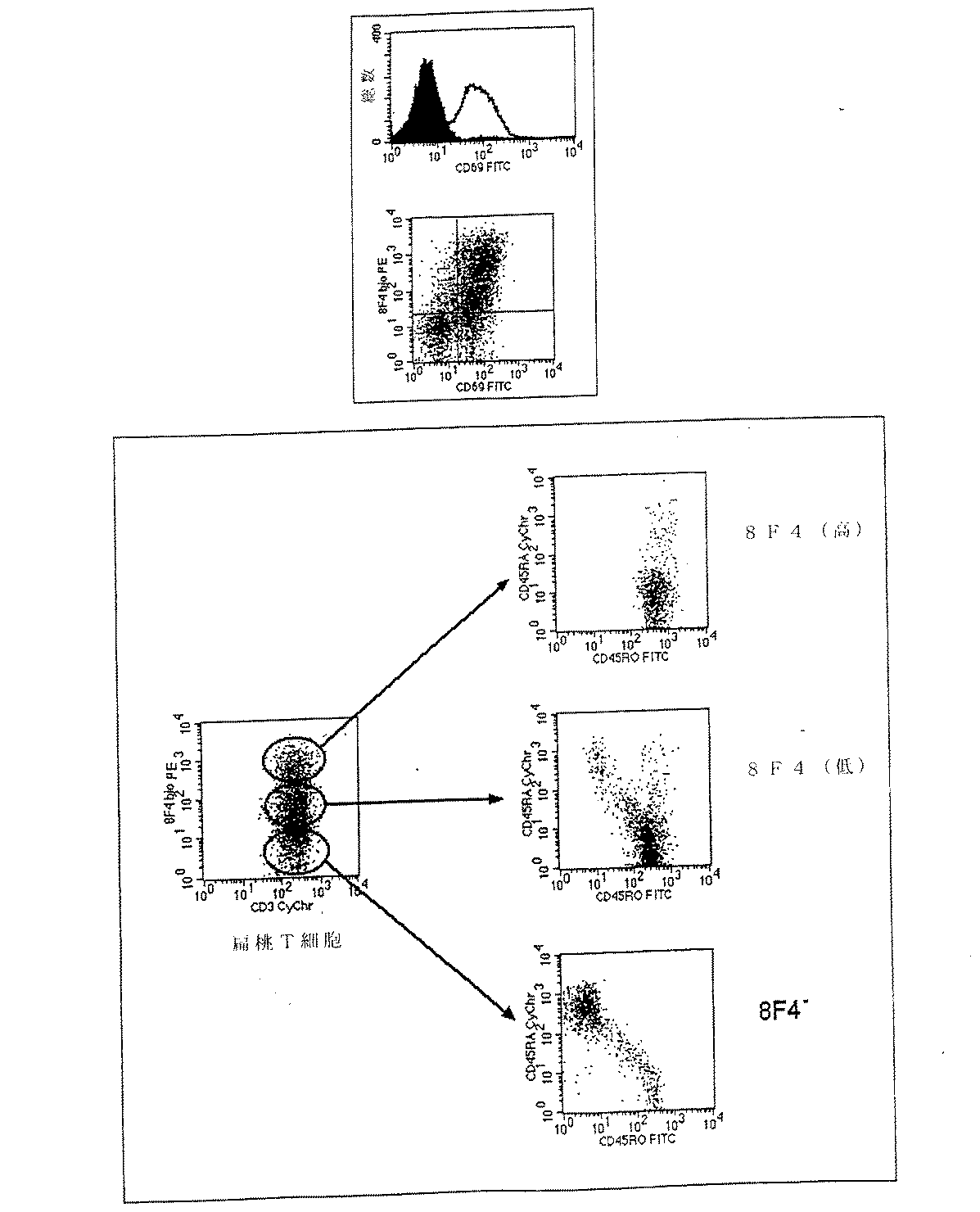
本発明のポリペプチド(以下、8F4分子または8F4とも呼ばれる)は、Tリンパ球、特にCD4+およびCD8+T細胞の双方が活性化された後にのみ発現される。非還元SDS−PAGEでは、8F4分子は約55〜60kDa(キロダルトン)の間の分子量を有する。この8F4分子は2つのペプチド鎖からなり、その2つのペプチド鎖は還元SDS−PAGEでは約27および29kDaの分子量を有する。8F4抗原は扁桃腺およびリンパ節のリンパ組織、特に胚細胞の中心部、抗体の生成時にTリンパ球とBリンパ球が相互作用する部位において活性化されたTリンパ球で組織学的に明らかに検出できる。ex vivoに単離された扁桃T細胞は約50〜80%が8F4抗原に関して陽性であり、活性化の進行の徴候を示す。8F4分子は休止中の、または活性化されたB細胞、顆粒球、単球、NK細胞および樹状細胞では検出されない。 The polypeptides of the present invention (hereinafter also referred to as 8F4 molecules or 8F4) are expressed only after activation of both T lymphocytes, in particular both CD4 + and CD8 + T cells. In non-reducing SDS-PAGE, the 8F4 molecule has a molecular weight between about 55-60 kDa (kilodalton). This 8F4 molecule consists of two peptide chains, which have a molecular weight of about 27 and 29 kDa in reduced SDS-PAGE. The 8F4 antigen is histologically revealed in lymphoid tissues of the tonsils and lymph nodes, particularly T lymphocytes activated in the central part of the germ cell, where T lymphocytes and B lymphocytes interact during antibody production. It can be detected. Approximately 50-80% of tonsillar T cells isolated ex vivo are positive for the 8F4 antigen, indicating signs of progression of activation. The 8F4 molecule is not detected on resting or activated B cells, granulocytes, monocytes, NK cells and dendritic cells.
8F4分子の重要な生物学的活性は、Tリンパ球に対するその共同刺激活性でである。この共同刺激活性はLinsley ea al., Journal of Experimental Medicine 176 (1992), 1595-604の方法によって測定できる。8F4分子の共同刺激活性は、免疫系による抗原認識の中枢増強要素として同定されているCD28分子の共同刺激活性と類似している。しかしながら8F4分子は多くの点でCD28とは異なっている。例えば、T細胞の表面における8F4分子の発現には誘導を要するが、CD28は構成的に発現する。また機能において検出可能な明瞭な違いもあり、CD28による共同刺激は多くのリンパ球、とりわけインターロイキン−2(IL−2)の過剰発現をもたらす。8F4による共同刺激もまた、リンホカインの分泌を増強するが、IL−2の分泌は増強しない。このように8F4分子の共同刺激活性は、CD28分子の活性とは異なっている。8F4による共同刺激は総てのサイトカイン遺伝子のスイッチを入れることはなく、in vivoにおける8F4による共同刺激は、例えばCD28受容体を介する共同刺激と比較すると有利である。さらに、8F4分子の誘導、発現、発現部位および機能は、共同刺激活性を有する他の公知の分子のいずれとも異なっている。 An important biological activity of the 8F4 molecule is its costimulatory activity on T lymphocytes. This costimulatory activity can be measured by the method of Linsley ea al., Journal of Experimental Medicine 176 (1992), 1595-604. The costimulatory activity of the 8F4 molecule is similar to the costimulatory activity of the CD28 molecule that has been identified as a central potentiator of antigen recognition by the immune system. However, the 8F4 molecule differs from CD28 in many ways. For example, expression of 8F4 molecules on the surface of T cells requires induction, but CD28 is constitutively expressed. There is also a distinct difference in function, and costimulation with CD28 results in overexpression of many lymphocytes, especially interleukin-2 (IL-2). Co-stimulation with 8F4 also enhances lymphokine secretion but not IL-2 secretion. Thus, the costimulatory activity of 8F4 molecules is different from that of CD28 molecules. Co-stimulation with 8F4 does not switch on all cytokine genes, and co-stimulation with 8F4 in vivo is advantageous compared to co-stimulation, for example via the CD28 receptor. Furthermore, the induction, expression, expression site and function of the 8F4 molecule is different from any other known molecule with costimulatory activity.
本発明の8F4分子は、Tリンパ球の中枢機能に強い共同刺激作用を有する、活性化されたT細胞上の新規な表面分子である。in vivoにおける発現は、とりわけウイルスおよび細菌に対する体液性および細胞性免疫防御の範囲で、T細胞と、B細胞または樹状細胞のような免疫系の他の細胞との共同作用における8F4分子の必須の関与を示している。 The 8F4 molecule of the present invention is a novel surface molecule on activated T cells that has a strong costimulatory effect on the central function of T lymphocytes. In vivo expression is essential for the 8F4 molecule in synergy between T cells and other cells of the immune system such as B cells or dendritic cells, especially in the range of humoral and cellular immune defenses against viruses and bacteria. Shows the involvement.
発現の後、8F4分子はin vitroにおいてTリンパ球の種々の機能に強い共同刺激作用を有する:
1.Tリンパ球の増殖の顕著な増強
2.Tリンパ球によるある種のサイトカイン合成の顕著な増強
3.Tリンパ球上およびTリンパ球内での制御分子、例えば表面分子およびサイトカインの極めて高い発現
4.T細胞によって誘導されるB細胞による抗体形成(IgMおよびIgG)における顕著な向上。
After expression, the 8F4 molecule has a strong costimulatory effect on various functions of T lymphocytes in vitro:
1. 1. Significant enhancement of
さらに本発明は、T細胞の共同刺激の生物学的活性を有し、かつ、図15(配列番号2)の199個のアミノ酸を含んでなる配列、またはその生物学的に有効な断片もしくは類似体と少なくとも40%の相同性を示すアミノ酸配列を有するポリペプチドを提供する。生物学的に有効な断片または類似体とは、T細胞リンパ球に対して同様に共同刺激作用を示すか、あるいは少なくともブロックの特徴の生物学的作用示す断片または類似体をいう。図15(配列番号2)の199個のアミノ酸を含んでなる配列と少なくとも60%の相同性を示すポリペプチド、またはその生物学的に有効な断片もしくは類似体が好ましい。特に好ましい具体例では、本発明のポリペプチドは、図15(配列番号2)の199個のアミノ酸を含んでなる配列と少なくとも80%の相同性を示すアミノ酸配列、またはその生物学的に有効な断片もしくは類似体を含んでなる。 Furthermore, the present invention has a T cell costimulatory biological activity and comprises a sequence comprising 199 amino acids of FIG. 15 (SEQ ID NO: 2), or a biologically effective fragment or analog thereof. A polypeptide having an amino acid sequence exhibiting at least 40% homology with the body is provided. A biologically effective fragment or analog refers to a fragment or analog that also exhibits a costimulatory effect on T cell lymphocytes, or at least a biological effect of a block characteristic. Preferred are polypeptides that exhibit at least 60% homology with the sequence comprising 199 amino acids of FIG. 15 (SEQ ID NO: 2), or biologically effective fragments or analogs thereof. In a particularly preferred embodiment, the polypeptide of the invention comprises an amino acid sequence that exhibits at least 80% homology with the sequence comprising 199 amino acids of FIG. 15 (SEQ ID NO: 2), or a biologically effective thereof. It comprises a fragment or analog.
特に好ましいポリペプチドはT細胞を共同刺激する生物学的活性を有し、かつ図15に示されるアミノ酸配列(配列番号2)、またはその生物学的に有効な断片もしくは類似体を含んでなる。 Particularly preferred polypeptides have the biological activity of costimulating T cells and comprise the amino acid sequence shown in FIG. 15 (SEQ ID NO: 2), or a biologically effective fragment or analog thereof.
本発明には8F4分子の対立遺伝子変異体、断片および類似体が含まれる。これらの変異体には、天然に存在している対立遺伝子変異体、1以上のアミノ酸が異なるアミノ酸で置換された置換類似体、1以上のアミノ酸を欠失した欠失類似体および、1以上のアミノ酸が付加された付加類似体が含まれる。1以上のアミノ酸の欠失および付加はポリペプチドの内部領域か、またはアミノもしくはカルボキシル末端のいずれで行われてもよい。 The present invention includes allelic variants, fragments and analogs of the 8F4 molecule. These variants include naturally occurring allelic variants, substituted analogs in which one or more amino acids are replaced with different amino acids, deleted analogs in which one or more amino acids are deleted, and one or more Additional analogs with amino acids added are included. One or more amino acid deletions and additions may be made either at the internal region of the polypeptide or at the amino or carboxyl terminus.
異種ポリペプチドと融合した本発明のポリペプチドも同様に包含される。 Also encompassed are polypeptides of the invention fused to heterologous polypeptides.
もう1つの具体例では、本発明は、本発明のポリペプチドまたはその生物学的に有効な断片もしくは類似体をコードするDNA配列に関する。 In another embodiment, the present invention relates to a DNA sequence encoding a polypeptide of the present invention or a biologically effective fragment or analog thereof.
これらのDNA配列には、配列番号1で示される配列(図16)ならびに生物学的活性を有する対立遺伝子変異体、断片および類似体が含まれる。 These DNA sequences include the sequence shown in SEQ ID NO: 1 (FIG. 16) and allelic variants, fragments and analogs having biological activity.
好ましいDNA配列は、T細胞を共同刺激する生物学的活性を有するポリペプチドをコードし、その配列は:
a)配列番号1で示されるDNA配列(図16)、およびその相補鎖
b)(a)の配列とハイブリダイズするDNA配列
c)遺伝子コードの縮重により(a)および(b)の配列とハイブリダイズするDNA配列
からなる群から選択される。前記のDNA配列はストリンジェントな条件下でともにハイブリダイズすることが好ましい。
A preferred DNA sequence encodes a polypeptide having biological activity that co-stimulates T cells, the sequence being:
a) DNA sequence shown in SEQ ID NO: 1 (FIG. 16) and its complementary strand b) DNA sequence hybridizing with the sequence of (a) c) The sequences of (a) and (b) due to the degeneracy of the gene code Selected from the group consisting of hybridizing DNA sequences. Preferably, the DNA sequences hybridize together under stringent conditions.
またこれらのDNA配列を含んでなるベクター、およびこれらのベクターで形質転換されるか、またはトランスフェクトされた宿主細胞も提供される。 Also provided are vectors comprising these DNA sequences, and host cells transformed or transfected with these vectors.
もう1つの具体例では、本発明は8F4分子に対するモノクロナール抗体に関する。本発明のモノクロナール抗体は、Milstein and Kohler, Nature 256 (1975), 495-497により記載されている常法で調製できる。本発明のモノクロナール抗体は特に、in vitroでホルボールミリステートアセテート(PMA)およびイオノマイシン(「2シグナル系」)で24時間活性化させたT細胞でマウスを免疫化することにより調製できる。免疫化したマウスの脾臓細胞を骨髄腫細胞と融合させる。8F4特異的モノクロナール抗体は休止中のものではなく、2つのシグナルで活性化されたTリンパ球を認識することにより同定される。さらに8F4特異的抗体は、常法で行われる検出法において1つのシグナル(PMAまたはイオノマイシンのいずれか)で刺激されたT細胞を染色しない。8F4特異的抗体は典型的な扁桃T細胞の染色パターンを示し、活性化されたTリンパ球の非還元SDS−PAGEでは約55〜60kDa、および還元SDS−PAGEで約27kDaおよび約29kDaの抗原を認識する。 In another embodiment, the invention relates to a monoclonal antibody against the 8F4 molecule. The monoclonal antibody of the present invention can be prepared by a conventional method described by Milstein and Kohler, Nature 256 (1975), 495-497. The monoclonal antibodies of the invention can be prepared in particular by immunizing mice with T cells activated in vitro with phorbol myristate acetate (PMA) and ionomycin (“2 signal system”) for 24 hours. Immunized mouse spleen cells are fused with myeloma cells. The 8F4-specific monoclonal antibody is not resting and is identified by recognizing T lymphocytes activated by two signals. Furthermore, 8F4-specific antibodies do not stain T cells stimulated with a single signal (either PMA or ionomycin) in detection methods performed in a conventional manner. The 8F4-specific antibody shows a typical staining pattern of tonsillar T cells, with about 55-60 kDa for non-reduced SDS-PAGE of activated T lymphocytes and about 27 kDa and about 29 kDa for reduced SDS-PAGE. recognize.
もう1つの具体例では、本発明は、本発明のモノクローナル抗体を産生するハイブリドーマ細胞に関する。 In another embodiment, the invention relates to a hybridoma cell that produces a monoclonal antibody of the invention.
もう1つの具体例では、本発明は、本発明のポリペプチド8F4の生物学的活性を阻害する物質の医薬としての使用に関する。本発明の8F4分子のモノクロナール抗体、天然または合成リガンド、アゴニストまたはアンタゴニストの使用が特に好ましい。これらの物質は免疫系が関与する疾患の予防または治療、特には自己免疫疾患の治療、または臓器移植における拒絶反応の予防を目的とした医薬として使用され得る。8F4抗原のその受容体との相互作用のブロックは予め活性化されたTリンパ球にのみ影響を及ぼすため、例えば臓器拒絶の予防に適している。本発明のもう1つの具体例は、本発明のポリペプチドの医薬としての使用に関する。本発明のポリペプチドは、特に免疫系が関与する疾患の予防または治療、特には癌、エイズ、喘息性疾患またはHCVもしくはHBV感染などの慢性ウイルス病の治療を目的として使用され得る。 In another embodiment, the invention relates to the use of a substance that inhibits the biological activity of the polypeptide 8F4 of the invention as a medicament. Particular preference is given to the use of monoclonal antibodies, natural or synthetic ligands, agonists or antagonists of the 8F4 molecule according to the invention. These substances can be used as medicaments for the purpose of preventing or treating diseases involving the immune system, particularly treating autoimmune diseases, or preventing rejection in organ transplantation. Blocking the interaction of the 8F4 antigen with its receptor affects only pre-activated T lymphocytes and is suitable, for example, for the prevention of organ rejection. Another embodiment of the invention relates to the use of a polypeptide of the invention as a medicament. The polypeptides of the present invention may be used for the prevention or treatment of diseases involving the immune system, particularly for the treatment of cancer, AIDS, asthmatic diseases or chronic viral diseases such as HCV or HBV infection.
同様に本発明のポリペプチドは、これらの細胞が例えばそのポリペプチドを構成的に発現するように常法で細胞に導入することができる。例えばそのポリペプチドをコードする核酸配列、またはそのポリペプチド、例えばcDNAまたはゲノムDNA、プロモーター、エンハンサーおよび核酸配列の発現に必要な他のエレメントを含んでなるベクターを細胞に挿入することができる。図16で示された8F4 cDNA(2641個のヌクレオチド)(配列番号1)またはその断片もしくは誘導体を本発明のポリペプチドまたはその断片の発現に使用することが好ましい。 Similarly, the polypeptides of the present invention can be introduced into cells by conventional methods such that these cells constitutively express the polypeptide, for example. For example, a nucleic acid sequence encoding the polypeptide, or a vector comprising the polypeptide, eg, cDNA or genomic DNA, a promoter, an enhancer, and other elements necessary for expression of the nucleic acid sequence can be inserted into the cell. The 8F4 cDNA (2641 nucleotides) (SEQ ID NO: 1) shown in FIG. 16 or a fragment or derivative thereof is preferably used for the expression of the polypeptide of the present invention or a fragment thereof.
本発明のポリペプチドはまた、例えばリポソームにより、後にその細胞表面にポリペプチドを形成する細胞へ導入することができる。これらの細胞は本発明に従い医薬として、特に多くの慢性感染症、例えばエイズ、喘息性疾患、または慢性ウイルス性肝炎(例えばHCV、HBV感染)の枠組み内にあるとき、ヒト免疫系の正確な調節を回復させるために、または例えば癌の治療のようなin viroまたはin vivoで免疫系を刺激するために使用できる。 The polypeptides of the invention can also be introduced, for example, by liposomes, into cells that later form the polypeptide on their cell surface. These cells are in accordance with the present invention as pharmaceuticals, particularly when within the framework of many chronic infections, such as AIDS, asthmatic diseases, or chronic viral hepatitis (eg HCV, HBV infection), the precise regulation of the human immune system Or to stimulate the immune system in vitro or in vivo, such as for example in the treatment of cancer.
もう1つの具体例では、本発明のポリペプチドを特異的に認識する物質が、免疫系が関与する疾患を診断するために使用される。この物質には特に、モノクロナール抗体、天然または合成リガンド、アゴニストまたはアンタゴニストが包含される。例えばELISA検出法、フローサイトメトリー、ウエスタンブロット、放射性免疫検定法、比濁分析法または組織化学的染色が診断のために使用可能である。本発明のポリペプチドを認識する物質はまた核酸配列もまた含んでおり、それはハイブリダイゼーションおよび/または核酸(RNA,DNA)増幅(例えば、PCR)に使用することが好ましい。 In another embodiment, a substance that specifically recognizes the polypeptide of the present invention is used to diagnose a disease involving the immune system. This material includes in particular monoclonal antibodies, natural or synthetic ligands, agonists or antagonists. For example, ELISA detection methods, flow cytometry, Western blots, radioimmunoassay methods, turbidimetric methods or histochemical staining can be used for diagnosis. The substance that recognizes the polypeptide of the present invention also includes a nucleic acid sequence, which is preferably used for hybridization and / or nucleic acid (RNA, DNA) amplification (eg, PCR).
もう1つの具体例では、本発明は、本発明のポリペプチドのT細胞へのシグナル変換経路に正または負の影響を及ぼす(調節する)物質、およびこれらの物質の医薬としての使用に関する。 In another embodiment, the present invention relates to substances that positively or negatively affect (modulate) the signal transduction pathway of the polypeptides of the present invention to T cells, and the use of these substances as pharmaceuticals.
もう1つの具体例では、本発明はT細胞表面の本発明のポリペプチドのアップレギュレーションを妨げる物質、およびその医薬としての使用に関する。 In another embodiment, the present invention relates to substances that interfere with the upregulation of the polypeptides of the present invention on the surface of T cells and their use as pharmaceuticals.
もう1つの具体例では、本発明のポリペプチドまたはその断片はトランスジェニック動物により発現される。 In another embodiment, the polypeptide of the invention or fragment thereof is expressed by a transgenic animal.
もう1つの具体例では、本発明は、本発明のポリペプチドをコードする遺伝子のスイッチが切られた(「ノックアウト」)トランスジェニック動物を包含する。 In another embodiment, the invention encompasses a transgenic animal in which the gene encoding the polypeptide of the invention is switched off (“knock out”).
以下、実施例により本発明を説明するが、それらに限定するものではない。 Hereinafter, the present invention will be described by way of examples, but is not limited thereto.
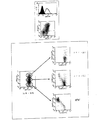
実施例1:8F4抗体の作製
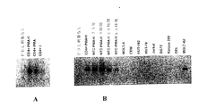
Balb/cマウスを、予めホルボールエステル、ホルボールミリステートアセテート(PMA)(Sigma,Deisenhofen)33ng/mlおよびCa2+イオノフォア、イオノマイシン(Sigma,Deisenhofen)20ng/mlで24時間活性化(いわゆる、「2−シグナル活性化」)させたヒトT細胞で免疫化した。3度追加抗原投与した後、マウスの脾臓細胞を骨髄腫P3X63Ag8.653(ATCC CRL−1580)と融合させ、標準法により抗体分泌ハイブリドーマを作製した;Peters and Baumgarten, Monoclonal Antibodies, Springer, Heidelberg, 1992を参照。得られた抗体をフローサイトメトリーにおいて休止中のT細胞に対して活性化されたものについてスクリーニングした。活性化され(「2シグナル活性化」)かつ休止中のT細胞をハイブリドーマ上清とともにインキュベートし、次いで蛍光標識二次抗体で標識した;Shapiro, Practical Flow Cytometry, Wiley-Liss, New York, 1995を参照。T細胞表面において1つの薬剤単独によってではなく、優先的にPMAおよびCa2+イオノフォア、イオノマイシンによってのみ誘導された分子(2シグナル分子)を認識する抗体だけをさらなる精製により選択した。得られた抗体をフローサイトメトリーにおいてT細胞の活性化分子に対する既知の抗体(表1参照)との類似点または相違点について調べた。これについての基準は前記の「2シグナル依存性」のほか、刺激されたT細胞における誘導の速度論および様々な細胞系における発現であった。
Example 1 Generation of 8F4 Antibody Balb / c mice were pre- treated with phorbol ester, phorbol myristate acetate (PMA) (Sigma, Deisenhofen) 33 ng / ml and Ca 2+ ionophore, ionomycin (Sigma, Deisenhofen) 20 ng / ml. Immunization with human T cells activated for 24 hours (so-called “2-signal activation”). After three additional doses, mouse spleen cells were fused with myeloma P3X63Ag8.653 (ATCC CRL-1580) to produce antibody-secreting hybridomas by standard methods; Peters and Baumgarten, Monoclonal Antibodies, Springer, Heidelberg, 1992 See The resulting antibodies were screened for those activated against resting T cells in flow cytometry. Activated ("2 signal activated") and resting T cells were incubated with hybridoma supernatants and then labeled with a fluorescently labeled secondary antibody; Shapiro, Practical Flow Cytometry, Wiley-Liss, New York, 1995 reference. Only antibodies recognizing molecules that were preferentially induced only by PMA and Ca 2+ ionophore, ionomycin (2 signal molecules) but not by one agent alone at the T cell surface were selected by further purification. The obtained antibodies were examined in flow cytometry for similarities or differences from known antibodies to T cell activation molecules (see Table 1). Criteria for this were the “2 signal dependence” described above, as well as induction kinetics in stimulated T cells and expression in various cell lines.
実施例2:8F4抗原の免疫沈降
活性化されたヒトT細胞由来の分子の表面を125Iで標準法によりヨウ素化し、抗体8F4で標準法により免疫沈降させた;Goding, Monoclonal Antibodies: Principle and Practice, Academic Press, London, 1996を参照。免疫沈降反応用の抗体をSchneider et al., Journal of Biological Chemistry 257 (1982), 10766-10769の方法によりprotein G(Pharmacia, Freiburgへ結合させた(8F4マトリックス)。このマトリックスをSchneider et al.により記載されるように洗浄した、前記参照。免疫沈降した8F4分子をSDS−PAGE(非還元および還元)により常法で分子量について解析した;Goding 、前記参照。
Example 2: Immunoprecipitation of 8F4 antigen Surfaces of activated human T cell-derived molecules were iodinated with 125 I by standard methods and immunoprecipitated with antibody 8F4 by standard methods; Goding, Monoclonal Antibodies: Principle and Practice , Academic Press, London, 1996. An antibody for immunoprecipitation was conjugated to protein G (Pharmacia, Freiburg (8F4 matrix) by the method of Schneider et al., Journal of Biological Chemistry 257 (1982), 10766-10769. This matrix was prepared by Schneider et al. Washed as described, supra .. Immunoprecipitated 8F4 molecules were analyzed for molecular weight by SDS-PAGE (non-reducing and reducing) routinely;
実施例3:フローサイトメトリー
8F4を有するT細胞をフローサイトメトリーにおいて標準法により解析した;Shapiro, Practical Flow Cytometry, Wiley-Liss, New York, 1995を参照。
Example 3: T cells with flow cytometry 8F4 were analyzed by standard methods in flow cytometry; see Shapiro, Practical Flow Cytometry, Wiley-Liss, New York, 1995.
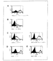
具体例3.1:CD4+T細胞における8F4抗原誘導後のフローサイトメトリー。
末梢血由来のCD4+T細胞を様々な薬剤により常法で刺激し、8F4分子の発現をフローサイトメトリーにおいて常法で調べた。T細胞の活性化時間は種々の薬剤で24時間〜144時間の間とした。活性化モード:ホルボールミリステートアセテート(PMA;33ng/ml)、イオノマイシン(200ng/ml)、フィトヘマグルチニン(PHA 1.5mg/ml)、OKT3(CD3に対するモノクロナール抗体)、混合リンパ球反応(MLR,CD4+T細胞50,000とB細胞100,000間)、mAk9.3(CD28に対するモノクロナール抗体)、ブドウ球菌エンテロトキシンB(SEB,0.1ng/ml)。解析により、種々の刺激はT細胞の8F4分子の誘導に好適であるが、発現強度が異なっていることが示された。最も有効な刺激は、非常に有効な薬理学的薬剤であるPMAおよびイオノマイシンの他、例えばMLRの補助細胞または共同刺激性mAk9.3などの共同刺激状態を示すものである。
Specific Example 3.1: Flow cytometry after induction of 8F4 antigen in CD4 + T cells.
Peripheral blood-derived CD4 + T cells were stimulated with various drugs in a conventional manner, and the expression of 8F4 molecules was examined by a conventional method in flow cytometry. The activation time of T cells was 24 hours to 144 hours with various drugs. Activation mode: phorbol myristate acetate (PMA; 33 ng / ml), ionomycin (200 ng / ml), phytohemagglutinin (PHA 1.5 mg / ml), OKT3 (monoclonal antibody against CD3), mixed lymphocyte reaction (MLR) , CD4 + T cells 50,000 and B cells 100,000), mAk 9.3 (monoclonal antibody against CD28), staphylococcal enterotoxin B (SEB, 0.1 ng / ml). Analysis showed that various stimuli were suitable for induction of 8F4 molecules in T cells, but differed in expression intensity. The most effective stimuli are those that exhibit costimulatory states such as MLR co-stimulatory cells or costimulatory mAk 9.3, in addition to the highly effective pharmacological agents PMA and ionomycin.
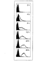
具体例3.2:PMAおよびイオノマイシンでの活性化後のCD4+T細胞における8F4抗原誘導の速度論
末梢血由来のCD4+T細胞をPMA(33ng/ml)およびイオノマイシン(200ng/ml)により常法で刺激し、0,4,8,12,24および48時間後、8F4分子の発現をフローサイトメトリーにおいて常法で調べた。この分子は4時間後でのみ表面上で検出可能であり、比較的初期のクラスの活性化抗原に属する。48時間後の抗原でもさらに強い発現がある。
Example 3.2: Kinetics of 8F4 antigen induction in CD4 + T cells after activation with PMA and ionomycin Peripheral blood derived CD4 + T cells were routinely treated with PMA (33 ng / ml) and ionomycin (200 ng / ml). After 0, 4, 8, 12, 24, and 48 hours, the expression of 8F4 molecules was examined in a conventional manner in flow cytometry. This molecule is detectable on the surface only after 4 hours and belongs to a relatively early class of activated antigens. There is even stronger expression in the antigen after 48 hours.
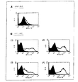
具体例3.3:「混合リンパ球反応」において8F4の誘導に関与する分子を同定するためのフローサイトメトリー
末梢血由来のCD4+T細胞50,000を同種扁桃B細胞100,000と6日間同時培養し(37℃、5.2%CO2、96ウェル丸底プレートに10%FCSの入ったPRMI 200μl)、次いで8F4分子の発現をフローサイトメトリーで調べた。培養の開始時に種々の抗体(抗CD80、抗CD86、抗MHCII;総て10mg/ml)を培地に添加し、8F4誘導のこれら分子への依存性を試験した。8F4の発現はCD86/CD28の相互作用のブロックによってのみ阻害でき、CD80のブロックによっては阻害できない。この場合のブロック作用はMHCIIのブロックよりいっそう強いものである(正の調節)。
Specific Example 3.3: Flow cytometry for identifying molecules involved in the induction of 8F4 in the “mixed lymphocyte reaction”. Peripheral blood-derived CD4 + T cells 50,000 with allogeneic tonsil B cells 100,000 for 6 days Co-cultured (37 ° C., 5.2% CO 2 , 200 μl of RPMI with 10% FCS in a 96-well round bottom plate) and then examined for expression of 8F4 molecules by flow cytometry. Various antibodies (anti-CD80, anti-CD86, anti-MHCII; total 10 mg / ml) were added to the medium at the start of culture to test the dependence of 8F4 induction on these molecules. Expression of 8F4 can only be inhibited by blocking the CD86 / CD28 interaction and not by blocking CD80. The blocking effect in this case is even stronger than the MHCII block (positive regulation).
具体例3.4:ヒト扁桃由来のTおよびB細胞の発現
種々の起源からの扁桃組織のTおよびB細胞を常法で精製し、8F4分子の発現をフローサイトメトリーにより調べた。B細胞ではシグナルが明らかに有意でなかったのに対し、扁桃T細胞の密度が約50〜80%の場合には8F4分子の発現がみられた。この場合、蛍光のレベルが異なり(それぞれ8F4が高いものと低いもの)、かつ種々の扁桃における発現が異なる2集団の同定が可能である。従って、例えばある扁桃は明らかに8F4の低い集団を示し、他の扁桃は明らかに8F4の高い集団を示す。
Specific Example 3.4: Expression of T and B cells derived from human tonsils T and B cells of tonsils from various sources were purified by conventional methods, and the expression of 8F4 molecules was examined by flow cytometry. The signal was clearly not significant in B cells, whereas the expression of 8F4 molecules was seen when the density of tonsillar T cells was about 50-80%. In this case, it is possible to identify two populations with different levels of fluorescence (high and low 8F4 respectively) and different expression in various tonsils. Thus, for example, one tonsil clearly shows a low 8F4 population and the other tonsil clearly shows a high 8F4 population.
具体例3.5:8F4分子の他の活性化マーカーとの同時発現
ヒト扁桃から精製したT細胞を、8F4分子の他の活性化マーカーとの同時発現について2色系フローサイトメトリーで解析した。扁桃において8F4はCD69と、ならびにCD45分子の変異体とともに同時発現する。この場合、8F4の高い細胞は明らかにCD45RO発現と相互関係があるが、一方8F4が陰性の細胞は表現型CD45RAを有する。CD45RAは主として、いわゆる「天然」T細胞により発現されるが、CD45ROはエフェクター細胞の機能に関与している。よって8F4+細胞は主として「成熟」T細胞である。CD45ROおよびCD45RAはCD45のイソ型である。
Specific Example 3.5: Co-expression with other activation markers of 8F4 molecule T cells purified from human tonsils were analyzed for co-expression with other activation markers of 8F4 molecule by two-color flow cytometry. In tonsils, 8F4 is co-expressed with CD69, as well as variants of the CD45 molecule. In this case, cells with high 8F4 clearly correlate with CD45RO expression, whereas cells negative for 8F4 have the phenotype CD45RA. CD45RA is mainly expressed by so-called “natural” T cells, whereas CD45RO is involved in the function of effector cells. Thus, 8F4 + cells are primarily “mature” T cells. CD45RO and CD45RA are isoforms of CD45.
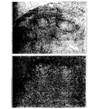
実施例4:8F4陽性細胞の扁桃における局在性
扁桃組織の凍結切片をAPAAP手法(アルカリ性ホスファターゼ−抗アルカリ性ホスファターゼ)において標準法により8F4抗体で染色した。8F4+細胞は扁桃の胚中心で優先的に見られるが、扁桃のT細胞ゾーンの一部分でも見られた。
Example 4: Frozen sections of localized tonsil tissue in tonsils of 8F4 positive cells were stained with 8F4 antibody by APAAP technique (alkaline phosphatase-antialkaline phosphatase) by standard methods. 8F4 + cells are preferentially found in tonsil germinal centers, but also in a portion of the tonsil T cell zone.
実施例5:Tリンパ球の共同刺激
96ウェルプレートにヤギ抗マウスIg抗体(20μg/ml)を塗布し、洗浄して、抗CD3モノクロナール抗体OKT3(種々の希釈の腹水)および本発明の8F4抗体(2μg/ml)を添加した。OKMl抗体または2A11抗体(双方2μg/ml)をイソタイプの対照として用いた。
Example 5: Co-stimulation of T lymphocytes A 96-well plate was coated with goat anti-mouse Ig antibody (20 μg / ml), washed, anti-CD3 monoclonal antibody OKT3 (various dilutions of ascites) and 8F4 of the invention Antibody (2 μg / ml) was added. OKMl antibody or 2A11 antibody (both 2 μg / ml) were used as isotype controls.
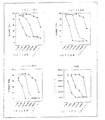
具体例5.1:8F4により共同刺激された後のTリンパ球の活性化分子の発現の増強
末梢血由来の精製CD4+T細胞を種々の濃度のモノクロナール抗体OKT3、同時に8F4抗体または同じイソタイプの非特異的抗体により活性化した。比較として、既知の最も強い共同刺激性抗体の1つである抗CD28抗体−9.3により共同刺激を行った。CD3によっては最適に共同刺激されるものの、mAk8F4およびmAk9.3双方によってもなお共同刺激作用が認められる。最適下限のOKT3領域、すなわち共同刺激無しではT細胞の完全な活性化がなされ得ない領域では、両抗体は4〜100の因子によって他の活性化抗原の発現を高めることができ、抗CD28抗体の作用はOKT3の希釈度が非常に高いときでもなお認識できる。このことは極めて弱いOKT3による刺激では、8F4抗原はもはや細胞表面にもたらされず、そのためmAk 8F4のいずれかでは架橋できないということに帰すことができる。
Example 5.1: Enhanced expression of T lymphocyte activation molecules after costimulation with 8F4 Peripheral blood-derived purified CD4 + T cells with various concentrations of monoclonal antibody OKT3, simultaneously with 8F4 antibody or the same isotype Of non-specific antibodies. For comparison, costimulation was performed with anti-CD28 antibody-9.3, one of the strongest known costimulatory antibodies. Although CD3 is optimally co-stimulated, both mAk8F4 and mAk 9.3 still have co-stimulatory effects. In the suboptimal OKT3 region, i.e., where complete activation of T cells cannot be achieved without co-stimulation, both antibodies can enhance the expression of other activated antigens by a factor of 4-100, and anti-CD28 antibodies This effect can still be recognized even when the dilution of OKT3 is very high. This can be attributed to the fact that upon stimulation with very weak OKT3, the 8F4 antigen is no longer brought to the cell surface and therefore cannot be cross-linked by any of the mAk 8F4.
具体例5.2:8F4の共同刺激作用とCD28の共同刺激作用との比較
精製CD8+T細胞を最適下限濃度のモノクロナール抗体OKT3で51時間刺激した。使用した共同刺激物質は抗体8F4、抗体9.3(抗体CD28)およびイソタイプの対照(各2μg/ml)であった。刺激時間が完了した後、T細胞増殖速度を3H−チミジンの組み込みにより求めた。同時に行った培養では上清を除去し、サイトカイン ATAC/リンフォタクチンおよびIL−2の濃度を決定した。8F4およびCD28はIL−2合成に関して、互いに顕著に異なっている。CD28共同刺激により、先行技術にも記載されるように(Chambers and Allison, Current Opinion in Immunology 9 (1997), 396-404)IL−2の分泌が極めて多くなる。これに対し、8F4によるIL−2産生は検出限界以下である。しかしながら、増殖については2つの混合物で比較できる、よってT細胞の自己分泌増殖は8F4の共同刺激における他の因子に帰すべきであろう。この2つの抗体はリンフォカインATACの分泌に関する共同刺激作用においてほとんど全く差がない。
Specific Example 5.2: Comparison of 8F4 costimulatory effect and CD28 costimulatory effect Purified CD8 + T cells were stimulated with monoclonal antibody OKT3 at the suboptimal concentration for 51 hours. The costimulators used were antibody 8F4, antibody 9.3 (antibody CD28) and isotype control (2 μg / ml each). After completion of the stimulation time, T cell proliferation rate was determined by 3 H-thymidine incorporation. Supernatants were removed from simultaneous cultures and the concentrations of cytokines ATAC / lymphotactin and IL-2 were determined. 8F4 and CD28 are significantly different from each other with respect to IL-2 synthesis. CD28 co-stimulation leads to very high IL-2 secretion, as described in the prior art (Chambers and Allison, Current Opinion in Immunology 9 (1997), 396-404). In contrast, IL-2 production by 8F4 is below the detection limit. However, proliferation can be compared in the two mixtures, so T cell autocrine proliferation should be attributed to other factors in 8F4 costimulation. The two antibodies have little difference in the costimulatory effect on secretion of lymphokine ATAC.
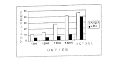
実施例6:8F4により共同刺激されたT細胞との相互作用後にB細胞によって合成された免疫グロブリンの測定
96ウェルプレートをヤギ抗マウスIg抗体(20μg/ml)で被覆し、抗CD3モノクローナル抗体OKT3(1:500〜1:80,000腹水)および本発明の8F4抗体(2μg/ml)を入れた。OKM1抗体または2A11抗体をイソタイプ対照として使用した。ある実験では、比較のためにCD28特異的抗体(「9.3」)を用いて共同刺激を行った;Hara et al., Journal of Experimental Medicine 161 (1985), 1513-1524を参照。末梢血由来の50,000個の精製した(Magnetbeads, Dynal, Hamburg)CD4+T細胞(純度>95%)および25,000個の同種扁桃B細胞(ヒツジ赤血球とロゼットを形成するT細胞による負の選抜、純度96%)をこのように予め処理した培養プレートの各ウェルにピペットで入れ、8日間共存培養した。この後、上清を採取してIgMおよびIgG型の分泌免疫グロブリンの濃度を常法にてELISA法で分析した;Nishioka and Lipsky, Journal of Immunology 153 (1994), 1027-1036を参照。
Example 6: Measurement of immunoglobulin synthesized by B cells after interaction with T cells costimulated with 8F4 A 96-well plate was coated with goat anti-mouse Ig antibody (20 μg / ml) and anti-CD3 monoclonal antibody OKT3 (1: 500-1: 80,000 ascites) and 8F4 antibody of the present invention (2 μg / ml) were added. OKM1 antibody or 2A11 antibody was used as an isotype control. In some experiments, costimulation was performed using a CD28 specific antibody ("9.3") for comparison; see Hara et al., Journal of Experimental Medicine 161 (1985), 1513-1524. 50,000 purified (Magnetbeads, Dynal, Hamburg) CD4 + T cells (> 95% purity) from peripheral blood and 25,000 allogeneic tonsil B cells (negative by T cells that form rosettes with sheep erythrocytes) (Purity of 96%) was pipetted into each well of the culture plate thus pretreated and co-cultured for 8 days. After this, the supernatant was collected and analyzed for the concentrations of IgM and IgG type secretory immunoglobulins by conventional ELISA methods; see Nishioka and Lipsky, Journal of Immunology 153 (1994), 1027-1036.
具体例6.1:T細胞の共同刺激後のB細胞によるIgMおよびIgG型の抗体合成の増強
末梢血由来の精製したCD4+T細胞を扁桃由来の同種B細胞とともに常法で8日間共存培養した。OKT3抗体によるT細胞の最適下限刺激では、8F4によるT細胞の共同刺激はIgMおよびIgG型免疫グロブリンの分泌を40だけ増強する。
Example 6.1: Enhancement of IgM and IgG-type antibody synthesis by B cells after co-stimulation of T cells Purified CD4 + T cells derived from peripheral blood were co-cultured with allogeneic B cells derived from tonsils in a conventional manner for 8 days did. At sub-optimal stimulation of T cells with OKT3 antibody, co-stimulation of T cells with 8F4 enhances secretion of IgM and IgG type immunoglobulins by 40.
実施例7:8F4による共同刺激後の末梢T細胞の活性化誘導性アポトーシスの阻害
末梢T細胞(常法においてナイロンウール付着によって精製した)をPHA(1.5mg/ml)で20時間刺激し、IL−2とともに6日間培養した。次いで、細胞をmAK 8F4(2μg/ml)による共同刺激を伴い、または伴わずにOKT3で再刺激した。フローサイトメトリー(FACS)においてヨー化プロピジウムでDNAを染色することによってアポトーシスを測定した。T細胞受容体複合体を介した最適下限の刺激を伴う8F4による共同刺激は、アポトーシス細胞の割合を4だけ減少できる。
Example 7: Inhibition of activation-induced apoptosis of peripheral T cells after co-stimulation with 8F4 Peripheral T cells (purified by nylon wool attachment in a conventional manner) were stimulated with PHA (1.5 mg / ml) for 20 hours, Cultured with IL-2 for 6 days. Cells were then restimulated with OKT3 with or without costimulation with mAK 8F4 (2 μg / ml). Apoptosis was measured by staining the DNA with propidium iodide in flow cytometry (FACS). Co-stimulation with 8F4 with suboptimal stimulation through the T cell receptor complex can reduce the proportion of apoptotic cells by 4.
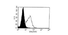
実施例8:8F4タンパク質をコードするcDNAのクローニング
フローサイトメトリーにおいて蛍光色素を結合させた8F4抗体で染色することによって、8F4抗原を構成的に発現する細胞系統(MOLT−4V)を同定した(図11)。MOLT−4V系統はヒトT細胞系統MOLT−4の変異体である(American Type Culture Collection(ATCC) CRL−1582)。
Example 8: Cloning of cDNA encoding 8F4 protein A cell line (MOLT-4V) that constitutively expresses the 8F4 antigen was identified by staining with 8F4 antibody conjugated with a fluorescent dye in flow cytometry (Fig. 11). The MOLT-4V line is a variant of the human T cell line MOLT-4 (American Type Culture Collection (ATCC) CRL-1582).
この細胞系統をモノクローナル抗体を用いる8F4抗原の予備精製に使用した。
細胞を回転培養瓶中で大量培養し(150l)、遠心分離により回収して溶解緩衝液(50mM トリス、pH8.0、150mM NaCl、1mM EDTA、1mM PMSF(Sigma、Deisenhofen)、1% NP−40(Boehringer, Mannheim))を用いて細胞性タンパク質を抽出した。細胞核および他の不溶性成分を超遠心分離によって除去した。このようにして得た細胞溶解物をセファロースCL4−B(Pharmacia, Freiburg)とともに2時間プレインキュベートしてセファロースに非特異的に結合するタンパク質を除去した。次いで前記実施例2に記載の8F4免疫親和性マトリックスとともにインキュベーションを行った(4℃で4時間)。マトリックスをカラムに詰め、次いで非特異的に結合しているタンパク質の完全な除去を行う条件(1.50mM トリス、pH8.0、300mM NaCl、1mM EDTA、1mM PMSF、0.5% NP−40;2.50mM トリス、pH8.0、150mM NaCl、1mM EDTA、1mM PMSF、0.5% NP−40、0.1% SDS;3.0.2M グリシン pH4.0、0.5% CHAPS(Merck, Darmstadt))下で数回洗浄した。0.2M グリシン、pH2.5、0.5% CHAPSを用いてマトリックスから8F4抗原を溶出させた。溶出液を限外濾過(Amicon Centricon 10, Millipore, Eschborn)によって濃縮した。
This cell line was used for prepurification of 8F4 antigen using monoclonal antibodies.
Cells are cultured in large volumes in rotating culture bottles (150 l), recovered by centrifugation and lysis buffer (50 mM Tris, pH 8.0, 150 mM NaCl, 1 mM EDTA, 1 mM PMSF (Sigma, Deisenhofen), 1% NP-40. (Boehringer, Mannheim)) was used to extract cellular proteins. Cell nuclei and other insoluble components were removed by ultracentrifugation. The cell lysate thus obtained was preincubated with Sepharose CL4-B (Pharmacia, Freiburg) for 2 hours to remove proteins that non-specifically bind to Sepharose. The incubation was then carried out with the 8F4 immunoaffinity matrix described in Example 2 (4 hours at 4 ° C.). Conditions under which the matrix is packed into a column followed by complete removal of non-specifically bound proteins (1.50 mM Tris, pH 8.0, 300 mM NaCl, 1 mM EDTA, 1 mM PMSF, 0.5% NP-40; 2. 50 mM Tris, pH 8.0, 150 mM NaCl, 1 mM EDTA, 1 mM PMSF, 0.5% NP-40, 0.1% SDS; 3.0.2 M glycine pH 4.0, 0.5% CHAPS (Merck, Darmstadt)) and washed several times. The 8F4 antigen was eluted from the matrix using 0.2M glycine, pH 2.5, 0.5% CHAPS. The eluate was concentrated by ultrafiltration (
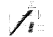
8F4分子のさらなる精製を実施するために、ニ次元ゲル電気泳動(非還元/還元)において分子のニ量体構造(図1を参照)を利用した。ほとんどのタンパク質は単量体として生じるので、それらがゲル電気泳動においては対角線上に移動するのに対し、8F4分子は第1の次元(非還元)では55〜60kDaに、また第2の次元(還元)では27および29kDaに移動する(図12)。 To perform further purification of the 8F4 molecule, the dimeric structure of the molecule (see FIG. 1) was utilized in two-dimensional gel electrophoresis (non-reduction / reduction). Most proteins occur as monomers, so they move diagonally in gel electrophoresis, whereas the 8F4 molecule is 55-60 kDa in the first dimension (non-reducing) and the second dimension ( In reduction, it moves to 27 and 29 kDa (FIG. 12).
予備分画のために、各場合において20×109細胞からの免疫沈降物を図12に関して前記に記載のように調製し、ニ次元ゲル電気泳動で分画し、ゲルをクマシーブルーG250(Biorad, Munich)で染色して図12において示された領域をゲルから個々に切り出した(それぞれ8F4−27kDaおよび8F4−29kDa)。 For pre-fractionation, immunoprecipitates from 20 × 10 9 cells in each case were prepared as described above with respect to FIG. 12, fractionated by two-dimensional gel electrophoresis, and the gel was coomassie blue G250 (Biorad , Munich) and the regions shown in FIG. 12 were individually cut from the gel (8F4-27 kDa and 8F4-29 kDa, respectively).
ペプチドのマイクロシーケンシングのために、各場合においてゲル4片からのタンパク質をトリプシンで消化してゲルから溶出させた。トリプシンによって生じた断片をHPLCによって分画し、個々の画分をEdman分解(Groettrup, M. et al. (1996), Eur. J. Immunol., 26: 863-869に詳細に記載された方法)に付した。 For peptide microsequencing, in each case, the protein from four pieces of gel was digested with trypsin and eluted from the gel. Fragments generated by trypsin were fractionated by HPLC, and individual fractions were separated by Edman degradation (Groettrup, M. et al. (1996), Eur. J. Immunol., 26: 863-869). )
8F4−29kDaサンプルの配列決定により、既知のタンパク質の断片に加え、いずれのタンパク質データベースにおいてもヒトとの相関性が認められないペプチド配列XRLTDVTが示された。 Sequencing of the 8F4-29 kDa sample showed a peptide sequence XRLTDVT that was not correlated with humans in any protein database in addition to known protein fragments.
タンパク質配列のDNA配列への明確な逆翻訳は可能でない。従って前記のペプチド配列の、17個のヌクレオチドを含むオリゴヌクレオチドへの逆翻訳は結果として2048通りの組合せを生ずる。しかしながら、特殊な方法(Wozney, J.M. (1990), Methods Enzymol. 182; 738-751)により縮重オリゴヌクレオチドを用いてcDNAバンクをスクリーニングすることは可能である。見出したペプチド配列をもとにして、2つのオリゴヌクレオチド(オリゴ1(配列番号3);MGN CTS ACN GAY GTN AC、512通りの組合せ;オリゴ2(配列番号4):MGN YTD ACN GAY GTN AC、1024通りの組合せ)を合成した。 Clear back translation of protein sequences into DNA sequences is not possible. Thus, reverse translation of the peptide sequence into an oligonucleotide containing 17 nucleotides results in 2048 combinations. However, it is possible to screen cDNA banks using degenerate oligonucleotides by special methods (Wozney, J.M. (1990), Methods Enzymol. 182; 738-751). Based on the found peptide sequence, two oligonucleotides (oligo 1 (SEQ ID NO: 3); MGN CTS ACN GAY GTN AC, 512 combinations; oligo 2 (SEQ ID NO: 4): MGN YTD ACN GAY GTN AC, 1024 combinations) were synthesized.
スクリーニングのために、タンパク質精製のためにも用いたMOLT−4V細胞系統からcDNAバンクを構築した。:
グアニジニウム/CsCl法(Chirgwin, J.M. et al. (1979), Biochemistry 18: 5294-5299)によって完全RNAを単離し、オリゴ−dT−セルロースカラム(Gibco BRL, Eggenstein)でmRNAを濃縮した。オリゴ−dT−プライマーを用いる市販のcDNA合成系(Gibco BRL, Eggenstein)を用いて、製造業者の使用説明書に従って第1および第2cDNA鎖の合成を行った。cDNAをEcoRIアダプターを介してLambda ZAPIIベクター(Stratagene, Heidelberg)に連結した。
For screening, a cDNA bank was constructed from the MOLT-4V cell line that was also used for protein purification. :
Complete RNA was isolated by the guanidinium / CsCl method (Chirgwin, JM et al. (1979), Biochemistry 18: 5294-5299), and mRNA was concentrated with an oligo-dT-cellulose column (Gibco BRL, Eggenstein). First and second cDNA strands were synthesized using a commercially available cDNA synthesis system (Gibco BRL, Eggenstein) using oligo-dT-primers according to the manufacturer's instructions. The cDNA was ligated to the Lambda ZAPII vector (Stratagene, Heidelberg) via an EcoRI adapter.
cDNAバンクを標準法(Vogeli, G, and kaytes, P.S. (1987), Methods Enzymol., 152: 407-515)によって平板培養し、ニトロセルロースフィルター(Optitran BA-S 85, Schleicher&Schuell, Dassel)上にLambda DNAを固定した。 cDNA banks are plated by standard methods (Vogeli, G, and kaytes, PS (1987), Methods Enzymol., 152: 407-515) and Lambda on nitrocellulose filters (Optitran BA-S 85, Schleicher & Schuell, Dassel). DNA was fixed.
前記のオリゴヌクレオチドをT4ポリヌクレオチドキナーゼ(NEBL、Schwalbach)およびγ−32P ATP(NEN Du Pont、Brussels)を用いて放射性標識した(Wallace, R.B. and Miyata, C.G. (1987), Methods Enzymol.,152: 432-442)。 The oligonucleotides were radiolabeled with T4 polynucleotide kinase (NEBL, Schwalbach) and γ- 32 P ATP (NEN DuPont, Brussels) (Wallace, RB and Miyata, CG (1987), Methods Enzymol., 152 : 432-442).
3Mのテトラメチル塩化アンモニウム(Roth、Karlsruhe)を含む縮重オリゴヌクレオチドに関して記載された緩衝液(Wozney, J.M. (1990), Methods Enzymol, 182: 738-751)中、48℃でフィルターのハイブリダイゼーションを行った。洗浄温度を50℃とし、前記参照文献に記載のようにフィルターを洗浄した。X線フィルム上でのこれらのフィルターの露光により、100,000個の平板培養ファージ当たり約50個の陽性クローンが現れた(図13)。 Filter hybridization at 48 ° C in buffer (Wozney, JM (1990), Methods Enzymol, 182: 738-751) described for degenerate oligonucleotides containing 3M tetramethylammonium chloride (Roth, Karlsruhe) went. The washing temperature was 50 ° C. and the filter was washed as described in the above reference. Exposure of these filters on X-ray film revealed approximately 50 positive clones per 100,000 plated phage (FIG. 13).
ベクターの製造業者(Stratagene, Heidelberg)によって記載された方法を用いるin vivo切り出しによって、それらをプラスミドベクターに導入することにより6個のクローンをさらに同定し、T3およびT7プライマーを用いて部分的に配列決定した(BigDyeターミネーターサイクルシークエンシングキット、Applied Biosystems, Foster City, USA)。1つのクローンは、求めたペプチド配列を翻訳時に正確に提供する配列を含んでいた。このクローンをノーザンブロットのハイブリダイゼーション(図14)に用いた(Kroczek, R.A. (1993), J. Chromatogr., 618, 133-145)。フローサイトメトリーによるモノクローナル抗体に対する研究から知られていたように、mRNAの発現パターンは8F4分子の発現に厳密に対応した。見出されたクローンは求めるcDNAの3’末端のみしか含んでいなかったので、5’側に対する断片を用いて完全な8F4cDNAを単離した。数個のクローンを両鎖に関して配列決定した。 Six clones were further identified by in vivo excision using the method described by the vector manufacturer (Stratagene, Heidelberg) by introducing them into a plasmid vector and partially sequenced using T3 and T7 primers (BigDye terminator cycle sequencing kit, Applied Biosystems, Foster City, USA). One clone contained a sequence that accurately provided the determined peptide sequence upon translation. This clone was used for Northern blot hybridization (FIG. 14) (Kroczek, R.A. (1993), J. Chromatogr., 618, 133-145). As known from studies on monoclonal antibodies by flow cytometry, the expression pattern of mRNA closely corresponded to the expression of 8F4 molecules. Since the found clone contained only the 3 'end of the sought cDNA, the fragment for the 5' side was used to isolate the complete 8F4 cDNA. Several clones were sequenced on both strands.
8F4cDNA(2641個のヌクレオチド)は図16および配列番号1で記載されている配列中に示されており、また図15および配列番号2で記載されている配列中に示されている199個のアミノ酸(ヌクレオチド68〜664)を有するタンパク質をコードする。cDNAバンク由来の数個の独立したクローンの配列決定により、本明細書に示される配列からのいくらかのずれが示されたが、これらは総て3’非翻訳領域中にある:
位置909−910:欠失
位置1631:T→C
位置2074:G→T
位置2440:G→C
位置2633:選択可能なポリアデニル化部位。
8F4 cDNA (2641 nucleotides) is shown in the sequence set forth in FIG. 16 and SEQ ID NO: 1, and 199 amino acids shown in the sequence set forth in FIG. 15 and SEQ ID NO: 2. It encodes a protein having (nucleotides 68-664). Sequencing of several independent clones from the cDNA bank showed some deviation from the sequences shown here, all in the 3 'untranslated region:
Position 909-910: Deletion position 1631: T → C
Position 2074: G → T
Position 2440: G → C
Position 2633: Selectable polyadenylation site.
表1:表1は使用した抗体(クローン)、それらの起源の供給源(供給源)、それらの特定の抗原に対する特異性(特異性)、および要すればそれらの標識化(標識)をまとめたものである。
Claims (27)
b)活性化されたCD4+およびCD8+Tリンパ球で生じるが、休止中の、もしくは活性化されたB細胞、顆粒球、単球、NK細胞または樹状細胞では生じず、かつ
c)2つのポリペプチド鎖を有し、非還元SDSポリアクリルアミドゲル電気泳動で測定した場合に約55〜60kDaの分子量を有し、その分子の2つのポリペプチド鎖が還元SDSポリアクリルアミドゲル電気泳動で測定した場合に約27kDaおよび約29kDaの分子量を有する、共同刺激性分子。 a) has the biological activity of costimulation of T cells,
b) occurs in activated CD4 + and CD8 + T lymphocytes but not in resting or activated B cells, granulocytes, monocytes, NK cells or dendritic cells, and c) 2 It has one polypeptide chain and has a molecular weight of about 55-60 kDa as measured by non-reducing SDS polyacrylamide gel electrophoresis, and the two polypeptide chains of that molecule were measured by reduced SDS polyacrylamide gel electrophoresis. Costimulatory molecules, sometimes having molecular weights of about 27 kDa and about 29 kDa.
a)配列番号1(図16)で示されるDNA配列およびその相補鎖、
b)(a)の配列とハイブリダイズするDNA配列、および
c)遺伝子コードの縮重のために(a)および(b)の配列とハイブリダイズするDNA配列
からなる群から選択される配列。 A DNA sequence encoding a costimulatory molecule having a T cell costimulatory biological activity comprising:
a) the DNA sequence shown in SEQ ID NO: 1 (FIG. 16) and its complementary strand,
b) a sequence selected from the group consisting of a DNA sequence that hybridizes with the sequence of (a), and c) a DNA sequence that hybridizes with the sequence of (a) and (b) due to the degeneracy of the genetic code.
Applications Claiming Priority (4)
| Application Number | Priority Date | Filing Date | Title |
|---|---|---|---|
| DE19741929 | 1997-09-23 | ||
| DE19741929.1 | 1997-09-23 | ||
| DE19821060.4 | 1998-05-11 | ||
| DE19821060A DE19821060A1 (en) | 1997-09-23 | 1998-05-11 | T cell co-stimulating polypeptide, monoclonal antibodies, and the production and use thereof |
Related Parent Applications (1)
| Application Number | Title | Priority Date | Filing Date |
|---|---|---|---|
| JP2000512857A Division JP4463418B2 (en) | 1997-09-23 | 1998-09-23 | T cell costimulatory polypeptides, monoclonal antibodies, and methods and uses thereof |
Publications (3)
| Publication Number | Publication Date |
|---|---|
| JP2010042007A true JP2010042007A (en) | 2010-02-25 |
| JP2010042007A5 JP2010042007A5 (en) | 2010-12-09 |
| JP5021008B2 JP5021008B2 (en) | 2012-09-05 |
Family
ID=7843330
Family Applications (1)
| Application Number | Title | Priority Date | Filing Date |
|---|---|---|---|
| JP2009211970A Expired - Fee Related JP5021008B2 (en) | 1997-09-23 | 2009-09-14 | T cell costimulatory polypeptides, monoclonal antibodies, and methods and uses thereof |
Country Status (3)
| Country | Link |
|---|---|
| US (6) | US7132099B2 (en) |
| JP (1) | JP5021008B2 (en) |
| DE (1) | DE19821060A1 (en) |
Cited By (1)
| Publication number | Priority date | Publication date | Assignee | Title |
|---|---|---|---|---|
| JP2013541332A (en) * | 2010-09-20 | 2013-11-14 | ビオエンテッヒ・アクチェンゲゼルシャフト | Antigen-specific T cell receptor and T cell epitope |
Families Citing this family (52)
| Publication number | Priority date | Publication date | Assignee | Title |
|---|---|---|---|---|
| US7112655B1 (en) | 1997-02-27 | 2006-09-26 | Japan Tobacco, Inc. | JTT-1 protein and methods of inhibiting lymphocyte activation |
| JP3521382B2 (en) | 1997-02-27 | 2004-04-19 | 日本たばこ産業株式会社 | Cell surface molecules that mediate cell-cell adhesion and signal transduction |
| DE19821060A1 (en) * | 1997-09-23 | 1999-04-15 | Bundesrepublik Deutschland Let | T cell co-stimulating polypeptide, monoclonal antibodies, and the production and use thereof |
| WO1999015553A2 (en) * | 1997-09-23 | 1999-04-01 | Bundesrepublik Deutschland Letztvertreten Durch Den Direktor Des Robert-Koch-Instituts | Costimulating t-cell polypeptide, monoclonal antibodies, their preparation and use |
| CA2346496A1 (en) * | 1998-10-07 | 2000-04-13 | Millennium Pharmaceuticals, Inc. | Novel th2-specific molecules and uses thereof |
| US7435796B1 (en) | 1999-02-03 | 2008-10-14 | Amgen Inc. | Antibodies which bind B7RP1 |
| US8624010B1 (en) | 1999-02-03 | 2014-01-07 | Steven K. Yoshinaga | Nucleic acids encoding B7RP1 |
| IL144635A0 (en) * | 1999-02-03 | 2002-05-23 | Amgen Inc | Novel polypeptides involved in immune response |
| US7708993B2 (en) * | 1999-02-03 | 2010-05-04 | Amgen Inc. | Polypeptides involved in immune response |
| DE19936035C2 (en) * | 1999-07-30 | 2002-11-21 | Univ Leipzig | Lymphozytenproliferationstestkit |
| JP3871503B2 (en) | 1999-08-30 | 2007-01-24 | 日本たばこ産業株式会社 | Immune disease treatment |
| JP4210454B2 (en) | 2001-03-27 | 2009-01-21 | 日本たばこ産業株式会社 | Inflammatory bowel disease treatment |
| RU2262511C2 (en) * | 2000-05-18 | 2005-10-20 | Джапан Тобакко, Инк. | Humanized monoclonal antibody raised against ailim, co-stimulating molecule for signal transfer and its pharmaceutical applying |
| JP3597140B2 (en) * | 2000-05-18 | 2004-12-02 | 日本たばこ産業株式会社 | Human monoclonal antibody against costimulatory molecule AILIM and pharmaceutical use thereof |
| JP4212278B2 (en) * | 2001-03-01 | 2009-01-21 | 日本たばこ産業株式会社 | Graft rejection inhibitor |
| WO2005068662A1 (en) * | 2003-12-30 | 2005-07-28 | Sigma-Aldrich Co. | Rapid preparation of nucleic acids by enzymatic digestion |
| CA2562764A1 (en) | 2004-04-23 | 2005-11-03 | Richard Kroczek | Method for the treatment of t cell mediated conditions by depletion of icos-positive cells in vivo |
| CN102740887B (en) | 2009-09-30 | 2015-04-15 | 斯隆凯特林防癌纪念中心 | Combination immunotherapy for the treatment of cancer |
| US9133436B2 (en) * | 2010-02-04 | 2015-09-15 | The Trustees Of The University Of Pennsylvania | ICOS critically regulates the expansion and function of inflammatory human Th17 cells |
| EA035351B1 (en) * | 2011-03-31 | 2020-06-01 | Инсэрм (Инститют Насиональ Де Ля Сантэ Э Де Ля Решерш Медикаль) | Antibodies directed against icos and uses thereof |
| CN116637183A (en) | 2012-12-03 | 2023-08-25 | 百时美施贵宝公司 | Enhancing anti-cancer activity of immunomodulatory Fc fusion proteins |
| US9458246B2 (en) | 2013-03-13 | 2016-10-04 | Amgen Inc. | Proteins specific for BAFF and B7RP1 |
| JOP20140087B1 (en) | 2013-03-13 | 2021-08-17 | Amgen Inc | Proteins specific for baff and b7rp1 and uses thereof |
| GB201403775D0 (en) | 2014-03-04 | 2014-04-16 | Kymab Ltd | Antibodies, uses & methods |
| MA41414A (en) | 2015-01-28 | 2017-12-05 | Centre Nat Rech Scient | ICOS AGONIST BINDING PROTEINS |
| JP6944924B2 (en) | 2015-03-23 | 2021-10-06 | ジョンス セラピューティクス, インコーポレイテッド | Antibodies to ICOS |
| EP3365372A1 (en) | 2015-10-22 | 2018-08-29 | Jounce Therapeutics, Inc. | Gene signatures for determining icos expression |
| US9567399B1 (en) | 2016-06-20 | 2017-02-14 | Kymab Limited | Antibodies and immunocytokines |
| IL263834B2 (en) | 2016-06-20 | 2024-01-01 | Kymab Ltd | Anti-pd-l1 antibodies |
| WO2018029474A2 (en) | 2016-08-09 | 2018-02-15 | Kymab Limited | Anti-icos antibodies |
| CN117736325A (en) | 2016-08-09 | 2024-03-22 | 科马布有限公司 | Isolated antibodies and uses thereof |
| WO2018083248A1 (en) | 2016-11-03 | 2018-05-11 | Kymab Limited | Antibodies, combinations comprising antibodies, biomarkers, uses & methods |
| TWI788340B (en) | 2017-04-07 | 2023-01-01 | 美商必治妥美雅史谷比公司 | Anti-icos agonist antibodies and uses thereof |
| GB201709808D0 (en) | 2017-06-20 | 2017-08-02 | Kymab Ltd | Antibodies |
| US11629189B2 (en) | 2017-12-19 | 2023-04-18 | Kymab Limited | Bispecific antibody for ICOS and PD-L1 |
| GB201721338D0 (en) | 2017-12-19 | 2018-01-31 | Kymab Ltd | Anti-icos Antibodies |
| TW202003565A (en) | 2018-03-23 | 2020-01-16 | 美商必治妥美雅史谷比公司 | Antibodies against MICA and/or MICB and uses thereof |
| WO2020243570A1 (en) | 2019-05-30 | 2020-12-03 | Bristol-Myers Squibb Company | Cell localization signature and combination therapy |
| CN114174538A (en) | 2019-05-30 | 2022-03-11 | 百时美施贵宝公司 | Multiple tumor gene signatures suitable for immunooncology therapy |
| JP2022534982A (en) | 2019-05-30 | 2022-08-04 | ブリストル-マイヤーズ スクイブ カンパニー | Cellular localization signatures and their uses |
| GB202007099D0 (en) | 2020-05-14 | 2020-07-01 | Kymab Ltd | Tumour biomarkers for immunotherapy |
| JP2023538955A (en) | 2020-08-31 | 2023-09-12 | ブリストル-マイヤーズ スクイブ カンパニー | Cellular localization signatures and immunotherapy |
| WO2022120179A1 (en) | 2020-12-03 | 2022-06-09 | Bristol-Myers Squibb Company | Multi-tumor gene signatures and uses thereof |
| US20220233689A1 (en) | 2020-12-28 | 2022-07-28 | Bristol-Myers Squibb Company | Methods of treating tumors |
| IL303648A (en) | 2020-12-28 | 2023-08-01 | Bristol Myers Squibb Co | Antibody compositions and methods of use thereof |
| JP2024514530A (en) | 2021-04-02 | 2024-04-02 | ザ リージェンツ オブ ザ ユニバーシティ オブ カリフォルニア | Antibodies against truncated CDCP1 and uses thereof |
| US20220396623A1 (en) | 2021-05-18 | 2022-12-15 | Kymab Limited | Uses of anti-icos antibodies |
| GB202107994D0 (en) | 2021-06-04 | 2021-07-21 | Kymab Ltd | Treatment of cancer |
| WO2023178329A1 (en) | 2022-03-18 | 2023-09-21 | Bristol-Myers Squibb Company | Methods of isolating polypeptides |
| WO2023222854A1 (en) | 2022-05-18 | 2023-11-23 | Kymab Limited | Uses of anti-icos antibodies |
| WO2023235847A1 (en) | 2022-06-02 | 2023-12-07 | Bristol-Myers Squibb Company | Antibody compositions and methods of use thereof |
| WO2024054992A1 (en) | 2022-09-09 | 2024-03-14 | Bristol-Myers Squibb Company | Methods of separating chelator |
Citations (1)
| Publication number | Priority date | Publication date | Assignee | Title |
|---|---|---|---|---|
| JPH1129599A (en) * | 1997-02-27 | 1999-02-02 | Japan Tobacco Inc | Cell surface molecule mediating intercellular adhesion and signal transduction |
Family Cites Families (10)
| Publication number | Priority date | Publication date | Assignee | Title |
|---|---|---|---|---|
| SE8801537D0 (en) | 1988-04-26 | 1988-04-26 | Ellco Food Ab | CELL CULTURE MEDIUM AND PROCEDURES FOR ITS PREPARATION |
| AU4416189A (en) | 1988-10-06 | 1990-05-01 | T Cell Sciences, Inc. | Therapeutic and diagnostic methods using soluble t cell surface molecules |
| US7112655B1 (en) | 1997-02-27 | 2006-09-26 | Japan Tobacco, Inc. | JTT-1 protein and methods of inhibiting lymphocyte activation |
| DE19821060A1 (en) * | 1997-09-23 | 1999-04-15 | Bundesrepublik Deutschland Let | T cell co-stimulating polypeptide, monoclonal antibodies, and the production and use thereof |
| WO1999015553A2 (en) | 1997-09-23 | 1999-04-01 | Bundesrepublik Deutschland Letztvertreten Durch Den Direktor Des Robert-Koch-Instituts | Costimulating t-cell polypeptide, monoclonal antibodies, their preparation and use |
| CA2346496A1 (en) * | 1998-10-07 | 2000-04-13 | Millennium Pharmaceuticals, Inc. | Novel th2-specific molecules and uses thereof |
| IL144635A0 (en) * | 1999-02-03 | 2002-05-23 | Amgen Inc | Novel polypeptides involved in immune response |
| JP4210454B2 (en) * | 2001-03-27 | 2009-01-21 | 日本たばこ産業株式会社 | Inflammatory bowel disease treatment |
| JP3597140B2 (en) * | 2000-05-18 | 2004-12-02 | 日本たばこ産業株式会社 | Human monoclonal antibody against costimulatory molecule AILIM and pharmaceutical use thereof |
| JP4212278B2 (en) * | 2001-03-01 | 2009-01-21 | 日本たばこ産業株式会社 | Graft rejection inhibitor |
-
1998
- 1998-05-11 DE DE19821060A patent/DE19821060A1/en not_active Withdrawn
-
2001
- 2001-04-02 US US09/823,307 patent/US7132099B2/en not_active Expired - Lifetime
- 2001-10-04 US US09/972,524 patent/US7125551B2/en not_active Expired - Lifetime
-
2003
- 2003-08-22 US US10/647,072 patent/US7306800B2/en not_active Expired - Fee Related
-
2007
- 2007-10-23 US US11/977,334 patent/US7722872B2/en not_active Expired - Fee Related
- 2007-10-23 US US11/977,342 patent/US20090074784A1/en not_active Abandoned
-
2009
- 2009-09-14 JP JP2009211970A patent/JP5021008B2/en not_active Expired - Fee Related
-
2010
- 2010-03-26 US US12/732,360 patent/US20120045447A1/en not_active Abandoned
Patent Citations (1)
| Publication number | Priority date | Publication date | Assignee | Title |
|---|---|---|---|---|
| JPH1129599A (en) * | 1997-02-27 | 1999-02-02 | Japan Tobacco Inc | Cell surface molecule mediating intercellular adhesion and signal transduction |
Cited By (2)
| Publication number | Priority date | Publication date | Assignee | Title |
|---|---|---|---|---|
| JP2013541332A (en) * | 2010-09-20 | 2013-11-14 | ビオエンテッヒ・アクチェンゲゼルシャフト | Antigen-specific T cell receptor and T cell epitope |
| JP2016136939A (en) * | 2010-09-20 | 2016-08-04 | ビオエンテッヒ・アクチェンゲゼルシャフトBioNTech AG | Antigen-specific t cell receptors and t cell epitopes |
Also Published As
| Publication number | Publication date |
|---|---|
| DE19821060A1 (en) | 1999-04-15 |
| US7722872B2 (en) | 2010-05-25 |
| US20020182667A1 (en) | 2002-12-05 |
| US20120045447A1 (en) | 2012-02-23 |
| US20090074784A1 (en) | 2009-03-19 |
| JP5021008B2 (en) | 2012-09-05 |
| US20080286283A1 (en) | 2008-11-20 |
| US7132099B2 (en) | 2006-11-07 |
| US7306800B2 (en) | 2007-12-11 |
| US20050261489A1 (en) | 2005-11-24 |
| US7125551B2 (en) | 2006-10-24 |
| US20020177191A1 (en) | 2002-11-28 |
Similar Documents
| Publication | Publication Date | Title |
|---|---|---|
| JP5021008B2 (en) | T cell costimulatory polypeptides, monoclonal antibodies, and methods and uses thereof | |
| JP4463418B2 (en) | T cell costimulatory polypeptides, monoclonal antibodies, and methods and uses thereof | |
| RU2203682C2 (en) | Pharmaceutical composition for immune disease treatment | |
| KR100884766B1 (en) | New dendritic cell co-stimulatory molecules | |
| US7517966B2 (en) | Triggering receptor involved in natural cytotoxicity mediated by human natural killer cells and antibodies that identify the same | |
| US6576754B2 (en) | CD100 antigen and uses therefor | |
| US20120301484A1 (en) | Regulatory t cell mediator proteins and uses thereof | |
| JPH09500788A (en) | B7-2: CTLA4 / CD28 counter receptor | |
| WO1994017184A1 (en) | Modulation of physiological responses of lymphocytes by cd38 or antibodies thereto | |
| US20230015969A1 (en) | Regulatory t cell mediator proteins and uses thereof | |
| EP1240326B1 (en) | Triggering receptor involved in natural cytotoxicity mediated by human natural killer cells and antibodies that identify this receptor | |
| WO1996020206A9 (en) | Secreted human fas antigen | |
| WO1996020206A1 (en) | Secreted human fas antigen | |
| AU2002323894B2 (en) | Costimulating t-cell polypeptide, monoclonal antibodies, their preparation and use | |
| CA2288307C (en) | Novel triggering receptor involved in natural cytotoxicity mediated by human natural killer cells and antibodies that identify the same |
Legal Events
| Date | Code | Title | Description |
|---|---|---|---|
| A521 | Request for written amendment filed |
Free format text: JAPANESE INTERMEDIATE CODE: A523 Effective date: 20101008 |
|
| A131 | Notification of reasons for refusal |
Free format text: JAPANESE INTERMEDIATE CODE: A131 Effective date: 20111021 |
|
| A601 | Written request for extension of time |
Free format text: JAPANESE INTERMEDIATE CODE: A601 Effective date: 20120119 |
|
| A602 | Written permission of extension of time |
Free format text: JAPANESE INTERMEDIATE CODE: A602 Effective date: 20120124 |
|
| A601 | Written request for extension of time |
Free format text: JAPANESE INTERMEDIATE CODE: A601 Effective date: 20120217 |
|
| A602 | Written permission of extension of time |
Free format text: JAPANESE INTERMEDIATE CODE: A602 Effective date: 20120222 |
|
| A601 | Written request for extension of time |
Free format text: JAPANESE INTERMEDIATE CODE: A601 Effective date: 20120315 |
|
| A602 | Written permission of extension of time |
Free format text: JAPANESE INTERMEDIATE CODE: A602 Effective date: 20120321 |
|
| A521 | Request for written amendment filed |
Free format text: JAPANESE INTERMEDIATE CODE: A523 Effective date: 20120423 |
|
| TRDD | Decision of grant or rejection written | ||
| A01 | Written decision to grant a patent or to grant a registration (utility model) |
Free format text: JAPANESE INTERMEDIATE CODE: A01 Effective date: 20120515 |
|
| A01 | Written decision to grant a patent or to grant a registration (utility model) |
Free format text: JAPANESE INTERMEDIATE CODE: A01 |
|
| A61 | First payment of annual fees (during grant procedure) |
Free format text: JAPANESE INTERMEDIATE CODE: A61 Effective date: 20120613 |
|
| R150 | Certificate of patent or registration of utility model |
Free format text: JAPANESE INTERMEDIATE CODE: R150 |
|
| FPAY | Renewal fee payment (event date is renewal date of database) |
Free format text: PAYMENT UNTIL: 20150622 Year of fee payment: 3 |
|
| R250 | Receipt of annual fees |
Free format text: JAPANESE INTERMEDIATE CODE: R250 |
|
| R250 | Receipt of annual fees |
Free format text: JAPANESE INTERMEDIATE CODE: R250 |
|
| R250 | Receipt of annual fees |
Free format text: JAPANESE INTERMEDIATE CODE: R250 |
|
| LAPS | Cancellation because of no payment of annual fees |