JP2009519104A - High intensity echo stimulation block needle - Google Patents
High intensity echo stimulation block needle Download PDFInfo
- Publication number
- JP2009519104A JP2009519104A JP2008545673A JP2008545673A JP2009519104A JP 2009519104 A JP2009519104 A JP 2009519104A JP 2008545673 A JP2008545673 A JP 2008545673A JP 2008545673 A JP2008545673 A JP 2008545673A JP 2009519104 A JP2009519104 A JP 2009519104A
- Authority
- JP
- Japan
- Prior art keywords
- needle
- nerve
- tip
- stimulation
- conduit
- Prior art date
- Legal status (The legal status is an assumption and is not a legal conclusion. Google has not performed a legal analysis and makes no representation as to the accuracy of the status listed.)
- Pending
Links
Images


Classifications
-
- A—HUMAN NECESSITIES
- A61—MEDICAL OR VETERINARY SCIENCE; HYGIENE
- A61B—DIAGNOSIS; SURGERY; IDENTIFICATION
- A61B8/00—Diagnosis using ultrasonic, sonic or infrasonic waves
- A61B8/08—Detecting organic movements or changes, e.g. tumours, cysts, swellings
- A61B8/0833—Detecting organic movements or changes, e.g. tumours, cysts, swellings involving detecting or locating foreign bodies or organic structures
- A61B8/0841—Detecting organic movements or changes, e.g. tumours, cysts, swellings involving detecting or locating foreign bodies or organic structures for locating instruments
-
- A—HUMAN NECESSITIES
- A61—MEDICAL OR VETERINARY SCIENCE; HYGIENE
- A61B—DIAGNOSIS; SURGERY; IDENTIFICATION
- A61B17/00—Surgical instruments, devices or methods, e.g. tourniquets
- A61B17/34—Trocars; Puncturing needles
- A61B17/3401—Puncturing needles for the peridural or subarachnoid space or the plexus, e.g. for anaesthesia
-
- A—HUMAN NECESSITIES
- A61—MEDICAL OR VETERINARY SCIENCE; HYGIENE
- A61B—DIAGNOSIS; SURGERY; IDENTIFICATION
- A61B8/00—Diagnosis using ultrasonic, sonic or infrasonic waves
- A61B8/08—Detecting organic movements or changes, e.g. tumours, cysts, swellings
- A61B8/0833—Detecting organic movements or changes, e.g. tumours, cysts, swellings involving detecting or locating foreign bodies or organic structures
-
- A—HUMAN NECESSITIES
- A61—MEDICAL OR VETERINARY SCIENCE; HYGIENE
- A61N—ELECTROTHERAPY; MAGNETOTHERAPY; RADIATION THERAPY; ULTRASOUND THERAPY
- A61N1/00—Electrotherapy; Circuits therefor
- A61N1/02—Details
- A61N1/04—Electrodes
- A61N1/05—Electrodes for implantation or insertion into the body, e.g. heart electrode
- A61N1/0551—Spinal or peripheral nerve electrodes
-
- A—HUMAN NECESSITIES
- A61—MEDICAL OR VETERINARY SCIENCE; HYGIENE
- A61B—DIAGNOSIS; SURGERY; IDENTIFICATION
- A61B90/00—Instruments, implements or accessories specially adapted for surgery or diagnosis and not covered by any of the groups A61B1/00 - A61B50/00, e.g. for luxation treatment or for protecting wound edges
- A61B90/39—Markers, e.g. radio-opaque or breast lesions markers
- A61B2090/3925—Markers, e.g. radio-opaque or breast lesions markers ultrasonic
-
- A—HUMAN NECESSITIES
- A61—MEDICAL OR VETERINARY SCIENCE; HYGIENE
- A61N—ELECTROTHERAPY; MAGNETOTHERAPY; RADIATION THERAPY; ULTRASOUND THERAPY
- A61N1/00—Electrotherapy; Circuits therefor
- A61N1/18—Applying electric currents by contact electrodes
- A61N1/32—Applying electric currents by contact electrodes alternating or intermittent currents
- A61N1/36—Applying electric currents by contact electrodes alternating or intermittent currents for stimulation
- A61N1/36014—External stimulators, e.g. with patch electrodes
- A61N1/36017—External stimulators, e.g. with patch electrodes with leads or electrodes penetrating the skin
-
- A—HUMAN NECESSITIES
- A61—MEDICAL OR VETERINARY SCIENCE; HYGIENE
- A61N—ELECTROTHERAPY; MAGNETOTHERAPY; RADIATION THERAPY; ULTRASOUND THERAPY
- A61N1/00—Electrotherapy; Circuits therefor
- A61N1/18—Applying electric currents by contact electrodes
- A61N1/32—Applying electric currents by contact electrodes alternating or intermittent currents
- A61N1/36—Applying electric currents by contact electrodes alternating or intermittent currents for stimulation
- A61N1/36014—External stimulators, e.g. with patch electrodes
- A61N1/36021—External stimulators, e.g. with patch electrodes for treatment of pain
Abstract
患者の末梢神経をブロック(遮断)する装置及び方法は、連続的な電気神経刺激及び二次元超音波を用いた神経の可視化を同時に利用する。患者内に挿入するための高輝度エコー刺激ブロック針を提供する。針には、中空の金属導管及び導管の軸に沿って伸びる略非導電性の被覆が含まれる。可視化を高めるために超音波の波を散乱及び反射することができるエコー源性表面が、針の長さの少なくとも一部に沿って伸びる。針を患者内に挿入し、針の先端を、同時の二次元超音波を用いた可視化及び電気神経刺激によって、神経の近位に最適に位置合わせする。針が神経に対して最適に配置された後、針の孔を通して患者に薬剤を注入し得る。 Devices and methods for blocking (blocking) a patient's peripheral nerve simultaneously utilize nerve visualization using continuous electrical nerve stimulation and two-dimensional ultrasound. A high intensity echo stimulation block needle for insertion into a patient is provided. The needle includes a hollow metal conduit and a substantially non-conductive coating extending along the axis of the conduit. An echogenic surface that can scatter and reflect ultrasound waves to enhance visualization extends along at least a portion of the length of the needle. A needle is inserted into the patient and the tip of the needle is optimally aligned proximal to the nerve by simultaneous visualization and electrical nerve stimulation using two-dimensional ultrasound. After the needle is optimally positioned relative to the nerve, the drug can be injected into the patient through the needle hole.
Description
本発明は、一般に、患者の末梢神経をブロック(遮断)して局所麻酔を発生させるために用いる装置に関し、より詳しくは、そのような使用に適した新規な高輝度エコー刺激ブロック針に関する。本発明はさらに、神経刺激と高められた二次元(2D)超音波可視化とを同時に利用した、神経をブロックする方法に関する。 The present invention relates generally to devices used to block (block) a patient's peripheral nerves to generate local anesthesia, and more particularly to a novel high intensity echo stimulation block needle suitable for such use. The invention further relates to a method of blocking nerves that simultaneously utilizes neural stimulation and enhanced two-dimensional (2D) ultrasound visualization.
1つ又は複数の末梢神経の経路に沿って局所麻酔剤を入れることによって、患者に局所麻酔を発生させることは、周知の医療行為である。この技術の成功は、臨床医が神経の非常に近くに局所麻酔剤を入れられる能力に大きく依存する。局所麻酔剤は、神経線維に沿ったシグナルの伝播を顕著に低下させるか又は完全に停止させるように神経と可逆的に相互作用する種類の薬剤を含む。そのような薬剤を、鼠径部内の大腿神経又は腋窩の腕神経叢もしくは頸部内の神経幹などの大きな神経幹に沿って入れた場合、その効果は、身体肢などの標的の身体構造を無感覚にする又は「麻痺」させることである。この現象は、歯科医院で歯の詰め物の設置に局所麻酔剤を用いた場合に患者が経験する現象に類似している。 It is a well-known medical practice to generate local anesthesia in a patient by placing a local anesthetic along one or more peripheral nerve pathways. The success of this technique is highly dependent on the ability of the clinician to place a local anesthetic very close to the nerve. Local anesthetics include a class of agents that interact reversibly with nerves to significantly reduce or completely stop the propagation of signals along nerve fibers. When such drugs are placed along large nerve trunks such as the femoral nerve in the groin or the arm plexus of the axilla or the nerve trunk in the neck, the effect is that the target body structure, such as the body limb, is lost. Sense or “paralyze”. This phenomenon is similar to the phenomenon experienced by patients when using a local anesthetic to install a dental filling in a dental office.
局所麻酔剤を鼠径部、頸部又は腋窩内に注入しようとする場合は、もちろん、関連する神経又は複数の神経に注入を行う前に位置を特定しておかなければならない。体表解剖学を全般的に理解することで、神経の通常の領域を最初に特定することが可能となる。歴史的には、神経は、神経線維と接触させる針により生じる感覚異常、又は疼痛を誘発させることによって位置を特定していた。これは、尺骨神経に皮膚と骨との間で圧力を加えることによってそれを刺激する、「尺骨突起部」を打つことによって経験する感覚に非常によく似ている。このプロセスを針で行った場合、神経線維を損傷させる危険性が高く、永久神経傷害がもたらされる可能性がある。そのような永久傷害の可能性が高いことが原因で、この技法は、ほとんど断念されている。 If a local anesthetic is to be injected into the groin, neck, or axilla, it must, of course, be located before making the injection into the relevant nerve or nerves. With a general understanding of body surface anatomy, it is possible to first identify the normal area of the nerve. Historically, nerves have been located by inducing sensory abnormalities or pain caused by needles in contact with nerve fibers. This is very similar to the sensation experienced by striking the “ulna protrusion”, which stimulates the ulna nerve by applying pressure between the skin and bone. If this process is performed with a needle, there is a high risk of damaging nerve fibers and can result in permanent nerve injury. Due to the high probability of such permanent injury, this technique has been largely abandoned.
現在、このような麻酔を達成するための神経ブロックの使用は、神経の位置を特定するために刺激針を神経と直接接触させることなく利用するところにまで進歩している。神経ブロックシステムには、神経への損傷を最小限にする特定の特徴が備わっている。第1の特徴は、針の末端を「B」斜端、およそ45度の角度で切断することである。この動作により、針を神経幹に向けた際に神経線維と衝突する頻度の低下がもたらされる。この針角度により、たとえば液剤又はカテーテルを皮膚下に導入するための皮膚穿刺に用いられる種類の針で見られるより一般的な鋭い先端と比較して、神経線維が転がって進路から離れることが可能となりやすくなる。第2の特徴は、針に、PTFEなどのプラスチックコーティングで被覆された針軸からなる同軸設計を備えることである。針軸を電極に接続し、針電極システムと接地皮膚電極を市販の神経刺激ボックスに接続する。 Currently, the use of nerve blocks to achieve such anesthesia has advanced to the point of using a stimulating needle without direct contact with the nerve to locate the nerve. The nerve block system has certain features that minimize damage to the nerve. The first feature is to cut the distal end of the needle at a “B” bevel, approximately 45 degrees angle. This action results in a reduced frequency of collision with nerve fibers when the needle is pointed at the nerve trunk. This needle angle allows the nerve fibers to roll away from the path compared to the more common sharp tip found, for example, with the type of needle used for skin puncture to introduce solutions or catheters under the skin It becomes easy to become. The second feature is that the needle has a coaxial design consisting of a needle shaft covered with a plastic coating such as PTFE. The needle shaft is connected to the electrode, and the needle electrode system and the ground skin electrode are connected to a commercially available nerve stimulation box.
針を患者の組織内に入れ、接地電極を患者の皮膚に、たとえば従来のEKG電極を用いて接続した際、電気回路が形成される。この回路により生じる神経刺激は、針を介して調節可能な電気エネルギーのパルスを送達することができる。針の先端が神経の非常に近くにある場合、運動神経線維が刺激されて、神経に神経支配されている筋肉が、電気回路中の電流の流れから生じる電気刺激によって単収縮を引き起こす。この機構は、死んだばかりのカエルの脚を神経の直接電気刺激によって単収縮させ、それにより神経を分布する、高等学校の生物学の実験で観察されるものと類似している。この神経刺激技術では、臨床医らは、事実上、神経組織を実際に穿刺せずに神経を位置特定することを試みている。この技術は、神経に永久損傷を引き起こす可能性のある様式で神経線維と実際に接触せずに、針を神経に近づけることを可能にしようとしている。 An electrical circuit is formed when the needle is placed in the patient's tissue and the ground electrode is connected to the patient's skin using, for example, a conventional EKG electrode. Neural stimulation generated by this circuit can deliver a pulse of adjustable electrical energy through the needle. When the needle tip is very close to the nerve, the motor nerve fibers are stimulated and the muscle innervated by the nerve causes twitch contraction by electrical stimulation resulting from the flow of current in the electrical circuit. This mechanism is similar to that observed in high school biology experiments where the dead frog's leg is twitched by direct electrical stimulation of the nerve, thereby distributing the nerve. With this nerve stimulation technique, clinicians are virtually trying to locate the nerve without actually puncturing the nerve tissue. This technique seeks to allow the needle to be close to the nerve without actually contacting the nerve fibers in a manner that can cause permanent damage to the nerve.
前述の技術による針挿入は臨床的判断に基づいており、したがって正確ではない。神経をブロックするために適切な筋肉を単収縮させるための必要な電流量は、針と神経との近さによって決定される。一般に、針が神経に近づくにつれて抵抗は典型的には最小限となるので、電流は少量で足りる。臨床的には、当該施術は典型的には2〜4Hzの刺激周波数で行い、薬剤注入のために針を神経の十分近くに持っていくために0.5ミリアンペア以下の最適電流を用いた。必要な実電圧は独自仕様であり、技法で利用した特定の末梢神経刺激装置の特性である。これは、疼痛なしで運動応答を生じる値に設定する。この技術の限界は、これが、ブロックする特定の神経の体表解剖学の一般的な理解に基づき、かつ皮膚下の神経の正確な位置情報なしで実施する、分かり難い技術であるということである。 Needle insertion with the aforementioned techniques is based on clinical judgment and is therefore not accurate. The amount of current required to twitch the appropriate muscle to block the nerve is determined by the proximity of the needle and the nerve. In general, as the needle approaches the nerve, resistance is typically minimal, so a small amount of current is sufficient. Clinically, the procedure was typically performed at a stimulation frequency of 2-4 Hz, and an optimal current of 0.5 milliamps or less was used to bring the needle close enough to the nerve for drug injection. The actual voltage required is proprietary and is a characteristic of the specific peripheral nerve stimulator used in the technique. This is set to a value that produces a motor response without pain. The limitation of this technique is that it is a confusing technique that is based on a general understanding of the surface anatomy of the specific nerve to block, and that is performed without accurate location information of the nerve under the skin .
超音波エネルギーは、2〜15MHzの範囲で発生させた高周波数音波を含む。一般的な医療行為では、ほとんどの用途には5〜12MHzの範囲が用いられ、これは、この範囲が最適な組織分解能及び貫通をもたらすからである。音波は一般的に圧電結晶を用いて発生させる。圧電結晶は電気刺激した際に超音波エネルギーを生じ、かつ反射した超音波エネルギーにも応答する。超音波エネルギーは律動的かつ時間固定されている。超音波エネルギーは典型的には反射し、この反射した超音波エネルギーは増幅可能である。反射した増幅されたエネルギーを測定することにより、臨床医が組織境界までの範囲又は距離を決定することが可能となる。二次元医療用超音波などの医療用超音波技術は、典型的には、圧電効果反射ヘッド、コンピュータ、電子部品、及び検査する組織の超音波の集積によって作成された生体構造を表示するためのモニタを用いる。 The ultrasonic energy includes high frequency sound waves generated in the range of 2-15 MHz. In general medical practice, the range of 5-12 MHz is used for most applications because this range provides optimal tissue resolution and penetration. Sound waves are generally generated using a piezoelectric crystal. Piezoelectric crystals produce ultrasonic energy when electrically stimulated and respond to reflected ultrasonic energy. The ultrasonic energy is rhythmic and time fixed. The ultrasonic energy is typically reflected and the reflected ultrasonic energy can be amplified. Measuring the reflected amplified energy allows the clinician to determine the range or distance to the tissue boundary. Medical ultrasound techniques, such as two-dimensional medical ultrasound, are typically used to display anatomy created by the integration of ultrasound in a piezoelectric effect reflective head, computer, electronic components, and tissue to be examined. Use a monitor.
二次元超音波技術は、典型的には位置合わせした一組の圧電結晶を備えた超音波ヘッドを用い、該結晶は、反射した超音波エネルギーに応答して電子的にスイッチを入れたり切ったりすることができる。超音波の発生と反射との間の時間遅延を用いて、作成された超音波平面に位置合わせされた組織の二次元図を構築することができる。圧電結晶のスイッチを電子的に入れたり切ったりした際、解剖学的形態の平面図が作成され、二次元超音波モニタ上に表示される。二次元超音波機器により、組織及び生体構造が縦軸方向及び横軸方向の両方で可視化されることが可能となる。超音波ヘッド内の個々の圧電結晶のスイッチの切替え順及びタイミングを制御することによって、組織を経時的方法で走査することができ、ひいては組織、ひいては動きのリアルタイム表示が作成される。 Two-dimensional ultrasonic technology typically uses an ultrasonic head with a set of aligned piezoelectric crystals that are electronically switched on and off in response to reflected ultrasonic energy. can do. A time delay between the generation and reflection of the ultrasound can be used to construct a two-dimensional view of the tissue aligned with the created ultrasound plane. When the piezoelectric crystal is switched on and off electronically, a plan view of the anatomy is created and displayed on the two-dimensional ultrasound monitor. Two-dimensional ultrasound equipment allows tissue and anatomy to be visualized both in the vertical and horizontal directions. By controlling the switching order and timing of the switches of the individual piezoelectric crystals in the ultrasonic head, the tissue can be scanned in a time-dependent manner, thus creating a real-time display of the tissue and thus the movement.
二次元超音波などの超音波技術は、近代医学で幅広く使用されている。そのような技術は現在、臨床医がブロックする神経をリアルタイムで見ることを可能にすることによって、末梢神経ブロックに用いられている。神経をブロックするために二次元超音波機器を用いることで、臨床医は皮膚の下を見ることができ、したがってブロックする神経の位置を見ることができる。これは、手順をより正確にし、臨床医が針を神経に対して所望の位置まで進めることを可能とする。その後、局所麻酔剤をブロックする神経の近くに入れることができる。 Ultrasound technology such as two-dimensional ultrasound is widely used in modern medicine. Such techniques are currently used for peripheral nerve blocks by allowing clinicians to see the blocking nerves in real time. By using a two-dimensional ultrasound device to block the nerve, the clinician can see under the skin and thus the location of the blocking nerve. This makes the procedure more accurate and allows the clinician to advance the needle to the desired position relative to the nerve. The local anesthetic can then be placed near the blocking nerve.
二次元超音波技術で用いられる従来の神経刺激ブロック針は、典型的には同軸設計のものである。これらの針は、金属材料、典型的には医療用グレードのスチールから作製される内部針部分を有する。プラスチックマトリックスが針の長さのほとんどを覆い、略針の近位末端から剥き出しの金属針のほぼ先端まで伸びる。この型の針の構造は、覆われていない金属針の先端からしか電流が出ることができないので、最大の電流密度を確実にする。針のプラスチック被覆が針の残りの部分を患者組織の残りの部分から絶縁して、電流が主に針の先端から出ることを確実にする。神経に近位の場合は、針の先端は、神経損傷を最小限にしながら電気刺激を用いて神経の位置を特定する。 Conventional nerve stimulation block needles used in 2D ultrasound technology are typically of coaxial design. These needles have an internal needle portion made from a metallic material, typically medical grade steel. A plastic matrix covers most of the needle length and extends from approximately the proximal end of the needle to approximately the tip of the bare metal needle. This type of needle structure ensures maximum current density because current can only come from the tip of an uncovered metal needle. The plastic coating of the needle insulates the rest of the needle from the rest of the patient tissue, ensuring that the current exits primarily from the needle tip. When proximal to the nerve, the needle tip uses electrical stimulation to locate the nerve while minimizing nerve damage.
従来の同軸針を二次元超音波技術で用いることの1つの主な欠点は、しばしば針が二次元超音波ビームの平面上で容易に見えないことである。超音波エネルギーの最大反射は、針が二次元超音波平面内で超音波の波の方向に対して90°の角度である場合に起こる。この角度が減少するにつれてシグナルが劣化し、最終的には針が二次元超音波平面内で見えなくなる。この効果により、針を狭い二次元超音波平面上で見えるようにしたままで、針を超音波ヘッド内で位置合わせし、組織生体構造を示し、針を3D構造内に進めることは、しばしば人間工学的に困難であるので、同軸刺激針の使用は問題となる。 One major drawback of using conventional coaxial needles in two-dimensional ultrasound technology is that the needle is often not easily visible in the plane of the two-dimensional ultrasound beam. Maximum reflection of ultrasonic energy occurs when the needle is at an angle of 90 ° to the direction of the ultrasonic wave in the two-dimensional ultrasonic plane. As this angle decreases, the signal degrades and eventually the needle becomes invisible in the two-dimensional ultrasound plane. Due to this effect, it is often human to align the needle in the ultrasound head, show the tissue anatomy, and advance the needle into the 3D structure while leaving the needle visible on a narrow two-dimensional ultrasound plane. The use of coaxial stimulation needles is problematic because of engineering difficulties.
既存の針と比較してエコー源性が高められ、従来の同軸針を用いた場合よりもシグナル劣化が少ない、二次元超音波技術で用いるための刺激針を提供することが望ましいであろう。さらに、電気神経刺激及び二次元超音波可視化のそれぞれの利点を組み合わせることによって、薬剤を末梢神経の近くに注入する、改善された機構を提供することが望ましいであろう。 It would be desirable to provide a stimulation needle for use in two-dimensional ultrasound technology that has enhanced echogenicity compared to existing needles and less signal degradation than when using conventional coaxial needles. Furthermore, it would be desirable to provide an improved mechanism for injecting drugs near the peripheral nerves by combining the respective advantages of electrical nerve stimulation and two-dimensional ultrasound visualization.
本発明は、従来技術の限界に取り組む。二次元超音波での使用に適したエコー源性表面を有する刺激針を提供する。エコー源性表面は、超音波ヘッドに戻る超音波の波の反射を向上させることによって、針の可視化を高める。 The present invention addresses the limitations of the prior art. A stimulating needle having an echogenic surface suitable for use with two-dimensional ultrasound is provided. The echogenic surface enhances needle visualization by improving the reflection of ultrasonic waves back to the ultrasonic head.
刺激針は、微小規模の変形などの凹凸を針の軸方向表面に沿って導入することによって形成する。凹凸の存在は、超音波エネルギーを超音波ヘッドへ反射させる針の能力を向上させることによって、針のエコー源性能力を向上させる。 The stimulating needle is formed by introducing irregularities such as microscale deformation along the axial surface of the needle. The presence of the irregularities improves the echogenic ability of the needle by improving the ability of the needle to reflect ultrasonic energy to the ultrasonic head.
一形態では、針は3つの個別の構成要素を有するように構成されている。第1の構成要素は、従来の方法で電極に電気接続され得る金属針軸である。針は、好ましくは周知の「B」斜端などの斜角の先端を有する。第2の構成要素は、PTFEなどのプラスチック同軸被覆材料である。プラスチック材料は、好ましくは金属針の長さのほとんどを覆うが、金属針の先端を被覆するまで伸びない。第3の構成要素は、PTFEプラスチックコーティング上に位置し、針を実質的に覆う軸方向の針被覆である。金属製であり得るこの被覆は、記載した2つの特性を有する。第1に、当該被覆は、超音波ヘッドに戻る超音波エネルギーの反射を向上させるための凹凸表面を有する。第2に、これは、針の先端の電気的絶縁を損なわないように、かつ針の先端における電流密度が最大となることを確実にするために、針の金属マトリックスから電気的に絶縁されている。追加又は代替の特徴として、同様の結果を果たすために、針の金属マトリックスは、プラスチック材料の絶縁の下にエコー源性表面を含んでいてもよい。 In one form, the needle is configured to have three individual components. The first component is a metal needle shaft that can be electrically connected to the electrode in a conventional manner. The needle preferably has a beveled tip, such as the well-known “B” bevel. The second component is a plastic coaxial coating material such as PTFE. The plastic material preferably covers most of the length of the metal needle, but does not stretch until it covers the tip of the metal needle. The third component is an axial needle covering located on the PTFE plastic coating and substantially covering the needle. This coating, which can be made of metal, has the two properties described. First, the coating has a concavo-convex surface for improving the reflection of ultrasonic energy back to the ultrasonic head. Second, it is electrically insulated from the needle's metal matrix so as not to compromise the electrical insulation of the needle tip and to ensure that the current density at the needle tip is maximized. Yes. As an additional or alternative feature, to achieve a similar result, the needle's metal matrix may include an echogenic surface under insulation of the plastic material.
したがって、本発明の一つの特徴は新規な高輝度エコー刺激ブロック針を含む。この針の使用により、末梢神経の超音波可視化と針を超音波可視化の下で神経に向かって進めながらの連続的な電気神経刺激とを同時に行うことが可能となる。高輝度エコー刺激ブロック針は末梢神経刺激装置に電気接続する。接地電極及び神経刺激装置を備える電気回路により、神経刺激によって末梢神経の位置を特定することができる。高輝度エコー刺激ブロック針は、薬剤を長期にわたり持続的に注入するためのカテーテルの導入にも用いられ得る。 Accordingly, one feature of the present invention includes a novel high intensity echo stimulation block needle. By using this needle, it is possible to simultaneously perform ultrasonic visualization of the peripheral nerve and continuous electrical nerve stimulation while the needle is advanced toward the nerve under ultrasonic visualization. The high intensity echo stimulation block needle is electrically connected to the peripheral nerve stimulator. With an electrical circuit including a ground electrode and a nerve stimulation device, the position of the peripheral nerve can be specified by nerve stimulation. High intensity echo stimulation block needles can also be used to introduce catheters for continuous infusion of medication over time.
別の特徴は、超音波可視化及び連続的な神経刺激を利用した、患者の神経をブロックする方法を含む。高輝度エコー刺激ブロック針を提供する。針には、軸部分及び遠位の先端部分を有する導電性の針導管が含まれる。略非導電性の被覆が導管の軸部分に沿って伸び、エコー源性の材料が非導電性の被覆の少なくとも一部分に沿って伸びる。針の先端を患者内に挿入し、神経の近位に位置合わせさせる。その後、超音波可視化による神経の可視化と電気神経刺激による神経の刺激とを同時に行うことによって、麻酔剤を注入される。 Another feature includes a method of blocking a patient's nerves using ultrasound visualization and continuous nerve stimulation. A high intensity echo stimulation block needle is provided. The needle includes a conductive needle conduit having a shaft portion and a distal tip portion. A substantially non-conductive coating extends along the axial portion of the conduit and an echogenic material extends along at least a portion of the non-conductive coating. The needle tip is inserted into the patient and aligned proximal to the nerve. Thereafter, an anesthetic is injected by simultaneously performing nerve visualization by ultrasonic visualization and nerve stimulation by electrical nerve stimulation.
本発明の原理の理解を促進するために、以下、図面に例示した実施形態に言及し、それを説明するために特定の用語を用いる。しかし、それによって本発明の範囲を限定することは意図せず、例示した装置の改変及びさらなる変形、ならびに例示される本発明の原理のさらなる応用は、本発明が関連する分野の当業者に通常想到されるものと企図されることを理解されたい。 In order to facilitate an understanding of the principles of the invention, reference will now be made to the embodiment illustrated in the drawings and specific language will be used to describe the same. It is not intended, however, to limit the scope of the invention thereby, and modifications and further variations of the illustrated apparatus, as well as further applications of the illustrated principles of the invention, will normally occur to those skilled in the art to which the present invention pertains. It should be understood that it is contemplated.
以下の記述では、用語「近位」及び「遠位」は、本発明の刺激ブロック針の対向する軸方向の末端、及びその様々な構成要素の軸方向末端を説明するために用いる。用語「近位」とは、その従来の意味で使用し、針の使用時に操作者に最も近い針(又はその構成要素)の末端をいう。用語「遠位」とは、その従来の意味で使用し、最初に患者内に挿入する、又は使用時に患者に最も近い、針(又はその構成要素)の末端をいう。 In the following description, the terms “proximal” and “distal” are used to describe the opposing axial ends of the stimulation block needle of the present invention and the axial ends of its various components. The term “proximal” is used in its conventional sense and refers to the end of the needle (or component thereof) that is closest to the operator when the needle is in use. The term “distal” is used in its conventional sense and refers to the end of a needle (or a component thereof) that is first inserted into the patient or closest to the patient in use.
図1は、本発明の一実施形態による高輝度エコー刺激ブロック針1を例示する。針1は、他の使用の中でもとりわけ、神経刺激及び二次元超音波可視化技術を組み合わせた末梢神経をブロックするプロセスで利用し得る。針1は、二次元超音波機器のエミッタレシーバアレイに向かう超音波の波の反射を増強させる凹凸表面を備え、したがって、二次元超音波機器を用いて針の可視化を高めることが可能となる。これにより、超音波又は末梢神経刺激を単独で用いて達成し得るよりも正確に、針を患者の組織内に挿入して末梢神経に向かって進めることが可能となる。この技術を利用して、たとえば患者の身体内の標的領域で局部又は局所麻酔を発生させるために、薬剤を神経の近くに注入し得る。 FIG. 1 illustrates a high intensity echo stimulation block needle 1 according to one embodiment of the present invention. The needle 1 may be utilized in a process of blocking peripheral nerves that combine neural stimulation and two-dimensional ultrasound visualization techniques, among other uses. The needle 1 has a concavo-convex surface that enhances the reflection of ultrasonic waves toward the emitter-receiver array of the two-dimensional ultrasonic instrument, and thus it is possible to increase the visualization of the needle using the two-dimensional ultrasonic instrument. This allows the needle to be inserted into the patient's tissue and advanced toward the peripheral nerve more accurately than can be achieved using ultrasound or peripheral nerve stimulation alone. Using this technique, drugs can be injected close to the nerve, for example, to generate local or local anesthesia at a target area within the patient's body.
図1に示す実施形態では、針1は中空の伸長した導管2を有する。刺激針で用いるための導管は当分野で周知であり、導管2はそのような目的のために典型的に利用される組成を有していてもよい。好ましくは、導管2は、医療用グレードのスチールなどの導電性金属又は金属合金から形成される細長い軸部分30及び導電性の遠位先端部分9を含む。遠位先端部分9は、軸30の形成に用いた金属もしくは合金と同じもの、又は異なる導電性材料で形成されていてよい。
In the embodiment shown in FIG. 1, the needle 1 has a hollow elongated conduit 2. Conduits for use with stimulating needles are well known in the art, and conduit 2 may have a composition typically utilized for such purposes. Preferably, the conduit 2 includes an
先端9は、針が患者の皮膚を穿刺し、組織を通って進むことを可能にするために十分な鋭さを有している。好ましくは、先端9は弓状又は斜角の先端であり、より好ましくは短い斜端を有する先端である。短い斜端の先端は特定の斜端角度に限定されないが、好ましい実施形態では、斜端角度は約45°である。当業者は、45°以外の角度(45°未満など)の斜角先端が特定の状況下で好ましい場合があり、そのような先端も本発明の範囲内にあることを理解されよう。通常は、短い斜端を有する先端は挿入時において、長い斜端を有する先端よりも大きな力を必要とする。しかし、この追加の力の加えることにより、先端を進める際に臨床医が組織の感触をより良好に「感じる」ことを可能にし、したがって先端の位置を同定することを補助する。当業者は様々な針の先端に精通しており、本発明の教示に鑑みて、特定の用途に満足のできる先端を選択する能力を適切に備えているであろう。 The tip 9 is sharp enough to allow the needle to puncture the patient's skin and advance through the tissue. Preferably, the tip 9 is an arcuate or beveled tip, more preferably a tip having a short bevel. The short bevel tip is not limited to a particular bevel angle, but in the preferred embodiment the bevel angle is about 45 °. Those skilled in the art will appreciate that beveled tips other than 45 ° (such as less than 45 °) may be preferred under certain circumstances, and such tips are also within the scope of the present invention. Usually, a tip with a short bevel requires more force during insertion than a tip with a long bevel. However, the addition of this additional force allows the clinician to “feel” the tissue feel better as the tip is advanced, thus assisting in identifying the tip location. Those skilled in the art are familiar with various needle tips and, in view of the teachings of the present invention, will suitably have the ability to select a tip that is satisfactory for a particular application.
非導電性の絶縁層7が導管2の少なくとも一部分を被覆する。好ましくは、絶縁層7は、導管軸部分30の実質的に全長を被覆するが、先端部分9を被覆しない。軸部分30上に絶縁被覆を備えることにより、軸が患者組織の残りの部分から絶縁され、先端部分9で最大の電流の出力を確実にする。好ましくは、被覆材料はPTFEなどのプラスチックである。絶縁層7は必ずしも軸30の全長に沿って伸びる必要はないが、最大電流密度が先端で提供されるようにこの配置が好ましい。
A non-conductive insulating layer 7 covers at least a portion of the conduit 2. Preferably, the insulating layer 7 covers substantially the entire length of the
示した好ましい実施形態では、非導電性の層7の少なくとも一部分は、エコー源性の覆いシース10によって被覆されている。シース10は、従来のジャケットもしくはチューブを含むか、又は代わりに層7の全体もしくは一部を被覆するコーティング層を含み得る。シースは好ましくは金属又は金属合金から形成され、凹凸表面を備える。示した実施形態では、凹凸表面はシース10の外部表面に沿って分布された複数の変形部32を含む。変形部32とは、超音波エネルギーを超音波ヘッドに戻して散乱及び/又は反射する針の能力を向上させて、それによって針のエコー源性能力を向上させる様式でシース表面に沿って形成される、欠陥である。変形部は、サンドブラスト、物理的変形、マイクロハンマリングなどの周知の方法によってシースの長さ方向に沿って形成し得る。当業者は、上述の技術に置換され得る、超音波の波の散乱及び/又は反射をもたらす型の表面変形を形成する多数の他の方法が存在することを理解されよう。変形部32は、組織を通る針の能力に関して、針の物理的特性に有害な影響を与えない。
In the preferred embodiment shown, at least a part of the non-conductive layer 7 is covered by an
変形部32などの欠陥の存在は、変形部と接触する超音波の波が複数の方向に、かつ従来の針よりもランダムな様式で進むことを引き起こす。超音波の波の散乱及び/又は反射の増加は、二次元超音波検査中の高輝度エコー針及び先端の進路の経時的な可視化を向上させる。この動作は、二次元超音波下における刺激針の軸方向、横方向及び経時的な分解能を顕著に向上させる。実施において、変形部は、超音波ヘッドに対する針の配向にかかわらず、二次元超音波下における針の可視性を向上させる。針は二次元超音波モニタ上にリアルタイムで現れ、針の位置、針の進路の先端、及び組織との相互作用がリアルタイムで目に見える。その結果、可視化エコー源性針を用いることで、二次元超音波モニタを用いて針を安全に神経に向かって進め得る。
The presence of a defect such as the
エコー源性シースを本明細書中では針の絶縁層の上に塗布したジャケット又はコーティングとして記載したが、この配置は必要ではない。代替実施形態では、エコー源性表面は、絶縁層の下に位置するカニューレ又は針の軸などの表面の変形部を含み得る。この場合、超音波ビームが絶縁層を通り、カニューレ又は軸の表面から反射して超音波ヘッドに戻る。さらなる代替実施形態では、エコー源性表面は、絶縁層の上及び下の変形部の組み合わせた効果から生じてもよい。 Although the echogenic sheath is described herein as a jacket or coating applied over the insulating layer of the needle, this arrangement is not necessary. In an alternative embodiment, the echogenic surface may include a surface deformation such as a cannula or needle shaft located under the insulating layer. In this case, the ultrasonic beam passes through the insulating layer, reflects off the surface of the cannula or shaft and returns to the ultrasonic head. In a further alternative embodiment, the echogenic surface may result from the combined effect of deformations above and below the insulating layer.
エコー源性層は、針の長さに沿って連続的である必要はない。むしろ、エコー源性層は針の軸に沿って非連続的であってよく、針の長さには、変形部の凹凸を有する又は有さない個々の長さが含まれ得る。この配置により針の表面に沿ってさらなるコントラストがもたらされ、それにより、臨床医が二次元超音波を用いて針の位置、進路、及び長さを描写することが可能となる。同様に、エコー源性層は単一種類のエコー源シグナルのみをもたらすような構造である必要はない。むしろ、層は、針の軸に沿って異なる種類のエコー源性シグナルを提供し、それにより針の表面に沿ってさらなるコントラスト及び/又は可視性をもたらすために、複数種類の欠陥又は変形部を有するような構造であってもよい。 The echogenic layer need not be continuous along the length of the needle. Rather, the echogenic layer may be discontinuous along the needle axis, and the length of the needle may include individual lengths with or without deformation irregularities. This arrangement provides additional contrast along the surface of the needle, which allows the clinician to describe the position, path and length of the needle using 2D ultrasound. Similarly, the echogenic layer need not be structured to provide only a single type of echo source signal. Rather, the layer provides multiple types of defects or deformations to provide different types of echogenic signals along the needle axis, thereby providing additional contrast and / or visibility along the needle surface. It may be the structure which has.
示した実施形態では、略管状の金属又はプラスチックハブ8を軸30の近位末端に係合させる。存在する場合は、ハブ8はシリンジ、チューブ、又は他の医療用装置と周知の様式で結合させるための大きさ及び形状である。エコー源性の覆いシース10は、好ましくは管状ハブ8及び中空の金属導管2から絶縁されている。針1を回路中で利用するためには、電極6を、一方の末端で金属導管2又はハブ8に電気接続し、他方の末端で従来の末梢神経刺激装置4に電気接続し得る(図3)。電極6の近位端は、末梢神経刺激装置4の出口との接続に適した型の機械的コネクタ11で終止し得る。
In the illustrated embodiment, a generally tubular metal or plastic hub 8 is engaged with the proximal end of the
図2は、図1の高輝度エコー刺激ブロック針1の遠位端又は先端9の拡大図である。示した実施形態では、針の先端9は約45°の角度で、すなわち、言い換えれば標準の針よりも鈍角で斜端切断されている。覆われていないエコー源性針の先端部分14にも、針の末端9から絶縁コーティング7まで伸びる表面を変形させることによってエコー源性を与えていてもよい。その結果、二次元超音波によって見ると、針の先端を、高輝度エコー刺激ブロック針1の残りの部分から区別することができる。 FIG. 2 is an enlarged view of the distal end or tip 9 of the high intensity echo stimulation block needle 1 of FIG. In the embodiment shown, the needle tip 9 is beveled at an angle of about 45 °, in other words at an obtuse angle than a standard needle. The uncovered echogenic needle tip portion 14 may also be rendered echogenic by deforming the surface extending from the needle distal end 9 to the insulating coating 7. As a result, the tip of the needle can be distinguished from the rest of the high intensity echo stimulation block needle 1 when viewed by two-dimensional ultrasound.
図3は、末梢神経ブロックのシステムを例示する。このシステムには、高輝度エコー刺激ブロック針1、医療用イメージング機構3、及び末梢神経刺激装置4が含まれる。好ましくは、イメージング機構は超音波機器を含み、より好ましくは二次元超音波機器を含む。当業者は、検出可能なビームのアレイを受信することができる他の医療用イメージング機構を、本明細書中の好ましい実施形態に記載の二次元超音波機構で置き換え得ることを理解されよう。末梢神経刺激装置は当分野で知られており、当業者は、本明細書の教示に鑑みて、適切な装置を容易に選択することができる。 FIG. 3 illustrates a peripheral nerve block system. The system includes a high intensity echo stimulation block needle 1, a medical imaging mechanism 3, and a peripheral nerve stimulation device 4. Preferably, the imaging mechanism includes an ultrasound instrument, more preferably a two-dimensional ultrasound instrument. One skilled in the art will appreciate that other medical imaging mechanisms that can receive an array of detectable beams can be replaced with the two-dimensional ultrasound mechanisms described in the preferred embodiments herein. Peripheral nerve stimulation devices are known in the art, and one of ordinary skill in the art can readily select an appropriate device in view of the teachings herein.
患者の末梢神経15を組織16のブロック中で図3に示す。患者の解剖学の残りの部分はシステムの理解を得るために無関係であるので、図中に示していない。示した実施形態では、末梢神経刺激装置4は2つの制御機構、すなわち、周波数制御ノブ17及びアンペア数又は電流制御ノブ18を有する。また、示した例中では、末梢神経刺激装置4は、回路が形成された際に電流を表示するための任意選択のデジタル表示器19を備える。 The patient's peripheral nerve 15 is shown in FIG. The rest of the patient's anatomy is not shown in the figure because it is irrelevant to gain an understanding of the system. In the embodiment shown, the peripheral nerve stimulation device 4 has two control mechanisms: a frequency control knob 17 and an amperage or current control knob 18. Also, in the example shown, the peripheral nerve stimulation device 4 comprises an optional digital display 19 for displaying current when a circuit is formed.
回路は、末梢神経刺激装置4から患者の皮膚21に設置するタイプの従来の電極20まで伸びる接地電極5、及び高輝度エコー刺激ブロック針1から末梢神経刺激装置4まで伸びる針電極6を取り付けることによって形成される。高輝度エコー刺激ブロック針1を患者組織22内に挿入する。それにより経路が形成されて、電子が末梢神経刺激装置4から針電極6を通って高輝度エコー刺激ブロック針1まで、ならびに軸30(患者から絶縁されている)及び針の先端9を通って流れる経路が形成される。電子は、患者の組織22を通り、皮膚電極20から患者を出て、接地電極5を介して末梢神経刺激装置4に戻る。末梢神経刺激装置4を作動させて回路を形成することによって末梢神経15の位置を特定する。、最も一般的には2〜4Hzの範囲の電気パルスを生じさせるように刺激装置の周波数ノブ17を調節する。刺激装置のアンペア数制御ノブ18を調節して、針を末梢神経15の領域内に進めた際に運動応答を誘発させる。一般的な神経の位置を検索するために、アンペア数は一般的に約2ミリアンペアに設定する。
The circuit attaches a ground electrode 5 extending from the peripheral nerve stimulator 4 to a
体表解剖学の一般知識を用いて、かつ二次元超音波機器3による誘導により、針を末梢神経15に向かって進める。図3に模式図を示す二次元超音波機器3は、典型的には、モニタ24、コンピュータ(示さず)、ケーブル又はヘッドコード25及び超音波ヘッド13を含む。超音波ヘッド13には、典型的に、一直線上にある一連の圧電効果結晶が含まれる。超音波ヘッド13は、一連の超音波ビームを送信し、反射したエネルギーを受信することができる。二次元超音波機器3内で反射したエネルギーを増幅し、加工し、結合することで、ヘッドの下の組織の二次元平面画像が得られる。
The needle is advanced toward the peripheral nerve 15 by using the general knowledge of the body surface anatomy and by the guidance by the two-dimensional ultrasonic device 3. 3 typically includes a
神経15は二次元超音波機器23の平面上で横方向及び軸方向に可視化され、二次元超音波機器3のモニタ24上に平面二次元画像として置き換わる。超音波機器ヘッド13からのシグナルは、二次元超音波ヘッドコード25を介して二次元超音波機器3によって受信される。末梢神経刺激装置4は、末梢神経刺激装置の特性に従って決定される、事前に決定された電位(たとえば、典型的には約数百ボルト)で矩形波DC電流を発生する。高輝度エコー刺激ブロック針1を末梢神経15に向かって進めると、神経を流れる電流による運動神経線維の刺激によって運動応答を誘発させる。ブロックされるべき適切な神経は、神経システムの解剖学の一般的な理解によって、特に、どの神経が電気刺激の結果、身体の特定部分を動かすかを認識することによって、決定することができる。
The nerve 15 is visualized in the lateral direction and the axial direction on the plane of the two-dimensional
運動応答は、ブロックされるそれぞれの神経について異なる。針を標的神経に向かって進めるにつれて、針の先端9と末梢神経15との間に存在する組織が減少して電流の流れに対する抵抗が低下するので、運動応答はより強くなる。針が神経に近づくにつれて、運動応答を誘発させるために必要な電流が下がるので、電流を下げる。臨床的には、電流が0.5ミリアンペア以下である場合、針の先端9は神経に十分近位であり、局所麻酔剤を注入することができるので、神経15に神経支配される領域を感度を下げ又は麻痺させるという所望の臨床反応が達成される。 The motor response is different for each blocked nerve. As the needle is advanced toward the target nerve, the tissue response between the needle tip 9 and the peripheral nerve 15 is reduced and the resistance to current flow is reduced, so the motor response is stronger. As the needle approaches the nerve, the current required to elicit a motor response decreases, so the current is lowered. Clinically, when the current is less than 0.5 milliamps, the needle tip 9 is sufficiently proximal to the nerve and can be injected with a local anesthetic so that the area innervated by the nerve 15 is sensitive. The desired clinical response of lowering or paralyzing is achieved.
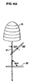
図4は、組織ブロック中の二次元超音波平面23における従来の同軸刺激ブロック針26の二次元超音波モニタ上の表示を示す。図4Aは、図4にほぼ類似の表示を示すが、本明細書中に引用した様々な角度に関する参照点を示す。同軸刺激ブロック針26は、超音波ビームに対して90°の角度に配向されている場合に、モニタ24上に最適に見える。しかし、二次元超音波モニタ24上で同軸針画像を解像する能力は、針が90°の配向からより小さな配向へと移動するにつれて劣化していき、最後には見えなくなる。図4Aは、指定した角度の基準系を提供する。図中、角度をつけた針26’は超音波モニタ24上で見えないことに留意されたい。示した例では、針26の配向を90°の配向から25°の角度まで、65°回転させた。プラスチックコーティングは超音波の波12を超音波ヘッド13に直接戻して反射するだけなので、この現象は鏡面反射によって引き起こされている。この反射は、ほぼ光が鏡から反射される場合に類似している。同軸ブロック針26の鏡面反射は、二次元超音波を用いた針の可視化を困難にする。一般的に、針は進めるにつれて見えなくなり、運動応答を誘発させるために必要なアンペア数を低下させる結果、末梢神経刺激のみでの針の先端位置の解像を許容することで二次元超音波の有用性を大きく低下させる。
FIG. 4 shows a display on a two-dimensional ultrasound monitor of a conventional coaxial
図5は、組織ブロック中の、本発明による高輝度エコー刺激ブロック針1の二次元超音波モニタ24上の表示を示す。高輝度エコー刺激ブロック針1は、本明細書中に上述したエコー源性層10を有し、エコー源性層は、超音波の波12の超音波ヘッド13に戻る反射を最大にするように構成されている。これにより、超音波の波の方向に対して90°の配向(たとえば垂直)から超音波の波と整列する配向に近づくように移動するにつれて、高輝度エコー刺激ブロック針1が可視化される。したがって、高輝度エコー刺激ブロック針画像28は、二次元超音波平面23中で、90°の角度及びより小さな角度のどちらにおいても、同軸針26で達し得るよりも高い分解能で可視化される。図5では、針画像28は、針が記載した垂直な配向から65°回転した後でも見える。
FIG. 5 shows the display on the two-dimensional ultrasound monitor 24 of the high intensity echo stimulation block needle 1 according to the invention in a tissue block. The high intensity echo stimulation block needle 1 has an
エコー源性層10は、二次元超音波平面23を進める間に高輝度エコー刺激針1が見えていることを可能にし、針が末梢神経15に近づく間に針の進路及び針の先端9がどちらも可視化されている。この反射は、エコー源性層10による超音波の波12の散乱により生じるものであるので、鏡面反射とは異なる。この場合、波の散乱は二次元超音波ヘッド13に向かって起こり、高輝度エコー刺激ブロック針画像28が二次元超音波平面23中で、様々な角度で可視化される。
The
針の進路、針の先端9、及びブロックする末梢神経15が見える能力を、末梢神経刺激と組み合わせることにより、以前可能であったよりも正確に末梢神経15に近づくことが可能となる。針の位置は、針の進路、針の先端9及び末梢神経15の同時可視化による二次元超音波を用いて解剖学的に解像され、加えて、針の位置は、電流を最小限にすることによって運動応答を誘発させるための末梢神経刺激を用いることで生理学的に解像されることができる。これにより、図4に示すように二次元超音波、末梢神経刺激及び同軸末梢刺激神経ブロック針を用いた場合と比較して、針の先端9を末梢神経15により近位に向けることが可能となる。その結果、超音波可視化又は末梢神経刺激を別々に用いて以前に可能であったよりも正確に薬剤を注入することができる。 Combining the path of the needle, the tip 9 of the needle, and the ability to see the blocking peripheral nerve 15 with peripheral nerve stimulation makes it possible to approach the peripheral nerve 15 more accurately than previously possible. The needle position is anatomically resolved using two-dimensional ultrasound with simultaneous visualization of the needle path, needle tip 9 and peripheral nerve 15, and in addition, the needle position minimizes current. Can be physiologically resolved by using peripheral nerve stimulation to elicit a motor response. As a result, as shown in FIG. 4, the tip 9 of the needle can be directed proximally by the peripheral nerve 15 as compared with the case of using a two-dimensional ultrasound, peripheral nerve stimulation and coaxial peripheral stimulation nerve block needle. Become. As a result, medication can be injected more accurately than previously possible using separate ultrasound visualization or peripheral nerve stimulation.
高輝度エコー刺激ブロック針1の使用は、二次元超音波を用いてカテーテルを針に通す機構、及び薬剤の連続投与のために針を取り外してカテーテルを留置するための機構も提供する。同様に、カテーテルは、カテーテルの素地中又はカテーテルの素地の先端に混合されたエコー源性の材料を有してもよく、これにより、カテーテルの可視化及びカテーテルが針中を前進、及び超音波を用いた正しい解剖学的位置が可能となる。さらに、カテーテルは、カテーテルの素地中に金属又は導電性のエコー源性の材料を混合させてもよく、これにより、カテーテルの素地を遠位端から近位端まで走る電極を利用することによって、末梢神経刺激装置と共に電気回路が形成されることが可能となる。あるいは、カテーテルを完全に又は部分的に生理食塩水などの導電性材料で満たすことができる。 The use of the high intensity echo stimulation block needle 1 also provides a mechanism for passing the catheter through the needle using two-dimensional ultrasound and a mechanism for removing the needle and placing the catheter for continuous administration of the drug. Similarly, the catheter may have an echogenic material mixed in the catheter body or at the tip of the catheter body, thereby allowing visualization of the catheter and advancement of the catheter through the needle and ultrasound. The correct anatomical position used is possible. Further, the catheter may be mixed with a metal or conductive echogenic material in the catheter body, thereby utilizing an electrode that runs from the distal end to the proximal end of the catheter body. An electrical circuit can be formed with the peripheral nerve stimulator. Alternatively, the catheter can be completely or partially filled with a conductive material such as saline.
したがって、前述の詳細な説明は限定するものではなく、例示的であるとされることを意図し、また、本発明の精神及び範囲を定義することを意図するのは、すべての均等物を含めた添付の特許請求の範囲であると理解されることを意図する。 Accordingly, the foregoing detailed description is intended to be illustrative rather than limiting and is intended to define the spirit and scope of the invention, including all equivalents thereof. It is intended that the appended claims be understood.
Claims (20)
軸部分及び先端部分を含む中空の針導管と、
前記針軸部分の少なくとも一部分を被覆する絶縁層と、
前記二次元超音波可視化中に発生された超音波の波の反射を高めるようにされた、前記針の長さに沿って配置されているエコー源性表面と、
を備える、患者の神経ブロックを行うために使用する高輝度エコー刺激ブロック針。 A high intensity echo stimulation block needle used to perform a patient's nerve block using electrical nerve stimulation and two-dimensional ultrasound visualization of the nerve simultaneously,
A hollow needle conduit including a shaft portion and a tip portion;
An insulating layer covering at least a portion of the needle shaft portion;
An echogenic surface disposed along the length of the needle, adapted to enhance reflection of ultrasonic waves generated during the two-dimensional ultrasonic visualization;
A high intensity echo stimulation block needle used to perform a patient's nerve block.
前記針の先端を患者内に挿入するステップと、
医療用イメージングを用いた可視化及び電気神経刺激によって前記針の先端を前記神経に近接して位置合わせするステップと、
薬剤を前記中空の針導管の孔を通して前記患者内に注入するステップと、
を含む、患者の神経ブロックを行なう方法。 A conductive hollow needle conduit, the conduit having a shaft portion and a distal tip, a generally non-conductive coating extending along the conduit shaft portion, and an echogenic surface of the length of the needle Providing a high intensity echo stimulation block needle extending along at least a portion;
Inserting the tip of the needle into a patient;
Aligning the tip of the needle close to the nerve by visualization and electrical nerve stimulation using medical imaging;
Injecting a drug into the patient through a hole in the hollow needle conduit;
A method of performing nerve block of a patient, comprising:
電気パルスを与える針と電気接続することができる末梢神経刺激装置と、
患者の組織のセグメント中の前記針の少なくとも一部分を可視化することができる医療用イメージング機構と
を含む、患者の神経をブロック(遮断)するために用いるシステム。 A conductive needle conduit, the conduit having a shaft portion and a distal tip, a substantially non-conductive coating extending along the conduit shaft portion, and an echogenic material at least the length of the needle A high-intensity echo stimulation block needle extending along a portion;
A peripheral nerve stimulator that can be electrically connected to a needle that provides an electrical pulse;
A system for use in blocking a patient's nerves, comprising a medical imaging mechanism capable of visualizing at least a portion of the needle in a segment of patient tissue.
Applications Claiming Priority (2)
| Application Number | Priority Date | Filing Date | Title |
|---|---|---|---|
| US74966405P | 2005-12-12 | 2005-12-12 | |
| PCT/US2006/046854 WO2007070374A2 (en) | 2005-12-12 | 2006-12-08 | Stimulating block needle comprising echogenic surface |
Publications (2)
| Publication Number | Publication Date |
|---|---|
| JP2009519104A true JP2009519104A (en) | 2009-05-14 |
| JP2009519104A5 JP2009519104A5 (en) | 2010-02-04 |
Family
ID=37964785
Family Applications (1)
| Application Number | Title | Priority Date | Filing Date |
|---|---|---|---|
| JP2008545673A Pending JP2009519104A (en) | 2005-12-12 | 2006-12-08 | High intensity echo stimulation block needle |
Country Status (4)
| Country | Link |
|---|---|
| US (1) | US20070179508A1 (en) |
| EP (2) | EP2308551A1 (en) |
| JP (1) | JP2009519104A (en) |
| WO (1) | WO2007070374A2 (en) |
Cited By (2)
| Publication number | Priority date | Publication date | Assignee | Title |
|---|---|---|---|---|
| JP2012513833A (en) * | 2008-12-30 | 2012-06-21 | ボストン サイエンティフィック サイムド, インコーポレイテッド | Enhanced echo generation for needles |
| JP2013502274A (en) * | 2009-08-19 | 2013-01-24 | クック メディカル テクノロジーズ エルエルシー | Echogenic electrosurgical instrument |
Families Citing this family (92)
| Publication number | Priority date | Publication date | Assignee | Title |
|---|---|---|---|---|
| US20120108918A1 (en) * | 2008-09-19 | 2012-05-03 | Physiosonics, Inc. | Acoustic Palpation Using Non-Invasive Ultrasound Techniques for Identification of Target Sites and Assessment of Chronic Pain Disorders |
| EP2308551A1 (en) | 2005-12-12 | 2011-04-13 | Cook Critical Care Incorporated | System for blocking a nerve comprising an echogenic stimulating needle and a drug delivery catheter |
| US20080058702A1 (en) * | 2005-12-12 | 2008-03-06 | Cook Critical Care Incorporated | Continuous nerve block assembly |
| US9498282B2 (en) * | 2007-02-09 | 2016-11-22 | Boston Scientific Scimed, Inc. | Medical probe with echogenic and insulative properties |
| FR2923372B1 (en) * | 2007-11-08 | 2010-10-29 | Theraclion | DEVICE AND METHOD FOR NON-INVASIVE REPORTING OF A STRUCTURE SUCH AS A NERVE. |
| JP5159326B2 (en) * | 2008-01-10 | 2013-03-06 | 株式会社東芝 | Ultrasonic diagnostic equipment |
| DE102008025878A1 (en) * | 2008-05-29 | 2009-12-10 | Pajunk Gmbh & Co. Kg Besitzverwaltung | Cannula, especially for regional anesthesia |
| US8137285B1 (en) * | 2008-08-26 | 2012-03-20 | Rhythmlink International, Llc | Monopolar stimulation probe system |
| US20100204567A1 (en) * | 2009-02-09 | 2010-08-12 | The Cleveland Clinic Foundation | Ultrasound-guided delivery of a therapy delivery device to a phrenic nerve |
| US8870773B2 (en) * | 2009-02-09 | 2014-10-28 | The Cleveland Clinic Foundation | Ultrasound-guided delivery of a therapy delivery device to a nerve target |
| EP2470103B1 (en) * | 2009-09-24 | 2014-01-01 | Boston Scientific Scimed, Inc. | Echogenic needle mechanism |
| WO2011163368A1 (en) | 2010-06-24 | 2011-12-29 | Cdw Investments, Llc | Hyperechogenic needles |
| US9254146B2 (en) | 2010-10-18 | 2016-02-09 | Avent, Inc. | Echogenic nerve block apparatus and system |
| US9265897B2 (en) * | 2011-01-26 | 2016-02-23 | Avent, Inc. | Method and corresponding kit for administering a paravertebral block |
| DE102012100292A1 (en) | 2012-01-13 | 2013-07-18 | Rm Temena Gmbh | Medical device |
| US11871901B2 (en) | 2012-05-20 | 2024-01-16 | Cilag Gmbh International | Method for situational awareness for surgical network or surgical network connected device capable of adjusting function based on a sensed situation or usage |
| GB2509750A (en) * | 2013-01-11 | 2014-07-16 | Teodor Goroszeniuk | Stimulating needle with plurality of electrode zones |
| CN105377324B (en) | 2013-05-31 | 2018-12-21 | 库克医学技术有限责任公司 | Access needle and die needle assemblies |
| US11504192B2 (en) | 2014-10-30 | 2022-11-22 | Cilag Gmbh International | Method of hub communication with surgical instrument systems |
| CN106388914A (en) * | 2016-11-22 | 2017-02-15 | 中国人民解放军总医院 | Ultrasonic interventional therapy device |
| EP3548136A4 (en) | 2016-12-01 | 2020-07-08 | Thimble Bioelectronics, Inc. D/B/A Enso | Neuromodulation device and method for use |
| US10751469B2 (en) * | 2017-04-07 | 2020-08-25 | Northwesten University | Computer controlled pediatric regional anesthesia |
| CN107126260B (en) * | 2017-07-18 | 2019-09-13 | 深圳开立生物医疗科技股份有限公司 | Method for ultrasonic imaging, system and supersonic imaging apparatus |
| US11510741B2 (en) | 2017-10-30 | 2022-11-29 | Cilag Gmbh International | Method for producing a surgical instrument comprising a smart electrical system |
| US11801098B2 (en) | 2017-10-30 | 2023-10-31 | Cilag Gmbh International | Method of hub communication with surgical instrument systems |
| US11413042B2 (en) | 2017-10-30 | 2022-08-16 | Cilag Gmbh International | Clip applier comprising a reciprocating clip advancing member |
| US11109878B2 (en) | 2017-10-30 | 2021-09-07 | Cilag Gmbh International | Surgical clip applier comprising an automatic clip feeding system |
| US11564756B2 (en) | 2017-10-30 | 2023-01-31 | Cilag Gmbh International | Method of hub communication with surgical instrument systems |
| US11911045B2 (en) | 2017-10-30 | 2024-02-27 | Cllag GmbH International | Method for operating a powered articulating multi-clip applier |
| US11903601B2 (en) | 2017-12-28 | 2024-02-20 | Cilag Gmbh International | Surgical instrument comprising a plurality of drive systems |
| US11818052B2 (en) | 2017-12-28 | 2023-11-14 | Cilag Gmbh International | Surgical network determination of prioritization of communication, interaction, or processing based on system or device needs |
| US11540855B2 (en) | 2017-12-28 | 2023-01-03 | Cilag Gmbh International | Controlling activation of an ultrasonic surgical instrument according to the presence of tissue |
| US11464535B2 (en) | 2017-12-28 | 2022-10-11 | Cilag Gmbh International | Detection of end effector emersion in liquid |
| US20190200981A1 (en) | 2017-12-28 | 2019-07-04 | Ethicon Llc | Method of compressing tissue within a stapling device and simultaneously displaying the location of the tissue within the jaws |
| US11389164B2 (en) | 2017-12-28 | 2022-07-19 | Cilag Gmbh International | Method of using reinforced flexible circuits with multiple sensors to optimize performance of radio frequency devices |
| US11464559B2 (en) | 2017-12-28 | 2022-10-11 | Cilag Gmbh International | Estimating state of ultrasonic end effector and control system therefor |
| US11832840B2 (en) | 2017-12-28 | 2023-12-05 | Cilag Gmbh International | Surgical instrument having a flexible circuit |
| US11576677B2 (en) | 2017-12-28 | 2023-02-14 | Cilag Gmbh International | Method of hub communication, processing, display, and cloud analytics |
| US11571234B2 (en) | 2017-12-28 | 2023-02-07 | Cilag Gmbh International | Temperature control of ultrasonic end effector and control system therefor |
| US11324557B2 (en) | 2017-12-28 | 2022-05-10 | Cilag Gmbh International | Surgical instrument with a sensing array |
| US10892995B2 (en) | 2017-12-28 | 2021-01-12 | Ethicon Llc | Surgical network determination of prioritization of communication, interaction, or processing based on system or device needs |
| US11864728B2 (en) | 2017-12-28 | 2024-01-09 | Cilag Gmbh International | Characterization of tissue irregularities through the use of mono-chromatic light refractivity |
| US11179175B2 (en) | 2017-12-28 | 2021-11-23 | Cilag Gmbh International | Controlling an ultrasonic surgical instrument according to tissue location |
| US11678881B2 (en) | 2017-12-28 | 2023-06-20 | Cilag Gmbh International | Spatial awareness of surgical hubs in operating rooms |
| US11419667B2 (en) | 2017-12-28 | 2022-08-23 | Cilag Gmbh International | Ultrasonic energy device which varies pressure applied by clamp arm to provide threshold control pressure at a cut progression location |
| US11559307B2 (en) | 2017-12-28 | 2023-01-24 | Cilag Gmbh International | Method of robotic hub communication, detection, and control |
| US11896322B2 (en) | 2017-12-28 | 2024-02-13 | Cilag Gmbh International | Sensing the patient position and contact utilizing the mono-polar return pad electrode to provide situational awareness to the hub |
| US11786251B2 (en) | 2017-12-28 | 2023-10-17 | Cilag Gmbh International | Method for adaptive control schemes for surgical network control and interaction |
| US11559308B2 (en) | 2017-12-28 | 2023-01-24 | Cilag Gmbh International | Method for smart energy device infrastructure |
| US11937769B2 (en) | 2017-12-28 | 2024-03-26 | Cilag Gmbh International | Method of hub communication, processing, storage and display |
| US11602393B2 (en) | 2017-12-28 | 2023-03-14 | Cilag Gmbh International | Surgical evacuation sensing and generator control |
| US11612444B2 (en) | 2017-12-28 | 2023-03-28 | Cilag Gmbh International | Adjustment of a surgical device function based on situational awareness |
| US11432885B2 (en) | 2017-12-28 | 2022-09-06 | Cilag Gmbh International | Sensing arrangements for robot-assisted surgical platforms |
| US11529187B2 (en) | 2017-12-28 | 2022-12-20 | Cilag Gmbh International | Surgical evacuation sensor arrangements |
| US11744604B2 (en) | 2017-12-28 | 2023-09-05 | Cilag Gmbh International | Surgical instrument with a hardware-only control circuit |
| US11857152B2 (en) | 2017-12-28 | 2024-01-02 | Cilag Gmbh International | Surgical hub spatial awareness to determine devices in operating theater |
| US11132462B2 (en) | 2017-12-28 | 2021-09-28 | Cilag Gmbh International | Data stripping method to interrogate patient records and create anonymized record |
| US11166772B2 (en) | 2017-12-28 | 2021-11-09 | Cilag Gmbh International | Surgical hub coordination of control and communication of operating room devices |
| US11364075B2 (en) | 2017-12-28 | 2022-06-21 | Cilag Gmbh International | Radio frequency energy device for delivering combined electrical signals |
| US10758310B2 (en) | 2017-12-28 | 2020-09-01 | Ethicon Llc | Wireless pairing of a surgical device with another device within a sterile surgical field based on the usage and situational awareness of devices |
| US11424027B2 (en) | 2017-12-28 | 2022-08-23 | Cilag Gmbh International | Method for operating surgical instrument systems |
| US11832899B2 (en) | 2017-12-28 | 2023-12-05 | Cilag Gmbh International | Surgical systems with autonomously adjustable control programs |
| US11423007B2 (en) | 2017-12-28 | 2022-08-23 | Cilag Gmbh International | Adjustment of device control programs based on stratified contextual data in addition to the data |
| US11026751B2 (en) | 2017-12-28 | 2021-06-08 | Cilag Gmbh International | Display of alignment of staple cartridge to prior linear staple line |
| US20190201146A1 (en) | 2017-12-28 | 2019-07-04 | Ethicon Llc | Safety systems for smart powered surgical stapling |
| US11659023B2 (en) | 2017-12-28 | 2023-05-23 | Cilag Gmbh International | Method of hub communication |
| US11202570B2 (en) | 2017-12-28 | 2021-12-21 | Cilag Gmbh International | Communication hub and storage device for storing parameters and status of a surgical device to be shared with cloud based analytics systems |
| US11633237B2 (en) | 2017-12-28 | 2023-04-25 | Cilag Gmbh International | Usage and technique analysis of surgeon / staff performance against a baseline to optimize device utilization and performance for both current and future procedures |
| US11013563B2 (en) | 2017-12-28 | 2021-05-25 | Ethicon Llc | Drive arrangements for robot-assisted surgical platforms |
| US11410259B2 (en) | 2017-12-28 | 2022-08-09 | Cilag Gmbh International | Adaptive control program updates for surgical devices |
| US20190201039A1 (en) | 2017-12-28 | 2019-07-04 | Ethicon Llc | Situational awareness of electrosurgical systems |
| US11109866B2 (en) | 2017-12-28 | 2021-09-07 | Cilag Gmbh International | Method for circular stapler control algorithm adjustment based on situational awareness |
| US11786245B2 (en) | 2017-12-28 | 2023-10-17 | Cilag Gmbh International | Surgical systems with prioritized data transmission capabilities |
| US11446052B2 (en) | 2017-12-28 | 2022-09-20 | Cilag Gmbh International | Variation of radio frequency and ultrasonic power level in cooperation with varying clamp arm pressure to achieve predefined heat flux or power applied to tissue |
| US11311306B2 (en) | 2017-12-28 | 2022-04-26 | Cilag Gmbh International | Surgical systems for detecting end effector tissue distribution irregularities |
| US11896443B2 (en) | 2017-12-28 | 2024-02-13 | Cilag Gmbh International | Control of a surgical system through a surgical barrier |
| US11589888B2 (en) | 2017-12-28 | 2023-02-28 | Cilag Gmbh International | Method for controlling smart energy devices |
| US11666331B2 (en) | 2017-12-28 | 2023-06-06 | Cilag Gmbh International | Systems for detecting proximity of surgical end effector to cancerous tissue |
| CN108095809B (en) * | 2018-02-05 | 2019-12-03 | 郑雪松 | A kind of puncture needle and drainage device for paracentesis pericardii |
| US11259830B2 (en) | 2018-03-08 | 2022-03-01 | Cilag Gmbh International | Methods for controlling temperature in ultrasonic device |
| US11457944B2 (en) | 2018-03-08 | 2022-10-04 | Cilag Gmbh International | Adaptive advanced tissue treatment pad saver mode |
| US11678927B2 (en) | 2018-03-08 | 2023-06-20 | Cilag Gmbh International | Detection of large vessels during parenchymal dissection using a smart blade |
| US11471156B2 (en) | 2018-03-28 | 2022-10-18 | Cilag Gmbh International | Surgical stapling devices with improved rotary driven closure systems |
| US11090047B2 (en) | 2018-03-28 | 2021-08-17 | Cilag Gmbh International | Surgical instrument comprising an adaptive control system |
| US11259806B2 (en) | 2018-03-28 | 2022-03-01 | Cilag Gmbh International | Surgical stapling devices with features for blocking advancement of a camming assembly of an incompatible cartridge installed therein |
| US20210361905A1 (en) * | 2018-04-19 | 2021-11-25 | Wake Forest University Health Sciences | A medical device for use in a nerve block procedure that obviates the need for injecting test doses and a method |
| US11317915B2 (en) | 2019-02-19 | 2022-05-03 | Cilag Gmbh International | Universal cartridge based key feature that unlocks multiple lockout arrangements in different surgical staplers |
| US11464511B2 (en) | 2019-02-19 | 2022-10-11 | Cilag Gmbh International | Surgical staple cartridges with movable authentication key arrangements |
| US11291444B2 (en) | 2019-02-19 | 2022-04-05 | Cilag Gmbh International | Surgical stapling assembly with cartridge based retainer configured to unlock a closure lockout |
| US11357503B2 (en) | 2019-02-19 | 2022-06-14 | Cilag Gmbh International | Staple cartridge retainers with frangible retention features and methods of using same |
| USD964564S1 (en) | 2019-06-25 | 2022-09-20 | Cilag Gmbh International | Surgical staple cartridge retainer with a closure system authentication key |
| US20230131115A1 (en) * | 2021-10-21 | 2023-04-27 | GE Precision Healthcare LLC | System and Method for Displaying Position of Echogenic Needles |
Citations (1)
| Publication number | Priority date | Publication date | Assignee | Title |
|---|---|---|---|---|
| JP2003190179A (en) * | 2001-12-27 | 2003-07-08 | Olympus Optical Co Ltd | Ultrasonic puncture needle |
Family Cites Families (18)
| Publication number | Priority date | Publication date | Assignee | Title |
|---|---|---|---|---|
| US4279252A (en) * | 1979-08-24 | 1981-07-21 | Martin Michael T | X-ray scaling catheter |
| US4401124A (en) * | 1981-08-13 | 1983-08-30 | Technicare Corporation | Reflection enhancement of a biopsy needle |
| DE3508013A1 (en) * | 1984-07-28 | 1986-02-06 | Peter 7730 Villingen-Schwenningen Krebs | COMBINATION NEEDLE FOR THE AXILLAERE PLEXUS-BRACHIALIS-ANESTHESIA |
| US4869259A (en) * | 1988-05-17 | 1989-09-26 | Vance Products Incorporated | Echogenically enhanced surgical instrument and method for production thereof |
| US4977897A (en) * | 1988-08-17 | 1990-12-18 | Robert Hurwitz | Amniocentesis needle with improved sonographic visibility |
| US5084022A (en) * | 1989-10-04 | 1992-01-28 | Lake Region Manufacturing Company, Inc. | Graduated guidewire |
| DE4310924C2 (en) * | 1993-04-02 | 1995-01-26 | Siemens Ag | Therapy device for the treatment of pathological tissue with ultrasound waves and a catheter |
| US5490521A (en) * | 1993-08-31 | 1996-02-13 | Medtronic, Inc. | Ultrasound biopsy needle |
| US5479938A (en) * | 1994-02-07 | 1996-01-02 | Cordis Corporation | Lumen diameter reference guidewire |
| EP0723786A1 (en) * | 1995-01-30 | 1996-07-31 | Cardiovascular Concepts, Inc. | Lesion measurement catheter and method |
| US5807304A (en) * | 1995-03-09 | 1998-09-15 | Cockburn; John F. | Medical needle for use in ultrasound imaging |
| DE69941534D1 (en) * | 1998-02-16 | 2009-11-26 | Philadelphia Health & Educatio | INTRALUMINAL CATHETER WITH A SCALE, AND METHODS FOR ITS APPLICATION |
| US6298256B1 (en) * | 1999-09-10 | 2001-10-02 | Frank-Egbert Meyer | Device and method for the location and catheterization of the surroundings of a nerve |
| US6620114B2 (en) * | 2000-10-05 | 2003-09-16 | Scimed Life Systems, Inc. | Guidewire having a marker segment for length assessment |
| US7065394B2 (en) * | 2001-12-12 | 2006-06-20 | Medtronic, Inc | Guide catheter |
| JP3967950B2 (en) * | 2002-04-10 | 2007-08-29 | ジーイー・メディカル・システムズ・グローバル・テクノロジー・カンパニー・エルエルシー | Puncture needle guide, ultrasonic probe, and ultrasonic imaging apparatus |
| US6936048B2 (en) * | 2003-01-16 | 2005-08-30 | Charlotte-Mecklenburg Hospital Authority | Echogenic needle for transvaginal ultrasound directed reduction of uterine fibroids and an associated method |
| EP2308551A1 (en) | 2005-12-12 | 2011-04-13 | Cook Critical Care Incorporated | System for blocking a nerve comprising an echogenic stimulating needle and a drug delivery catheter |
-
2006
- 2006-12-08 EP EP10191017A patent/EP2308551A1/en not_active Withdrawn
- 2006-12-08 EP EP06845014A patent/EP1960039A2/en not_active Withdrawn
- 2006-12-08 JP JP2008545673A patent/JP2009519104A/en active Pending
- 2006-12-08 US US11/635,931 patent/US20070179508A1/en not_active Abandoned
- 2006-12-08 WO PCT/US2006/046854 patent/WO2007070374A2/en active Application Filing
Patent Citations (1)
| Publication number | Priority date | Publication date | Assignee | Title |
|---|---|---|---|---|
| JP2003190179A (en) * | 2001-12-27 | 2003-07-08 | Olympus Optical Co Ltd | Ultrasonic puncture needle |
Cited By (3)
| Publication number | Priority date | Publication date | Assignee | Title |
|---|---|---|---|---|
| JP2012513833A (en) * | 2008-12-30 | 2012-06-21 | ボストン サイエンティフィック サイムド, インコーポレイテッド | Enhanced echo generation for needles |
| US9521993B2 (en) | 2008-12-30 | 2016-12-20 | Boston Scientific Scimed, Inc. | Echogenic enhancement for a needle |
| JP2013502274A (en) * | 2009-08-19 | 2013-01-24 | クック メディカル テクノロジーズ エルエルシー | Echogenic electrosurgical instrument |
Also Published As
| Publication number | Publication date |
|---|---|
| US20070179508A1 (en) | 2007-08-02 |
| WO2007070374A2 (en) | 2007-06-21 |
| EP1960039A2 (en) | 2008-08-27 |
| EP2308551A1 (en) | 2011-04-13 |
| WO2007070374A3 (en) | 2007-08-30 |
Similar Documents
| Publication | Publication Date | Title |
|---|---|---|
| JP2009519104A (en) | High intensity echo stimulation block needle | |
| EP2152185B1 (en) | Continuous nerve block assembly | |
| US11925512B2 (en) | Methods and systems for controlled deployment of needles in tissue | |
| US5885219A (en) | Interrogation device and method | |
| JP4436092B2 (en) | Alignment system for nerve treatment in the brain | |
| US9364164B2 (en) | Non-invasive device and method for locating a structure such as a nerve | |
| US20050240126A1 (en) | Ultrasound guided high intensity focused ultrasound treatment of nerves | |
| Foley et al. | Image-guided HIFU neurolysis of peripheral nerves to treat spasticity and pain | |
| US20100256483A1 (en) | Devices and methods for tissue navigation | |
| WO2005055849A1 (en) | Ultrasonically marked system for therapy delivery | |
| US11253729B2 (en) | External ultrasound generating treating device for spinal cord and/or spinal nerve treatment, apparatus comprising such device and method | |
| US20200146718A1 (en) | Neurovascular puncture-avoidant sheath | |
| JP2019518544A (en) | Electroacoustic imaging-guided therapy | |
| RU163436U1 (en) | DEVICE FOR ADMINISTRATION OF A LIQUID MEDICINE | |
| UA128996U (en) | BREAST CELL BREEDING METHOD | |
| McDonald | Computer driven needle probe enables therapy for painful neuropathies | |
| Gupta et al. | Equipment and Usage of Ultrasound | |
| Hawkinberry II et al. | An introduction to ultrasonic guided axillary brachial plexus neuroblockade | |
| NIAZI et al. | Ultrasound-Guided Interscalene Block | |
| JPH0852152A (en) | Ultrasonic medical treatment device |
Legal Events
| Date | Code | Title | Description |
|---|---|---|---|
| A521 | Written amendment |
Free format text: JAPANESE INTERMEDIATE CODE: A523 Effective date: 20091207 |
|
| A621 | Written request for application examination |
Free format text: JAPANESE INTERMEDIATE CODE: A621 Effective date: 20091207 |
|
| A131 | Notification of reasons for refusal |
Free format text: JAPANESE INTERMEDIATE CODE: A131 Effective date: 20111213 |
|
| A977 | Report on retrieval |
Free format text: JAPANESE INTERMEDIATE CODE: A971007 Effective date: 20111215 |
|
| A02 | Decision of refusal |
Free format text: JAPANESE INTERMEDIATE CODE: A02 Effective date: 20120508 |