JP2009504153A - Method and / or apparatus for oligonucleotide design and / or nucleic acid detection - Google Patents
Method and / or apparatus for oligonucleotide design and / or nucleic acid detection Download PDFInfo
- Publication number
- JP2009504153A JP2009504153A JP2008525967A JP2008525967A JP2009504153A JP 2009504153 A JP2009504153 A JP 2009504153A JP 2008525967 A JP2008525967 A JP 2008525967A JP 2008525967 A JP2008525967 A JP 2008525967A JP 2009504153 A JP2009504153 A JP 2009504153A
- Authority
- JP
- Japan
- Prior art keywords
- nucleic acid
- target nucleic
- probe
- oligonucleotide
- probes
- Prior art date
- Legal status (The legal status is an assumption and is not a legal conclusion. Google has not performed a legal analysis and makes no representation as to the accuracy of the status listed.)
- Pending
Links
Images


Classifications
-
- C—CHEMISTRY; METALLURGY
- C12—BIOCHEMISTRY; BEER; SPIRITS; WINE; VINEGAR; MICROBIOLOGY; ENZYMOLOGY; MUTATION OR GENETIC ENGINEERING
- C12Q—MEASURING OR TESTING PROCESSES INVOLVING ENZYMES, NUCLEIC ACIDS OR MICROORGANISMS; COMPOSITIONS OR TEST PAPERS THEREFOR; PROCESSES OF PREPARING SUCH COMPOSITIONS; CONDITION-RESPONSIVE CONTROL IN MICROBIOLOGICAL OR ENZYMOLOGICAL PROCESSES
- C12Q1/00—Measuring or testing processes involving enzymes, nucleic acids or microorganisms; Compositions therefor; Processes of preparing such compositions
- C12Q1/68—Measuring or testing processes involving enzymes, nucleic acids or microorganisms; Compositions therefor; Processes of preparing such compositions involving nucleic acids
- C12Q1/6813—Hybridisation assays
-
- G—PHYSICS
- G16—INFORMATION AND COMMUNICATION TECHNOLOGY [ICT] SPECIALLY ADAPTED FOR SPECIFIC APPLICATION FIELDS
- G16B—BIOINFORMATICS, i.e. INFORMATION AND COMMUNICATION TECHNOLOGY [ICT] SPECIALLY ADAPTED FOR GENETIC OR PROTEIN-RELATED DATA PROCESSING IN COMPUTATIONAL MOLECULAR BIOLOGY
- G16B99/00—Subject matter not provided for in other groups of this subclass
-
- C—CHEMISTRY; METALLURGY
- C12—BIOCHEMISTRY; BEER; SPIRITS; WINE; VINEGAR; MICROBIOLOGY; ENZYMOLOGY; MUTATION OR GENETIC ENGINEERING
- C12Q—MEASURING OR TESTING PROCESSES INVOLVING ENZYMES, NUCLEIC ACIDS OR MICROORGANISMS; COMPOSITIONS OR TEST PAPERS THEREFOR; PROCESSES OF PREPARING SUCH COMPOSITIONS; CONDITION-RESPONSIVE CONTROL IN MICROBIOLOGICAL OR ENZYMOLOGICAL PROCESSES
- C12Q1/00—Measuring or testing processes involving enzymes, nucleic acids or microorganisms; Compositions therefor; Processes of preparing such compositions
- C12Q1/68—Measuring or testing processes involving enzymes, nucleic acids or microorganisms; Compositions therefor; Processes of preparing such compositions involving nucleic acids
- C12Q1/6806—Preparing nucleic acids for analysis, e.g. for polymerase chain reaction [PCR] assay
-
- C—CHEMISTRY; METALLURGY
- C12—BIOCHEMISTRY; BEER; SPIRITS; WINE; VINEGAR; MICROBIOLOGY; ENZYMOLOGY; MUTATION OR GENETIC ENGINEERING
- C12Q—MEASURING OR TESTING PROCESSES INVOLVING ENZYMES, NUCLEIC ACIDS OR MICROORGANISMS; COMPOSITIONS OR TEST PAPERS THEREFOR; PROCESSES OF PREPARING SUCH COMPOSITIONS; CONDITION-RESPONSIVE CONTROL IN MICROBIOLOGICAL OR ENZYMOLOGICAL PROCESSES
- C12Q1/00—Measuring or testing processes involving enzymes, nucleic acids or microorganisms; Compositions therefor; Processes of preparing such compositions
- C12Q1/68—Measuring or testing processes involving enzymes, nucleic acids or microorganisms; Compositions therefor; Processes of preparing such compositions involving nucleic acids
- C12Q1/6811—Selection methods for production or design of target specific oligonucleotides or binding molecules
-
- G—PHYSICS
- G16—INFORMATION AND COMMUNICATION TECHNOLOGY [ICT] SPECIALLY ADAPTED FOR SPECIFIC APPLICATION FIELDS
- G16B—BIOINFORMATICS, i.e. INFORMATION AND COMMUNICATION TECHNOLOGY [ICT] SPECIALLY ADAPTED FOR GENETIC OR PROTEIN-RELATED DATA PROCESSING IN COMPUTATIONAL MOLECULAR BIOLOGY
- G16B25/00—ICT specially adapted for hybridisation; ICT specially adapted for gene or protein expression
- G16B25/20—Polymerase chain reaction [PCR]; Primer or probe design; Probe optimisation
Abstract
(I)増幅される少なくとも1つの標的核酸の領域を同定および/または選択ステップと、ここで、前記領域が、平均増幅効率(AE)よりも高いAEを有し、(II)選択される領域にハイブリダイズすることができる少なくとも1つのオリゴヌクレオチドを設計するステップとを、任意の順序で含む核酸検出のための少なくとも1つのオリゴヌクレオチドを設計する方法が提供される。(i)少なくとも1つの生体試料を提供するステップと、(ii)生体試料中に含まれる核酸を増幅するステップと、(iii)生体試料中に標的核酸が存在する場合、少なくとも1つの標的核酸にハイブリダイズすることができる少なくとも1つのオリゴヌクレオチドを提供するステップと、(iv)前記オリゴヌクレオチドを増幅された核酸に接触させ、標的核酸にハイブリダイズしたオリゴヌクレオチドを検出するステップとを含む少なくとも1つの標的核酸を検出する方法もまた、提供される。具体的には、本方法は、少なくとも1つのヒト生体試料中の、少なくとも1つの病原体、例えばウイルスの存在を検出するためのものである。プローブは支持体、例えばマイクロアレイ上に配置してもよい。 (I) identifying and / or selecting a region of at least one target nucleic acid to be amplified, wherein said region has an AE higher than an average amplification efficiency (AE) and (II) a region to be selected A method of designing at least one oligonucleotide for nucleic acid detection comprising, in any order, designing at least one oligonucleotide capable of hybridizing to (I) providing at least one biological sample; (ii) amplifying the nucleic acid contained in the biological sample; and (iii) if the target nucleic acid is present in the biological sample, the at least one target nucleic acid Providing at least one oligonucleotide capable of hybridizing; and (iv) contacting the oligonucleotide with the amplified nucleic acid and detecting the oligonucleotide hybridized to the target nucleic acid. A method for detecting a target nucleic acid is also provided. Specifically, the method is for detecting the presence of at least one pathogen, such as a virus, in at least one human biological sample. The probe may be placed on a support, such as a microarray.
Description
本発明は、オリゴヌクレオチド設計および/または核酸検出の分野に関する。本発明の方法、装置および/または製品は、病原体の検出、例えばウイルスの検出のために使用してもよい。 The present invention relates to the field of oligonucleotide design and / or nucleic acid detection. The methods, devices and / or products of the present invention may be used for pathogen detection, eg, virus detection.
ヒト患者および集団におけるウイルス性および細菌性病原体の正確かつ迅速な検出は、医学的および疫学的に非常に重要である。歴史的に、診断技術は、細胞培養継代および種々の免疫学的アッセイまたは染色手順に依存してきた。感染性疾患因子の正確かつ感度の高い検出は、この分野における進歩の長い歴史にもかかわらず、現在でも依然として困難である。疾患症状から診断までの遅れの問題、およびこれらの手法による微生物の検出限界にもかかわらず、微生物学研究室において、培養および抗体ベースの検出の典型的な方法は、依然として中心的な役割を果たしている。感染のより速い診断によって、例えばより早期の適切な抗菌治療の実施によって、罹患率および死亡率が減少し得る。過去数十年間で、これを達成するための種々の方法が提案されており、PCRおよびマイクロアレイベースの技術を含む核酸検出に基づくものが、最も見込みがあると思われている。具体的には、PCRベースのアッセイが実施されて、より高い検出感度で疑いのある病原体をより迅速に診断することが可能になった。しかしながら、臨床上の実施において、病原因子は、しばしば未確認のままであり、無数の様式で検出を免れる。例えば、いくつかのウイルスは、培養を施行できない。またある時には、患者の試料は、従来の技術による病原体検出には、質が悪過ぎるか、または力価が不十分である場合がある。さらに、PCRベースおよび抗体ベースの手法はともに、PCRプライマー結合部位の変化および抗原連続変異を生じる天然の遺伝子多様化だけのために、疑いのある病原体を認識できない場合がある。 Accurate and rapid detection of viral and bacterial pathogens in human patients and populations is of great medical and epidemiological significance. Historically, diagnostic techniques have relied on cell culture passages and various immunological assays or staining procedures. Accurate and sensitive detection of infectious disease factors is still difficult today despite the long history of progress in this field. Despite the problem of delay from disease symptoms to diagnosis, and the limit of detection of microorganisms by these techniques, typical methods of culture and antibody-based detection still play a central role in microbiology laboratories. Yes. With a faster diagnosis of infection, for example by the implementation of appropriate antimicrobial treatment earlier, morbidity and mortality can be reduced. In the past few decades, various methods have been proposed to achieve this, and those based on nucleic acid detection, including PCR and microarray-based techniques, appear to be the most promising. Specifically, PCR-based assays have been performed that have made it possible to diagnose suspected pathogens more rapidly with higher detection sensitivity. However, in clinical practice, virulence factors often remain unidentified and escape detection in a myriad of ways. For example, some viruses cannot be cultured. At other times, patient samples may be too poor or titered for pathogen detection by conventional techniques. Furthermore, both PCR-based and antibody-based approaches may not be able to recognize suspected pathogens solely due to natural gene diversification resulting in altered PCR primer binding sites and antigen-continuous mutations.
複数の病原体を並行して検出する能力を有するDNAおよびオリゴヌクレオチドマイクロアレイが記載されてきた(Wang et al.,2002;Urisman et al.,2005)。しかしながら、未解決の技術的問題によって、臨床設定におけるそれらの日常的な使用が妨げられている。例えば、増幅およびクロスハイブリダイゼーションによるアーチファクトに照らして病原体の「サイン」を比較するための最も情報を与えるプローブをどのようにして選択するか?どのレベルの蛍光シグナルおよびサインプローブによって、病原体が検出されるか?最適化された検出アルゴリズムの精度および感度は何か?(Striebel et al.,2003;Bodrossy and Sessitsch,2004;Vora et al.,2004)。 DNA and oligonucleotide microarrays have been described that have the ability to detect multiple pathogens in parallel (Wang et al., 2002; Urisman et al., 2005). However, unresolved technical problems prevent their daily use in clinical settings. For example, how to select the most informative probe for comparing pathogen “signatures” in the light of amplification and cross-hybridization artifacts? What level of fluorescent signal and signature probe can detect pathogens? What is the accuracy and sensitivity of the optimized detection algorithm? (Striebel et al., 2003; Bodrosy and Sessitsch, 2004; Vora et al., 2004).
したがって、この技術の分野において、核酸の検出の代替的および改良された方法が必要である。具体的には、病原体の検出のための代替的および/または改良された診断方法が必要とされる。 Accordingly, there is a need in the art for alternative and improved methods of nucleic acid detection. In particular, alternative and / or improved diagnostic methods for the detection of pathogens are needed.
本発明は、上述の問題を扱い、具体的には、オリゴヌクレオチド設計の方法、装置および/または製品を提供する。具体的には、オリゴヌクレオチドプローブおよび/またはプライマー設計の方法、装置および/または製品が提供される。核酸検出の方法、装置および/または製品もまた提供される。 The present invention addresses the above-described problems and specifically provides methods, devices and / or products for oligonucleotide design. Specifically, methods, apparatus and / or products for oligonucleotide probe and / or primer design are provided. Nucleic acid detection methods, devices and / or products are also provided.
第一の態様によると、本発明は、
(I)増幅される少なくとも1つの標的核酸の少なくとも1つの領域を同定および/または選択するステップと、ここで、領域が平均増幅効率(AE)よりも高いAEを有し、
(II)選択された領域にハイブリダイズすることができる少なくとも1つのオリゴヌクレオチドを設計するステップと
を任意の順序で含む、核酸検出のための少なくとも1つのオリゴヌクレオチドを設計する方法を提供する。
According to a first aspect, the present invention provides:
(I) identifying and / or selecting at least one region of at least one target nucleic acid to be amplified, wherein the region has an AE that is higher than the average amplification efficiency (AE);
(II) Designing at least one oligonucleotide for nucleic acid detection comprising, in any order, designing at least one oligonucleotide capable of hybridizing to a selected region.
少なくとも1つのオリゴヌクレオチドは、少なくとも1つのプローブおよび/またはプライマーであってもよい。具体的には、ステップ(I)において、AEのスコアは、標的核酸またはその少なくとも1つの領域の範囲上の各位置iについて判定され、次いで、平均AEスコアが得られる。平均よりも高いAEスコアを示す領域を、増幅される標的核酸の領域として選択してもよい。具体的には、選択される領域のAEは、増幅効率スコア(AES)として計算してもよく、これは、フォワードプライマーriが位置iに結合することができ、リバースプライマーrjが標的核酸の位置jに結合できる確率であり、|i−j|は、増幅されることが所望される標的核酸の領域である。具体的には、領域|i−j|は10000bp以下、より具体的には5000bp以下、または1000bp以下、例えば500bp以下であってもよい。具体的には、フォワードおよびリバースプライマーは、ランダムプライマーであってもよい。 The at least one oligonucleotide may be at least one probe and / or primer. Specifically, in step (I), an AE score is determined for each position i on the range of the target nucleic acid or at least one region thereof, and then an average AE score is obtained. A region showing an AE score higher than the average may be selected as a region of the target nucleic acid to be amplified. Specifically, AE area to be selected may be calculated as amplification efficiency score (AES), which can forward primer r i is bound to position i, the reverse primer r j is the target nucleic acid Where | i−j | is the region of the target nucleic acid that is desired to be amplified. Specifically, the region | i−j | may be 10000 bp or less, more specifically 5000 bp or less, or 1000 bp or less, for example, 500 bp or less. Specifically, the forward and reverse primers may be random primers.
別の態様によると、ステップ(I)は、標的核酸の各位置についての幾何学的増幅バイアスの効果を判定すること、および平均増幅効率(AE)よりも高いAEを有する領域として増幅される少なくとも1つの領域を選択することを含む。例えば、幾何学的増幅バイアスは、PCRバイアスである。 According to another aspect, step (I) comprises determining the effect of a geometric amplification bias for each position of the target nucleic acid, and at least amplified as a region having an AE higher than the average amplification efficiency (AE) Including selecting one region. For example, the geometric amplification bias is a PCR bias.
ステップ(I)で選択される領域にハイブリダイズすることができる少なくとも1つのオリゴヌクレオチドを設計するステップ(II)は、当分野で公知の任意のオリゴヌクレオチド設計技術に従って行ってもよい。具体的には、選択される領域にハイブリダイズすることができるオリゴヌクレオチドは、以下の基準:
(a)選択されたオリゴヌクレオチドは、40%〜60%のCG含有量を有する;
(b)隣接モデルに基づいて計算される、最も高い自由エネルギーを有することによってオリゴヌクレオチドが選択される;
(c)標的核酸vaおよびvbのオリゴヌクレオチドsaおよびオリゴヌクレオチドsbサブストリングを仮定すると、saと任意の長さmのサブストリングsbとの間のハミング距離に基づいて、ならびに/またはsaおよびオリゴヌクレオチドsbの最長の共通するサブストリングに基づいてsaが選択される;
(d)標的核酸vaに特異的な長さmの任意のオリゴヌクレオチドsaについて、標的核酸と異なる核酸のいずれの領域ともいかなるヒットも有さない場合、オリゴヌクレオチドsaが選択され、長さmのオリゴヌクレオチドsaが標的核酸と異なる核酸とのヒットを有する場合、最小の最大アライメント長および/または最少の数のヒットを有する長さmのオリゴヌクレオチドsaが選択される;ならびに
(e)piが、増幅された標的核酸の位置iにハイブリダイズすると予測される場合、標的核酸の位置iのオリゴヌクレオチドpiが選択される
の少なくとも1つに従って、選択および設計してもよい。
Step (II) of designing at least one oligonucleotide capable of hybridizing to the region selected in step (I) may be performed according to any oligonucleotide design technique known in the art. Specifically, oligonucleotides that can hybridize to a selected region can have the following criteria:
(A) the selected oligonucleotide has a CG content of 40% to 60%;
(B) the oligonucleotide is selected by having the highest free energy calculated based on the adjacent model;
Assuming oligonucleotides s a and oligonucleotide s b substring (c) a target nucleic acid v a and v b, based on the Hamming distance between the substring s b of s a and any length m, and s a is selected / or on the basis of s a and oligonucleotide s b longest common substring of;
For any oligonucleotide s a a (d) target nucleic v specific length a m, if with any region of a nucleic acid different from the target nucleic acid does not have any hits, oligonucleotides s a is selected, the length If the with a hit with a nucleic acid different oligonucleotides s a is the target nucleic acid m, oligonucleotides s a length m with a minimum maximum alignment length and / or hit the least number of are selected; and ( e) If p i is predicted to hybridize to position i of the amplified target nucleic acid, it may be selected and designed according to at least one of the oligonucleotides p i at position i of the target nucleic acid being selected .
具体的には、オリゴヌクレオチドは、プローブおよび/またはプライマーであってもよい。 Specifically, the oligonucleotide may be a probe and / or a primer.
したがって、オリゴヌクレオチドを設計するために、上述の基準の2つ以上を用いてもよい。例えば、全ての基準(a)〜(e)を適用することによって、オリゴヌクレオチドを設計してもよい。本明細書で言及されないが明らかに当業者の知識の範囲内である他の基準もまた、使用してもよい。 Thus, two or more of the above criteria may be used to design the oligonucleotide. For example, oligonucleotides may be designed by applying all criteria (a)-(e). Other criteria that are not mentioned herein but are clearly within the knowledge of those skilled in the art may also be used.
具体的には、基準(e)に基づいて、P(pi|va)>λであり、式中λが0.5であり、P(pi|va)が、piが標的核酸vaの位置iにハイブリダイズする確率である場合、標的核酸vaの位置iのオリゴヌクレオチドpiが選択される。より具体的には、λは0.8である。 Specifically, based on criterion (e), P (p i | v a )> λ, where λ is 0.5 and P (p i | v a ) is the target of p i If the probability of hybridizing to the position i of the nucleic v a, oligonucleotides p i at position i of a target nucleic acid v a is selected. More specifically, λ is 0.8.
具体的には、

本発明の別の態様によると、上述のようにオリゴヌクレオチドを設計する方法は、選択および設計されたオリゴヌクレオチドを調製するステップをさらに含む。オリゴヌクレオチドは、少なくとも1つのプローブおよび/またはプライマーであってもよく、当分野で公知の任意の標準的な方法、例えば、化学合成またはフォトリソグラフィーによって調製してもよい。 According to another aspect of the invention, the method of designing an oligonucleotide as described above further comprises preparing a selected and designed oligonucleotide. The oligonucleotide may be at least one probe and / or primer and may be prepared by any standard method known in the art, such as chemical synthesis or photolithography.
別の態様によると、本発明は、
(i)少なくとも1つの生体試料を提供するステップと、
(ii)生体試料中に含まれる核酸を増幅するステップと、
(iii)生体試料中に標的核酸が存在する場合、少なくとも1つの標的核酸にハイブリダイズすることができる少なくとも1つのオリゴヌクレオチドを提供するステップと、ここで本明細書に記載される本発明の任意の態様による方法を用いることによって、前記オリゴヌクレオチドが設計および/または調製され、
(iv)前記オリゴヌクレオチドを増幅された核酸と接触させる、および/または標的核酸にハイブリダイズしたオリゴヌクレオチドを検出するステップと
を含む、少なくとも1つの標的核酸を検出する方法を提供する。
According to another aspect, the present invention provides:
(I) providing at least one biological sample;
(Ii) amplifying the nucleic acid contained in the biological sample;
(Iii) providing at least one oligonucleotide capable of hybridizing to at least one target nucleic acid when the target nucleic acid is present in a biological sample; and any of the inventions described herein By using the method according to the embodiments, the oligonucleotide is designed and / or prepared,
(Iv) contacting the oligonucleotide with the amplified nucleic acid and / or detecting the oligonucleotide hybridized to the target nucleic acid, to provide a method of detecting at least one target nucleic acid.
具体的には、オリゴヌクレオチドはプローブである。 Specifically, the oligonucleotide is a probe.
増幅ステップ(ii)は、ランダムプライマーの存在下で行ってもよい。例えば、増幅ステップ(ii)は、少なくとも1つのランダムフォワードプライマー、少なくとも1つのランダムリバースプライマーおよび/または2つより多いランダムプライマーの存在下で行ってもよい。当分野で公知の任意の増幅方法を用いてもよい。例えば、増幅方法は、RT−PCRである。 The amplification step (ii) may be performed in the presence of a random primer. For example, the amplification step (ii) may be performed in the presence of at least one random forward primer, at least one random reverse primer and / or more than two random primers. Any amplification method known in the art may be used. For example, the amplification method is RT-PCR.
具体的には、位置iに結合しているフォワードランダムプライマーおよび標的核酸vaの位置jに結合しているリバースランダムプライマーは、標的核酸vaの各位置iについて、
増幅ステップは、フォワードおよびリバースプライマーを含んでもよく、フォワードおよびリバースプライマーのそれぞれは、5’−3’配向で、不変のプライマー頭部および可変のプライマー尾部を含んでもよく、少なくとも可変の尾部は、標的核酸vaの一部にハイブリダイズする。具体的には、増幅ステップは、配列番号1のヌクレオチド配列またはそのバリアントもしくは誘導体を有するフォワードおよび/またはリバースランダムプライマーを含んでもよい。 The amplification step may include forward and reverse primers, each of the forward and reverse primers may be in a 5′-3 ′ orientation and include an invariant primer head and a variable primer tail, at least the variable tail hybridizes to a portion of the target nucleic acid v a. Specifically, the amplification step may comprise forward and / or reverse random primers having the nucleotide sequence of SEQ ID NO: 1 or variants or derivatives thereof.
生体試料は、哺乳動物から、例えばヒトから採取された任意の試料であってもよい。生体試料は、組織、血清、鼻咽頭洗浄液、唾液、他の任意の体液、血液、尿、便等であってもよい。増幅ステップを行う前に、生体試料を処置して、生体試料中に含まれる核酸を取り除いてもよい。標的核酸は、検出されることが意図される任意の核酸であってもよい。検出される標的核酸は、少なくとも、生体試料にとって外因性の核酸であってもよい。したがって、生体試料がヒト由来である場合、検出される外因性標的核酸(生体試料中に存在する場合)は、ヒト由来でない核酸である。本発明の態様によると、検出される標的核酸は、少なくとも、病原体ゲノムまたはその断片である。病原体核酸は、少なくとも、ウイルス、寄生生物、もしくは細菌の核酸、またはその断片であってもよい。 The biological sample may be any sample collected from a mammal, for example, a human. The biological sample may be tissue, serum, nasopharyngeal lavage fluid, saliva, any other body fluid, blood, urine, feces and the like. Prior to performing the amplification step, the biological sample may be treated to remove nucleic acids contained in the biological sample. The target nucleic acid may be any nucleic acid that is intended to be detected. The target nucleic acid to be detected may be at least a nucleic acid exogenous to the biological sample. Therefore, when the biological sample is derived from human, the exogenous target nucleic acid to be detected (when present in the biological sample) is a nucleic acid that is not derived from human. According to an aspect of the invention, the target nucleic acid to be detected is at least a pathogen genome or a fragment thereof. The pathogen nucleic acid may be at least a viral, parasite, or bacterial nucleic acid, or a fragment thereof.
したがって、本発明は、少なくとも、存在する場合、生体試料中の標的核酸の検出の方法を提供する。方法は、生体試料中の病原体の存在の検出のための診断方法であってもよい。例えば、生体試料がヒトから得られる場合、標的核酸は、生体試料中に存在する場合、ヒト由来でない。 Accordingly, the present invention provides a method of detecting a target nucleic acid in a biological sample, at least when present. The method may be a diagnostic method for the detection of the presence of a pathogen in a biological sample. For example, when the biological sample is obtained from a human, the target nucleic acid is not derived from a human when present in the biological sample.
本発明のいずれかの方法に従って設計および/または調製されたオリゴヌクレオチドは、溶液中で使用するか、または不溶性支持体上に配置してもよい。例えば、当分野で公知の任意の技術に従って、オリゴヌクレオチドプローブを、不溶性支持体上に塗布するか、スポットするか、または印刷してもよい。支持体は、マイクロアレイ、バイオチップ、膜/合成表面、固体支持体またはゲルであってもよい。
次いで、プローブを、生体試料の核酸と接触させ、存在する場合、標的核酸とプローブがハイブリダイズし、標的核酸の存在が検出される。具体的には、検出ステップ(iv)において、vaにハイブリダイズするプローブのシグナル強度の平均値は、統計的に、vaに含まれないプローブの平均よりも高く、それによって、生体試料中のvaの存在が示される。
Oligonucleotides designed and / or prepared according to any method of the present invention may be used in solution or placed on an insoluble support. For example, oligonucleotide probes may be coated, spotted or printed on an insoluble support according to any technique known in the art. The support may be a microarray, biochip, membrane / synthetic surface, solid support or gel.
The probe is then contacted with the nucleic acid of the biological sample and, if present, the target nucleic acid and probe are hybridized and the presence of the target nucleic acid is detected. Specifically, in the detection step (iv), v the mean value of signal intensities of the hybridized probes in a is statistically higher than the mean of the probes that are not included in v a, whereby the biological sample the presence of the v a is shown.
より具体的には、検出ステップに(iv)において、vaにハイブリダイズするプローブのシグナル強度の平均値は、統計的に、vaに含まれないプローブの平均値よりも高く、方法は、高いシグナル強度を有する検出方法において使用されるプローブの割合に対する、高いシグナル強度を有するvaに含まれないプローブの割合の相対的差異を計算するステップをさらに含み、プローブvaのシグナル強度の密度分布は、vaに含まれないプローブのものよりも正に歪んでおり、それによって、生体試料中のvaの存在が示される。 More specifically, in (iv) the detection step, v the mean value of signal intensities of the hybridized probes in a is statistically higher than the mean value of the probe that is not included in v a, method, further comprising the step of calculating a relative difference ratio, the ratio of v is not included in a probe with high signal intensity of the probes used in the detection method having high signal intensities, the density of the signal intensity of the probe v a distribution, v is distorted positively than that of the probe that is not included in a, whereby the presence of v a biological sample is shown.
例えば、検出ステップ(iv)において、そのプローブシグナル強度の密度分布が正規でない場合、すなわち、0.05以下のアンダーソン・ダーリン検定値および/または0.1以下のt検定の値および/または1.0以上、好ましくは5.0以上の重み付きカルバック・ライブラー情報量の値によって示すと、より正に歪んでいる場合に、生体試料中の少なくとも1つの標的核酸が検出される。具体的には、t検定値は0.05以下である。 For example, in the detection step (iv), if the probe signal intensity density distribution is not normal, ie an Anderson-Darlin test value of 0.05 or less and / or a t-test value of 0.1 or less and / or When indicated by a value of the weighted cullback library information amount of 0 or more, preferably 5.0 or more, at least one target nucleic acid in a biological sample is detected when more positive distortion occurs. Specifically, the t test value is 0.05 or less.
より具体的には、検出ステップ(iv)の方法は、重み付きカルバック・ライブラー(WKL)情報量スコア:
式中、Qa(j)は、瓶bjに見られるPaにおけるプローブのシグナル強度の累積分布関数であり、
![]()
Where Q a (j) is the cumulative distribution function of the probe signal intensity at P a found in bottle b j ,
![]()
例えば、標的核酸vaの非存在を表す各サインプローブセット(SPS)は、正規分布したシグナル強度(0.05以下のアンダーソン・ダーリン検定値によって評価される)および/または5より小さい重み付きカルバック・ライブラー(WKL)情報量スコアを有する。少なくとも1つの標的核酸vaの存在を表す各サインプローブセット(SPS)は、正に歪んでいるシグナル強度分布および/または5より大きい重み付きカルバック・ライブラー(WKL)情報量スコアを有する。 For example, the sign probe sets representing absence of the target nucleic acid v a (SPS) is (are assessed by 0.05 Anderson Darling test value) normally distributed signal intensity and / or less than 5 Weighted Kullback • Have a Liver (WKL) information score. At least one target nucleic v each sign probe sets representing the presence of a (SPS) has a positively distorted and signal intensity distribution and / or greater than 5 Weighted Kullback-Leibler (WKL) the amount of information score.
本方法は、WKLスコアの分布に関してアンダーソン・ダーリン検定を行うことをさらに含み、P>0.05の結果は、それによって、標的核酸vaの非存在を示すか、または、P<0.05の結果は、それによって、標的核酸vaの存在を示す。また、さらなるアンダーソン・ダーリン検定を行って、それによって、さらなる重感染している標的核酸の存在を示してもよい。別の態様によると、本発明は、少なくとも1つの標的核酸vaへの、少なくとも1つのオリゴヌクレオチドプローブ(プローブは、当分野で公知の任意の本発明の方法に必ずしも限定されない方法に従って、選択および設計される)のハイブリダイゼーションを検出することを含む、標的核酸vaの存在を判定する方法を提供し、vaにハイブリダイズするプローブのシグナル強度の平均値は、vaに含まれないプローブの平均値よりも統計的に高く、それによって、vaの存在が示される。具体的には、vaにハイブリダイズするプローブのシグナル強度の平均値は、vaに含まれないプローブの平均値よりも統計的に高いことを特徴とし、本方法は、高いシグナル強度を有する検出方法において使用されるプローブの割合に対する、高いシグナル強度を有するvaに含まれないプローブの割合の相対的差異を計算するステップをさらに含む方法であって、プローブvaのシグナル強度の密度分布は、vaに含まれないプローブのものよりも正に歪んでおり、それによって、vaの存在が示される方法である。より具体的には、生体試料中の標的核酸の存在は、0.1以下のt検定の値および/または0.05以下のアンダーソン・ダーリン検定値および/または1.0以上、好ましくは5.0以上の、重み付きカルバック・ライブラー情報量の値によって示される。例えば、t検定値は0.05以下であってもよい。 The method further includes performing the Anderson Darling test with respect to the distribution of WKL scores, P> 0.05 and the result is thereby either indicating the absence of the target nucleic acid v a, or, P <0.05 the result is thereby indicating the presence of the target nucleic acid v a. A further Anderson-Darling assay may also be performed, thereby indicating the presence of additional superinfected target nucleic acids. According to another aspect, the present invention is to at least one target nucleic acid v a, at least one oligonucleotide probe (probe according not necessarily limited way to the method of any known present invention in the art, the selection and and detecting hybridization of to) designed to provide a method of determining the presence of a target nucleic acid v a, v the mean value of signal intensities of probes which hybridize to a can, v not included in a probe mean values statistically higher than in, whereby, the presence of v a is shown. Specifically, v the mean value of signal intensities of the hybridized probes in a can, v than the average value of the probe that is not included in a characterized by statistically high, the method has a high signal intensity for the proportion of the probes used in the detection method, a method step further comprising the calculating the relative difference of the proportion of v is not included in a probe with high signal intensity, the density distribution of signal intensities of probes v a is, v is distorted positively than that of the probe that is not included in a, whereby, the presence of v a is the way indicated. More specifically, the presence of the target nucleic acid in the biological sample is a t-test value of 0.1 or less and / or an Anderson-Darlin test value of 0.05 or less and / or 1.0 or more, preferably 5. It is indicated by a value of the weighted cullback library information amount of 0 or more. For example, the t test value may be 0.05 or less.
別の態様によると、本発明は、
(i)少なくとも1つの生体試料を提供するステップと、
(ii)生体試料中に含まれる少なくとも1つの核酸を増幅するステップと、
(iii)生体試料中に標的核酸が存在する場合、少なくとも1つの標的核酸にハイブリダイズすることができる少なくとも1つのオリゴヌクレオチドプローブを提供するステップと、
(iv)前記オリゴヌクレオチドを増幅された核酸と接触させ、標的核酸にハイブリダイズしたオリゴヌクレオチドを検出するステップと、ここで、vaにハイブリダイズするオリゴヌクレオチドのシグナル強度の平均値が、vaに含まれないオリゴヌクレオチドの平均値よりも統計的に高く、それによって生体試料中のvaの存在が示されるステップと
を含む、少なくとも1つの標的核酸を検出する方法を提供する。
According to another aspect, the present invention provides:
(I) providing at least one biological sample;
(Ii) amplifying at least one nucleic acid contained in the biological sample;
(Iii) providing at least one oligonucleotide probe capable of hybridizing to at least one target nucleic acid when the target nucleic acid is present in the biological sample;
(Iv) is contacted with the nucleic acid an oligonucleotide which is amplified, and detecting the oligonucleotide hybridized to a target nucleic acid, wherein, v the mean value of signal intensities of the hybridizing oligonucleotide a is, v a statistically higher than the average value of the oligonucleotides which are not included in, thereby; and presence of v a biological sample is indicated, it provides a method of detecting at least one target nucleic acid.
具体的には、オリゴヌクレオチドは、オリゴヌクレオチドプローブである。 Specifically, the oligonucleotide is an oligonucleotide probe.
ステップ(iv)において、vaにハイブリダイズするプローブのシグナル強度の平均値は、vaに含まれないプローブの平均値よりも統計的に高いことを特徴とし、本方法は、高いシグナル強度を有する、検出方法において使用されるプローブの割合に対する、高いシグナル強度を有するvaに含まれないプローブの割合の相対的差異を計算するステップをさらに含む方法であって、プローブvaのシグナル強度の密度分布は、vaに含まれないプローブのものよりも正に歪んでおり、それによって、生体試料中のvaの存在が示される方法である。具体的には、ステップ(iv)において、生体試料中の少なくとも1つの標的核酸の存在は、0.1以下のt検定の値および/または0.05以下のアンダーソン・ダーリン検定値および/または1.0以上、好ましくは5.0以上の重み付きカルバック・ライブラー情報量の値によって示される。t検定値は、0.05以下であってもよい。検出される核酸は、生体試料の核酸にとって外因性の核酸である。検出される標的核酸は、少なくとも1つの病原体ゲノムまたはその断片であってもよい。病原体核酸は、ウイルス、寄生生物、もしくは細菌由来の少なくとも1つの核酸、またはその断片であってもよい。具体的には、試料がヒトから得られる場合、標的核酸は、生体試料中に存在する場合、ヒトゲノム由来でない。プローブは、不溶性支持体上に配置してもよい。支持体は、マイクロアレイ、バイオチップ、または膜/合成表面であってもよい。 In step (iv), v the mean value of signal intensities of the hybridized probes in a can, v than the average value of the probe that is not included in a characterized by statistically high, the method of high signal intensity has, for the ratio of the probe used in the detection method, a method step further comprising the calculating the relative difference of the proportion of v is not included in a probe with high signal intensity, the signal intensity of the probes v a density distribution, v is distorted positively than that of the probe that is not included in a, whereby a method for the presence of v a biological sample is shown. Specifically, in step (iv), the presence of at least one target nucleic acid in the biological sample is a t-test value of 0.1 or less and / or an Anderson-Darlin test value of 0.05 or less and / or 1 It is indicated by a value of the weighted cullback library information amount of 0.0 or more, preferably 5.0 or more. The t test value may be 0.05 or less. The nucleic acid to be detected is a nucleic acid that is exogenous to the nucleic acid of the biological sample. The target nucleic acid to be detected may be at least one pathogen genome or a fragment thereof. The pathogen nucleic acid may be at least one nucleic acid from a virus, parasite, or bacterium, or a fragment thereof. Specifically, when the sample is obtained from a human, the target nucleic acid is not derived from a human genome when present in a biological sample. The probe may be placed on an insoluble support. The support may be a microarray, biochip, or membrane / synthetic surface.
本発明は、本発明の方法を行うための装置を含む本発明の装置を提供する。具体的には、装置は、核酸検出および/または増幅のためにオリゴヌクレオチドを設計するためであってもよく、装置は、増幅される少なくとも1つの標的核酸の少なくとも1つの領域を同定および/または選択するよう構成され、ここで前記領域は、平均増幅効率(AE)よりも高いAEを有し、同定および/または選択される領域にハイブリダイズすることができる少なくとも1つのオリゴヌクレオチドが設計される。より具体的には、装置は、少なくとも1つの生体試料を提供するステップ;生体試料に含まれる核酸を増幅するステップ;生体試料中に存在する場合、少なくとも1つの標的核酸にハイブリダイズすることができる少なくとも1つのオリゴヌクレオチドを提供し、オリゴヌクレオチドは、本発明に従って構成された装置に従って、設計および/または調製されるステップ;ならびにオリゴヌクレオチドを増幅された核酸と接触させる、および/または標的核酸にハイブリダイズしたオリゴヌクレオチドを検出するステップのいずれか1つを含む少なくとも1つの標的核酸を検出するよう構成してもよい。 The present invention provides an apparatus of the present invention including an apparatus for performing the method of the present invention. Specifically, the device may be for designing oligonucleotides for nucleic acid detection and / or amplification, wherein the device identifies and / or identifies at least one region of at least one target nucleic acid to be amplified. At least one oligonucleotide is designed wherein the region has an AE higher than the average amplification efficiency (AE) and is capable of hybridizing to the identified and / or selected region . More specifically, the device can provide at least one biological sample; amplify nucleic acid contained in the biological sample; and can hybridize to at least one target nucleic acid if present in the biological sample. Providing at least one oligonucleotide, wherein the oligonucleotide is designed and / or prepared according to an apparatus constructed in accordance with the present invention; and contacting the oligonucleotide with the amplified nucleic acid and / or hybridizing to the target nucleic acid It may be configured to detect at least one target nucleic acid comprising any one of the steps of detecting soy oligonucleotides.
本発明はまた、本発明の方法を行うために構成された少なくとも1つのコンピュータプログラム製品を提供する。本発明の装置の構成を記憶する少なくとも1つの電子記憶媒体もまた提供される。一態様によると、本発明は、本発明の方法を行うよう構成されたソフトウエアを含む取り外し可能な電子記憶媒体を提供する。具体的には、取り外し可能な電子記憶媒体は、少なくとも1つのオリゴヌクレオチドプローブおよび/もしくはプライマーを設計するため、ならびに/または少なくとも1つの標的核酸を検出するためのWKL情報量スコアおよび/またはアンダーソン・ダーリン検定を判定するよう構成されたソフトウエアを含んでもよい。より具体的には、ソフトウエア構成を含む取り外し可能な電子記憶装置は、本発明に従って定義されるように、WKL、アンダーソン・ダーリン検定、プローブを設計することおよび/または標的核酸を検出することを含んでもよい。したがって、上述のように構成されたソフトウエアもまた提供される。 The present invention also provides at least one computer program product configured to perform the method of the present invention. At least one electronic storage medium storing the configuration of the apparatus of the present invention is also provided. According to one aspect, the present invention provides a removable electronic storage medium that includes software configured to perform the method of the present invention. In particular, the removable electronic storage medium is a WKL information score and / or Anderson's score for designing at least one oligonucleotide probe and / or primer and / or for detecting at least one target nucleic acid. Software configured to determine a darling test may be included. More specifically, a removable electronic storage device that includes a software configuration is capable of designing WKL, Anderson-Darlin assay, designing probes and / or detecting target nucleic acids as defined in accordance with the present invention. May be included. Therefore, software configured as described above is also provided.
本明細書で言及される書誌参照は、便宜上、参考文献のリストの形態で列挙し、実施例の最後に加えている。このような書誌参照の内容全体は、参照によって本明細書に組み込まれる。 Bibliographic references mentioned in this specification are, for convenience, listed in the form of a list of references and added to the end of the examples. The entire contents of such bibliographic references are incorporated herein by reference.
本発明は、従来技術の課題を扱い、具体的には、オリゴヌクレオチド設計の少なくとも1つの方法、装置および/または製品を提供する。具体的には、プローブおよび/またはプライマー設計の方法、装置および/または製品が提供される。核酸検出の方法、装置および/または製品もまた提供される。 The present invention addresses the problems of the prior art and specifically provides at least one method, apparatus and / or product of oligonucleotide design. Specifically, methods, apparatus and / or products for probe and / or primer design are provided. Nucleic acid detection methods, devices and / or products are also provided.
オリゴヌクレオチドハイブリダイゼーションマイクロアレイを病原体の存在を判定するための手段として使用する概念は提案されているが、相当な障害が残っており、したがって、これらのマイクロアレイの日常的な使用が妨げられている(Striebel,H.M.,2003)。これらの障害としては、プローブ設計およびデータ分析が挙げられる(Striebel,H.M.,2003;Bodrossy,L.およびSessitsch,A.,2004;Vora,G.J.et al.,2004)。本発明者らは、細部に至るプローブ選択にもかかわらず、コンピュータ内で設計された最良のプローブは、必ずしも患者試料に十分ハイブリダイズしないことを試験的なマイクロアレイにおいて観察した。本発明者らは、患者材料に一貫して十分ハイブリダイズするプローブを作製するために、最適な設計の予測の判断材料を判定するようなプローブ設計の新規なおよび/または改良された方法を開発することが必要であることに気づいた。具体的には、実施例の部分に記載されているように、本発明者らは、35のウイルスゲノム全体にタイリングされた重複する40塩基長のプローブを含む、マイクロアレイを作製した。しかしながら、本発明は、この特定の適用、プローブの長さ、および標的核酸の型に限定されない。 The concept of using oligonucleotide hybridization microarrays as a means to determine the presence of pathogens has been proposed, but considerable obstacles remain, thus preventing the routine use of these microarrays ( Striebel, HM, 2003). These obstacles include probe design and data analysis (Striebel, HM, 2003; Bodrossy, L. and Sessitsch, A., 2004; Vora, GJ et al., 2004). The inventors have observed in a pilot microarray that despite the selection of probes in detail, the best probes designed in a computer do not necessarily hybridize well to patient samples. We have developed a new and / or improved method of probe design to determine the optimal design prediction criteria to create probes that consistently and sufficiently hybridize to patient material. I realized it was necessary to do. Specifically, as described in the Examples section, we created a microarray containing overlapping 40 base-length probes tiled across 35 viral genomes. However, the present invention is not limited to this particular application, probe length, and target nucleic acid type.
本発明の特定の態様によると、本発明者らは、どのようにして、支持体、具体的にはマイクロアレイ基盤を標的核酸検出、具体的には病原体検出における実行可能な手段になるように最適化するかを説明している。本発明者らはまた、融解温度、プローブのGC含有量、二次構造、ハミング距離、ヒトゲノムに対する類似性、ランダムPCR増幅効率におけるPCRプライマータグの効果、および/または配列多型の効果を含むプローブ設計の予測の判断材料を同定した。プローブおよび/またはプライマー設計の方法および基準の開発に、これらの結果は考慮され、および/または組み込まれた。より具体的な態様によると、本発明者らは、病原体であるかどうかわからない標的核酸の存在を正確に予測できる、データ分析アルゴリズムを開発した。例えば、病原体は、ウイルス、細菌および/または寄生生物であってもよいが、これらに限定されない。プローブが理想的に設計されなくて、アルゴリズムを使用してもよい。この検出アルゴリズムは、プローブ設計方法論と組み合わせて予測の信頼度レベルを有意に向上させる(表6および7参照)。 According to a particular aspect of the present invention, the inventors have optimized how to make a support, specifically a microarray substrate, a viable means in target nucleic acid detection, specifically pathogen detection It explains how to make it. We also include probes that include melting temperature, probe GC content, secondary structure, Hamming distance, similarity to the human genome, the effect of PCR primer tags on random PCR amplification efficiency, and / or the effect of sequence polymorphisms. The judgment materials for design prediction were identified. These results have been taken into account and / or incorporated into the development of probe and / or primer design methods and standards. According to a more specific aspect, the inventors have developed a data analysis algorithm that can accurately predict the presence of a target nucleic acid that is not known to be a pathogen. For example, the pathogen may be, but is not limited to, a virus, bacteria and / or parasite. The probe may not be ideally designed and an algorithm may be used. This detection algorithm, in combination with the probe design methodology, significantly improves the confidence level of prediction (see Tables 6 and 7).
特定の態様によると、本発明の方法は、あり得る病原体の予測を必要としない場合があるが、ほとんどの公知のヒトウイルス、細菌および/または寄生生物、ならびにいくつかの新しい種を公平に検出することができる場合がある。ゲノムまたはその断片は、生物の染色体中の全遺伝物質として定義される。特定の生物の染色体中の遺伝物質由来のDNAは、ゲノムDNAである。ゲノムライブラリーは、生物の全ゲノムを表すランダムに生成された重複するDNA断片の組から作製されたクローンの収集物である。本発明のこの検出基盤の背後にある論拠は、ウイルス、細菌および/または寄生生物の各種が、それらのゲノムの一次配列内に特有の分子特性を含むことである。これらの特徴的な領域の同定によって、個々の種、およびいくつかの場合、個々の系の、特異的検出のための合理的なオリゴヌクレオチドプローブ設計が可能になる。科および属のメンバーの間で最も高度に保存された領域を表すオリゴヌクレオチド(オリゴ)プローブの同時の設計および/または調製によって、いくつかの新しい病原体の検出および部分的な特徴づけが可能になる。さらに、単一の支持体中にこのようなプローブ全てを含むことによって、臨床的試料に同時に重感染している複数のウイルス、細菌および/または寄生生物の検出が可能になる場合がある。支持体は、不溶性支持体、具体的には固体支持体であってもよい(例えば、マイクロアレイまたはバイオチップアッセイ)。 According to certain embodiments, the methods of the invention may not require the prediction of possible pathogens, but fairly detect most known human viruses, bacteria and / or parasites, and some new species You may be able to. A genome or fragment thereof is defined as the total genetic material in an organism's chromosomes. DNA derived from genetic material in the chromosome of a particular organism is genomic DNA. A genomic library is a collection of clones made from a set of randomly generated overlapping DNA fragments representing the entire genome of an organism. The rationale behind this detection platform of the present invention is that viruses, bacteria and / or parasite species contain unique molecular properties within the primary sequence of their genome. Identification of these characteristic regions allows rational oligonucleotide probe design for specific detection of individual species, and in some cases individual systems. Simultaneous design and / or preparation of oligonucleotide (oligo) probes that represent the most highly conserved regions among family and genus members allow the detection and partial characterization of several new pathogens . Furthermore, including all such probes in a single support may allow detection of multiple viruses, bacteria and / or parasites that are simultaneously superinfected in a clinical sample. The support may be an insoluble support, specifically a solid support (eg, a microarray or biochip assay).
特定の態様によると、本発明は、オリゴヌクレオチドプローブが設計される様式、ならびに/またはマイクロアレイによって生成されるデータがどのように解釈および分析されるかに依存して、診断手段として使用してもよい。 According to certain embodiments, the present invention may be used as a diagnostic tool, depending on the manner in which the oligonucleotide probes are designed and / or how the data generated by the microarray is interpreted and analyzed. Good.
<増幅効率の判定>
第一の態様によると、本発明は、
(i)増幅される少なくとも1つの標的核酸の少なくとも1つの領域を同定および/または選択するステップと、ここで、前記領域が平均増幅効率(AE)よりも高いAEを有し、
(ii)同定および/または選択された領域にハイブリダイズすることができる少なくとも1つのオリゴヌクレオチドプローブを設計するステップと
を任意の順序で含む、核酸検出のためのオリゴヌクレオチドプローブを設計する方法を提供する。
<Determination of amplification efficiency>
According to a first aspect, the present invention provides:
(I) identifying and / or selecting at least one region of at least one target nucleic acid to be amplified, wherein said region has an AE that is higher than an average amplification efficiency (AE);
(Ii) providing a method of designing an oligonucleotide probe for nucleic acid detection comprising, in any order, designing at least one oligonucleotide probe capable of hybridizing to an identified and / or selected region. To do.
具体的には、ステップ(i)において、AEのスコアは、標的核酸の範囲またはその領域の各位置iについて判定され、平均AEが得られる。増幅される標的核酸の領域として、平均よりも高いAEを示す領域が選択される。具体的には、選択される領域のAEは、フォワードプライマーriが位置iに結合することができ、リバースプライマーrjが標的核酸の位置jに結合することができる確率である、増幅効率スコア(AE)として計算してもよく、および|i−j|は、増幅されることが所望される標的核酸の領域である。具体的には、領域|i−j|は、10000bp以下、より具体的には5000bp以下、または1000bp以下、例えば500bp以下であってもよい。具体的には、フォワードおよび/またはリバースプライマーはランダムプライマーであってもよい。 Specifically, in step (i), the AE score is determined for each position i in the range of the target nucleic acid or region thereof, and an average AE is obtained. As a region of the target nucleic acid to be amplified, a region showing AE higher than the average is selected. Specifically, the AE of the selected region is the amplification efficiency score, which is the probability that forward primer r i can bind to position i and reverse primer r j can bind to position j of the target nucleic acid. (AE) and | i−j | is the region of the target nucleic acid that is desired to be amplified. Specifically, the region | i−j | may be 10000 bp or less, more specifically 5000 bp or less, or 1000 bp or less, for example, 500 bp or less. Specifically, the forward and / or reverse primer may be a random primer.
別の態様によると、増幅される標的核酸の領域の同定および/または選択のステップ(i)は、標的核酸の各位置についての幾何学的増幅バイアスの効果を判定すること、および平均増幅効率(AE)よりも高いAEを有する領域として増幅される領域を選択することを含む。幾何学的増幅バイアスは、核酸のいくつかの領域が、他の領域よりも効率的に増幅される可能性として定義してもよい。例えば、幾何学的増幅バイアスは、PCRバイアスである。 According to another aspect, the step (i) of identifying and / or selecting a region of the target nucleic acid to be amplified comprises determining the effect of a geometric amplification bias for each location of the target nucleic acid and determining the average amplification efficiency ( Selecting a region to be amplified as a region having a higher AE than AE). Geometric amplification bias may be defined as the likelihood that some regions of the nucleic acid are amplified more efficiently than others. For example, the geometric amplification bias is a PCR bias.
<増幅効率のモデリング>
患者試料内にどの標的核酸(例えば病原体)が存在するかは未知なので、増幅ステップおよび/または逆転写(RT)プロセスの間にランダムプライマーを用いて、存在する全てのRNAの、DNAへの公平な逆転写を確実にしてもよい。本発明の目的のために、当分野で公知の任意のランダム増幅方法を用いてもよい。本明細書において、ランダム増幅方法は、RT−PCRであってもよい。
<Modeling of amplification efficiency>
Since it is unknown which target nucleic acids (eg, pathogens) are present in a patient sample, random primers are used during the amplification step and / or reverse transcription (RT) process to ensure that all RNA present is equitable to DNA Such reverse transcription may be ensured. For the purposes of the present invention, any random amplification method known in the art may be used. In the present specification, the random amplification method may be RT-PCR.
しかしながら、本発明の方法がRT−PCRに限定されないことが、当業者に明らかになる。具体的には、RT−PCR手法は、プライマー−二量体結合によって引き起こされるシグナルの不正確さおよびRT−PCRプロセスにおける乏しい増幅効率の影響を受けやすい場合がある(Bustin,S.A et al.,2004)。この障害を克服するために、本発明者らは、ランダムプライマーを用いることによって、RT−PCRプロセスをモデリングした。 However, it will be apparent to those skilled in the art that the method of the present invention is not limited to RT-PCR. Specifically, RT-PCR techniques may be susceptible to signal inaccuracies caused by primer-dimer binding and poor amplification efficiency in the RT-PCR process (Bustin, SA et al. , 2004). To overcome this obstacle, we modeled the RT-PCR process by using random primers.
本発明の特定の態様によると、増幅ステップは、フォワードおよびリバースプライマーを含み、フォワードおよびリバースプライマーのそれぞれは、5’−3’配向で、不変のプライマー頭部および可変のプライマー尾部を含み、少なくとも可変の尾部は、標的核酸vaの一部にハイブリダイズする。固定されたプライマー頭部のサイズおよび可変のプライマー尾部のものは、本発明の方法の目的に適した、塩基長で任意のサイズであってもよい。固定された頭部は、10〜30塩基長、具体的には15〜25塩基長、例えば17塩基長であってもよい。可変の尾部は、1〜20塩基長、具体的には5〜15塩基長、例えば9塩基長であってもよい。これらのフォワードおよびリバースプライマーの例を、図1に示す。より具体的には、増幅ステップは、ヌクレオチド配列5’−GTTTCCCAGTCACGATANNNNNNNNN−3’(配列番号1)を有するフォワードおよび/またはリバースランダムプライマーを含んでもよく、Nは、A、T、C、およびGまたはその誘導体のいずれか1つである。
According to a particular aspect of the invention, the amplification step comprises forward and reverse primers, each of the forward and reverse primers in a 5′-3 ′ orientation, comprising an invariant primer head and a variable primer tail, The variable tail hybridizes to a portion of the target nucleic acid va. The size of the fixed primer head and the variable primer tail may be any size in base length suitable for the purposes of the method of the invention. The fixed head may be 10 to 30 bases long, specifically 15 to 25 bases long, for example 17 bases long. The variable tail may be 1 to 20 bases long, specifically 5 to 15 bases long, for example 9 bases long. Examples of these forward and reverse primers are shown in FIG. More specifically, the amplification step may comprise a forward and / or reverse random primer having the
特定の実施形態によると、図1でも例示されているが、本発明者らは、以下のようにランダムRT−PCRプロセスをモデリングした。vaを、試料中の実際のウイルスとする。RT−PCRプロセスにおいて使用したランダムプライマーは、好ましくは、形態(5’−GTTTCCCAGTCACGATANNNNNNNN−3’)(配列番号1、および具体的には配列番号2〜7)の、固定された17塩基長の頭部および可変の9塩基長の尾部を有する26塩基長のプライマーであった。しかしながら、本発明のプライマーが配列番号1〜7の配列および図1に限定されないことは、当業者に明らかである。実際、プライマーの、特にその頭部および可変の尾部のヌクレオチドサイズは、上述の範囲内で変化および選択してもよい。vaの位置iとjの間の領域におけるRT−PCR産物を得るために、本発明者らは、(1)位置iに結合しているフォワードプライマー、(2)|i−j|≦10000、および(3)位置jに結合しているリバースプライマーを要した。具体的には、|i−j|は、増幅されることが所望される標的核酸の領域であり、5000bp以下、より具体的には1000以下、例えば500bp以下であってもよい。RT−PCR産物の質は、フォワードプライマーおよび/またはリバースプライマーがvaにどれだけよく結合するかに依存する。いくつかのランダムプライマーは、他のものよりもよくvaに結合することができる。このようなプライマー、およびそれらがどこでvaに結合するかの同定によって、vaの特定の領域がどの程度増幅されそうかの目安が提供される。この手法を用いて、vaの各位置についての増幅効率スコア(AES)を計算する増幅効率モデルが提供される。 According to a particular embodiment, which is also illustrated in FIG. 1, we modeled a random RT-PCR process as follows. Let v a be the actual virus in the sample. The random primer used in the RT-PCR process is preferably a fixed 17 base long head in the form (5′-GTTTCCCAGCTCAGATANNNNNNNN-3 ′) (SEQ ID NO: 1 and specifically SEQ ID NO: 2-7). And a 26 base primer with a variable 9 base long tail. However, it will be apparent to those skilled in the art that the primers of the present invention are not limited to the sequences of SEQ ID NOs: 1-7 and FIG. Indeed, the nucleotide size of the primer, particularly its head and variable tail, may vary and be selected within the above-mentioned range. v To obtain RT-PCR products in the region between positions i and j of a, the present inventors have found that the forward primer bound to (1) position i, (2) | i- j | ≦ 10000 And (3) required a reverse primer bound to position j. Specifically, | i−j | is a region of the target nucleic acid that is desired to be amplified, and may be 5000 bp or less, more specifically 1000 or less, for example, 500 bp or less. The quality of the RT-PCR products, the forward primer and / or reverse primer is dependent on how well coupled to v a. Some random primer can bind to the well v a than others. Such primers, and one of the identification they where binding to v a, v specific area extent amplified likely some indication of a is provided. Using this approach, v amplification efficiency model to compute the amplification efficiency score (AES) for each position of a is provided.
標的核酸vaの特定の位置iについて、Pf(i)およびPr(i)は、ランダムプライマーriが、それぞれフォワードプライマーおよびリバースプライマーとしてvaの位置iに結合することができる確率である。簡単にするために、ランダムプライマーの最後の9ヌクレオチドがvaのリバース相補物のサブストリング(フォワードプライマー)またはvaのサブストリング(リバースプライマー)である場合に、ランダムプライマーがvaにだけ結合することができると仮定する。これを図1に示す。確立したプライマー設計基準(Wu,D.Y.et al.,1991)に基づいて、riが有意なプライマー−二量体を形成するか極度の融解温度を有する場合に、Pf(i)は低いと推定した。他方では、riがいかなる有意なプライマー−二量体を形成せず、最適な融解温度を有する場合、Pf(i)は高くなる。ランダムプライマーの頭部がvaに類似している場合に、これによって結合が助けられ、より高いPf(i)を生じる場合があることに留意されたい。同様に、Pr(i)を計算した。 For a particular position i of the target nucleic acid va, P f (i) and P r (i) are the probabilities that the random primer r i can bind to position i of va as a forward primer and a reverse primer, respectively. is there. For simplicity, in the case of sub-strings of the reverse complement of the last 9 nucleotides v a random primer (forward primer) or v a substring (reverse primer), a random primer only v a binding Suppose you can. This is shown in FIG. Based on established primer design criteria (Wu, DY et al., 1991), P f (i) when r i forms a significant primer-dimer or has an extreme melting temperature. Was estimated to be low. On the other hand, if r i does not form any significant primer-dimer and has an optimal melting temperature, P f (i) will be high. When the head of the random primers is similar to v a, thereby coupling the help, it should be noted that there may occur a higher P f (i). Similarly, P r (i) was calculated.
フォワードプライマーとしてのvaの位置iでのランダムプライマーriの結合は、位置iの上流の少なくとも10000ヌクレオチドについて、RT−PCR産物の質に影響を及ぼす。同様に、リバースプライマーとしてのvaの位置iでのランダムプライマーriの結合は、位置iの下流の少なくとも10000ヌクレオチドについて、RT−PCR産物の質に影響を及ぼす。したがって、増幅効率スコア、vaの各位置iについてのAESiは、これを増幅する全てのフォワードおよびリバースプライマー対の複合効果を考慮することによって計算することができ、
したがって、Zは、10000bp以下、5000bp以下、1000bp以下または500bp以下であってもよい。ウイルスの異なる領域によって提示されるシグナル強度の変化がそれらの対応する増幅効率スコアと直接相関を有するかどうかを検証するために、ヒトに影響を及ぼす一般的な病原体、ヒト呼吸器多核体ウイルスB(RSV−B)に関して、いくつかのマイクロアレイ実験(具体的な場合において、合計5つのマイクロアレイ実験)を行った。 Therefore, Z may be 10,000 bp or less, 5000 bp or less, 1000 bp or less, or 500 bp or less. To verify whether changes in signal intensity presented by different regions of the virus have a direct correlation with their corresponding amplification efficiency scores, a common pathogen affecting humans, the human respiratory multinucleated virus B For (RSV-B), several microarray experiments (in a specific case, a total of 5 microarray experiments) were performed.
<増幅効率についてのRT−PCRのモデリング>
(Sung et al.,2003;CSB)の方法の改良である本発明の方法によると、逆転写のために使用されるプライマーは、固定されたオリゴヌクレオチドタグ(頭部)およびランダムなオリゴヌクレオチド尾部を含む。理論的には、ランダムなオリゴヌクレオチド尾部は、患者試料中の全ての核酸に無差別に結合して、第一の鎖合成を開始するべきである。第二の鎖合成の後、全ての逆転写された配列は、固定されたオリゴヌクレオチドタグ(頭部)を両端に有する。これらの配列を、プライマーとして固定されたオリゴヌクレオチドタグ(頭部)を用いて少なくとも長さ10000bpのPCR産物を生じてPCRによって増幅する。具体的には、増幅されたPCR産物の大部分は、長さが500〜1000bpの間である。特定の実施形態によると、逆転写(RT)のために使用される26塩基長のプライマーは、9塩基長のランダムな尾部を有する固定された17塩基長のタグ:5’−GTTTCCCAGTCACGATANNNNNNNNN−3’(配列番号1)を含む。
<RT-PCR modeling for amplification efficiency>
(Sung et al., 2003; CSB), according to the method of the present invention, the primers used for reverse transcription are immobilized oligonucleotide tag (head) and random oligonucleotide tail. including. Theoretically, the random oligonucleotide tail should indiscriminately bind to all nucleic acids in the patient sample to initiate the first strand synthesis. After the second strand synthesis, all reverse transcribed sequences have a fixed oligonucleotide tag (head) at both ends. These sequences are amplified by PCR using a oligonucleotide tag (head) immobilized as a primer to produce a PCR product of at least 10,000 bp in length. Specifically, the majority of amplified PCR products are between 500 and 1000 bp in length. According to a specific embodiment, the 26 base primer used for reverse transcription (RT) is a fixed 17 base tag with a 9 base random tail: 5′-GTTTCCCCAGTCACGATANNNNNNNNNN-3 ′ (SEQ ID NO: 1) is included.
本発明者らのモデルにおいて、vaは、臨床試料中の病原体を示す。vaの位置iおよびjによって定義されるゲノム中の任意の領域において、例えば500〜1000bpの少なくとも1つのPCR産物を生成することは、500=|i−j|=10000、および具体的には500=|i−j|=1000であるように、位置Iに結合しているフォワードプライマーおよびアンチセンス方向で位置jに結合しているリバースプライマーを必要とする。プライマーの結合親和性は、少なくとも2つの要因、(1)プライマー二量体形成、および(2)ウイルスvaに対するプライマーのハイブリダイゼーション親和性によって判定される。10000ヌクレオチド、具体的には1000または500ヌクレオチド内で理想的なプライマー結合配置を有することによってうまく増幅されることができるゲノム領域は、vaの各位置の増幅効率スコア(AES)を計算することによって予測することができる(図1)。
In our model, v a denotes a pathogen in a clinical sample. v In any region of the genome which is defined by the location i and j of a, for example, to generate at least one
<増幅効率スコア(AES)>
vaの各位置iについて、Pf(i)およびPr(i)を、ランダムプライマーriが、それぞれフォワードプライマーおよびリバースプライマーとしてvaの位置iに結合することができる確率とする。簡単にするために、本発明者らは、プライマーのランダムな尾部のヌクレオチド(例えば、図1に示されるようなランダムプライマーの最後の9ヌクレオチド)がvaのリバース相補物のサブストリング(フォワードプライマー)またはvaのサブストリング(リバースプライマー、図1)である場合に、ランダムプライマーがvaにのみ結合できると仮定する。確立したプライマー設計基準(Wu and Ugozzoli,1991)に基づいて、本発明者らは、riが有意なプライマー−二量体を形成するか、極端な融解温度を有する場合に、Pf(i)が低いと推定した。他方では、riが、いかなる有意なプライマー−二量体も形成せず、最適な融解温度を有した場合、Pf(i)は高い。ランダムプライマーの固定されたオリゴヌクレオチドタグ(頭部)(例えば、図1に示されるような固定された17塩基長のタグ)は、vaと類似しており、これはまた、結合を助け、より高いPf(i)を生じる場合がある。同様に、Pf(i)を計算した。
<Amplification efficiency score (AES)>
v For each position i of a, a P f (i) and P r (i), a random primer r i is a probability that can bind to a position i of v a as forward and reverse primers, respectively. For simplicity, we have a reverse complement substring (forward primer) where the random tail nucleotide of the primer (eg, the last 9 nucleotides of the random primer as shown in FIG. 1) is va. ) Or a substring of v a (reverse primer, FIG. 1), assume that the random primer can only bind to v a . Based on established primer design criteria (Wu and Ugozzoli, 1991), the inventors have determined that P f (i) when r i forms a significant primer-dimer or has an extreme melting temperature. ) Was low. On the other hand, if r i does not form any significant primer-dimer and has the optimal melting temperature, P f (i) is high. Random primers immobilized oligonucleotide tag (head) (e.g., a fixed 17 bases long tags, as shown in FIG. 1) is similar to the v a, which also helps bond, May result in higher P f (i). Similarly, P f (i) was calculated.
フォワードプライマーとしてのvaの位置iでのランダムプライマーriの結合は、位置Iの上流のヌクレオチド(例えば、位置iの上流の500〜1000ヌクレオチド)についてのRT−PCR産物の質に影響を及ぼす。同様に、リバースプライマーとしての、vaの位置iでのランダムプライマーriの結合は、位置Iの下流のヌクレオチド(例えば、位置iの下流の500〜1000ヌクレオチドについて)についてのRT−PCR産物の質および被覆に影響を及ぼす。vaの位置xを仮定する。位置iおよびjにある全ての効果的なプライマー対は、それぞれxでのRT−PCR産物の質に寄与する。i=x=jおよびi−j=10000であることに留意されたい。例えば、500〜1000塩基対長である場合、本発明者らのRT−PCR産物から500=i−j=1000である。したがって、vaの各位置xについての増幅効率スコア、AESXは、これを増幅する全てのプライマー対の複合効果を考慮することによって計算することができる。
<成功するRT−PCRを予測するAES閾値>
ウイルスvaのためのプローブ選択のための増幅効率スコアの閾値を、AES値vaの累積分布関数によって判定する。Xを、vaの全てのプローブのAES値を表す確率変数とする。kを、vaにおけるプローブの数とする。このとき、本発明者らは、AES値がx以下である確率を
プローブ選択のために、P(pi|va)≧λである場合にプローブpiを選択する。本発明者らの実験において、本発明者らは、λ=0.8に設定した。この閾値(上位20%AES)で、本発明者らは、期待されるプローブの50%より多くが、異なる臨床試料に再現性良くハイブリダイズすることを観察した。より高いAES(例えば、上位10%AES)を有するプローブを用いることによって、再現性が向上するが、これによって、種のレベルで10未満までいくつかのゲノムに残っている特有のプローブの数が減少し、結果として、アレイが病原体を特異的に同定する能力が損なわれる。したがって、上位20%AESを用いた。
<AES threshold for predicting successful RT-PCR>
The threshold value for amplification efficiency scores for probe selection for viruses v a, determines the cumulative distribution function of the AES values v a. The X, a random variable representing the AES values of all probes of v a. Let k be the number of probes in va. At this time, the inventors calculated the probability that the AES value is less than or equal to x.
For probe selection, select probe p i if P (p i | v a ) ≧ λ. In our experiments, we set λ = 0.8. At this threshold (top 20% AES), we observed that more than 50% of the expected probes hybridize reproducibly to different clinical samples. Reproducibility is improved by using probes with higher AES (eg, top 10% AES), but this reduces the number of unique probes remaining in some genomes to less than 10 at the species level. And as a result, the ability of the array to specifically identify pathogens is impaired. Therefore, the top 20% AES was used.
<病原体検出マイクロアレイ上でのクロスハイブリダイゼーション閾値の経験的判定>
<プローブ設計>
選択される領域にハイブリダイズすることができるオリゴヌクレオチドプローブの設計のステップ(ii)は、当分野で公知のプローブ設計技術のいずれか1つに選択してもよい。以下の説明はプローブ設計に関するが、プライマーの設計、具体的にはRT−PCRのためのプライマーの設計にも、同じ原理が適用されることが当業者に明らかになる。
<Empirical determination of cross-hybridization threshold on pathogen detection microarray>
<Probe design>
The step (ii) of designing an oligonucleotide probe that can hybridize to a selected region may be selected by any one of the probe design techniques known in the art. Although the following description relates to probe design, it will be apparent to those skilled in the art that the same principles apply to primer design, specifically to primer design for RT-PCR.
例えば、各vi∈Vについての標的核酸(例えば、ウイルスゲノム)V={v1,v2,・・・,vn}のセットを仮定すると、以下の条件を満たす長さmのプローブのセット(viのサブストリングである)を、例えば以下の、
(a)均質性、感度および特異性の確立されたプローブ設計基準(Sung,W.K.et al.,2003,CSB)
(b)ヒトゲノムゲノムに対する有意な配列類似性はない
(c)本明細書に記載されているように、例えばRT−PCRによってAEスコアを用いて効率的に増幅される
の少なくとも1つを考慮して設計してもよい。
For example, assuming a set of target nucleic acids (eg, viral genome) V = {v 1 , v 2 ,..., V n } for each v i ∈V, a probe of length m that satisfies the following condition: A set (which is a substring of v i ), for example:
(A) Established probe design criteria of homogeneity, sensitivity and specificity (Sung, KK et al., 2003, CSB)
(B) There is no significant sequence similarity to the human genome genome. (C) Consider at least one that is efficiently amplified using AE scores, eg, by RT-PCR, as described herein. May be designed.
クロスハイブリダイゼーションによるアーチファクトによって引き起こされるノイズの多い信号は、具体的には核酸の複合混合物中に存在する稀な病原体配列の同定のためのマイクロアレイデータの解釈への主な障害を提示する。例えば、臨床材料において、宿主組織に由来するもの等の混入している核酸配列は、配列相補性のある閾値を超える病原体特異的マイクロアレイプローブとクロスハイブリダイズする。これは、誤った結論につながる偽陽性シグナルを生じる場合がある。同様に、病原体配列は、その特異的プローブに結合することに加えて、他の非標的プローブ(すなわち、他の病原体を検出するよう設計されている)とクロスハイブリダイズする場合がある。この後者の現象は、問題があるように見えるがこのようなクロスハイブリダイゼーションを正確に予測することができる程度まで病原体同定のための有用な情報を提供することができた。アニーリング可能性および配列特異性を評価するための種々の評価指標で、マイクロアレイプローブは、昔から、(公知の標的への)最大の特異的ハイブリダイゼーションと(非特異的配列への)最小のクロスハイブリダイゼーションを確実にするよう設計されてきた。しかしながら、実際には、本発明者らは、多くのプローブは、最適なコンピュータ内でのパラメータを用いて設計されているが、不明な理由のために期待に従って機能しないことを見出した。 Noisy signals caused by cross-hybridization artifacts present a major obstacle to the interpretation of microarray data, specifically for the identification of rare pathogen sequences present in complex mixtures of nucleic acids. For example, in clinical material, contaminating nucleic acid sequences, such as those derived from host tissue, cross-hybridize with pathogen-specific microarray probes that exceed a sequence complementary threshold. This can result in false positive signals leading to false conclusions. Similarly, a pathogen sequence may cross-hybridize with other non-target probes (ie, designed to detect other pathogens) in addition to binding to its specific probe. This latter phenomenon seemed problematic but could provide useful information for pathogen identification to the extent that such cross-hybridization could be accurately predicted. With various metrics to assess annealing potential and sequence specificity, microarray probes have traditionally been maximally specific hybridization (to known targets) and minimal crossover (to nonspecific sequences). It has been designed to ensure hybridization. In practice, however, the inventors have found that many probes are designed with optimal in-computer parameters, but do not function as expected for unknown reasons.
アレイベースの病原体検出の動力学を体系的に調査するために、本発明者らは、Nimblegenアレイ合成技術(Nuwaysir et al.,2002)を用いてオリゴヌクレオチドアレイを作製した。アレイを設計して、各ゲノムの全長にわたって平均8塩基の分解能でタイリングされた40塩基長のプローブを用いて35までのRNAウイルスを検出した(53,555プローブ、図6、表1)。 In order to systematically investigate the kinetics of array-based pathogen detection, we created oligonucleotide arrays using the Nimblegen array synthesis technique (Nuwaysir et al., 2002). Arrays were designed to detect up to 35 RNA viruses using 40 base length probes tiled with an average of 8 base resolution over the entire length of each genome (53,555 probes, FIG. 6, Table 1).
各ウイルスプローブについての7つの複製、ならびにアレイ合成およびハイブリダイゼーションのための対照配列とともに(下記のように)、アレイは、合計390,482のプローブを含んだ。 The array contained a total of 390,482 probes, with seven replicates for each viral probe, and control sequences for array synthesis and hybridization (as described below).
<均質性、感度および特異性>
均質性は、類似した融解温度を有するプローブの選択を要する。低いCG含有量を有するプローブが、信頼できるハイブリダイゼーションシグナル強度を生じないこと、および高いCG含有量を有するプローブが非特異的結合を通して高いシグナル強度を生じる性向を有するがわかった。したがって、選択されたプローブのCG含有量が40%〜60%であるべきであることが確立できた。
<Homogeneity, sensitivity and specificity>
Homogeneity requires the selection of probes with similar melting temperatures. It has been found that probes with low CG content do not produce reliable hybridization signal intensity and that probes with high CG content have a propensity to produce high signal intensity through non-specific binding. Therefore, it could be established that the CG content of the selected probe should be 40% -60%.
したがって、本発明は、40%〜60%のCG含有量を有するプローブを選択することを含む核酸検出のためのオリゴヌクレオチドプローブを設計する方法を提供する。 Accordingly, the present invention provides a method for designing oligonucleotide probes for nucleic acid detection comprising selecting probes having a CG content of 40% to 60%.
用語「ハイブリダイゼーション」は、オリゴプローブが標的核酸またはその一部に非共有結合して、安定した二本鎖を形成するプロセスをいう。三重ハイブリダイゼーションもまた、理論的に可能である。 The term “hybridization” refers to the process by which an oligo probe binds non-covalently to a target nucleic acid or portion thereof to form a stable duplex. Triple hybridization is also theoretically possible.
ハイブリダイゼーションプローブは、標的核酸の相補鎖に塩基特異的に結合することができるオリゴヌクレオチドである。特異的にハイブリダイズすることは、分子が、DNAまたはRNAの複合混合物(例えば、全細胞)中に配列が存在する場合に、ストリンジェントな条件下で、実質的に、または唯一、特定のヌクレオチド配列に、結合、二重化、またはハイブリダイズすることをいう。ハイブリダイゼーション、例えば対立遺伝子特異的プローブハイブリダイゼーションは、一般に、ストリンジェントな条件下で行われる。例えば、塩濃度が約1モル以下(M)、温度が少なくとも25℃、例えば、750mM NaCl、50mM リン酸Na、5mM EDTA、pH7.4(5×SSPE)および約25℃〜約30℃の温度である条件。ハイブリダイゼーションは、通常、ストリンジェントな条件下で、例えば1M以下の塩濃度および少なくとも25℃の温度で行われる。ストリンジェントな条件下については、例えば、その全体が上述の全ての目的のために参照によって本明細書に組み込まれる、Sambrook and Russel,Molecular Cloning:A Laboratory Manual,Cold Springs Harbor Laboratory,New York(2001)も参照されたい。 A hybridization probe is an oligonucleotide that can bind base-specifically to a complementary strand of a target nucleic acid. Specifically hybridizing means that a molecule is substantially or solely under a stringent condition when a sequence is present in a complex mixture of DNA or RNA (eg, whole cells). Refers to binding, duplexing, or hybridizing to a sequence. Hybridization, such as allele-specific probe hybridization, is generally performed under stringent conditions. For example, a salt concentration of about 1 mole or less (M) and a temperature of at least 25 ° C., such as a temperature of about 750 mM NaCl, 50 mM Na phosphate, 5 mM EDTA, pH 7.4 (5 × SSPE) and about 25 ° C. to about 30 ° C. Is a condition. Hybridization is usually performed under stringent conditions, for example, at a salt concentration of 1M or less and at a temperature of at least 25 ° C. For stringent conditions, see, for example, Sambrook and Russel, Molecular Cloning: A Laboratory Manual, Cold Springs Harbor Laboratory, New York, which is incorporated herein by reference in its entirety for all purposes described above. See also
感度は、少量のmRNAを検出するために、有意な二次構造を形成することができないプローブが選択されることを要する。したがって、隣接モデルに基づいて計算される最も高い自由エネルギーを有するプローブが選択される(SantaLucia,J.,Jr.,et al.,1996)。 Sensitivity requires that probes that cannot form significant secondary structure be selected in order to detect small amounts of mRNA. Therefore, the probe with the highest free energy calculated based on the neighboring model is selected (SantaLucia, J., Jr., et al., 1996).
したがって、本発明は、核酸検出のための少なくとも1つのオリゴヌクレオチドプローブを設計する方法を提供し、プローブは、隣接モデルに基づいて計算される最も高い自由エネルギーを有することによって選択される。 Thus, the present invention provides a method of designing at least one oligonucleotide probe for nucleic acid detection, where the probe is selected by having the highest free energy calculated based on the neighboring model.
特異性は、ウイルスゲノムに最も特有のプローブの選択を要する。これは、他の非標的核酸(例えば、ウイルスゲノム)とのプローブのクロスハイブリダイゼーションを最小限にすることである。標的核酸vaおよびvbのプローブsaおよびプローブsbサブストリングを仮定すると、saが、saと標的核酸vb由来の任意の長さmのサブストリングsbとの間のハミング距離ならびに/またはsaおよびプローブsbの最も長い共通するサブストリングに基づいて選択される。具体的には、saおよびsbを、それぞれウイルスゲノムvaおよびvb由来の長さmのサブストリングとし、ここで(va≠vb)である。 Specificity requires the selection of probes that are most specific to the viral genome. This is to minimize cross-hybridization of the probe with other non-target nucleic acids (eg, viral genome). Assuming a probe s a and probes s b substring of the target nucleic acid v a and v b, s a is, the Hamming distance between the substring s b of any length m from s a target nucleic acid v b and / or s a and based on the longest common substring of probes s b is selected. Specifically, s a and s b are substrings of length m derived from the viral genomes v a and v b , respectively, where (v a ≠ v b ).
設計されるプローブの長さは、本発明の目的のために有用な任意の長さであってよい。プローブは、100塩基長未満、例えば20〜80塩基長、25〜60塩基長、例えば40塩基長であってもよい。ハミング距離および/または最も長い共通するサブストリングもまた変化してもよい。 The length of the probe designed can be any length useful for the purposes of the present invention. The probe may be less than 100 bases long, such as 20-80 bases long, 25-60 bases long, such as 40 bases long. The Hamming distance and / or the longest common substring may also vary.
Kaneの基準によると(Kane,M.D.et al.,2000)、saは、
(a)saとウイルスゲノムvb由来の任意の長さmのサブストリングsbとの間のハミング距離が0.25mより多く、
(b)saおよびsbの最も長い共通するサブストリングが15未満である
場合、vaに特異的である。
According to the criteria of Kane (Kane, M.D.et al., 2000), s a will,
(A) Hamming distance between the s a and the viral genome v b substring s b of any length m from the more than 0.25 m,
(B) if the longest common substring of s a and s b is less than 15, which is specific for v a.
ハミング距離についてのカットオフ値は、所望されるストリンジェンシーに従って選択してもよい。所望される特定のストリンジェンシーに従ってハミング距離カットオフをどのようにして選択するかは当業者に明らかになる。本明細書に記載されるプローブ設計の特定の実施例によると、本発明者らは、特定のプローブについての他の標的核酸に関して10より大きく、および保存されたプローブについては10より小さい、好ましくは5より小さいハミング距離カットオフを使用した。特定のプローブでは、特定の標的核酸にのみハイブリダイズするプローブが示されるが、保存されたプローブでは、標的核酸のファミリーの任意のメンバーにハイブリダイズすることができるプローブが示される。 The cutoff value for the Hamming distance may be selected according to the desired stringency. It will be clear to those skilled in the art how to select the Hamming distance cutoff according to the particular stringency desired. According to certain examples of probe designs described herein, we have greater than 10 for other target nucleic acids for a particular probe and less than 10 for conserved probes, preferably A Hamming distance cutoff of less than 5 was used. Certain probes show probes that hybridize only to specific target nucleic acids, while conserved probes show probes that can hybridize to any member of the family of target nucleic acids.
したがって、本発明はまた、核酸検出のためのオリゴヌクレオチドプローブを設計する方法を提供し、生体試料中に含まれる標的核酸vaおよびvbのプローブsaおよびsbサブストリングを仮定すると、saは、saと標的核酸vb由来の任意の長さmのサブストリングsbの間のハミング距離が0.25mより多く、saおよびプローブsbの最も長い共通するサブストリングが15未満である場合に選択される。 Accordingly, the present invention also provides a method of designing an oligonucleotide probe for nucleic acid detection, assuming the probes s a and s b substrings of target nucleic v a and v b is contained in a biological sample, s a is, it s a and the Hamming distance between substrings s b of any length m from the target nucleic acid v b is more than 0.25 m, the longest common substring of s a and probes s b is less than 15 Is selected.
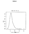
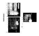
ヒトRNA由来のクロスハイブリダイゼーションの複雑性なしにアレイハイブリダイゼーション動力学を研究するために、SARSコロナウイルスおよびデング熱血清型1ウイルスRNAを感染細胞系の培地から精製し、逆転写し、ウイルス特異的プライマーを用いてPCR増幅した(Wong et al.,2004)。各ゲノムcDNAを、(配列決定によって確認されるように)その全体を増幅し、Cy3で標識し、マイクロアレイ上で別々にハイブリダイズさせた。SARS試料は、検出閾値を十分超えた蛍光(Cy3)シグナルを提示している(平均アレイシグナル強度よりも標準偏差の2倍を超える強度のプローブシグナル強度によって判定される;図7A)全3,805のSARS特異的プローブで、SARSタイリングプローブに十分ハイブリダイズした。コロナウイルス科の他のメンバーならびにピコナウイルス科およびパラミクソウイルス科のいくつかの種についてのみ観察すると、他の病原体プローブセットとのクロスハイブリダイゼーションは最小であり、SARSが他の公知のウイルスと配列相同性をほとんど共有していないという観察と一致した(Ksiazek et al.,2003)。他方では、1型デング熱のハイブリダイゼーションパターンは、より複雑であった(図7B)。まず、本発明者らは、配列多型のために、1型デング熱プローブセットへのハイブリダイゼーションが部分的に不完全である(すなわち、シグナルのない領域)ことを観察した。アレイ上でハイブリダイズされた1型デング熱試料を、1944年のハワイの分離株(ATCCカタログ番号VR−1254)から培養したが、アレイプローブセットは、1990年にシンガポールで単離されたS275/90株の配列(Fu et al.,1992)に基づく。cDNA標識とハイブリダイズできなかった1型デング熱プローブは、それぞれ、標的配列との少なくとも3つのミスマッチ(15塩基の範囲内に)を含んだ。第二に、本発明者らは、アレイ上に存在するほとんど全てのウイルスプローブセット、具体的には他のフラビウイルス科メンバーのプローブと、ある程度のクロスハイブリダイゼーションが起こったことを観察し、4型デング熱血清型が60〜70%の相同性を共有していることと一致した。ハイブリダイゼーションシグナル出力とアニーリング特異性との間の関係を理解するために、本発明者らは、まず、類似性の2つの尺度:プローブハミング距離(HD)および最大連続マッチ(MCM)を用いて、全てのプローブ配列を各ウイルスゲノムと比較した。HDは、類似した配列については低いスコアで、2つの配列の全体の類似性距離を評価する(Hamming,1950)。MCMは、類似した配列については高いスコアで、正確な適合である連続した塩基の数を評価する(Kane et al.,2000)。
To study array hybridization kinetics without the complexity of human RNA-derived cross-hybridization, SARS coronavirus and
本発明者らは、ハワイの1型デング熱分離株に関連して、各プローブについてHDおよびMCMスコアを計算し、これらのスコアが、それぞれプローブシグナル強度に対して反比例および直接の相関があることを観察した。ハワイの1型デング熱ゲノムに対して高い類似性を有する、すなわちHD=2(n=942)またはMCM=27(n=627)のアレイ上の全てのプローブは、バックグラウンドより3対数分高く、メジアンシグナル強度でハイブリダイズした。プローブの98%は0〜4の低HD範囲または18〜40の高いMCM範囲で検出可能であったが、メジアンプローブシグナル強度は、配列距離の各増分で減少した。メジアンシグナル強度は、それぞれ43%および46%の検出可能なプローブで、HD=7およびMCM=15でバックグラウンドレベルまで激減した。プローブの大部分(>96%、n>51,000)は、8〜21の間のHDスコアおよび/または0〜15の間のMCMスコアを有し、それぞれその1.23%および1.57%が検出可能であった。
We calculated HD and MCM scores for each probe in relation to
理想的な、クロスハイブリダイゼーション類似性閾値は、特定の病原体を同定する全てのプローブが、病原体配列の多型の存在下であっても、常にバックグラウンドノイズより高い検出可能なシグナル強度を有するものである。最適な類似性閾値HD=4およびMCM=18で、98%を超えるプローブが、バックグラウンドよりも2対数分高いメジアンシグナル強度で検出することができたが、閾値をHD=5およびMCM=17まで1段階下に調節することによって、わずか約85%のプローブ検出、およびバックグラウンドよりも約1.2対数分高いメジアンシグナル強度が生じた(図8)。
An ideal cross-hybridization similarity threshold is one in which all probes that identify a specific pathogen always have a detectable signal intensity above background noise, even in the presence of polymorphisms of the pathogen sequence It is. With optimal similarity thresholds HD = 4 and MCM = 18, more than 98% of the probes could be detected with a
これらの最適なHDおよびMCM閾値を用いてクロスハイブリダイゼーションを予測して、本発明者らは、全てのプローブを、所与の病原体を最も検出しそうなグループに分けた。本発明者らは、これらのグループを、特異的サインプローブセット(SPS)と呼び、アレイ上に示される35の病原体ゲノムのそれぞれについてのSPSを定義した(表2)。 Using these optimal HD and MCM thresholds to predict cross-hybridization, we divided all probes into groups most likely to detect a given pathogen. We referred to these groups as specific signature probe sets (SPS) and defined the SPS for each of the 35 pathogen genomes shown on the array (Table 2).
各病原体のSPSは、そのゲノム配列に由来するタイリングプローブ(HD=0、MCM=40)、ならびに他の病原体に由来するクロスハイブリダイズしているプローブを含んだ(HD=4、MCM=18)。 The SPS of each pathogen included tiling probes derived from its genomic sequence (HD = 0, MCM = 40) as well as cross-hybridized probes derived from other pathogens (HD = 4, MCM = 18). ).
本発明者らは、次に、本発明者らのSPSプローブの性能に影響を及ぼす確率のある他の非特異的ハイブリダイゼーション現象を考慮した。例えば、本発明者らは、プローブシグナルと%GC含有量との間の一般的な関係を観察した。以前の観察と一致して、本発明者らは、40%未満のGCのプローブが、減少したシグナル強度を生じるが、60%を超えるGC含有量のプローブは、より高いシグナル強度を示すことを見出した(Wong et al.,2004;Maskos and Southern,1993)。したがって、本発明者らは、さらなる選択フィルターとして%GC含有量を利用し、それによって、最適なHDおよびMCM値にもかかわらず、40%未満のGCおよび60%を超えるGCを有するプローブを本発明者らのSPSから排除した。 We next considered other non-specific hybridization phenomena that have the potential to affect the performance of our SPS probes. For example, we observed a general relationship between probe signal and% GC content. Consistent with previous observations, we have found that probes with less than 40% GC produce a reduced signal intensity, while probes with a GC content greater than 60% show higher signal intensity. (Wong et al., 2004; Maskos and Southern, 1993). Thus, we utilize% GC content as an additional selection filter, thereby presenting probes with less than 40% GC and more than 60% GC despite optimal HD and MCM values. Excluded from our SPS.
<ヒトゲノムに対する配列類似性>
検出される標的核酸がヒトから(例えば、ウイルスゲノムを含むヒト試料)抽出される場合、ヒトゲノムに対して高い相同性を有するプローブもまた避けられるべきである。したがって、標的核酸vaに特異的な長さmの任意のプローブsaについて、標的核酸と異なる核酸のいずれの領域ともいかなるヒットも有さない場合、プローブsaが選択され、長さmのプローブsaが標的核酸と異なる核酸とヒットを有する場合、最も小さい最大アライメント長および/または最少の数のヒットを有する長さmのプローブsaが選択される。具体的には、任意の長さmのプローブsaについて、BLASTアルゴリズムで、ヒトゲノムとのsaのヒットが見られる(Altschul,S.F.et al.,1997)。(W=15)のBLAST文字列サイズおよび100の期待値を用いて、全てのヒットを見出した。ヒトゲノムといかなるヒットも有さない、すなわち、vaに特異的である場合、saを選択する。しかしながら、vaの長さmの全てのサブストリングがヒトゲノムとヒットを有する場合、最も小さい最大アライメント長および最少の数のヒットを有するものを選択した。
<Sequence similarity to the human genome>
If the target nucleic acid to be detected is extracted from a human (eg, a human sample containing a viral genome), probes with high homology to the human genome should also be avoided. Thus, for any probes s a target nucleic acid v a specific length m, if with any region of a nucleic acid different from the target nucleic acid does not have any hits, probes s a is selected, the length m If the probe s a have a different nucleic acid and hit the target nucleic acid, probes s a length m with a number hits the smallest maximum alignment length and / or minimum is selected. Specifically, the probe s a of any length m, with BLAST algorithm, hit s a with the human genome is observed (Altschul, S.F.et al., 1997 ). All hits were found using a BLAST string size of (W = 15) and an expected value of 100. The human genome and does not have any hits, i.e., if the v a is specific, it selects s a. However, all substrings of length m of v a may have human genome and the hit was selected to have a hit of the smallest maximum alignment length and minimum number.
さらに、ヒト配列とのクロスハイブリダイゼーションはまた、結果を複雑にする可能性があったので、本発明者らは、15の文字列サイズを用いたBLASTによって(Altschul et al.,1997)、全てのプローブをヒトゲノム集合(17を構築)(International Human Genome Sequencing Consortium.Initial sequencing and analysis of the human genome.Nature 409(6822)、860−921(2001))と比較した。SPSから100の期待値を有するプローブをさらにフィルタリングした(上記の表2参照)。 In addition, since cross-hybridization with human sequences could also complicate the results, we performed BLAST with a string size of 15 (Altschul et al., 1997), all Human genome assembly (constructed 17) (International Human Genome Sequencing Consortium. Initial sequencing and analysis of the human genome. Nature 409 (6822), 860-921 (2001)). Probes with an expected value of 100 from the SPS were further filtered (see Table 2 above).
したがって、本発明は、核酸検出のためのオリゴヌクレオチドプローブを設計する方法を提供し、標的核酸vaに特異的な長さmの任意のプローブsaについて、標的核酸と異なる核酸のいずれの領域ともいかなるヒットも有さない場合、プローブsaが選択され、長さmのプローブsaが標的核酸と異なる核酸とヒットを有する場合、最も小さい最大アライメント長および/または最少の数のヒットを有する長さmのプローブsaが選択される。 Accordingly, the present invention provides a method of designing an oligonucleotide probe for nucleic acid detection, for any probes s a target nucleic acid v specific length a m, any region of the nucleic acid different from the target nucleic acid and if neither have any hits, probes s a is selected, if the probe s a length m has a different nucleic acid and hit the target nucleic acid, having a hit of the smallest maximum alignment length and / or minimum number probe s a length m is selected.
さらに、オリゴヌクレオチドプローブの設計はまた、本発明のAESによって行ってもよい。具体的には、本発明は、piが増幅された標的核酸の位置iにハイブリダイズすると予測される場合に標的核酸の位置iのプローブpiが選択される、プローブを選択および/または設計する方法を提供する。 Furthermore, the design of oligonucleotide probes may also be performed by the AES of the present invention. Specifically, the present invention provides a probe p i of position i of the target nucleic acid is selected if p i is predicted hybridize to the position i of a target nucleic acid amplified, selection and / or design probes Provide a way to do it.
具体的には、選択される領域にハイブリダイズすることができるオリゴヌクレオチドプローブは、以下の基準:
(a)選択されたプローブが、40%〜60%のCG含有量を有する;
(b)隣接モデルに基づいて計算される最も高い自由エネルギーを有することによってプローブが選択される;
(c)標的核酸vaおよびvbのプローブsaおよびプローブsbサブストリングを仮定すると、saと標的核酸vb由来の任意の長さmのサブストリングsbの間のハミング距離および/もしくはsaおよびプローブsbの最も長い共通するサブストリングに基づいて、saが選択される;
(d)標的核酸vaに特異的な長さmの任意のプローブsaについて、標的核酸と異なる核酸のいずれの領域ともいかなるヒットも有さない場合、プローブsaが選択され、長さmのプローブsaが標的核酸と異なる核酸とヒットを有する場合、最も小さい最大アライメント長および/もしくは最少の数のヒットを有する長さmのプローブsaが選択される;ならびに/または
(e)piが増幅された標的核酸の位置iにハイブリダイズすると予測される場合、標的核酸の位置iのプローブpiが選択される
の少なくとも1つに従って、選択および/または設計してもよい。
Specifically, an oligonucleotide probe that can hybridize to a selected region has the following criteria:
(A) the selected probe has a CG content of 40% -60%;
(B) the probe is selected by having the highest free energy calculated based on the adjacent model;
(C) assuming the probe s a and probes s b substring of the target nucleic acid v a and v b, the Hamming distance between substrings s b of any length m from s a target nucleic acid v b and / or s a and based on the longest common substring of probes s b, s a is selected;
For any probes s a a (d) target nucleic v specific length a m, if with any region of a nucleic acid different from the target nucleic acid does not have any hits, probes s a is selected, the length m when the probe s a a have different nucleic acid and hit the target nucleic acid, probes s a length m with a number hits the smallest maximum alignment length and / or minimum is selected; and / or (e) p If i is expected to hybridize to position i of the amplified target nucleic acid, it may be selected and / or designed according to at least one of the probes p i at position i of the target nucleic acid being selected.
本発明の特定の態様によると、オリゴヌクレオチドプローブを設計するために、上述の基準の2つ以上を用いてもよい。例えば、全ての基準(a)〜(e)を適用することによって、プローブを設計してもよい。本明細書で明らかに言及されていないが当業者に明らかな他の基準もまた用いてもよい。 In accordance with certain aspects of the invention, two or more of the above criteria may be used to design oligonucleotide probes. For example, the probe may be designed by applying all the criteria (a) to (e). Other criteria not explicitly mentioned herein but apparent to those skilled in the art may also be used.
具体的には、基準(e)に基づいて、P(pi|va)>λであり、式中λが0.5であり、P(pi|va)が、piが標的核酸vaの位置iにハイブリダイズしなければならない確率である場合、標的核酸vaの位置iのプローブpiが選択される。より具体的には、λは0.8である。 Specifically, based on criterion (e), P (p i | v a )> λ, where λ is 0.5 and P (p i | v a ) is the target of p i If the probability of the position i of the nucleic v a must hybridize, probe p i at position i of a target nucleic acid v a is selected. More specifically, λ is 0.8.
別の態様によると、本発明は、
別の態様によると、AESを用いて、ランダムプライマータグを設計して、ランダムPCRによって試料のランダム増幅を容易にすることもできる(病原体の検出、遺伝子発現の検出、クローンDNAライブラリーの構築、および当業者がランダムPCRを採用する他の用途等の用途における使用のために)。 According to another aspect, AES can be used to design random primer tags to facilitate random amplification of samples by random PCR (pathogen detection, gene expression detection, clonal DNA library construction, And for use in applications such as other applications where one skilled in the art employs random PCR).
<支持体上のオリゴヌクレオチドプローブの合成>
本発明の別の態様によると、上述のように少なくとも1つのオリゴヌクレオチドプローブを選択および/または設計する方法は、選択および/または設計されたプローブを調製するステップをさらに含む。プローブを設計することは、その配列を理解すること、および/または任意の適した手段によって、例えばソフトウエアを使用することによってそれを設計することを含む。プローブを調製するステップは、その物理的調製を含む。プローブは、当分野で公知の任意の標準的な方法に従って調製してもよい。例えば、プローブは、化学的に合成するか、またはクローニングによって調製してもよい。例えば、Sambrook and Russel,2001に記載されているように。
<Synthesis of oligonucleotide probe on support>
According to another aspect of the invention, the method of selecting and / or designing at least one oligonucleotide probe as described above further comprises the step of preparing the selected and / or designed probe. Designing the probe includes understanding its sequence and / or designing it by any suitable means, for example by using software. The step of preparing the probe includes its physical preparation. Probes may be prepared according to any standard method known in the art. For example, the probe may be chemically synthesized or prepared by cloning. For example, as described in Sambrook and Russel, 2001.
本発明の任意の実施形態に従って調製された、支持体、例えばマイクロアレイまたはバイオチップもまた提供される。 Supports such as microarrays or biochips prepared according to any embodiment of the present invention are also provided.
本発明の任意の方法に従って設計および調製されたプローブは、溶液中で使用するか、または不溶性支持体上に配置してもよい。例えば、当分野で公知の任意の技術に従って、不溶性支持体に塗布するか、スポットするか、または印刷してもよい。支持体は、固体支持体またはゲルであってもよい。プローブを塗布された支持体は、マイクロアレイまたはバイオチップであってもよい。 Probes designed and prepared according to any method of the invention may be used in solution or placed on an insoluble support. For example, it may be applied to, spotted or printed on an insoluble support according to any technique known in the art. The support may be a solid support or a gel. The support coated with the probe may be a microarray or a biochip.
より具体的には、本発明は、最初の組織試料から調製されたPCR増幅cDNA(具体的には、ランダムPCR増幅cDNA)からの、病原体、例えばウイルスおよび/または細菌病原体の迅速な検出および同定のためのオリゴマイクロアレイハイブリダイゼーションベースの手法を提供する。 More specifically, the present invention provides for the rapid detection and identification of pathogens, such as viral and / or bacterial pathogens, from PCR amplified cDNA (specifically random PCR amplified cDNA) prepared from an initial tissue sample. An oligo-microarray hybridization-based approach is provided for.
以下の説明において、プローブの調製は、マイクロアレイに特に関連して行われる。しかしながら、支持体、ならびにプローブは、本出願の内容全体にわたる任意の記載に従って調製してもよい。具体的には、「アレイ」は、合成または生合成のいずれで調製されてもよい分子の意図的に作られた収集物である。アレイ中の分子は、互いに同一でも異なってもよい。アレイは、種々の形式、例えば、可溶性分子のライブラリー、樹脂ビーズ、シリカチップ、または他の固体支持体に繋ぎ止められた化合物のライブラリーの形をとってもよい。アレイプレートまたはプレートは、ウェルと呼ばれる、液体の通過に耐性があって領域または空間を形成する物理的障壁によって、各アレイが他のアレイから分離されている複数のアレイを有する物体である。 In the following description, probe preparation is performed with particular reference to microarrays. However, the support, as well as the probe, may be prepared according to any description throughout the contents of this application. Specifically, an “array” is an intentionally created collection of molecules that may be prepared either synthetically or biosynthetically. The molecules in the array may be the same or different from each other. The array may take various forms, for example, a library of compounds anchored to a library of soluble molecules, resin beads, silica chips, or other solid supports. An array plate or plate is an object, called a well, having a plurality of arrays in which each array is separated from the other by a physical barrier that resists the passage of liquid and forms a region or space.
<試料調製およびマイクロアレイへのハイブリダイゼーション>
生体試料は、哺乳動物、例えばヒトから採取される、任意の試料であってもよい。生体試料は、血液、体液、唾液、尿、便等であってもよい。生体試料は、増幅ステップを行う前に生体試料中に含まれる核酸を取り除くよう処理してもよい。標的核酸は、検出されることが意図される任意の核酸であってもよい。検出される標的核酸は、少なくとも、生体試料の核酸にとって外因性である核酸であってもよい。したがって、生体試料がヒトに由来する場合、検出される外因性標的核酸(生体試料中に存在する場合)は、ヒト由来でない核酸である。本発明の態様によると、検出される標的核酸は、少なくとも、病原体ゲノムまたはその断片である。病原体核酸は、少なくとも、ウイルス、寄生生物、もしくは細菌由来の核酸、またはその断片であってもよい。
<Sample preparation and hybridization to microarray>
The biological sample may be any sample collected from a mammal, such as a human. The biological sample may be blood, body fluid, saliva, urine, stool or the like. The biological sample may be treated to remove nucleic acids contained in the biological sample before performing the amplification step. The target nucleic acid may be any nucleic acid that is intended to be detected. The target nucleic acid to be detected may be at least a nucleic acid that is exogenous to the nucleic acid of the biological sample. Therefore, when the biological sample is derived from a human, the exogenous target nucleic acid to be detected (when present in the biological sample) is a nucleic acid that is not derived from a human. According to an aspect of the invention, the target nucleic acid to be detected is at least a pathogen genome or a fragment thereof. The pathogen nucleic acid may be at least a virus, parasite, or bacterial nucleic acid, or a fragment thereof.
本発明の態様によると、標的核酸検出分析の方法が提供される。検出されることが所望される生体試料由来の標的核酸は、任意の標的核酸、RNAおよび/またはDNAであってもよい(例えば、mRNAおよび/またはcDNA)。より具体的には、検出される標的核酸は、病原体または非病原体であってもよい。例えば、少なくとも1つのウイルス、少なくとも1つの細菌および/または少なくとも1つの寄生生物のゲノムまたはその断片であってもよい。選択および/または調製されるプローブは、当業者に公知の任意の標準的な技術に従って支持体上に配置、塗布および/または固定されてもよい。支持体は、不溶性支持体、例えば固体支持体であってもよい(具体的には、マイクロアレイおよび/またはバイオチップ)。 According to an aspect of the invention, a method for target nucleic acid detection analysis is provided. The target nucleic acid from the biological sample that is desired to be detected can be any target nucleic acid, RNA and / or DNA (eg, mRNA and / or cDNA). More specifically, the target nucleic acid to be detected may be a pathogen or a non-pathogen. For example, it may be the genome of at least one virus, at least one bacterium and / or at least one parasite or a fragment thereof. The probes selected and / or prepared may be placed, applied and / or immobilized on a support according to any standard technique known to those skilled in the art. The support may be an insoluble support, such as a solid support (specifically, a microarray and / or a biochip).
特定の実施例によると、確立されたプロトコルおよび商業的キットを用いて、患者試料、例えば組織、血清、鼻咽頭洗浄液、便からRNAおよびDNAを抽出した。例えば、核酸抽出のためのQiagenキットを用いてもよい。あるいは、フェノール/クロロホルムもまた、DNAおよび/またはRNAの抽出のために用いてもよい。例えばSambrookおよびRussel,2001に記載されているような、当分野で公知の任意の技術を用いてもよい。Bohlander et al.,1992およびWang et al.,2003によって記載されたプロトコルに基づいて、タグを付けたランダムプライマーを用いてRNAをcDNAに逆転写した。次いで、ランダムPCRによってcDNAを増幅した。マイクロアレイへの試料の断片化、標識およびハイブリダイゼーションは、Wong et al.,2004によって記載されているように行った。 According to certain examples, RNA and DNA were extracted from patient samples such as tissue, serum, nasopharyngeal lavage fluid, stool using established protocols and commercial kits. For example, a Qiagen kit for nucleic acid extraction may be used. Alternatively, phenol / chloroform may also be used for DNA and / or RNA extraction. Any technique known in the art may be used, for example as described in Sambrook and Russel, 2001. Bohlander et al. , 1992 and Wang et al. , 2003, RNA was reverse transcribed to cDNA using tagged random primers. The cDNA was then amplified by random PCR. Fragmentation, labeling and hybridization of samples to microarrays are described in Wong et al. , 2004.
<マイクロアレイ合成>
実施例部分に記載されている特定の実験によると、本発明者らは、シンガポールにおけるウイルス疾患の最も一般的な原因を代表するいくつかのウイルスゲノムを選択した。Genbankからダウンロードした全ゲノム配列を使用して、ゲノム全体にわたってタイリングされたオーバーラップ5塩基の解像度の40塩基長のプローブを生成した。Nimblegen技術(Nuwaysir,E.F.et al.,2002)を用いて、各ウイルスプローブの7つの複製を、マイクロアレイ上に直接合成した。マイクロアレイ上でプローブをランダムに分配して、ハイブリダイゼーションのアーチファクトの効果を最小限にした。試料の、プローブへの非特異的ハイブリダイゼーションを制御するために、10,000のオリゴヌクレオチドプローブをマイクロアレイ上に設計および合成した。これらの10,000のオリゴヌクレオチドは、ヒトゲノムに対して、または病原性ゲノムに対していかなる配列類似性も有さなかった。これらは40〜60%のCG含有量を有するランダムプローブであった。これらのプローブは、バックグラウンドシグナル強度を評価した。陽性対照として、免疫応答において公知または推定される機能を有するヒト遺伝子に対する400のオリゴヌクレオチドプローブをアレイ上で合成した。合計およそ380,000のプローブについて、植物ウイルス、PMMVを陰性対照として含めた。以下の説明において、病原体検出チップ分析(PDCとも呼ばれる)に関連して、本発明が、より具体的に説明される。しかしながら、分析(方法)は、この特定の実施形態に限定されず、本出願の内容全体にわたって記載されるような本発明のいくつかの態様を包含する。
<Microarray synthesis>
According to specific experiments described in the Examples section, we have selected several viral genomes representing the most common causes of viral disease in Singapore. The whole genome sequence downloaded from Genbank was used to generate a 40 base long probe with resolution of overlapping 5 bases tiled across the entire genome. Using Nimblegen technology (Nuwaysir, EF et al., 2002), seven replicas of each viral probe were synthesized directly on the microarray. Probes were randomly distributed on the microarray to minimize the effects of hybridization artifacts. 10,000 oligonucleotide probes were designed and synthesized on the microarray to control non-specific hybridization of the sample to the probe. These 10,000 oligonucleotides did not have any sequence similarity to the human genome or to the pathogenic genome. These were random probes with a CG content of 40-60%. These probes were evaluated for background signal intensity. As a positive control, 400 oligonucleotide probes for human genes with functions known or presumed in the immune response were synthesized on the array. Plant virus, PMMV, was included as a negative control for a total of approximately 380,000 probes. In the following description, the present invention will be described more specifically in connection with pathogen detection chip analysis (also referred to as PDC). However, the analysis (method) is not limited to this particular embodiment, but encompasses several aspects of the invention as described throughout the content of the present application.
<標的核酸を検出する方法>
別の態様によると、本発明は、
(i)生体試料を提供するステップと、
(ii)生体試料中に含まれる核酸を増幅するステップと、
(iii)生体試料中に存在する場合、少なくとも1つの標的核酸にハイブリダイズすることができる少なくとも1つのオリゴヌクレオチドプローブを提供するステップと、ここで、前記プローブが、本明細書に記載される本発明の任意の態様による方法を用いることによって調製され、
(iv)プローブを増幅された核酸と接触させ、少なくとも1つの標的核酸にハイブリダイズしたプローブを検出するステップと
を含む、少なくとも1つの標的核酸を検出する方法を提供する。
<Method of detecting target nucleic acid>
According to another aspect, the present invention provides:
(I) providing a biological sample;
(Ii) amplifying the nucleic acid contained in the biological sample;
(Iii) providing at least one oligonucleotide probe capable of hybridizing to at least one target nucleic acid when present in a biological sample, wherein said probe is a book described herein; Prepared by using a method according to any aspect of the invention,
(Iv) contacting the probe with the amplified nucleic acid and detecting the probe hybridized to the at least one target nucleic acid, to provide a method of detecting at least one target nucleic acid.
増幅ステップ(ii)は、ランダムな、部分的にランダムな(すなわち、固定された部分およびランダムな部分を含む)または特異的なプライマーの存在下で行ってもよい。具体的には、増幅ステップ(II)は、少なくとも1つのランダムプライマーの存在下で行ってもよい。より具体的には、少なくとも1つのランダムフォワードプライマーおよび/または少なくとも1つのランダムリバースプライマーの存在下で。例えば、増幅ステップ(ii)は、2つより多いランダムプライマーの存在下で行ってもよい。当分野で公知の任意の増幅方法を用いてもよい。例えば、増幅方法は、RT−PCRである。 Amplification step (ii) may be performed in the presence of random, partially random (ie, including fixed and random portions) or specific primers. Specifically, the amplification step (II) may be performed in the presence of at least one random primer. More specifically, in the presence of at least one random forward primer and / or at least one random reverse primer. For example, the amplification step (ii) may be performed in the presence of more than two random primers. Any amplification method known in the art may be used. For example, the amplification method is RT-PCR.
具体的には、本発明者らは、増幅効率スコア(AES)に基づいて、標的核酸にハイブリダイズしたプローブを検出する方法を開発した。これは、本明細書で、本発明のアルゴリズムとも呼ばれる。具体的には、
増幅ステップは、フォワードおよびリバースプライマーを含んでもよく、フォワードおよびリバースプライマーのそれぞれは、5’−3’配向で、不変のプライマー頭部および可変のプライマー尾部を含んでもよく、少なくとも可変の尾部は、標的核酸vaの一部にハイブリダイズする。具体的には、増幅ステップは、配列番号1〜7のいずれかのヌクレオチド配列、またはそのバリアントもしくは誘導体を有するフォワードおよび/またはリバースランダムプライマーを含んでもよい。 The amplification step may include forward and reverse primers, each of the forward and reverse primers may be in a 5′-3 ′ orientation and include an invariant primer head and a variable primer tail, at least the variable tail hybridizes to a portion of the target nucleic acid v a. Specifically, the amplification step may include forward and / or reverse random primers having any nucleotide sequence of SEQ ID NOs: 1-7, or variants or derivatives thereof.
生体試料は、哺乳動物、例えばヒトから採取された任意の試料であってもよい。生体試料は、組織、血清、鼻咽頭洗浄液、唾液、任意の他の体液、血液、尿、便等であってもよい。増幅ステップを行う前に、生体試料を処置して、生体試料中に含まれる核酸を取り除いてもよい。標的核酸は、検出されることが意図される任意の核酸であってもよい。検出される標的核酸は、少なくとも、生体試料の核酸にとって外因性である核酸であってもよい。したがって、生体試料がヒト由来である場合、検出される外因性標的核酸(生体試料中に存在する場合)は、ヒト由来でない核酸である。 The biological sample may be any sample collected from a mammal such as a human. The biological sample may be tissue, serum, nasopharyngeal lavage fluid, saliva, any other body fluid, blood, urine, feces and the like. Prior to performing the amplification step, the biological sample may be treated to remove nucleic acids contained in the biological sample. The target nucleic acid may be any nucleic acid that is intended to be detected. The target nucleic acid to be detected may be at least a nucleic acid that is exogenous to the nucleic acid of the biological sample. Therefore, when the biological sample is derived from human, the exogenous target nucleic acid to be detected (when present in the biological sample) is a nucleic acid that is not derived from human.
本発明の態様によると、検出される標的核酸は、少なくとも、病原体ゲノムまたはその断片である。病原体核酸は、少なくとも、ウイルス、寄生生物、もしくは細菌由来の核酸、またはその断片であってもよい。 According to an aspect of the invention, the target nucleic acid to be detected is at least a pathogen genome or a fragment thereof. The pathogen nucleic acid may be at least a virus, parasite, or bacterial nucleic acid, or a fragment thereof.
したがって、本発明は、少なくとも、生体試料中に存在する場合、標的核酸の検出の方法を提供する。方法は、生体試料中の病原体の存在の検出のための診断方法であってもよい。例えば、生体試料がヒトから得られる場合、標的核酸は、生体試料中に存在する場合、ヒト由来でない。 Accordingly, the present invention provides a method for detecting a target nucleic acid, at least when present in a biological sample. The method may be a diagnostic method for the detection of the presence of a pathogen in a biological sample. For example, when the biological sample is obtained from a human, the target nucleic acid is not derived from a human when present in the biological sample.
本発明の任意の方法に従って設計および/または調製されたプローブは、溶液中で用いてもよく、または、不溶性支持体上に配置してもよい。例えば、当分野で公知の任意の技術に従って不溶性支持体上に、塗布するか、スポットするか、または印刷してもよい。プローブを塗布された支持体は、固体支持体またはゲルであってもよい。具体的には、マイクロアレイまたはバイオチップであってもよい。 Probes designed and / or prepared according to any method of the invention may be used in solution or placed on an insoluble support. For example, it may be coated, spotted or printed on an insoluble support according to any technique known in the art. The support coated with the probe may be a solid support or a gel. Specifically, it may be a microarray or a biochip.
次いで、プローブを生体試料の核酸に接触させ、存在する場合、標的核酸とプローブがハイブリダイズし、標的核酸の存在が検出される。具体的には、検出ステップ(iv)において、vaにハイブリダイズするプローブのシグナル強度の平均値は、vaに含まれないプローブの平均値よりも統計的に高く、それによって、生体試料中のvaの存在が示される。 The probe is then contacted with the nucleic acid of the biological sample and, if present, the target nucleic acid and probe are hybridized and the presence of the target nucleic acid is detected. Specifically, in the detection step (iv), v the mean value of signal intensities of the hybridized probes in a statistically higher than the average value of the probe that is not included in v a, whereby the biological sample the presence of the v a is shown.
より具体的には、検出ステップ(iv)において、vaにハイブリダイズするプローブのシグナル強度の平均値は、vaに含まれないプローブの平均値よりも統計的に高いことを特徴とし、本方法は、高いシグナル強度を有する検出方法において使用されるプローブの割合に対する、高いシグナル強度を有するvaに含まれないプローブの割合の相対的差異を計算するステップをさらに含む方法であって、プローブvaのシグナル強度の密度分布は、vaに含まれないプローブのものよりも正に歪んでおり、それによって、生体試料中のvaの存在が示される方法である。 More specifically, in the detection step (iv), v the mean value of signal intensities of the hybridized probes in a features a statistically higher than the mean value of the probe that is not included in v a, the The method further comprises the step of calculating the relative difference of the proportion of probes not included in va having a high signal intensity relative to the proportion of probes used in the detection method having a high signal intensity comprising: v density distribution of signal intensities of a is, v is distorted positively than that of the probe that is not included in a, whereby a method for the presence of v a biological sample is shown.
例えば、検出ステップ(iv)において、生体試料中の標的核酸の存在は、0.1以下のt検定の値および/または0.05以下のアンダーソン・ダーリン検定値および/または1.0以上、好ましくは5.0以上の重み付きカルバック・ライブラー情報量の値によって示される。具体的には、t検定値は0.05以下である。 For example, in the detection step (iv), the presence of the target nucleic acid in the biological sample is a t-test value of 0.1 or less and / or an Anderson-Darlin test value of 0.05 or less and / or 1.0 or more, preferably Is indicated by a value of the weighted cullback library information amount of 5.0 or more. Specifically, the t test value is 0.05 or less.
別の態様によると、本発明は、標的核酸vaへのプローブのハイブリダイゼーションを検出することを含む標的核酸vaの存在を判定する方法を提供し、vaにハイブリダイズするプローブのシグナル強度の平均値は、vaに含まれないプローブの平均値よりも統計的に高く、それによって、vaの存在が示される。具体的には、vaにハイブリダイズするプローブのシグナル強度の平均値は、vaに含まれないプローブの平均値よりも統計的に高いことを特徴とし、本方法は、高いシグナル強度を有する検出方法において使用されるプローブの割合に対する、高いシグナル強度を有するvaに含まれないプローブの割合の相対的差異を計算するステップをさらに含む方法であって、プローブvaのシグナル強度の密度分布は、vaに含まれないプローブのものよりも正に歪んでおり、それによって、vaの存在が示される方法である。より具体的には、生体試料中の標的核酸の存在は、0.1以下のt検定の値および/または0.05以下のアンダーソン・ダーリン検定値および/または1.0以上、好ましくは5.0以上の重み付きカルバック・ライブラー情報量の値によって示される。例えば、t検定値は、0.05以下であってもよい。 According to another aspect, the present invention, the signal intensity of the probes provides a method of determining the presence of a target nucleic acid v a comprising detecting the hybridization of the probe to the target nucleic acid v a, hybridize to v a the average value of, v statistically higher than the average value of the probe that is not included in a, whereby, the presence of v a is shown. Specifically, v the mean value of signal intensities of the hybridized probes in a can, v than the average value of the probe that is not included in a characterized by statistically high, the method has a high signal intensity for the proportion of the probes used in the detection method, a method step further comprising the calculating the relative difference of the proportion of v is not included in a probe with high signal intensity, the density distribution of signal intensities of probes v a is, v is distorted positively than that of the probe that is not included in a, whereby, the presence of v a is the way indicated. More specifically, the presence of the target nucleic acid in the biological sample is a t-test value of 0.1 or less and / or an Anderson-Darlin test value of 0.05 or less and / or 1.0 or more, preferably 5. It is indicated by a value of a weighted cullback library information amount of 0 or more. For example, the t test value may be 0.05 or less.
別の態様によると、本発明は、
(i)少なくとも1つの生体試料を提供するステップと、
(ii)生体試料中に含まれる核酸を増幅するステップと、
(iii)生体試料中に存在する場合、少なくとも1つの標的核酸にハイブリダイズすることができる少なくとも1つのオリゴヌクレオチドプローブを提供するステップと、
(iv)プローブを増幅された核酸と接触させ、標的核酸にハイブリダイズしたプローブを検出するステップと、ここで、vaにハイブリダイズするプローブのシグナル強度の平均値が、vaに含まれないプローブの平均値よりも統計的に高く、それによって生体試料中のvaの存在が示されるステップと
を含む、少なくとも1つの標的核酸を検出する方法を提供する。
According to another aspect, the present invention provides:
(I) providing at least one biological sample;
(Ii) amplifying the nucleic acid contained in the biological sample;
(Iii) providing at least one oligonucleotide probe capable of hybridizing to at least one target nucleic acid when present in a biological sample;
(Iv) is contacted with a nucleic acid amplified with a probe, and detecting the hybridized probe to the target nucleic acid, where, v the mean value of signal intensities of the hybridized probes in a is not included in v a statistically higher than the average value of the probe, thereby; and presence of v a biological sample is indicated, it provides a method of detecting at least one target nucleic acid.
ステップ(iv)において、vaにハイブリダイズするプローブのシグナル強度の平均値は、vaに含まれないプローブの平均値よりも統計的に高いことを特徴とし、本方法は、高いシグナル強度を有する検出方法において使用されるプローブの割合に対する、高いシグナル強度を有するvaに含まれないプローブの割合の相対的差異を計算するステップをさらに含む方法であって、プローブvaのシグナル強度の密度分布は、vaに含まれないプローブのものよりも正に歪んでおり、それによって、生体試料中のvaの存在が示される方法である。具体的には、ステップ(iv)において、生体試料中の標的核酸の存在は、0.1以下のt検定の値および/または0.05以下のアンダーソン・ダーリン検定値および/または1.0以上、好ましくは5.0以上の重み付きカルバック・ライブラー情報量の値によって示される。t検定値は0.05以下であってもよい。検出される核酸は、生体試料の核酸にとって外因性の核酸である。検出される標的核酸は、少なくとも、病原体ゲノムまたはその断片であってもよい。病原体核酸は、少なくとも、ウイルス、寄生生物、もしくは細菌由来の核酸、またはその断片であってもよい。具体的には、試料がヒトから得られる場合、標的核酸は、生体試料中に存在する場合、ヒトゲノム由来でない。プローブは、不溶性支持体上に配置してもよい。支持体は、マイクロアレイまたはバイオチップであってもよい。 In step (iv), v the mean value of signal intensities of the hybridized probes in a can, v than the average value of the probe that is not included in a characterized by statistically high, the method of high signal intensity for the proportion of the probes used in the detection method having, a v method further comprising the step of calculating a relative difference of the proportion of probes that are not included in a having a high signal strength, the density of the signal intensity of the probe v a distribution, v is distorted positively than that of the probe that is not included in a, whereby a method for the presence of v a biological sample is shown. Specifically, in step (iv), the presence of the target nucleic acid in the biological sample is a t-test value of 0.1 or less and / or an Anderson-Darlin test value of 0.05 or less and / or 1.0 or more. , Preferably indicated by a value of the weighted Cullback library information amount of 5.0 or more. The t test value may be 0.05 or less. The nucleic acid to be detected is a nucleic acid that is exogenous to the nucleic acid of the biological sample. The target nucleic acid to be detected may be at least a pathogen genome or a fragment thereof. The pathogen nucleic acid may be at least a virus, parasite, or bacterial nucleic acid, or a fragment thereof. Specifically, when the sample is obtained from a human, the target nucleic acid is not derived from a human genome when present in a biological sample. The probe may be placed on an insoluble support. The support may be a microarray or a biochip.
<RSV−Bの鋳型配列を用いた試験>
ウイルスの異なる領域によって提示されるシグナル強度の変動がそれらの対応する増幅効率スコアと直接的な相関を有するかどうかを検証するために、ヒトに影響を及ぼす一般的な病原体、ヒト呼吸器多核体ウイルスB(RSV−B)に関して、合計5つのマイクロアレイ実験を行った。
<Test using RSV-B template sequence>
A common pathogen that affects humans, the human respiratory multinucleate, to verify whether fluctuations in signal intensity presented by different regions of the virus have a direct correlation with their corresponding amplification efficiency scores A total of 5 microarray experiments were performed on virus B (RSV-B).
次に、NCBIから得られたRSV−Bの鋳型配列(NC_001781)に、上述のようなプローブ設計基準を適用した。これは、各マイクロアレイ上に斑点をつけられた1948のプローブが得られた。RSV−Bについての増幅効率マップもまた、実際の実験の前に計算し、図2に示す。この図は、平均AESよりも高いAESを有するピークを示し、より高い増幅の確率を有するRSV−Bの領域が示される。 Next, the probe design criteria as described above were applied to the RSV-B template sequence (NC — 001781) obtained from NCBI. This resulted in 1948 probes spotted on each microarray. The amplification efficiency map for RSV-B was also calculated before the actual experiment and is shown in FIG. This figure shows a peak with an AES higher than the average AES, indicating a region of RSV-B with a higher probability of amplification.
ヒト呼吸器多核体ウイルスB(RSV−B)を含む5つの試料を用いて、独立したマイクロアレイ実験を行った。1つのこのような実験についての得られたシグナル強度を、図3に示す。 Independent microarray experiments were performed using five samples containing human respiratory polynuclear virus B (RSV-B). The resulting signal intensity for one such experiment is shown in FIG.
各実験について、1948のプローブのシグナル強度を、減少順で順位付けし、それらの対応するAES値と関連付けた。p値は、平均で2.2e-16未満であるとわかった。これは、RSV−Bの位置iのプローブのシグナル強度とAESiとの間の相関が全くランダムでないことを示す。さらなる調査によって、5つ全ての実験において一貫して高いシグナル強度を生じる約300のプローブが、90パーセンタイルレベルで増幅効率スコアを有することが明らかになった。 For each experiment, the signal intensity of 1948 probes was ranked in decreasing order and correlated with their corresponding AES values. The p-value was found to be less than 2.2e -16 on average. This indicates that the correlation between the signal intensity of the probe at position i of RSV-B and AES i is not random at all. Further investigation revealed that about 300 probes that consistently produced high signal intensity in all five experiments had an amplification efficiency score at the 90th percentile level.
説明された増幅効率モデルはRSV−Bゲノムに関して功を奏することを示してきたが、本発明の方法が他のウイルスゲノムにも拡張することができることを示すことが望まれた。ヒトメタニューモウイルス(HMPV)に関して、別のマイクロアレイ実験を行った。今回は、マイクロアレイ上に1705のプローブがあった。ここでも、HMPVについての増幅効率マップを計算した。この実験において、シグナル強度と増幅効率スコアとの間の相関試験によって、1.335e-9のp値が示された。 Although the amplification efficiency model described has been shown to work for the RSV-B genome, it was desired to show that the method of the invention can be extended to other viral genomes. Another microarray experiment was performed on human metapneumovirus (HMPV). This time, there were 1705 probes on the microarray. Again, an amplification efficiency map for HMPV was calculated. In this experiment, a correlation test between signal intensity and amplification efficiency score indicated a p-value of 1.335e- 9 .
したがって、本発明の増幅効率モデルは、説明された実験設定において、ウイルスゲノムの異なる領域によって生成されたシグナルの相対的強度を予測することができる。低い増幅効率スコアを有する領域由来のプローブは、シグナル強度を生じないか、または低いシグナル強度を生じる傾向が高い。これは、マイクロアレイ上で偽陰性を生じる。このようなプローブは、マイクロアレイデータの分析を複雑にし、これは、低いシグナル強度を有するプローブはその標的ゲノムが存在しないか、または単に増幅されなかったためである場合があるので、これがさらに複雑になる。そのようなものとして、適度に高い増幅効率スコアを有する領域におけるプローブを選択して、ランダムプライマーを用いたRT−PCRプロセスによって引き起こされる不正確性を最小限にするべきである。 Thus, the amplification efficiency model of the present invention can predict the relative intensities of signals generated by different regions of the viral genome in the described experimental setting. Probes from regions with low amplification efficiency scores tend not to produce signal intensity or produce low signal intensity. This produces a false negative on the microarray. Such a probe complicates the analysis of microarray data, which is further complicated because a probe with low signal intensity may be because its target genome may not be present or simply amplified. . As such, probes in regions with reasonably high amplification efficiency scores should be selected to minimize inaccuracies caused by the RT-PCR process with random primers.
ウイルスvaのプローブ選択のための増幅効率スコアの閾値は、AES値vaの累積分布関数によって判定される。Xを、全てのプローブvaのAES値を表す確率変数とする。kを、vaにおけるプローブの数とする。次いで、本発明者らは、AES値がx以下である確率を、P(X≦x)=c/kとし、式中cは、x以下であるAES値を有するプローブの数である。vaの位置iのプローブpiについて、xiを、その対応するAES値とする。プローブのシグナル強度はそのAES値と高度に相関があるので、本発明者らは、vaの存在下でpiが高いシグナル強度を有する確率であるP(pi|va)を、P(X≦xi)であると推定した。したがって、
プローブ選択のために、P(pi|va)>λの場合に、プローブpiが選択される。本実験において、λはλ=0.8として設定される。 For probe selection, probe p i is selected if P (p i | v a )> λ. In this experiment, λ is set as λ = 0.8.
したがって、本発明はまた、プローブ設計および/または標的核酸検出の方法を提供し、P(pi|va)>λの場合、標的核酸vaの位置iのプローブpiが選択され、式中、λは0.75であり、P(pi|va)は、piがvaの存在下で高いシグナル強度を有する確率である。より具体的には、
<標的核酸検出分析>
以下の説明において、病原体検出チップ分析(PDCとも呼ばれる)に関連して、本発明をより具体的に説明する。しかしながら、分析(方法)は、この特定の実施形態に限定されず、本出願の内容全体にわたって記載されるような、本発明のいくつかの態様を包含する。したがって、具体的には、ウイルスゲノムのセットV={v1,v2,・・・,vn}のために設計される長さmのプローブのセットP={p1,P2,・・・,pI}を有するPDC仮定すると、病原体検出チップ分析の問題は、チップデータに基づいて試料中に存在するウイルスを検出することである。本明細書で、チップデータは、PDC上のプローブシグナルによって提供される集合的な情報をいう。したがって、チップデータD={d1,d2,・・・,dx}は、PDC上のプローブセットPの対応するシグナルのセットである。
<Target nucleic acid detection analysis>
In the following description, the present invention will be described more specifically in connection with pathogen detection chip analysis (also called PDC). However, the analysis (method) is not limited to this particular embodiment, but encompasses some aspects of the invention as described throughout the content of the present application. Thus, specifically, a set of probes of length m P = {p 1 , P 2 ,... Designed for a set of viral genomes V = {v 1 , v 2 ,..., V n }. Assuming PDC with..., P I }, the problem with pathogen detection chip analysis is to detect viruses present in the sample based on chip data. In this specification, chip data refers to collective information provided by a probe signal on a PDC. Therefore, the chip data D = {d 1 , d 2 ,..., D x } is a set of signals corresponding to the probe set P on the PDC.
試料を考慮すると、存在する場合、試料中に何の病原体が存在するか、いくつの異なる病原体が存在するのかは、全く不明である。しかしながら、ウイルスvaが実際に試料中にある場合、vaのプローブのシグナル強度は、他のウイルス由来のプローブのシグナル強度と有意に異なるべきである。特に、他のウイルスと比較してvaのより高い割合のプローブが、高いシグナル強度を有するべきである。したがって、vaにおけるプローブのシグナル強度の平均値が、vaに含まれないプローブのものよりも統計的に高いべきであると期待される。 When considering the sample, it is completely unknown what pathogens are present in the sample and how many different pathogens are present when present. However, when in the viral v a really sample, v signal intensities of the probes of a should differ significantly from the signal intensity of the probes from other viruses. In particular, a higher proportion of probes of va compared to other viruses should have a high signal intensity. Accordingly, the average value of signal intensities of probes in v a is expected statistically at higher should than that of the probe that is not included in v a.
したがって、本発明は、vaにハイブリダイズするプローブのシグナル強度の平均値がvaに含まれないプローブの平均値よりも統計的に高く、これが生体試料中のvaの存在を示す方法を提供する。 Accordingly, the present invention is, v a mean value of signal intensities of probes that hybridize v statistically higher than the average value of the probe that is not included in a, this is a way to indicate the presence of a v a biological sample provide.
しかしながら、統計的により高い平均値を有することは、試料中にvaが存在すると結論付けるには、依然として不十分である場合がある。好ましくは、さらなるステップが必要とされる場合がある。本発明者らは、高いシグナル強度を有するPDC上のプローブの割合に対する、高いシグナル強度を有するvaに含まれないプローブの割合の相対的差異を計算する必要がある。これは、vaに含まれるプローブのシグナル強度の分布が、vaに含まれないプローブのものよりも正に歪んでいるという観察に基づく(図4Aの矢印参照。比較のために、図4B参照)。 However, statistically have a higher average value, the conclude that v a is present in the sample may still insufficient. Preferably further steps may be required. The present inventors have found that it is necessary to calculate the v relative difference of the proportion of probes that are not included in a with respect to the proportion of probe on the PDC with a high signal strength, high signal intensity. This distribution of signal intensities of probes contained in the v a is, v based on the observation that distorted positively than that of the probe that is not included in a (for see arrow. Comparison of FIGS. 4A, 4B reference).
上述の観察に基づいて、ウイルスの存在に関するチップデータDを、以下のように分析した。各ウイルスva∈Vについて、本発明者らは、片側t検定(Goulden,C.H.,1956)を用いて、vaに含まれるプローブのシグナル強度の平均値が、vaに含まれないプローブのシグナル強度のものよりも統計的に高いかどうかを判定した。したがって、t統計を計算した:
差異の有意性を試験するために、有意性のレベルを0.05に設定した。これは、vaに含まれるプローブのシグナル強度の平均値がvaに含まれないプローブのシグナル強度のものよりも高いという仮説が、taのp値が0.05未満である場合にのみ許容されることを意味する。この場合、vaは、試料中に存在する見込みがある。 In order to test the significance of the differences, the level of significance was set to 0.05. This, v hypothesis that higher than that of the signal intensities of probes average value of signal intensities of probes contained in a is not included in v a is only if p value of t a is less than 0.05 Means allowed. In this case, v a, it is expected to be present in the sample.
ウイルスのシグナル強度の分布が他のウイルスのものと異なるかどうかを本発明者らが知ることができるt検定のみでは、特定のウイルスが試料中にあるかどうかを判定するのに十分でない。2つの分布がどれだけ類似または相違しているかを知ることも必須である。真の分布とモデル分布との間の類似性を測定するのに用いることができる尺度は、カルバック・ライブラー情報量(Kullback and Leiber,1951)(相対エントロピーとしても公知)である。この適用において、vaにおけるプローブのシグナル強度の確率分布は真の分布であるが、Pにおける全てのプローブのシグナル強度の確率分布はモデル分布である。Paをvaにおけるプローブのセットとする。PaおよびPのシグナル強度の確率分布のカルバック・ライブラー(KL)情報量は、
試料中に存在するウイルスは、母集団のものよりも高いシグナル強度を有するので、これは、KL(Pa‖P)>0の場合にvaが試料中に存在する確率を有することを含意する。したがって、KL(Pa‖P)の値が大きいほど、2つの確率分布はより異なり、vaが実際に試料中に存在する見込みが高くなる。 Virus present in the sample, because it has a higher signal strength than that of the population, which implies that it has a probability of v a is present in the sample in the case of KL (P a ‖P)> 0 To do. Therefore, the larger the value of KL (P a ‖P), the more different the two probability distributions, and the higher the probability that v a actually exists in the sample.
2つの確率分布の全てのxにわたるカルバック・ライブラー情報量が集合的な差異であることに留意することが重要である。したがって、カルバック・ライブラー情報量は確率分布の変化を見出すのには優れているが、拡散を見出すのには常にそれほど優れているわけではなく、これは確率分布の尾部に、より影響を及ぼす。図4(A、B)に説明されているように、確率分布の尾部は、ウイルスが試料中に存在するかどうかについてのほとんどの情報を提供する。したがって、カルバック・ライブラー情報量統計は、このような観察をより正確に反映するように改良されなければならない。 It is important to note that the amount of cullback liber information across all x of the two probability distributions is a collective difference. Therefore, the amount of information on Cullback-Liber is good for finding changes in the probability distribution, but not always good for finding diffusion, which has more influence on the tail of the probability distribution. . As illustrated in FIG. 4 (A, B), the tail of the probability distribution provides most information about whether virus is present in the sample. Therefore, the Cullback library information statistic must be improved to more accurately reflect such observations.
その感度を尾部の外に増大させるために、本発明者らは、カルバック・ライブラー情報量に安定化および重み付きされた統計、アンダーソン・ダーリン統計を導入した(Stephens,M.A.(1974).EDF Statistics for Goodness of Fit and Some Comparisons,Journal of the American Statistical Association,Vol 69,pp.730−737)。したがって、重み付きカルバック・ライブラー情報量(WKL)は、
経験的試験から、ウイルスがない試料中で、0.05の有意性レベルでt検定を通過するウイルスが5.0未満のWKLを有することが示される。実際にウイルスが存在する試料中で、実際のウイルスは、0.05の有意性レベルでt検定を通過するだけでなく、5.0以上のWKLを有する唯一のウイルスである。したがって、本発明者らは、試料中に存在するウイルスについての重み付きカルバック・ライブラー情報量閾値を、5.0に設定した。この解析フレームワークを図5に示す。 Empirical testing shows that in samples without virus, viruses that pass the t-test at a significance level of 0.05 have a WKL of less than 5.0. In samples where virus is actually present, the actual virus is the only virus that not only passes the t-test with a significance level of 0.05, but also has a WKL of 5.0 or greater. Therefore, the inventors set the weighted cullback library information threshold for viruses present in the sample to 5.0. This analysis framework is shown in FIG.
<本発明の方法を行う装置および/または製品>
本発明において提供されるアルゴリズムおよび/または方法を行うことできるソフトウエアをどのようにして構成するかは、当業者に周知である。したがって、本発明はまた、本発明の任意の実施形態によるアルゴリズムおよび/または方法を行うよう構成されたソフトウエアおよび/またはコンピュータプログラム製品も提供する。少なくとも1つの電子記憶媒体もまた提供される。電子記憶媒体は、コンピュータハードドライブ、CD−ROM、フラッシュメモリー素子(例えば、USBメモリ)、フロッピー(登録商標)ディスク、または当分野の任意の他の電子記憶媒体であってもよい。ソフトウエアは、パソコン、メインフレーム、および任意の計算処理装置上で実行されてもよく、具体的な構成は、当業者に公知である。
<Apparatus and / or product for performing the method of the present invention>
It is well known to those skilled in the art how to configure software capable of performing the algorithms and / or methods provided in the present invention. Accordingly, the present invention also provides software and / or computer program products configured to perform algorithms and / or methods according to any embodiment of the present invention. At least one electronic storage medium is also provided. The electronic storage medium may be a computer hard drive, CD-ROM, flash memory device (eg, USB memory), floppy disk, or any other electronic storage medium in the art. The software may be executed on a personal computer, a main frame, and any calculation processing device, and a specific configuration is known to those skilled in the art.
本発明が例によってのみ説明されてきたこと、ならびに本発明の精神および範囲から逸脱することなく設計における種々の変更を行ってもよいことは、当業者に理解される。 It will be appreciated by those skilled in the art that the present invention has been described by way of example only and that various changes in design may be made without departing from the spirit and scope of the invention.
ここで、本発明を一般的に説明してきたが、例示によって提供され、本発明を限定することを意図されない、以下の実施例を参照して、より容易にこれが理解される。 Although the present invention has been generally described herein, it will be more readily understood with reference to the following examples, which are provided by way of illustration and are not intended to limit the invention.
当分野で公知で、具体的に説明されていない標準的な分子生物学技術は、一般に、Sambrook and Russel,Molecular Cloning:A Laboratory Manual,Cold Springs Harbor Laboratory,New York(2001)に記載されているように従った。 Standard molecular biology techniques known in the art and not specifically described are generally described in Sambrook and Russel, Molecular Cloning: A Laboratory Manual, Cold Springs Harbor Laboratory, New York (2001). I followed.
<マイクロアレイ合成>
本発明者らは、シンガポールにおけるウイルス疾患の最も一般的な原因を代表する35のウイルスゲノムを選択した(上記の表1参照)。
<Microarray synthesis>
We selected 35 viral genomes representing the most common causes of viral disease in Singapore (see Table 1 above).
NCBI生物分類データベース(http://www.ncbi.nlm.nih.gov/Taxonomy/taxonomyhome.html/)から全ゲノム配列をダウンロードして、ゲノム全体にわたってタイリングされたオーバーラップ平均8塩基の解像度の40塩基長のプローブ配列を生成した。Nimblegen専売特許の技術(Nuwaysir et al.,2002)を用いて、マイクロアレイ上に直接、各ウイルスプローブの7つの複製を合成した。マイクロアレイ上にプローブをランダムに分布させて、ハイブリダイゼーションアーチファクトを最小限にした。プローブへの試料の非特異的ハイブリダイゼーションを制御するため、およびバックグラウンドシグナルを測定するために、マイクロアレイ上に10,000のオリゴヌクレオチドプローブを設計および合成した。それらはヒトゲノムまたは病原体ゲノムに対する配列類似性がなく、40〜60%のGC含有量を有するランダムプローブである。陽性対照として、免疫応答において公知または推測される機能を有するヒト遺伝子に対する400のオリゴヌクレオチドプローブを、アレイ上で合成した。植物ウイルス、PMMVを、合計390,482のプローブで陰性対照として含めた。
Download the entire genome sequence from the NCBI taxonomic database (http://www.ncbi.nlm.nih.gov/Taxonomic/taxonomyhome.html/) with an average resolution of 8 bases overlapped across the
<試料調製、マイクロアレイハイブリダイゼーションおよび染色>
デング熱細胞系(ATCC #VR−1254)をATCC推奨のように培養し、Sin850 SARS細胞系を記載されているように(Vega et al.,2004)培養した。臨床材料(鼻咽頭洗浄液)を、インドネシアの小児科集団から得て、RNAゾル(Leedo Medical Laboratories,Inc.,Friendswood,TX)中、−80℃で保存した。全ては特定の呼吸器疾患の臨床兆候を示す、肺炎の疑いのある7〜38月齢の間の患者であった。製造業者の使用説明書に従って(Smalling et al.,2002;Tang et al.,1999)、RNAゾルでRNAを抽出した。抽出されたRNAを、RNA保存溶液(Ambion、米国)に再懸濁し、必要になるまで−80℃で保存した。Bohlander et al.and Wang et al.(Wang et al.,2002;Bohlander et al.,1992)によって記載されたプロトコルに基づいて、タグを付けられたランダムプライマーを用いてRNAをcDNAに逆転写した。次いでこれまでに記載されているように(Wong et al.,2002)、cDNAをランダムPCRによって増幅し、断片化し、ビオチン標識で末端標識し、マイクロアレイ上にハイブリダイズさせ、染色した。本発明者らの最初の実験において本発明者らは、プローブGC含有量によって、シグナル強度がプローブGC含有量に正比例して増加する、シグナル強度測定におけるアーチファクトが生じる可能性があることを見出した。0.82MのTMACをNimblegenの専売特許のTMACハイブリダイゼーションバッファーに加えることによって、このアーチファクトが排除された。
<Sample preparation, microarray hybridization and staining>
Dengue cell lines (ATCC # VR-1254) were cultured as recommended by ATCC and the Sin850 SARS cell line was cultured as described (Vega et al., 2004). Clinical material (nasopharyngeal lavage fluid) was obtained from an Indonesian pediatric population and stored at −80 ° C. in RNA sol (Leedo Medical Laboratories, Inc., Friendswood, TX). All were patients between the ages of 7 and 38 months suspected of having pneumonia, showing clinical signs of certain respiratory diseases. RNA was extracted with RNA sol according to the manufacturer's instructions (Smalling et al., 2002; Tang et al., 1999). Extracted RNA was resuspended in RNA stock solution (Ambion, USA) and stored at −80 ° C. until needed. Bohlander et al. and Wang et al. RNA was reverse transcribed into cDNA using tagged random primers based on the protocol described by (Wang et al., 2002; Bohlander et al., 1992). The cDNA was then amplified by random PCR, fragmented, end-labeled with a biotin label, hybridized on a microarray, and stained as previously described (Wong et al., 2002). In our initial experiments, we found that probe GC content can cause artifacts in signal intensity measurements where the signal intensity increases in direct proportion to probe GC content. . This artifact was eliminated by adding 0.82M TMAC to Nimblegen's proprietary TMAC hybridization buffer.
<RSVおよびhMPVについてのリアルタイム診断用RT−PCR>
2μlの精製された患者RNA、5UのMuLV逆転写酵素、8Uの組換えRNアーゼ阻害剤、UNGを含まない10μlの2XユニバーサルPCRマスターミックス(全てApplied Biosystemsから)、0.9μMのプライマーおよび0.2μMのプローブを含有する20μlの反応混合物。ABI Prism 7900HT配列検出システム(Applied Biosystems)において、リアルタイムRT−PCR反応を行った。48℃で30分間RTを行い、続いてDNAポリメラーゼの活性化のために95℃で10分間行った。95℃で15秒間および60℃で1分間の40サイクルによってRT産物の増幅を行った。陰性対照およびプラスミドクローンの段階希釈(陽性対照)を各PCRアッセイに含んだ。増幅の間、各熱サイクルで蛍光発光をモニタリングした。閾値(CT)は、有意な蛍光が最初に検出されるサイクルを表す。既知の濃度の対照プラスミドを用いることによって、CT値をコピー数に変換した。RSVについては、2.61×10⁹コピーが11.897のCT値を有したが、hMPVについては、7.51×10⁹コピーが10.51のCT値を有した。
<RT-PCR for real-time diagnosis of RSV and hMPV>
2 μl purified patient RNA, 5 U MuLV reverse transcriptase, 8 U recombinant RNase inhibitor, 10 μl 2X universal PCR master mix without UNG (all from Applied Biosystems), 0.9 μM primers and 0. 20 μl of reaction mixture containing 2 μM probe. Real-time RT-PCR reactions were performed on an ABI Prism 7900HT sequence detection system (Applied Biosystems). RT was performed at 48 ° C. for 30 minutes, followed by 10 minutes at 95 ° C. for DNA polymerase activation. The RT product was amplified by 40 cycles of 95 ° C. for 15 seconds and 60 ° C. for 1 minute. Negative controls and serial dilutions of plasmid clones (positive controls) were included in each PCR assay. During amplification, fluorescence emission was monitored at each thermal cycle. The threshold (CT) represents the cycle in which significant fluorescence is first detected. CT values were converted to copy number by using a known concentration of control plasmid. For RSV, 2.61 × 10 9 copies had a CT value of 11.897, whereas for hMPV, 7.51 × 10 9 copies had a CT value of 10.51.
<コロナウイルスおよびライノウイルスについての一段階診断用RT−PCR>
陽性対照としての使用のために、ヒトコロナウイルスOC43、229Eおよびライノウイルス16の凍結した生きた培養物を、ATCC(Cat# VR−1558,VR−740,VR−283)から購入した。製造業者の使用説明書に従ってRNAミニキット(Qiagen,Germany)を用いて、これらの培養物からRNAを抽出した。以下の診断用プライマー対:パンコロナウイルス(Cor−FW,Cor−RV)、OC43(OC43−FW,OC43−RV)、229E(229E−FW,229E−RV)、ライノウイルス(アンプリマー1,アンプリマー2)を用いて、これまでに記載されているように試料を増幅した(Moes et al.,2005;Deffernez et al.,2004)。
<RT-PCR for one-step diagnosis of coronavirus and rhinovirus>
Frozen live cultures of human coronavirus OC43, 229E and
<病原体マイクロアレイデータの分析>
本発明者らの病原体マイクロアレイは、35のウイルスゲノムV={v1,v2,・・・,v35}について異なるプローブハイブリダイゼーションサインに分けられた、40塩基長のプローブのセットP={p1,p2,・・・,ps}を含有する。病原体核酸のハイブリダイゼーションの際に、プローブセットPに対応するプローブシグナル強度データのセットD={d1,d2,・・・,ds}が生じる。
<Analysis of pathogen microarray data>
Our pathogen microarray consists of a set of 40 base long probes P = {, divided into different probe hybridization signatures for 35 viral genomes V = {v 1 , v 2 ,..., V 35 }. p 1 , p 2 ,..., p s }. Upon hybridization of the pathogen nucleic acid, a probe signal intensity data set D = {d 1 , d 2 ,..., D s } corresponding to the probe set P is generated.
<片側T検定>
ウイルスvaが存在する場合、次いでそのハイブリダイゼーションサインを含むプローブ(vaに含まれるプローブ)は、t統計(片側T検定)によって判定すると、vaに含まれないプローブよりも統計的に高いシグナル強度を有するべきである。
If the virus v a is present, then the probe containing the hybridization sign (v probes contained in a), when determined by t-statistic (one-sided T-test), statistically higher than the probe that is not included in v a Should have signal intensity.
有意性のレベルを0.05に設定した。これは、vaに含まれるプローブのシグナル強度の平均値がvaに含まれないプローブのシグナル強度のものよりも高いという仮説を、taのp値が0.05未満である場合にのみ許容することを意味する。この場合vaは、試料中に存在する見込みがある。しかしながらT検定の検出の方法は、多くの偽陽性信号を生じる。 The level of significance was set to 0.05. This, v the hypothesis that higher than that of the signal intensities of probes average value of signal intensities of the probes is not included in v a contained in a, only when the p-value of t a is less than 0.05 It means to allow. In this case v a will, there is expected to be present in the sample. However, the method of detection for the T-test produces many false positive signals.
<PDA v.1>
PDA v.1は、重み付きカルバック・ライブラー検定およびZスコア変換(WKLスコア)で始まり、正規性についてのアンダーソン・ダーリン検定が続く、一連の統計試験を含む。
<PDA v. 1>
PDA v. 1 includes a series of statistical tests that begin with a weighted Cullback-Liber test and a Z score transformation (WKL score) followed by an Anderson-Darlin test for normality.
ウイルスvaを考慮する。Paをウイルスvaのプローブのセットとし、
![]()
![]()
![]()
![]()
![]()
![]()
確率分布の尾部のシグナル差異を比較するために、本発明者らは、
![]()
![]()
![]()
![]()
確率分布の尾部でのカルバック・ライブラー情報量の感度をさらに安定化および/または増大させるために、2つの修正を行った。まず本発明者らは、アンダーソン・ダーリン型重み付き関数をカルバック・ライブラー情報量に導入した。これによって分布の中央部よりも尾部に、より多くの重み付けが生じた。次いで本発明者らは、それらの確率密度関数の代わりに、2つの対応する累積分布関数に統計を適用した。本発明者らは、本発明者らの改善されたカルバック・ライブラー情報量を重み付きカルバック・ライブラー情報量(WKLスコア)と呼ぶ。 Two modifications were made to further stabilize and / or increase the sensitivity of the Cullback-Librer information content at the tail of the probability distribution. First, the present inventors have introduced an Anderson-Darling type weighted function to the amount of information on the Cullback library. This resulted in more weighting at the tail than at the center of the distribution. We then applied the statistics to the two corresponding cumulative distribution functions instead of their probability density functions. The inventors refer to the improved amount of information of the Cullbuck libra information of the present inventors as the weighted amount of cullback libra information (WKL score).
![]()
![]()
![]()
![]()
したがってハイブリダイズした各試料については、本発明者らはVに含まれる各ウイルスvaのWKLスコアを計算した。次に本発明者らは、Vに含まれる全てのウイルスvaのWKLスコアの分布が試料中にウイルスが存在しない場合、およそ正規であることを主張した。本発明者らはブートストラッププロセスによって、本発明者らの主張が正しいかどうかを経験的に検証した:nをVにおけるウイルスの数とする。k=1,・・・,nである、Vに含まれる各ウイルスvkについて本発明者らは、置換によってvkの「乱された」シグナル強度分布を形成して、実際のデータセットDからランダムに|vk|プローブシグナル強度を選択した。このような分布はvkが試料D中に存在しない状況を模倣することができる。その後n個のウイルスのセットについて、n個のWKLスコアが生じる。次に本発明者らは、95%信頼区間での正規性についてのアンダーソン・ダーリン検定によって、n個のWKLスコアが正規分布に従うかどうかを調べた。ブートストラップを100,000回繰り返した。分布は99%を超える回で正規であることがわかった。(NB:本発明者らのマイクロアレイ上に示される35のウイルスゲノムがあったので、n=35) For each sample hybridized Accordingly, the present inventors calculated the WKL score for each virus v a contained V. Then the present inventors have found that when the distribution of WKL scores of all viruses v a contained V does not contain viruses in a sample were claimed to be approximately normal. We have empirically verified whether our claim is correct by the bootstrap process: Let n be the number of viruses in V. For each virus v k contained in V, where k = 1,..., n, we form a “disturbed” signal intensity distribution of v k by substitution to produce the actual data set D Randomly select | v k | probe signal intensity. Such a distribution can mimic the situation where v k is not present in sample D. Thereafter, for a set of n viruses, n WKL scores are generated. The inventors then examined whether the n WKL scores follow a normal distribution by the Anderson-Darlin test for normality at the 95% confidence interval. The bootstrap was repeated 100,000 times. The distribution was found to be normal at over 99%. (NB: Since there were 35 viral genomes shown on our microarray, n = 35)
上述の議論に基づいて本発明者らは、以下の帰無仮説および対立仮説を作成することによって、試料がウイルスを含有するかどうかを試験することができる。
H0:WKLスコアの分布は正規であり、すなわちウイルスは試料中に存在しない。
H1:WKLスコアの分布は正規でなく、すなわち少なくとも1つのウイルスが試料中に存在する。
<定義>
アンダーソン・ダーリン検定は、
H0:データは特定分布に従う。
Ha:データは特定分布に従わない。
検定統計:アンダーソン・ダーリン検定統計は、
A2=−N−S
として定義され、
式中、
有意性レベル:α
臨界領域:アンダーソン・ダーリン検定についての臨界値は、試験されている特定分布に依存する。いくつかの特定分布(正規、対数正規、指数関数、ワイブル、ロジスティック、極値分布1型)についての表集計値および式は、公開されている(Stephens,1974,1976,1977,1979)。検定は片側検定であり、分布が特定の形態であるという仮説は、検定統計Aが臨界値よりも大きい場合に拒絶される。
として定義される。
Based on the above discussion, we can test whether a sample contains virus by creating the following null and alternative hypotheses.
The distribution of H 0 : WKL score is normal, ie no virus is present in the sample.
The distribution of H 1 : WKL scores is not normal, ie at least one virus is present in the sample.
<Definition>
Anderson Darling test
H 0 : The data follows a specific distribution.
H a : Data does not follow a specific distribution.
Test statistics: Anderson Darling test statistics
A 2 = −N−S
Defined as
Where
Significance level: α
Critical region: The critical value for the Anderson-Darling test depends on the particular distribution being tested. Tabulated values and formulas for some specific distributions (normal, lognormal, exponential, Weibull, logistic, extreme value distribution type 1) are published (Stephens, 1974, 1976, 1977, 1979). The test is a one-sided test and the hypothesis that the distribution is of a particular form is rejected if the test statistic A is greater than the critical value.
Is defined as
本発明者らは、WKLスコアの分布に関する正規性についてのアンダーソン・ダーリン検定の適用を進め、95%の信頼区間を有するH0を拒絶する。WKLスコアの分布が正規でない場合本発明者らは、次いで範囲外のWKLスコアを有するウイルスを排除し、再びアンダーソン・ダーリン検定を適用する。H0が許容されるまでこのプロセスを繰り返す(重感染している病原体の存在を同定するため)。 We proceed with the application of the Anderson-Darlin test for normality with respect to the distribution of WKL scores and reject H 0 with a 95% confidence interval. If the WKL score distribution is not normal, we then eliminate viruses with out-of-range WKL scores and apply the Anderson Darling test again. This process is repeated until H 0 is tolerated (to identify the presence of superinfected pathogens).
本発明者らはH0がバックグラウンドWKL分布として許容される場合に、WKLスコアの分布を表示する。したがって排除されたウイルスは、それらのWKLスコアがバックグラウンドWKL分布に従わないので、試料中に存在する可能性が非常に高い。 We display the WKL score distribution when H 0 is allowed as the background WKL distribution. Thus, the excluded viruses are very likely to be present in the sample because their WKL score does not follow the background WKL distribution.
本発明者らの実験において本発明者らは、ウイルスを含有する試料中で、所与のWKLスコアで非正規分布が偶然によって起こる可能性であるPが非常に低い、すなわちP<1.0×10⁻⁶であることを観察した(WKLスコアのZスコア変換によって得られる)。本発明者らのウイルス検出アルゴリズムについての擬似コードを以下に示す。 In our experiments, we have very low P, which is the chance that a non-normal distribution will occur by chance in a given WKL score in a sample containing virus. It was observed to be × 10 −6 (obtained by Z score conversion of WKL score). Pseudo code for our virus detection algorithm is shown below.
<ウイルス検出アルゴリズム>
ウイルスセットVおよびプローブセットPを有する病原体マイクロアレイデータDを仮定し、
Vpresent=Fとし、
Vに含まれる全てのvについて、DWKLを
![]()
1.正規性についてのアンダーソン・ダーリン検定でDWKLの正規性を判定する。DWKLが有意性レベル0.05の正規分布である場合、Vpresentを返す。そうでなければステップ2に進む。
2.最も高いWKL(Pa‖Pa')を有するウイルスvaをDWKLから見つける。
Vpresent=Vpresent∪{va};DWKL=DWKL−{WKL(Pa‖Pa')}とする;ステップ1に進む。
3.検出されたSPSを除去し、WKL分布が正規であることを検証する。
4.分布が正規でない場合、ステップ2に戻って重感染している病原体を見つける。
<Virus detection algorithm>
Assuming pathogen microarray data D with virus set V and probe set P,
V present = F,
D WKL for all v in V
![]()
1. The normality of D WKL is determined by the Anderson-Darling test for normality. If D WKL is a normal distribution with a significance level of 0.05, V present is returned. Otherwise, go to
2. Find virus v a having the highest WKL (P a ‖P a ') from D WKL.
V present = V present ∪ {v a }; D WKL = D WKL − {WKL (P a ‖P a ′ )};
3. Remove the detected SPS and verify that the WKL distribution is normal.
4). If the distribution is not normal, return to step 2 to find the superinfected pathogen.
<ゲノム規模での増幅バイアスの予測>
臨床材料中の未知の病原体を同定するためには、プライマー特異的増幅よりもランダムプライマー増幅が好ましい。しかしながら、既知の病原体を同定するためのランダムプライミング増幅を用いた最初の実験において本発明者らは、しばしば配列多型によって説明されない、ゲノム領域に渡る不完全なハイブリダイゼーションを観察した(図7C)。ゲノム二次構造、プローブ二次構造およびプローブGC含有量もまた、これらの低シグナル強度プローブを説明することができなかった。したがって本発明者らは、不完全なハイブリダイゼーションが、ランダムプライマーが逆転写(RT)ステップでウイルスゲノムに結合する能力の差異から生じるPCRバイアスによるかもしれないという仮説を立てた。本発明者らの実験において使用されるランダムプライマーは、固定された17塩基長の配列(5’−GTTTCCCAGTCACGATA)(配列番号1)でタグを付けられたランダムノナマー(3’)で構成される26塩基長であり(図1も参照)、ここで固定された5’タグの目的は、RT産物のPCRを容易にして、10000bp未満のPCR断片、具体的には500〜1000bpのPCR断片を生じることであった(Pang et al.,2005;Wang et al.,2002;Wang et al.,2003)。この現象を研究するために本発明者らは、アルゴリズム(AES)を設計して実験データを用いてRT−PCRプロセスをモデリングした。成功するRT−PCRはプライマーが鋳型に結合する能力に依存する。鋳型との間の二量体およびヘアピン形成等のプライマー内二次構造形成。タグとノナマーとの間の二量体およびヘアピン形成等のプライマー内二次構造形成、ならびにプローブ融解温度は、結合効率に影響を及ぼすことが公知である(Nguyen and Southern,2000;Ratushna et al.,2005)。
<Prediction of genome-wide amplification bias>
Random primer amplification is preferred over primer-specific amplification to identify unknown pathogens in clinical material. However, in initial experiments using random priming amplification to identify known pathogens, we observed incomplete hybridization across the genomic region, often not explained by sequence polymorphisms (FIG. 7C). . Genomic secondary structure, probe secondary structure and probe GC content also failed to account for these low signal intensity probes. We therefore hypothesized that incomplete hybridization may be due to PCR bias resulting from differences in the ability of random primers to bind to the viral genome in a reverse transcription (RT) step. The random primer used in our experiments consists of a random nonamer (3 ′) tagged with a fixed 17 base long sequence (5′-GTTTCCCAGCTCACATA) (SEQ ID NO: 1) The purpose of the 5 ′ tag, which is 26 bases long (see also FIG. 1), is to facilitate the PCR of the RT product and to generate PCR fragments of less than 10,000 bp, specifically 500-1000 bp PCR fragments. (Pang et al., 2005; Wang et al., 2002; Wang et al., 2003). To study this phenomenon, we designed an algorithm (AES) and modeled the RT-PCR process using experimental data. Successful RT-PCR depends on the ability of the primer to bind to the template. Secondary structure formation within the primer such as dimer and hairpin formation between templates. Intraprimary secondary structure formation such as dimer and hairpin formation between tag and nonamer, and probe melting temperature are known to affect binding efficiency (Nguyen and Southern, 2000; Ratushna et al. 2005).
ランダムプライマーミックス中のノナマーがウイルスゲノムの配列を完璧に補完すると仮定して、アルゴリズムは500〜1000bpの産物が、ゲノム中の可能性のある各開始位置から生じることができる確率を判定する。したがって1000塩基のスライディングウインドウの各ヌクレオチドについて、これがうまく増幅される確率は、その増幅効率スコア(AES;上述の増幅効率スコア参照)に反映される。アルゴリズムを検証するために本発明者らは、ハイブリダイゼーションシグナル強度をRSVゲノムについての全1,948のSPSプローブについて順位付けし、それらをそれらのAES値と比較した。RSVゲノム全体で本発明者らは、AESがハイブリダイゼーションシグナル強度と極めて関連していることを観察し(Fisherの直接確率法P=2.2×10⁻¹⁶)、AESとプローブ検出との間の強い相関が示された(図12)。メタニューモウイルスについての1,705のSPSプローブを用いた別の比較から類似した結果、P=1.3×10⁻⁹が示された。臨床試料中のSPSプローブ検出の予測におけるAESの重要性を図10に示す。とりわけ本発明者らは、AESのより高い値が特に上位20%のAES値において、より大きい割合の検出可能なプローブと相関していたことを観察した。したがってHD、MCM、%GCおよび配列独自性は、プローブ性能の有益なパラメータであるが、それらはPCRバイアスを考慮せず、したがってAESなしで考えた場合、プローブ性能の不十分な予測の判断材料である。病原体SPSの選択における第一のフィルターとしての上位20パーセンタイルのAESを用いることによって、より高いWKLスコアおよび偽陽性信号の排除によって明らかなように病原体予測が有意に向上した(表3)。 Assuming that the nonamers in the random primer mix perfectly complement the sequence of the viral genome, the algorithm determines the probability that a 500-1000 bp product can result from each possible starting position in the genome. Thus, for each nucleotide in a 1000 base sliding window, the probability that it is successfully amplified is reflected in its amplification efficiency score (AES; see amplification efficiency score above). To validate the algorithm, we ranked the hybridization signal intensity for all 1,948 SPS probes for the RSV genome and compared them to their AES values. Throughout the RSV genome, we observe that AES is highly related to hybridization signal intensity (Fisher's direct probability method P = 2.2 × 10 −16 ) and between AES and probe detection. Strong correlation was shown (FIG. 12). Similar results from another comparison with 1,705 SPS probes for metapneumovirus showed P = 1.3 × 10 −9 . The importance of AES in predicting SPS probe detection in clinical samples is shown in FIG. In particular, we observed that higher values of AES were correlated with a greater proportion of detectable probes, especially at the top 20% AES values. Thus, HD, MCM,% GC and sequence uniqueness are valuable parameters of probe performance, but they do not take into account PCR bias, and therefore, a judgment for poor prediction of probe performance when considered without AES It is. By using the top 20th percentile AES as the first filter in the selection of pathogen SPS, pathogen prediction was significantly improved as evidenced by higher WKL scores and elimination of false positive signals (Table 3).
AESの重要性から、最適化されたRT−PCRプライマータグを用いることによって、増幅効率およびそれに続くプローブ検出が向上することができることが示唆された。したがって本発明者らは、ランダムに生じた17塩基長のタグ配列を用いてAESスコアを計算し、最も大きいAESスコアの全体的増大を生じる上位3つの最も相違するプライマーを選択した(図13)。AES最適化プライマーを用いて本発明者らは、向上したPCR効率および検出感度で、臨床試料由来のメタニューモウイルスおよびRSVを増幅した(図14、表5)。 The importance of AES suggested that amplification efficiency and subsequent probe detection could be improved by using optimized RT-PCR primer tags. We therefore calculated the AES score using a randomly generated 17 base long tag sequence and selected the top 3 most different primers that produced the overall increase in the largest AES score (FIG. 13). . Using AES optimized primers, we amplified metapneumovirus and RSV from clinical samples with improved PCR efficiency and detection sensitivity (Figure 14, Table 5).
<PDA v.1−病原体を検出するためのアルゴリズム>
臨床材料はしばしば、ゲノム増幅のために準最適である。それらは低いウイルス価を有するか、アレイ上の参考菌株由来の配列多型を有するか、または重感染している病原体を有する場合がある。マイクロアレイはまた、非特異的ハイブリダイゼーションおよび他のアーチファクト由来の内部雑音を有する。したがってマイクロアレイデータを解釈することは、プローブシグナル強度プロフィールをSPSに適合させるか、または単純な統計的手法(例えば、T検定、ANOVA等)を用いる簡単な問題ではない。この問題を扱うために本発明者らは、コンピュータ内で予測されるSPSと関連してプローブシグナル強度の分布を分析して、ハイブリダイズした試料中に存在する病原体を同定する、ロバスト統計ソフトウエア、PDA v.1を確立した(上記参照)。
<PDA v. 1—Algorithm for detecting pathogens>
Clinical material is often suboptimal for genome amplification. They may have a low viral titer, have a sequence polymorphism from a reference strain on the array, or have a pathogen that is superinfected. Microarrays also have internal noise from non-specific hybridization and other artifacts. Interpreting microarray data is therefore not a simple matter of fitting probe signal intensity profiles to SPS or using simple statistical techniques (eg, T-test, ANOVA, etc.). To address this issue, we have analyzed the probe signal intensity distribution in relation to the predicted SPS in a computer to identify pathogens present in the hybridized sample. PDA v. 1 was established (see above).
アレイ上の全てのプローブについてのシグナル強度は正規分布に分類されるが、試料中に存在する病原体SPSを含むプローブの大部分は右に歪んだ分布を生じる非常に強力なシグナル強度を有するという本発明者らの観察に基づいて、本発明者らは、プローブシグナル強度の分布を分析することによって、病原体の存在を検出することができると推測した(図9A)。各SPSについてシグナル強度分布の尾部を調べることによっても、試料中で重感染している病原体の存在を同定することが可能になる。 The signal intensities for all probes on the array are categorized into a normal distribution, but most of the probes containing the pathogen SPS present in the sample have a very strong signal intensity resulting in a skewed distribution to the right. Based on our observations, we speculated that the presence of pathogens could be detected by analyzing the distribution of probe signal intensity (FIG. 9A). By examining the tail of the signal intensity distribution for each SPS, it is also possible to identify the presence of a pathogen that is superinfected in the sample.
したがって、PDA v.1は、2つの部分を含む。(1)各病原体SPSにおけるプローブのプローブシグナル強度を評価するための重み付きカルバック・ライブラー情報量(WKL;本発明者らの、改良されたカルバック・ライブラー検定)、および(2)各SPSについてのWKLスコアの分布が正規であるかどうかを判定するためのアンダーソン・ダーリン検定。 Therefore, PDA v. 1 includes two parts. (1) Weighted Cullback library information (WKL; our improved Cullback library test) for assessing the probe signal intensity of the probe in each pathogen SPS, and (2) each SPS Anderson-Darlin test to determine if the distribution of WKL scores for is normal.
元々のカルバック・ライブラーは確率分布の尾部の差異を確実に判定することができず、プローブ/ゲノムの数および各シグナル強度瓶のサイズに高度に依存している(Kullback and Leibler,1951)。本発明者らはアンダーソン・ダーリン統計を組み込んで、各分布の尾部により多くの重みを与えることによって、および元々の確率分布の代わりに累積分布関数を用いることによって、これらの欠点を克服した(Anderson and Darling,1952)。本発明者らは本発明者らの改良されたKL情報量を、重み付きカルバック・ライブラー情報量(WKL)と呼ぶ。 The original Cullback library cannot reliably determine the tail difference of the probability distribution and is highly dependent on the number of probes / genome and the size of each signal intensity bottle (Kullback and Leibler, 1951). We have overcome these drawbacks by incorporating Anderson-Darling statistics, giving more weight to the tail of each distribution, and by using the cumulative distribution function instead of the original probability distribution (Anderson) and Darling, 1952). We refer to our improved amount of KL information as the weighted Cullback-Liblar information amount (WKL).
![]()
![]()
![]()
![]()
<33の臨床患者試料に関する病原体診断>
本発明者らは図11に示されるワークフローに従って、33の臨床材料を本発明者らの病原体マイクロアレイ基盤にハイブリダイズすることによって、本発明者らの基盤を評価した。これらのうち27の標本は、予めRSV A、RSV−Bまたはメタニューモウイルスと診断されていた。本発明者らの基盤は21/27の試料から病原体を正確に検出した。ウイルスが検出されなかった6つの試料(偽陰性)は、リアルタイムPCRによる検出限界(<10ウイルスコピー/反応)であり、このような低いウイルス量は患者の深刻な疾患に関与する病原因子である可能性は低かった。これらのうち2つは、マイクロアレイによってライノウイルスに感染していると正しく診断された。未知の病原体によって引き起こされた深刻な呼吸器疾患を有する別の6人の患者のスクリーニングにおいて、マイクロアレイは試料のうちの1つにおいて、病原因子(ライノウイルス)を同定した(上記の表4)。リアルタイムPCRによってこれらの結果を検証した。期待されたように本発明者らは、非ウイルス性病因を有する肺炎患者から抽出された試料をハイブリダイズさせた場合、いかなる病原体も検出しなかった。
<Pathogen diagnosis for 33 clinical patient samples>
We evaluated our platform by hybridizing 33 clinical materials to our pathogen microarray platform according to the workflow shown in FIG. Of these, 27 specimens had been previously diagnosed with RSV A, RSV-B or metapneumovirus. Our platform accurately detected pathogens from 21/27 samples. Six samples in which no virus was detected (false negatives) were limits of detection by real-time PCR (<10 viral copies / reaction), and such low viral load is a virulence factor responsible for the patient's serious disease The possibility was low. Two of these were correctly diagnosed as infected with rhinovirus by microarray. In a screen of another 6 patients with serious respiratory illness caused by an unknown pathogen, microarray identified a virulence factor (rhinovirus) in one of the samples (Table 4 above). These results were verified by real-time PCR. As expected, we did not detect any pathogens when hybridized samples extracted from pneumonia patients with non-viral etiology.
<データ解析>
Axon 4000bスキャナーおよびGenepix 4ソフトウエア(Axon Instruments)を用いて、5μm分解能でマイクロアレイをスキャンした。Nimblescan 2.1ソフトウエア(NimbleGen Systems)を用いてシグナル強度を抽出した。自動化スクリプトを使用して本発明者らは、各プローブの7つの複製からメジアンシグナル強度(ハイブリダイゼーションアーチファクトを排除するため)および標準偏差を計算した。プローブシグナル強度をゲノムによって分類し、配列順に配置し、次いでJava(登録商標) Treeview(http://jtreeview.sourceforge.net)でシグナル強度を図式的に見るために、CDT形式に再フォーマットした。並行してPDA v.1を用いてプローブメジアンシグナル強度を分析して、どの病原体が存在するか、および関連する予測の信頼水準を判定した。本発明者らは実験を行って、実験結果に対するプローブ設計の効果を示し、次いで本発明による分析アルゴリズムのロバスト性を示した。
<Data analysis>
The microarray was scanned with 5 μm resolution using an Axon 4000b scanner and
<実験結果に対するプローブ設計の効果>
ヒトに影響を及ぼす35のウイルス由来の53555の40塩基長のプローブを含有するPDCを、4つの独立したマイクロアレイ実験に使用した。これらの53555のプローブを、各ウイルスの5bpのタイリングに基づいて選択し、プローブ設計基準のいずれにも供さなかった。したがって本発明者らは、CG含有量、クロスハイブリダイゼーションおよび不十分な増幅によって生じる誤差が、うまく設計されたプローブを有するPDCのものよりも有意に高いと期待した。本発明者らは4つの実験についてのこのような不利な設定において、本発明者らの分析アルゴリズムを試験した。
<Effect of probe design on experimental results>
A PDC containing 53555 40-base probes from 35 viruses affecting humans was used in four independent microarray experiments. These 53555 probes were selected based on the 5 bp tiling of each virus and were not subjected to any of the probe design criteria. The inventors therefore expected that the errors caused by CG content, cross-hybridization and poor amplification were significantly higher than those of PDC with well-designed probes. We tested our analysis algorithm in this unfavorable setting for four experiments.
本実施例において、未知の病原体を含むヒト試料をランダムプローブを用いたRT−PCRプロセスによって増幅し、次いでPDC上にハイブリダイズさせた。本発明者らは、本発明者らのPDC上の35のウイルスのそれぞれについてのプローブを、0.05の有意性レベルの片側t検定に供し、チップ上の全てのプローブのシグナル強度に対してそれらのシグナル強度の重み付きカルバック・ライブラー(WKL)情報量を計算して、各実験について試料中にどのウイルスがあるかを判定した。ウエットラボPCRによって本発明者らのプログラムによる分析の精度の確認を行って、試料中の実際のウイルスを同定した。表6の4つの実験についての本発明者らの分析の結果およびそれらの対応するPCR検証を、表6に示す。 In this example, a human sample containing an unknown pathogen was amplified by an RT-PCR process using a random probe and then hybridized on PDC. We subject the probes for each of the 35 viruses on our PDC to a one-sided t-test with a significance level of 0.05, against the signal intensity of all probes on the chip. The weighted Cullback library (WKL) information content of their signal intensity was calculated to determine which virus was in the sample for each experiment. The accuracy of the analysis by our program was confirmed by wet lab PCR to identify the actual virus in the sample. The results of our analysis for the four experiments in Table 6 and their corresponding PCR validation are shown in Table 6.
本結果から分析アルゴリズムが、最初の3つの実験において試験された試料中の実際のウイルスを正確に予測することが示される(上記の表6に示される結果)。さらに本発明者らは最後の実験において、試料がウイルスを有さないことを推測することができた。本発明者らが有意性のレベルが0.05のt検定を使用しただけの場合、各試料について存在すると検出されたウイルスの数が下記の表7に示されることに留意されたい。 The results show that the analysis algorithm accurately predicts the actual virus in the samples tested in the first three experiments (results shown in Table 6 above). Furthermore, the inventors were able to infer in the last experiment that the sample had no virus. Note that if we only used a t-test with a significance level of 0.05, the number of viruses detected to be present for each sample is shown in Table 7 below.
t検定を通過するウイルスの重み付きカルバック・ライブラー情報量を用いることによって本発明者らは、全ての偽陽性ウイルスを除去して、実際のウイルスを同定することができた。したがって本発明者らの分析アルゴリズムは、高レベルの雑音下でウイルスを確実に判定することができる。 By using the weighted cullback library information amount of the virus that passed the t-test, we were able to remove all false positive viruses and identify the actual virus. Our analysis algorithm can therefore reliably determine viruses under high levels of noise.
次に本発明者らは、本発明者らの分析結果に対する、プローブ設計基準を適用したPDCを用いることの効果を調べた。まず35のウイルスのそれぞれについての増幅効率マップを計算した。次いで元々のPDC上のちょうど53555のプローブを、プローブ設計基準に供した。極端なレベルのCG含有量、ヒトおよび非標的ウイルスに対する高い類似性、ならびに低い増幅効率スコアを有するプローブをチップから除去した。合計10955のプローブを、第二の組の実験のために保持した。第一の組の実験で使用された試料を用いて、本発明者らは新しいチップで、下記の表8の4つの実験を繰り返した。実験結果を表8に示す。 Next, the present inventors examined the effect of using the PDC to which the probe design criteria were applied to the analysis results of the present inventors. First, the amplification efficiency map for each of the 35 viruses was calculated. Then just 53555 probes on the original PDC were subjected to probe design criteria. Probes with extreme levels of CG content, high similarity to human and non-target viruses, and low amplification efficiency scores were removed from the chip. A total of 10955 probes were retained for the second set of experiments. Using the samples used in the first set of experiments, we repeated the four experiments in Table 8 below with a new chip. Table 8 shows the experimental results.
以下の組の実験において分析アルゴリズムは、3つの試料、および陰性試料においても、実際のウイルスを正確に検出した。本発明者らのチップのための優れたプローブを設計した後、実験1、2および3における実際のウイルスの重み付きカルバック・ライブラー情報量は、プローブ設計のない対応する実験のものよりも大きかった。これは実際のウイルス由来のシグナル強度が、PDCにおけるバックグラウンドノイズよりも比較的高かったことを意味する。このことから、本発明者らのプローブ設計基準によってPDCからいくつかの不良なプローブが除去され、それによってより正確な分析が生じたことが示された。
In the following set of experiments, the analytical algorithm accurately detected the actual virus in three samples and also in the negative sample. After designing a superior probe for our chip, the actual virus weighted Cullback library information in
ここでも有意性のレベルが0.05のt検定を用いただけの場合、下記の表9に示される4つの実験の結果を示す。このとき各試料について存在すると検出されたウイルスの数を表9に示す。 Again, if only a t-test with a significance level of 0.05 is used, the results of four experiments shown in Table 9 below are shown. The number of viruses detected to be present for each sample at this time is shown in Table 9.
表9から、プローブ設計によって試料35259_324および35179_122についてのt検定によって検出された偽陽性ウイルスの数が減少したことがわかる。より重要な点は、実際のウイルスについての重み付きカルバック・ライブラー情報量が4つ全ての試料において増大したことである。これはPDCにプローブ設計基準が適用された場合に、実際のウイルスのシグナルがバックグラウンドシグナルよりも差異があったことを意味する。 From Table 9, it can be seen that the probe design reduced the number of false positive viruses detected by t-test for samples 35259_324 and 35179_122. More importantly, the amount of weighted cullback library information about the actual virus has increased in all four samples. This means that when the probe design criteria were applied to the PDC, the actual viral signal differed from the background signal.
結論として本発明者らは、0.05の有意性レベルの片側t検定を用い、それに続いて各ウイルスのシグナル強度についての重み付きカルバック・ライブラー情報量を計算して、本発明者らがPDC上のデータを正確に分析し、高い確率で試料中の実際の病原体を判定することができたことを示した。分析アルゴリズムは高レベルのノイズ下でもうまく機能するが、本発明者らは、分析の精度が上述のプローブ設計基準を用いることによって改良されて、PDCについてのプローブの優れたセットが選択されることを示した。 In conclusion, we used a one-sided t-test with a significance level of 0.05, followed by calculating the weighted Kalbach library information for each virus signal intensity. Accurate analysis of the data on the PDC showed that it was possible to determine the actual pathogen in the sample with high probability. Although the analysis algorithm works well under high levels of noise, we have found that the accuracy of the analysis is improved by using the probe design criteria described above and a good set of probes for the PDC is selected. showed that.
<プローブ設計および病原体検出のための代替的方法>
マイクロアレイ上のクロスハイブリダイゼーションを予測するのに利用可能なアルゴリズムはほとんどなく、1つのアルゴリズム、E−Predictだけが、マイクロアレイ上で病原体を検出することについて報告および検証されている(Urisman et al.,2005;Li et al.,2005)。E−Predictは、ハイブリダイゼーションサインを各マイクロアレイプローブについてのハイブリダイゼーションの理論的自由エネルギーに由来する、予測されるサインと適合させる。しかしながらE−predictを用いて本発明者らのマイクロアレイを分析すると、多くの偽陽性信号を生じた(上記の表5参照)。例えばE−Predictは、RSV患者412においてコロナウイルスを検出した(図15)。パンコロナウイルスプライマーならびにOC43および229Eコロナウイルスのための特定の診断用プライマーを用いた診断用PCRによって、患者412のコロナウイルスの非存在が確認された(上記の表4参照)。本発明者らは、E−Predictを用いた偽陽性信号が、ヒトまたはRSVゲノムとクロスハイブリダイズするコロナウイルスプローブから生じたという仮説を立てた。実際に最も高いシグナル強度を有する50のコロナウイルスプローブの85%がヒトゲノムとクロスハイブリダイズすると予測され、65%がRSVに関して17未満のHDを有し、これは家族性クロスハイブリダイゼーションについての本発明者らの12のHD閾値のすぐ上である。さらにE−Predictを最適化して、ヒトゲノムへのクロスハイブリダイゼーションが重要な考慮すべき事柄であるタイリングアレイの代わりに、ウイルスゲノム領域の間で高度に保存されたプローブを含有するマイクロアレイ上で機能させた。したがってこれらの2つの要因−異なるマイクロアレイ設計戦略およびヒトゲノムへのクロスハイブリダイゼーションは、本発明者らの基盤に関してE−predictの貧弱な性能に寄与するようである。本発明者らのE−predictでの経験から、PDA v1はそれらは異なるプローブ長について設計され、他の適用および基盤について最適化されているので、他のアルゴリズムと比較するのは公正ではない。
<Alternative methods for probe design and pathogen detection>
Few algorithms are available to predict cross-hybridization on microarrays, and only one algorithm, E-Predict, has been reported and validated for detecting pathogens on microarrays (Urisman et al.,). 2005; Li et al., 2005). E-Predict matches the hybridization signature with the expected signature derived from the theoretical free energy of hybridization for each microarray probe. However, analysis of our microarray using E-predict resulted in many false positive signals (see Table 5 above). For example, E-Predict detected coronavirus in RSV patient 412 (FIG. 15). Diagnostic PCR using pancoronavirus primers and specific diagnostic primers for OC43 and 229E coronavirus confirmed the absence of coronavirus in patient 412 (see Table 4 above). We hypothesized that a false positive signal using E-Predict originated from a coronavirus probe that cross-hybridizes with the human or RSV genome. In fact, 85% of the 50 coronavirus probes with the highest signal intensity are predicted to cross-hybridize with the human genome and 65% have an HD of less than 17 for RSV, which is the present invention for familial cross-hybridization Just above their 12 HD threshold. In addition, E-Predict can be optimized to function on microarrays containing highly conserved probes between viral genomic regions instead of tiling arrays where cross-hybridization to the human genome is an important consideration I let you. Thus, these two factors—different microarray design strategies and cross-hybridization to the human genome appear to contribute to the poor performance of E-predict on our basis. From our experience with E-predict, PDA v1 is not fair to compare with other algorithms because they are designed for different probe lengths and optimized for other applications and infrastructure.
<結論>
クロスハイブリダイゼーション閾値を経験的に判定することによって、本発明者らは、臨床試料中に存在する特定のウイルスとよくハイブリダイズするプローブのみを含むコンピュータ内での病原体サインプローブセットを作製した。AESアルゴリズムによってユニバーサルプライマータグを設計して、ウイルスゲノム全体を効率的に増幅することが可能になった。PDA v.1検出アルゴリズムとともに、臨床試料由来のマイクロアレイ上に示される病原体のいずれかを確実に同定することができる。この手法によって、各病原体ハイブリダイゼーションサインの経験的な検証の必要性が排除され、病原体同定の強力な診断の基盤となる、10000を越える病原体についてのプローブを含有する将来のマイクロアレイが可能になる。
本発明者らは、病原体検出マイクロアレイの設計および分析を最適化し、病院におけるそれらの使用を容易にした。本発明者らは、ランダムPCRにおいて日常的に使用されるプライマータグが偏っており、病原体ゲノムの不均一な増幅を生じることを発見した。この偏りは、本発明者らのAESアルゴリズムを用いてプライマーを設計することによって避けることができる。本発明者らのコンピュータ内でのサインプローブセットによって、どのプローブがアレイ上に示されるいずれかの病原体にハイブリダイズするかを正確に予測することができる。PDA v.1検出アルゴリズムとともに、この手法によって、各病原体ハイブリダイゼーションサインの経験的な検証の必要性が排除され、病原体同定のための強力な診断の基盤となる10000を越える病原体についてのプローブを含有する将来のマイクロアレイが可能になる。
<Conclusion>
By empirically determining the cross-hybridization threshold, we have generated in-computer pathogen signature probe sets that contain only probes that hybridize well with specific viruses present in clinical samples. Designing universal primer tags with the AES algorithm enabled efficient amplification of the entire viral genome. PDA v. With one detection algorithm, any of the pathogens shown on a clinical sample-derived microarray can be reliably identified. This approach eliminates the need for empirical validation of each pathogen hybridization signature and allows future microarrays containing probes for over 10,000 pathogens that provide a powerful diagnostic basis for pathogen identification.
The inventors have optimized the design and analysis of pathogen detection microarrays to facilitate their use in hospitals. The inventors have discovered that primer tags routinely used in random PCR are biased, resulting in heterogeneous amplification of the pathogen genome. This bias can be avoided by designing primers using our AES algorithm. The signature probe set in our computer allows us to accurately predict which probes will hybridize to any pathogens shown on the array. PDA v. Together with one detection algorithm, this approach eliminates the need for empirical validation of each pathogen hybridization signature and contains future probes containing probes for over 10,000 pathogens that provide a powerful diagnostic basis for pathogen identification. Microarray becomes possible.
ここで本発明者らは、ウイルス増幅効率、ハイブリダイゼーションシグナル出力、標的−プローブアニーリング特異性、および特注設計のマイクロアレイ基盤を用いた病原体検出の再現性の間の、複雑な関係の体系的調査の結果を報告する。本発明者らの発見によって、最適な病原体サインプローブセット(SPS)のコンピュータ内での予測の新規の方法論の基礎が形成され、(マイクロアレイハイブリダイゼーションの前の)ウイルス増幅効率を左右する要因に光明が投じられ、ウイルス増幅効率スコア(AES)と最適なプローブ選択との間の重要な関連が示される。最後に本発明者らは、ウイルス価の範囲全体で臨床材料中の病原体を迅速かつ再現性良く同定することができる、新しい統計ベースの病原体検出アルゴリズム(PDA)を説明する。 Here we are a systematic investigation of a complex relationship between virus amplification efficiency, hybridization signal output, target-probe annealing specificity, and reproducibility of pathogen detection using a custom designed microarray platform. Report the results. Our findings have formed the basis for a new methodology for in-computer prediction of optimal pathogen signature probe sets (SPS), which highlights the factors that affect virus amplification efficiency (prior to microarray hybridization). And an important link between virus amplification efficiency score (AES) and optimal probe selection is shown. Finally, we describe a new statistically based pathogen detection algorithm (PDA) that can quickly and reproducibly identify pathogens in clinical material across a range of viral titers.
本発明者らは、公的に利用可能なデータベースから得られたウイルスゲノム配列を用いて、少なくとも4000のウイルスコピーが存在するかどうかの高い程度の確実性で、臨床試料中のウイルスを検出することの実行可能性を示した(上記の表3参照)。その感度は抗原検出方法のものに近づき、それによって臨床的に意義のある検出手段になる(Liu et al.,2005;Marra et al.,2003)。コンピュータ内で病原体ハイブリダイゼーションサインを正確に予測できることによって、最初にアレイを純粋な病原体試料にハイブリダイズさせることによる経験的な検証を必要とする現在のマイクロアレイ方法に対し、相当な進展が提供される。アレイ上に示される病原体の特異的同定に加えて、PDA v.1によってアレイ上に特異的に示されないそれらのゲノムについて、病原体の綱、科または属の同定が可能になる(HDおよびMCMの閾値を緩めることによって)。この情報はしばしば、病院における治療法の決定に十分である。AES最適化タグで、本発明者らは非AES最適化タグを用いて増幅した場合に、以前は検出することができなかった臨床試料由来のウイルスを同定することができた。したがってAESによるタグの選択によって、PCR効率および検出の感度が増大した。本発明のアルゴリズムは、DNAライブラリーの生成および再配列決定のためのRNAの強化等の、他のタグベースのPCR用途に適用してもよい。 We use viral genome sequences obtained from publicly available databases to detect viruses in clinical samples with a high degree of certainty that at least 4000 virus copies are present. (See Table 3 above). Its sensitivity approaches that of the antigen detection method, thereby making it a clinically meaningful detection means (Liu et al., 2005; Marra et al., 2003). The ability to accurately predict pathogen hybridization signatures in a computer provides considerable progress over current microarray methods that require empirical verification by first hybridizing the array to a pure pathogen sample. . In addition to the specific identification of the pathogens shown on the array, PDA v. For those genomes that are not specifically shown on the array by 1, it will be possible to identify the class, family or genus of the pathogen (by relaxing the thresholds for HD and MCM). This information is often sufficient for hospital treatment decisions. With AES optimized tags, we were able to identify viruses from clinical samples that were not previously detectable when amplified using non-AES optimized tags. Thus, selection of tags by AES increased PCR efficiency and detection sensitivity. The algorithm of the present invention may be applied to other tag-based PCR applications, such as RNA enhancement for DNA library generation and resequencing.
Claims (93)
(II)同定および/または選択された領域にハイブリダイズすることができる少なくとも1つのオリゴヌクレオチドを設計するステップと
を任意の順序で含む、核酸検出のための少なくとも1つのオリゴヌクレオチドを設計する方法。 (I) identifying and / or selecting at least one region of at least one target nucleic acid to be amplified, wherein the region has an AE that is higher than the average amplification efficiency (AE);
(II) A method of designing at least one oligonucleotide for nucleic acid detection comprising, in any order, designing at least one oligonucleotide capable of hybridizing to an identified and / or selected region.
(a)選択されたオリゴヌクレオチドは、40%〜60%のCG含有量を有する;
(b)オリゴヌクレオチドは、隣接モデルに基づいて計算される、最も高い自由エネルギーを有することによって選択される;
(c)標的核酸vaおよびvbのオリゴヌクレオチドsaおよびオリゴヌクレオチドsbサブストリングを仮定すると、saはsaと標的核酸vb由来の任意の長さmのサブストリングsbとの間のハミング距離ならびに/またはsaおよびオリゴヌクレオチドsbの最も長い共通するサブストリングに基づいて選択される;
(d)標的核酸vaに特異的な長さmの任意のオリゴヌクレオチドsaについて、標的核酸と異なる核酸のいずれの領域ともいかなるヒットも有さない場合、オリゴヌクレオチドsaが選択され、長さmのオリゴヌクレオチドsaが標的核酸と異なる核酸とヒットを有する場合、最も小さい最大アライメント長および/または最少の数のヒットを有する長さmのオリゴヌクレオチドsaが選択される;ならびに
(e)piが、増幅された標的核酸の位置iにハイブリダイズすると予測される場合、標的核酸の位置iのオリゴヌクレオチドpiが選択される
の少なくとも1つに従って選択および設計される、請求項1〜7のいずれか一項に記載の方法。 At least one oligonucleotide capable of hybridizing to the selected region is based on the following criteria:
(A) the selected oligonucleotide has a CG content of 40% to 60%;
(B) the oligonucleotide is selected by having the highest free energy calculated based on the adjacent model;
Of (c) a target nucleic acid v a and v b Assuming oligonucleotides s a and oligonucleotide s b substring, s a is the s a and the target nucleic acid v b substring s b of any length m from It is selected based on the Hamming distance and / or s a and the longest common substring of the oligonucleotide s b between;
(D) For any oligonucleotide s a of length m specific to the target nucleic acid v a , if there is no hit in any region of the nucleic acid different from the target nucleic acid, the oligonucleotide s a is selected and the length If the with different nucleic acid and the hit oligonucleotide s a is the target nucleic acid m, oligonucleotides s a length m with a number hits the smallest maximum alignment length and / or minimum is selected; and (e 2) If p i is predicted to hybridize to position i of the amplified target nucleic acid, it is selected and designed according to at least one of the oligonucleotides p i at position i of the target nucleic acid being selected. The method as described in any one of -7.
(ii)生体試料中に含まれる核酸を増幅するステップと、
(iii)生体試料中に標的核酸が存在する場合、少なくとも1つの標的核酸にハイブリダイズすることができる少なくとも1つのオリゴヌクレオチドを提供するステップと、ここで請求項1〜12のいずれか一項に記載の方法に従って前記オリゴヌクレオチドが設計および/または調製され、
(iv)前記オリゴヌクレオチドを増幅された核酸と接触させるステップおよび/または標的核酸にハイブリダイズしたオリゴヌクレオチドを検出するステップと
を含む、少なくとも1つの標的核酸を検出する方法。 (I) providing at least one biological sample;
(Ii) amplifying the nucleic acid contained in the biological sample;
(Iii) providing at least one oligonucleotide capable of hybridizing to at least one target nucleic acid when the target nucleic acid is present in a biological sample, wherein: Said oligonucleotides are designed and / or prepared according to the methods described,
(Iv) contacting at least one target nucleic acid comprising contacting the oligonucleotide with an amplified nucleic acid and / or detecting an oligonucleotide hybridized to the target nucleic acid.
式中、
Where
(ii)生体試料中に含まれる少なくとも1つの核酸を増幅するステップと、
(iii)生体試料中に標的核酸が存在する場合、少なくとも1つの標的核酸にハイブリダイズすることができる少なくとも1つのオリゴヌクレオチドプローブを提供するステップと、
(iv)プローブを増幅された核酸と接触させ、標的核酸にハイブリダイズしたプローブを検出するステップと、ここで、vaにハイブリダイズするプローブのシグナル強度の平均値が、vaに含まれないプローブの平均値よりも統計的に高く、それによって生体試料中のvaの存在が示されるステップと
を含む、少なくとも1つの標的核酸を検出する方法。 (I) providing at least one biological sample;
(Ii) amplifying at least one nucleic acid contained in the biological sample;
(Iii) providing at least one oligonucleotide probe capable of hybridizing to at least one target nucleic acid when the target nucleic acid is present in the biological sample;
(Iv) is contacted with a nucleic acid amplified with a probe, and detecting the hybridized probe to the target nucleic acid, where, v the mean value of signal intensities of the hybridized probes in a is not included in v a mean values statistically higher than the probe, the method thereby comprising the step of the presence of v a biological sample is indicated, for detecting at least one target nucleic acid.
(II)同定および/または選択される領域にハイブリダイズすることができる少なくとも1つのオリゴヌクレオチドを設計する
ように構成された、核酸検出のための少なくとも1つのオリゴヌクレオチドを設計するための装置。 (I) identifying and / or selecting at least one region of at least one target nucleic acid to be amplified, wherein said region has an AE higher than the average amplification efficiency (AE), and (II) identification and / or Or an apparatus for designing at least one oligonucleotide for nucleic acid detection configured to design at least one oligonucleotide capable of hybridizing to a selected region.
(a)選択されたオリゴヌクレオチドが、40%〜60%のCG含有量を有する;
(b)オリゴヌクレオチドが、隣接モデルに基づいて計算される最も高い自由エネルギーを有することによって選択される;
(c)標的核酸vaおよびvbのオリゴヌクレオチドsaおよびオリゴヌクレオチドsbサブストリングを仮定すると、saがsaと標的核酸vb由来の任意の長さmのサブストリングsbとの間のハミング距離および/またはsaおよびオリゴヌクレオチドsbの最も長い共通するサブストリングに基づいて選択される;
(d)標的核酸vaに特異的な長さmの任意のオリゴヌクレオチドsaについて、標的核酸と異なる核酸のいずれの領域ともいかなるヒットも有さない場合、オリゴヌクレオチドsaが選択され、長さmのオリゴヌクレオチドsaが標的核酸と異なる核酸とヒットを有する場合、最も小さい最大アライメント長および/または最少の数のヒットを有する長さmのオリゴヌクレオチドsaが選択される;ならびに
(e)標的核酸の位置iの少なくとも1つのオリゴヌクレオチドpiが、piが、増幅された標的核酸の位置iにハイブリダイズすると予測される場合に選択される
の少なくとも1つに従って選択および/または設計される、請求項53〜57のいずれか一項に記載の装置。 Oligonucleotides that are capable of hybridizing to the selected region have the following criteria:
(A) the selected oligonucleotide has a CG content of 40% to 60%;
(B) the oligonucleotide is selected by having the highest free energy calculated based on the adjacent model;
(C) of the target nucleic acid v a and v b Assuming oligonucleotides s a and oligonucleotide s b substring, s a is the substring s b of any length m from s a target nucleic acid v b Selected based on the Hamming distance between and / or the longest common substring of s a and oligonucleotide s b ;
For any oligonucleotide s a a (d) target nucleic v specific length a m, if with any region of a nucleic acid different from the target nucleic acid does not have any hits, oligonucleotides s a is selected, the length If the with different nucleic acid and the hit oligonucleotide s a is the target nucleic acid m, oligonucleotides s a length m with a number hits the smallest maximum alignment length and / or minimum is selected; and (e ) Selection and / or design of at least one oligonucleotide p i at position i of the target nucleic acid according to at least one selected if p i is predicted to hybridize to position i of the amplified target nucleic acid 58. The device according to any one of claims 53 to 57.
(ii)生体試料中に含まれる核酸を増幅するステップと、
(iii)生体試料中に標的核酸が存在する場合、少なくとも1つの標的核酸にハイブリダイズすることができる少なくとも1つのオリゴヌクレオチドを提供するステップと、ここで前記オリゴヌクレオチドが、請求項53〜63のいずれか一項に記載の装置に従って構成されている装置に従って設計および/または調製され、
(iv)オリゴヌクレオチドを増幅された核酸と接触させる、および/または標的核酸にハイブリダイズしたオリゴヌクレオチドを検出するステップと
のいずれか1つのステップを含む、少なくとも1つの標的核酸を検出するよう構成された装置。 (I) providing at least one biological sample;
(Ii) amplifying the nucleic acid contained in the biological sample;
(Iii) providing at least one oligonucleotide capable of hybridizing to at least one target nucleic acid when the target nucleic acid is present in a biological sample, wherein said oligonucleotide comprises Designed and / or prepared according to a device configured in accordance with the device according to any one of the above,
(Iv) contacting at least one target nucleic acid comprising contacting the oligonucleotide with the amplified nucleic acid and / or detecting the oligonucleotide hybridized to the target nucleic acid. Equipment.
Applications Claiming Priority (2)
| Application Number | Priority Date | Filing Date | Title |
|---|---|---|---|
| US11/202,023 US20070042388A1 (en) | 2005-08-12 | 2005-08-12 | Method of probe design and/or of nucleic acids detection |
| PCT/SG2006/000224 WO2007021250A2 (en) | 2005-08-12 | 2006-08-08 | Method and/or apparatus of oligonucleotide design and/or nucleic acid detection |
Publications (2)
| Publication Number | Publication Date |
|---|---|
| JP2009504153A true JP2009504153A (en) | 2009-02-05 |
| JP2009504153A5 JP2009504153A5 (en) | 2009-09-24 |
Family
ID=37757981
Family Applications (1)
| Application Number | Title | Priority Date | Filing Date |
|---|---|---|---|
| JP2008525967A Pending JP2009504153A (en) | 2005-08-12 | 2006-08-08 | Method and / or apparatus for oligonucleotide design and / or nucleic acid detection |
Country Status (7)
| Country | Link |
|---|---|
| US (3) | US20070042388A1 (en) |
| EP (1) | EP1922418A4 (en) |
| JP (1) | JP2009504153A (en) |
| KR (1) | KR20080052585A (en) |
| CN (1) | CN101292044B (en) |
| AU (1) | AU2006280489B2 (en) |
| WO (1) | WO2007021250A2 (en) |
Cited By (3)
| Publication number | Priority date | Publication date | Assignee | Title |
|---|---|---|---|---|
| JP2006246888A (en) * | 2005-03-08 | 2006-09-21 | Samsung Electronics Co Ltd | Method for designing primer-probe set, primer-probe set designed by the same, kit comprising the set, recorded medium readable by computor obtained by recording program for executing the method on computer, and method for identification of target sequence using the same |
| WO2019117192A1 (en) * | 2017-12-15 | 2019-06-20 | 東洋鋼鈑株式会社 | Method for designing probe for detecting single nucleotide polymorphism and probe set |
| US11319602B2 (en) | 2017-02-07 | 2022-05-03 | Tcm Biotech Internationl Corp. | Probe combination for detection of cancer |
Families Citing this family (12)
| Publication number | Priority date | Publication date | Assignee | Title |
|---|---|---|---|---|
| US20070042388A1 (en) * | 2005-08-12 | 2007-02-22 | Wong Christopher W | Method of probe design and/or of nucleic acids detection |
| US20100159533A1 (en) * | 2008-11-24 | 2010-06-24 | Helicos Biosciences Corporation | Simplified sample preparation for rna analysis |
| WO2011046614A2 (en) * | 2009-10-16 | 2011-04-21 | The Regents Of The University Of California | Methods and systems for phylogenetic analysis |
| US20110152109A1 (en) * | 2009-12-21 | 2011-06-23 | Gardner Shea N | Biological sample target classification, detection and selection methods, and related arrays and oligonucleotide probes |
| WO2012173636A1 (en) * | 2011-06-16 | 2012-12-20 | University Of Rochester | Hiv incidence assays with high sensitivity and specificity |
| SG11201407107VA (en) * | 2012-05-08 | 2014-11-27 | Adaptive Biotechnologies Corp | Compositions and method for measuring and calibrating amplification bias in multiplexed pcr reactions |
| CN105780129B (en) * | 2014-12-15 | 2019-06-11 | 天津华大基因科技有限公司 | Target area sequencing library construction method |
| US10787699B2 (en) * | 2017-02-08 | 2020-09-29 | Microsoft Technology Licensing, Llc | Generating pluralities of primer and payload designs for retrieval of stored nucleotides |
| CN109097450B (en) * | 2018-08-30 | 2022-05-13 | 江苏省疾病预防控制中心 | Nucleic acid sequence independent full RNA amplification method |
| KR102020614B1 (en) * | 2018-09-11 | 2019-09-11 | 한국과학기술정보연구원 | Primer set for diagnosing tuberculosis and uses thereof |
| WO2020054906A1 (en) * | 2018-09-11 | 2020-03-19 | 한국과학기술정보연구원 | Method for designing primer for detecting target gene |
| CN115101128B (en) * | 2022-06-29 | 2023-09-15 | 纳昂达(南京)生物科技有限公司 | Method for evaluating off-target risk of hybridization capture probe |
Citations (2)
| Publication number | Priority date | Publication date | Assignee | Title |
|---|---|---|---|---|
| JP2000194554A (en) * | 1998-12-25 | 2000-07-14 | Nec Corp | Arithmetic processor |
| WO2004017068A1 (en) * | 2002-08-12 | 2004-02-26 | Hitachi High-Technologies Corporation | Method of detecting nucleic acid by using dna microarrays and nucleic acid detection apparatus |
Family Cites Families (4)
| Publication number | Priority date | Publication date | Assignee | Title |
|---|---|---|---|---|
| US20030096986A1 (en) * | 2001-10-25 | 2003-05-22 | Affymetrix, Incorporated | Methods and computer software products for selecting nucleic acid probes |
| GB2377017A (en) * | 2001-06-28 | 2002-12-31 | Animal Health Inst | Detection of foot and mouth disease virus |
| US20040259124A1 (en) * | 2003-02-19 | 2004-12-23 | Affymetrix, Inc. | Methods for oligonucleotide probe design |
| US20070042388A1 (en) * | 2005-08-12 | 2007-02-22 | Wong Christopher W | Method of probe design and/or of nucleic acids detection |
-
2005
- 2005-08-12 US US11/202,023 patent/US20070042388A1/en not_active Abandoned
-
2006
- 2006-08-08 CN CN2006800369768A patent/CN101292044B/en not_active Expired - Fee Related
- 2006-08-08 AU AU2006280489A patent/AU2006280489B2/en not_active Ceased
- 2006-08-08 JP JP2008525967A patent/JP2009504153A/en active Pending
- 2006-08-08 US US11/990,290 patent/US8234079B2/en not_active Expired - Fee Related
- 2006-08-08 KR KR1020087006089A patent/KR20080052585A/en not_active Application Discontinuation
- 2006-08-08 EP EP06769707A patent/EP1922418A4/en not_active Withdrawn
- 2006-08-08 WO PCT/SG2006/000224 patent/WO2007021250A2/en active Application Filing
-
2012
- 2012-07-13 US US13/549,032 patent/US20120309643A1/en not_active Abandoned
Patent Citations (2)
| Publication number | Priority date | Publication date | Assignee | Title |
|---|---|---|---|---|
| JP2000194554A (en) * | 1998-12-25 | 2000-07-14 | Nec Corp | Arithmetic processor |
| WO2004017068A1 (en) * | 2002-08-12 | 2004-02-26 | Hitachi High-Technologies Corporation | Method of detecting nucleic acid by using dna microarrays and nucleic acid detection apparatus |
Non-Patent Citations (1)
| Title |
|---|
| JPN6012063253; Proc. IEEE Comput. Soc. Bioinform. Conf. vol.2, 2003, pp.65-74 * |
Cited By (3)
| Publication number | Priority date | Publication date | Assignee | Title |
|---|---|---|---|---|
| JP2006246888A (en) * | 2005-03-08 | 2006-09-21 | Samsung Electronics Co Ltd | Method for designing primer-probe set, primer-probe set designed by the same, kit comprising the set, recorded medium readable by computor obtained by recording program for executing the method on computer, and method for identification of target sequence using the same |
| US11319602B2 (en) | 2017-02-07 | 2022-05-03 | Tcm Biotech Internationl Corp. | Probe combination for detection of cancer |
| WO2019117192A1 (en) * | 2017-12-15 | 2019-06-20 | 東洋鋼鈑株式会社 | Method for designing probe for detecting single nucleotide polymorphism and probe set |
Also Published As
| Publication number | Publication date |
|---|---|
| US20090053708A1 (en) | 2009-02-26 |
| US20120309643A1 (en) | 2012-12-06 |
| AU2006280489B2 (en) | 2012-05-24 |
| US20070042388A1 (en) | 2007-02-22 |
| EP1922418A2 (en) | 2008-05-21 |
| WO2007021250A2 (en) | 2007-02-22 |
| US8234079B2 (en) | 2012-07-31 |
| CN101292044A (en) | 2008-10-22 |
| KR20080052585A (en) | 2008-06-11 |
| CN101292044B (en) | 2012-11-07 |
| WO2007021250A3 (en) | 2007-07-05 |
| EP1922418A4 (en) | 2010-02-03 |
| AU2006280489A1 (en) | 2007-02-22 |
Similar Documents
| Publication | Publication Date | Title |
|---|---|---|
| JP2009504153A (en) | Method and / or apparatus for oligonucleotide design and / or nucleic acid detection | |
| Sengupta et al. | Molecular detection and identification of influenza viruses by oligonucleotide microarray hybridization | |
| US8232058B2 (en) | Multiplex detection of respiratory pathogens | |
| James et al. | LamPORE: rapid, accurate and highly scalable molecular screening for SARS-CoV-2 infection, based on nanopore sequencing | |
| US8980555B2 (en) | Rapid genotyping analysis and devices thereof | |
| Wong et al. | Optimization and clinical validation of a pathogen detection microarray | |
| AU2006259666B2 (en) | Multiplexed polymerase chain reaction for genetic sequence analysis | |
| Létant et al. | Multiplexed reverse transcriptase PCR assay for identification of viral respiratory pathogens at the point of care | |
| US20080228406A1 (en) | System and method for fungal identification | |
| Lin et al. | Universal detection and identification of avian influenza virus by use of resequencing microarrays | |
| US20050136395A1 (en) | Methods for genetic analysis of SARS virus | |
| JP2023519919A (en) | Assays to detect pathogens | |
| US20090246754A1 (en) | Optimized probes and primers and methods of using same for the detection and quantitation of bk virus | |
| US20090258342A1 (en) | Optimized probes and primers and methods of using same for the detection, quantification and grouping of hiv-1 | |
| WO2011145614A1 (en) | Method for designing probe for detecting nucleic acid reference material, probe for detecting nucleic acid reference material, and nucleic acid detection system having probe for detecting nucleic acid reference material | |
| WO2022020259A1 (en) | Methods and devices for detecting and sequencing sars-cov-2 | |
| US20110014598A1 (en) | Optimized probes and primers and method of using same for the detection of herpes simplex virus | |
| US10190176B2 (en) | Primers, probes, and methods for mycobacterium tuberculosis specific diagnosis | |
| Jabado et al. | Nonparametric methods for the analysis of single-color pathogen microarrays | |
| CN115044711A (en) | Method for identifying novel coronavirus variant strain by real-time fluorescence PCR | |
| US20110256537A1 (en) | Optimized oligonucleotides and methods of using same for the screening, detection, isolation, quantitation, monitoring and sequencing of prostate cancer associated viruses and host biomarkers | |
| JP2009509499A (en) | Multiple polymerase chain reaction for gene sequence analysis |
Legal Events
| Date | Code | Title | Description |
|---|---|---|---|
| A521 | Request for written amendment filed |
Free format text: JAPANESE INTERMEDIATE CODE: A523 Effective date: 20090807 |
|
| A621 | Written request for application examination |
Free format text: JAPANESE INTERMEDIATE CODE: A621 Effective date: 20090807 |
|
| A131 | Notification of reasons for refusal |
Free format text: JAPANESE INTERMEDIATE CODE: A131 Effective date: 20111209 |
|
| A601 | Written request for extension of time |
Free format text: JAPANESE INTERMEDIATE CODE: A601 Effective date: 20120309 |
|
| A602 | Written permission of extension of time |
Free format text: JAPANESE INTERMEDIATE CODE: A602 Effective date: 20120316 |
|
| A521 | Request for written amendment filed |
Free format text: JAPANESE INTERMEDIATE CODE: A523 Effective date: 20120409 |
|
| A131 | Notification of reasons for refusal |
Free format text: JAPANESE INTERMEDIATE CODE: A131 Effective date: 20121204 |
|
| A02 | Decision of refusal |
Free format text: JAPANESE INTERMEDIATE CODE: A02 Effective date: 20130423 |