JP2008546474A - Method and system for treating BPH using electroporation - Google Patents
Method and system for treating BPH using electroporation Download PDFInfo
- Publication number
- JP2008546474A JP2008546474A JP2008518194A JP2008518194A JP2008546474A JP 2008546474 A JP2008546474 A JP 2008546474A JP 2008518194 A JP2008518194 A JP 2008518194A JP 2008518194 A JP2008518194 A JP 2008518194A JP 2008546474 A JP2008546474 A JP 2008546474A
- Authority
- JP
- Japan
- Prior art keywords
- tissue site
- bph tissue
- electroporation
- temperature
- voltage
- Prior art date
- Legal status (The legal status is an assumption and is not a legal conclusion. Google has not performed a legal analysis and makes no representation as to the accuracy of the status listed.)
- Pending
Links
Images


Classifications
-
- A—HUMAN NECESSITIES
- A61—MEDICAL OR VETERINARY SCIENCE; HYGIENE
- A61N—ELECTROTHERAPY; MAGNETOTHERAPY; RADIATION THERAPY; ULTRASOUND THERAPY
- A61N1/00—Electrotherapy; Circuits therefor
- A61N1/18—Applying electric currents by contact electrodes
- A61N1/32—Applying electric currents by contact electrodes alternating or intermittent currents
- A61N1/327—Applying electric currents by contact electrodes alternating or intermittent currents for enhancing the absorption properties of tissue, e.g. by electroporation
-
- A—HUMAN NECESSITIES
- A61—MEDICAL OR VETERINARY SCIENCE; HYGIENE
- A61N—ELECTROTHERAPY; MAGNETOTHERAPY; RADIATION THERAPY; ULTRASOUND THERAPY
- A61N1/00—Electrotherapy; Circuits therefor
- A61N1/02—Details
- A61N1/04—Electrodes
- A61N1/0404—Electrodes for external use
- A61N1/0408—Use-related aspects
- A61N1/0412—Specially adapted for transcutaneous electroporation, e.g. including drug reservoirs
-
- A—HUMAN NECESSITIES
- A61—MEDICAL OR VETERINARY SCIENCE; HYGIENE
- A61B—DIAGNOSIS; SURGERY; IDENTIFICATION
- A61B18/00—Surgical instruments, devices or methods for transferring non-mechanical forms of energy to or from the body
- A61B2018/00571—Surgical instruments, devices or methods for transferring non-mechanical forms of energy to or from the body for achieving a particular surgical effect
- A61B2018/00613—Irreversible electroporation
-
- A—HUMAN NECESSITIES
- A61—MEDICAL OR VETERINARY SCIENCE; HYGIENE
- A61N—ELECTROTHERAPY; MAGNETOTHERAPY; RADIATION THERAPY; ULTRASOUND THERAPY
- A61N1/00—Electrotherapy; Circuits therefor
- A61N1/02—Details
- A61N1/04—Electrodes
- A61N1/0404—Electrodes for external use
- A61N1/0472—Structure-related aspects
- A61N1/0476—Array electrodes (including any electrode arrangement with more than one electrode for at least one of the polarities)
Landscapes
- Health & Medical Sciences (AREA)
- Life Sciences & Earth Sciences (AREA)
- Engineering & Computer Science (AREA)
- Nuclear Medicine, Radiotherapy & Molecular Imaging (AREA)
- Biophysics (AREA)
- Biomedical Technology (AREA)
- Radiology & Medical Imaging (AREA)
- Animal Behavior & Ethology (AREA)
- General Health & Medical Sciences (AREA)
- Public Health (AREA)
- Veterinary Medicine (AREA)
- Bioinformatics & Cheminformatics (AREA)
- Electrotherapy Devices (AREA)
- Surgical Instruments (AREA)
Abstract
前立腺の良性前立腺肥大(BPH)を治療するためのシステム。少なくとも第1及び第2の単極電極は、患者の前立腺のBPH組織部位に又はその付近に導入されるように構成される。電圧パルス発生器は、第1及び第2の単極電極に結合される。電圧パルス発生器は、BPH組織部位における細胞のエレクトロポレーションを誘起させて、BPH組織部位の細胞壊死を生じさせるのに十分であるが、BPH組織部位の大部分に熱による損傷効果をもたらすには不十分な電気パルスを第1及び第2の単極電極間に印加するように構成される。 A system for treating benign prostatic hyperplasia (BPH) of the prostate. At least the first and second monopolar electrodes are configured to be introduced at or near the BPH tissue site of the patient's prostate. A voltage pulse generator is coupled to the first and second monopolar electrodes. The voltage pulse generator is sufficient to induce electroporation of cells at the BPH tissue site, resulting in cell necrosis of the BPH tissue site, but to cause a thermal damage effect on the majority of the BPH tissue site. Is configured to apply an insufficient electrical pulse between the first and second monopolar electrodes.
Description
本発明は、一般に、エレクトロポレーションに関し、より具体的には、エレクトロポレーションを用いた患者のBPH組織部位を治療するためのシステム及び方法に関する。 The present invention relates generally to electroporation, and more particularly to a system and method for treating a patient's BPH tissue site using electroporation.
(関連出願の相互参照)
本出願は、この出願の全てが引用により本明細書に完全に組み入れられる、当該出願と同じ日付で出願された、米国特許出願第11/165,961号(代理人整理番号42218−0002)、米国特許出願第11/165,881号(代理人整理番号42218−0003)、及び米国特許出願第11/165,908号(代理人整理番号42218−0004)に関連する。
(Cross-reference of related applications)
This application is a US patent application Ser. No. 11 / 165,961 (Attorney Docket No. 42218-0002) filed on the same date as that application, all of which is hereby incorporated by reference in its entirety. Related to U.S. Patent Application No. 11 / 165,881 (Attorney Docket No. 42218-0003) and U.S. Patent Application No. 11 / 165,908 (Attorney Docket No. 42218-0004).
エレクトロポレーションは、細胞膜を特定の電気パルスにさらすことによって、細胞膜を透過性にする現象と定義される(Weaver,J.C.and Y.A.Chizmadzhev,Theory of electroporation:a review.Bioelectrochem.Bioenerg.,1996.41:p.135−60)。膜の透過化は、使用される電気パラメータの関数として可逆的又は不可逆的とすることができる。可逆的エレクトロポレーションにおいて、細胞膜は、パルスが停止した後、ある時間で再封し、細胞は生存する。不可逆的エレクトロポレーションにおいて、細胞膜は再封せず、細胞は溶解する。(Dev,S.B.,Rabussay,D.P.,Widera,G.,Hofmann,G.A.,Medical applications of electroporation,IEEE Transactions of Plasma Science,Vol 28 No 1,Feb 2000,pp206−223)。
誘起された電界による細胞膜の絶縁破壊、すなわち不可逆的エレクトロポレーションは、1970年代初期に最初に確認された(Neumann,E.and K.Rosenheck,Permeability changes induced by electric impulses in vesicular membranes.J.Membrane Biol.,1972.10:p.279−290;Crowley,J.M.,Electrical beakdown of biomolecular lipid membranes as an electromechanical instability.Biophysical Journal,1973.13:p.711−724;Zimmermann,U.,J.Vienken,and G.Pilwat,Dielectric breakdown of cell membranes,.Biophysical Journal,1974.14(11):p.881−899)。膜の再封する能力、すなわち可逆的エレクトロポレーションは、1970年代後半に別途発見された(Kinoshita Jr、K.and T.Y.Tsong,Hemolysis of human erythrocytes by a transient electric field.Proc.Natl.Acad.Sci.USA,1977.74(5):p.1923−1927;Baker,P.F.and D.E.Knight,Calcium−dependent exocytosis in bovine adrenal medullary cells with leaky plasma membranes.Nature,1978.276:p.620−622;Gauger,B.and F.W.Bentrup,A Study of Dielectric Membrane Breakdown in the Fucus Egg,.J.Membrane Biol.,1979.48(3):p.249−264)。
Electroporation is defined as a phenomenon that makes a cell membrane permeable by exposing the cell membrane to a specific electrical pulse (Weaver, JC and YA Chizmadzev, Theory of electroporation: a review. Bioelectrochem. Bioenerg., 1996.41: p.135-60). The permeation of the membrane can be reversible or irreversible as a function of the electrical parameters used. In reversible electroporation, the cell membrane reseals some time after the pulse stops and the cells survive. In irreversible electroporation, the cell membrane does not reseal and the cells lyse. (Dev, SB, Rabusay, DP, Widera, G., Hofmann, GA, Medical applications of electronics, IEEE Transactions of Plasma 23, Vol. .
Cell membrane dielectric breakdown, i.e., irreversible electroporation, induced by an induced electric field was first confirmed in the early 1970s (Neumann, E. and K. Rosenhack, Permeability changes inductive impulses in vesicular membranes. Biol., 1972.10: p. 279-290; Vienken, and G.Pilwat, Dielectric breakdown of cell membranes, .Biophysical Journal, 1974.14 (11): p.881-899). The ability to re-seal the membrane, ie reversible electroporation, was discovered separately in the late 1970s (Kinoshita Jr, K. and TY Tsong, Hemolysis of human erythrocytes, by a transient electric field. Proc. Acad.Sci.USA, 1977.74 (5): p.1923-1927; Baker, PF and DE Knight, Calcium-dependent ectotoxicity in bovine adrenerable cells. 276: 620-622; Gauguer, B. and FW Bentru. p, A Study of Dielectric Membrane Breakdown in the Fucus Egg, J. Membrane Biol., 1979.48 (3): p.249-264).
エレクトロポレーションの機構は、まだ完全には理解されていない。電界が細胞膜の周囲の電気化学ポテンシャルを変化させ、分極された細胞膜の脂質二重層において不安定性を誘起させると考えられている。次に、不安定な膜は、その形状を変更させて、場合によっては膜を通るナノスケールの細孔である水路を形成し、したがって「エレクトロポレーション」という用語が充てられる。(Chang,D.C.,et al.,Guide to Electroporation and Electrofusion.1992,San Diego,CA:Academic Press,Inc.)。ここで、質量輸送は、電気化学的制御下でこれらのチャネルを通して行われ得る。細胞膜が透過化となる機構が何であろうとも、エレクトロポレーションは、細胞膜にわたる質量輸送を高めるための重要な方法となった。
エレクトロポレーションの細胞膜の透過化特性についての第1の重要な用途は、Neumannによるものである(Neumann,E.,et al.,Gene transfer into mouse lyoma cells by electroporation in high electric fields.J.EMBO,1982.1:p.841−5)。彼は、可逆的エレクトロポレーションを細胞に適用することによって、細胞膜を十分に透過化することが可能であり、その結果、通常は細胞に侵入するにはあまりにも大きい巨大分子である遺伝子がエレクトロポレーション後に細胞に侵入することができることを示した。この手法の目的は遺伝子を組み込む生存細胞を有することであるため、可逆的エレクトロポレーションの電気的パラメータを使用することは、この手法の成功を決める鍵となる。
The mechanism of electroporation is not yet fully understood. It is believed that the electric field changes the electrochemical potential around the cell membrane and induces instabilities in the lipid bilayer of the polarized cell membrane. The unstable membrane then changes its shape, possibly forming a water channel that is a nanoscale pore through the membrane, and thus is devoted to the term “electroporation”. (Chang, DC, et al., Guide to Electroporation and Electrofusion. 1992, San Diego, CA: Academic Press, Inc.). Here, mass transport can take place through these channels under electrochemical control. Whatever the mechanism by which the cell membrane becomes permeabilized, electroporation has become an important way to enhance mass transport across the cell membrane.
The first important application for the permeabilization properties of electroporation cell membranes is by Neumann (Neumann, E., et al., Gene transfer into mouse lyoma cells by high electric in JEEL. , 1982.1: p. 841-5). By applying reversible electroporation to cells, he can fully permeabilize the cell membrane, so that genes that are macromolecules that are usually too large to invade cells It was shown that cells can enter after poration. Since the purpose of this approach is to have viable cells that incorporate the gene, the use of reversible electroporation electrical parameters is key to the success of this approach.
この発見に続いて、エレクトロポレーションは、蛍光染料、薬物、及び放射性トレーサーのような小型分子から、抗体、酵素、核酸、HMWデキストラン、及びDNAのような高分子量の分子までの、細胞膜を通常は通過させないか、或いは細胞膜の通過が困難である化学種を細胞内に導入するか又は細胞から抽出するための、医療及びバイオテクノロジーにおける種々の用途で細胞膜を可逆的に透過化するためによく使用されるようになった。
体外での細胞についての研究に続いて、可逆的エレクトロポレーションは、組織中の細胞の透過化のために使用され始めた。Heller,R.,R.Gilbert,and M.J.Jaroszeski,Clinical applications of electrochemotherapy.Advanced drug delivery reviews,1999.35:p.119−129。細胞のエレクトロポレーションは、現在、小型薬物及び巨大分子を身体の特定領域の細胞内に導入するための最小侵襲外科技術として人気を高めつつある。この技術は、薬物又は巨大分子を患部領域に注入し、電極を標的組織の中又はその周囲に配置して、組織中に可逆的透過化電界を発生し、これにより薬物又は巨大分子を患部領域の細胞内に導入することによって達成される(Mir,L.M.,Therapeutic perspectives of in vivo cell electropermeabilization.Bioelectrochemistry,2001,53:p.1−10)。
Following this discovery, electroporation usually involves cell membranes, from small molecules such as fluorescent dyes, drugs, and radioactive tracers to high molecular weight molecules such as antibodies, enzymes, nucleic acids, HMW dextran, and DNA. Is often used to reversibly permeabilize cell membranes in various applications in medicine and biotechnology to introduce or extract chemical species that do not pass through or are difficult to pass through the cell membrane. Came to be used.
Following research on cells in vitro, reversible electroporation began to be used for permeabilization of cells in tissues. Heller, R.A. , R. Gilbert, and M.M. J. et al. Jaroszeski, Clinical applications of electrochemistry. Advanced drug delivery reviews, 1999.35: p. 119-129. Cell electroporation is currently gaining popularity as a minimally invasive surgical technique for introducing small drugs and macromolecules into cells in specific areas of the body. This technique injects a drug or macromolecule into the affected area and places an electrode in or around the target tissue to generate a reversible permeation electric field in the tissue, thereby delivering the drug or macromolecule to the affected area. (Mir, LM, Therapeutic perspectives of in vivo cell permeabilization. Bioelectrochemistry, 2001, 53: p. 1-10).
望ましくない組織をアブレーションするためのエレクトロポレーションの使用は、1987年にOkino及びMohriによって、並びに1991年にMir他によって導入された。彼らは、ブレオマイシン及びシスプラチナのような、癌治療のための薬物が存在し、これらは癌細胞のアブレーションにおいては極めて有効であるが、細胞膜を貫通することは困難であると認識していた。さらに、ブレオマイシンのような、これらの薬物の幾つかは、再生しない正常細胞に影響を与えることなく再生する癌細胞に選択的に影響を与える能力を有する。Okino及びMori並びにMir他は、電気パルスと不透過性の抗癌剤とを組み合わせることがその薬物による治療の効果を大幅に高めたことを別途発見した(Okino,M.and H.Mohri,Effects of a high−voltage electrical impulse and an anticancer drug on in vivo growing tumors.Japanese Journal of Cancer Research,1987.78(12):p.1319−21;Mir,L.M.,et al.,Electrochemotherapy potentiation of antitumour effect of bleomycin by local electric pulses.European Journal of Cancer,1991.27:p.68−72)。Mir他は、すぐに、有望な結果を示した臨床試験を続行し、治療用電気化学療法を作った(Mir,L.M.,et al.,Electrochemotherapy,a novel antitumor treatment:first clinical trial.C.R.Acad.Sci.,1991.Ser.III 313(613−8))。 The use of electroporation to ablate unwanted tissue was introduced by Okino and Mohri in 1987 and by Mir et al. In 1991. They recognized that there are drugs for cancer treatment, such as bleomycin and cisplatinum, which are very effective at ablating cancer cells, but difficult to penetrate the cell membrane. In addition, some of these drugs, such as bleomycin, have the ability to selectively affect cancer cells that regenerate without affecting normal cells that do not regenerate. Okino and Mori and Mir et al. Separately discovered that the combination of an electric pulse and an impermeable anticancer agent significantly enhanced the effectiveness of the drug treatment (Okino, M. and H. Mohri, Effects of a high-voltage electrical impulse and an anticancer drug on in vivo growing tumours. Japan journal of Cancer Research, 1987. 78. (12): p.13-19-2. of blemycin by local elect ric pulses. European Journal of Cancer, 1999.27: p.68-72). Mir et al. Immediately followed a clinical trial that showed promising results and created a therapeutic electrochemotherapy (Mir, LM, et al., Electrochemotherapeutic, anovate anti-tremement: first clinical trial. CR Acad.Sci., 1991. Ser.III 313 (613-8)).
現在、エレクトロポレーションについての主要な治療的インビボ用途は、細胞毒性非透過性薬物を透過化電気パルスと組み合わせる抗腫瘍電気化学療法(ECT)、及び非ウィルス遺伝子療法の形としての電気遺伝子療法(EGT)、並びに経皮的薬物送達である(Mir,L.M.,Therapeutic perspectives of in vivo cell electropermeabilization.Bioelectrochemistry,2001.53:p.1−10)。電気化学療法及び電気遺伝子療法についての研究は、最近、幾つかの出版物において要約されている(Jaroszeski,M.J.,et al.,In vivo gene delivery by electroporation.Advanced applications of electrochemistry,1999.35:p.131−137;Heller,R.,R.Gilbert,and M.J.Jaroszeski,Clinical applications of electrochemotherapy.Advanced drug delivery reviews,1999.35:p.119−129;Mir,L.M.,Therapeutic perspectives of in vivo cell electropermeabilization.Bioelectrochemistry,2001.53:p.1−10;Davalos,R.V.,Real Time Imaging for Molecular Medicine through electrical Impedace Tomography of Electroporation,in Mechanical Engineering.2002,University of California at Berkeley:Berkeley.p.237)。最近の記事は、5つの癌研究センターにおいて実施された臨床試験結果を要約したものである。基底細胞癌腫、悪性黒色腫、腺癌、並びに頭部及び頸部の扁平上皮細胞癌は、総数291の腫瘍について治療された(Mir,L.M.,et al.,Effective treatment of cutaneous and subcutaneous malignant tumours by electrochemotherapy.British Journal of Cancer,1998.77(12):p.2336−2342)。 Currently, the main therapeutic in vivo applications for electroporation are anti-tumor electrochemotherapy (ECT), which combines cytotoxic impermeable drugs with permeabilized electrical pulses, and electrogene therapy as a form of non-viral gene therapy ( EGT), as well as transdermal drug delivery (Mir, LM, Therapeutic perspectives of in vivo cell permeabilization. Bioelectrochemistry, 2001.53: p. 1-10). Research on electrochemotherapy and electrogene therapy has recently been summarized in several publications (Jaroszeski, MJ, et al., In vivo gene delivery by electroporation. Advanced applications of electrochemistry. 99. Chemistry. 35: p. 131-137; Heller, R., R. Gilbert, and MJ Jaroszeski, Clinical applications of electrochemotherapy. Advanced drug delivery reviews, 1999. 35: M. , Therapeutic perspectives of in vivo cell electropermeabilization.Bioelectrochemistry, 2001.53: p.1-10; Davalos, R.V., Real Time Imaging for Molecular Medicine through electrical Impedace Tomography of Electroporation, in Mechanical Engineering.2002, University of California at Berkeley: Berkeley P.237). A recent article summarizes the results of clinical trials conducted at five cancer research centers. Basal cell carcinoma, malignant melanoma, adenocarcinoma, and squamous cell carcinoma of the head and neck were treated for a total of 291 tumors (Mir, LM, et al., Effective treatment of cutaneous and subcutaneous). maligant tumors by electrochemotherapeutic. British Journal of Cancer, 1998.77 (12): pp. 2336-2342).
電気化学療法は、組織を局所的にアブレーションし、最小限の有害な副作用及び高い反応率でそれらの組織学的型にかかわらず腫瘍を治療するための有望な最小侵襲外科技術である(Dev,S.B.,et al.,Medical Applications of Electroporation.IEEE Transactions on Plasma Science,2000.28(1):p.206−223;Heller,R.,R.Gilbert,and M.J.Jaroszeski,Clinical applications of electrochemotherapy.Advanced drug delivery reviews,1999.35:p.119−129)。望ましくない組織への電極の挿入、細胞への細胞毒性薬物の注入、及び可逆的エレクトロポレーション・パラメータの適用によって実行される電気化学療法は、高温治療法と非選択的化学療法の両方の適用の容易さからの利益を得て、高温療法と非選択的化学療法の両方について、匹敵する結果をもたらす。 Electrochemotherapy is a promising minimally invasive surgical technique for locally ablating tissue and treating tumors with minimal adverse side effects and high response rates regardless of their histological type (Dev, S.B., et al., Medical Applications of Electroporation.IEEE Transactions on Plasma Science, 200.28 (1): p.206-223, Heller, R., R. Gilbert, and M.J.ars. applications of electrochemotherapy. Advanced drug delivery reviews, 1999.35: p.119-129). Electrochemotherapy performed by inserting electrodes into undesired tissue, injecting cytotoxic drugs into cells, and applying reversible electroporation parameters is an application of both high-temperature and non-selective chemotherapy The benefits of ease of use are comparable with both hyperthermia and non-selective chemotherapy.
不可逆的エレクトロポレーションを細胞に誘起させる電気パルスの適用である、不可逆的エレクトロポレーションは、組織アブレーションに対しても考慮される(Davalos,R.V.,Real Time Imaging for Molecular Medicine through electrical Impedance Tomography of Electroporation,in Mechanical Engeering.2002,PhD Thesis,University of California at Berkeley:Berkeley,Davalos,R.,L.Mir,Rubinsky B.,“Tissue ablation with irreversible electroporation” in print Feb 2005 Annals of Biomedical Eng,)。不可逆的エレクトロポレーションは、重要な最小侵襲性外科技術になる可能性がある。しかしながら、体の外面又は体の外面の近くではなく、深く体内に入って使用されるときは、深く体内に入って行われる全ての最小侵襲性外科技術に典型的な、それを厳密にモニタリングし、制御することができないという欠点を有する。不可逆的エレクトロポレーションが組織アブレーションにおいて普通の技術になるためには、直接フィードバックによって制御可能である必要がある。このことは、周囲の組織に影響を与えることなく標的領域が適切に治療されることを保証するために必要である。本発明は、医用画像形成の形態のこうした問題への解決法を提供する。 Irreversible electroporation, which is the application of electrical pulses to induce irreversible electroporation in cells, is also considered for tissue ablation (Davalos, RV, Real Time Imaging for Molecular Medicine through electrical impedance). Tomography of Electroporation, in Mechanical Engineering. 2002, PhD Thesis, University of California at Berkeley: Berkeley, Davaros, R., L., Ru. rotation "in print Feb 2005 Anals of Biomedical Eng,). Irreversible electroporation can be an important minimally invasive surgical technique. However, when used deeply into the body rather than at or near the outer surface of the body, it is closely monitored that is typical of all minimally invasive surgical techniques performed deeply into the body. , Has the disadvantage that it cannot be controlled. In order for irreversible electroporation to become a common technique in tissue ablation, it needs to be controllable by direct feedback. This is necessary to ensure that the target area is properly treated without affecting the surrounding tissue. The present invention provides a solution to these problems in the form of medical imaging.
医用画像形成は、Onik及びRubinskyのグループによって1980年代初期に導入されてから、最小及び非侵襲性治療に必須の態様となった(G.Onik,C.Cooper,H.I.Goldenberg,A.A.Moss,B.Rubinsky,and M.Christianson,“Ultrasonic Characteristics of Frozen Liver”,Cryobiology,21,pp.321−328,1984,J.C.Gilbert,G.M.Onik,W.Haddick,and B.Rubinsky,“The Use of Ultrasound Imaging for Monitoring Cryosurgery”,Proceedings 6th Annual Conference,IEEE Engineering in Medicine and Biology,107−112,1984 G.Onik,J.Gilbert,W.K.Haddick,R.A.Filly,P.W.Collen,B.Rubinsky,and L.Farrel,“Sonographic Monitoring of Hepatitc Cryosurgery,Experimental Animal Model”,American J.of Roentgenology,May 1985,pp.1043−1047)。医用画像形成は、画像形成技術を用いて分布を形成する、組織についての種々の物理的特性のマップの作成を伴う。例えば、x線を用いる際には、種々の組織のx線吸収特性のマップが作成され、超音波においては、組織についての圧力波反射特性のマップが作成され、磁気共鳴画像形成においては、プロトン密度のマップが作成され、光画像形成においては、組織についての光子散乱特性か又は吸収特性のいずれかのマップが作成され、電気インピーダンス断層撮影法、又は誘導インピーダンス断層撮影法、或いはマイクロ波断層撮影法においては、電気インピーダンスのマップが作成される。 Medical imaging has been an essential aspect of minimal and non-invasive treatment since it was introduced by the Onik and Rubinsky group in the early 1980's (G. Onik, C. Cooper, HI Goldenberg, A. et al. A. Moss, B. Rubinsky, and M. Christianson, "Ultrasonic Characteristic of Frozen Liver", Cryobiology, 21, pp. 321-328, 1984, J. C. Gilbert, G. M. O. B. Rubinsky, “The Use of Ultraimaging for Monitoring Cryosurgery”, Proceedings 6th An ual Conference, IEEE Engineering in Medicine and Biology, 107-112, 1984 G. Onik, J. Gilbert, W. K. Haddick, R. A. Filly, P. W. Collen, B. Rubinsky and B. RubinS. “Sonographic Monitoring of Hepatic Cryosurgery, Experimental Animal Model”, American J. of Roentgenology, May 1985, pp. 1043-1047). Medical imaging involves the creation of a map of various physical properties about a tissue that forms a distribution using imaging techniques. For example, when using x-rays, a map of x-ray absorption characteristics of various tissues is created, and in ultrasound, a map of pressure wave reflection characteristics of tissues is created. In magnetic resonance imaging, protons are generated. A map of density is created, and in photoimaging, a map of either photon scattering or absorption properties for the tissue is created, and electrical impedance tomography, induced impedance tomography, or microwave tomography In the method, a map of electrical impedance is created.
最小侵襲性外科治療は、最小侵襲性手段によって、望ましい組織の変化をもたらすことを伴う。多くの場合、最小侵襲性外科治療は、種々の手段によって特定の望ましくない組織のアブレーションのために用いられる。例えば、冷凍外科治療においては、望ましくない組織が冷凍され、高周波アブレーション、集束超音波、電気及びマイクロ波の高熱においては、組織が加熱され、アルコール・アブレーションにおいては、タンパク質が変性させられ、レーザー・アブレーションにおいては、電子エネルギーを高めるために光子が供給される。画像形成により最小侵襲性外科治療の効果を検出し、モニタリングするためには、これらは画像形成技術がモニタリングする物理的特性に変化を生じさせるべきである。
細胞膜中のナノ細孔の形成は、細胞の電気インピーダンス特性を変化させる効果がある(Huang,Y,Rubinsky,B.,“Micro−electroporation:improving the efficiency and understanding of electrical permeabilization of cells”Biomedical Microdevices,Vol 3,145−150,2000.(Discussed in“Nature Biotechnology”Vol 18.pp368,April 2000),B.Rubinsky,Y.Huang.“Controlled electroporation and mass transfer across cell membranes”,US patent No.6300108,Oct 9,2001)。
Minimally invasive surgical treatment involves bringing about desirable tissue changes by minimally invasive means. Often, minimally invasive surgical treatment is used for ablation of certain undesirable tissues by various means. For example, in cryosurgery, undesired tissue is frozen, in high frequency ablation, focused ultrasound, electrical and microwave high heat, the tissue is heated, in alcohol ablation, proteins are denatured and laser In ablation, photons are supplied to increase electron energy. In order to detect and monitor the effects of minimally invasive surgical treatments through imaging, they should cause changes in the physical properties monitored by the imaging technique.
The formation of nanopores in the cell membrane has the effect of changing the electrical impedance properties of the cells (Huang, Y, Rubinsky, B., “Micro-electroporation: improving the electrical and effective permeabiliZed perm ebil iz efi ral ef eral ef eral ef velop ri mer bil iz e bel efi s e m e nt i e s e m e p e r e m e p e n e p e n i n a p e n e m e n e t e n e i n e m e n e n e n e s e n e i n e n e n e n e n e n e n e s e n i n e n e n i n e n e n e n i n e n e n e n i n e n e n i n e n i n e n e n i n e n e n i n e n e n i n e n e n i n e n e n i n e n e n i n e n c e e n. Vol 3, 145-150, 2000. (Discussed in “Nature Biotechnology” Vol 18. pp368, April 2000), B. Rubinsky, Y. Huang. “Controlled electrotranslation and mass transcriber. mbranes ", US patent No. 6300108, Oct 9, 2001).
その後、組織の電気特性をマッピングする画像形成技術である、電気インピーダンス断層撮影法が開発された。この概念は、実験的研究及び分析的研究によって証明された(Davalos,R.V.,Rubinsky,B.,Otten,D.M.,“A feasibility study for electrical impedance tomography as a means to monitor tissue electroporation in molecular medicine”IEEE Trans of Biomedical Engineering.Vol.49,No.4 pp400−404,2002,B.Rubinsky,Y.Huang.“Electrical Impedance Tomography to control electroporation”US patent No.6,387,671,May 14,2002)。
エレクトロポレーションを用いたBPHを治療するための改善されたシステム及び方法に対する必要性が存在する。
Later, electrical impedance tomography, an imaging technique that maps the electrical properties of tissues, was developed. This concept has been proved by experimental and analytical studies (Davalos, R.V., Rubinsky, B., Otten, DM, “A feasibility study for electrical astronomy as a measurable ability to measure as a measure. in molecular medicine “IEEE Transof of Biomedical Engineering. Vol. 49, No. 4 pp400-404, 2002, B. Rubinsky, Y. Huang.“ Electrical Impedance Tomography. 1, May 14, 2002).
There is a need for improved systems and methods for treating BPH using electroporation.
したがって、本発明の目的は、エレクトロポレーションを用いたBPH組織部位を治療するための改善されたシステム及び方法を提供することである。
本発明の別の目的は、BPH組織部位の大部分に熱による損傷効果をもたらすことなく、BPH組織部位における細胞のエレクトロポレーションを誘起させるのに十分な電気パルスを使用するエレクトロポレーションを用いたBPH組織部位を治療するためのシステム及び方法を提供することである。
本発明のさらに別の目的は、リアルタイム・モニタリングによるエレクトロポレーションを用いたBPH組織部位を治療するためのシステム及び方法を提供することである。
Accordingly, it is an object of the present invention to provide an improved system and method for treating BPH tissue sites using electroporation.
Another object of the present invention is to use electroporation that uses sufficient electrical pulses to induce electroporation of cells in the BPH tissue site without causing a thermal damage effect on the majority of the BPH tissue site. A system and method for treating a BPH tissue site that has been present.
Yet another object of the present invention is to provide a system and method for treating BPH tissue sites using electroporation with real-time monitoring.
本発明のさらなる目的は、エレクトロポレーションが電気インピーダンスのモニタリングによる制御された方法で行われるエレクトロポレーションを用いたBPH組織部位を治療するためのシステム及び方法を提供することである。
本発明のまたさらなる目的は、制御された電圧強度及び持続時間による制御された方法で行われるエレクトロポレーションを用いたBPH組織部位を治療するためのシステム及び方法を提供することである。
本発明の別の目的は、電圧の大きさの適切な選択による制御された方法で行われるエレクトロポレーションを用いたBPH組織部位を治療するためのシステム及び方法を提供することである。
It is a further object of the present invention to provide a system and method for treating BPH tissue sites using electroporation in which electroporation is performed in a controlled manner by electrical impedance monitoring.
A still further object of the present invention is to provide a system and method for treating BPH tissue sites using electroporation performed in a controlled manner with controlled voltage strength and duration.
Another object of the present invention is to provide a system and method for treating BPH tissue sites using electroporation performed in a controlled manner with appropriate selection of voltage magnitude.
本発明のさらに別の目的は、電圧印加時間の適切な選択による制御された方法で行われるエレクトロポレーションを用いたBPH組織部位を治療するためのシステム及び方法を提供することである。
本発明のさらなる目的は、エレクトロポレーションと、BPH組織部位及び直腸並びに尿道といった遠隔部位の細胞に供給された試験電圧を測定するように構成されたモニタリング電極とを用いたBPH組織部位を治療するためのシステム及び方法を提供することである。
本発明のまたさらなる目的は、細胞膜において制御された細孔形成を与える制御された方法で行われるエレクトロポレーションを用いたBPH組織部位を治療するためのシステム及び方法を提供することである。
Yet another object of the present invention is to provide a system and method for treating BPH tissue sites using electroporation performed in a controlled manner with appropriate selection of voltage application time.
It is a further object of the present invention to treat BPH tissue sites using electroporation and monitoring electrodes configured to measure test voltages supplied to cells at remote sites such as BPH tissue sites and rectum and urethra. A system and method is provided.
A still further object of the present invention is to provide a system and method for treating BPH tissue sites using electroporation performed in a controlled manner that provides controlled pore formation in the cell membrane.
本発明のさらに別の目的は、周囲の組織を保持しながら、BPH組織部位の細胞において組織効果をもたらす制御された方法で行われるエレクトロポレーションを用いたBPH組織部位を治療するためのシステム及び方法を提供することである。
本発明の別の目的は、エレクトロポレーションを用いたBPH組織部位を治療するための、並びにBPH組織部位における細胞のエレクトロポレーションの開始を検知するためのシステム及び方法を提供することである。
本発明のさらに別の目的は、細胞膜にわたる質量輸送が調節され、制御されるようにエレクトロポレーションが行われるエレクトロポレーションを用いたBPH組織部位を治療するためのシステム及び方法を提供することである。
Yet another object of the present invention is a system for treating a BPH tissue site using electroporation performed in a controlled manner that produces a tissue effect in cells of the BPH tissue site while retaining surrounding tissue and Is to provide a method.
Another object of the present invention is to provide a system and method for treating a BPH tissue site using electroporation and for detecting the onset of electroporation of cells at the BPH tissue site.
Yet another object of the present invention is to provide a system and method for treating BPH tissue sites using electroporation where electroporation is performed such that mass transport across the cell membrane is regulated and controlled. is there.
本発明のさらなる目的は、エレクトロポレーションと、BPH組織部位の周囲に境界を形成して、細胞壊死の体積測定領域を形成する電極アレイとを用いたBPH組織部位を治療するためのシステム及び方法を提供することである。 A further object of the present invention is a system and method for treating a BPH tissue site using electroporation and an electrode array that forms a boundary around the BPH tissue site to form a volumetric area of cell necrosis. Is to provide.
本発明のこれらの及び他の目的は、前立腺の良性前立腺肥大(BPH)を治療するためのシステムにおいて達成される。少なくとも第1及び第2の単極電極は、患者の前立腺のBPH組織部位に又はその付近に導入されるように構成される。電圧パルス発生器は、第1及び第2の単極電極に結合される。電圧パルス発生器は、BPH組織部位における細胞のエレクトロポレーションを誘起させて、BPH組織部位の細胞壊死を生じさせるのに十分であるが、BPH組織部位の大部分に熱による損傷効果をもたらすには不十分な電気パルスを第1及び第2の単極電極間に印加するように構成される。
本発明の別の実施形態において、前立腺のBPHを治療するためのシステムが提供される。双極電極は、患者の前立腺のBPH部位に又はその付近に導入されるように構成される。電圧パルス発生器は、双極電極に結合される。電圧パルス発生器は、BPH組織部位における細胞のエレクトロポレーションを誘起させて、BPH組織部位の細胞壊死を生じさせるのに十分であるが、BPH組織部位の大部分に熱による損傷効果をもたらすには不十分な電気パルスを双極電極に印加するように構成される。
These and other objects of the invention are achieved in a system for treating benign prostatic hyperplasia (BPH) of the prostate. At least the first and second monopolar electrodes are configured to be introduced at or near the BPH tissue site of the patient's prostate. A voltage pulse generator is coupled to the first and second monopolar electrodes. The voltage pulse generator is sufficient to induce electroporation of cells at the BPH tissue site, resulting in cell necrosis of the BPH tissue site, but to cause a thermal damage effect on the majority of the BPH tissue site. Is configured to apply an insufficient electrical pulse between the first and second monopolar electrodes.
In another embodiment of the present invention, a system for treating prostate BPH is provided. The bipolar electrode is configured to be introduced at or near the BPH site of the patient's prostate. A voltage pulse generator is coupled to the bipolar electrode. The voltage pulse generator is sufficient to induce electroporation of cells at the BPH tissue site, resulting in cell necrosis of the BPH tissue site, but to cause a thermal damage effect on the majority of the BPH tissue site. Is configured to apply insufficient electrical pulses to the bipolar electrode.
本発明の別の実施形態において、前立腺のBPHを治療するための方法が提供される。少なくとも第1及び第2の単極電極は、患者のBPH組織部位に導入される。少なくとも第1及び第2の単極電極は、BPH組織部位に又はその付近に位置決めされる。電界は、制御された方法でBPH組織部位に印加される。電界は、BPH組織部位における細胞のエレクトロポレーションを生じさせるのに十分で、かつBPH組織部位の大部分に熱による損傷をもたらす量より少ない。
本発明の別の実施形態において、前立腺のBPHを治療するための方法が提供される。双極電極は、患者のBPH組織部位に導入される。双極電極は、BPH組織部位に又はその付近に位置決めされる。電界は、制御された方法でBPH組織部位に印加される。電界は、BPH組織部位における細胞のエレクトロポレーションを生じさせるのに十分で、かつBPH組織部位の大部分に熱による損傷をもたらす量より少ない。
In another embodiment of the present invention, a method for treating prostate BPH is provided. At least first and second monopolar electrodes are introduced into the patient's BPH tissue site. At least the first and second monopolar electrodes are positioned at or near the BPH tissue site. The electric field is applied to the BPH tissue site in a controlled manner. The electric field is sufficient to cause cell electroporation at the BPH tissue site and less than the amount that causes thermal damage to the majority of the BPH tissue site.
In another embodiment of the present invention, a method for treating prostate BPH is provided. Bipolar electrodes are introduced into the patient's BPH tissue site. The bipolar electrode is positioned at or near the BPH tissue site. The electric field is applied to the BPH tissue site in a controlled manner. The electric field is sufficient to cause cell electroporation at the BPH tissue site and less than the amount that causes thermal damage to the majority of the BPH tissue site.
(定義)
この「可逆的エレクトロポレーション」という用語は、細胞にわたる電気パルスの印加による細胞膜の透過化を含む。「可逆的エレクトロポレーション」において、細胞膜の透過化は、パルスの印加後に停止し、細胞膜の透過性は正常に戻るか、又は少なくとも細胞が生存可能なレベルに戻る。したがって、細胞は、「可逆的エレクトロポレーション」を生き延びる。それは、化学物質、DNA、又は他の物質を細胞に導入するための手段として使用することができる。
この「不可逆的エレクトロポレーション」という用語もまた、細胞にわたる電気パルスの印加による細胞膜の透過化を含む。しかしながら、「不可逆的エレクトロポレーション」において、細胞膜の透過化は、パルスの印加後に停止せず、細胞膜の透過性は正常に戻らず、これにより細胞は生存不能である。したがって、細胞は、「不可逆的エレクトロポレーション」を生き延びず、細胞死は、細胞膜の破壊によって引き起こされ、単に細胞成分の内部摂動によって引き起こされるものではない。細胞膜内の開口部が生じ、及び/又はサイズが拡大されて、細胞膜にわたる物質の制御された正常な流れの決定的な破壊をもたらす。細胞膜は、細胞に残り、侵入するものを調整する能力が極めて特殊化している。不可逆的エレクトロポレーションは、細胞が補償することができないような方法でそうした調節能力を破壊し、これにより細胞が死滅する。
(Definition)
The term “reversible electroporation” includes permeabilization of the cell membrane by application of an electrical pulse across the cell. In “reversible electroporation”, the permeabilization of the cell membrane stops after the application of the pulse, and the permeability of the cell membrane returns to normal or at least returns to a level where the cells are viable. Thus, the cells survive “reversible electroporation”. It can be used as a means for introducing chemicals, DNA, or other substances into cells.
This term “irreversible electroporation” also includes permeabilization of the cell membrane by application of an electrical pulse across the cell. However, in “irreversible electroporation”, the permeabilization of the cell membrane does not stop after the application of the pulse, and the permeability of the cell membrane does not return to normal, which makes the cells non-viable. Thus, cells do not survive “irreversible electroporation” and cell death is caused by disruption of the cell membrane, not simply by internal perturbations of cellular components. Openings in the cell membrane are created and / or enlarged in size, resulting in a critical disruption of the controlled normal flow of material across the cell membrane. Cell membranes are highly specialized in their ability to regulate what remains and enters cells. Irreversible electroporation destroys such regulatory ability in a way that the cell cannot compensate, thereby killing the cell.
「超音波」とは、圧力波が圧電性結晶を用いて組織に送り込まれる組織を画像形成するために使用される方法である。組織反射によって引き起こされる結果としての戻り波が、画像に変換される。
「MRI」とは、無線パルスによって引き起こされる水素分子の摂動を用いて、画像を形成する画像形成モダリティである。
「CT」とは、x線ビームの減衰を用いて、画像を形成する画像形成モダリティである。
“Ultrasound” is a method used to image tissue in which pressure waves are delivered to the tissue using piezoelectric crystals. The resulting return wave caused by the tissue reflex is converted into an image.
“MRI” is an imaging modality that forms an image using perturbations of hydrogen molecules caused by radio pulses.
“CT” is an image forming modality that forms an image using attenuation of an x-ray beam.
「光画像形成」とは、遠赤外線までの可視範囲内の周波数を有する電磁波が組織に送り込まれ、組織の反射及び/又は吸収特性が再構成される画像形成法である。
「電気インピーダンス断層撮影法」とは、組織にわたり電流を印加し、電流及び電位を測定することによって、組織の電気インピーダンス特性が再構成される画像形成技術である。
本発明によれば、医療分野において用いられる特定の画像形成技術は、エレクトロポレーション・パルスによって影響を受けた組織の画像を形成するために使用される。画像は、不可逆的エレクトロポレーションを行う工程中に形成され、アブレーションされる組織にエレクトロポレーションを集中させ、神経のような組織をアブレーションしないようにするために使用される。本発明の工程は、画像形成装置の画像形成経路内に、針電極のような電極を配置することによって実行することができる。電極が作動させられるときに、画像装置は、エレクトロポレーションを受ける組織の画像を形成する。所与の組織領域上のエレクトロポレーションの効果及び範囲は、画像形成技術を用いてリアルタイムに求めることができる。
“Optical image formation” is an image forming method in which electromagnetic waves having a frequency in the visible range up to far-infrared rays are sent to a tissue to reconstruct the reflection and / or absorption characteristics of the tissue.
“Electric impedance tomography” is an image forming technique in which an electrical impedance characteristic of a tissue is reconstructed by applying a current across the tissue and measuring the current and potential.
In accordance with the present invention, specific imaging techniques used in the medical field are used to form images of tissue affected by electroporation pulses. The image is formed during the irreversible electroporation process and is used to concentrate the electroporation on the ablated tissue and not ablate tissue such as nerves. The process of the present invention can be performed by disposing an electrode such as a needle electrode in the image forming path of the image forming apparatus. When the electrode is activated, the imaging device forms an image of the tissue undergoing electroporation. The effect and extent of electroporation on a given tissue region can be determined in real time using imaging techniques.
可逆的エレクトロポレーションは、可逆的エレクトロポレーションのみを誘起させる正確な範囲の値内の電気的パラメータを必要とする。可逆的エレクトロポレーション装置が設計されるときに、(エレクトロポレーションの開始と不可逆的エレクトロポレーションの開始との間の)こうした正確で比較的狭い範囲の値を達成するためには、それらは一対になって、又は特定の上方値と下方値とによって制限されるこれらの正確なパルスの供給を可能にする正確に制御された構成で一般的に作動するように設計されている。これとは対照的に、不可逆的エレクトロポレーションにおいて、制限は、パルスの下方値により集中し、この値は、不可逆的エレクトロポレーションを誘起させるのに十分高いものであるべきである。
より高い値は、それらが燃焼を誘起させるものではないという条件で使用することができる。したがって、設計原理は、幾つの電極が使用されても、最も離れたものの間の電気的パラメータが少なくとも不可逆的エレクトロポレーションの値であることが唯一の制約であるというようなものである。エレクトロポレーションされた領域内及び電極内で、より高い勾配が存在する場合には、このことはプローブ効果を減少させない。これらの原理から、アブレーションされるどのような不規則形状の領域も、接地電極によりこの領域を取り囲み、中央の電極から電気パルスを与えることによって、治療することができる非常に効果的な設計を用いることができる。治療領域の周囲に接地電極を使用すると、別の電位値となり、これは、電流によって治療されることが意図される領域以外の組織を保護し、重要な安全対策である。原則として、漂遊電流から組織領域をさらに保護するためには、アブレーションされることになる領域の周囲に2つの接地電極層を配置することが可能である。電極は無限に長くすることができ、アブレーションされることが望ましくない領域を良好に抱え込むように湾曲させることもできることを強調すべきである。
Reversible electroporation requires electrical parameters within a precise range of values that only induce reversible electroporation. In order to achieve such an accurate and relatively narrow range of values (between the start of electroporation and the start of irreversible electroporation) when reversible electroporation devices are designed, they are Designed to generally operate in a precisely controlled configuration that allows the delivery of these precise pulses in pairs or limited by specific upper and lower values. In contrast, in irreversible electroporation, the limits are concentrated by the lower value of the pulse, and this value should be high enough to induce irreversible electroporation.
Higher values can be used provided that they do not induce combustion. Thus, the design principle is that no matter how many electrodes are used, the only constraint is that the electrical parameter between the furthest ones is at least the value of irreversible electroporation. This does not diminish the probe effect if there is a higher gradient within the electroporated region and within the electrode. From these principles, any irregularly shaped region to be ablated uses a very effective design that can be treated by surrounding this region with a ground electrode and applying an electrical pulse from the central electrode. be able to. The use of a ground electrode around the treatment area results in another potential value, which protects tissue outside the area intended to be treated by the current and is an important safety measure. In principle, to further protect the tissue area from stray currents, it is possible to place two ground electrode layers around the area to be ablated. It should be emphasized that the electrodes can be infinitely long and can be curved to better enclose areas that are not desired to be ablated.
本発明の1つの実施形態において、電気パルスをBPH組織部位に印加するための方法が提供される。パルスは、周囲の細胞を損傷することなく細胞の不可逆的エレクトロポレーションをもたらすように電極間に印加され、電流が数回に分けて印加される。エネルギー波は、画像形成装置のエネルギー波が電極間に位置決めされた領域を通過し、細胞の不可逆的エレクトロポレーションが画像を形成するような方法で画像形成装置のエネルギー波を生じさせるように画像形成装置から放出される。
不可逆的エレクトロポレーションの場合のパルス長についての代表値は、約5マイクロ秒から約62,000ミリ秒、又は約75マイクロ秒から約20,000ミリ秒、或いは約100マイクロ秒±10マイクロ秒の範囲内である。これは、1マイクロ秒又はそれ以下である、細胞内(ナノ秒)電気操作において一般に使用されるパルス長より大幅に長く、2002年1月24日に公開された米国公開出願第2002/0010491号を参照されたい。パルス長は、リアルタイムの画像形成に基づいて調整することができる。
In one embodiment of the invention, a method is provided for applying an electrical pulse to a BPH tissue site. The pulse is applied between the electrodes to provide irreversible electroporation of the cells without damaging the surrounding cells, and the current is applied in several portions. The energy wave is generated in such a way that the energy wave of the imaging device passes through the area positioned between the electrodes and the irreversible electroporation of the cells produces an image of the energy of the imaging device in a way that forms an image. Released from the forming device.
Typical values for pulse length for irreversible electroporation are from about 5 microseconds to about 62,000 milliseconds, or from about 75 microseconds to about 20,000 milliseconds, or from about 100 microseconds ± 10 microseconds. Is within the range. This is significantly longer than the pulse length commonly used in intracellular (nanosecond) electrical manipulation, which is 1 microsecond or less, and US Published Application No. 2002/0010491, published January 24, 2002. Please refer to. The pulse length can be adjusted based on real-time image formation.
パルスは、不可逆的エレクトロポレーションの場合、電圧が約100V/cmから7,000V/cmまで、又は200V/cmから2000V/cmまで、或いは300V/cmから1000V/cmまで、約600V/cm±10%である。これは、約10,000V/cmである、細胞内電気操作において使用されるものより大幅に低く、2002年1月24日に公開された米国特許出願第2002/0010491号を参照されたい。電圧は、リアルタイムの画像形成情報に基づいて単独に又はパルス長と共に調節することができる。
上記の電圧は、電圧勾配(1センチメートル当たりの電圧)である。電極は、異なる形状及びサイズとすることができ、かつ互いに異なる距離に位置決めすることができる。形状は、円形、楕円形、正方形、矩形、又は不規則な形等とすることができる。ある電極から別の電極までの距離は、0.5cmから10cmまで、1cmから5cmまで、又は2cmから3cmとすることができる。電極は、0.1−5平方cm又は1−2平方cmの表面積を有することができる。
For irreversible electroporation, the pulse is about 100 V / cm to 7,000 V / cm, or 200 V / cm to 2000 V / cm, or 300 V to 1000 V / cm, about 600 V / cm ± 10%. This is significantly lower than that used in intracellular electrical manipulation, which is about 10,000 V / cm, see US patent application 2002/0010491 published on January 24, 2002. The voltage can be adjusted alone or with the pulse length based on real-time imaging information.
The voltage is a voltage gradient (voltage per centimeter). The electrodes can be of different shapes and sizes and can be positioned at different distances from each other. The shape can be a circle, an ellipse, a square, a rectangle, an irregular shape, or the like. The distance from one electrode to another can be from 0.5 cm to 10 cm, from 1 cm to 5 cm, or from 2 cm to 3 cm. The electrode can have a surface area of 0.1-5 square cm or 1-2 square cm.
電極のサイズ、形状及び距離は、変えることができるので、使用される電圧及びパルス持続時間を変化させることができ、画像形成情報に基づいて調整することができる。当業者であれば、所望の度合いのエレクトロポレーションを取得し、かつ周囲の細胞への熱による損傷を回避するように、本開示及び画像形成によりこれらのパラメータを調整するであろう。
温熱効果は、不可逆的エレクトロポレーションにおいて使用されるものより大幅に長い電気パルスを必要とする(Davalos,R.V.,B.Rubinsky,and L.M.Mir,Theoretical analysis of the thermal effects during in vivo tissue electroporation.Bioelectrochemistry,2003.Vol61(1−2):p.99−107)。組織アブレーションのために不可逆的エレクトロポレーションを使用する場合には、不可逆的エレクトロポレーション・パルスが周囲の組織への熱による損傷効果を引き起こすほど大きくなり、不可逆的エレクトロポレーションによってアブレーションされるBPH組織部位の範囲は温熱効果によってアブレーションされるものと大幅な相関関係がないという懸念が存在し得る。こうした状況下では、不可逆的エレクトロポレーションは、それが温熱アブレーションと重ね合わせて作用することになるので、有効なBPH組織部位のアブレーション・モダリティと考えることはできない。ある程度は、この問題は、画像形成技術を用いた本発明によって取り組まれる。
Since the size, shape and distance of the electrodes can be varied, the voltage and pulse duration used can be varied and adjusted based on the imaging information. Those skilled in the art will adjust these parameters with this disclosure and imaging to obtain the desired degree of electroporation and to avoid thermal damage to surrounding cells.
The thermal effect requires significantly longer electrical pulses than those used in irreversible electroporation (Davalos, R. V., B. Rubinsky, and L. M. Mir, Theoretic analysis of the effects of the effects. in vivo tissue electroporation. Bioelectrochemistry, 2003. Vol 61 (1-2): p. 99-107). When using irreversible electroporation for tissue ablation, the irreversible electroporation pulse is so large that it causes a thermal damage effect on the surrounding tissue and the BPH ablated by irreversible electroporation There may be concerns that the extent of the tissue site is not significantly correlated with that ablated by the thermal effect. Under these circumstances, irreversible electroporation cannot be considered an effective BPH tissue site ablation modality because it will act in tandem with thermal ablation. To some extent, this problem is addressed by the present invention using image forming techniques.
本発明の1つの態様において、画像形成装置は、超音波技術、X線技術、磁気共鳴映像法(MRI)、光画像形成法、電気インピーダンス断層撮影法、電気誘導インピーダンス断層撮影法、及びマイクロ波断層撮影法を含む、任意の医用画像形成装置である。工程内の異なるポイントにおいて異なる画像形成技術の組み合わせを使用することが可能である。
例えば、ある種の画像形成技術を用いて、BPH組織部位を正確に突きとめることができ、第2の種類の画像形成技術を用いて、BPH組織部位に対する電極の配置を確認することができる。そして、さらに別の種類の画像形成技術を用いて、リアルタイムで不可逆的エレクトロポレーションの電流の画像を形成することができる。したがって、例えば、MRI技術を用いて、BPH組織部位を正確に突きとめることができる。電極は、X線画像形成技術を用いて良好に位置決めされるように配置し、識別することができる。エレクトロポレーション・パルスによってもたらされるBPH組織部位の範囲を求めるために超音波技術を用いながら、不可逆的エレクトロポレーションを行うために電流を印加することができる。計算及び画像形成の解像度内では、超音波上に形成される画像の範囲は、不可逆的にエレクトロポレーションされるように計算された領域に対応することが見出された。組織構造の解像度内では、超音波画像によって形成される画像は、組織学的に検査される、アブレーションされるBPH組織部位の範囲に対応する。
In one aspect of the present invention, an image forming apparatus includes ultrasonic technology, X-ray technology, magnetic resonance imaging (MRI), optical imaging, electrical impedance tomography, electrical induction impedance tomography, and microwave. Any medical image forming apparatus including tomography. It is possible to use a combination of different imaging techniques at different points in the process.
For example, a certain type of image forming technique can be used to accurately locate the BPH tissue site, and a second type of image forming technology can be used to confirm the placement of the electrodes relative to the BPH tissue site. Then, still another type of image forming technique can be used to form an image of irreversible electroporation current in real time. Thus, for example, the BPH tissue site can be accurately located using MRI technology. The electrodes can be positioned and identified to be well positioned using X-ray imaging techniques. A current can be applied to perform irreversible electroporation while using ultrasound techniques to determine the extent of the BPH tissue site provided by the electroporation pulse. Within the computational and imaging resolution, it was found that the range of images formed on the ultrasound corresponds to the area calculated to be irreversibly electroporated. Within the resolution of the tissue structure, the image formed by the ultrasound image corresponds to the extent of the ablated BPH tissue site that is histologically examined.
画像形成によって不可逆的エレクトロポレーションの効果をすぐに確認することができるため、周囲の組織への望ましくない損傷の量を制限し、行われるエレクトロポレーションの量を制限することが可能である。さらに、画像形成技術を使用することによって、工程中に電極を再位置決めすることが可能である。電極の再位置決めは、一度、二度、又は所望のBPH組織部位における所望の度合いの不可逆的エレクトロポレーションを得るのに必要な複数の回数行うことができる。
本発明の1つの実施形態によれば、幾つかのステップを含む方法を実行することができる。第1のステップにおいて、不可逆的エレクトロポレーションによって治療されるBPH組織部位領域が画像形成される。次に、アブレーションされるBPH組織部位が電極間に位置決めされて、電極は、BPH組織部位内に配置される。電極が適切に配置されていることを確認するために、この時点で画像形成を行うこともできる。電極が適切に配置された後で、電流パルスが、これらの2つの電極間に通され、パルシング電流は、周囲の組織への損傷を最小にし、BPH組織部位の所望の不可逆的エレクトロポレーションを達成するように設計されている。不可逆的エレクトロポレーションが行わる間、画像形成技術が使用され、その画像形成技術が、リアルタイムに行われる不可逆的エレクトロポレーションを画像形成する。これが行われる間、所望の度合いのエレクトロポレーションを達成するように、電流量及びパルス数を調整することができる。さらに、不可逆的エレクトロポレーションを対象とし、所望のBPH組織部位をアブレーションすることを可能にするように、1つ又はそれ以上の電極を再位置決めすることができる。
Since the effect of irreversible electroporation can be immediately confirmed by imaging, it is possible to limit the amount of undesirable damage to the surrounding tissue and to limit the amount of electroporation performed. Furthermore, it is possible to reposition the electrodes during the process by using imaging techniques. The repositioning of the electrodes can be performed once, twice, or as many times as necessary to obtain the desired degree of irreversible electroporation at the desired BPH tissue site.
According to one embodiment of the present invention, a method including several steps can be performed. In the first step, the BPH tissue site area to be treated by irreversible electroporation is imaged. Next, the ablated BPH tissue site is positioned between the electrodes, and the electrode is placed within the BPH tissue site. Image formation can also be performed at this point to confirm that the electrodes are properly positioned. After the electrodes are properly placed, a current pulse is passed between these two electrodes, and the pulsing current minimizes damage to the surrounding tissue and provides the desired irreversible electroporation of the BPH tissue site. Designed to achieve. During irreversible electroporation, an imaging technique is used that images irreversible electroporation that occurs in real time. While this is done, the amount of current and the number of pulses can be adjusted to achieve the desired degree of electroporation. Further, one or more electrodes can be repositioned to target irreversible electroporation and allow ablation of the desired BPH tissue site.
図1を参照すると、本発明の1つの実施形態は、患者のBPH組織部位を治療するための、全体的に10と示された、システムを提供する。
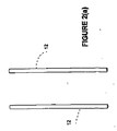
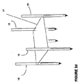
図2(a)−2(d)に示されるように、2つ又はそれ以上の単極電極12、1つ又はそれ以上の双極電極14、又は電極アレイ16を用いることができる。1つの実施形態において、少なくとも第1及び第2の単極電極12は、患者のBPH組織部位に又はその付近に導入されるように構成される。3つ又はそれ以上の単極電極12を用いることができることが認識される。電極アレイ16は、BPH組織部位に対して実質的に取り囲む状態になるように構成される。電極アレイ16は、テンプレート17を用いて、電極の各々を位置決めし、及び/又は保持することができる。テンプレート17は、電極アレイ16の幾何形状を維持することができる。医師が電極の配置及び深さを決定することができる。図3に示すように、電極アレイ16は、BPH組織部位の周囲に境界を形成して、細胞壊死の体積測定領域を形成する。本質的には、電極アレイ16は、治療領域を電極アレイ16からの範囲にし、内向きの方向に延びる。電極アレイ16は、所定の幾何形状を有することができ、関連する電極の各々は、経皮的か又は患者にインサイチュで移植された状態のいずれかで、BPH組織部位に個々に又は同時に配備することができる。
Referring to FIG. 1, one embodiment of the present invention provides a system, generally indicated as 10, for treating a patient's BPH tissue site.
As shown in FIGS. 2 (a)-2 (d), two or more
1つの実施形態において、単極電極12は、約5mmから10cmまでの距離だけ分離され、円形断面の幾何形状を有する。モニタリング・プローブ、脂肪吸引のために使用されるもののような吸引プローブ、流体導入プローブ等を含む、1つ又はそれ以上の付加的なプローブ18を備えることができる。各々の双極電極14は、多数の電極バンド20を有することができる。電極バンド20の間隔及び厚さは、電界の形状を最適化するように選択される。1つの実施形態において、間隔は、典型的には、約1mmから5cmまでであり、電極バンド20の厚さは、0.5mmから5cmまでとすることができる。
再び図1を参照すると、電圧パルス発生器22は、電極12、14、及びアレイ16に結合されている。電圧パルス発生器22は、第1及び第2の単極電極12、双極電極14並びにアレイ16間に十分な電気パルスを印加して、BPH組織部位に細胞のエレクトロポレーションを誘起させ、BPH組織部位の細胞壊死を生じさせるように構成されている。しかしながら、印加される電気パルスは、BPH組織部位の大部分に熱による損傷効果をもたらすには不十分である。
In one embodiment, the
Referring again to FIG. 1, the voltage pulse generator 22 is coupled to the
電極12、14及びアレイ16は、各々がケーブルを介して電圧パルス発生器22に接続されている。スイッチング装置24を含むことができる。ソフトウェアを有するスイッチング装置24は、多数の電極12、14及びアレイ16の同時又は個々の作動を与える。スイッチング装置24は、電圧パルス発生器22に結合されている。1つの実施形態において、BPH組織部位に対して選択されたパターンで予め選択された電極12、14及びアレイ16間に生じる電界を生じさせるために、電極12、14及びアレイ16を個々に作動させるための手段が提供される。個々の電極12、14及びアレイ16間の電気信号のスイッチングは、これらに限定されるものではないが、手動で、機械的に、電気的に、プログラム化されたデジタル・コンピュータにより制御された回路等によってといった様々な異なる手段で達成することができる。1つの実施形態において、各々の電極12、14及びアレイ16は、個々に制御される。
BPH組織部位における細胞の細胞膜を恒久的に破壊するために、ある持続時間及び大きさでパルスが印加される。細胞にわたる電圧に対する、BPH組織部位における細胞を通る電流の比を検出することができ、次いで、BPH組織部位への印加電圧の大きさは、電圧に対する電流の比の変化により調整される。
The
In order to permanently break the cell membrane of the cells at the BPH tissue site, a pulse is applied for a certain duration and magnitude. The ratio of the current through the cells at the BPH tissue site to the voltage across the cells can be detected, and then the magnitude of the applied voltage to the BPH tissue site is adjusted by the change in the ratio of current to voltage.
1つの実施形態において、BPH組織部位における細胞のエレクトロポレーションの開始は、電流を測定することによって検出される。別の実施形態において、BPH組織部位における細胞の細胞膜上のエレクトロポレーションの効果のモニタリングがモニタリングされる。モニタリングは、超音波、CTスキャン、MRI等を用いて画像モニタリングによって実行することができる。
他の実施形態において、モニタリングは、モニタリング電極18を用いて達成される。1つの実施形態において、モニタリング電極18は、モニタリング針への優先電流の流れを防止するために用いることができる高インピーダンス針である。高インピーダンス針は、BPH組織部位に隣接して又はその内部に、すなわち重要な位置に位置決めされる。これは、熱的モニタリングにおいて熱電対を配置するのと同様な概念及び位置決めである。供給される全エレクトロポレーション・パルスの前に、提案された全エレクトロポレーション・パルスのわずかである「試験パルス」が供給され、このパルスは、一例に過ぎず、限定するものではないが、10%等とすることができる。この試験パルスは、不可逆的エレクトロポレーションを生じさせない範囲内であることが好ましい。
モニタリング電極18は、その位置の試験電圧を測定する。次に、測定された電圧は、その関係が線形であるので、1つの実施形態においては、例えば、10を掛けるといった、全パルスの間のモニタリング電極18によって確認されるものへまた外挿される。BPH組織部位における可能性のある合併症についてモニタリングする場合には、不可逆的エレクトロポレーションの知られているレベルの下になる電圧の外挿は、モニタリングが行われているBPH組織部位が安全であることを示す。エレクトロポレーションの妥当性についてそのBPH組織部位においてモニタリングする場合には、この外挿は、組織の不可逆的エレクトロポレーションに適している電圧の知られているレベルの上になる。
In one embodiment, the onset of electroporation of cells at the BPH tissue site is detected by measuring current. In another embodiment, monitoring of the effect of electroporation on the cell membrane of cells at the BPH tissue site is monitored. Monitoring can be performed by image monitoring using ultrasound, CT scan, MRI, or the like.
In other embodiments, monitoring is accomplished using monitoring electrodes 18. In one embodiment, the monitoring electrode 18 is a high impedance needle that can be used to prevent preferential current flow to the monitoring needle. The high impedance needle is positioned adjacent to or within the BPH tissue site, i.e., at a critical location. This is a similar concept and positioning as placing a thermocouple in thermal monitoring. A "test pulse", which is a fraction of the proposed total electroporation pulse, is supplied before the total electroporation pulse delivered, this pulse is only an example and is not limiting, It can be 10%. This test pulse is preferably within a range that does not cause irreversible electroporation.
The monitoring electrode 18 measures the test voltage at that position. The measured voltage is then extrapolated to that identified by the monitoring electrode 18 during the entire pulse, in one embodiment, for example, multiplying by 10, since the relationship is linear. When monitoring for possible complications at a BPH tissue site, extrapolation of the voltage below a known level of irreversible electroporation is safe for the BPH tissue site being monitored. Indicates that there is. When monitoring at the BPH tissue site for the validity of electroporation, this extrapolation is above a known level of voltage suitable for irreversible electroporation of the tissue.
双極電極14が経直腸的に配置される1つの実施形態においては、モニタリング電極18は、双極電極14に不可欠なものであり、作動状態の双極電極14に対して遠位か又は近位のいずれかに配置される。モニタリング電極18は、双極電極14から一定の距離である。別の実施形態においては、モニタリング電極18は、双極電極14が配置されているシース上に取り付けられる。次に、双極電極14からの距離は、画像形成及び直腸粘膜のようなモニタリングされる構造体に基づいて変化させ、位置決めすることができる。別の実施形態においては、モニタリング電極18は、双極電極14が配置されている生検ガイド上に取り付けられる。モニタリング電極18は、ガイドの先端に配置され、双極電極14が配置されているときに、直腸粘膜に当接する。
BPH組織部位における細胞の細胞膜上のエレクトロポレーションの効果は、電流を測定することによって検出することができる。
In one embodiment where the
The effect of electroporation on the cell membrane of cells at the BPH tissue site can be detected by measuring the current.
種々の実施形態において、エレクトロポレーションは、リアルタイム・モニタリングによって、BPH組織部位の細胞の細胞膜において制御された細孔形成を与えて、電気インピーダンス等のモニタリングにより、周囲の組織を保持しながらBPH組織部位の細胞において組織効果をもたらすように、制御された方法で実行される。
エレクトロポレーションは、印加電圧の強度及び持続時間を制御することによる制御された方法で、リアルタイム制御の有無にかかわらず実行することができる。これに加えて、エレクトロポレーションは、細胞膜にわたる質量輸送の調整及び制御を与えるように実行される。制御された方法でのエレクトロポレーションの実行は、電圧の大きさの適切な選択、電圧印加時間の適切な選択等の選択によって達成することができる。
In various embodiments, electroporation provides controlled pore formation in the cell membrane of cells at the BPH tissue site by real-time monitoring, and maintains the surrounding tissue by monitoring electrical impedance or the like while retaining the surrounding tissue. Performed in a controlled manner to produce tissue effects in the cells at the site.
Electroporation can be performed with or without real-time control in a controlled manner by controlling the strength and duration of the applied voltage. In addition to this, electroporation is performed to provide coordination and control of mass transport across the cell membrane. Performing electroporation in a controlled manner can be accomplished by selection such as appropriate selection of voltage magnitude, appropriate selection of voltage application time, and the like.
システム10は、BPH組織部位の温度を制御するように機能する制御盤26を含むことができる。本発明の1つの実施形態において、制御盤26は、コントローラからそのプログラムを受け取る。プログラミングは、コントローラ28にパーソナル・コンピュータが使用される場合には、C又はBASIC(登録商標)のようなコンピュータ言語、或いは、コントローラ28にマイクロプロセッサが使用される場合には、アセンブリ言語の形とすることができる。ユーザ指定の温度の制御は、コントローラ28においてプログラムすることができる。
コントローラ28は、コンピュータ、デジタル又はアナログ処理装置、プログラム可能論理アレイ、配線論理回路、特定用途向け集積回路(ASIC)、或いは他の適切な装置を含むことができる。1つの実施形態において、コントローラ28は、要求とおりに、適切なRAM又はROMモジュール付きのマイクロプロセッサを含む。コントローラ28をユーザ・インタフェース30に結合して、ユーザとデータを交換することができる。ユーザは、電極12、14及びアレイ16に印加される望ましいパルシング・パターン及び対応する温度プロフィールを入力するためにユーザ・インタフェース30を作動することができる。
The system 10 can include a control board 26 that functions to control the temperature of the BPH tissue site. In one embodiment of the present invention, the control board 26 receives the program from the controller. Programming may be in the form of a computer language such as C or BASIC® if a personal computer is used for the
The
一例として、ユーザ・インタフェース30は、文字数字キーパッド、タッチ・スクリーン、コンピュータ・マウス、プッシュ・ボタン式及び/又はトグル・スイッチ、或いは人間のユーザから入力を受け取るための別の適切なコンポーネントを含むことができる。ユーザ・インタフェース30は、CRTスクリーン、LEDスクリーン、LCDスクリーン、液晶ディスプレイ、プリンタ、ディスプレイ・パネル、音響スピーカー、又は人間のユーザにデータを送るための別の適切なコンポーネントをさらに含むことができる。制御盤26は、コントローラの入力を受け取るように機能することができ、電圧パルス発生器22によって駆動することができる。
種々の実施形態において、電圧パルス発生器22は、各々のパルスが約5マイクロ秒から約62秒まで、90マイクロ秒から110マイクロ秒まで、100マイクロ秒等の持続時間だけ印加されるように構成される。これらに限定されるものではないが、約1個から15個までのパルス、持続時間毎に約100マイクロ秒の約8個のパルス等を含む、様々な異なるパルス数を印加することができる。1つの実施形態において、パルスは、約50ボルト/cmから約8000ボルト/cmまでの範囲内でBPH組織部位において電圧勾配を生じさせるように印加される。
By way of example, the user interface 30 includes a alphanumeric keypad, a touch screen, a computer mouse, push button and / or toggle switches, or another suitable component for receiving input from a human user. be able to. The user interface 30 may further include a CRT screen, LED screen, LCD screen, liquid crystal display, printer, display panel, acoustic speaker, or another suitable component for sending data to a human user. The control board 26 can function to receive controller inputs and can be driven by the voltage pulse generator 22.
In various embodiments, the voltage pulse generator 22 is configured such that each pulse is applied for a duration of about 5 microseconds to about 62 seconds, 90 microseconds to 110 microseconds, such as 100 microseconds. Is done. A variety of different pulse numbers can be applied including, but not limited to, about 1 to 15 pulses, about 8 pulses of about 100 microseconds per duration, and the like. In one embodiment, the pulse is applied to create a voltage gradient at the BPH tissue site within a range from about 50 volts / cm to about 8000 volts / cm.
種々の実施形態において、BPH組織部位において摂氏100度又はそれより下、BPH組織部位において摂氏75度又はそれより下、BPH組織部位において摂氏60度又はそれより下、BPH組織部位において摂氏50度又はそれより下等に温度を維持するために、BPH組織部位がモニタリングされ、パルスが調整される。BPH組織部位への熱的効果の発生を最小にするために、温度が制御される。これらの温度は、温度に基づいて電流対電圧の比を調節することによって制御することができる。
本発明の1つの実施形態においては、システム10は、BPH組織部位における細胞のエレクトロポレーションによりBPHを治療して、尿管の周りのBPH組織部位に細胞壊死を生じさせるように使用される。システム10は、BPH組織部位における筋繊維及び神経に沿ってエレクトロポレーション・パルスを供給し、尿管の周りのBPH組織部位にある量の壊死細胞を生成する。筋繊維の緊張を上昇させる、これらの神経の破壊もまた実現される。結果としてもたらされる壊死組織はマクロファージにより除去される。本発明と併せてエレクトロポレーションを用いることにより、BPH組織部位における細胞、関連する神経の除去がもたらされ、BPH組織部位の総体積が減少して、尿道に対する圧力の減少及び前立腺の弛緩が生じる。エレクトロポレーションは、尿道括約筋及び前立腺並びに、隣接する組織及び器官における他の組織を取っておくように制御して適用される。
第1及び第2の単極電極12又はそれ以上、双極電極14、或いは電極アレイ16は、患者の直腸壁、腹膜、又は尿道を通して導入される。エレクトロポレーションは、超音波、CTスキャン、MRI等、又はモニタリング電極18を用いた画像モニタリングによって位置決めされ、モニタリングされる。電極12、14又はアレイ16の各々は絶縁部分を有することができ、電圧パルス発生器22に接続されている。
In various embodiments, 100 degrees Celsius or less at the BPH tissue site, 75 degrees Celsius or less at the BPH tissue site, 60 degrees Celsius or less at the BPH tissue site, 50 degrees Celsius at the BPH tissue site, or To maintain the temperature below it, the BPH tissue site is monitored and the pulse is adjusted. The temperature is controlled to minimize the occurrence of thermal effects on the BPH tissue site. These temperatures can be controlled by adjusting the current to voltage ratio based on the temperature.
In one embodiment of the invention, the system 10 is used to treat BPH by electroporation of cells at the BPH tissue site to cause cell necrosis at the BPH tissue site around the ureter. System 10 delivers electroporation pulses along the muscle fibers and nerves at the BPH tissue site, producing a quantity of necrotic cells at the BPH tissue site around the ureter. The destruction of these nerves, which increases muscle fiber tension, is also realized. The resulting necrotic tissue is removed by macrophages. The use of electroporation in conjunction with the present invention results in the removal of cells and associated nerves at the BPH tissue site, reducing the total volume of the BPH tissue site, reducing pressure on the urethra and prostate relaxation. Arise. Electroporation is applied in a controlled manner to reserve other tissues in the urethral sphincter and prostate and adjacent tissues and organs.
First and second
(例1)
BPH組織部位の領域を画像形成する。患者の直腸壁を通して鋭くされた遠位端を有する2つの双極電極12をBPH組織部位に導入する。アブレーションされるBPH組織部位領域を2つの電極12間に位置決めする。画像形成を用いて、単極電極が適切に配置されることを確認する。2つの単極電極を、BPH組織部位の種々の位置において約5mmから10cmまでの距離だけ分離する。各々5マイクロ秒から約62秒までの持続時間でパルスを印加する。超音波を用いてモニタリングを行う。BPH組織部位をモニタリングする。モニタリングに応じて、摂氏100度を超えない温度を維持するように、パルスを調整する。約50ボルト/cmから約1000ボルト/cmまでの範囲内のBPH組織部位における電圧勾配を生成する。約1cm×0.5cmのBPH組織部位の体積が、細胞壊死を受ける。
(Example 1)
Image the region of the BPH tissue site. Two
(例2)
BPH組織部位の領域を画像形成する。2つの単極電極12を患者の尿管を通してBPH組織部位に導入する。アブレーションされるBPH組織部位領域を2つの単極電極12間に位置決めする。画像形成を用いて、単極電極が適切に配置されることを確認する。2つの単極電極12を、BPH組織部位の種々の位置において約5mmから10cmまでの距離だけ分離する。各々90マイクロ秒から約110秒までの持続時間でパルスを印加する。CTスキャンを用いてモニタリングを行う。BPH組織部位をモニタリングする。モニタリングに応じて、摂氏75度を超えない温度を維持するように、パルスを調整する。約50ボルト/cmから約5000ボルト/cmまでの範囲内のBPH組織部位における電圧勾配を生成する。BPH組織部位の体積が、細胞壊死を受ける。
(Example 2)
Image the region of the BPH tissue site. Two
(例3)
BPH組織部位の領域を画像形成する。電極アレイ16を患者の腹膜を通してBPH組織部位に導入する。電極アレイ16は、BPHに対して取り囲む状態で位置決めする。画像形成を用いて、電極が適切に配置されることを確認する。各々約100マイクロ秒の持続時間でパルスを印加する。モニタリング電極18を用いる。供給される全エレクトロポレーション・パルスの前に、提案された全エレクトロポレーション・パルスの約10%の試験パルスを供給する。この試験パルスは、不可逆的エレクトロポレーションを生じさせるものではない。BPH組織部位領域をモニタリングする。モニタリングに応じて、摂氏60度を超えない温度を維持するように、パルスを調整する。約50ボルト/cmから約8000ボルト/cmまでの範囲内のBPH組織部位における電圧勾配を生成する。BPH組織部位の体積が、細胞壊死を受ける。
(Example 3)
Image the region of the BPH tissue site. The
(例4)
BPH組織部位領域を画像形成する。患者の直腸壁を通して、鋭くされた遠位端を有する、単一の双極電極14をBPH組織部位に導入する。モニタリング電極18を生体ガイドの先端に配置して、双極電極14が配置されるときに直腸粘膜に当接させる。画像形成を用いて、双極電極14が適切に配置されることを確認する。各々約5マイクロ秒から約62秒の持続時間でパルスを印加する。超音波を用いてモニタリングを行う。BPH組織部位をモニタリングする。モニタリングに応じて、摂氏100度を超えない温度を維持するように、パルスを調整する。約50ボルト/cmから約1000ボルト/cmまでの範囲内のBPH組織部位における電圧勾配を生成する。BPH組織部位の体積が、細胞壊死を受ける。
(Example 4)
The BPH tissue site region is imaged. A single
(例5)
BPH組織部位領域を画像形成する。患者の直腸壁を通して電極アレイ16をBPH組織部位に導入し、BPH組織部位の周りに位置決めする。画像形成を用いて、電極アレイ16が適切に配置されることを確認する。各々約90マイクロ秒から約110マイクロ秒の持続時間でパルスを印加する。CTスキャンを用いてモニタリングを行う。BPH組織部位をモニタリングする。モニタリングに応じて、摂氏75度を超えない温度を維持するように、パルスを調整する。約50ボルト/cmから約5000ボルト/cmまでの範囲内のBPH組織部位における電圧勾配を生成する。BPH組織部位の体積が、細胞壊死を受ける。
(Example 5)
The BPH tissue site region is imaged. An
(例6)
BPH組織部位領域を画像形成する。患者の腹膜を通して電極アレイ16をBPH組織部位に導入し、BPH組織部位に対して取り囲む状態で位置決めする。画像形成を用いて、電極アレイ16が適切に配置されることを確認する。各々約100マイクロ秒の持続時間でパルスを印加する。モニタリング電極18を用いる。供給される全エレクトロポレーション・パルスの前に、提案された全エレクトロポレーション・パルスの約10%の試験パルスを供給する。この試験パルスは、不可逆的エレクトロポレーションを生じさせるものではない。BPH組織部位領域をモニタリングする。モニタリングに応じて、摂氏60度を超えない温度を維持するように、パルスを調整する。約50ボルト/cmから約8000ボルト/cmまでの範囲内のBPH組織部位における電圧勾配を生成する。BPH組織部位の体積が、細胞壊死を受ける。
本発明の実施形態の上記の説明は、例示及び説明の目的のために呈示された。排他的であるか又は本発明を開示された正確な形態に限定することが意図されるものではない。明らかに、多くの修正及び変形が当業者には明らかであろう。本発明の範囲は、特許請求の範囲及びそれらの同等物によって定義されることが意図される。
(Example 6)
The BPH tissue site region is imaged. The
The foregoing descriptions of embodiments of the present invention have been presented for purposes of illustration and description. It is not intended to be exhaustive or to limit the invention to the precise form disclosed. Obviously, many modifications and variations will be apparent to practitioners skilled in this art. It is intended that the scope of the invention be defined by the claims and their equivalents.
Claims (148)
患者の前立腺のBPH組織部位に又はその付近に導入されるように構成された少なくとも第1及び第2の単極電極と、
前記第1及び第2の単極電極に結合され、前記BPH組織部位に細胞のエレクトロポレーションを誘起させて、該BPH組織部位の細胞壊死を生じさせるには十分であるが、該BPH組織部位の大部分に熱による損傷効果をもたらすには不十分な電気パルスを該第1及び第2の単極電極間に印加するように構成された電圧パルス発生器と、
を含むことを特徴とするシステム。 A system for treating benign prostatic hyperplasia (BPH) of the prostate,
At least first and second monopolar electrodes configured to be introduced at or near a BPH tissue site of a patient's prostate;
Coupled to the first and second monopolar electrodes and sufficient to induce cell electroporation at the BPH tissue site to cause cell necrosis of the BPH tissue site; A voltage pulse generator configured to apply an electrical pulse between the first and second monopolar electrodes that is insufficient to cause a thermal damage effect to a majority of
A system characterized by including.
患者の前立腺のBPH組織部位に又はその付近に導入されるように構成された双極電極と、
前記第1及び第2の単極電極に結合され、前記BPH組織部位に細胞のエレクトロポレーションを誘起させて、該BPH組織部位の細胞壊死を生じさせるには十分であるが、該BPH組織部位の大部分に熱による損傷効果をもたらすには不十分な電気パルスを前記双極電極に印加するように構成された電圧パルス発生器と、
を含むことを特徴とするシステム。 A system for treating benign prostatic hyperplasia (BPH) of the prostate,
A bipolar electrode configured to be introduced at or near a BPH tissue site in a patient's prostate;
Coupled to the first and second monopolar electrodes and sufficient to induce cell electroporation at the BPH tissue site to cause cell necrosis of the BPH tissue site; A voltage pulse generator configured to apply an electrical pulse to the bipolar electrode that is insufficient to cause a thermal damage effect on a majority of
A system characterized by including.
少なくとも第1及び第2の単極電極を患者のBPH組織部位に導入し、
前記BPH組織部位に又はその付近に前記少なくとも第1及び第2の単極電極を位置決めし、
前記BPH組織部位において細胞のエレクトロポレーションを生じさせるのに十分な量で、かつ該BPH組織部位の大部分に熱損傷をもたらすより少ない量で、制御された方法により電界を該BPH組織部位に印加する
ことを含むことを特徴とする方法。 A method for treating benign prostatic hyperplasia of the prostate,
Introducing at least first and second monopolar electrodes into the patient's BPH tissue site;
Positioning the at least first and second monopolar electrodes at or near the BPH tissue site;
An electric field is applied to the BPH tissue site in a controlled manner in an amount sufficient to cause electroporation of cells at the BPH tissue site and in a lesser amount that causes thermal damage to the majority of the BPH tissue site. Applying the method.
前記BPH組織部位において摂氏100度又はそれより下の温度を維持するように前記パルスを調整する
ことをさらに含むことを特徴とする請求項70に記載の方法。 Monitoring the temperature of the BPH tissue site;
71. The method of claim 70, further comprising adjusting the pulse to maintain a temperature of 100 degrees Celsius or less at the BPH tissue site.
前記BPH組織部位において摂氏75度又はそれより下の温度を維持するように前記パルスを調整する
ことをさらに含むことを特徴とする請求項70に記載の方法。 Monitoring the temperature of the BPH tissue site;
71. The method of claim 70, further comprising adjusting the pulse to maintain a temperature at or below 75 degrees Celsius at the BPH tissue site.
前記BPH組織部位において摂氏60度又はそれより下の温度を維持するように前記パルスを調整する
ことをさらに含むことを特徴とする請求項70に記載の方法。 Monitoring the temperature of the BPH tissue site;
71. The method of claim 70, further comprising adjusting the pulse to maintain a temperature of 60 degrees Celsius or less at the BPH tissue site.
前記BPH組織部位において摂氏50度又はそれより下の温度を維持するように前記パルスを調整する
ことをさらに含むことを特徴とする請求項70に記載の方法。 Monitoring the temperature of the BPH tissue site;
71. The method of claim 70, further comprising adjusting the pulse to maintain a temperature of 50 degrees Celsius or less at the BPH tissue site.
患者のBPH組織部位に双極電極を導入し、
前記BPH組織部位に又はその付近に前記双極電極を位置決めし、
前記BPH組織部位において細胞のエレクトロポレーションを生じさせるのに十分な量で、かつ該BPH組織部位の大部分に熱損傷をもたらすより少ない量で、制御された方法により電界を該BPH組織部位に印加する
ことを含むことを特徴とする方法。 A method for treating benign prostatic hyperplasia of the prostate,
Introducing a bipolar electrode into the patient's BPH tissue site,
Positioning the bipolar electrode at or near the BPH tissue site;
An electric field is applied to the BPH tissue site in a controlled manner in an amount sufficient to cause electroporation of cells at the BPH tissue site and in a lesser amount that causes thermal damage to the majority of the BPH tissue site. Applying the method.
前記BPH組織部位において摂氏100度又はそれより下の温度を維持するように前記パルスを調整する
ことをさらに含むことを特徴とする請求項105に記載の方法。 Monitoring the temperature of the BPH tissue site;
106. The method of claim 105, further comprising adjusting the pulse to maintain a temperature of 100 degrees Celsius or less at the BPH tissue site.
前記BPH組織部位において摂氏75度又はそれより下の温度を維持するように前記パルスを調整する
ことをさらに含むことを特徴とする請求項105に記載の方法。 Monitoring the temperature of the BPH tissue site;
106. The method of claim 105, further comprising adjusting the pulse to maintain a temperature of 75 degrees Celsius or less at the BPH tissue site.
前記BPH組織部位において摂氏60度又はそれより下の温度を維持するように前記パルスを調整する
ことをさらに含むことを特徴とする請求項105に記載の方法。 Monitoring the temperature of the BPH tissue site;
106. The method of claim 105, further comprising adjusting the pulse to maintain a temperature of 60 degrees Celsius or less at the BPH tissue site.
前記BPH組織部位において摂氏50度又はそれより下の温度を維持するように前記パルスを調整する
ことをさらに含むことを特徴とする請求項105に記載の方法。 Monitoring the temperature of the BPH tissue site;
106. The method of claim 105, further comprising adjusting the pulse to maintain a temperature of 50 degrees Celsius or less at the BPH tissue site.
Applications Claiming Priority (2)
| Application Number | Priority Date | Filing Date | Title |
|---|---|---|---|
| US11/166,974 US8114070B2 (en) | 2005-06-24 | 2005-06-24 | Methods and systems for treating BPH using electroporation |
| PCT/US2006/021812 WO2007001751A1 (en) | 2005-06-24 | 2006-06-05 | Methods and systems for treating bph using electroporation |
Publications (2)
| Publication Number | Publication Date |
|---|---|
| JP2008546474A true JP2008546474A (en) | 2008-12-25 |
| JP2008546474A5 JP2008546474A5 (en) | 2009-07-23 |
Family
ID=37568577
Family Applications (1)
| Application Number | Title | Priority Date | Filing Date |
|---|---|---|---|
| JP2008518194A Pending JP2008546474A (en) | 2005-06-24 | 2006-06-05 | Method and system for treating BPH using electroporation |
Country Status (5)
| Country | Link |
|---|---|
| US (2) | US8114070B2 (en) |
| EP (1) | EP1898993A1 (en) |
| JP (1) | JP2008546474A (en) |
| CA (1) | CA2612530A1 (en) |
| WO (1) | WO2007001751A1 (en) |
Families Citing this family (66)
| Publication number | Priority date | Publication date | Assignee | Title |
|---|---|---|---|---|
| US6994706B2 (en) * | 2001-08-13 | 2006-02-07 | Minnesota Medical Physics, Llc | Apparatus and method for treatment of benign prostatic hyperplasia |
| AU2004311842C1 (en) | 2003-12-24 | 2011-01-06 | The Regents Of The University Of California | Tissue ablation with irreversible electroporation |
| US7601149B2 (en) | 2005-03-07 | 2009-10-13 | Boston Scientific Scimed, Inc. | Apparatus for switching nominal and attenuated power between ablation probes |
| US9549739B2 (en) | 2005-05-20 | 2017-01-24 | Neotract, Inc. | Devices, systems and methods for treating benign prostatic hyperplasia and other conditions |
| US7645286B2 (en) | 2005-05-20 | 2010-01-12 | Neotract, Inc. | Devices, systems and methods for retracting, lifting, compressing, supporting or repositioning tissues or anatomical structures |
| US7758594B2 (en) | 2005-05-20 | 2010-07-20 | Neotract, Inc. | Devices, systems and methods for treating benign prostatic hyperplasia and other conditions |
| US8628542B2 (en) | 2005-05-20 | 2014-01-14 | Neotract, Inc. | Median lobe destruction apparatus and method |
| US10195014B2 (en) | 2005-05-20 | 2019-02-05 | Neotract, Inc. | Devices, systems and methods for treating benign prostatic hyperplasia and other conditions |
| US8668705B2 (en) | 2005-05-20 | 2014-03-11 | Neotract, Inc. | Latching anchor device |
| US10925587B2 (en) | 2005-05-20 | 2021-02-23 | Neotract, Inc. | Anchor delivery system |
| US8603106B2 (en) | 2005-05-20 | 2013-12-10 | Neotract, Inc. | Integrated handle assembly for anchor delivery system |
| CA2632970C (en) * | 2006-01-03 | 2012-01-24 | Steven W. Kovalcheck | System for dissociation and removal of proteinaceous tissue |
| US20080132885A1 (en) * | 2006-12-01 | 2008-06-05 | Boris Rubinsky | Methods for treating tissue sites using electroporation |
| US20090076502A1 (en) * | 2007-09-14 | 2009-03-19 | Lazure Technologies, Llc. | Prostate cancer ablation |
| US11272979B2 (en) | 2008-04-29 | 2022-03-15 | Virginia Tech Intellectual Properties, Inc. | System and method for estimating tissue heating of a target ablation zone for electrical-energy based therapies |
| US11254926B2 (en) | 2008-04-29 | 2022-02-22 | Virginia Tech Intellectual Properties, Inc. | Devices and methods for high frequency electroporation |
| US9283051B2 (en) | 2008-04-29 | 2016-03-15 | Virginia Tech Intellectual Properties, Inc. | System and method for estimating a treatment volume for administering electrical-energy based therapies |
| US10238447B2 (en) | 2008-04-29 | 2019-03-26 | Virginia Tech Intellectual Properties, Inc. | System and method for ablating a tissue site by electroporation with real-time monitoring of treatment progress |
| US8992517B2 (en) | 2008-04-29 | 2015-03-31 | Virginia Tech Intellectual Properties Inc. | Irreversible electroporation to treat aberrant cell masses |
| US9867652B2 (en) | 2008-04-29 | 2018-01-16 | Virginia Tech Intellectual Properties, Inc. | Irreversible electroporation using tissue vasculature to treat aberrant cell masses or create tissue scaffolds |
| US10117707B2 (en) | 2008-04-29 | 2018-11-06 | Virginia Tech Intellectual Properties, Inc. | System and method for estimating tissue heating of a target ablation zone for electrical-energy based therapies |
| US10702326B2 (en) | 2011-07-15 | 2020-07-07 | Virginia Tech Intellectual Properties, Inc. | Device and method for electroporation based treatment of stenosis of a tubular body part |
| US8926606B2 (en) | 2009-04-09 | 2015-01-06 | Virginia Tech Intellectual Properties, Inc. | Integration of very short electric pulses for minimally to noninvasive electroporation |
| US9198733B2 (en) | 2008-04-29 | 2015-12-01 | Virginia Tech Intellectual Properties, Inc. | Treatment planning for electroporation-based therapies |
| US10245098B2 (en) | 2008-04-29 | 2019-04-02 | Virginia Tech Intellectual Properties, Inc. | Acute blood-brain barrier disruption using electrical energy based therapy |
| US10272178B2 (en) | 2008-04-29 | 2019-04-30 | Virginia Tech Intellectual Properties Inc. | Methods for blood-brain barrier disruption using electrical energy |
| CA2722296A1 (en) | 2008-04-29 | 2009-11-05 | Virginia Tech Intellectual Properties, Inc. | Irreversible electroporation to create tissue scaffolds |
| CN102421386A (en) * | 2009-03-31 | 2012-04-18 | 安吉戴尼克公司 | System and method for estimating a treatment region for a medical treatment device and for interactively planning a treatment of a patient |
| US11382681B2 (en) | 2009-04-09 | 2022-07-12 | Virginia Tech Intellectual Properties, Inc. | Device and methods for delivery of high frequency electrical pulses for non-thermal ablation |
| US11638603B2 (en) | 2009-04-09 | 2023-05-02 | Virginia Tech Intellectual Properties, Inc. | Selective modulation of intracellular effects of cells using pulsed electric fields |
| WO2010138919A2 (en) | 2009-05-28 | 2010-12-02 | Angiodynamics, Inc. | System and method for synchronizing energy delivery to the cardiac rhythm |
| US9895189B2 (en) | 2009-06-19 | 2018-02-20 | Angiodynamics, Inc. | Methods of sterilization and treating infection using irreversible electroporation |
| US20110118729A1 (en) * | 2009-11-13 | 2011-05-19 | Alcon Research, Ltd | High-intensity pulsed electric field vitrectomy apparatus with load detection |
| US20110135626A1 (en) * | 2009-12-08 | 2011-06-09 | Alcon Research, Ltd. | Localized Chemical Lysis of Ocular Tissue |
| US20110144562A1 (en) * | 2009-12-14 | 2011-06-16 | Alcon Research, Ltd. | Localized Pharmacological Treatment of Ocular Tissue Using High-Intensity Pulsed Electrical Fields |
| WO2011081897A1 (en) * | 2009-12-15 | 2011-07-07 | Alcon Research, Ltd. | High-intensity pulsed electric field vitrectomy apparatus |
| US20110202052A1 (en) * | 2010-02-12 | 2011-08-18 | Daniel Gelbart | System for treating benign prostatic hyperplasia |
| US8546979B2 (en) | 2010-08-11 | 2013-10-01 | Alcon Research, Ltd. | Self-matching pulse generator with adjustable pulse width and pulse frequency |
| US20120089009A1 (en) * | 2010-10-11 | 2012-04-12 | Omary Reed A | Methods and apparatus to deliver nanoparticles to tissue using electronanotherapy |
| US9700368B2 (en) | 2010-10-13 | 2017-07-11 | Angiodynamics, Inc. | System and method for electrically ablating tissue of a patient |
| WO2012088149A2 (en) | 2010-12-20 | 2012-06-28 | Virginia Tech Intellectual Properties, Inc. | High-frequency electroporation for cancer therapy |
| US9078665B2 (en) | 2011-09-28 | 2015-07-14 | Angiodynamics, Inc. | Multiple treatment zone ablation probe |
| US10292801B2 (en) | 2012-03-29 | 2019-05-21 | Neotract, Inc. | System for delivering anchors for treating incontinence |
| US10130353B2 (en) | 2012-06-29 | 2018-11-20 | Neotract, Inc. | Flexible system for delivering an anchor |
| US10154869B2 (en) | 2013-08-02 | 2018-12-18 | Gary M. Onik | System and method for creating radio-frequency energy electrical membrane breakdown for tissue ablation |
| CA2932765A1 (en) | 2013-12-05 | 2015-06-11 | Rfemb Holdings, Llc | Cancer immunotherapy by radiofrequency electrical membrane breakdown (rf-emb) |
| EP3143124A4 (en) | 2014-05-12 | 2018-01-17 | Virginia Tech Intellectual Properties, Inc. | Selective modulation of intracellular effects of cells using pulsed electric fields |
| WO2016100325A1 (en) | 2014-12-15 | 2016-06-23 | Virginia Tech Intellectual Properties, Inc. | Devices, systems, and methods for real-time monitoring of electrophysical effects during tissue treatment |
| US10271893B2 (en) | 2014-12-15 | 2019-04-30 | Medtronic Ablation Frontiers Llc | Timed energy delivery |
| KR102128856B1 (en) | 2015-01-30 | 2020-07-02 | 알에프이엠비 홀딩스, 엘엘씨 | system for ablating undesirable soft tissue in a living subject using radio frequency electrical membrane breakdown |
| WO2016126778A1 (en) * | 2015-02-04 | 2016-08-11 | Rfemb Holdings, Llc | Radio-frequency electrical membrane breakdown for the treatment of benign prostatic hyperplasia |
| CN109069624A (en) | 2016-01-15 | 2018-12-21 | 瑞美控股有限责任公司 | The immunization therapy of cancer |
| CN114098949B (en) | 2016-06-27 | 2023-12-19 | 盖能适治疗股份有限公司 | Generator and catheter with electrode |
| US10905492B2 (en) | 2016-11-17 | 2021-02-02 | Angiodynamics, Inc. | Techniques for irreversible electroporation using a single-pole tine-style internal device communicating with an external surface electrode |
| US11717337B2 (en) * | 2016-11-29 | 2023-08-08 | St. Jude Medical, Cardiology Division, Inc. | Electroporation systems and catheters for electroporation systems |
| US11045648B2 (en) | 2017-11-14 | 2021-06-29 | Boston Scientific Scimed, Inc. | Irreversible electroporation through a combination of substance injection and electrical field application |
| US11607537B2 (en) | 2017-12-05 | 2023-03-21 | Virginia Tech Intellectual Properties, Inc. | Method for treating neurological disorders, including tumors, with electroporation |
| SG11202005766XA (en) | 2017-12-23 | 2020-07-29 | Neotract Inc | Expandable tissue engagement apparatus and method |
| JP2021508533A (en) | 2017-12-26 | 2021-03-11 | ギャラリー,インコーポレイテッド | Optimizing energy delivery for a variety of applications |
| CN111615371B (en) | 2018-01-23 | 2024-02-27 | 波士顿科学国际有限公司 | Enhanced needle array and treatment for tumor ablation |
| US11311329B2 (en) | 2018-03-13 | 2022-04-26 | Virginia Tech Intellectual Properties, Inc. | Treatment planning for immunotherapy based treatments using non-thermal ablation techniques |
| US11925405B2 (en) | 2018-03-13 | 2024-03-12 | Virginia Tech Intellectual Properties, Inc. | Treatment planning system for immunotherapy enhancement via non-thermal ablation |
| US11950835B2 (en) | 2019-06-28 | 2024-04-09 | Virginia Tech Intellectual Properties, Inc. | Cycled pulsing to mitigate thermal damage for multi-electrode irreversible electroporation therapy |
| CN114945338A (en) | 2019-10-15 | 2022-08-26 | 波士顿科学国际有限公司 | Control system and user interface for an ablation system |
| JP2024509644A (en) | 2021-04-07 | 2024-03-04 | ビーティーエル メディカル デベロップメント エー.エス. | Pulsed field ablation device and method |
| IL309432A (en) | 2021-07-06 | 2024-02-01 | Btl Medical Dev A S | Pulsed field ablation device and method |
Citations (7)
| Publication number | Priority date | Publication date | Assignee | Title |
|---|---|---|---|---|
| US20010044596A1 (en) * | 2000-05-10 | 2001-11-22 | Ali Jaafar | Apparatus and method for treatment of vascular restenosis by electroporation |
| JP2002095760A (en) * | 2000-04-06 | 2002-04-02 | Varian Medical Systems Inc | Multipurpose template and needle for delivery and monitoring of multiple minimally dilation therapy |
| US20030060856A1 (en) * | 2001-08-13 | 2003-03-27 | Victor Chornenky | Apparatus and method for treatment of benign prostatic hyperplasia |
| JP2003520574A (en) * | 1999-07-21 | 2003-07-08 | ザ・レジェンツ・オブ・ザ・ユニバーシティー・オブ・カリフォルニア | Electrical impedance tomography for controlling electroporation |
| US20040059389A1 (en) * | 2002-08-13 | 2004-03-25 | Chornenky Victor I. | Apparatus and method for the treatment of benign prostatic hyperplasia |
| JP2004525726A (en) * | 2001-05-10 | 2004-08-26 | リタ メディカル システムズ インコーポレイテッド | RF tissue resection device and method |
| JP2004532654A (en) * | 2000-05-22 | 2004-10-28 | マーク・アンド・カンパニー・インコーポレイテッド | Drug administration system performance evaluation system and method |
Family Cites Families (132)
| Publication number | Priority date | Publication date | Assignee | Title |
|---|---|---|---|---|
| US1653819A (en) | 1926-08-07 | 1927-12-27 | Northcott Ephraim | Electrotherapeutical apparatus |
| DE863111C (en) | 1951-07-03 | 1953-01-15 | Walter Hallegger | Instrument for transcutaneous and subcutaneous heating and iontophoresis and method of its use |
| US4016886A (en) | 1974-11-26 | 1977-04-12 | The United States Of America As Represented By The United States Energy Research And Development Administration | Method for localizing heating in tumor tissue |
| US4407943A (en) | 1976-12-16 | 1983-10-04 | Millipore Corporation | Immobilized antibody or antigen for immunoassay |
| CH617590A5 (en) | 1977-05-27 | 1980-06-13 | Carba Ag | |
| DE2800039C2 (en) | 1978-01-02 | 1984-06-20 | Horst Dr.Med. 6700 Ludwigshafen Kief | Acupuncture device |
| US5385544A (en) | 1992-08-12 | 1995-01-31 | Vidamed, Inc. | BPH ablation method and apparatus |
| US5370675A (en) | 1992-08-12 | 1994-12-06 | Vidamed, Inc. | Medical probe device and method |
| GB8408529D0 (en) | 1984-04-03 | 1984-05-16 | Health Lab Service Board | Concentration of biological particles |
| US4946793A (en) | 1986-05-09 | 1990-08-07 | Electropore, Inc. | Impedance matching for instrumentation which electrically alters vesicle membranes |
| US5098843A (en) | 1987-06-04 | 1992-03-24 | Calvin Noel M | Apparatus for the high efficiency transformation of living cells |
| US5389069A (en) | 1988-01-21 | 1995-02-14 | Massachusetts Institute Of Technology | Method and apparatus for in vivo electroporation of remote cells and tissue |
| ATE131081T1 (en) | 1988-01-21 | 1995-12-15 | Massachusetts Inst Technology | MOLECULAR TRANSPORT THROUGH TISSUES USING ELECTROPORATION. |
| EP0346513A1 (en) | 1988-06-15 | 1989-12-20 | Etama Ag | Assembly for electrotherapy |
| ES2012944A6 (en) | 1989-01-09 | 1990-04-16 | Tomas Justribo Jose Ramon | A device for the administration of medication by iontopheresis for local - regional treatment. |
| CS275292B2 (en) | 1989-02-22 | 1992-02-19 | Cvut Fakulta Elektrotechnick | Private rehabilitation apparatus with ion transcutaneous acceleration |
| DE4000893A1 (en) | 1990-01-15 | 1991-07-18 | Bosch Gmbh Robert | Multichannel appts. for electro-simulation - provides several current circuits for patient with electrodes applying pulse signals |
| US5134070A (en) | 1990-06-04 | 1992-07-28 | Casnig Dael R | Method and device for cell cultivation on electrodes |
| US5193537A (en) | 1990-06-12 | 1993-03-16 | Zmd Corporation | Method and apparatus for transcutaneous electrical cardiac pacing |
| US5052391A (en) | 1990-10-22 | 1991-10-01 | R.F.P., Inc. | High frequency high intensity transcutaneous electrical nerve stimulator and method of treatment |
| US5389096A (en) * | 1990-12-18 | 1995-02-14 | Advanced Cardiovascular Systems | System and method for percutaneous myocardial revascularization |
| US5173158A (en) | 1991-07-22 | 1992-12-22 | Schmukler Robert E | Apparatus and methods for electroporation and electrofusion |
| US5328451A (en) | 1991-08-15 | 1994-07-12 | Board Of Regents, The University Of Texas System | Iontophoretic device and method for killing bacteria and other microbes |
| US5425752A (en) | 1991-11-25 | 1995-06-20 | Vu'nguyen; Dung D. | Method of direct electrical myostimulation using acupuncture needles |
| US6210402B1 (en) | 1995-11-22 | 2001-04-03 | Arthrocare Corporation | Methods for electrosurgical dermatological treatment |
| US6500173B2 (en) | 1992-01-07 | 2002-12-31 | Ronald A. Underwood | Methods for electrosurgical spine surgery |
| US6132419A (en) | 1992-05-22 | 2000-10-17 | Genetronics, Inc. | Electroporetic gene and drug therapy |
| US5318563A (en) | 1992-06-04 | 1994-06-07 | Valley Forge Scientific Corporation | Bipolar RF generator |
| US5273525A (en) | 1992-08-13 | 1993-12-28 | Btx Inc. | Injection and electroporation apparatus for drug and gene delivery |
| US5634899A (en) | 1993-08-20 | 1997-06-03 | Cortrak Medical, Inc. | Simultaneous cardiac pacing and local drug delivery method |
| GB9226376D0 (en) | 1992-12-18 | 1993-02-10 | British Tech Group | Tomography |
| JPH08506259A (en) | 1993-02-02 | 1996-07-09 | ヴィーダメッド インコーポレイテッド | Transurethral needle excision device and method |
| US5792187A (en) | 1993-02-22 | 1998-08-11 | Angeion Corporation | Neuro-stimulation to control pain during cardioversion defibrillation |
| US5403311A (en) | 1993-03-29 | 1995-04-04 | Boston Scientific Corporation | Electro-coagulation and ablation and other electrotherapeutic treatments of body tissue |
| FR2703253B1 (en) | 1993-03-30 | 1995-06-23 | Centre Nat Rech Scient | APPLICATOR OF ELECTRIC PULSES FOR TREATING BIOLOGICAL TISSUES. |
| US5439440A (en) | 1993-04-01 | 1995-08-08 | Genetronics, Inc. | Electroporation system with voltage control feedback for clinical applications |
| US5702359A (en) | 1995-06-06 | 1997-12-30 | Genetronics, Inc. | Needle electrodes for mediated delivery of drugs and genes |
| US6832996B2 (en) | 1995-06-07 | 2004-12-21 | Arthrocare Corporation | Electrosurgical systems and methods for treating tissue |
| US5464437A (en) | 1993-07-08 | 1995-11-07 | Urologix, Inc. | Benign prostatic hyperplasia treatment catheter with urethral cooling |
| US5533999A (en) | 1993-08-23 | 1996-07-09 | Refractec, Inc. | Method and apparatus for modifications of visual acuity by thermal means |
| US5458625A (en) | 1994-05-04 | 1995-10-17 | Kendall; Donald E. | Transcutaneous nerve stimulation device and method for using same |
| US5836905A (en) | 1994-06-20 | 1998-11-17 | Lemelson; Jerome H. | Apparatus and methods for gene therapy |
| JPH11505435A (en) | 1995-01-17 | 1999-05-21 | ヘールライン・クリストフ | Balloon catheter for preventing restenosis of blood vessels after angioplasty and method for constructing the balloon catheter |
| US5720921A (en) | 1995-03-10 | 1998-02-24 | Entremed, Inc. | Flow electroporation chamber and method |
| US5810762A (en) | 1995-04-10 | 1998-09-22 | Genetronics, Inc. | Electroporation system with voltage control feedback for clinical applications |
| US6041252A (en) | 1995-06-07 | 2000-03-21 | Ichor Medical Systems Inc. | Drug delivery system and method |
| US6607529B1 (en) | 1995-06-19 | 2003-08-19 | Medtronic Vidamed, Inc. | Electrosurgical device |
| US5919142A (en) | 1995-06-22 | 1999-07-06 | Btg International Limited | Electrical impedance tomography method and apparatus |
| US5983131A (en) | 1995-08-11 | 1999-11-09 | Massachusetts Institute Of Technology | Apparatus and method for electroporation of tissue |
| EP0776678A1 (en) | 1995-11-30 | 1997-06-04 | Hewlett-Packard Company | System for administering transcutaneous cardiac pacing with transcutaneous electrical nerve stimuli |
| US6010613A (en) | 1995-12-08 | 2000-01-04 | Cyto Pulse Sciences, Inc. | Method of treating materials with pulsed electrical fields |
| US6090106A (en) | 1996-01-09 | 2000-07-18 | Gyrus Medical Limited | Electrosurgical instrument |
| US6016452A (en) | 1996-03-19 | 2000-01-18 | Kasevich; Raymond S. | Dynamic heating method and radio frequency thermal treatment |
| US5778894A (en) | 1996-04-18 | 1998-07-14 | Elizabeth Arden Co. | Method for reducing human body cellulite by treatment with pulsed electromagnetic energy |
| SE509241C2 (en) | 1996-07-18 | 1998-12-21 | Radinvent Ab | Devices for electrodynamic radiation therapy of tumor diseases |
| US6102885A (en) | 1996-08-08 | 2000-08-15 | Bass; Lawrence S. | Device for suction-assisted lipectomy and method of using same |
| US6106521A (en) | 1996-08-16 | 2000-08-22 | United States Surgical Corporation | Apparatus for thermal treatment of tissue |
| US5991697A (en) | 1996-12-31 | 1999-11-23 | The Regents Of The University Of California | Method and apparatus for optical Doppler tomographic imaging of fluid flow velocity in highly scattering media |
| WO1998033451A1 (en) | 1997-02-04 | 1998-08-06 | National Aeronautics And Space Administration | Multimodality instrument for tissue characterization |
| US5873849A (en) | 1997-04-24 | 1999-02-23 | Ichor Medical Systems, Inc. | Electrodes and electrode arrays for generating electroporation inducing electrical fields |
| US6085115A (en) | 1997-05-22 | 2000-07-04 | Massachusetts Institite Of Technology | Biopotential measurement including electroporation of tissue surface |
| US5978704A (en) | 1997-06-03 | 1999-11-02 | Uab Research Foundation | Method and apparatus for treating cardiac arrhythmia |
| US6216034B1 (en) | 1997-08-01 | 2001-04-10 | Genetronics, Inc. | Method of programming an array of needle electrodes for electroporation therapy of tissue |
| US6055453A (en) | 1997-08-01 | 2000-04-25 | Genetronics, Inc. | Apparatus for addressing needle array electrodes for electroporation therapy |
| US5999847A (en) | 1997-10-21 | 1999-12-07 | Elstrom; John A. | Apparatus and method for delivery of surgical and therapeutic agents |
| US20020055731A1 (en) | 1997-10-24 | 2002-05-09 | Anthony Atala | Methods for promoting cell transfection in vivo |
| US6208893B1 (en) | 1998-01-27 | 2001-03-27 | Genetronics, Inc. | Electroporation apparatus with connective electrode template |
| US6009347A (en) | 1998-01-27 | 1999-12-28 | Genetronics, Inc. | Electroporation apparatus with connective electrode template |
| US5947142A (en) * | 1998-01-30 | 1999-09-07 | Pgi International, Ltd. | Breakaway coupling |
| US6440127B2 (en) | 1998-02-11 | 2002-08-27 | Cosman Company, Inc. | Method for performing intraurethral radio-frequency urethral enlargement |
| CA2229528A1 (en) | 1998-02-13 | 1999-08-13 | Shailesh Mehta | Apparatus and method for analyzing particles |
| KR100304197B1 (en) * | 1998-03-30 | 2001-11-30 | 윤종용 | Method for manufacturing silicon on insulator |
| SE513814C2 (en) | 1998-03-31 | 2000-11-06 | Aditus Medical Ab | Device for the treatment of diseases with electric fields |
| US6219577B1 (en) | 1998-04-14 | 2001-04-17 | Global Vascular Concepts, Inc. | Iontophoresis, electroporation and combination catheters for local drug delivery to arteries and other body tissues |
| US6159163A (en) | 1998-05-07 | 2000-12-12 | Cedars-Sinai Medical Center | System for attenuating pain during bone marrow aspiration and method |
| CA2331532A1 (en) | 1998-05-08 | 1999-11-18 | Genetronics, Inc. | Electrically induced vessel vasodilation |
| EP2428250A1 (en) | 1998-07-13 | 2012-03-14 | Genetronics, Inc. | Skin and muscle-targeted gene therapy by pulsed electrical field |
| US6212433B1 (en) | 1998-07-28 | 2001-04-03 | Radiotherapeutics Corporation | Method for treating tumors near the surface of an organ |
| EP1102559B1 (en) | 1998-09-30 | 2003-06-04 | Cygnus, Inc. | Method and device for predicting physiological values |
| US6611706B2 (en) | 1998-11-09 | 2003-08-26 | Transpharma Ltd. | Monopolar and bipolar current application for transdermal drug delivery and analyte extraction |
| AU1727400A (en) | 1998-11-16 | 2000-06-05 | United States Surgical Corporation | Apparatus for thermal treatment of tissue |
| US6090016A (en) * | 1998-11-18 | 2000-07-18 | Kuo; Hai Pin | Collapsible treader with enhanced stability |
| US6351674B2 (en) | 1998-11-23 | 2002-02-26 | Synaptic Corporation | Method for inducing electroanesthesia using high frequency, high intensity transcutaneous electrical nerve stimulation |
| US6261831B1 (en) | 1999-03-26 | 2001-07-17 | The United States Of America As Represented By The Secretary Of The Air Force | Ultra-wide band RF-enhanced chemotherapy for cancer treatmeat |
| US6738663B2 (en) | 1999-04-09 | 2004-05-18 | Oncostim, A Minnesota Corporation | Implantable device and method for the electrical treatment of cancer |
| US6627421B1 (en) | 1999-04-13 | 2003-09-30 | Imarx Therapeutics, Inc. | Methods and systems for applying multi-mode energy to biological samples |
| US7053063B2 (en) | 1999-07-21 | 2006-05-30 | The Regents Of The University Of California | Controlled electroporation and mass transfer across cell membranes in tissue |
| US6300108B1 (en) | 1999-07-21 | 2001-10-09 | The Regents Of The University Of California | Controlled electroporation and mass transfer across cell membranes |
| US6927049B2 (en) | 1999-07-21 | 2005-08-09 | The Regents Of The University Of California | Cell viability detection using electrical measurements |
| US6403348B1 (en) | 1999-07-21 | 2002-06-11 | The Regents Of The University Of California | Controlled electroporation and mass transfer across cell membranes |
| US6326177B1 (en) * | 1999-08-04 | 2001-12-04 | Eastern Virginia Medical School Of The Medical College Of Hampton Roads | Method and apparatus for intracellular electro-manipulation |
| US20020010491A1 (en) | 1999-08-04 | 2002-01-24 | Schoenbach Karl H. | Method and apparatus for intracellular electro-manipulation |
| US20030078499A1 (en) | 1999-08-12 | 2003-04-24 | Eppstein Jonathan A. | Microporation of tissue for delivery of bioactive agents |
| US6613211B1 (en) | 1999-08-27 | 2003-09-02 | Aclara Biosciences, Inc. | Capillary electrokinesis based cellular assays |
| JP4676042B2 (en) * | 1999-10-01 | 2011-04-27 | 帝國製薬株式会社 | Topical analgesic / anti-inflammatory patch containing felbinac |
| US6493592B1 (en) | 1999-12-01 | 2002-12-10 | Vertis Neuroscience, Inc. | Percutaneous electrical therapy system with electrode position maintenance |
| KR100375657B1 (en) | 2000-06-21 | 2003-03-15 | 주식회사 몸앤맘 | Apparatus and method for eliminating a fat mass in human body |
| JP2002033538A (en) * | 2000-07-13 | 2002-01-31 | Mitsubishi Electric Corp | Semiconductor laser excitation solid-state laser device |
| US6669691B1 (en) | 2000-07-18 | 2003-12-30 | Scimed Life Systems, Inc. | Epicardial myocardial revascularization and denervation methods and apparatus |
| CA2416581A1 (en) * | 2000-07-25 | 2002-04-25 | Rita Medical Systems, Inc. | Apparatus for detecting and treating tumors using localized impedance measurement |
| US6795728B2 (en) | 2001-08-17 | 2004-09-21 | Minnesota Medical Physics, Llc | Apparatus and method for reducing subcutaneous fat deposits by electroporation |
| US6892099B2 (en) | 2001-02-08 | 2005-05-10 | Minnesota Medical Physics, Llc | Apparatus and method for reducing subcutaneous fat deposits, virtual face lift and body sculpturing by electroporation |
| US6697670B2 (en) | 2001-08-17 | 2004-02-24 | Minnesota Medical Physics, Llc | Apparatus and method for reducing subcutaneous fat deposits by electroporation with improved comfort of patients |
| US6702808B1 (en) | 2000-09-28 | 2004-03-09 | Syneron Medical Ltd. | Device and method for treating skin |
| US20050043726A1 (en) | 2001-03-07 | 2005-02-24 | Mchale Anthony Patrick | Device II |
| EP1377680B1 (en) | 2001-04-12 | 2011-10-05 | Imperial Innovations Limited | Diagnosis and treatment of breast cancer based upon scn5a |
| WO2002087692A1 (en) | 2001-04-26 | 2002-11-07 | The Procter & Gamble Company | A method and apparatus for the treatment of cosmetic skin conditioins |
| US6832111B2 (en) | 2001-07-06 | 2004-12-14 | Hosheng Tu | Device for tumor diagnosis and methods thereof |
| EP1429678B1 (en) | 2001-09-28 | 2006-03-22 | Rita Medical Systems, Inc. | Impedance controlled tissue ablation apparatus |
| AU2002362441A1 (en) | 2001-10-01 | 2003-04-14 | Am Discovery, Incorporated | Devices for treating atrial fibrilation |
| FR2830767B1 (en) | 2001-10-12 | 2004-03-12 | Optis France Sa | DEVICE FOR DELIVERING DRUGS BY IONTOPHORESIS OR INTROCULAR ELECTROPORATION |
| RU2323017C2 (en) | 2001-10-24 | 2008-04-27 | Пауэр Пэйпер Лтд. | Method and device for carrying out controlled delivery of active substances into skin |
| US6807444B2 (en) | 2001-11-05 | 2004-10-19 | Hosheng Tu | Apparatus and methods for monitoring tissue impedance |
| WO2003047684A2 (en) | 2001-12-04 | 2003-06-12 | University Of Southern California | Method for intracellular modifications within living cells using pulsed electric fields |
| AU2003220143A1 (en) | 2002-03-11 | 2003-09-29 | Altea Therapeutics Corporation | Transdermal integrated actuator device, methods of making and using same |
| US6912417B1 (en) | 2002-04-05 | 2005-06-28 | Ichor Medical Systmes, Inc. | Method and apparatus for delivery of therapeutic agents |
| US8175711B2 (en) | 2002-04-08 | 2012-05-08 | Ardian, Inc. | Methods for treating a condition or disease associated with cardio-renal function |
| US7653438B2 (en) | 2002-04-08 | 2010-01-26 | Ardian, Inc. | Methods and apparatus for renal neuromodulation |
| US20030204161A1 (en) * | 2002-04-25 | 2003-10-30 | Bozidar Ferek-Petric | Implantable electroporation therapy device and method for using same |
| US6780178B2 (en) | 2002-05-03 | 2004-08-24 | The Board Of Trustees Of The Leland Stanford Junior University | Method and apparatus for plasma-mediated thermo-electrical ablation |
| US6801804B2 (en) | 2002-05-03 | 2004-10-05 | Aciont, Inc. | Device and method for monitoring and controlling electrical resistance at a tissue site undergoing iontophoresis |
| ES2289307T3 (en) | 2002-05-06 | 2008-02-01 | Covidien Ag | BLOOD DETECTOR TO CONTROL AN ELECTROCHIRURGICAL UNIT. |
| EP1515775A4 (en) | 2002-05-07 | 2010-03-03 | Oncostim Inc | Method and device for treating concer with electrical therapy in conjunction with chemotherapeutic agents and radiation therapy |
| US7063698B2 (en) | 2002-06-14 | 2006-06-20 | Ncontact Surgical, Inc. | Vacuum coagulation probes |
| US6972014B2 (en) | 2003-01-04 | 2005-12-06 | Endocare, Inc. | Open system heat exchange catheters and methods of use |
| US7211083B2 (en) | 2003-03-17 | 2007-05-01 | Minnesota Medical Physics, Llc | Apparatus and method for hair removal by electroporation |
| AU2004311842C1 (en) | 2003-12-24 | 2011-01-06 | The Regents Of The University Of California | Tissue ablation with irreversible electroporation |
| US20050261672A1 (en) | 2004-05-18 | 2005-11-24 | Mark Deem | Systems and methods for selective denervation of heart dysrhythmias |
| US7261710B2 (en) | 2004-10-13 | 2007-08-28 | Medtronic, Inc. | Transurethral needle ablation system |
| US20060264752A1 (en) | 2005-04-27 | 2006-11-23 | The Regents Of The University Of California | Electroporation controlled with real time imaging |
| JP4841346B2 (en) * | 2006-02-16 | 2011-12-21 | 日本碍子株式会社 | Electron emitter |
| US20080052786A1 (en) * | 2006-08-24 | 2008-02-28 | Pei-Cheng Lin | Animal Model of Prostate Cancer and Use Thereof |
-
2005
- 2005-06-24 US US11/166,974 patent/US8114070B2/en active Active
-
2006
- 2006-06-05 CA CA002612530A patent/CA2612530A1/en not_active Abandoned
- 2006-06-05 JP JP2008518194A patent/JP2008546474A/en active Pending
- 2006-06-05 WO PCT/US2006/021812 patent/WO2007001751A1/en active Application Filing
- 2006-06-05 EP EP06772212A patent/EP1898993A1/en not_active Withdrawn
-
2009
- 2009-07-27 US US12/510,011 patent/US20090292342A1/en not_active Abandoned
Patent Citations (7)
| Publication number | Priority date | Publication date | Assignee | Title |
|---|---|---|---|---|
| JP2003520574A (en) * | 1999-07-21 | 2003-07-08 | ザ・レジェンツ・オブ・ザ・ユニバーシティー・オブ・カリフォルニア | Electrical impedance tomography for controlling electroporation |
| JP2002095760A (en) * | 2000-04-06 | 2002-04-02 | Varian Medical Systems Inc | Multipurpose template and needle for delivery and monitoring of multiple minimally dilation therapy |
| US20010044596A1 (en) * | 2000-05-10 | 2001-11-22 | Ali Jaafar | Apparatus and method for treatment of vascular restenosis by electroporation |
| JP2004532654A (en) * | 2000-05-22 | 2004-10-28 | マーク・アンド・カンパニー・インコーポレイテッド | Drug administration system performance evaluation system and method |
| JP2004525726A (en) * | 2001-05-10 | 2004-08-26 | リタ メディカル システムズ インコーポレイテッド | RF tissue resection device and method |
| US20030060856A1 (en) * | 2001-08-13 | 2003-03-27 | Victor Chornenky | Apparatus and method for treatment of benign prostatic hyperplasia |
| US20040059389A1 (en) * | 2002-08-13 | 2004-03-25 | Chornenky Victor I. | Apparatus and method for the treatment of benign prostatic hyperplasia |
Also Published As
| Publication number | Publication date |
|---|---|
| US8114070B2 (en) | 2012-02-14 |
| WO2007001751A1 (en) | 2007-01-04 |
| CA2612530A1 (en) | 2007-01-04 |
| US20090292342A1 (en) | 2009-11-26 |
| US20060293713A1 (en) | 2006-12-28 |
| EP1898993A1 (en) | 2008-03-19 |
Similar Documents
| Publication | Publication Date | Title |
|---|---|---|
| US8114070B2 (en) | Methods and systems for treating BPH using electroporation | |
| US8603087B2 (en) | Methods and systems for treating restenosis using electroporation | |
| JP2008543492A (en) | Method and system for treating tumors using electroporation | |
| US11033321B2 (en) | Tissue ablation with irreversible electroporation | |
| US20120071872A1 (en) | Systems for Treating Tissue Sites Using Electroporation | |
| JP2008543493A (en) | Method and system for treating adipose tissue sites using electroporation | |
| US20080132885A1 (en) | Methods for treating tissue sites using electroporation | |
| AU2006239295B2 (en) | Electroporation controlled with real time imaging | |
| AU2017279765A1 (en) | Tissue ablation with irreversible electroporation |
Legal Events
| Date | Code | Title | Description |
|---|---|---|---|
| A711 | Notification of change in applicant |
Free format text: JAPANESE INTERMEDIATE CODE: A711 Effective date: 20090107 |
|
| A521 | Request for written amendment filed |
Free format text: JAPANESE INTERMEDIATE CODE: A523 Effective date: 20090602 |
|
| A621 | Written request for application examination |
Free format text: JAPANESE INTERMEDIATE CODE: A621 Effective date: 20090602 |
|
| A977 | Report on retrieval |
Free format text: JAPANESE INTERMEDIATE CODE: A971007 Effective date: 20111124 |
|
| A131 | Notification of reasons for refusal |
Free format text: JAPANESE INTERMEDIATE CODE: A131 Effective date: 20111128 |
|
| A02 | Decision of refusal |
Free format text: JAPANESE INTERMEDIATE CODE: A02 Effective date: 20120521 |