JP2007509690A - Long bone head prosthesis to protect cartilage - Google Patents
Long bone head prosthesis to protect cartilage Download PDFInfo
- Publication number
- JP2007509690A JP2007509690A JP2006537551A JP2006537551A JP2007509690A JP 2007509690 A JP2007509690 A JP 2007509690A JP 2006537551 A JP2006537551 A JP 2006537551A JP 2006537551 A JP2006537551 A JP 2006537551A JP 2007509690 A JP2007509690 A JP 2007509690A
- Authority
- JP
- Japan
- Prior art keywords
- head
- long bone
- prosthesis
- prosthetic
- bipolar
- Prior art date
- Legal status (The legal status is an assumption and is not a legal conclusion. Google has not performed a legal analysis and makes no representation as to the accuracy of the status listed.)
- Pending
Links
Images


Classifications
-
- A—HUMAN NECESSITIES
- A61—MEDICAL OR VETERINARY SCIENCE; HYGIENE
- A61F—FILTERS IMPLANTABLE INTO BLOOD VESSELS; PROSTHESES; DEVICES PROVIDING PATENCY TO, OR PREVENTING COLLAPSING OF, TUBULAR STRUCTURES OF THE BODY, e.g. STENTS; ORTHOPAEDIC, NURSING OR CONTRACEPTIVE DEVICES; FOMENTATION; TREATMENT OR PROTECTION OF EYES OR EARS; BANDAGES, DRESSINGS OR ABSORBENT PADS; FIRST-AID KITS
- A61F2/00—Filters implantable into blood vessels; Prostheses, i.e. artificial substitutes or replacements for parts of the body; Appliances for connecting them with the body; Devices providing patency to, or preventing collapsing of, tubular structures of the body, e.g. stents
- A61F2/02—Prostheses implantable into the body
- A61F2/30—Joints
- A61F2/30767—Special external or bone-contacting surface, e.g. coating for improving bone ingrowth
- A61F2/30771—Special external or bone-contacting surface, e.g. coating for improving bone ingrowth applied in original prostheses, e.g. holes or grooves
-
- A—HUMAN NECESSITIES
- A61—MEDICAL OR VETERINARY SCIENCE; HYGIENE
- A61F—FILTERS IMPLANTABLE INTO BLOOD VESSELS; PROSTHESES; DEVICES PROVIDING PATENCY TO, OR PREVENTING COLLAPSING OF, TUBULAR STRUCTURES OF THE BODY, e.g. STENTS; ORTHOPAEDIC, NURSING OR CONTRACEPTIVE DEVICES; FOMENTATION; TREATMENT OR PROTECTION OF EYES OR EARS; BANDAGES, DRESSINGS OR ABSORBENT PADS; FIRST-AID KITS
- A61F2/00—Filters implantable into blood vessels; Prostheses, i.e. artificial substitutes or replacements for parts of the body; Appliances for connecting them with the body; Devices providing patency to, or preventing collapsing of, tubular structures of the body, e.g. stents
- A61F2/02—Prostheses implantable into the body
- A61F2/30—Joints
- A61F2/32—Joints for the hip
- A61F2/36—Femoral heads ; Femoral endoprostheses
- A61F2/3609—Femoral heads or necks; Connections of endoprosthetic heads or necks to endoprosthetic femoral shafts
-
- A—HUMAN NECESSITIES
- A61—MEDICAL OR VETERINARY SCIENCE; HYGIENE
- A61F—FILTERS IMPLANTABLE INTO BLOOD VESSELS; PROSTHESES; DEVICES PROVIDING PATENCY TO, OR PREVENTING COLLAPSING OF, TUBULAR STRUCTURES OF THE BODY, e.g. STENTS; ORTHOPAEDIC, NURSING OR CONTRACEPTIVE DEVICES; FOMENTATION; TREATMENT OR PROTECTION OF EYES OR EARS; BANDAGES, DRESSINGS OR ABSORBENT PADS; FIRST-AID KITS
- A61F2/00—Filters implantable into blood vessels; Prostheses, i.e. artificial substitutes or replacements for parts of the body; Appliances for connecting them with the body; Devices providing patency to, or preventing collapsing of, tubular structures of the body, e.g. stents
- A61F2/02—Prostheses implantable into the body
- A61F2/30—Joints
- A61F2002/30001—Additional features of subject-matter classified in A61F2/28, A61F2/30 and subgroups thereof
- A61F2002/30316—The prosthesis having different structural features at different locations within the same prosthesis; Connections between prosthetic parts; Special structural features of bone or joint prostheses not otherwise provided for
- A61F2002/30535—Special structural features of bone or joint prostheses not otherwise provided for
-
- A—HUMAN NECESSITIES
- A61—MEDICAL OR VETERINARY SCIENCE; HYGIENE
- A61F—FILTERS IMPLANTABLE INTO BLOOD VESSELS; PROSTHESES; DEVICES PROVIDING PATENCY TO, OR PREVENTING COLLAPSING OF, TUBULAR STRUCTURES OF THE BODY, e.g. STENTS; ORTHOPAEDIC, NURSING OR CONTRACEPTIVE DEVICES; FOMENTATION; TREATMENT OR PROTECTION OF EYES OR EARS; BANDAGES, DRESSINGS OR ABSORBENT PADS; FIRST-AID KITS
- A61F2250/00—Special features of prostheses classified in groups A61F2/00 - A61F2/26 or A61F2/82 or A61F9/00 or A61F11/00 or subgroups thereof
- A61F2250/0058—Additional features; Implant or prostheses properties not otherwise provided for
Abstract
【課題】 長骨用体内人工補装具および患者の長骨の頸部骨折を治療するための方法を提供する。
【解決手段】 この長骨用体内人工補装具は、細いステム部位に接続された基本的に球状の頭部領域を有し、この頭部領域が粗面化外表面を有することを特徴とする。この治療方法は、長骨頭部外殻の保持により特徴づけられるものである。PROBLEM TO BE SOLVED: To provide a long bone endoprosthesis and a method for treating a neck fracture of a long bone of a patient.
The long bone endoprosthesis has a basically spherical head region connected to a thin stem region, and the head region has a roughened outer surface. . This treatment method is characterized by the retention of the long bone head shell.
Description
本発明は、新規な型の人工補装具およびこの人工補装具を用いる大腿骨および上腕骨頸部骨折の治療方法に関する。特に、本発明の人工補装具は、長骨の生来の関節軟骨および軟骨下骨が保護されるような処置において長骨頸部骨折を治療するのに使用することができる。 The present invention relates to a novel type of prosthesis and a method for treating femoral and humeral neck fractures using the prosthesis. In particular, the prosthesis of the present invention can be used to treat long bone neck fractures in procedures where the long bones native articular cartilage and subchondral bone are protected.
大腿骨および上腕骨の頸部の骨折は、整形外科医にとって常に大きなチャレンジがつきまとい、その治療方法およびそれにより得られる結果に関する限り、今日においても多くの点で“未解決の骨折”のままとなっている。 Fractures in the neck of the femur and humerus have always been a great challenge for orthopedic surgeons and remain “unresolved fractures” in many respects today, as far as their treatment methods and results are obtained. ing.
米国において、1998年度に約280,000件の股関節部の骨折が発生している。全国骨粗しょう症財団(National Osteoporosis Foundation)は骨粗しょう症による股関節部骨折の治療管理のための健康介護経費が1995年度において総額87億ドルに達したと報告している。この額は骨粗しょう症骨折全体の治療コストの63%であり、全ての骨折介護の費用の43%に相当するものである。更に、2020年までに、米国において股関節部骨折の発病件数が毎年500,000以上新たに発生すると予測されている。更に、これらの患者を治療するための費用が年間160億ドルに達すると予測されている。 In the United States, about 280,000 hip fractures occurred in 1998. The National Osteoporosis Foundation reports that health care costs for the management of hip fracture caused by osteoporosis totaled $ 8.7 billion in 1995. This amount is 63% of the total cost of treatment for osteoporotic fractures and corresponds to 43% of all fracture care costs. Furthermore, by 2020, it is predicted that more than 500,000 new hip fractures will occur each year in the United States. In addition, the cost to treat these patients is projected to reach $ 16 billion annually.
阻血性骨壊死および骨癒合不全の高い割合は、大腿頸部の脱臼骨折における一般的な合併症となっている(ガーデン(Garden)分類の第3および4期)。脱臼骨折でない場合でも、大腿頸部の骨折が満足な方法で治療されるという保証はない。治療上の観点においてこれらの骨折の不確定な性質の重要な理由の1つは、外科医が阻血性骨壊死に対し十分にコントロールできないということである。その理由は大腿頸部骨折に続いて起きる大腿頭への血流障害である。 A high proportion of ischemic osteonecrosis and osteosynthesis failure has become a common complication of femoral neck dislocation fractures (Garden classification stages 3 and 4). Even if it is not a dislocation fracture, there is no guarantee that a femoral neck fracture will be treated in a satisfactory manner. One important reason for the uncertain nature of these fractures from a therapeutic point of view is that surgeons have insufficient control over ischemic osteonecrosis. The reason is a blood flow disturbance to the femoral head that occurs following a femoral neck fracture.
大腿頸部骨折は、通常、完全に関節包内性のものであり、(全ての関節包内性骨折に共通して)骨折部を浸している滑液が癒合プロセスを妨害することがある。更に、大腿頸部が実質的に骨膜層を有していないという事実からして、全ての癒合は骨内において行わなければならない。最後に、滑液中の欠陥形成阻害因子もまた、骨折修復を阻害することがある。これら全ての因子が、前述の大腿頭への不安定な血液供給と一緒になって、癒合を予想困難なものとし、骨癒合不全の可なり高い発生率をもたらすものとなる。 Femoral neck fractures are usually completely intracapsular, and synovial fluid that soaks the fracture (common to all intracapsular fractures) can interfere with the healing process. Furthermore, due to the fact that the femoral neck has substantially no periosteal layer, all fusion must be performed within the bone. Finally, defect formation inhibitors in synovial fluid can also inhibit fracture repair. All these factors, together with the aforementioned unstable blood supply to the femoral head, make fusion difficult to predict and lead to a fairly high incidence of bone union failure.
僅かな治療上の選択が、長骨頸部骨折の治療管理として利用可能である。この選択肢として最も一般的に使用されているものは、以下のものである。 A few therapeutic options are available for treatment management of long bone neck fractures. The most commonly used options are as follows:
1.骨接合術: 骨折固定;および
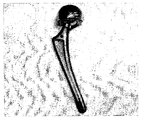
2.半関節形成術: 大腿頭を、一体型ブロック又はモジュール大腿ステム部に取着させた大腿頭(通常、金属)で置換する。最もポピュラーに使用されている人工補装具は、トンプソン(Thompson)、オースチン‐ムア(Austin-Moore)、双極(バイポーラ型)および単極人工補装具である。バイポーラ型の大腿用人工補装具の典型例を図1に示す。
1. Osteosynthesis: fracture fixation; and Hemiarthroplasty: The femoral head is replaced with a femoral head (usually metal) attached to an integral block or module femoral stem. The most commonly used prostheses are Thompson, Austin-Moore, bipolar (bipolar) and monopolar prostheses. A typical example of a bipolar thigh prosthesis is shown in FIG.
多くの外科医は、この第2の選択肢を推奨する。すなわち、一次人工骨頭置換術の使用であり、高年齢の外来患者の治療に使用される。人工補装具の使用は骨癒合不全および阻血性骨壊死の防止に役立つが、それらの使用は他の多くの合併症を伴う。 Many surgeons recommend this second option. That is, it is the use of primary artificial head replacement and is used to treat older outpatients. Although the use of prosthetic devices helps prevent bone union failure and ischemic osteonecrosis, their use is associated with many other complications.
新規の大腿頸部骨折の治療管理で、人工補装具を使用する場合の知られている欠点の1つに、臼蓋窩びらん(侵食)の結果として生じる痛みがある。この合併症はしばしば極めて厳しいものであり、熟練技術者をして以下のようにコメントせざるを得ないようなものである。 One of the known disadvantages of using prosthetic devices in the management of new femoral neck fractures is the pain resulting from acetabular erosion (erosion). This complication is often very severe and requires a skilled technician to comment as follows:
“頭部又は頸部を犠牲にして、金属製の異物で置換をすることは多くの患者にとって正しい処置とは言えない。半分以上のケースにおいて、利用可能な最良の物質は臼蓋窩内にあり、その区別することなく除去することは避けるべきである。”(Salvatore; “Campbell’s Operative Orthopedics”、第9版)。 “Replacement with a metal foreign body at the expense of the head or neck is not the correct treatment for many patients. In more than half of the cases, the best available material is in the acetabulum. Yes, you should avoid removing them without distinction. ”(Salvatore;“ Campbell's Operative Orthopedics ”, 9th edition).
従来の人工補装具の使用に伴う更なる主な問題は、臼蓋窩と、人工補装具頭部との間の不適合から生じる機能不全である。更に、人工補装具の移植後に、その頭部の固い金属と、臼蓋窩のかなり柔らかな表面との間に関節化がなされるという事実から外傷的合併症も発生する。 A further major problem with the use of conventional prosthetic devices is dysfunction resulting from incompatibility between the acetabulum and the prosthetic head. In addition, traumatic complications also arise from the fact that after implantation of the prosthesis, articulation is made between the hard metal of its head and the fairly soft surface of the acetabulum.
本発明の目的は、長骨頸部骨折の治療に使用することができる改善された人工補装具を提供することである。 It is an object of the present invention to provide an improved prosthesis that can be used to treat long bone neck fractures.
本発明の他の目的は、長骨頸部骨折の治療での使用において、長骨関節基端側の2つの生来の関節表面のいずれをも失わせることを要しない人工補装具を提供することである。 Another object of the present invention is to provide a prosthesis that does not require the loss of either of the two native articular surfaces on the proximal side of the long bone joint for use in the treatment of a long bone neck fracture. It is.
本発明の更なる目的は、生来の関節表面を保持しつつ体内人工補装具の使用を可能にする長骨骨折の治療方法を提供することである。 It is a further object of the present invention to provide a method for treating long bone fractures that allows the use of an endoprosthesis while retaining the natural articular surface.
本発明の更なる目的は、従来の人工補装具および治療方法に関連する欠点および問題を克服することができる人工補装具および治療方法を提供することである。 It is a further object of the present invention to provide a prosthesis and treatment method that can overcome the disadvantages and problems associated with conventional prosthesis and treatment methods.
本発明の更なる目的および利点は以下の記載から明らかになるであろう。 Further objects and advantages of the present invention will become apparent from the following description.
大腿骨および上腕頸部の骨折を、新規な体内人工補装具の使用を伴う外科方法により治療することができることを意外にも見出された。この体内人工補装具の頭部(人工骨頭部)は、海綿骨の殆どを除去した後の患者の大腿骨および上腕頭部の外側層を含む外殻(shell)状のキャビティ(洞)内に挿入自在となっている。このようにして、長骨頭の軟骨質関節表面は、その下層の軟骨下骨と共に保存され、長骨の球窩(ボール・ソケット)関節の生来の関節表面が維持される。 It has been unexpectedly found that femoral and cervical fractures can be treated by surgical methods involving the use of novel endoprostheses. The head of this endoprosthesis (artificial bone head) is in a shell-like cavity (sinus) containing the outer layer of the patient's femur and humeral head after most of the cancellous bone has been removed. It can be inserted freely. In this way, the cartilage joint surface of the long bone head is preserved along with the underlying subchondral bone, and the natural joint surface of the long bone ball and socket joint is maintained.
このように、本発明は主として、長骨用体内人工補装具に向けられたものであり、これは実質的に球状の頭部領域を有し、これが細いステム部位に接続されており、この頭部領域が粗面化外表面を有していることを特徴とするものである。 Thus, the present invention is primarily directed to a long bone endoprosthesis, which has a substantially spherical head region that is connected to a thin stem site, The partial region has a roughened outer surface.
本発明の体内人工補装具の好ましい1実施例において、この粗面化された人工補装具の外表面は算術平均粗さ(Rα)が0.05μmから500μmの範囲となっている。より好ましくは、このRαの値は40μmから200μmの範囲とする。最も好ましくは、このRαの値は50μmである。本発明の目的のため、このパラメータRαは国際標準化機構(ISO)468(“表面粗さパラメータ: 要件を特定するためのそれら値および一般的ルール”)に従って定義されるものとする。 In a preferred embodiment of the endoprosthesis according to the present invention, the outer surface of the roughened prosthesis has an arithmetic average roughness (Rα) in the range of 0.05 μm to 500 μm. More preferably, the value of Rα is in the range of 40 μm to 200 μm. Most preferably, the value of Rα is 50 μm. For purposes of the present invention, this parameter Rα shall be defined according to the International Organization for Standardization (ISO) 468 (“Surface Roughness Parameters: Their Values and General Rules for Specifying Requirements”).
他の好ましい実施例において、この粗面化された人工補装具の外表面は、複数のへこみ、線条、スロット、溝、穴、窪みおよび突起からなる群から選択される1又はそれ以上の表面特徴を有する。これらの特徴は、成形/注入成形法、機械的切削および(小さい径の表面突起の場合)グリットブラスト法を含む任意の標準的手法により人工補装具の頭部表面に付加することができる。特に好ましい実施例として、この粗面化された人工補装具の外表面は以下に詳述するように1又はそれ以上の溝又はスロットを備えたものとする。 In another preferred embodiment, the outer surface of the roughened prosthesis is one or more surfaces selected from the group consisting of a plurality of indentations, filaments, slots, grooves, holes, depressions and protrusions. Has characteristics. These features can be added to the prosthetic head surface by any standard technique including molding / injection molding, mechanical cutting and grit blasting (for small diameter surface protrusions). In a particularly preferred embodiment, the roughened prosthetic device outer surface is provided with one or more grooves or slots as detailed below.
本発明にいうところの“長骨”とは主に大腿骨および上腕骨を指すものである。 In the present invention, “long bone” mainly refers to the femur and humerus.
本発明の人工補装具の好ましい1実施例として、人工補装具の頭部および頸部は一体形のユニット(以下、単一ブロック(体)と呼ぶ)として作られる。 As a preferred embodiment of the prosthesis of the present invention, the head and neck of the prosthesis are made as a unitary unit (hereinafter referred to as a single block (body)).
他の好ましい1実施例として、人工補装具の頭部領域および頸部は2個の別々のモジュラユニットとして作られる。本発明のこの形態の特に好ましい実施例において、この別体の頭部は双極(バイポーラ型)人工骨頭部からなるものである。 As another preferred embodiment, the head region and neck of the prosthesis are made as two separate modular units. In a particularly preferred embodiment of this aspect of the invention, the separate head comprises a bipolar (bipolar) artificial bone head.
本発明の他の形態は、人工補装具頭部部材に向けられたものであり、これは実質的に球状体からなり、この先端側に窪みが形成され、人工大腿ステム部がこの窪みに挿入、接続されるようになっており、更に、この体内人工骨頭部が先に定義、説明したような粗面化外表面を有していることを特徴とするものである。 Another aspect of the present invention is directed to the prosthetic head member, which is substantially made of a spherical body, has a recess formed at the distal end, and the artificial femoral stem is inserted into the recess. Furthermore, this body artificial bone head has a roughened outer surface as defined and explained above.
すなわち、本発明の体内人工補装具の好ましい1実施例において、この粗面化された人工補装具の外表面は算術平均粗さ(Rα)が0.05μmから500μmの範囲となっている。より好ましくは、このRαの値は40μmから200μmの範囲とする。最も好ましくは、このRαの値は50μmである。 That is, in one preferred embodiment of the endoprosthesis of the present invention, the outer surface of the roughened prosthesis has an arithmetic average roughness (Rα) in the range of 0.05 μm to 500 μm. More preferably, the value of Rα is in the range of 40 μm to 200 μm. Most preferably, the value of Rα is 50 μm.
他の好ましい実施例において、この粗面化された人工補装具頭部の外表面は、複数のへこみ、線条、スロット、溝、穴、窪みおよび突起からなる群から選択される1又はそれ以上の表面特徴を有する。本発明の1つの好ましい実施例において、上述の表面凹凸形状の平均深さ又は高さは0.05μmから5000μmの範囲となっている。より好ましくは、この平均値は400μmから2000μmの範囲とする。この平均深さ又は高さの最も好まし値は1000μmである。他の好ましい実施例にとして、この外面形状がスロット、溝および穴の場合は、その深さは1μmから、人工補装具頭部の外表面の材料の最大厚みまでの範囲となる。この最大深さの値の場合、人工補装具頭部がスロット、溝、穴、窪みにより実際に穿孔されていて、人工補装具頭部の外表面から、この頭部の内側表面又はキャビティへ連通する通路が形成されることになる。 In another preferred embodiment, the outer surface of the roughened prosthetic head is one or more selected from the group consisting of a plurality of indentations, filaments, slots, grooves, holes, depressions and protrusions. Surface characteristics. In one preferred embodiment of the present invention, the average depth or height of the surface irregularities described above is in the range of 0.05 μm to 5000 μm. More preferably, this average value is in the range of 400 μm to 2000 μm. The most preferred value for this average depth or height is 1000 μm. As another preferred embodiment, if the outer surface shape is a slot, groove or hole, the depth ranges from 1 μm to the maximum thickness of the material on the outer surface of the prosthetic head. For this maximum depth value, the prosthetic head is actually perforated with slots, grooves, holes, and depressions, and communicates from the outer surface of the prosthetic head to the inner surface or cavity of the head. A passage is formed.
本発明の更なる好ましい実施例において、粗面化外側表面を有する前記人工補装具頭部はバイポーラ型の人工補装具頭部として構成され、外側頭部(粗面化外側表面を有する)と、この外側頭部の内面近傍の中間部と、この中間部の内面に連節された内側頭部とを有する。 In a further preferred embodiment of the invention, the prosthetic head having a roughened outer surface is configured as a bipolar prosthetic head, the outer head (having a roughened outer surface), An intermediate portion near the inner surface of the outer head portion and an inner head portion articulated on the inner surface of the intermediate portion.
更に、本発明は、ここに開示し、規定した長骨用人工補装具頭部と、この頭部に接合可能な体内人工大腿ステム部とを具備してなる体内人工補装具システム部を包含するものである。 The present invention further includes an endoprosthesis system portion comprising a long bone prosthesis head disclosed and defined herein and an endoprosthetic femoral stem portion connectable to the head. Is.
他の形態として、本発明は、治療を要する患者の長骨の頸部骨折を治療するための方法にも向けられている。その方法は:
a)前記長骨の頭部から海綿骨の殆ど又は全てを除去し、長骨頭部外殻を形成する段階と;
b)人工補装具ステム部を受入れるための長骨通路を作る段階と;
c)前記人工補装具ステム部を前記長骨通路内に挿入し、適宜、接合させる段階と;
d)長骨用人工補装具頭部を前記長骨頭部外殻内に挿入し、適宜、接合させる段階と;
e)前記長骨頭の窪んだ部分に前記ステム部部分を復位させる段階と;
を具備してなる。
In another form, the present invention is also directed to a method for treating a long neck neck fracture in a patient in need of treatment. Here's how:
a) removing most or all of the cancellous bone from the long bone head to form a long bone head shell;
b) creating a long bone passage for receiving the prosthetic stem portion;
c) inserting the prosthetic stem portion into the long bone passage and appropriately joining;
d) inserting a long bone prosthesis head into the long bone head shell and appropriately joining;
e) repositioning the stem portion to the recessed portion of the long bone head;
It comprises.
本発明は更に、治療を要する患者の長骨の頸部骨折を治療するための方法にも向けられたものであって、その方法は:
a)軟骨および軟骨下骨に障害を生じさせない安全な手法を用いて、人体の本来の位置から生来の長骨頭を除去する段階と;
b)人体外で頭部外殻を手術トレー上に準備する段階と;
c)人工補装具ステム部部位を受入れるための長骨通路を作る段階と;
d)接合剤を用い、又は用いずに、前記人工補装具ステム部部位を前記長骨通路内に挿入する段階と;
e)段階(b)で形成された頭部外殻に前記長骨用人工補装具頭部を接合させる段階と;
f)取着された前記頭部外殻と共に、製造された長骨用人工補装具頭部を治療対象の患者の関節内に復位させ、該長骨用人工補装具頭部の先端部を該人工補装具ステム部部位に接合させる段階と;
を具備してなる。
The present invention is further directed to a method for treating a long neck cervical fracture of a patient in need of treatment, the method comprising:
a) removing the natural long bone head from its natural location using a safe technique that does not damage the cartilage and subchondral bone;
b) preparing the outer shell on the surgical tray outside the human body;
c) creating a long bone passage for receiving the prosthetic stem site;
d) inserting the prosthetic stem portion with or without a bonding agent into the long bone passageway;
e) joining the long bone prosthesis head to the head shell formed in step (b);
f) The produced prosthetic head for the long bone is moved back into the joint of the patient to be treated together with the attached outer shell of the head, and the distal end of the prosthetic head for the long bone is Joining to the prosthesis stem site;
It comprises.
本発明は更に、治療を要する患者の長骨の頸部骨折を治療するための方法にも向けられたものであって、その方法は:
a)軟骨および軟骨下骨に障害を生じさせない安全な手法を用いて、人体の本来の位置から生来の長骨頭を除去する段階と;
b)人体外で頭部外殻を手術トレー上に準備する段階と;
c)単一ブロックの人工補装具のステム部部位を受入れるための長骨通路を作る段階と;
d)接合剤を用い、又は用いずに、前記単一ブロックの人工補装具のステム部部位を前記長骨通路内に挿入する段階と;
e)段階(b)で形成された頭部外殻に前記単一ブロックの長骨用人工補装具頭部部位を接合させる段階と;
f)取着された前記頭部外殻と共に、用意された前記単一ブロックの長骨用人工補装具頭部を治療対象の患者の関節内に復位させる段階と;
を具備してなる。
The present invention is further directed to a method for treating a long neck cervical fracture of a patient in need of treatment, the method comprising:
a) removing the natural long bone head from its natural location using a safe technique that does not damage the cartilage and subchondral bone;
b) preparing the outer shell on the surgical tray outside the human body;
c) creating a long bone passage for receiving a stem portion of a single block prosthesis;
d) inserting the stem portion of the single block prosthesis into the long bone passage with or without a bonding agent;
e) joining the single block long bone prosthesis head portion to the head shell formed in step (b);
f) repositioning the prepared single block long bone prosthesis head with the attached outer shell into the joint of the patient to be treated;
It comprises.
本発明は更に、治療を要する患者の長骨の頸部骨折を治療するための方法にも向けられたものであって、その方法は:
a)前記長骨の頭部から海綿骨の殆ど又は全てを除去し、長骨頭部外殻を形成する段階と;
b)人工補装具ステム部を受入れるための長骨通路を作る段階と;
c)前記人工補装具ステム部を前記長骨通路内に挿入し、適宜、接合させる段階と;
d)段階(a)で形成された頭部外殻に前記バイポーラ型の人工補装具の外側頭部を接合させる段階と;
e)前記人工補装具ステム部部位のトラニオンを人工長骨内側頭部の内側キャビティ内に配置する段階と;
f)前記人工長骨内側頭部を前記人工長骨外側頭部の内側キャビティ内に挿入する段階と;
g)前記バイポーラ型の頭部アッセンブリーを、固定リングを閉じることにより固定する段階と;
を具備してなる。
The present invention is further directed to a method for treating a long neck cervical fracture of a patient in need of treatment, the method comprising:
a) removing most or all of the cancellous bone from the long bone head to form a long bone head shell;
b) creating a long bone passage for receiving the prosthetic stem portion;
c) inserting the prosthetic stem portion into the long bone passage and appropriately joining;
d) joining the outer head of the bipolar prosthesis to the head shell formed in step (a);
e) placing the trunnion at the prosthetic stem portion within the medial cavity of the medial prosthetic long bone;
f) inserting the medial long bone prosthesis into the medial cavity of the prosthetic long bone lateral head;
g) fixing the bipolar head assembly by closing a fixing ring;
It comprises.
本発明は更に、治療を要する患者の長骨の頸部骨折を治療するための方法にも向けられたものであって、その方法は:
a)軟骨および軟骨下骨に障害を生じさせない安全な手法を用いて、人体の本来の位置から生来の長骨頭を除去する段階と;
b)人体外で頭部外殻を手術トレー上に準備する段階と;
c)人工補装具ステム部部位を受入れるための長骨通路を作る段階と;
d)接合剤を用い、又は用いずに、前記人工補装具ステム部部位を前記長骨通路内に挿入する段階と;
e)段階(b)で形成された頭部外殻にバイポーラ型の人工補装具の外側頭部を接合させる段階と;
f)前記人工補装具ステム部部位のトラニオンを人工長骨内側頭部の内側キャビティ内に配置する段階と;
g)前記人工長骨内側頭部を前記人工長骨外側頭部の内側キャビティ内に挿入する段階と;
h)バイポーラ型の頭部アッセンブリーを、固定リングを閉じることにより固定する段階と;
i)取着された前記頭部外殻と共に、用意された前記長骨用人工補装具頭部を治療対象の患者の関節内に復位させる段階と;
を具備してなる。
The present invention is further directed to a method for treating a long neck cervical fracture of a patient in need of treatment, the method comprising:
a) removing the natural long bone head from its natural location using a safe technique that does not damage the cartilage and subchondral bone;
b) preparing the outer shell on the surgical tray outside the human body;
c) creating a long bone passage for receiving the prosthetic stem site;
d) inserting the prosthetic stem portion with or without a bonding agent into the long bone passageway;
e) joining the outer head of the bipolar prosthesis to the head shell formed in step (b);
f) placing the trunnion of the prosthetic stem portion within the medial cavity of the medial prosthetic long bone;
g) inserting the medial long bone prosthesis into the medial cavity of the prosthetic long bone lateral head;
h) fixing the bipolar head assembly by closing the fixing ring;
i) repositioning the prepared long bone prosthesis head with the attached outer shell into the joint of the patient to be treated;
It comprises.
本発明は更に、治療を要する患者の長骨の頸部骨折を治療するための方法にも向けられたものであって、その方法は:
a)軟骨および軟骨下骨に障害を生じさせない安全な手法を用いて、人体の本来の位置から生来の長骨頭を除去する段階と;
b)人体外で頭部外殻を手術トレー上に準備する段階と;
c)単一ブロックバイポーラ型の人工補装具のステム内側部位を受入れるための長骨通路を作る段階と;
d)接合剤を用い、又は用いずに、前記単一ブロックバイポーラ型の人工補装具のステム内側部位を前記長骨通路内に挿入する段階と;
e)段階(b)で形成された頭部外殻にバイポーラ型の人工補装具の外側頭部を接合させる段階と;
f)取着された前記頭部外殻と共に、用意された前記バイポーラ型の人工補装具の外側頭部を治療対象の患者の関節内に復位させる段階と;
g)前記バイポーラ型の人工補装具の内側頭部を前記バイポーラ型の人工補装具の外側頭部の内側キャビティ内に挿入する段階と;
h)前記バイポーラ型の頭部アッセンブリーを、固定リングを閉じることにより固定する段階と;
を具備してなる。
The present invention is further directed to a method for treating a long neck cervical fracture of a patient in need of treatment, the method comprising:
a) removing the natural long bone head from its natural location using a safe technique that does not damage the cartilage and subchondral bone;
b) preparing the outer shell on the surgical tray outside the human body;
c) creating a long bone passage for receiving the inner stem portion of a single block bipolar prosthesis;
d) inserting the stem inner portion of the single-block bipolar prosthesis into the long bone passage with or without a bonding agent;
e) joining the outer head of the bipolar prosthesis to the head shell formed in step (b);
f) repositioning the outer head of the prepared bipolar prosthesis with the attached outer shell into the joint of the patient to be treated;
g) inserting the inner head of the bipolar prosthesis into the inner cavity of the outer head of the bipolar prosthesis;
h) securing the bipolar head assembly by closing a retaining ring;
It comprises.
本発明は更に、治療を要する患者の長骨の頸部骨折を治療するための方法にも向けられたものであって、その方法は:
a)軟骨および軟骨下骨に障害を生じさせない安全な手法を用いて、人体の本来の位置から生来の長骨頭を除去する段階と;
b)人体外で頭部外殻を手術トレー上に準備する段階と;
c)単一ブロックのバイポーラ型の人工補装具のステム内側部位を受入れるための長骨通路を作る段階と;
d)接合剤を用い、又は用いずに、前記単一ブロックのバイポーラ型の人工補装具のステム内側部位を前記長骨通路内に挿入する段階と;
e)前記バイポーラ型の人工補装具の内側頭部を前記バイポーラ型の人工補装具の外側頭部の内側キャビティ内に挿入する段階と;
f)前記バイポーラ型の頭部アッセンブリーを、固定リングを閉じることにより固定する段階と;
g)段階(b)で形成された頭部外殻をバイポーラ型の人工補装具の外側頭部に接合させる段階と;
h)取着された前記バイポーラ型の人工補装具と共に、用意された前記バイポーラ型の人工補装具の外側頭部を治療対象の患者の関節内に復位させる段階と;
を具備してなる。
The present invention is further directed to a method for treating a long neck cervical fracture of a patient in need of treatment, the method comprising:
a) removing the natural long bone head from its natural location using a safe technique that does not damage the cartilage and subchondral bone;
b) preparing the outer shell on the surgical tray outside the human body;
c) creating a long bone passageway for receiving the inner stem portion of a single block bipolar prosthesis;
d) inserting the stem inner portion of the single-block bipolar prosthesis into the long bone passage with or without a bonding agent;
e) inserting the inner head of the bipolar prosthesis into the inner cavity of the outer head of the bipolar prosthesis;
f) fixing the bipolar head assembly by closing a fixing ring;
g) joining the head shell formed in step (b) to the outer head of a bipolar prosthesis;
h) with the attached bipolar prosthesis, repositioning the outer head of the prepared bipolar prosthesis into the joint of the patient to be treated;
It comprises.
本発明は更に、治療を要する患者の長骨の頸部骨折を治療するための方法にも向けられたものであって、その方法は:
a)前記長骨の頭部から海綿骨の殆ど又は全てを除去し、長骨頭部外殻を形成する段階と;
b)単一ブロックバイポーラ型の人工補装具のステム内側部位を受入れるための長骨通路を作る段階と;
c)接合剤を用い、又は用いずに、前記単一ブロックバイポーラ型の人工補装具のステム内側部位を前記長骨通路内に挿入する段階と;
d)前記バイポーラ型の人工補装具の内側頭部を前記バイポーラ型の人工補装具の外側頭部の内側キャビティ内に挿入する段階と;
e)前記バイポーラ型の頭部アッセンブリーを、固定リングを閉じることにより固定する段階と;
f)段階(b)で形成された頭部外殻にバイポーラ型の人工補装具の外側頭部を接合させる段階と;
を具備してなる。
The present invention is further directed to a method for treating a long neck cervical fracture of a patient in need of treatment, the method comprising:
a) removing most or all of the cancellous bone from the long bone head to form a long bone head shell;
b) creating a long bone passage for receiving the inner stem portion of a single block bipolar prosthesis;
c) inserting the stem inner portion of the single block bipolar prosthesis into the long bone passage with or without a bonding agent;
d) inserting the inner head of the bipolar prosthesis into the inner cavity of the outer head of the bipolar prosthesis;
e) fixing the bipolar head assembly by closing a fixing ring;
f) joining the outer head of the bipolar prosthesis to the head shell formed in step (b);
It comprises.
上記治療方法はいずれも大腿骨および上腕頸部の双方の骨折の治療に適用することができる。 Any of the above treatment methods can be applied to the treatment of both femoral and humeral neck fractures.
本発明の前記並びに他の特徴および利点は以下の好ましい態様である例示的、非制限的実施例から明らかになるであろう。 The above and other features and advantages of the present invention will become apparent from the following preferred, illustrative, non-limiting examples.
本発明の体内人工補装具の基本的特徴は、接合すべき頭部領域の最適な機械的接合を可能とし、それにより大腿骨頭又は上腕骨頭の外殻キャビティの内面への最適な機械的接合を可能とすべく、その頭部領域(又は少なくともその一部)が粗面化非研磨外側層を有するという事実である。これは従来の長骨頭およびステム用人工補装具との比較において著しく対照的なものである。つまり、従来の人工補装具では頭部領域が研磨表面を有することが特徴となっているからであり、その目的は生来の大腿骨頭又は上腕骨頭の生来の軟骨質関節表面を置換するためである。球窩(ボール・ソケット)関節の生来の関節表面をそのままにしておくことは、幾つかの臨床的利点、例えば臼蓋窩びらん(侵食)の防止、従来のアプローチで見られたボール・ソケットの不適合の問題の回避、痛みの減少などがもたらされる。これら利点の多くは解剖学上のこのような関節表面保つことが長骨頭と、関節内の窩との間の生来の隙間を保持するという事実からもたらされるものである。長骨頭の関節軟骨を保持し完全な状態を維持するには、この組織を浸している滑液により組織の栄養学的要求が満たされる事実が必要である点に、留意すべきである。 The basic features of the endoprosthesis of the present invention allow for optimal mechanical joining of the head region to be joined, thereby providing optimum mechanical joining to the inner surface of the outer shell cavity of the femoral head or humeral head. The fact is that the head region (or at least part of it) has a roughened non-polished outer layer, if possible. This is in stark contrast to conventional long bone head and stem prostheses. That is, the conventional prosthesis is characterized by the fact that the head region has a polished surface, the purpose of which is to replace the natural cartilage joint surface of the natural femoral head or humeral head. . Keeping the natural articular surface of the ball socket joints has several clinical advantages, such as prevention of acetabular erosion (erosion), ball socket sockets seen with conventional approaches This will help avoid non-conformance problems and reduce pain. Many of these benefits stem from the fact that maintaining such an anatomical joint surface preserves the natural gap between the long bone head and the fossa in the joint. It should be noted that maintaining the articular cartilage of the long bone head and maintaining its integrity requires the fact that the synovial fluid soaking the tissue meets the nutritional requirements of the tissue.
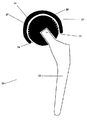
本発明による典型的な人工大腿骨が図2に模式的に示されている。20として示したこの人工補装具は、基本的に2つの部位からなる。すなわち、人工補装具の頭部領域21と、ステム領域22である。この図における側面図から、ステム領域の基端部23が前記頭部領域内の長細い窪み24内に挿入されていることが分かるであろう。患者の関節軟骨およびそれに関連する軟骨下骨を含む大腿骨頭外殻25は、生物学的適合性セメント26により人工補装具頭部領域21に接合されている。本図に示す実施例の人工補装具頭部領域21の表面粗さは、周方向に配列された一連のスロット27により提供されている。図示のように、生物学的適合性セメント26はこれらスロット内に浸透することができ、それにより人工補装具頭部領域21と、大腿骨頭外殻25との間の接合強度を増大させている。
A typical artificial femur according to the present invention is schematically shown in FIG. This prosthetic device shown as 20 basically consists of two parts. That is, the
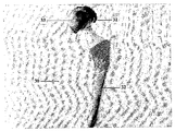
図3は本発明の人工補装具(30として大略的に示す)を写真で示すものであり、その頭部31およびステム32の領域を組立てた後のものである。この頭部31の表面の殆どは頭部31領域を覆う大腿骨頭外殻33により隠されている。
FIG. 3 is a photograph showing the prosthetic device of the present invention (shown schematically as 30), after assembling the region of the
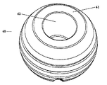
図4は、図3に示す人工補装具の頭領域(40として大略的に示す)であって、ステム領域の挿入前の状態を示している。この頭領域の粗面化外側表面41の僅かな部分が、大腿骨頭外殻42の下位(先端)周辺部下方に延びている。この表面には円形開口部43が穿設され、これに人工補装具のステム領域が挿入されることに留意されたい。
FIG. 4 is a head region (shown schematically as 40) of the prosthetic device shown in FIG. 3 and shows a state before insertion of the stem region. A small portion of the roughened outer surface 41 of this head region extends below the lower (tip) periphery of the
本発明の好ましい1実施例として、この粗面化外表面を有する人工補装具頭部はバイポーラ型の人工補装具のものでもよい。当業者に周知のように、長骨頸部骨折の治療管理に使用されるバイポーラ型の人工補装具は以下の構成部材を有するものである。 As a preferred embodiment of the invention, the prosthetic head having this roughened outer surface may be a bipolar prosthetic. As is well known to those skilled in the art, a bipolar prosthesis used for the treatment management of a long bone neck fracture has the following components.
1. 関節窩(例えば、臼蓋窩)表面と関節化される外側頭部。本発明の場合、この外側頭部は、ここに記載されているように、用意された大腿骨頭への取着を可能にするために、粗面化された外側表面を有する。 1. A lateral head articulated with a glenoid (eg, acetabular) surface. In the case of the present invention, this outer head has a roughened outer surface to allow attachment to a prepared femoral head, as described herein.
2. 外側頭部の内方キャビティ内に固定される中間層又は部分。一般に、この中間部分は、金属よりも柔らかな材質(ポリエチレンなど)から作られる。その他、この部分は外側頭部と同じ材質により構成され、外側頭部との一体的ユニットとしてもよい。(本発明のバイポーラ型の人工補装具の製作において使用される材料の詳細については以下に説明する) 2. An intermediate layer or portion that is secured within the inner cavity of the outer head. In general, this intermediate part is made of a material softer than metal (such as polyethylene). In addition, this part is comprised with the same material as an outer side head, and is good also as an integral unit with an outer side head. (Details of the materials used in the fabrication of the bipolar prosthesis of the present invention are described below)
3. 内側頭部であり、その外側表面は上記中間部分の内面と移動自在に関節化され、その内方キャビティは大腿骨ステムのトラニオンに動かないように固定されている。図8は、外側頭部85の内方キャビティ内での、内側頭部81の中間部分83との連節状態を示している。
3. The inner head, the outer surface of which is articulated movably with the inner surface of the intermediate portion, and its inner cavity is fixed against movement of the femoral stem trunnion. FIG. 8 shows a state of articulation with the intermediate portion 83 of the
4. 固定リングであり、その機能は、外側頭部の内方キャビティ内で内側頭部の前記中間部分との移動自在な接触状態を維持することである。図9は、固定リング97を閉じた後の、内側頭部91と、外側頭部95との相対的配置(下方から見て)を示している。
4). A locking ring, whose function is to maintain a movable contact with the intermediate part of the inner head within the inner cavity of the outer head. FIG. 9 shows the relative arrangement (viewed from below) of the
本発明の他の好ましい実施例として、粗面化された外側表面を有する人工補装具頭部(人工骨頭部)が、単一ブロックのバイポーラ型の人工補装具の一部となっている。この場合、人工補装具の内側頭部および大腿骨ステムは、単一の一体的ユニットとして提供される。前述のバイポーラ型の人工補装具頭部の場合と同様に、この単一ブロックユニットの内側頭部は、外側頭部のキャビティ内で前記中間層との移動自在な接触状態が維持され、前記固定リングにより所定位置にて固定される。 In another preferred embodiment of the invention, a prosthetic head having a roughened outer surface (artificial bone head) is part of a single block bipolar prosthesis. In this case, the prosthetic medial head and femoral stem are provided as a single integral unit. As in the case of the bipolar prosthesis head described above, the inner head of the single block unit is maintained in a movable contact state with the intermediate layer in the cavity of the outer head, and the fixed It is fixed in place by a ring.
本発明の体内人工補装具は、コバルト系合金(例えば、コバルト‐クロム)、チタン、チタン系合金、ステンレス鋼、又はこれら金属の組合せから作製することができる。更に、この体内人工補装具は、非金属材料、例えば生物学的適合性のプラスチック又はポリマー(例えば、ポリウレタン、ポリエチレン)、更に、前述の金属よりも柔らかな生物学的適合性合成材料、およびセラミックなどの硬質材料から構成された部材を含むものであってもよい。この列挙したものは、より一般的な材料の幾つかを例示することを意図したものであり、限定的なものと理解されるべきではない。上述した種々の異なる材料の組合せも、本発明の範疇に包含されるものである。例えば、人工補装具頭部をプラスチック本体と接触させた金属トラニオンで構成してもよい。これら材料の組合せの他の例は、人工補装具頭部の中央部分を金属又は合金で構成し、これに対し、外側部分(大腿骨頭外殻に固定された外側表面および前記金属又は合金に接合された内部表面を有する)を非金属、すなわちプラスチック材料から構成してもよい。 The endoprosthesis of the present invention can be made from a cobalt-based alloy (eg, cobalt-chromium), titanium, a titanium-based alloy, stainless steel, or a combination of these metals. In addition, the endoprosthesis includes non-metallic materials, such as biocompatible plastics or polymers (eg, polyurethane, polyethylene), biocompatible synthetic materials that are softer than the aforementioned metals, and ceramics. The member comprised from hard materials, such as, may be included. This list is intended to illustrate some of the more general materials and should not be construed as limiting. Combinations of the various different materials described above are also encompassed within the scope of the present invention. For example, the prosthetic head may be composed of a metal trunnion in contact with the plastic body. Another example of a combination of these materials is that the central part of the prosthesis head is made of metal or alloy, while the outer part (the outer surface fixed to the femoral head shell and joined to the metal or alloy). May be made of a non-metallic, ie plastic material.
本発明の人工補装具の頭部でバイポーラ型の構造のものの場合、外側頭部(すなわち、大腿骨頭外殻に固定された粗面化表面を有する部分)は金属又は合金で構成してもよい。その他、この外側頭部をセラミック又はポリマー材料から構成してもよい。この外側頭部の内側に設けられた中間層は一般にはポリエチレンから構成される。しかし、他の実施例として、この層を外側頭部の材質と同じ材質から構成し、それにより外側頭部と共に単一ユニットとして構成することができる。外側頭部と同様に、内側頭部は金属、ポリマー又はセラミック材料から構成することができる。 In the case of the bipolar prosthesis head of the present invention, the outer head (ie, the portion having a roughened surface fixed to the femoral head shell) may be made of metal or alloy. . In addition, the outer head may be made of a ceramic or polymer material. The intermediate layer provided inside the outer head is generally made of polyethylene. However, as another example, this layer can be constructed from the same material as the outer head, thereby forming a single unit with the outer head. Like the outer head, the inner head can be composed of a metal, polymer or ceramic material.
所望の物理的特性(硬度、弾力、弾性など)を有する人工補装具を得るため、種々の材料の多くの異なる組合せを適宜選択することができる。 Many different combinations of various materials can be selected as appropriate to obtain a prosthesis having the desired physical properties (hardness, elasticity, elasticity, etc.).
本発明の体内人工補装具の外部寸法は実質的に、従来の人工大腿骨および人工上腕骨のものと同一でよい。すなわち、実質的に球状の頭部を有する本発明の人工補装具の場合、その球状頭部の直径は一般に22から40mmの範囲である。より好ましくは、その球状頭部の直径は、28から38mmの範囲である。しかし、実際には、この頭部の径はこの好ましい範囲から外れたものでもよい。すなわち、場合によっては、12mmの小さいもの、あるいは60mmの大きいものでもよい。しかし、本発明の人工補装具は球状の頭部領域を有するものに限定されない。それ以外に、非球形、多数の側面を有する頭部を有する人工補装具も本発明の範疇に包含される。好ましくは、このような多数の側面を有する頭部は3又はそれ以上の側面を有するものとする。より好ましくは、このような多数の側面を有する頭部が4(例えば、四角形、矩形及び/又は台形)から8(すなわち、八角形)の数の側面を有するものとする。しかし、本発明の実施に適した他のタイプの多数の側面を有する頭部形状のものも、本発明の範疇に包含されるものである。このような適当な形状の例(上述の例に加えて)には、規則的なピラミッド形、不規則な多面形、星形、“ハリネズミ形”などが含まれるが、これらに限定されない。更に他の頭部形状、例えば円錐形、裁頭円錐形、これらの変形および組合せも有効に使用することができ、これらも本発明の範疇に包含されるものである。 The external dimensions of the endoprosthesis of the present invention may be substantially the same as that of a conventional artificial femur and artificial humerus. That is, for the prosthetic device of the present invention having a substantially spherical head, the diameter of the spherical head is generally in the range of 22 to 40 mm. More preferably, the diameter of the spherical head is in the range of 28 to 38 mm. In practice, however, the diameter of the head may deviate from this preferred range. That is, depending on the case, it may be as small as 12 mm or as large as 60 mm. However, the prosthetic device of the present invention is not limited to one having a spherical head region. In addition, an artificial prosthesis having a non-spherical head having a plurality of side surfaces is also included in the scope of the present invention. Preferably, such a head having a number of sides has three or more sides. More preferably, such a head having multiple sides has 4 (eg, square, rectangular and / or trapezoidal) to 8 (ie, octagonal) side surfaces. However, other types of head shapes having multiple sides suitable for the practice of the present invention are also encompassed within the scope of the present invention. Examples of such suitable shapes (in addition to the examples above) include, but are not limited to, regular pyramids, irregular polyhedrons, stars, “hedgehogs”, and the like. Still other head shapes, such as cones, frustoconicals, variations and combinations thereof, can be used effectively and are within the scope of the present invention.
本発明の体内人工補装具の外部形状および寸法は、対応する従来の人工補装具と同様でもよいが、本発明で請求している体内人工補装具は、研磨されていない頭部領域を有する点並びに上述のような、あるいは後述する特異な表面特徴を1又はそれ以上、頭部領域に設けることにより更に適宜粗面化している点で従来のものとは区別されるものである。 The external shape and dimensions of the endoprosthesis of the present invention may be similar to the corresponding conventional prosthesis, but the endoprosthesis claimed in the present invention has an unpolished head region. In addition, the present invention is distinguished from the conventional one in that one or more unique surface features as described above or described later are provided in the head region to further roughen the surface appropriately.
この粗面化した人工補装具頭部表面は、幾つかの異なる方法、すなわち、成形/注入成形技法、機械的切削および(小さい径の表面突起の場合)グリットブラスト法を含む任意の標準的手法により形成することができる。特に好ましい実施例は、この粗面化された人工補装具の外表面が以下の実施例に詳述するように1又はそれ以上のスロットを備えたものである。 This roughened prosthetic head surface can be applied in any standard manner including several different methods: molding / injection molding techniques, mechanical cutting and (for small diameter surface projections) grit blasting methods. Can be formed. A particularly preferred embodiment is one where the outer surface of the roughened prosthesis is provided with one or more slots as detailed in the examples below.
長骨頭から海綿骨の殆ど又は全てを除去した後に形成された頭部“外殻”内に、人工補装具の頭部を接合するのに使用されるセメント(接合剤)としては、種々のタイプのものを使用することができる。適当なセメントおよび糊の例としては、パラコス(Palacos)セメント、シンプレックス(Simplex)、CMWおよびセメンテク(Cementech)などがある。 There are various types of cement (bonding agent) used to join the head of the prosthesis in the head “shell” formed after removing most or all of the cancellous bone from the long bone head. Can be used. Examples of suitable cements and glues include Palacos cement, Simplex, CMW and Cementtech.
外科的手法:
本発明の体内人工補装具は、長骨頸部骨折の外科的処置で使用することができる。以下の手法は大腿骨頸部骨折の治療管理において、ここに開示した人工補装具を利用する外科的処置の1例を示すものである。
Surgical procedure:
The endoprosthesis of the present invention can be used in surgical procedures for long bone neck fractures. The following method shows an example of a surgical procedure using the prosthetic device disclosed herein in the treatment management of a femoral neck fracture.
1. 股関節への標準的アプローチ。
2. 股関節包の広い展開。
3. 大腿骨頭骨折周辺部の識別。
4. 大腿骨頭をリダクションクランプ又は同様の器具で保持し、骨折面を関節空間から離す。
5. 高速バー(ドリル)又は他の任意の従来の臼蓋窩リーマーを用いて、関節空間での回転力を避けながら、海綿骨を大腿骨頭から取り除く。
6. 軟骨下骨の薄い層(2−3mm)をそのままにしておき、大腿骨頭“外殻”を形成する(大腿骨頭関節軟骨と共に)。
7. 接合剤を付した又は付していない大腿骨ステムのため、標準的手法で大腿骨通路を用意する。
1. Standard approach to the hip joint.
2. Wide deployment of the hip capsule.
3. Identification of the area around the femoral head fracture.
4). The femoral head is held with a reduction clamp or similar device and the fracture surface is moved away from the joint space.
5). A high speed bar (drill) or any other conventional acetabular reamer is used to remove the cancellous bone from the femoral head while avoiding rotational forces in the joint space.
6). Leave a thin layer of subchondral bone (2-3 mm) to form the femoral head “shell” (with femoral head articular cartilage).
7). For the femoral stem with or without a bonding agent, a femoral passage is prepared using standard techniques.
8a. 大腿骨通路にセメントを挿入し、このステムを大腿骨通路に導入する。セメントの重合化が完了するまで、大腿骨通路内で、このステムを所定位置にて保持する:若しくは
8b. セメントを使用せずに、大腿骨ステムを大腿骨通路に挿入する。
9. 大腿骨頭“外殻”内にセメントを挿入し、人工補装具頭部を大腿骨頭“外殻”に挿入し、セメントの重合化が完了するまで注意深く冷却させる。
10. ステムトラニオンを人工補装具頭部内に復位させる。
11. 股関節包を閉じる。
8a. Insert cement into the femoral passage and introduce the stem into the femoral passage. Hold the stem in place in the femoral passage until cement polymerization is complete: or 8b. Insert the femoral stem into the femoral passage without using cement.
9. The cement is inserted into the femoral head “shell” and the prosthetic head is inserted into the femoral head “shell” and allowed to cool carefully until cement polymerization is complete.
10. Reposition the stem trunnion into the prosthetic head.
11. Close the hip capsule.
他の外科手法も有効に使用することができる。そのような別法の1例は、外科的展開の直後に大腿骨頭を転位させることを含むものである。ついで、この大腿骨頭を体内から除去し、以降、基本的に前記同様の手法が直ちに行われる。これ等の種々の外科的アプローチを、人工長骨の異なるタイプのもの(2個構成人工補装具、単一ブロック人工補装具、バイポーラ型の人工補装具および単一ブロックバイポーラ型の人工補装具)の使用との組合せで採用することができる。ここに記述した異なるタイプの外科的手法との関連で、これらのタイプの人工補装具を使用することに関する段階については先に開示、定義した通りである。 Other surgical techniques can also be used effectively. One example of such an alternative involves translating the femoral head immediately after surgical deployment. Then, the femoral head is removed from the body, and thereafter, basically the same method as described above is immediately performed. These various surgical approaches are for different types of prosthetic long bones (two piece prosthesis, single block prosthesis, bipolar prosthesis and single block bipolar prosthesis) Can be used in combination with The steps involved in using these types of prostheses in the context of the different types of surgical procedures described herein are as previously disclosed and defined.
以下の実施例は説明を目的としたものであり、本発明をより具体的に説明するためのものである。本発明はこれら実施例に開示されている特定の具体例に制限されるものではない。 The following examples are for illustrative purposes and are intended to illustrate the present invention more specifically. The present invention is not limited to the specific embodiments disclosed in these examples.
本発明による粗面化され、軟骨を残す長骨用人工補装具頭部
本発明の長骨用人工補装具頭部の好ましい1具体例において、粗面化外表面は、その内方に切削されたスロット又は溝の存在により提供されている。図5から7は本発明の典型的な体内人工大腿骨頭を示しており、この場合、表面粗面化は、そのようなスロット又は溝の存在により提供されている。
In a preferred embodiment of the long bone prosthesis head of the present invention, the roughened outer surface is cut inwardly in accordance with the present invention. Provided by the presence of a slot or groove. FIGS. 5-7 illustrate a typical endoprosthetic femoral head of the present invention, where surface roughening is provided by the presence of such slots or grooves.
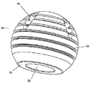
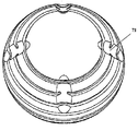
図5を参照すると、研磨されていないステンレス鋼製人工大腿骨頭(50として大略的に示している)がほぼ球状をなし、平坦化切頭ベース51を有し、これに適当なサイズの体内人工大腿骨ステムの基端を受入れるための円形開口部52が設けられている。図示の人工大腿骨頭において、人工大腿骨頭50の幾何学的中心が平坦化切頭ベース51上のほぼ11mmのところに位置している。この実施例での実質的球状の人工大腿骨頭50の外径は32mmとなっている。
Referring to FIG. 5, an unpolished stainless steel prosthetic femoral head (shown schematically as 50) is substantially spherical and has a flattened
本図に示した人工大腿骨頭は、2つの異なるタイプの表面特徴の存在により特徴づけられている。第1に、6個からなる一連の周方向の溝53が形成され、その最下位(先端)のものは、ほぼ球状の頭部の“赤道”(すなわち、人工大腿骨頭を垂直にし、平坦化切頭ベース51を下方に位置させたときの最も長い水平な周方向ライン)を画定するラインの下方4.6mmのところに位置している。前記溝の上方に隣接する周方向の溝は、前記赤道ラインの下方1mmの距離のところに位置している。残る4本の周方向の溝は前記赤道ラインの上方に位置し、この赤道ラインから(下から上に向けて順に)、2.6mm、6.1mm、9.3mmおよび12mmの距離のところに離間させて設けられている。各周方向溝53の隣接する周方向溝との間の離間角度は5度となっている。各周方向溝53は平均深さが1mmとなっている。各周方向溝53の開口端部の幅は1.2mmであり、そのテーパーした内方端部の幅は0.8mmとなっている。
The artificial femoral head shown in this figure is characterized by the presence of two different types of surface features. First, a series of six
この図示の人工大腿骨頭の第2の異なる表面特徴は、垂直方向のスロット54(図5には、その2個が示されている)である。本図から、これらのスロットは仮想長手方向ラインに沿って配置されていることが理解されよう。これらのスロットは3つの最も上位(近位)の周方向溝53と90°で交差する長さを有している。これら垂直方向スロットは、球状頭部の幾何学的中心に対し34度の角度で人工大腿骨頭に配向させた5mm径ドリルを用いて形成される。各スロットの上端は、球状頭部の上位(近位)極部から9mmのところに位置し、その下端は前記赤道ラインの上3.5mmのところに位置している。図7に明示するように、このような垂直方向スロットが合計4個、人工大腿骨頭に設けられている。なお、図7では、これらスロットは71として示されている。本図からこれら垂直方向スロットは互いに等間隔で配置されていることが理解されよう。
The second different surface feature of the illustrated artificial femoral head is a vertical slot 54 (two of which are shown in FIG. 5). From this figure it will be understood that these slots are arranged along a virtual longitudinal line. These slots have a length that intersects the three uppermost (proximal)
図6は、図5,7に示したものと同じ人工補装具頭部(大略的に60として示されている)の下位部分(すなわち、遠位表面が最も上にある)を概略的に描いたものである。本図において、切頭ベース61に円形開口部62が穿設されていることが示されている。この円形開口部62は人工補装具ステム部位(図示しない)を受入れ、保持するためのものである。この開口部は、基本的に円錐形の内部空間につながっており、この円錐形の外方表面は直径が14mmであり、その内方ベースの直径が12mmとなっている。
FIG. 6 schematically depicts the lower portion (ie, with the distal surface on top) of the same prosthetic head (shown generally as 60) as shown in FIGS. It is a thing. In this figure, it is shown that a
本発明のこれら実施例は説明を目的としたものであり、当業者が本発明の趣旨および特許請求の範囲を逸脱することなく、多くの変更、変形および適応性を加えて本発明を実施し得るであろうことを理解されたい。 These embodiments of the present invention are intended to be illustrative, and those skilled in the art can implement the present invention with many modifications, variations and adaptations without departing from the spirit and scope of the present invention. Please understand that you will get.
20、30 人工補装具
21、31 人工補装具頭部領域
22、32 ステム領域
23 ステム領域の基端部
24 窪み
25 大腿骨頭外殻
26 セメント
27 スロット
33、42 大腿骨頭外殻
40 人工補装具の頭領域
41 粗面化外側表面
43、52、62 円形開口部
53 周方向溝
54、71 垂直方向のスロット
61 切頭ベース
20, 30
Claims (39)
a)前記長骨の頭部から海綿骨の殆ど又は全てを除去し、長骨頭部外殻を形成する段階と;
b)人工補装具ステム部を受入れるための長骨通路を作る段階と;
c)前記人工補装具ステム部を前記長骨通路内に挿入し、適宜、接合させる段階と;
d)長骨用人工補装具頭部を前記長骨頭部外殻内に挿入し、適宜、接合させる段階と;
e)前記長骨頭の窪んだ部分に前記ステム部部分を復位させる段階と;
を具備してなることを特徴とする方法。 A method for treating a neck fracture of a long bone in a patient in need of treatment comprising:
a) removing most or all of the cancellous bone from the long bone head to form a long bone head shell;
b) creating a long bone passage for receiving the prosthetic stem portion;
c) inserting the prosthetic stem portion into the long bone passage and appropriately joining;
d) inserting a long bone prosthesis head into the long bone head shell and appropriately joining;
e) repositioning the stem portion to the recessed portion of the long bone head;
The method characterized by comprising.
a)軟骨および軟骨下骨に障害を生じさせない安全な手法を用いて、人体の本来の位置から生来の長骨頭を除去する段階と;
b)人体外で頭部外殻を手術トレー上に準備する段階と;
c)人工補装具ステム部部位を受入れるための長骨通路を作る段階と;
d)接合剤を用い、又は用いずに、前記人工補装具ステム部部位を前記長骨通路内に挿入する段階と;
e)段階(b)で形成された頭部外殻に前記長骨用人工補装具頭部を接合させる段階と;
f)取着された前記頭部外殻と共に、製造された長骨用人工補装具頭部を治療対象の患者の関節内に復位させ、該長骨用人工補装具頭部の先端部を該人工補装具ステム部部位に接合させる段階と;
を具備してなることを特徴とする方法。 A method for treating a neck fracture of a long bone in a patient in need of treatment comprising:
a) removing the natural long bone head from its natural location using a safe technique that does not damage the cartilage and subchondral bone;
b) preparing the outer shell on the surgical tray outside the human body;
c) creating a long bone passage for receiving the prosthetic stem site;
d) inserting the prosthetic stem portion with or without a bonding agent into the long bone passageway;
e) joining the long bone prosthesis head to the head shell formed in step (b);
f) The produced prosthetic head for the long bone is moved back into the joint of the patient to be treated together with the attached outer shell of the head, and the distal end of the prosthetic head for the long bone is Joining to the prosthesis stem site;
The method characterized by comprising.
a)軟骨および軟骨下骨に障害を生じさせない安全な手法を用いて、人体の本来の位置から生来の長骨頭を除去する段階と;
b)人体外で頭部外殻を手術トレー上に準備する段階と;
c)単一ブロックの人工補装具のステム部部位を受入れるための長骨通路を作る段階と;
d)接合剤を用い、又は用いずに、前記単一ブロックの人工補装具のステム部部位を前記長骨通路内に挿入する段階と;
e)段階(b)で形成された頭部外殻に前記単一ブロックの長骨用人工補装具頭部部位を接合させる段階と;
f)取着された前記頭部外殻と共に、用意された前記単一ブロックの長骨用人工補装具頭部を治療対象の患者の関節内に復位させる段階と;
を具備してなることを特徴とする方法。 A method for treating a neck fracture of a long bone in a patient in need of treatment comprising:
a) removing the natural long bone head from its natural location using a safe technique that does not damage the cartilage and subchondral bone;
b) preparing the outer shell on the surgical tray outside the human body;
c) creating a long bone passage for receiving a stem portion of a single block prosthesis;
d) inserting the stem portion of the single block prosthesis into the long bone passage with or without a bonding agent;
e) joining the single block long bone prosthesis head portion to the head shell formed in step (b);
f) repositioning the prepared single block long bone prosthesis head with the attached outer shell into the joint of the patient to be treated;
The method characterized by comprising.
a)前記長骨の頭部から海綿骨の殆ど又は全てを除去し、長骨頭部外殻を形成する段階と;
b)人工補装具ステム部を受入れるための長骨通路を作る段階と;
c)前記人工補装具ステム部を前記長骨通路内に挿入し、適宜、接合させる段階と;
d)段階(a)で形成された頭部外殻にバイポーラ型の人工補装具の外側頭部を接合させる段階と;
e)前記人工補装具ステム部部位のトラニオンを人工長骨内側頭部の内側キャビティ内に配置する段階と;
f)前記人工長骨内側頭部を前記人工長骨外側頭部の内側キャビティ内に挿入する段階と;
g)前記バイポーラ型の頭部アッセンブリーを、固定リングを閉じることにより固定する段階と;
を具備してなることを特徴とする方法。 A method for treating a neck fracture of a long bone in a patient in need of treatment comprising:
a) removing most or all of the cancellous bone from the long bone head to form a long bone head shell;
b) creating a long bone passage for receiving the prosthetic stem portion;
c) inserting the prosthetic stem portion into the long bone passage and appropriately joining;
d) joining the outer head of the bipolar prosthesis to the head shell formed in step (a);
e) placing the trunnion at the prosthetic stem portion within the medial cavity of the medial prosthetic long bone;
f) inserting the medial long bone prosthesis into the medial cavity of the prosthetic long bone lateral head;
g) fixing the bipolar head assembly by closing a fixing ring;
The method characterized by comprising.
a)軟骨および軟骨下骨に障害を生じさせない安全な手法を用いて、人体の本来の位置から生来の長骨頭を除去する段階と;
b)人体外で頭部外殻を手術トレー上に準備する段階と;
c)人工補装具ステム部部位を受入れるための長骨通路を作る段階と;
d)接合剤を用い、又は用いずに、前記人工補装具ステム部部位を前記長骨通路内に挿入する段階と;
e)段階(b)で形成された頭部外殻にバイポーラ型の人工補装具の外側頭部を接合させる段階と;
f)前記人工補装具ステム部部位のトラニオンを人工長骨内側頭部の内側キャビティ内に配置する段階と;
g)前記人工長骨内側頭部を前記人工長骨外側頭部の内側キャビティ内に挿入する段階と;
h)前記バイポーラ型の頭部アッセンブリーを、固定リングを閉じることにより固定する段階と;
i)取着された前記頭部外殻と共に、用意された前記長骨用人工補装具頭部を治療対象の患者の関節内に復位させる段階と;
を具備してなることを特徴とする方法。 A method for treating a neck fracture of a long bone in a patient in need of treatment comprising:
a) removing the natural long bone head from its natural location using a safe technique that does not damage the cartilage and subchondral bone;
b) preparing the outer shell on the surgical tray outside the human body;
c) creating a long bone passage for receiving the prosthetic stem site;
d) inserting the prosthetic stem portion with or without a bonding agent into the long bone passageway;
e) joining the outer head of the bipolar prosthesis to the head shell formed in step (b);
f) placing the trunnion of the prosthetic stem portion within the medial cavity of the medial prosthetic long bone;
g) inserting the medial long bone prosthesis into the medial cavity of the prosthetic long bone lateral head;
h) securing the bipolar head assembly by closing a retaining ring;
i) repositioning the prepared long bone prosthesis head with the attached outer shell into the joint of the patient to be treated;
The method characterized by comprising.
a)軟骨および軟骨下骨に障害を生じさせない安全な手法を用いて、人体の本来の位置から生来の長骨頭を除去する段階と;
b)人体外で頭部外殻を手術トレー上に準備する段階と;
c)単一ブロックバイポーラ型の人工補装具のステム内側部位を受入れるための長骨通路を作る段階と;
d)接合剤を用い、又は用いずに、前記単一ブロックバイポーラ型の人工補装具のステム内側部位を前記長骨通路内に挿入する段階と;
e)段階(b)で形成された頭部外殻にバイポーラ型の人工補装具の外側頭部を接合させる段階と;
f)取着された前記頭部外殻と共に、用意された前記バイポーラ型の人工補装具の外側頭部を治療対象の患者の関節内に復位させる段階と;
g)前記バイポーラ型の人工補装具の内側頭部を前記バイポーラ型の人工補装具の外側頭部の内側キャビティ内に挿入する段階と;
h)前記バイポーラ型の頭部アッセンブリーを、固定リングを閉じることにより固定する段階と;
を具備してなることを特徴とする方法。 A method for treating a neck fracture of a long bone in a patient in need of treatment comprising:
a) removing the natural long bone head from its natural location using a safe technique that does not damage the cartilage and subchondral bone;
b) preparing the outer shell on the surgical tray outside the human body;
c) creating a long bone passage for receiving the inner stem portion of a single block bipolar prosthesis;
d) inserting the stem inner portion of the single-block bipolar prosthesis into the long bone passage with or without a bonding agent;
e) joining the outer head of the bipolar prosthesis to the head shell formed in step (b);
f) repositioning the outer head of the prepared bipolar prosthesis with the attached outer shell into the joint of the patient to be treated;
g) inserting the inner head of the bipolar prosthesis into the inner cavity of the outer head of the bipolar prosthesis;
h) securing the bipolar head assembly by closing a retaining ring;
The method characterized by comprising.
a)軟骨および軟骨下骨に障害を生じさせない安全な手法を用いて、人体の本来の位置から生来の長骨頭を除去する段階と;
b)人体外で頭部外殻を手術トレー上に準備する段階と;
c)単一ブロックバイポーラ型の人工補装具のステム内側部位を受入れるための長骨通路を作る段階と;
d)接合剤を用い、又は用いずに、前記単一ブロックバイポーラ型の人工補装具のステム内側部位を前記長骨通路内に挿入する段階と;
e)前記バイポーラ型の人工補装具の内側頭部を前記バイポーラ型の人工補装具の外側頭部の内側キャビティ内に挿入する段階と;
f)前記バイポーラ型の頭部アッセンブリーを、固定リングを閉じることにより固定する段階と;
g)段階(b)で形成された頭部外殻をバイポーラ型の人工補装具の外側頭部に接合させる段階と;
h)取着された前記バイポーラ型の人工補装具と共に、用意された前記バイポーラ型の人工補装具の外側頭部を治療対象の患者の関節内に復位させる段階と;
を具備してなることを特徴とする方法。 A method for treating a neck fracture of a long bone in a patient in need of treatment comprising:
a) removing the natural long bone head from its natural location using a safe technique that does not damage the cartilage and subchondral bone;
b) preparing the outer shell on the surgical tray outside the human body;
c) creating a long bone passage for receiving the inner stem portion of a single block bipolar prosthesis;
d) inserting the stem inner portion of the single-block bipolar prosthesis into the long bone passage with or without a bonding agent;
e) inserting the inner head of the bipolar prosthesis into the inner cavity of the outer head of the bipolar prosthesis;
f) fixing the bipolar head assembly by closing a fixing ring;
g) joining the head shell formed in step (b) to the outer head of a bipolar prosthesis;
h) with the attached bipolar prosthesis, repositioning the outer head of the prepared bipolar prosthesis into the joint of the patient to be treated;
The method characterized by comprising.
a)前記長骨の頭部から海綿骨の殆ど又は全てを除去し、長骨頭部外殻を形成する段階と;
b)単一ブロックバイポーラ型の人工補装具のステム内側部位を受入れるための長骨通路を作る段階と;
c)接合剤を用い、又は用いずに、前記単一ブロックバイポーラ型の人工補装具のステム内側部位を前記長骨通路内に挿入する段階と;
d)前記バイポーラ型の人工補装具の内側頭部を前記バイポーラ型の人工補装具の外側頭部の内側キャビティ内に挿入する段階と;
e)前記バイポーラ型の頭部アッセンブリーを、固定リングを閉じることにより固定する段階と;
f)段階(b)で形成された頭部外殻にバイポーラ型の人工補装具の外側頭部を接合させる段階と;
を具備してなることを特徴とする方法。 A method for treating a neck fracture of a long bone in a patient in need of treatment comprising:
a) removing most or all of the cancellous bone from the long bone head to form a long bone head shell;
b) creating a long bone passage for receiving the inner stem portion of a single block bipolar prosthesis;
c) inserting the stem inner portion of the single block bipolar prosthesis into the long bone passage with or without a bonding agent;
d) inserting the inner head of the bipolar prosthesis into the inner cavity of the outer head of the bipolar prosthesis;
e) fixing the bipolar head assembly by closing a fixing ring;
f) joining the outer head of the bipolar prosthesis to the head shell formed in step (b);
The method characterized by comprising.
Applications Claiming Priority (2)
| Application Number | Priority Date | Filing Date | Title |
|---|---|---|---|
| IL15863303A IL158633A0 (en) | 2003-10-28 | 2003-10-28 | Cartilage-preserving long bone head prosthesis |
| PCT/IL2004/000980 WO2005039439A2 (en) | 2003-10-28 | 2004-10-27 | Cartilage-preserving long bone head prosthesis |
Publications (2)
| Publication Number | Publication Date |
|---|---|
| JP2007509690A true JP2007509690A (en) | 2007-04-19 |
| JP2007509690A5 JP2007509690A5 (en) | 2007-12-13 |
Family
ID=34044239
Family Applications (1)
| Application Number | Title | Priority Date | Filing Date |
|---|---|---|---|
| JP2006537551A Pending JP2007509690A (en) | 2003-10-28 | 2004-10-27 | Long bone head prosthesis to protect cartilage |
Country Status (7)
| Country | Link |
|---|---|
| EP (1) | EP1682049A2 (en) |
| JP (1) | JP2007509690A (en) |
| CN (1) | CN1897894A (en) |
| AU (1) | AU2004283563A1 (en) |
| CA (1) | CA2544151A1 (en) |
| IL (2) | IL158633A0 (en) |
| WO (1) | WO2005039439A2 (en) |
Cited By (1)
| Publication number | Priority date | Publication date | Assignee | Title |
|---|---|---|---|---|
| JP2011512908A (en) * | 2008-02-25 | 2011-04-28 | スミス アンド ネフュー インコーポレーテッド | Method and system for mapping a femoral head for registration of an artificial acetabulum |
Families Citing this family (5)
| Publication number | Priority date | Publication date | Assignee | Title |
|---|---|---|---|---|
| IL172588A0 (en) | 2005-12-14 | 2006-04-10 | Presrv Ltd | Faceted long bone head prosthesis |
| US8870876B2 (en) | 2009-02-13 | 2014-10-28 | Tarsus Medical Inc. | Methods and devices for treating hallux valgus |
| US8277459B2 (en) | 2009-09-25 | 2012-10-02 | Tarsus Medical Inc. | Methods and devices for treating a structural bone and joint deformity |
| US8652141B2 (en) | 2010-01-21 | 2014-02-18 | Tarsus Medical Inc. | Methods and devices for treating hallux valgus |
| US8696719B2 (en) | 2010-06-03 | 2014-04-15 | Tarsus Medical Inc. | Methods and devices for treating hallux valgus |
Family Cites Families (4)
| Publication number | Priority date | Publication date | Assignee | Title |
|---|---|---|---|---|
| US4687487A (en) * | 1978-07-21 | 1987-08-18 | Association Suisse Pour La Recherches Horlogere | Joint implant |
| US5192323A (en) * | 1990-11-05 | 1993-03-09 | Zimmer, Inc. | Method of surface hardening orthopedic implant devices |
| AU736216B2 (en) * | 1997-03-27 | 2001-07-26 | Smith & Nephew, Inc. | Method of surface oxidizing zirconium alloys and resulting product |
| US6527808B1 (en) * | 2000-10-11 | 2003-03-04 | Zimmer Technology, Inc. | Constrained socket for use with a ball-and-socket joint |
-
2003
- 2003-10-28 IL IL15863303A patent/IL158633A0/en unknown
-
2004
- 2004-10-27 JP JP2006537551A patent/JP2007509690A/en active Pending
- 2004-10-27 CA CA002544151A patent/CA2544151A1/en not_active Abandoned
- 2004-10-27 EP EP04791842A patent/EP1682049A2/en not_active Withdrawn
- 2004-10-27 CN CNA2004800389601A patent/CN1897894A/en active Pending
- 2004-10-27 WO PCT/IL2004/000980 patent/WO2005039439A2/en active Application Filing
- 2004-10-27 AU AU2004283563A patent/AU2004283563A1/en not_active Abandoned
-
2006
- 2006-04-27 IL IL175262A patent/IL175262A0/en unknown
Cited By (1)
| Publication number | Priority date | Publication date | Assignee | Title |
|---|---|---|---|---|
| JP2011512908A (en) * | 2008-02-25 | 2011-04-28 | スミス アンド ネフュー インコーポレーテッド | Method and system for mapping a femoral head for registration of an artificial acetabulum |
Also Published As
| Publication number | Publication date |
|---|---|
| CN1897894A (en) | 2007-01-17 |
| AU2004283563A1 (en) | 2005-05-06 |
| IL175262A0 (en) | 2006-09-05 |
| IL158633A0 (en) | 2004-05-12 |
| WO2005039439A2 (en) | 2005-05-06 |
| EP1682049A2 (en) | 2006-07-26 |
| CA2544151A1 (en) | 2005-05-06 |
| WO2005039439A3 (en) | 2006-05-11 |
Similar Documents
| Publication | Publication Date | Title |
|---|---|---|
| US20060259148A1 (en) | Cartilage-preserving long bone head prosthesis | |
| US20210236292A1 (en) | System and method for implanting a secondary glenoid prosthesis | |
| US9445911B2 (en) | Bone preparation tool kit and associated method | |
| EP1138283B1 (en) | Femoral hip prosthesis | |
| CN102883686B (en) | Locally hip prosthesis | |
| US10507113B2 (en) | Reverse total hip replacement | |
| JP5551074B2 (en) | Artificial joint | |
| US6488716B1 (en) | Anatomic femoral prosthesis for total hip arthroplasty | |
| JP2004526493A (en) | Implant locking system | |
| NZ571450A (en) | A glenoid plate for a reverse shoulder prosthesis | |
| JP2006095300A (en) | Extended articulation prosthesis adapter and associated method | |
| US20090005879A1 (en) | Prosthesis | |
| JP5646599B2 (en) | Surgical prosthesis | |
| JP2004130113A (en) | Cemented prosthetic kit | |
| JP2007509690A (en) | Long bone head prosthesis to protect cartilage | |
| JP4169973B2 (en) | Artificial organ, prosthetic stem, and method of manufacturing the prosthesis | |
| WO2007069250A2 (en) | Faceted long bone head prosthesis | |
| Kowaleski | Total hip replacement. | |
| UA53583A (en) | Device for making endoprosthesis of proximal part of femur |
Legal Events
| Date | Code | Title | Description |
|---|---|---|---|
| A521 | Request for written amendment filed |
Free format text: JAPANESE INTERMEDIATE CODE: A523 Effective date: 20071025 |
|
| A621 | Written request for application examination |
Free format text: JAPANESE INTERMEDIATE CODE: A621 Effective date: 20071025 |
|
| A131 | Notification of reasons for refusal |
Free format text: JAPANESE INTERMEDIATE CODE: A131 Effective date: 20090915 |
|
| A977 | Report on retrieval |
Free format text: JAPANESE INTERMEDIATE CODE: A971007 Effective date: 20090917 |
|
| A02 | Decision of refusal |
Free format text: JAPANESE INTERMEDIATE CODE: A02 Effective date: 20100223 |