JP2007236802A - Material for implant inclination - Google Patents
Material for implant inclination Download PDFInfo
- Publication number
- JP2007236802A JP2007236802A JP2006066291A JP2006066291A JP2007236802A JP 2007236802 A JP2007236802 A JP 2007236802A JP 2006066291 A JP2006066291 A JP 2006066291A JP 2006066291 A JP2006066291 A JP 2006066291A JP 2007236802 A JP2007236802 A JP 2007236802A
- Authority
- JP
- Japan
- Prior art keywords
- surface layer
- bone
- porosity
- implant
- gradient material
- Prior art date
- Legal status (The legal status is an assumption and is not a legal conclusion. Google has not performed a legal analysis and makes no representation as to the accuracy of the status listed.)
- Pending
Links
Images


Landscapes
- Prostheses (AREA)
- Materials For Medical Uses (AREA)
Abstract
Description
本発明は、股関節の大腿骨頭壊死、膝関節の膝骨頭壊死などの関節骨軟骨障害の治療や、靱帯の骨関節付着部の補強などに用いられるインプラント傾斜材料に関する。 The present invention relates to an implant gradient material used for the treatment of osteoarticular cartilage disorders such as femoral head necrosis of the hip joint and knee head necrosis of the knee joint, and reinforcement of the bone joint attachment portion of the ligament.
従来、大きく破壊、損傷した硬骨、軟骨部分の再建、再生あるいは補強のために、再生医療の諸技術が検討されている。そして、本質的に、ある形を持った損傷部位の再建には、組織の再生が完了するまでの期間、外部からの力学的負荷や、細胞学的、生理学的侵襲を避けて、求められる形を形成、維持させて、再建を完成させるための足場(Scaffold)が必要であることは広く認識されている。 Conventionally, various techniques of regenerative medicine have been studied for the reconstruction, regeneration, or reinforcement of bones and cartilage that have been greatly destroyed or damaged. In essence, the reconstruction of a damaged site with a certain shape requires the form required to avoid mechanical load from the outside, cytological and physiological invasion during the period until tissue regeneration is completed. It is widely recognized that a scaffold is needed to form, maintain and complete the reconstruction.
現在、股関節や膝関節などの関節骨軟骨に異常があり該軟骨を修復、再生、再建する必要がある場合に使用される足場材料について、種々のアイデアが示されている。しかしながら、関節骨頭の壊死部の治療、再建や、靱帯の骨関節付着部を強化するための足場となるに足り得るものは、材料学的な困難さから、いまだ開発されていない。理由は、それが用いられる部位が、異質の機能と物質からなり、軟骨と硬骨が動きを伴って接触する非連続的骨結合部位である境界面、即ち、関節の位置に適用すべき足場であるからである。 At present, various ideas have been presented for scaffold materials used when there is an abnormality in articular cartilage such as a hip joint and a knee joint and the cartilage needs to be repaired, regenerated and reconstructed. However, what is sufficient to serve as a scaffold for treating and reconstructing the necrotic part of the joint bone head and strengthening the bone-joint attachment part of the ligament has not been developed due to material difficulties. The reason for this is that the site where it is used is a scaffold that should be applied to the interface, i.e. the location of the joint, which is a discontinuous osteosynthesis site where the cartilage and bone contact with movement, consisting of different functions and materials. Because there is.
斯かる、足場の開発策の一つとして、軟骨代替物と硬骨代替物を組み合わせて一体化した補綴材料を作製し、関節頭骨の部分には硬骨代替物を、関節軟骨の部分には軟骨代替物を埋入、固定することが考えられる。しかし、この両方の代替物が一体物でなく、別々の分かれたものの組み合わせから成る場合は、軟骨と硬骨の組織の連続的な接続的移行の形態が得られず、また、関節の稼動時に、両者が剥離する問題が生ずる。したがって、関節部位に用いるに値する足場材料は、硬骨に納まる部位は硬骨と生体組織学的、力学的によく親和性を持ち、軟骨に納まる部位は軟骨と生体組織学的、力学的によく親和性を持つことにより、可動界面である骨関節に在って、脱転しないように安定に保たれるものでなければならない。 As one of the development measures for such a scaffold, a prosthetic material is produced by combining a cartilage substitute and a bone substitute, and a bone substitute is used for the joint skull and a cartilage substitute for the articular cartilage. It is conceivable to insert and fix objects. However, if both of these alternatives are not monolithic and consist of a combination of separate parts, a continuous connective transition form of cartilage and bone tissue cannot be obtained, and when the joint is in operation, There arises a problem that the two peel off. Therefore, the scaffold material worthy of being used for the joint part has a good affinity for bone in the bone and biohistologically and mechanically, and the part in the cartilage has a good affinity for the cartilage and biohistologically and mechanically. It must be stable in the bone joint, which is a movable interface, so that it does not fall out.
この場合、目的とする補綴材が金属や、セラミック、ポリマーなどの生体内で非吸収性であれば、経時的に生体組織と置き換わらず、長期埋入時に感染や、機械的な故障のような問題発生の危惧を抱え続ける。それゆえ、生体組織と置き換わりつつ、形を再建しながら、ついには、自らは生体内に分解吸収され、消失する、生体活性と生体内分解吸収性を合わせ持つ必要がある。そして、緻密質と多孔質の部分の両方の形態を同時に共有し、しかも、軟骨側を代替する多孔質の部分は、軟骨表面に近いほど、高開放率であり、硬骨を代替する緻密質の部分は、緻密質に埋入している部分ほど、低開放率であることが、力学的にも、生理学的にも望ましい。 In this case, if the target prosthetic material is non-absorbable in the living body such as metal, ceramic, polymer, etc., it will not be replaced with living tissue over time, and infection or mechanical failure may occur during long-term implantation. Continuing to worry about the occurrence of new problems. Therefore, it is necessary to have both the bioactivity and the biodegradability that can be decomposed and absorbed in the living body and disappear in the living body while being replaced with the living tissue and reconstructing the shape. In addition, the porous part that shares both the dense and porous parts at the same time, and the porous part that substitutes for the cartilage side has a higher open rate as it is closer to the cartilage surface, and the dense part that substitutes for the bone. It is desirable from a mechanical and physiological viewpoint that the portion of the portion that is densely embedded has a lower open rate.
すなわち、関節骨軟骨障害等の治療、再建の足場は、多孔質部は細胞が速やかに侵入し、足場の分解に伴って軟骨組織が表層部に誘導形成されて生体組織と交換し、緻密層は分解されるまでのある程度の期間、硬骨を伝導、密着して、十分な強度を維持するが、最終的には全て分解されて硬骨組織と完全置換するような材料の開発が求められる。 That is, scaffolds for the treatment and reconstruction of articular osteochondral disorders, etc., in the porous part, cells rapidly invade, the cartilage tissue is induced and formed in the surface layer part with the degradation of the scaffold, and exchanges with the biological tissue, the dense layer For a certain period until it is decomposed, the bone is conducted and closely adhered to maintain a sufficient strength, but finally, development of a material that is completely decomposed and completely replaces the bone tissue is required.
ところで、本発明者は、海綿骨の表層(外側)に皮質骨を備えた生体骨の欠損部分を修復、再建するインプラント材料として、その内部に連続気孔を有し且つ生体活性なバイオセラミックス粉体を含んだ生体内分解吸収性ポリマーよりなる立体状の多孔体の表面の一部に、生体活性なバイオセラミックス粉体を含んだ生体内分解吸収性ポリマーよりなる緻密質な表皮層を重ねて一体化した人工骨を既に提案した(特許文献1)。 By the way, the present inventor, as an implant material for repairing and reconstructing a defect portion of a living bone having cortical bone on the surface layer (outside) of the cancellous bone, has a continuous pore inside and is a bioactive bioceramic powder. A dense skin layer made of biodegradable absorbable polymer containing bioactive bioceramic powder is laminated on a part of the surface of a three-dimensional porous material made of biodegradable absorbable polymer containing An artificial bone has already been proposed (Patent Document 1).
このインプラント材料は、上記生体骨の内層部の海綿骨欠損部分に充当して立体状の多孔体を埋入し、表層部の皮質骨欠損部分に充当して緻密質な表皮層を埋入するものである。これは自家移植(autograft)骨片、同種移植(allograft)骨片の代替材料として好適に使用される人工骨である。けれども、異質の機能と物質からなり、軟骨と硬骨が動きを伴って接触する非連続的骨結合部位である境界面、即ち、関節の位置に適応すべき足場として相応しいものでない。
本発明は上記事情の下になされたもので、股関節や膝関節などの関節骨軟骨に異常があり該軟骨を修復、再生、再建する必要がある場合、つまり関節骨頭の壊死部の治療、再建や、靱帯の骨関節付着部を強化する場合に使用される一時的補綴・足場材料として、当該医療分野で希求されている上述の物性ないし機能を備えて骨関節に安定的に埋入固定できるインプラント傾斜材料を提供することを解決課題としている。 The present invention has been made under the above circumstances, and when there is an abnormality in the articular bone cartilage such as the hip joint and the knee joint, it is necessary to repair, regenerate and reconstruct the cartilage, that is, treatment and reconstruction of the necrotic part of the joint bone head. As a temporary prosthesis / scaffold material used to strengthen the bone joint attachment part of the ligament, it can be stably embedded and fixed in the bone joint with the above-mentioned physical properties and functions that are desired in the medical field. The problem to be solved is to provide an implant gradient material.
上記の課題を解決するため、本発明に係る第一のインプラント傾斜材料は、生体内吸収性、かつ、生体活性なバイオセラミックス粉体を含んだ生体内分解吸収性ポリマーの連続気孔質複合体からなり、気孔率が10〜90%の範囲内で該複合体の片側の表層部から反対側の表層部に近づくほど高くなるように順次連続的に変化していることを特徴とするものである。
そして、本発明に係る第二のインプラント傾斜材料は、生体内吸収性、かつ、生体活性なバイオセラミックス粉体を含んだ生体内分解吸収性ポリマーの連続気孔質複合体からなり、気孔率が10〜90%の範囲内で該複合体の中間部から両側の表層部に近づくほど、又は、該複合体の中央部から周囲の表層部に近づくほど高くなるように順次連続的に変化していることを特徴とするものである。
In order to solve the above problems, the first implant gradient material according to the present invention is a bioresorbable and bioactive bioceramic powder-containing biodegradable absorbable polymer continuous porous composite. In the range of 10 to 90%, the porosity is successively changed so as to increase from the surface layer portion on one side to the surface layer portion on the opposite side. .
And the 2nd implant inclination material which concerns on this invention consists of a continuous porous composite body of the biodegradable absorptive polymer containing the bioresorbable and bioactive bioceramics powder, and the porosity is 10 Within the range of ˜90%, it is successively changed so as to become higher as it approaches the surface layer part on both sides from the intermediate part of the complex or from the center part of the complex to the surrounding surface layer part. It is characterized by this.
第一のインプラント傾斜材料にあっては、バイオセラミックス粉体の含有率が、30〜80質量%の範囲内で、連続気孔質複合体の片側の表層部から反対側の表層部に近づくほど高くなるように順次連続的に変化していることが好ましい。また、第二のインプラント傾斜材料にあっては、バイオセラミックス粉体の含有率が、30〜80質量%の範囲内で、連続気孔質複合体の中間部から両側の表層部に近づくほど、又は、該複合体の中央部から周囲の表層部に近づくほど高くなるように順次連続的に変化していることが好ましい。そして、これらのインプラント傾斜材料はいずれも、連続気孔質複合体に、生物学的骨成長因子であるBMP(Bone Morphogenic Protein)、TGF−β(Transforming Growth Factor-b)、EP4(Prostanoid Receptor)、b−FGF(basic Fibroblast Growth Factor)、PRP(platelet-rich plasma)などの少なくとも一種、及び/又は、生体由来の骨芽細胞が含浸されていることが好ましい。 In the first implant gradient material, the content of the bioceramic powder is higher in the range of 30 to 80% by mass as it approaches the surface layer on the opposite side from the surface layer on one side of the continuous porous composite. It is preferable to change continuously so that it may become. Moreover, in the second implant gradient material, the content of the bioceramic powder is within the range of 30 to 80% by mass, and the closer to the surface layer parts on both sides from the middle part of the continuous porous composite, or It is preferable that the thickness of the composite is continuously changed so as to increase from the central portion toward the surrounding surface layer portion. All of these implant gradient materials are formed into a continuous pore composite, BMP (Bone Morphogenic Protein), TGF-β (Transforming Growth Factor-b), EP4 (Prostanoid Receptor), biological bone growth factors, It is preferable to be impregnated with at least one of b-FGF (basic fibroblast growth factor), PRP (platelet-rich plasma) and / or living osteoblasts.
このような本発明のインプラント傾斜材料は、関節骨軟骨障害の治療、再生又は靱帯の骨関節付着部の再建もしくは補強に用いられ、関節頭と関節軟骨が接している膝、股、足、肩、肘、脊椎(頚椎、腰椎)などの関節部に一時的な補綴材、足場および生物学的骨形成因子の除放のための担持体として適用される。 Such an implant gradient material of the present invention is used for treatment, regeneration, or reconstruction or reinforcement of a bone-joint attachment portion of a ligament, and the joint head and the joint cartilage are in contact with the knee, crotch, foot, and shoulder. It is applied to joints such as elbows and spine (cervical vertebrae, lumbar vertebrae) as temporary prosthetic materials, scaffolds and carriers for the release of biological osteogenic factors.
本発明のインプラント傾斜材料は、連続気孔質複合体の気孔率の高い表層部の連続気孔に体液が浸透し易いため、表層部の表面に接触する体液や連続気孔に浸透する体液によって表面と内部から速やかに加水分解される。そして、この気孔率の高い表層部は骨芽細胞も侵入し易いため、加水分解に伴って(軟)骨組織が生体活性なバイオセラミックス粉体が引き金となって気孔率の高い表層部から内部へ誘導形成されて、短期間で表層部やこれに連なる気孔率の比較的高い内層部が(軟)骨組織と置換する。一方、連続気孔質複合体の気孔率の低い片側の表層部や中間部や中央部は強度があり、加水分解が気孔率の高い表層部よりも遥かに遅く、加水分解がある程度進行するまでの期間、強度を維持し、最終的には全てが分解されて、生体活性なバイオセラミックス粉体により生体骨が伝導形成されて、骨組織と置換する。そして、この連続気孔質複合体に含まれるバイオセラミックス粉体は生体内吸収性であるため、置換、再生された(軟)骨組織に残存、堆積することもない。また、軟組織や血管内に浸出することもない。このように、本発明のインプラント傾斜材料は、関節骨軟骨障害の治療などに使用される足場材料として要求される物性ないし機能を有するものであるため、例えば、第一のインプラント傾斜材料を、関節骨頭の壊死部を切除した部分に、気孔率の高い表層部が関節骨頭表面の軟骨側となるように埋入固定すると、気孔率の高い表層部やこれに連なる気孔率の比較的高い内層部が早期に誘導形成された軟骨組織と全置換して消失し、強度を有する気孔率の低い反対側の部分も最終的には伝導形成された硬骨組織と全置換して消失し、バイオセラミックス粉体も完全に吸収されて、関節骨頭の壊死した硬骨部分と軟骨部分が再生される。また、第二のインプラント傾斜材料を関節骨頭の切除部分に埋入すると、上記の作用効果に加えて、切除部分の硬骨と接する気孔率の高い表層部に硬骨組織が速やかに誘導形成され、インプラント傾斜材料が短期間で関節骨頭の切除部分と結合して固定される。 In the implant gradient material of the present invention, the body fluid easily penetrates into the continuous pores of the surface layer portion having a high porosity of the continuous porous composite, so that the body fluid contacting the surface of the surface layer portion and the body fluid penetrating the continuous pores are Is rapidly hydrolyzed. And since the surface part with high porosity is easy for osteoblasts to invade, the (soft) bone tissue is triggered by bio-ceramics powder that is bioactive during hydrolysis, and the inside of the surface part with high porosity is In a short period of time, the surface layer portion and the inner layer portion having a relatively high porosity connected thereto are replaced with (soft) bone tissue in a short period of time. On the other hand, the surface part, middle part and central part on one side with low porosity of the continuous porous composite are strong, hydrolysis is much slower than the surface part with high porosity, and hydrolysis proceeds to some extent. The strength is maintained for a period of time, and finally, everything is decomposed, and the living bone is formed by conduction with the bioactive bioceramic powder to replace the bone tissue. And since the bioceramics powder contained in this continuous porous composite is in vivo absorbable, it does not remain or deposit in the replaced and regenerated (soft) bone tissue. Moreover, it does not leach into soft tissues or blood vessels. Thus, since the implant gradient material of the present invention has physical properties or functions required as a scaffold material used for the treatment of osteoarticular cartilage disorders, for example, the first implant gradient material is used as the joint material. If the surface layer part with high porosity is embedded and fixed in the part where the necrotic part of the bone head is excised, the surface layer part with high porosity and the inner layer part with relatively high porosity connected thereto are fixed. Disappears by replacing the cartilage tissue that was induced and formed early, and disappearing by replacing the bone part with the low porosity, which has strength, with the total replacement with the bone tissue formed by conduction. The body is also completely absorbed and the necrotic bone and cartilage parts of the joint head are regenerated. Moreover, when the second implant gradient material is embedded in the excised portion of the joint bone head, in addition to the above-described effects, the bone tissue is rapidly induced and formed in the surface layer portion having a high porosity in contact with the bone of the excised portion. The tilted material is fixed in combination with the excised part of the articular head in a short period of time.
バイオセラミックス粉体の含有率は、連続気孔質複合体の全体に亘って一定していてもよいが、バイオセラミックス粉体の含有率が30〜80質量%の範囲内で連続気孔質複合体の片側の表層部から反対側の表層部に近づくほど高くなるように順次連続的に変化しているインプラント傾斜材料や、バイオセラミックス粉体の含有率が30〜80質量%の範囲内で、連続気孔質複合体の中間部から両側の表層部に近づくほど、又は、該複合体の中央部から周囲の表層部に近づくほど高くなるように順次連続的に変化しているインプラント傾斜材料は、バイオセラミックス粉体の含有率が高い表層部の生体活性が高くなるため、表層部における骨芽細胞や(軟)骨組織の誘導形成が特に活発になり、(軟)骨組織との置換が一層促進される。 The content of the bioceramic powder may be constant throughout the continuous porous composite, but the content of the bioceramic powder is within the range of 30 to 80% by mass of the continuous porous composite. Within the range of the content of the implant gradient material or bioceramics powder that is successively changed so as to increase from the surface layer portion on one side to the surface layer portion on the opposite side, the continuous pores The implant gradient material which is successively changed so as to increase from the middle part of the porous composite to the surface layer part on both sides or from the center part of the composite to the surrounding surface part part is bioceramics. Since the bioactivity of the surface layer portion with a high powder content increases, the induction formation of osteoblasts and (soft) bone tissue in the surface layer portion becomes particularly active, and the replacement with (soft) bone tissue is further promoted. The
また、連続気孔質複合体に生物学的骨成長因子であるBMP、TGF−β、EP4、b−FGF、PRPの少なくとも一種、及び/又は、生体由来の骨芽細胞が含浸されているインプラント傾斜材料は、骨芽細胞の増殖、成長が大幅に促進されるため、(軟)骨組織の形成が旺盛になって一層速やかに再生される。 In addition, an implant gradient in which a continuous pore complex is impregnated with at least one of biological bone growth factors BMP, TGF-β, EP4, b-FGF, and PRP and / or living osteoblasts Since the material greatly promotes the proliferation and growth of osteoblasts, the formation of (soft) bone tissue is vigorous and is regenerated more rapidly.
以下、図面を参照して本発明の具体的な実施形態を詳述する。 Hereinafter, specific embodiments of the present invention will be described in detail with reference to the drawings.
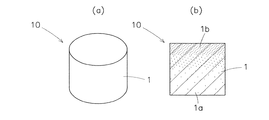
図1の(a)は本発明の一実施形態に係るインプラント傾斜材料の斜視図、(b)は同インプラント傾斜材料の断面図、図2は同インプラント傾斜材料の一使用例の説明図である。 1A is a perspective view of an implant gradient material according to an embodiment of the present invention, FIG. 1B is a cross-sectional view of the implant gradient material, and FIG. 2 is an explanatory diagram of an example of use of the implant gradient material. .
このインプラント傾斜材料10は、生体内吸収性、かつ、生体活性なバイオセラミックス粉体を含んだ生体内分解吸収性ポリマーの連続気孔質複合体1からなるものであって、その気孔率が10〜90%の範囲内で該複合体の片側の表層部1a(下面側の表層部)から反対側の表層部(上面側の表層部)に近づくほど高くなるように順次連続的に変化している。この連続気孔質複合体1からなるインプラント傾斜材料は、図1の(a)に示すように円柱状に形成されているが、これに限定されるものではなく、埋入される関節部位に応じて、四角柱状、楕円柱状、平板状その他の種々の形状となし得るものであり、また、その大きさも埋入される関節部位に応じて最適の大きさとすればよいものである。
This
この連続気孔質複合体1の原料となる生体内分解吸収性ポリマーとしては、結晶性のポリ−L−乳酸やポリグリコール酸なども使用されるが、この連続気孔質複合体1の気孔率の高い表層部1aやこれに連なる気孔率の比較的高い内層部は軟骨程度の強度と柔軟性が要求され、且つ、速やかに分解して生体(軟)骨との結合や全置換が早期に行われることが必要なものであるから、連続気孔質複合体1の原料となる生体内分解吸収性ポリマーとしては、安全で、分解が速く、あまり脆くない、非晶質もしくは結晶と非晶の混在したポリ−D,L−乳酸、L−乳酸とD,L−乳酸の共重合体、乳酸とグリコール酸の共重合体、乳酸とカプロラクトンの共重合体、乳酸とエチレングリコールの共重合体、乳酸とパラ−ジオキサノンの共重合体などが適しており、これらは単独で、或いは二種以上混合して使用される。これらの生体内分解吸収性ポリマーは、連続気孔質複合体1に要求される強度や生体内での分解吸収の期間などを考慮すると、5万〜60万程度の粘度平均分子量を有するものが好ましく使用される。
Crystalline poly-L-lactic acid, polyglycolic acid, or the like is also used as the biodegradable absorbable polymer that is a raw material of the continuous
上記のインプラント傾斜材料10を構成する連続気孔質複合体1は、内部に連続気孔を有し、この連続気孔の内面や複合体1の表面にバイオセラミックス粉体が一部露出した状態で含有されているものである。そして、この連続気孔質複合体1の気孔率は、上述したように、10〜90%の範囲内、好ましくは20〜80%の範囲内で、片側の表層部1a(下面側の表層部)から反対側の表層部1b(上面側の表層部)に近づくほど高くなるように順次連続的に変化している。連続気孔は気孔全体の50〜90%、なかんずく70〜90%を占めることが好ましい。また、連続気孔の孔径は50〜600μmの範囲内、好ましくは100〜400μmの範囲内に調整されており、気孔率の高い片側の表層部1bに近づくほど、孔径が大きくなっている。
The continuous
このように気孔率等が傾斜していると、連続気孔質複合体1の気孔率の高い片側の表層部1b(以下、高気孔率表層部と記す)は、体液の浸透が容易で速やかに加水分解され、しかも、骨芽細胞が侵入し易く、後述するように生体活性なバイオセラミックス粉体の含有率が高いことと相俟って、早期に(軟)骨組織が誘導形成されて生体(軟)骨と結合、置換される。高気孔率表層部1bの気孔率が90%を上回り、孔径が600μmよりも大きくなると、高気孔率表層部1bの物理的な強度が低下して脆くなるので好ましくない。また、連続気孔が気孔全体の50%を下回り、且つ、孔径が50μmよりも小さくなると、体液や骨芽細胞の侵入が困難になり、加水分解や骨組織の誘導形成が遅くなって、生体(軟)骨との結合や置換に要する時間が長くなるので好ましくない。ただし、上記の好適な孔径と併存して1〜0.1μmのサブミクロン程度微細な連続気孔が存在すると、骨誘導性が発現されることが見出されている。
When the porosity or the like is inclined as described above, the
一方、連続気孔質複合体1の気孔率が低い反対側(下面側)の表層部1a(以下、低気孔率表層部と記す)は、気孔率が低くなるほど強度が向上することになるが、関節部に足場として適応されるインプラント傾斜材料には極端に大きい強度が要求されないので、低気孔率表層部1aの気孔率を零に近づける必要はない。そこで、低気孔率表層部1aの気孔率の下限を10%、好ましくは20%として、足場に適した強度を付与すると共に、加水分解や(軟)骨細胞との全置換に要する時間を短縮できるようにしている。
On the other hand, the strength of the
この連続気孔質複合体1に含有させるバイオセラミックス粉体としては、生体活性があり、生体内吸収性で生体に全吸収されて骨組織と完全に置換され、良好な骨伝導(誘導)能と良好な生体親和性を有する、未仮焼かつ未焼成のハイドロキシアパタイト、ジカルシウムホスフェート、トリカルシウムホスフェート、テトラカルシウムホスフェート、オクタカルシウムホスフェート、カルサイト、セラバイタル、ジオプサイト、天然珊瑚等の粉体が好ましく使用される。その中でも、未仮焼かつ未焼成のハイドロキシアパタイト、トリカルシウムホスフェート、オクタカルシウムホスフェートは、生体活性が極めて高く、骨伝導能に優れ、為害性が低く、短期間で生体に吸収されるので、最適である。
The bioceramic powder to be contained in the continuous
これらのバイオセラミックス粉体は、生体内分解吸収性ポリマーへの分散性や生体への吸収性を考慮すると、30μm以下、好ましくは10μm以下、更に好ましくは0.1〜5μm程度の粒径を有するものが使用される。特に、0.1〜5μm程度の粒径を有するバイオセラミックス粉体は、生体への吸収性が良好であることに加えて、後述する方法で連続気孔質複合体1を作製する際にスプレー等の手段で形成される繊維を短く切断する心配もないので好ましく使用される。
These bioceramic powders have a particle size of about 30 μm or less, preferably about 10 μm or less, more preferably about 0.1 to 5 μm in consideration of dispersibility in the biodegradable absorbable polymer and absorbability to the living body. Things are used. In particular, the bioceramic powder having a particle size of about 0.1 to 5 μm has good absorbability to the living body, and sprays and the like when producing the continuous
連続気孔質複合体1のバイオセラミックス粉体の含有率は、連続気孔質複合体1の全体に亘って一定していてもよいが、30〜80質量%の範囲内で、低気孔率表層部1aから高気孔率表層部1bに近づくほど高くなるように順次連続的に変化していることが好ましい。即ち、低気孔率表層部1aから高気孔率表層部1bに近づくにつれて、バイオセラミックス粉体/生体内分解吸収性ポリマーの質量比率が30/70〜80/20の範囲内で順次連続的に大きくなるように変化していることが好ましい。このようにバイオセラミックス粉体の含有率が傾斜していると、高気孔率表層部1bの生体活性が大きく、骨芽細胞や(軟)骨組織の誘導形成が特に活発になるため、生体(軟)骨との結合や置換が一層促進される。
The content of the bioceramic powder of the continuous
高気孔率表層部1bにおけるバイオセラミックス粉体の含有率が80質量%を上回ると、高気孔率表層部1bの物理的強度の低下を招くという不都合が生じ、低気孔率表層部1aにおける含有率が30質量%を下回ると、低気孔率表層部1aのバイオセラミックス粉体による(軟)骨組織の誘導形成が不活発になるため、生体(軟)骨との結合や全置換に時間がかかり過ぎるという不都合が生じる。バイオセラミックス粉体の更に好ましい含有率の上限は70質量%である。
When the content ratio of the bioceramic powder in the high porosity
この連続気孔質複合体1には、生物学的骨成長因子であるBMP(Bone Morphogenic Protein)、TGF−β(Transforming Growth Factor-b)、EP4(Prostanoid Receptor)、b−FGF(basic Fibroblast Growth Factor)、PRP(platelet-rich plasma)などの少なくとも一種、及び/又は、生体由来の骨芽細胞を含浸させることが好ましい。これらの生物学的骨成長因子や骨芽細胞を含浸させると、骨芽細胞の増殖、成長が大幅に促進され、極く短期間(1週間程度)で連続気孔質複合体1の高気孔率表層部1bに(軟)骨組織が形成されるようになり、その後、連続気孔質複合体1の全体が(軟)骨組織と全置換されて生体(軟)骨が修復、再建される。上記の因子のうち、TGF−β、b−FGFは軟骨の成長に特に有効であり、BMP、EP4は硬骨の成長に特に有効であるから、再生すべき生体骨が軟骨の場合はTGF−βやb−FGFを、再生すべき生体骨が硬骨の場合はBMPやEP4を含浸させることが好ましい。また、PRPは、血小板が豊富に濃縮された血漿であり、これを添加すると、新生骨の形成が促進される。なお、場合によっては、IL−1、TNF−α、TNF−β、IFN−γなどの他の成長因子や薬剤を含浸させてもよい。
This
また、この連続気孔質複合体1の表面には、コロナ放電、プラズマ処理、過酸化水素処理などの酸化処理を施してもよく、このような酸化処理を施すと、連続気孔質複合体2の表面の濡れ特性が改善され、骨芽細胞が該複合体2の連続気孔内に一層効果的に侵入して成長するため、生体(軟)骨との結合や全置換が更に促進される利点がある。
Further, the surface of the continuous
上記の連続気孔質複合体1よりなるインプラント傾斜材料は、例えば、次の方法で製造される。まず、揮発性溶媒に生体内分解吸収性ポリマーを溶解すると共に、バイオセラミックス粉体を混合して懸濁液を調製し、この懸濁液をスプレー等の手段で繊維化して繊維の絡み合った繊維集合体を形成する。そして、この繊維集合体をメタノール、エタノール、イソプロパノール、ジクロロエタン(メタン)、クロロホルムなどの揮発性溶剤に浸漬して膨潤または半溶融状態とし、これを加圧して図1に示すような円柱状の多孔質の繊維融着集合体となし、この繊維融着集合体をの繊維を収縮、融合させながら実質的に繊維状の形態を消失させてマトリクス化し、繊維間空隙が丸みを有する連続気孔となった連続気孔質複合体に形態変化させる。その場合、上記のように繊維集合体を揮発性溶剤に浸漬して膨潤又は半溶融状態とし,これを加圧して繊維融着集合体とする際に、繊維集合体の量を片側(下面側)から反対側(上面側)に近づくほど少なくすると、片側の表層部1aから反対側の表層部1bに向かって気孔率が順次連続的に大きくなる連続気孔質複合体1を得ることができる。また、バイオセラミックス粉体の含有率が低気孔率表層部1aから高気孔率表層部1bに近づくほど高くなる連続気孔質複合体を製造する場合は、バイオセラミックス粉体の混合量が異なる数種類の懸濁液を調製して、バイオセラミックス粉体の含有率が異なる数種類の繊維集合体を形成し、これらの繊維集合体をバイオセラミックス粉体の含有率が低いものから順々に重ねて膨潤又は半溶融状態となして加圧すればよい。
The implant gradient material made of the continuous
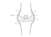
以上のようなインプラント傾斜材料10を用いて、例えば膝骨頭壊死などの関節骨軟骨障害を治療する場合は、図2に示すように、膝骨頭2の壊死部分を切除して、その切除部分3にインプラント傾斜材料10(好ましくは硬骨の成長に有効なBMP、EP4あるいはPRPを低気孔率表層部1aやこれに連なる気孔率の比較的低い内層部に含有させ、軟骨に有効な生物学的成長因子であるTGF−β、b−FGFを高気孔率表層部1bやこれに連なる気孔率の比較的高い内層部に含有させたもの)を、高気孔率表層部1bが軟骨4側となるように面一に埋入固定する。このようにインプラント傾斜材料10を埋入すると、高気孔率表層部1bがその表面に接触する体液や内部の連続気孔に浸透する体液によって表面と内部から速やかに加水分解され、生体活性なバイオセラミックス粉体によって、軟骨4と接する高気孔率表層部1bの周側面に軟骨組織が極く短期間で誘導形成されて、高気孔率表層部1bと軟骨4が結合し、その後、高気孔率表層部1bやこれに連なる気孔率の比較的高い内層部がすみやかに軟骨組織と全置換されて消失する。一方、連続気孔質複合体1の低気孔率表層部1aやこれに連なる気孔率の比較的低い内層部は、加水分解がある程度進行するけれども、高気孔率表層部1bが軟骨組織に置換される頃までは十分な強度を維持する。そして、加水分解の更なる進行に伴って、膝関節頭2の硬骨組織がバイオセラミックス粉体の骨伝導能により低気孔率表層部1aやこれに連なる気孔率の比較的低い内層部に伝導形成され、最終的には硬骨組織と置換されて消失する。また、この連続気孔質複合体1に含まれていたバイオセラミックス粉体も、完全に吸収されて消失する。これによって、膝関節骨軟骨障害部分は完全に修復、再建される。
In the case of treating an articular osteochondral disorder such as knee osteonecrosis using the
図3は、本発明の他の実施形態に係るインプラント傾斜材料の断面図である。 FIG. 3 is a cross-sectional view of an implant gradient material according to another embodiment of the present invention.
このインプラント傾斜材料11は、前述したインプラント傾斜材料10と同様に、生体内吸収性かつ生体活性なバイオセラミックス粉体を含んだ生体内分解吸収性ポリマーの連続気孔質複合体1からなる円柱状のものであるが、その気孔率が10〜90%の範囲内、好ましくは20〜80%の範囲内で、連続気孔質複合体1の中央部1cから周囲の表層部1dに近づくほど高くなるように順次連続的に変化している点、及び、バイオセラミックス粉体が30〜80質量%の範囲内で、連続気孔質複合体の中央部1cから周囲の表層部1dに近づくほど高くなるように順次連続的に変化している点で、前述したインプラント傾斜材料10と相違している。
This
このようなインプラント傾斜材料11を関節骨の切除部分3に埋入すると、前述したインプラント傾斜材料10の作用効果に加えて、切除部分3の内面に接触する気孔率及びバイオセラミックス粉体含有率の高い表層部1dに硬骨組織が速やかに伝導形成され、短期間で関節骨の切除部分3の内面と結合して固定されるという利点がある。
When such an
図4は、本発明の更に他の実施形態に係るインプラント傾斜材料の断面図である。 FIG. 4 is a cross-sectional view of an implant gradient material according to still another embodiment of the present invention.
このインプラント傾斜材料12も、前述したインプラント傾斜材料10と同様に、生体内吸収性かつ生体活性なバイオセラミックス粉体を含んだ生体内分解吸収性ポリマーの連続気孔質複合体1からなるものであるが、角柱状に形成されている点、及び、気孔率が10〜90%の範囲内、好ましくは20〜80%の範囲内で、連続気孔質複合体1の中間部1eから上下両側の表層部1f,1fに近づくほど高くなるように順次連続的に変化している点、及び、バイオセラミックス粉体が30〜80質量%の範囲内で、連続気孔質複合体1の中間部1eから上下両側の表層部1f,1fに近づくほど高くなるように順次連続的に変化している点で、前述したインプラント傾斜材料10と相違している。
This
このようなインプラント傾斜材料12は、例えば、図5に示すように、脊椎、腰椎、頸椎などの関節部の椎骨5,5間にスペーサとして挿入される。このように挿入すると、上下の椎骨5,5に接する気孔率及びバイオセラミックス粉体含有率の高い表層部1f,1fは速やかに加水分解し、上下の椎骨5,5から骨組織が表層部1f,1fに伝導形成されて、短期間で結合するため、インプラント傾斜材料12が関節部から脱転することはない。そして、表層部1f,1fは早期に骨組織と全置換して消失し、中間部1eはある程度の期間、強度を維持するが、その後、骨組織の伝導形成が進行し、最終的には骨組織と全置換して消失する。
For example, as shown in FIG. 5, the
上記のように椎骨間にスペーサとして挿入されるインプラント傾斜材料12は、気孔率の高い上下の表層部1f,1fの厚みが大きくなると、該表層部1f,1fが上下から圧縮されて椎骨5,5の上下間隔が小さくなる恐れがあるので、上下の表層部1f,1fの厚みは0.1〜2.0mm程度と薄くするのがよい。また、このような恐れを完全になくすためには、このインプラント傾斜材料12を90度回転させて低気孔率の中間部1eの左右両側に高気孔率の表層部1f,1fを位置させた状態で椎骨5,5間に挿入すれば良い。このようにすると、強度のある低気孔率の中間部1eによって上下の椎骨5,5の間隔を確実に保ったまま、左右両側の高気孔率表層部1f,1fの上下端面により上下の椎骨5,5と早期に橋渡し状態で結合してインプラント複合材料12の脱転を防止することができる。
As described above, the
図6は、本発明の更に他の実施形態に係るインプラント傾斜材料の断面図である。 FIG. 6 is a cross-sectional view of an implant gradient material according to still another embodiment of the present invention.
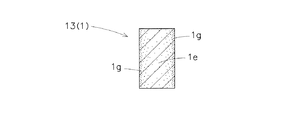
このインプラント傾斜材料13も、前述したインプラント傾斜材料10と同様に、生体内吸収性かつ生体活性なバイオセラミックス粉体を含んだ生体内分解吸収性ポリマーの連続気孔質複合体1からなるものであるが、靱帯の骨関節付着部の再建、補強に適合するようにピース状(小片状)に形成されている点、及び、気孔率が10〜90%の範囲内、好ましくは20〜80%の範囲内で、連続気孔質複合体1の中間部1eから左右両側の表層部1g,1gに近づくほど高くなるように順次連続的に変化している点、及び、バイオセラミックス粉体が30〜80質量%の範囲内で、連続気孔質複合体1の中間部1eから左右両側の表層部1g,1gに近づくほど高くなるように順次連続的に変化している点で、前述したインプラント傾斜材料10と相違している。
This
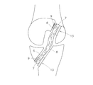
このインプラント傾斜材料13は、図9に示すように、靱帯の骨関節付着部の再建もしくは補強に使用されるものである。即ち、図9に示すように骨関節の双方の骨に孔6,6をあけ、靱帯8の両端部7,7を孔6,6に通し、靱帯8の両端部7,7と孔6の片側内面との間にインプラント傾斜材料13を挟んで、該両端部7,7と孔6の反対側内面との間にインターフェアランススクリュー9をねじ込んで固定すればよい。このようにすると、インプラント傾斜材料14の両側の高気孔率表層部1g,1gが靱帯8の両端部7、7と孔6の内面に結合し、その後すみやかに骨組織と全置換して消失すると共に、強度のある低気孔率中間部1eもやがては骨組織に全置換して消失するため、靱帯8の両端部7,7は全置換した骨組織を介して孔6に結合される。その場合、インターフェアランススクリュー9が生体活性なバイオセラミックス粉体を含む生体内分解吸収性ポリマーからなるものであれば、このスクリュー9もやがては骨組織に置換されて孔6の内面と靱帯8の両端部7,7に結合するため、靱帯付着部分の固定強度は一層向上することになる。
As shown in FIG. 9, the
尚、上記のインプラント傾斜材料11,12,13のいずれにも、前述の生物学的骨成長因子や生体由来の骨芽細胞を含浸させることが好ましいことは言うまでもない。
Needless to say, any of the
1 連続気孔質複合体
1a 片側の表層部
1b 反対側の表層部
1c 中央部
1d 周囲の表層部
1e 中間部
1f 上下両側の表層部
1g 左右両側の表層部
2 関節骨頭
3 関節骨頭の切除部分
4 軟骨
8 靱帯
10,11,12,13 インプラント傾斜材料
DESCRIPTION OF
Claims (7)
Priority Applications (9)
| Application Number | Priority Date | Filing Date | Title |
|---|---|---|---|
| JP2006066291A JP2007236802A (en) | 2006-03-10 | 2006-03-10 | Material for implant inclination |
| CA002643586A CA2643586A1 (en) | 2006-03-10 | 2007-03-08 | Implant composite material |
| EP07738054A EP2005975A4 (en) | 2006-03-10 | 2007-03-08 | Composite implant material |
| CNA2007800086265A CN101400381A (en) | 2006-03-10 | 2007-03-08 | Composite implant material |
| AU2007225892A AU2007225892A1 (en) | 2006-03-10 | 2007-03-08 | Composite implant material |
| US12/282,205 US20090157194A1 (en) | 2006-03-10 | 2007-03-08 | Implant composite material |
| PCT/JP2007/054564 WO2007105600A1 (en) | 2006-03-10 | 2007-03-08 | Composite implant material |
| KR1020087022123A KR20080108447A (en) | 2006-03-10 | 2007-03-08 | Composite implant material |
| TW096108172A TW200803803A (en) | 2006-03-10 | 2007-03-09 | Composite implant material |
Applications Claiming Priority (1)
| Application Number | Priority Date | Filing Date | Title |
|---|---|---|---|
| JP2006066291A JP2007236802A (en) | 2006-03-10 | 2006-03-10 | Material for implant inclination |
Publications (1)
| Publication Number | Publication Date |
|---|---|
| JP2007236802A true JP2007236802A (en) | 2007-09-20 |
Family
ID=38582922
Family Applications (1)
| Application Number | Title | Priority Date | Filing Date |
|---|---|---|---|
| JP2006066291A Pending JP2007236802A (en) | 2006-03-10 | 2006-03-10 | Material for implant inclination |
Country Status (2)
| Country | Link |
|---|---|
| JP (1) | JP2007236802A (en) |
| CN (1) | CN101400381A (en) |
Cited By (8)
| Publication number | Priority date | Publication date | Assignee | Title |
|---|---|---|---|---|
| JP2009095522A (en) * | 2007-10-18 | 2009-05-07 | National Institute For Materials Science | Porous scaffold material |
| JP2009119206A (en) * | 2007-11-11 | 2009-06-04 | Univ Nagoya | Multi-layer porous scaffold and its manufacture process |
| JP2010167040A (en) * | 2009-01-21 | 2010-08-05 | Ngk Spark Plug Co Ltd | Medicine slowly releasing body |
| JP2012501810A (en) * | 2008-09-12 | 2012-01-26 | アーティキュリンクス, インコーポレイテッド | Tether-based orthopedic device delivery method |
| WO2012036286A1 (en) * | 2010-09-16 | 2012-03-22 | 国立大学法人大阪大学 | Artificial bone, artificial bone manufacturing device, and artificial bone manufacturing method |
| JP2013531515A (en) * | 2010-05-24 | 2013-08-08 | エピサーフ メディカル エービー | Cartilage treatment implant |
| JP2019034071A (en) * | 2017-08-21 | 2019-03-07 | 日本特殊陶業株式会社 | Vertebral body spacer |
| JP2022068212A (en) * | 2016-03-24 | 2022-05-09 | ロケート・バイオ・リミテッド | Scaffold material, methods, and uses |
Families Citing this family (10)
| Publication number | Priority date | Publication date | Assignee | Title |
|---|---|---|---|---|
| AT516116B1 (en) | 2014-08-05 | 2016-05-15 | Dietmar Dr Sonnleitner | Method for producing a multilayer film |
| CN105534619A (en) * | 2016-01-28 | 2016-05-04 | 江苏奥康尼医疗科技发展有限公司 | Cartilage repair and compensation prosthesis |
| CN108451673A (en) * | 2017-02-20 | 2018-08-28 | 中国科学院沈阳自动化研究所 | A kind of Invasive lumbar fusion device material-structure |
| JP7402393B2 (en) * | 2017-09-03 | 2023-12-21 | エボニック オペレーションズ ゲーエムベーハー | Biocompatible polymer powders for additive manufacturing |
| CN108969152A (en) * | 2018-09-06 | 2018-12-11 | 北京安颂科技有限公司 | A kind of acetabular component and artificial hip joint |
| CN109528356B (en) * | 2019-01-21 | 2021-01-26 | 北京爱康宜诚医疗器材有限公司 | Composite insert |
| CN111281610B (en) * | 2019-12-30 | 2023-08-18 | 雅博尼西医疗科技(苏州)有限公司 | Porous structure and connection structure of substrate |
| WO2021183778A1 (en) * | 2020-03-12 | 2021-09-16 | Smith & Nephew, Inc. | Tissue repair implant and compositions and method of implantation |
| CN114642771A (en) * | 2020-12-21 | 2022-06-21 | 立心(深圳)医疗器械有限公司 | Artificial ligament with traction unit and preparation method thereof |
| CN112957539B (en) * | 2021-02-01 | 2022-05-13 | 北京大学第三医院(北京大学第三临床医学院) | Zinc-manganese-magnesium alloy interface screw for reconstruction and fixation of anterior cruciate ligament |
Citations (4)
| Publication number | Priority date | Publication date | Assignee | Title |
|---|---|---|---|---|
| JPH02271856A (en) * | 1989-04-14 | 1990-11-06 | Mitsubishi Materials Corp | Ceramics artificial bone and production thereof |
| JP2000157626A (en) * | 1998-11-27 | 2000-06-13 | Takiron Co Ltd | Concentration gradient material |
| JP2001206787A (en) * | 2000-01-19 | 2001-07-31 | Natl Inst For Research In Inorganic Materials Mext | Calcium phosphate porous sintered compact and method of producing the same |
| WO2003045460A1 (en) * | 2001-11-27 | 2003-06-05 | Takiron Co., Ltd. | Implant material and process for producing the same |
-
2006
- 2006-03-10 JP JP2006066291A patent/JP2007236802A/en active Pending
-
2007
- 2007-03-08 CN CNA2007800086265A patent/CN101400381A/en active Pending
Patent Citations (4)
| Publication number | Priority date | Publication date | Assignee | Title |
|---|---|---|---|---|
| JPH02271856A (en) * | 1989-04-14 | 1990-11-06 | Mitsubishi Materials Corp | Ceramics artificial bone and production thereof |
| JP2000157626A (en) * | 1998-11-27 | 2000-06-13 | Takiron Co Ltd | Concentration gradient material |
| JP2001206787A (en) * | 2000-01-19 | 2001-07-31 | Natl Inst For Research In Inorganic Materials Mext | Calcium phosphate porous sintered compact and method of producing the same |
| WO2003045460A1 (en) * | 2001-11-27 | 2003-06-05 | Takiron Co., Ltd. | Implant material and process for producing the same |
Cited By (9)
| Publication number | Priority date | Publication date | Assignee | Title |
|---|---|---|---|---|
| JP2009095522A (en) * | 2007-10-18 | 2009-05-07 | National Institute For Materials Science | Porous scaffold material |
| JP2009119206A (en) * | 2007-11-11 | 2009-06-04 | Univ Nagoya | Multi-layer porous scaffold and its manufacture process |
| JP2012501810A (en) * | 2008-09-12 | 2012-01-26 | アーティキュリンクス, インコーポレイテッド | Tether-based orthopedic device delivery method |
| JP2010167040A (en) * | 2009-01-21 | 2010-08-05 | Ngk Spark Plug Co Ltd | Medicine slowly releasing body |
| JP2013531515A (en) * | 2010-05-24 | 2013-08-08 | エピサーフ メディカル エービー | Cartilage treatment implant |
| WO2012036286A1 (en) * | 2010-09-16 | 2012-03-22 | 国立大学法人大阪大学 | Artificial bone, artificial bone manufacturing device, and artificial bone manufacturing method |
| JP2022068212A (en) * | 2016-03-24 | 2022-05-09 | ロケート・バイオ・リミテッド | Scaffold material, methods, and uses |
| JP2019034071A (en) * | 2017-08-21 | 2019-03-07 | 日本特殊陶業株式会社 | Vertebral body spacer |
| JP7002885B2 (en) | 2017-08-21 | 2022-01-20 | 日本特殊陶業株式会社 | Vertebral spacer |
Also Published As
| Publication number | Publication date |
|---|---|
| CN101400381A (en) | 2009-04-01 |
Similar Documents
| Publication | Publication Date | Title |
|---|---|---|
| JP2007236802A (en) | Material for implant inclination | |
| US20220062004A1 (en) | Porous composite biomaterials and related methods | |
| US11179243B2 (en) | Implantable devices | |
| JP2007236803A (en) | Implant composite material | |
| KR100903761B1 (en) | Implant material and process for producing the same | |
| CA2503904C (en) | Devices for treating defects in the tissue of a living being | |
| EP2802363B1 (en) | Composite device that combines porous metal and bone stimuli | |
| WO2007105600A1 (en) | Composite implant material | |
| Zhang et al. | Repair of rabbit femoral condyle bone defects with injectable nanohydroxyapatite/chitosan composites | |
| EP2675490B1 (en) | Non-resorbable polymer - ceramic composite implant materials | |
| JP2011515162A (en) | Methods, devices and compositions for adhering hydrated polymer implants to bone | |
| JP2006517842A (en) | Apparatus and method for in situ forming intervertebral fusion | |
| Gao et al. | Morphological and biomechanical difference in healing in segmental tibial defects implanted with Biocoral® or tricalcium phosphate cylinders | |
| JP4170744B2 (en) | Biomaterial for artificial cartilage | |
| JP2008029745A (en) | Bone joint material and bone joint material set for enhancement of bone adhesion | |
| JP2008029746A (en) | Interference screw for fixing tendon or ligament, and fixation set for tendon or ligament | |
| Taylor et al. | Recent advances in bone graft technologies | |
| Tanner | Hard tissue applications of biocomposites | |
| US20070173949A1 (en) | Bonding system for orthopedic implants | |
| JP5145375B2 (en) | Filler for bonding between implant and living tissue | |
| Barua et al. | Fiber nanobiocompositions for cranioplasty and other orthopedic applications | |
| JP4280968B2 (en) | Implant complex | |
| JP4988996B2 (en) | Filler for bonding between implant and living tissue | |
| JP4327432B2 (en) | Implant material | |
| Joji et al. | Biomaterials and Structural Fat Grafting |
Legal Events
| Date | Code | Title | Description |
|---|---|---|---|
| A621 | Written request for application examination |
Free format text: JAPANESE INTERMEDIATE CODE: A621 Effective date: 20090210 |
|
| A131 | Notification of reasons for refusal |
Free format text: JAPANESE INTERMEDIATE CODE: A131 Effective date: 20120118 |
|
| A02 | Decision of refusal |
Free format text: JAPANESE INTERMEDIATE CODE: A02 Effective date: 20120530 |