JP2007236414A - Ultrasound endoscope - Google Patents
Ultrasound endoscope Download PDFInfo
- Publication number
- JP2007236414A JP2007236414A JP2006058708A JP2006058708A JP2007236414A JP 2007236414 A JP2007236414 A JP 2007236414A JP 2006058708 A JP2006058708 A JP 2006058708A JP 2006058708 A JP2006058708 A JP 2006058708A JP 2007236414 A JP2007236414 A JP 2007236414A
- Authority
- JP
- Japan
- Prior art keywords
- distal end
- ultrasonic
- observation
- treatment instrument
- endoscope
- Prior art date
- Legal status (The legal status is an assumption and is not a legal conclusion. Google has not performed a legal analysis and makes no representation as to the accuracy of the status listed.)
- Pending
Links
- 238000002604 ultrasonography Methods 0.000 title description 13
- 238000003780 insertion Methods 0.000 claims abstract description 46
- 230000037431 insertion Effects 0.000 claims abstract description 46
- 230000005540 biological transmission Effects 0.000 claims description 28
- 230000003287 optical effect Effects 0.000 claims description 26
- XLYOFNOQVPJJNP-UHFFFAOYSA-N water Substances O XLYOFNOQVPJJNP-UHFFFAOYSA-N 0.000 description 29
- 238000005286 illumination Methods 0.000 description 20
- 238000005452 bending Methods 0.000 description 13
- 238000009795 derivation Methods 0.000 description 5
- 230000000007 visual effect Effects 0.000 description 5
- 238000010586 diagram Methods 0.000 description 4
- 210000002784 stomach Anatomy 0.000 description 4
- 239000012530 fluid Substances 0.000 description 2
- 239000007787 solid Substances 0.000 description 2
- 230000009471 action Effects 0.000 description 1
- 238000003491 array Methods 0.000 description 1
- 230000008901 benefit Effects 0.000 description 1
- 238000003745 diagnosis Methods 0.000 description 1
- 230000006872 improvement Effects 0.000 description 1
- 239000000463 material Substances 0.000 description 1
- 239000002184 metal Substances 0.000 description 1
- 230000004048 modification Effects 0.000 description 1
- 238000012986 modification Methods 0.000 description 1
- 230000002093 peripheral effect Effects 0.000 description 1
- 230000009467 reduction Effects 0.000 description 1
- 239000011347 resin Substances 0.000 description 1
- 229920005989 resin Polymers 0.000 description 1
Images
Classifications
-
- A—HUMAN NECESSITIES
- A61—MEDICAL OR VETERINARY SCIENCE; HYGIENE
- A61B—DIAGNOSIS; SURGERY; IDENTIFICATION
- A61B8/00—Diagnosis using ultrasonic, sonic or infrasonic waves
- A61B8/12—Diagnosis using ultrasonic, sonic or infrasonic waves in body cavities or body tracts, e.g. by using catheters
-
- A—HUMAN NECESSITIES
- A61—MEDICAL OR VETERINARY SCIENCE; HYGIENE
- A61B—DIAGNOSIS; SURGERY; IDENTIFICATION
- A61B1/00—Instruments for performing medical examinations of the interior of cavities or tubes of the body by visual or photographical inspection, e.g. endoscopes; Illuminating arrangements therefor
- A61B1/012—Instruments for performing medical examinations of the interior of cavities or tubes of the body by visual or photographical inspection, e.g. endoscopes; Illuminating arrangements therefor characterised by internal passages or accessories therefor
- A61B1/018—Instruments for performing medical examinations of the interior of cavities or tubes of the body by visual or photographical inspection, e.g. endoscopes; Illuminating arrangements therefor characterised by internal passages or accessories therefor for receiving instruments
-
- A—HUMAN NECESSITIES
- A61—MEDICAL OR VETERINARY SCIENCE; HYGIENE
- A61B—DIAGNOSIS; SURGERY; IDENTIFICATION
- A61B8/00—Diagnosis using ultrasonic, sonic or infrasonic waves
- A61B8/44—Constructional features of the ultrasonic, sonic or infrasonic diagnostic device
- A61B8/4444—Constructional features of the ultrasonic, sonic or infrasonic diagnostic device related to the probe
- A61B8/445—Details of catheter construction
Landscapes
- Health & Medical Sciences (AREA)
- Life Sciences & Earth Sciences (AREA)
- Surgery (AREA)
- Medical Informatics (AREA)
- Biophysics (AREA)
- Pathology (AREA)
- Radiology & Medical Imaging (AREA)
- Engineering & Computer Science (AREA)
- Biomedical Technology (AREA)
- Heart & Thoracic Surgery (AREA)
- Physics & Mathematics (AREA)
- Molecular Biology (AREA)
- Nuclear Medicine, Radiotherapy & Molecular Imaging (AREA)
- Animal Behavior & Ethology (AREA)
- General Health & Medical Sciences (AREA)
- Public Health (AREA)
- Veterinary Medicine (AREA)
- Optics & Photonics (AREA)
- Ultra Sonic Daignosis Equipment (AREA)
- Endoscopes (AREA)
Abstract
【課題】挿入部の挿入方向前方の光学的な観察を行え、挿入方向前方深部の超音波画像に描出される目的部位に向けて、処置具の導出を確実に行える超音波内視鏡を提供すること。
【解決手段】超音波内視鏡1は、体腔内に挿入される挿入部2の先端に、可撓管部2cより前方側に配設された先端硬質部2aと、 先端硬質部2aの長手方向中心軸L1の前方側に対して平行な面を走査する超音波振動子部10と、 超音波振動子部10の先端側端面に開口して長手方向中心軸L4が先端硬質部の長手方向中心軸L1に平行な処置具挿通用チャンネル孔27とを具備する。
【選択図】図5Provided is an ultrasonic endoscope that can optically observe the front of an insertion portion in the insertion direction and can reliably lead out a treatment tool toward a target portion depicted in an ultrasonic image deep in the front of the insertion direction. To do.
An ultrasonic endoscope 1 includes a distal end hard portion 2a disposed on a front side of a flexible tube portion 2c at a distal end of an insertion portion 2 to be inserted into a body cavity, and a longitudinal length of the distal end rigid portion 2a. The ultrasonic transducer section 10 that scans a plane parallel to the front side of the direction center axis L1 and the longitudinal center axis L4 is the longitudinal direction of the distal end hard section that opens at the distal end side end surface of the ultrasonic transducer section 10 And a treatment instrument insertion channel hole 27 parallel to the central axis L1.
[Selection] Figure 5
Description
本発明は、挿入部の先端部に観察光学系と、処置具導出口と、コンベックス型の超音波送受信部とを備える超音波内視鏡に関する。 The present invention relates to an ultrasonic endoscope provided with an observation optical system, a treatment instrument outlet, and a convex ultrasonic transmission / reception unit at a distal end portion of an insertion unit.
従来より、体腔内を超音波診断する超音波内視鏡として、コンベックス型の超音波送受信部を有するものが知られている。コンベックス型の超音波送受信部は、複数の振動子アレイを凸型の円弧状に配列して構成される。 2. Description of the Related Art Conventionally, as an ultrasonic endoscope for ultrasonic diagnosis in a body cavity, one having a convex type ultrasonic transmission / reception unit is known. The convex-type ultrasonic transmission / reception unit is configured by arranging a plurality of transducer arrays in a convex arc shape.
コンベックス型の超音波送受信部を有する超音波内視鏡として、例えば特許文献1の超音波内視鏡がある。この超音波内視鏡は、先端硬質部に観察光学系に加えて、超音波走査領域を前方斜視とする超音波送受信部を備えている。
しかしながら、特許文献1の超音波内視鏡は、前方斜視の超音波走査領域に対して処置具を突出させる構成である。つまり、処置具は、挿入部の挿入方向に対して斜めに導出される。 However, the ultrasonic endoscope of Patent Document 1 has a configuration in which the treatment tool is protruded with respect to the ultrasonic scanning region of the front perspective view. That is, the treatment instrument is led out obliquely with respect to the insertion direction of the insertion portion.
このため、図11に示すように胃内において、処置具100を胃壁101を介して処置部位102に向けて導出させるとき、まず、術者は、破線に示すように内視鏡挿入部110を傾け、その状態で超音波送受信部111の振動子レンズ面112を胃壁101に密着させる。次に、術者は、その密着状態を保持しつつ、処置具100を処置部位102に向けて導入する操作を行う。このとき、内視鏡挿入部110の挿入方向(押圧方向)は破線の矢印A方向に、処置具100の導出方向は破線の矢印B方向になる。
Therefore, when the
このため、処置具100を内視鏡挿入部110から突出させたとき、導出方向と、挿入方向とが一致していないことによって、実線に示すように内視鏡挿入部110が移動されることがある。すると、内視鏡挿入部110の挿入方向が実線の矢印C方向に、処置具100の導入方向が実線の矢印D方向に変化される。つまり、処置具100の導出方向が処置部位102から外れて、所望の処置が困難になる。
For this reason, when the
本発明は上記事情に鑑みてなされたものであり、挿入部の挿入方向前方の光学的な観察を行え、挿入方向前方深部の超音波画像に描出される目的部位に向けて、処置具の導出を確実に行える超音波内視鏡を提供することを目的にしている。 The present invention has been made in view of the above circumstances, and allows the optical observation of the insertion portion forward in the insertion direction to be performed, and the derivation of the treatment tool toward the target site depicted in the ultrasound image of the deep portion in the insertion direction forward. An object of the present invention is to provide an ultrasonic endoscope capable of reliably performing the above.
本発明の超音波内視鏡は、体腔内に挿入される挿入部の先端に、可撓管部より前方側に配設された先端硬質部と、前記先端硬質部の長手方向中心軸の前方側に対して平行な面を走査する超音波振動子部と、前記超音波振動子部の先端側端面に開口して孔長手方向中心軸が前記先端硬質部の長手方向中心軸に平行な処置具挿通用チャンネル孔とを具備する。 An ultrasonic endoscope according to the present invention includes a distal end rigid portion disposed on the front side of a flexible tube at a distal end of an insertion portion to be inserted into a body cavity, and a front side of a longitudinal central axis of the distal end rigid portion. An ultrasonic transducer unit that scans a plane parallel to the side, and a treatment that opens at the end surface on the distal end side of the ultrasonic transducer unit and that the central axis in the longitudinal direction of the hole is parallel to the central axis in the longitudinal direction of the distal rigid unit A tool insertion channel hole.
この構成によれば、挿入方向前方深部の超音波画像を得られる。そして、超音波画像に描出される目的部位に対して、処置具導出口から処置具を導出させたとき、処置具の導出される方向と挿入部の挿入方向とが略一致する。したがって、処置具を導出させる際の力量が効率良く、該処置具に伝達される。 According to this configuration, it is possible to obtain an ultrasonic image of a deep part in the front in the insertion direction. Then, when the treatment instrument is derived from the treatment instrument outlet for the target site depicted in the ultrasound image, the direction in which the treatment instrument is derived substantially coincides with the insertion direction of the insertion portion. Therefore, the amount of power for deriving the treatment tool is efficiently transmitted to the treatment tool.
本発明によれば、挿入方向前方深部の超音波画像に描出される目的部位に向けて、処置具の導出を確実に行える超音波内視鏡を実現できる。 ADVANTAGE OF THE INVENTION According to this invention, the ultrasonic endoscope which can derive | lead-out a treatment tool reliably toward the target site | part drawn on the ultrasonic image of the deep part ahead of an insertion direction is realizable.
以下、図面を参照して本発明の実施形態を説明する。 Hereinafter, embodiments of the present invention will be described with reference to the drawings.
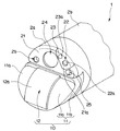
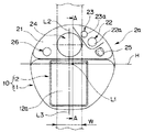
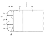
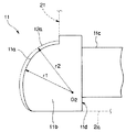
図1乃至図10は本発明の一実施形態に係り、図1は超音波内視鏡の構成を説明する図、図2は超音波内視鏡の先端部を示す斜視図、図3は図2に示す先端部を正面から見た正面図、図4は図2に示す先端部を上側から見た上面図、図5は図3のA−A線断面図、図6は複数の圧電素子を配列して構成される超音波送受信部、及び超音波送受信部の超音波観測領域と、処置具導出口から導出された処置具との関係を説明する図、図7はアーチファクトが現れた超音波画像例を示す図、図8は図6に示す超音波送受信部で描出された超音波画像例を示す図、図9はノーズピースの組織当接面と超音波送受信部の振動子レンズ面との関係を説明する図、図10は超音波内視鏡の作用を説明する図である。 1 to 10 relate to an embodiment of the present invention, FIG. 1 is a diagram for explaining the configuration of an ultrasonic endoscope, FIG. 2 is a perspective view showing a distal end portion of the ultrasonic endoscope, and FIG. FIG. 4 is a top view of the tip portion shown in FIG. 2 as viewed from above, FIG. 5 is a cross-sectional view taken along line AA in FIG. 3, and FIG. 6 is a plurality of piezoelectric elements. FIG. 7 is a diagram for explaining the relationship between the ultrasonic transmission / reception unit configured by arranging the ultrasonic transmission / reception unit, the ultrasonic observation region of the ultrasonic transmission / reception unit, and the treatment instrument derived from the treatment instrument outlet. FIG. FIG. 8 is a diagram showing an example of an ultrasound image drawn by the ultrasound transmission / reception unit shown in FIG. 6, and FIG. 9 is a tissue contact surface of the nosepiece and a transducer lens surface of the ultrasound transmission / reception unit. FIG. 10 is a diagram for explaining the operation of the ultrasonic endoscope.
図1に示すように本実施形態の超音波内視鏡1は、体腔内に挿入される細長の挿入部2と、この挿入部2の基端に設けられた操作部3と、この操作部3の側部から延出するユニバーサルコード4とを備えて構成されている。ユニバーサルコード4の基端部には内視鏡コネクタ5が設けられている。内視鏡コネクタ5の側部からは超音波ケーブル6が延出されている。超音波ケーブル6の基端部には超音波コネクタ7が設けられている。
As shown in FIG. 1, an ultrasonic endoscope 1 according to this embodiment includes an
挿入部2は、先端側から順に硬質部材で形成された先端硬質部2aと、湾曲自在に構成された湾曲部2bと、この湾曲部2bの基端から操作部3の先端に至る長尺で可撓性を有する可撓管部2cとを連接して構成されている。なお、符号10は超音波振動子部であり、後述するコンベックス型の超音波送受信部を備えている。超音波送受信部は、挿入軸方向に対して前方方向を走査する超音波観測領域10Aを形成する。言い換えれば、超音波振動子部10は前方方向を走査する超音波観測領域10Aを有している。
The
操作部3には湾曲操作を行うためのアングルノブ3aが設けられている。また、操作部3には送気及び送水の操作を行う送気送水ボタン3bと、吸引を行う吸引ボタン3cとが設けられている。さらに、操作部3には処置具を体腔内に導くための処置具挿入口3dが設けられている。
The
図2に示すように挿入部2の先端硬質部2aには超音波による音響的画像情報を得るための超音波振動子部10が設けられている。超音振動子部10は、筐体であるノーズピース11と、超音波送受信部12とを備えて構成されている。超音波送受信部12はノーズピース11の略中央部に形成された切り欠き部に一体的に配設されている。
As shown in FIG. 2, the distal end
ノーズピース11を構成する組織当接面11a、及び超音波送受信部12の振動子レンズ面12aは、先端硬質部2aの先端面21から突出して構成されている。
The
一方、先端硬質部2aの先端面21には、観察光学系22を構成する観察窓22aと、照明光学系23を構成する照明窓23aと、穿刺針等の処置具が導出される処置具導出口(以下、導出口と略記する)24と、観察窓22aに向けて水や空気等の流体を噴出する送気送水ノズル25と、前方に向けて送水を行うための副送水チャンネル口26とが設けられている。
On the other hand, on the
すなわち、本実施形態の超音波内視鏡1において先端面21は、図3に示すように、先端硬質部2aの長手方向中心軸L1を通過する水平線Hより上側を内視鏡観察のための領域、下側を超音波観測のための領域に分割されている。
That is, in the ultrasonic endoscope 1 of the present embodiment, as shown in FIG. 3, the
そして、導出口24の垂直方向中心線L2と、超音波送受信部12の振動子レンズ面12aの垂直方向中心線L3とが略一直線上に配置される構成になっている。
The vertical center line L2 of the
また、導出口24の径寸法は、振動子レンズ面12aから放射される超音波によって形成される、二点鎖線に示す超音波観測領域10Aの幅寸法W内に収まる大きさで形成されている。このことによって、導出口24から導出される処置具は、確実に超音波観測領域10A内を移動する。
The diameter of the
観察窓22a、照明窓23a、及び送気送水ノズル25は、導出口24に対して例えば図中右側である一面側にまとめて配置されている。また、観察窓22a、照明窓23a、及び送気送水ノズル25は、超音波観測領域10Aより外側に配置されている。
The
そして、観察窓22a、照明窓23a、送気送水ノズル25のうち、該送気送水ノズル25の配置位置を、超音波観測領域10Aから最も離れた位置となるように設定している。これは、図4に示すように送気送水ノズル25は、先端硬質部2aの先端面21に対して突出して設けられるためである。つまり、突出した送気送水ノズル25が超音波観測領域10Aに近接配置されたことにより、振動子レンズ面12aから放射された超音波が送気送水ノズル25で反射されて、超音波画像中に送気送水ノズルの画像が写り込むおそれがあるためである。
Of the
また、本実施形態においては、照明窓23a、観察窓22a、及び送気送水ノズル25の配置位置を、観察性能の向上、洗滌性の向上、及び内視鏡先端部外径寸法の小径化を図る目的を考慮して、一直線上に配置している。
Further, in the present embodiment, the arrangement positions of the
そして、観察窓22aについては、その配置位置を、観察光学系22の観察視野範囲(図5の一点鎖線で示す符号22Aの範囲参照)を考慮して超音波振動子部10に対して離れた図中上方向位置としている。このことによって、内視鏡観察の際、超音波振動子部10によって観察視野が遮られて光学像の(内視鏡画像)の一部が欠ける不具合が解消される。
The
一方、照明窓23aについては、その配置位置は、照明光学系23の照明光照射範囲(図5の二点鎖線で示す符号23Aの範囲参照)を考慮して超音波振動子部10から離れた、観察窓22aよりさらに外周の上方向位置としている。このことによって、内視鏡観察の際、観察画像中に超音波振動子部10の影が映り込む不具合が解消される。
On the other hand, the arrangement position of the
なお、観察窓22a、及び照明窓23aは、先端面21より僅かに突出して構成された観察部用先端面21a内に設けられている。また、副送水チャンネル口26は、観察窓22a、照明窓23a、及び送気送水ノズル25が配置されている一面側とは逆方向の他面側であって、超音波観測領域10Aより外側に配置されている。
Note that the
図5に示すように先端硬質部2aの基端側には湾曲部2bを構成する先端湾曲駒8aが接続固定されている。先端湾曲駒8aには複数の湾曲駒が連接されている。先端湾曲駒8aの所定位置には、上下左右用の湾曲ワイヤ8wのそれぞれの先端部が固設されている。したがって、術者が、アングルノブ3aを適宜操作することにより、その操作に対応する湾曲ワイヤ8wが牽引弛緩されて、湾曲部2bが湾曲さ動作するようになっている。
As shown in FIG. 5, a distal bending piece 8a constituting the bending
これら複数の湾曲駒は湾曲ゴム8gによって被覆されている。湾曲ゴム8gの先端部は、先端硬質部2aに設けられる糸巻き接着部8hによって一体的に固定されている。
The plurality of bending pieces are covered with a bending rubber 8g. The distal end portion of the curved rubber 8g is integrally fixed by a
先端硬質部2aの先端面21、及び観察部用先端面21aは先端硬質部2aの長手方向中心軸L1に対して直交して構成されている。先端硬質部2aには処置具導出口24を構成する処置具挿通用チャンネル孔(以下、処置具用孔と略記する)27、及び配置孔30が形成されている。なお、先端硬質部2aには、孔27、30の他に図示は省略しているが、観察光学系が設けられる貫通孔、照明光学系が設けられる貫通孔、送気送水ノズル25から噴出される流体を供給する送気送水用の貫通孔、副送水チャンネル口26を構成する貫通孔等が備えられている。
The
処置具用孔27の長手方向中心軸L4は、先端硬質部2aの長手方向中心軸L1に対して略平行に形成されている。配置孔30の長手方向中心軸L5は、先端硬質部2aの長手方向中心軸L1に対して略平行に形成されている。また、超音波内視鏡1に備えられる観察光学系の光軸L6、及び照明光学系の光軸L7は、先端硬質部2aの長手方向中心軸L1に対して平行である。したがって、本実施形態の超音波内視鏡1に備えられている観察光学系は、観察視野を前方正面、言い換えれば先端硬質部2aの長手方向中心軸L1の前方側である挿入方向に設定した、いわゆる直視型である。
The longitudinal center axis L4 of the
処置具用孔27の基端側には所定量傾斜して形成されたチューブ連結パイプ28の一端部が連通されている。チューブ連結パイプ28は、その他端部に処置具挿通用チャンネルを構成するチャンネルチューブ29の一端部を連通配置している。チャンネルチューブ29の他端部は、前記処置具挿入口3dに連通している。
One end of a
そして、処置具挿入口3dを介して挿通された処置具はチャンネルチューブ29、チューブ連結パイプ28、処置具用孔27内をスムーズに移動して処置具導出口24から導出される。処置具導出口24から導出される処置具は、先端硬質部2aの長手方向中心軸L1に対して平行に、挿入部挿入方向である前方に向けて導出される。
Then, the treatment instrument inserted through the treatment
つまり、処置具用孔27内に例えば処置具として穿刺針の先端部を配置した状態において、穿刺針を構成する針管を突出させる。すると、針管は、処置具導出口24から先端硬質部2aの長手方向中心軸L1に対して略平行に、観察窓22aを通して観察されている前方正面に向かって突出される。
That is, in the state where the distal end portion of the puncture needle is disposed as the treatment instrument in the
配置孔30には、ノーズピース11の取付部11cが配置される。取付部11cの基端部には絶縁チューブ35の先端部が連通固定されている。絶縁チューブ35の内部には、超音波送受信部12を構成する複数の圧電素子からそれぞれ延出する複数の信号線をひとまとめにした超音波ケーブル34が挿通される。絶縁チューブ35は、挿入部2内を挿通して他端部を操作部3まで延出している。超音波ケーブル34は、挿入部2、操作部3、ユニバーサルコード4、内視鏡コネクタ5、超音波ケーブル6内を挿通して超音波コネクタ7まで延出している。
In the
図5、図6に示すようにノーズピース11の組織当接部の中央部には、超音波振動子部10がある。超音波送受信部12は、例えば複数の圧電素子9と、振動子レンズ面12aとで構成されている。複数の圧電素子9は凸型の円弧を形成するように配列されている。
As shown in FIGS. 5 and 6, the
図6に示すように超音波送受信部12の円弧を形成する複数の圧電素子9を配設して構成された円弧の中心O1は、先端硬質部2aの先端面21より基端側に位置するように構成されている。また、円弧状に配列されている複数の圧電素子9は、一端側である突出口側近傍に超音波を放射する第1圧電素子9Fから他端側である最終圧電素子9Lまで所定間隔で配列されている。
As shown in FIG. 6, the center O1 of the arc formed by arranging a plurality of
第1圧電素子9Fの音軸LFの方向は、先端硬質部2aの先端面21(具体的には導出口24を備える先端面21を規準にしている)に対して角度θ1だけ先端側に傾いて設定されている。
The direction of the sound axis LF of the first
また、第1圧電素子9Fの音軸LFの方向を角度θ1だけ傾けて設定する際、第1圧電素子9Fの指向角θ2を考慮に入れている。具体的には、図中の二点鎖線で囲まれた指向角内に超音波を反射しうる材質、例えば金属や硬質樹脂である先端硬質部2aの少なくとも一部、或いは送気送水ノズル25の少なくとも一部等、が入り込まないように角度θ1を設定している。少なくとも、角度θ1は、指向角θ2の半分の角度よりも大きく設定される。
Further, when the direction of the sound axis LF of the first
先端硬性部2aが指向角の範囲内にある場合、図7に示すようなアーチファクト42が現れる。、しかし、本願の構成によればアーチファクトが、出現せず、図8に示すように処置具像41aが超音波画像40中に明瞭に描写される。これにより、病変部43に対して処置具41を正確に導入することができる。
When the distal end
一方、最終圧電素子9Lの音軸LLの方向は、先端硬質部2aの長手方向中心軸L1に対して平行、又は角度θ3だけ前方にいくにしたがって拡開するように設定している。
On the other hand, the direction of the sound axis LL of the final piezoelectric element 9L is set so as to expand in parallel with the longitudinal center axis L1 of the distal end
このことによって、処置具導出口24から突出された処置具41が先端硬質部2aの長手方向中心軸L1に対して略平行に前方に向かって突出されたとき、処置具41は超音波観測領域10A内を移動し続ける。このため、図8に示すように処置具導出口24から処置具41が僅かに突出された状態から該処置具41が病変部43に穿刺されるまでの処置具像41aが超音波画像40中に明瞭に描写される。
As a result, when the
図2、図4、図5、及び図9に示すようにノーズピース11は、組織当接部11bと取付部11cとを備えている。組織当接部11bは、円弧状の組織当接面11aを備えている。取付部11cは、配置孔30内に配設される。組織当接部11bの基端面は、突き当て面11dであって、段部36の平面部36aに当接配置される。平面部36aは、配置孔30の開口(不図示)を備える。
As shown in FIGS. 2, 4, 5, and 9, the
ノーズピース11の突き当て面11d側から先端に至る外径寸法は、図2乃至図4に示すように先端硬質部2aの先端外径寸法と略同寸法に設定されている。したがって、組織当接面11aを備える組織当接部11bの剛性の向上を図れる。言い換えれば、ノーズピース11の組織当接部11bの強度が増して、安定した突き当てが可能になる。
The outer diameter dimension from the butting
また、図9に示すようにノーズピース11に設けられた組織当接面11aの円弧の半径r1は、超音波送受信部12を構成する二点鎖線に示す振動子レンズ面12aの円弧の半径r2と同寸法、または、僅かに小径に設定されている。また、組織当接面11aの円弧の中心O2、及び前記振動子レンズ面12aの円弧の中心O1は、水平線Hに平行な軸上に位置するように設定されている。
Further, as shown in FIG. 9, the radius r1 of the arc of the
これらのことによって、超音波送受信部12の振動子レンズ面12aと、該超音波送受信部12を挟んで設けられた組織当接部11bの組織当接面11aとが略面一致に構成される。この構成によれば、超音波観察のために超音波振動子部10を体組織に押し当てたとき、組織当接面11aと振動子レンズ面12aとが略均一に体組織に密着する。したがって、超音波振動子部10を体組織に対して安定した状態で押し当て超音波観察像を得ることができる。
As a result, the
また、超音波観測下において、処置具を穿刺する際、図10に示すように先端硬質部2aの長手方向中心軸L1と略同方向である矢印Pで示す挿入部2の挿入方向と、矢印Qで示す処置具41の穿刺方向とが一致している。このことによって、術者が、処置具41を導入する操作を行った際、処置具41に係る力量が効率良く導入力量に変換されて、処置具41の導入性(穿刺性)を向上させることができる。
Further, when puncturing the treatment tool under ultrasonic observation, as shown in FIG. 10, the insertion direction of the
尚、本発明は、以上述べた実施形態のみに限定されるものではなく、発明の要旨を逸脱しない範囲で種々変形実施可能である。 The present invention is not limited to the above-described embodiments, and various modifications can be made without departing from the spirit of the invention.
ところで、前記特許文献1の超音波内視鏡は、観察光学系等を有する先端硬質部の先端側に超音波送受信部が突出している。このため、光学的な観察においては、観察視野範囲の一部が超音波送受信部によって遮られてしまう不具合、或いは光学像中に超音波振動子部の影が生じて観察が妨げられるおそれがあった。一方、超音波観察においては、送気送水ノズル等によって超音波が反射されて、超音波画像中にアーチファクトが発生する不具合が生じる等のおそれがあった。 By the way, in the ultrasonic endoscope of Patent Document 1, the ultrasonic transmission / reception unit protrudes from the distal end side of the distal end hard portion having an observation optical system or the like. For this reason, in the optical observation, there is a possibility that a part of the observation visual field range is blocked by the ultrasonic transmission / reception unit, or that the observation is hindered by the shadow of the ultrasonic transducer unit in the optical image. It was. On the other hand, in ultrasonic observation, there is a possibility that an ultrasonic wave is reflected by an air / water supply nozzle or the like, resulting in a problem that an artifact is generated in the ultrasonic image.
このため、観察光学系を通しての観察、及び超音波振動子部によって得られる超音波画像による観察を、良好に行える超音波内視鏡が望まれている。 For this reason, there is a demand for an ultrasonic endoscope that can satisfactorily perform observation through an observation optical system and observation using an ultrasonic image obtained by an ultrasonic transducer section.
[付記1]
体腔内に挿入される挿入部を構成する可撓管部より前方側に配設された先端硬質部に、前記先端硬質部の長手方向中心軸の前方側に対して平行な面を走査する超音波振動子部と、前記超音波振動子部の前方側走査面に対して処置具を導出させる処置具導出口を構成する、孔長手方向中心軸が前記先端硬質部の長手方向中心軸に平行な処置具挿通用チャンネル孔とを具備する超音波内視鏡において、
先端硬質部の先端面は、観察光学系を構成する観察窓、照明光学系を構成する照明窓、及び少なくとも前記観察窓の表面に流体を噴出する送気送水ノズルを備え、
前記送気送水ノズルは、前記超音波振動子部の有する超音波観測領域外に配置され、
前記観察窓は、前記超音波振動子部の有する超音波観測領域外であって、かつ該超音波振動子部を前記観察光学系の観察視野範囲外にする位置に配置され、
前記照明窓は、前記超音波振動子部の有する超音波観測領域外であって、かつ前記観察窓よりさらに外周側に配置されることを特徴とする超音波内視鏡。
[Appendix 1]
The tip hard part disposed on the front side of the flexible tube constituting the insertion part inserted into the body cavity is scanned with a surface parallel to the front side of the longitudinal center axis of the tip hard part. The longitudinal axis of the hole constituting the treatment instrument outlet for deriving the treatment instrument with respect to the ultrasonic transducer part and the front scanning surface of the ultrasonic transducer part is parallel to the longitudinal central axis of the hard tip part. An ultrasonic endoscope having a channel hole for inserting a treatment instrument,
The distal end surface of the distal end hard portion includes an observation window that constitutes an observation optical system, an illumination window that constitutes an illumination optical system, and an air / water supply nozzle that ejects fluid to at least the surface of the observation window,
The air / water supply nozzle is disposed outside the ultrasonic observation region of the ultrasonic transducer unit,
The observation window is disposed outside the ultrasonic observation region of the ultrasonic transducer unit and at a position where the ultrasonic transducer unit is outside the observation visual field range of the observation optical system,
The ultrasound endoscope, wherein the illumination window is disposed outside an ultrasound observation region of the ultrasound transducer section and further on the outer peripheral side than the observation window.
この構成によれば、超音波振動子部によって観察視野の一部が欠ける不具合、および観察視野内に超音波振動子部の影が生じる不具合が防止される。また、超音波振動子から出射された超音波が送気送水ノズル等で反射されて超音波画像中にアーチファクトが発生することが防止される。 According to this configuration, a problem that a part of the observation visual field is missing due to the ultrasonic transducer part and a problem that the shadow of the ultrasonic vibrator part occurs in the observation visual field are prevented. Further, it is possible to prevent the ultrasonic wave emitted from the ultrasonic transducer from being reflected by the air / water supply nozzle or the like and causing artifacts in the ultrasonic image.
[付記2]
前記観察窓、前記照明窓、及び前記送気送水ノズルのうち、該送気送水ノズルを前記超音波走査面から最も遠い位置に配置させる付記1に記載の超音波内視鏡。
[Appendix 2]
The ultrasonic endoscope according to supplementary note 1, wherein the air supply / water supply nozzle is disposed at a position farthest from the ultrasonic scan plane among the observation window, the illumination window, and the air supply / water supply nozzle.
[付記3]
前記観察窓、前記照明窓、及び前記送気送水ノズルは、一直線上に配置される付記1、又は付記2に記載の超音波内視鏡。
[Appendix 3]
The ultrasound endoscope according to appendix 1 or
1…超音波内視鏡 2a…先端硬質部 9、9F、9L…圧電素子
10…超音波振動子部 10A…超音波観測領域 11……ノーズピース
11a…組織当接面 11b…組織当接部 11c…取付部
11d…突き当て面 12…超音波送受信部 12a…振動子レンズ面
21…先端面 22…観察光学系 22a…観察窓 23…照明光学系
23a…照明窓 24…処置具導出口 25…送気送水ノズル
27…処置具用孔 41…処置具
DESCRIPTION OF SYMBOLS 1 ...
Claims (4)
可撓管部より前方側に配設された先端硬質部と、
前記先端硬質部の長手方向中心軸の前方側に対して平行な面を走査する超音波振動子部と、
前記超音波振動子部の先端側端面に開口して孔長手方向中心軸が前記先端硬質部の長手方向中心軸に平行な処置具挿通用チャンネル孔と、
を具備することを特徴とする超音波内視鏡。 The tip of the insertion part inserted into the body cavity is
A hard tip portion disposed on the front side of the flexible tube portion;
An ultrasonic transducer portion that scans a plane parallel to the front side of the longitudinal center axis of the distal end hard portion; and
A treatment instrument insertion channel hole that opens to the distal end side end surface of the ultrasonic transducer section and whose hole longitudinal central axis is parallel to the longitudinal central axis of the distal rigid section;
An ultrasonic endoscope comprising:
前記ノーズピースは、前記先端硬質部の先端面から突出して、前記振動子レンズ面に面一致で構成される組織当接面を有する組織当接部と、前記先端硬質部に取り付けられる取付部とを有することを特徴とする請求1に記載の超音波内視鏡。 The ultrasonic transducer unit includes a nose piece which is a housing, and an ultrasonic transmission / reception unit in which piezoelectric elements are arranged,
The nose piece protrudes from the distal end surface of the distal end hard portion and has a tissue abutting portion having a tissue abutting surface configured to be flush with the vibrator lens surface, and an attachment portion attached to the distal end hard portion. The ultrasonic endoscope according to claim 1, comprising:
Priority Applications (5)
| Application Number | Priority Date | Filing Date | Title |
|---|---|---|---|
| JP2006058708A JP2007236414A (en) | 2006-03-03 | 2006-03-03 | Ultrasound endoscope |
| CN2007800072633A CN101394792B (en) | 2006-03-03 | 2007-03-01 | Ultrasonic endoscope |
| EP07737608.5A EP1992292A4 (en) | 2006-03-03 | 2007-03-01 | ULTRASOUND ENDOSCOPE |
| PCT/JP2007/053928 WO2007100050A1 (en) | 2006-03-03 | 2007-03-01 | Ultrasonic endoscope |
| US12/203,663 US20090005689A1 (en) | 2006-03-03 | 2008-09-03 | Ultrasound endoscope |
Applications Claiming Priority (1)
| Application Number | Priority Date | Filing Date | Title |
|---|---|---|---|
| JP2006058708A JP2007236414A (en) | 2006-03-03 | 2006-03-03 | Ultrasound endoscope |
Publications (2)
| Publication Number | Publication Date |
|---|---|
| JP2007236414A true JP2007236414A (en) | 2007-09-20 |
| JP2007236414A5 JP2007236414A5 (en) | 2008-10-30 |
Family
ID=38459149
Family Applications (1)
| Application Number | Title | Priority Date | Filing Date |
|---|---|---|---|
| JP2006058708A Pending JP2007236414A (en) | 2006-03-03 | 2006-03-03 | Ultrasound endoscope |
Country Status (5)
| Country | Link |
|---|---|
| US (1) | US20090005689A1 (en) |
| EP (1) | EP1992292A4 (en) |
| JP (1) | JP2007236414A (en) |
| CN (1) | CN101394792B (en) |
| WO (1) | WO2007100050A1 (en) |
Cited By (2)
| Publication number | Priority date | Publication date | Assignee | Title |
|---|---|---|---|---|
| US8211007B2 (en) | 2008-05-15 | 2012-07-03 | Olympus Medical Systems Corp. | Lymph node removing method |
| JP2014018282A (en) * | 2012-07-13 | 2014-02-03 | Olympus Medical Systems Corp | Ultrasonic endoscope |
Families Citing this family (38)
| Publication number | Priority date | Publication date | Assignee | Title |
|---|---|---|---|---|
| US11547275B2 (en) | 2009-06-18 | 2023-01-10 | Endochoice, Inc. | Compact multi-viewing element endoscope system |
| US9706903B2 (en) | 2009-06-18 | 2017-07-18 | Endochoice, Inc. | Multiple viewing elements endoscope system with modular imaging units |
| US10165929B2 (en) | 2009-06-18 | 2019-01-01 | Endochoice, Inc. | Compact multi-viewing element endoscope system |
| US12137873B2 (en) | 2009-06-18 | 2024-11-12 | Endochoice, Inc. | Compact multi-viewing element endoscope system |
| US11864734B2 (en) | 2009-06-18 | 2024-01-09 | Endochoice, Inc. | Multi-camera endoscope |
| US11278190B2 (en) | 2009-06-18 | 2022-03-22 | Endochoice, Inc. | Multi-viewing element endoscope |
| US9901244B2 (en) | 2009-06-18 | 2018-02-27 | Endochoice, Inc. | Circuit board assembly of a multiple viewing elements endoscope |
| US9554692B2 (en) | 2009-06-18 | 2017-01-31 | EndoChoice Innovation Ctr. Ltd. | Multi-camera endoscope |
| US9101268B2 (en) | 2009-06-18 | 2015-08-11 | Endochoice Innovation Center Ltd. | Multi-camera endoscope |
| US9492063B2 (en) | 2009-06-18 | 2016-11-15 | Endochoice Innovation Center Ltd. | Multi-viewing element endoscope |
| US9642513B2 (en) | 2009-06-18 | 2017-05-09 | Endochoice Inc. | Compact multi-viewing element endoscope system |
| US9402533B2 (en) | 2011-03-07 | 2016-08-02 | Endochoice Innovation Center Ltd. | Endoscope circuit board assembly |
| US9872609B2 (en) | 2009-06-18 | 2018-01-23 | Endochoice Innovation Center Ltd. | Multi-camera endoscope |
| US9713417B2 (en) | 2009-06-18 | 2017-07-25 | Endochoice, Inc. | Image capture assembly for use in a multi-viewing elements endoscope |
| US8926502B2 (en) | 2011-03-07 | 2015-01-06 | Endochoice, Inc. | Multi camera endoscope having a side service channel |
| US9101287B2 (en) | 2011-03-07 | 2015-08-11 | Endochoice Innovation Center Ltd. | Multi camera endoscope assembly having multiple working channels |
| CN101803905A (en) * | 2010-03-16 | 2010-08-18 | 广州市番禺区胆囊病研究所 | Integrated rigid ultrasonic arthroscope system |
| US12220105B2 (en) | 2010-06-16 | 2025-02-11 | Endochoice, Inc. | Circuit board assembly of a multiple viewing elements endoscope |
| EP3718466B1 (en) | 2010-09-20 | 2023-06-07 | EndoChoice, Inc. | Endoscope distal section comprising a unitary fluid channeling component |
| US9560953B2 (en) | 2010-09-20 | 2017-02-07 | Endochoice, Inc. | Operational interface in a multi-viewing element endoscope |
| US12204087B2 (en) | 2010-10-28 | 2025-01-21 | Endochoice, Inc. | Optical systems for multi-sensor endoscopes |
| US20130296649A1 (en) | 2010-10-28 | 2013-11-07 | Peer Medical Ltd. | Optical Systems for Multi-Sensor Endoscopes |
| US11889986B2 (en) | 2010-12-09 | 2024-02-06 | Endochoice, Inc. | Flexible electronic circuit board for a multi-camera endoscope |
| EP3420886B8 (en) | 2010-12-09 | 2020-07-15 | EndoChoice, Inc. | Flexible electronic circuit board multi-camera endoscope |
| CN107361721B (en) | 2010-12-09 | 2019-06-18 | 恩多巧爱思创新中心有限公司 | Flexible electronic circuit board for multi-cam endoscope |
| CN103491854B (en) | 2011-02-07 | 2016-08-24 | 恩多卓斯创新中心有限公司 | Multicomponent cover for many cameras endoscope |
| CN103415260B (en) * | 2011-10-27 | 2015-02-04 | 奥林巴斯医疗株式会社 | Ultrasonic observation device |
| CA2798729A1 (en) | 2011-12-13 | 2013-06-13 | Peermedical Ltd. | Rotatable connector for an endoscope |
| EP3659491A1 (en) | 2011-12-13 | 2020-06-03 | EndoChoice Innovation Center Ltd. | Removable tip endoscope |
| US9560954B2 (en) | 2012-07-24 | 2017-02-07 | Endochoice, Inc. | Connector for use with endoscope |
| WO2014038638A1 (en) * | 2012-09-05 | 2014-03-13 | オリンパスメディカルシステムズ株式会社 | Ultrasonic endoscope |
| US9986899B2 (en) | 2013-03-28 | 2018-06-05 | Endochoice, Inc. | Manifold for a multiple viewing elements endoscope |
| US9993142B2 (en) | 2013-03-28 | 2018-06-12 | Endochoice, Inc. | Fluid distribution device for a multiple viewing elements endoscope |
| US10499794B2 (en) | 2013-05-09 | 2019-12-10 | Endochoice, Inc. | Operational interface in a multi-viewing element endoscope |
| WO2018003180A1 (en) | 2016-06-29 | 2018-01-04 | オリンパス株式会社 | Ultrasonic endoscope |
| CN109715073A (en) * | 2016-09-15 | 2019-05-03 | 奥林巴斯株式会社 | Ultrasonic endoscope and ultrasonic endoscope system |
| CN114533130B (en) | 2017-03-31 | 2024-07-05 | 富士胶片株式会社 | Ultrasonic endoscope |
| US10925629B2 (en) | 2017-09-18 | 2021-02-23 | Novuson Surgical, Inc. | Transducer for therapeutic ultrasound apparatus and method |
Citations (8)
| Publication number | Priority date | Publication date | Assignee | Title |
|---|---|---|---|---|
| JPH08126642A (en) * | 1994-11-01 | 1996-05-21 | Asahi Optical Co Ltd | Apex part of ultrasonic endoscope |
| JPH09140709A (en) * | 1995-11-24 | 1997-06-03 | Fuji Photo Optical Co Ltd | Ultrasonic diagnostic device |
| JPH10118072A (en) * | 1996-10-17 | 1998-05-12 | Olympus Optical Co Ltd | Intra-celom ultrasonic probe apparatus |
| JPH11137555A (en) * | 1997-11-10 | 1999-05-25 | Olympus Optical Co Ltd | Ultrasonic diagnostic apparatus |
| JP2000041985A (en) * | 1998-07-29 | 2000-02-15 | Asahi Optical Co Ltd | Sector scanning coelomic ultrasonic probe |
| JP2000139928A (en) * | 1998-11-12 | 2000-05-23 | Nippon Dempa Kogyo Co Ltd | Ultrasonic probe |
| JP2001087265A (en) * | 1999-09-22 | 2001-04-03 | Fuji Photo Optical Co Ltd | Endoscope attachable ultrasonic inspection device |
| JP2005168770A (en) * | 2003-12-10 | 2005-06-30 | Olympus Corp | Endoscope |
Family Cites Families (11)
| Publication number | Priority date | Publication date | Assignee | Title |
|---|---|---|---|---|
| AUPM413594A0 (en) * | 1994-02-28 | 1994-03-24 | Psk Connectors Pty. Ltd. | Endoscope cleaning system |
| JPH08131442A (en) | 1994-11-04 | 1996-05-28 | Olympus Optical Co Ltd | Ultrasonic endoscope |
| JP3106930B2 (en) * | 1995-09-25 | 2000-11-06 | 富士写真光機株式会社 | Ultrasound endoscope |
| US5938612A (en) * | 1997-05-05 | 1999-08-17 | Creare Inc. | Multilayer ultrasonic transducer array including very thin layer of transducer elements |
| JPH11276422A (en) * | 1998-03-31 | 1999-10-12 | Fuji Photo Optical Co Ltd | Ultrasonic endoscope |
| US6461304B1 (en) * | 1999-03-30 | 2002-10-08 | Fuji Photo Optical Co., Ltd. | Ultrasound inspection apparatus detachably connected to endoscope |
| JP3671764B2 (en) * | 1999-09-24 | 2005-07-13 | フジノン株式会社 | Endoscope removable electronic scanning ultrasonic inspection system |
| JP2001212146A (en) * | 2000-02-04 | 2001-08-07 | Fuji Photo Optical Co Ltd | Intra-corporeal ultrasonic inspecting device |
| JP4370120B2 (en) * | 2003-05-26 | 2009-11-25 | オリンパス株式会社 | Ultrasound endoscope and ultrasound endoscope apparatus |
| JP4618410B2 (en) * | 2004-07-06 | 2011-01-26 | 富士フイルム株式会社 | Ultrasound endoscope |
| JP4586456B2 (en) | 2004-08-20 | 2010-11-24 | 富士ゼロックス株式会社 | Toner supply device and image forming apparatus |
-
2006
- 2006-03-03 JP JP2006058708A patent/JP2007236414A/en active Pending
-
2007
- 2007-03-01 CN CN2007800072633A patent/CN101394792B/en active Active
- 2007-03-01 EP EP07737608.5A patent/EP1992292A4/en not_active Withdrawn
- 2007-03-01 WO PCT/JP2007/053928 patent/WO2007100050A1/en active Application Filing
-
2008
- 2008-09-03 US US12/203,663 patent/US20090005689A1/en not_active Abandoned
Patent Citations (8)
| Publication number | Priority date | Publication date | Assignee | Title |
|---|---|---|---|---|
| JPH08126642A (en) * | 1994-11-01 | 1996-05-21 | Asahi Optical Co Ltd | Apex part of ultrasonic endoscope |
| JPH09140709A (en) * | 1995-11-24 | 1997-06-03 | Fuji Photo Optical Co Ltd | Ultrasonic diagnostic device |
| JPH10118072A (en) * | 1996-10-17 | 1998-05-12 | Olympus Optical Co Ltd | Intra-celom ultrasonic probe apparatus |
| JPH11137555A (en) * | 1997-11-10 | 1999-05-25 | Olympus Optical Co Ltd | Ultrasonic diagnostic apparatus |
| JP2000041985A (en) * | 1998-07-29 | 2000-02-15 | Asahi Optical Co Ltd | Sector scanning coelomic ultrasonic probe |
| JP2000139928A (en) * | 1998-11-12 | 2000-05-23 | Nippon Dempa Kogyo Co Ltd | Ultrasonic probe |
| JP2001087265A (en) * | 1999-09-22 | 2001-04-03 | Fuji Photo Optical Co Ltd | Endoscope attachable ultrasonic inspection device |
| JP2005168770A (en) * | 2003-12-10 | 2005-06-30 | Olympus Corp | Endoscope |
Cited By (2)
| Publication number | Priority date | Publication date | Assignee | Title |
|---|---|---|---|---|
| US8211007B2 (en) | 2008-05-15 | 2012-07-03 | Olympus Medical Systems Corp. | Lymph node removing method |
| JP2014018282A (en) * | 2012-07-13 | 2014-02-03 | Olympus Medical Systems Corp | Ultrasonic endoscope |
Also Published As
| Publication number | Publication date |
|---|---|
| CN101394792B (en) | 2012-08-08 |
| EP1992292A1 (en) | 2008-11-19 |
| EP1992292A4 (en) | 2013-08-28 |
| US20090005689A1 (en) | 2009-01-01 |
| CN101394792A (en) | 2009-03-25 |
| WO2007100050A1 (en) | 2007-09-07 |
Similar Documents
| Publication | Publication Date | Title |
|---|---|---|
| JP2007236414A (en) | Ultrasound endoscope | |
| EP1992291B1 (en) | Ultrasonic endoscope | |
| EP2027818B1 (en) | Ultrasound probe and ultrasound endoscope including ultrasound probe | |
| US11317786B2 (en) | Endoscope | |
| JP7086015B2 (en) | Endoscopic ultrasound | |
| US20240374238A1 (en) | Ultrasonic endoscope and manufacturing method of ultrasonic endoscope | |
| JP5513589B2 (en) | Ultrasound endoscope | |
| JP7203798B2 (en) | ultrasound endoscope | |
| JP5165499B2 (en) | Convex-type ultrasound endoscope | |
| JP4647968B2 (en) | Ultrasound endoscope | |
| US11986343B2 (en) | Ultrasonic endoscope | |
| JP2002306489A (en) | Distal end of ultrasonic endoscope for treatment | |
| JP3735239B2 (en) | Ultrasound endoscope | |
| JP2005334187A (en) | Ultrasonic probe | |
| JP2018171258A (en) | Ultrasonic endoscope | |
| JP2000185044A (en) | Tip end of ultrasonic endoscope | |
| JP2014018282A (en) | Ultrasonic endoscope |
Legal Events
| Date | Code | Title | Description |
|---|---|---|---|
| A521 | Written amendment |
Free format text: JAPANESE INTERMEDIATE CODE: A523 Effective date: 20071112 |
|
| A621 | Written request for application examination |
Free format text: JAPANESE INTERMEDIATE CODE: A621 Effective date: 20071112 |
|
| A521 | Written amendment |
Free format text: JAPANESE INTERMEDIATE CODE: A523 Effective date: 20080911 |
|
| A131 | Notification of reasons for refusal |
Free format text: JAPANESE INTERMEDIATE CODE: A131 Effective date: 20101026 |
|
| A521 | Written amendment |
Free format text: JAPANESE INTERMEDIATE CODE: A523 Effective date: 20101217 |
|
| A131 | Notification of reasons for refusal |
Free format text: JAPANESE INTERMEDIATE CODE: A131 Effective date: 20110628 |
|
| A521 | Written amendment |
Free format text: JAPANESE INTERMEDIATE CODE: A523 Effective date: 20110811 |
|
| A02 | Decision of refusal |
Free format text: JAPANESE INTERMEDIATE CODE: A02 Effective date: 20120703 |


