JP2006504426A - Qualitative difference screening for detection of RNA splice sites - Google Patents
Qualitative difference screening for detection of RNA splice sites Download PDFInfo
- Publication number
- JP2006504426A JP2006504426A JP2004547929A JP2004547929A JP2006504426A JP 2006504426 A JP2006504426 A JP 2006504426A JP 2004547929 A JP2004547929 A JP 2004547929A JP 2004547929 A JP2004547929 A JP 2004547929A JP 2006504426 A JP2006504426 A JP 2006504426A
- Authority
- JP
- Japan
- Prior art keywords
- rna
- cdna
- oligonucleotide
- sample
- nucleic acid
- Prior art date
- Legal status (The legal status is an assumption and is not a legal conclusion. Google has not performed a legal analysis and makes no representation as to the accuracy of the status listed.)
- Pending
Links
Images


Classifications
-
- C—CHEMISTRY; METALLURGY
- C12—BIOCHEMISTRY; BEER; SPIRITS; WINE; VINEGAR; MICROBIOLOGY; ENZYMOLOGY; MUTATION OR GENETIC ENGINEERING
- C12N—MICROORGANISMS OR ENZYMES; COMPOSITIONS THEREOF; PROPAGATING, PRESERVING, OR MAINTAINING MICROORGANISMS; MUTATION OR GENETIC ENGINEERING; CULTURE MEDIA
- C12N15/00—Mutation or genetic engineering; DNA or RNA concerning genetic engineering, vectors, e.g. plasmids, or their isolation, preparation or purification; Use of hosts therefor
- C12N15/09—Recombinant DNA-technology
- C12N15/10—Processes for the isolation, preparation or purification of DNA or RNA
- C12N15/1034—Isolating an individual clone by screening libraries
- C12N15/1072—Differential gene expression library synthesis, e.g. subtracted libraries, differential screening
-
- C—CHEMISTRY; METALLURGY
- C12—BIOCHEMISTRY; BEER; SPIRITS; WINE; VINEGAR; MICROBIOLOGY; ENZYMOLOGY; MUTATION OR GENETIC ENGINEERING
- C12Q—MEASURING OR TESTING PROCESSES INVOLVING ENZYMES, NUCLEIC ACIDS OR MICROORGANISMS; COMPOSITIONS OR TEST PAPERS THEREFOR; PROCESSES OF PREPARING SUCH COMPOSITIONS; CONDITION-RESPONSIVE CONTROL IN MICROBIOLOGICAL OR ENZYMOLOGICAL PROCESSES
- C12Q1/00—Measuring or testing processes involving enzymes, nucleic acids or microorganisms; Compositions therefor; Processes of preparing such compositions
- C12Q1/68—Measuring or testing processes involving enzymes, nucleic acids or microorganisms; Compositions therefor; Processes of preparing such compositions involving nucleic acids
- C12Q1/6809—Methods for determination or identification of nucleic acids involving differential detection
-
- C—CHEMISTRY; METALLURGY
- C12—BIOCHEMISTRY; BEER; SPIRITS; WINE; VINEGAR; MICROBIOLOGY; ENZYMOLOGY; MUTATION OR GENETIC ENGINEERING
- C12Q—MEASURING OR TESTING PROCESSES INVOLVING ENZYMES, NUCLEIC ACIDS OR MICROORGANISMS; COMPOSITIONS OR TEST PAPERS THEREFOR; PROCESSES OF PREPARING SUCH COMPOSITIONS; CONDITION-RESPONSIVE CONTROL IN MICROBIOLOGICAL OR ENZYMOLOGICAL PROCESSES
- C12Q1/00—Measuring or testing processes involving enzymes, nucleic acids or microorganisms; Compositions therefor; Processes of preparing such compositions
- C12Q1/68—Measuring or testing processes involving enzymes, nucleic acids or microorganisms; Compositions therefor; Processes of preparing such compositions involving nucleic acids
- C12Q1/6876—Nucleic acid products used in the analysis of nucleic acids, e.g. primers or probes
- C12Q1/6883—Nucleic acid products used in the analysis of nucleic acids, e.g. primers or probes for diseases caused by alterations of genetic material
- C12Q1/6886—Nucleic acid products used in the analysis of nucleic acids, e.g. primers or probes for diseases caused by alterations of genetic material for cancer
-
- C—CHEMISTRY; METALLURGY
- C12—BIOCHEMISTRY; BEER; SPIRITS; WINE; VINEGAR; MICROBIOLOGY; ENZYMOLOGY; MUTATION OR GENETIC ENGINEERING
- C12Q—MEASURING OR TESTING PROCESSES INVOLVING ENZYMES, NUCLEIC ACIDS OR MICROORGANISMS; COMPOSITIONS OR TEST PAPERS THEREFOR; PROCESSES OF PREPARING SUCH COMPOSITIONS; CONDITION-RESPONSIVE CONTROL IN MICROBIOLOGICAL OR ENZYMOLOGICAL PROCESSES
- C12Q2600/00—Oligonucleotides characterized by their use
- C12Q2600/106—Pharmacogenomics, i.e. genetic variability in individual responses to drugs and drug metabolism
-
- C—CHEMISTRY; METALLURGY
- C12—BIOCHEMISTRY; BEER; SPIRITS; WINE; VINEGAR; MICROBIOLOGY; ENZYMOLOGY; MUTATION OR GENETIC ENGINEERING
- C12Q—MEASURING OR TESTING PROCESSES INVOLVING ENZYMES, NUCLEIC ACIDS OR MICROORGANISMS; COMPOSITIONS OR TEST PAPERS THEREFOR; PROCESSES OF PREPARING SUCH COMPOSITIONS; CONDITION-RESPONSIVE CONTROL IN MICROBIOLOGICAL OR ENZYMOLOGICAL PROCESSES
- C12Q2600/00—Oligonucleotides characterized by their use
- C12Q2600/136—Screening for pharmacological compounds
-
- C—CHEMISTRY; METALLURGY
- C12—BIOCHEMISTRY; BEER; SPIRITS; WINE; VINEGAR; MICROBIOLOGY; ENZYMOLOGY; MUTATION OR GENETIC ENGINEERING
- C12Q—MEASURING OR TESTING PROCESSES INVOLVING ENZYMES, NUCLEIC ACIDS OR MICROORGANISMS; COMPOSITIONS OR TEST PAPERS THEREFOR; PROCESSES OF PREPARING SUCH COMPOSITIONS; CONDITION-RESPONSIVE CONTROL IN MICROBIOLOGICAL OR ENZYMOLOGICAL PROCESSES
- C12Q2600/00—Oligonucleotides characterized by their use
- C12Q2600/142—Toxicological screening, e.g. expression profiles which identify toxicity
Abstract
本発明は、2つの生理学的状況の間の、選択的スプライシング及び/又はRNA転写されたゲノム領域に位置した挿入、欠失と関連した定性的な差を表す核酸領域を同定及び/又はクローニングするための方法であって、試験状況に由来するRNAと基準状況に由来するcDNAとのハイブリッド形成及び/又はその逆、又は試験状況に由来するcDNAと基準状況に由来するcDNAとの二本鎖ハイブリッド形成を含む方法;及び定性的な差を表す核酸を同定及び/又はクローニングするための方法に関する。本発明は、上記方法により得ることができる2つの生理学的状況間の定性的な差を表す核酸の組成物又はバンク、及び関心のある遺伝子又は分子を同定するため又は更に例えば薬理ゲノミクスの方法におけるプローブとしてのそれらの使用及び分子の治療的効果及び/又は毒性効果に対する分子のプロフィル化に関する。本発明は更に分子の毒性及び/又は効能を予言するためのマーカーとして及び薬理ゲノミクスにおけるマーカーとしてのRNAのスプライシングの調節異常の使用に関する。The present invention identifies and / or clones nucleic acid regions that represent qualitative differences associated with alternative splicing and / or insertions, deletions located in RNA transcribed genomic regions between two physiological situations. Method for hybridization of RNA derived from a test situation and cDNA derived from a reference situation and / or vice versa, or a double-stranded hybrid between a cDNA derived from a test situation and a cDNA derived from a reference situation And a method for identifying and / or cloning nucleic acids that exhibit qualitative differences. The present invention relates to a composition or bank of nucleic acids that represents a qualitative difference between the two physiological situations obtainable by the above method, and to identify a gene or molecule of interest or further eg in a method of pharmacogenomics It relates to their use as probes and to the profiling of molecules against the therapeutic and / or toxic effects of the molecules. The invention further relates to the use of RNA splicing dysregulation as a marker for predicting the toxicity and / or efficacy of molecules and as a marker in pharmacogenomics.
Description
本発明は、バイオテクノロジー医学、生物学及び生化学の分野に関する。その適用は、ヒトの健康、動物及び植物のケアーを目的とする。更に特定的には、本発明は、治療的関心のある分子を同定するための新規なスクリーニング方法及び新規な遺伝子治療ツールを開発することができる、核酸配列を同定することを可能とし、そして更に本発明は分子の毒性及び効能並びに薬理ゲノムデータに関する情報を提供する。 The present invention relates to the fields of biotechnology medicine, biology and biochemistry. Its application is aimed at human health, animal and plant care. More specifically, the present invention makes it possible to identify nucleic acid sequences that can develop new screening methods and novel gene therapy tools for identifying molecules of therapeutic interest, and further The present invention provides information regarding molecular toxicity and efficacy and pharmacogenomic data.
本発明は、比較されるべき2つの異なる状態に由来するRNA、特に病気の器官又は組織とその健康な同等物に由来するRNA間の定性的な差(qualitative differences)を証明することに基づく核酸配列を同定するための独創的な一連の方法を主として説明する。更に特定的には、これらの方法は、病理条件及び健康な条件に関して又は比較することが望まれる2つの生理学的条件に関して差異的にスプライシングされる(differentially spliced)オルタナティブエキソン及びイントロン(alternative exons and introns)を特異的にクローニングすることを意図する。RNAにおけるこれらの定性的な差は、RNAに転写されるべき領域における挿入又は欠失の如きゲノム変化(genome alterations)によることもありうる。この一連の方法は略称DATAS:選択的スプライシングによる転写物の差異分析(Differential Analysis of Transcripts with Alternative Splicing)として同定される。 The present invention relates to nucleic acids based on proving qualitative differences between RNA from two different states to be compared, in particular RNA from diseased organs or tissues and their healthy equivalents. The original series of methods for identifying sequences is mainly described. More specifically, these methods are differentially spliced alternative exons and introns with respect to pathological conditions and healthy conditions or with respect to two physiological conditions that it is desired to compare. ) Is specifically cloned. These qualitative differences in RNA can be due to genomic alterations such as insertions or deletions in the region to be transcribed into RNA. This series of methods is identified as the abbreviation DATAS: Differential Analysis of Transcripts with Alternative Splicing.
与えれた疾患の根底となる又は疾患に結び付けられる遺伝子発現変化の特徴付けは、新規な治療標的及び独創的な診断ツールの発見に関する実質的な希望を生じさせる。しかしながら、ゲノム又は相補的DNA配列の同定によって得られる情報は、ポジショナルクローニングによろうが定量的差異スクリーニング(quantitative differential screening)技術によろうが、調べている疾患に関する調節の欠陥に関与する機能に関するものは、もしあったとしても、僅かであり、そして関与する機能的ドメインに関して得られる情報は更に少ない。本発明は、2つの異なる病態生理学的条件の間で発生するRNAスプライシングの差を同定することを目的とする一連の独創的方法を記載する。このような差の同定は、定性的な差に関する情報を与えるが、これまでに記載された技術の場合のように定量的な差に関しては情報を与えない。ゆえに、本発明で開示される技術は、「定性的差異スクリーニング("qualitarive differential screening")」又はDATASという用語の下ですべて包含される。本発明の方法は、新規な標的又は治療的産物を同定するため、学的研究及び/又は診断用ツールを考案するため、核酸ライブラリーを構築するため及び例えば化合物の毒物学的プロフィル又は効能を決定するための方法を開発するために使用することができる。 Characterization of gene expression changes that underlie or are linked to a given disease raises substantial hope for the discovery of new therapeutic targets and creative diagnostic tools. However, the information gained from the identification of genomic or complementary DNA sequences, whether by positional cloning or by quantitative differential screening techniques, relates to functions involved in regulatory defects related to the disease being investigated. Things are little, if any, and less information is gained about the functional domains involved. The present invention describes a series of ingenious methods aimed at identifying differences in RNA splicing that occur between two different pathophysiological conditions. Such identification of differences provides information about qualitative differences, but not information about quantitative differences as in the techniques described so far. Thus, the techniques disclosed in the present invention are all encompassed under the term “qualitative differential screening” or DATAS. The methods of the invention can be used to identify new targets or therapeutic products, to devise scientific research and / or diagnostic tools, to construct nucleic acid libraries, and to determine toxicological profiles or efficacy of compounds, for example. Can be used to develop a method for determining.
本発明の第1の目的は、更に特定的には、2つの生物学的試料間で発生する定性的な遺伝的差異に相当する核酸領域を同定及び/又はクローニングする方法であって、第1の生物学的試料に由来する二本鎖cDNA又はRNAの集団を第2の生物学的試料に由来するcDNAの集団とハイブリッド形成させることを含む方法に基づいている(図1A)。 The first object of the present invention is more particularly a method for identifying and / or cloning nucleic acid regions corresponding to qualitative genetic differences that occur between two biological samples, comprising: Based on a method comprising hybridizing a population of double-stranded cDNA or RNA from one biological sample with a population of cDNA from a second biological sample (FIG. 1A).
上記したとおり、定性的な遺伝的差異は、RNAスプライシングの変化又はRNAに転写されるゲノムの領域における又は欠失及び/又は挿入によることがありうる。 As noted above, qualitative genetic differences can be due to changes in RNA splicing or in regions of the genome that are transcribed into RNA or due to deletions and / or insertions.
第1の態様では、ハイブリッド形成は、第1の生物学的試料に由来するRNAと第2の生物学的試料に由来するcDNA(一本鎖又は二本鎖の)との間で行われる。 In the first aspect, hybridization is performed between RNA from a first biological sample and cDNA (single stranded or double stranded) from a second biological sample.
他の態様では、ハイブリッド形成は、第1の生物学的試料に由来する二本鎖cDNAと第2の生物学的試料に由来するcDNA(二本鎖又は好ましくは一本鎖の)との間で行われる。 In other aspects, the hybridization is between a double stranded cDNA from a first biological sample and a cDNA (double stranded or preferably single stranded) from a second biological sample. Done in
本発明の更に特定の目的は、2つの生理学的条件間で発生する差異的にスプライシングされた(differentially spliced)核酸領域を同定するための方法であって、試験条件に由来するRNA又は二本鎖cDNAの集団を基準条件から生じるcDNAの集団とハイブリッド形成させそして差異スプライシングイベント(differential splicing events)に相当する核酸を同定することを含む方法を提供することである。 A more specific object of the present invention is a method for identifying a differentially spliced nucleic acid region occurring between two physiological conditions, comprising RNA or double stranded from the test conditions It is to provide a method comprising hybridizing a population of cDNAs with a population of cDNAs resulting from a reference condition and identifying nucleic acids corresponding to differential splicing events.
本発明の他の特定の目的は、2つの生理学的条件間で発生する差異スプライシングされた核酸領域を同定するための方法であって、試験条件からのcDNAの第1集団を第2の(例えば、基準)条件からのcDNAの第2集団とハイブリッド形成させ、そして形成されたハイブリッドから、差異的なスプライシングイベントに相当する核酸を同定することを含む方法を提供することである。更に特定の態様では、cDNAの第1集団は一本鎖でありそして第2集団は二本鎖又は一本鎖である。該集団は典型的には、その組成又は配列が少なくとも部分的に未知である複数の異なるポリヌクレオチド配列を含む。しかしながら、特定の態様では、第1集団は選ばれたcDNA、即ち、関心のある1つ又はそれより多くの選ばれた遺伝子又はRNAに相当する1つ又はそれより多くのcDNAを含む。この特定の態様では、種々の病態生理学的状況からの選ばれた遺伝子の生物学的に関係のあるスプライシング形態を同定することができる。 Another particular object of the invention is a method for identifying differentially spliced nucleic acid regions that occur between two physiological conditions, wherein a first population of cDNA from a test condition is a second (eg, , Criteria) to provide a method comprising hybridizing with a second population of cDNA from conditions and identifying from the formed hybrids nucleic acids corresponding to differential splicing events. In a more specific embodiment, the first population of cDNA is single stranded and the second population is double stranded or single stranded. The population typically comprises a plurality of different polynucleotide sequences whose composition or sequence is at least partially unknown. However, in certain embodiments, the first population comprises a selected cDNA, ie, one or more cDNAs corresponding to one or more selected genes or RNAs of interest. In this particular embodiment, biologically relevant splicing forms of selected genes from various pathophysiological situations can be identified.
本発明の他の目的は、2つの生理学的条件間で発生する差異スプライシングされた核酸をクローニングするための方法であって、試験条件から誘導されるRNA又は二本鎖cDNAの集団を基準条件から生じるcDNAの集団とハイブリッド形成させそして差異スプライシングイベントに相当する核酸をクローニングすることを含む方法を提供することである。 Another object of the invention is a method for cloning a differentially spliced nucleic acid that occurs between two physiological conditions, wherein a population of RNA or double stranded cDNA derived from the test conditions is derived from the reference conditions. To provide a method comprising cloning a nucleic acid corresponding to a differential splicing event and hybridizing with a resulting population of cDNAs.
本発明の他の目的は、2つの生理学的条件間で発生する差異スプライシングされた核酸をクローニングするための方法であって、試験条件から誘導されるcDNAの集団、該集団は複数の異なるDNA配列を含む、を、基準条件から生じるcDNAの集団、該集団は複数の異なるDNA配列を含む、とハイブリッド形成させ、そして形成されたハイブリッドから対を形成していない領域(unpaired region)を含む核酸、該核酸は差異スプライシングされたドメインに相当する、をクローニングすることを含む方法を提供することである。 Another object of the present invention is a method for cloning a differentially spliced nucleic acid that occurs between two physiological conditions, comprising a population of cDNAs derived from test conditions, the population comprising a plurality of different DNA sequences. A cDNA population resulting from a reference condition, the population comprising a plurality of different DNA sequences, and a nucleic acid comprising an unpaired region from the formed hybrid; It is intended to provide a method comprising cloning the nucleic acid corresponding to a differentially spliced domain.
特定の態様では、本発明に従う核酸同定及び/又はクローニングの方法は、
(a)第1の試料(試験条件)に由来するRNAを第2の試料(基準条件)に由来するcDNAとハイブリッド形成させること、
(b)第2の試料(基準条件)に由来するRNAを第1の試料(試験条件)に由来するcDNAとハイブリッド形成させること、及び
(c)段階(a)及び(b)で形成されたハイブリッドから、定性的な遺伝的差異(qualitative genetic differences)に相当するこれらの核酸を同定及び/又はクローニングすること、
からなる2つのハイブリッド形成を平行して行うことを含む。
In a particular embodiment, the method of nucleic acid identification and / or cloning according to the invention comprises
(A) hybridizing RNA from the first sample (test conditions) with cDNA from the second sample (reference conditions);
(B) hybridizing RNA from the second sample (reference conditions) with cDNA from the first sample (test conditions); and (c) formed in steps (a) and (b) Identifying and / or cloning these nucleic acids from hybrids corresponding to qualitative genetic differences;
Two parallel formations consisting of:
本発明は、核酸ライブラリーの作成、このようにして作成された核酸及び核酸ライブラリー並びに後記する生物学/バイオテクノロジーのすべての分野におけるこのような物質の使用も指向する。 The present invention is also directed to the creation of nucleic acid libraries, the nucleic acids and nucleic acid libraries thus created and the use of such materials in all fields of biology / biotechnology as described below.
この点で、本発明は、2つの生物学的試料間で発生する定性的な差を表すプロフィル化された核酸組成物又はライブラリーを作成する方法であって、第1の生物学的試料に由来するRNAを第2の生物学的試料から生じるcDNAとハイブリッド形成させることを含む方法も指向する。 In this regard, the present invention is a method of creating a profiled nucleic acid composition or library that represents a qualitative difference that occurs between two biological samples, the method comprising: Also directed is a method comprising hybridizing the derived RNA with cDNA derived from a second biological sample.
本発明は、更に、cDNA組成物をRNAとハイブリッド形成させること又はその逆、を含むcDNA組成物をプロフィル化するための方法にも関する。 The invention further relates to a method for profiling a cDNA composition comprising hybridizing the cDNA composition with RNA or vice versa.
上記したとおり、本発明は、特に、生理学的状態を表す核酸を同定及びクローニングするための方法に関する。更に、同定及び/又はクローニングされた核酸は、これらの核酸が観察される生理学的状態に強い程度に一般に関与しているという点で生理学的状態の定性的特徴を表す。ゆえに、本発明の定性的方法は、病態生理学的状態の出現に機能的役割を果たす遺伝子エレメント又はそのタンパク質産生物の直接の探求を与える。 As noted above, the present invention is particularly directed to methods for identifying and cloning nucleic acids that exhibit physiological conditions. Furthermore, the identified and / or cloned nucleic acids represent a qualitative characteristic of the physiological state in that they are generally involved to a strong degree in the observed physiological state. Thus, the qualitative methods of the present invention provide a direct search for genetic elements or protein products thereof that play a functional role in the emergence of pathophysiological conditions.
本発明の方法は、異なる生理学的状態に属する、一方ではRNA又はcDNAと他方ではcDNAの間のクロスハイブリッド形成からなる最初の段階に部分的に基づいている。この又はこれらのクロスハイブリッド形成法は、有利には、形成されたハイブリッドにおいて、対を形成していない領域、即ち、与えられた生理学的条件なおけるRNAには存在するが他の生理学的条件からのRNAには存在しない領域を証明することを可能とする。このような領域は、生理学的状態に典型的な選択的形態のスプライシングに本質的に相当するが、挿入又は欠失のような遺伝子変化の反映でもありえ、したがって下記するとおりの治療学及び診断学の分野で特に有用な遺伝子エレメントを形成する。ゆえに、本発明は、特に、クロスハイブリッド形成(1つ又は複数)の後に形成された複合体を保持してそれから定性的な差に相当する領域を推定することからなる。この方法は、ハイブリッド形成(1つ又は複数)の後形成されたハイブリッドを捨ててハイブリッド形成しなかった核酸のみを保存する、当業者に知られている定量的サブトラクション技術(Sargent and Dawid(1983), Science, 222: 135-139; Davis et al.(1984), PNAS, 81: 2194-2198; Duguid and Dinauer(1990), Nucl.Acid.Res., 18: 2789-2792; Diatchenko et al., (1996), PNAS, 93: 6025-6030)とは区別することができる。 The method of the invention is based in part on the first step, consisting of cross-hybridization between RNA or cDNA on the one hand and cDNA on the other hand, belonging to different physiological states. This or these cross-hybridization methods are advantageously used in the formed hybrid from unpaired regions, i.e. RNA present in a given physiological condition but from other physiological conditions. It is possible to prove a region that does not exist in the RNA. Such regions essentially correspond to alternative forms of splicing typical of physiological conditions, but can also be a reflection of genetic changes such as insertions or deletions and are therefore therapeutic and diagnostic as described below. Gene elements that are particularly useful in the field of The present invention thus consists in particular of preserving the complex formed after cross-hybridization (s) and then deducing a region corresponding to a qualitative difference. This method is a quantitative subtraction technique known to those skilled in the art (Sargent and Dawid (1983)) that discards hybrids formed after hybridization (s) and stores only unhybridized nucleic acids. , Science, 222: 135-139; Davis et al. (1984), PNAS, 81: 2194-2198; Duguid and Dinauer (1990), Nucl. Acid. Res., 18: 2789-2792; Diatchenko et al., (1996), PNAS, 93: 6025-6030).
第1の態様では、本発明は、試験試料のRNAを基準試料のcDNAとハイブリッド形成させることを含む関心のある核酸を同定するための方法に関する。このハイブリッド形成法は、形成された複合体において、調べている条件間の定性的な遺伝的差異を同定すること及びかくして例えば試験条件に特徴的なスプライシングを同定及び/又はクローニングすることを可能とする。 In a first aspect, the present invention relates to a method for identifying a nucleic acid of interest comprising hybridizing RNA of a test sample with cDNA of a reference sample. This hybridization method makes it possible to identify in the formed complex qualitative genetic differences between the conditions being examined and thus to identify and / or clone splicing characteristic of eg test conditions. To do.
ゆえに、本発明の第1の変法に従えば、該方法は、基準条件に比べて生理学的試験条件において発生するスプライシングイベントに特徴的な核酸集団を発生させること(図1A、1B)を可能とする。後に説明するとおり、この集団は、核酸のクローニング及び特徴付け、診断、スクリーニング、治療及び抗体産生におけるそれらの使用、又は完全タンパク質もしくはタンパク質の断片の合成のために使用することができる。この集団は、後に示されるとおり種々の適用分野で使用することができるライブラリーを発生させるため及び標識されたプローブを発生させるために使用することもできる(図1D)。 Thus, according to the first variant of the invention, the method is capable of generating a population of nucleic acids characteristic of splicing events occurring in physiological test conditions compared to reference conditions (FIGS. 1A, 1B) And As will be explained later, this population can be used for the cloning and characterization of nucleic acids, their use in diagnosis, screening, therapy and antibody production, or for the synthesis of complete proteins or protein fragments. This population can also be used to generate libraries that can be used in various fields of application as shown later and to generate labeled probes (FIG. 1D).
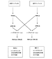
本発明の他の変法に従えば、本方法は、基準条件に由来するRNAと試験条件に由来するcDNA間で平行して行われる前記した第1のハイブリッド形成及び第2のハイブリッド形成を含む。この変法は、1つは基準条件に対する試験条件の定性的特徴を表しそして他方が試験条件に対する基準条件の定性的特徴を表す2つの核酸集団を発生させることを可能とする(図1C)ので特に有利である。これらの2つの集団は、以下にさらに完全に説明されるとおり特定の生理学的条件の遺伝学的フィンガープリント(genetic fingerprints)として役立つ核酸ソースとして又はライブラリーとして利用することもできる(図1D)。 According to another variant of the invention, the method comprises the first hybridization and the second hybridization described above performed in parallel between the RNA derived from the reference conditions and the cDNA derived from the test conditions. . This variation allows one to generate two nucleic acid populations, one representing the qualitative characteristics of the test conditions relative to the reference conditions and the other representing the qualitative characteristics of the reference conditions relative to the test conditions (FIG. 1C). Particularly advantageous. These two populations can also be utilized as nucleic acid sources or as libraries that serve as genetic fingerprints of specific physiological conditions, as described more fully below (FIG. 1D).
更なる態様では、本発明は、試験試料からのDNAsを基準試料の二本鎖cDNAとハイブリッド形成させることを含む関心のある核酸を同定するための方法に関する。このハイブリッド形成法は、形成された複合体において、調べている条件間の定性的な遺伝的差異を同定すること及びかくして例えば試験条件に特徴的なスプライシングを同定及び/又はクローニングすることを可能とする。後に開示されるとおり、この態様は、それがオルタナティブイントロン及びエキソン(alternative introns and exons)のみならず、同じ核酸ライブラリー内で、エキソン又はイントロンの欠失により形成される特異的連結部(specific junctions)も示すという点で有利である。更に、得られる配列は、オルタナティブイントロン及びエキソンのフランキング配列(flanking sequences)に関する情報も提供する。かくして、本発明は、スプライシングされなかった領域に関する情報なしに、選択的スプライシングが破壊されそしてスプライシングされた遺伝子の一部のみが保持される、US5,929,535に開示された技術の如き先行技術とは明らかに区別される。かくして、US5,929,535に開示された方法は適当なスプライスオリゴヌクレオチドのデザインを可能とすることができない。 In a further aspect, the present invention relates to a method for identifying a nucleic acid of interest comprising hybridizing DNAs from a test sample with double stranded cDNA of a reference sample. This hybridization method makes it possible to identify in the formed complex qualitative genetic differences between the conditions being examined and thus to identify and / or clone splicing characteristic of eg test conditions. To do. As disclosed later, this embodiment is directed to specific junctions formed by deletion of exons or introns in the same nucleic acid library as well as alternative introns and exons. ) Is also advantageous. In addition, the resulting sequences provide information on alternative introns and exon flanking sequences. Thus, the present invention provides prior art, such as the technique disclosed in US 5,929,535, in which alternative splicing is disrupted and only a portion of the spliced gene is retained without information regarding the unspliced region. Is clearly distinguished. Thus, the method disclosed in US Pat. No. 5,929,535 cannot allow the design of appropriate splice oligonucleotides.
本発明は、すべてのタイプの生物学的試料に適用することができる。特に、生物学的試料は、核酸を含有するいかなる細胞、器官、組織試料又は生検材料等であってもよい。器官、組織又は生検材料の場合には、試料は成分細胞へのアクセスを容易にするために培養させることができる。試料は、哺乳動物(特にヒト)、植物、バクテリア及び下等真核生成物(酵母、カビ細胞(fungal cells)等)から誘導されることができる。関係のある材料は、特に、腫瘍生検(tumor biopsy)、神経変性兆候を示す神経変性プラーク(neurodegenerative plaque)又は大脳ゾーン生検(cerebral zone biopsy)、皮膚試料、採血により得られる血液試料、結腸生検(colorectal biopsy)、気管支肺胞洗浄に由来する生検材料等により例示される。細胞の例は、注目すべきことに、筋肉細胞、肝細胞、繊維芽細胞、神経細胞、表皮及び皮膚細胞、血液細胞、例えばB及びTリンパ球、マスト細胞、単球、顆粒球及びマクロファージを含む。 The present invention can be applied to all types of biological samples. In particular, the biological sample may be any cell, organ, tissue sample or biopsy material that contains nucleic acid. In the case of an organ, tissue or biopsy, the sample can be cultured to facilitate access to the component cells. Samples can be derived from mammals (particularly humans), plants, bacteria and lower eukaryotic products (yeasts, fungal cells, etc.). Relevant materials include, among others, tumor biopsy, neurodegenerative plaque showing signs of neurodegeneration or cerebral zone biopsy, skin sample, blood sample obtained by blood sampling, colon Illustrated by biopsy (colorectal biopsy), biopsy material derived from bronchoalveolar lavage, and the like. Examples of cells include notably muscle cells, hepatocytes, fibroblasts, neurons, epidermis and skin cells, blood cells such as B and T lymphocytes, mast cells, monocytes, granulocytes and macrophages. Including.
上記したとおり、本発明に従う定性的差異スクリーニングは、他の用途のためにクローニング又は使用されるべき、基準生理学的条件(条件A)に対する与えられた生理学的条件(条件B)に特徴的な核酸の同定を可能とする。例示として、調査されるべき生理学的条件A及びBは下記の中から選ぶことができる:

A−RNA集団
本発明は、全RNA又はメッセンジャーRNAを使用することにより行うことができる。これらのRNAは、当業者に熟知されているいかなる慣用の分子生物学法によっても調製することができる。このような方法は、一般に、細胞、組織又は試料溶解(lysis)及び抽出法によるRNA回収を含む。これは、特に、カオトロピック剤、例えばチオシアン酸グアニジウム(RNAに影響を与えないで細胞を破壊する)による処理、次いで溶媒(例えば、フェノール、クロロホルム)によるRNA抽出により行うことができる。このような方法は当該技術分野で周知である(Maniatis et al., Chomczynski et al. (1987), Anal.Biochem., 162: 156参照)。これらの方法は、商業的に入手可能なキット、例えば、全RNA用のUS73750キット(Amersham)又はRneasy kit(Qujagen)を使用することにより容易に実施することができる。RNAは完全に純粋な状態にあることは必要ではなく、そして特に調製物中に残っている微量のゲノムDNA又は他の細胞成分(タンパク質等)は、それらがRNA安定性に有意に影響を与えない限りそして比較下の種々の試料の調製方法が同じである限り、妨害しないであろう。場合により、全RNA調製物の代わりにメッセンジャーRNAを使用することが更に可能である。これらは、標準方法に従い、ポリT配列によって、生物学的試料から直接又は全RNAから単離することができる。この点で、メッセンジャーRNAの調製は、例えば、US72700キット(Amersham)又はオリゴ(dT)ビーズ(Dynal)の使用を伴うキットの如き商業的に入手可能なキットを使用して行うことができる。RNA調製の有利な方法は、細胞質ゾルRNAを抽出し、次いで細胞質ゾルポリA+RNAを抽出することからなる。スプライシングされていないエキソン及びイントロンを有するプレメッセンジャーRNAにより汚染されていない細胞質ゾルRNAの選択的調製を可能とするキットは、商業的に入手可能である。これは、特にQuiagenにより市販されているRneasyキット(カタログ番号の例:74103)についてそうである。RNAは、前もって調製されたライブラリー又は他の試料から直接得ることもでき及び/又は適当な条件下に貯蔵された収集物から入手可能である。
A-RNA population The present invention can be carried out by using total RNA or messenger RNA. These RNAs can be prepared by any conventional molecular biology method familiar to those skilled in the art. Such methods generally include RNA recovery by cell, tissue or sample lysis and extraction methods. This can be done in particular by treatment with a chaotropic agent such as guanidinium thiocyanate (disrupting cells without affecting the RNA) followed by RNA extraction with a solvent (eg phenol, chloroform). Such methods are well known in the art (see Maniatis et al., Chomczynski et al. (1987), Anal. Biochem., 162: 156). These methods can be easily performed by using commercially available kits such as US73750 kit (Amersham) or Rneasy kit (Qujagen) for total RNA. It is not necessary for the RNA to be completely pure, and in particular the trace amounts of genomic DNA or other cellular components (such as proteins) that remain in the preparation can significantly affect RNA stability. Unless otherwise and as long as the various sample preparation methods under comparison are the same, they will not interfere. In some cases, it is further possible to use messenger RNA instead of total RNA preparation. These can be isolated directly from biological samples or from total RNA by poly T sequences according to standard methods. In this regard, messenger RNA can be prepared using commercially available kits such as, for example, the US72700 kit (Amersham) or kits involving the use of oligo (dT) beads (Dynal). An advantageous method of RNA preparation consists of extracting cytosolic RNA and then extracting cytosolic poly A + RNA. Kits that allow the selective preparation of cytosolic RNA uncontaminated by pre-messenger RNA with unspliced exons and introns are commercially available. This is especially true for the Rneasy kit marketed by Quiagen (catalog number example: 74103). RNA can be obtained directly from pre-prepared libraries or other samples and / or can be obtained from collections stored under appropriate conditions.
一般に、使用されるRNA調製物は、有利には少なくとも0.1μgのRNA、好ましくは少なくとも0.5μgのRNAを含む。量は、本発明の実施を変化させないように保ちながら、使用される特定の細胞及び方法に依存して変わることができる。十分な量のRNA(好ましくは少なくとも0.1μg)を得るために、一般に少なくとも105個の細胞を含む生物学的試料を使用することが推奨される。この点で、典型的な生検試料は一般に105〜108個の細胞を含みそして典型的なペトリ皿(直径6〜10cm)上での細胞培養物は、十分な量のRNAが容易に得られうるように、約106個の細胞を含有する。 In general, the RNA preparation used advantageously comprises at least 0.1 μg RNA, preferably at least 0.5 μg RNA. The amount can vary depending on the particular cell and method used, while keeping the practice of the invention unchanged. In order to obtain a sufficient amount of RNA (preferably at least 0.1 μg), it is generally recommended to use a biological sample containing at least 10 5 cells. In this regard, a typical biopsy sample generally contains 10 5 to 10 8 cells and cell cultures on a typical Petri dish (6-10 cm in diameter) can be easily obtained with sufficient amounts of RNA. As can be obtained, it contains about 10 6 cells.
RNA調製物は、その場で使用することができ、又は好ましくは、後に使用するために溶液として又は冷凍された状態において冷所に貯蔵することができる。 The RNA preparation can be used in situ or, preferably, can be stored in a cold place as a solution or in a frozen state for later use.
B−cDNA集団
本発明の範囲内で使用されるcDNAは、慣用の分子生物学技術に従う逆転写により得ることができる。特にManiatis et alを参照されたい。逆転写は、一般に酵素、逆転写酵素及びプライマーを使用して行われる。
B-cDNA population The cDNA used within the scope of the present invention can be obtained by reverse transcription according to conventional molecular biology techniques. See in particular Maniatis et al. Reverse transcription is generally performed using enzymes, reverse transcriptases and primers.
この点で、多くの逆転写酵素が文献に記載されておりそして商業的に入手可能である(1483188キット、Boehringer)。最も普通に使用される逆転写酵素の例は、鳥類ウイルスAMV(ニワトリ骨髄芽球症ウイルス(Avian Myeloblastosis Virus))に由来する逆転写酵素及びマウス白血病ウイルスMMLV(モロニーマウス白血病ウイルス(Moloney Murine Leukemia Virus))からの逆転写酵素を含む。逆転写活性を有するある種の熱安定性DNAポリメラーゼ、例えば、Thermus flavus及びThermus thermophilus HB-8(商業的に入手可能;Promegaカタログ番号 M1941及びM2101)から単離された熱安定性DNAポリメラーゼに言及することも価値がある。有利な変法に従えば、本発明は、AMV逆転写酵素を使用して行われる。何故ならば、42℃で活性な(37℃で活性なMMLVの逆転写酵素と対照的に)この酵素は、伸長を停止させる可能性のあるRNA二次構造を脱安定化させ、そしてそれゆえより大きい長さのRNAの逆転写を可能としそしてRNAのはるかにより忠実なコピーであるcDNA調製物を高収率で与えるからである。 In this regard, many reverse transcriptases have been described in the literature and are commercially available (1483188 kit, Boehringer). Examples of the most commonly used reverse transcriptases are reverse transcriptase derived from the avian virus AMV (chicken myeloblastosis virus) and mouse leukemia virus MMLV (Moloney Murine Leukemia Virus). )) From reverse transcriptase. Reference to certain thermostable DNA polymerases with reverse transcription activity, eg, thermostable DNA polymerases isolated from Thermus flavus and Thermus thermophilus HB-8 (commercially available; Promega catalog numbers M1941 and M2101) It is also worth doing. According to an advantageous variant, the present invention is carried out using AMV reverse transcriptase. Because it is active at 42 ° C. (as opposed to the MMLV reverse transcriptase active at 37 ° C.), this enzyme destabilizes RNA secondary structures that may stop elongation and hence This is because it allows reverse transcription of larger lengths of RNA and gives high yields of cDNA preparations that are much more faithful copies of RNA.
本発明の更なる有利な変法に従えば、RNアーゼH活性を欠いた逆転写酵素が使用される。このタイプの酵素の使用は、いくつかの利点、特にcDNA合成の収率を増加させ、そして新しく合成されたcDNAとのヘテロ二本鎖の形成にその後参加させられるRNAのいかなる劣化も回避するという利点を有し、それによって場合によりcDNAのフェノール抽出を省くことを可能とする。RNアーゼH活性に欠けた逆転写酵素は、欠失(1つ又は複数)及び/又は突然変異誘発によりいかなる逆転写酵素からも調製されうる。更に、このような酵素も商業的に入手可能である(例えば、Life Technologies,カタログ番号 18053-017)。 According to a further advantageous variant of the invention, reverse transcriptase lacking RNase H activity is used. The use of this type of enzyme increases several yields, particularly the yield of cDNA synthesis, and avoids any degradation of the RNA that subsequently participates in heteroduplex formation with newly synthesized cDNA. Has the advantage that it makes it possible to omit the phenol extraction of the cDNA. A reverse transcriptase lacking RNase H activity can be prepared from any reverse transcriptase by deletion (s) and / or mutagenesis. In addition, such enzymes are also commercially available (eg, Life Technologies, catalog number 18053-017).
逆転写酵素に適用される操作条件((濃度及び温度)は当業者には周知である。特に、10mMの最適なMg2+濃度の存在下に、一般に10〜30単位の酵素が1つの反応で使用される。 The operating conditions (concentration and temperature) applied to the reverse transcriptase are well known to those skilled in the art, especially 10-30 units of enzyme in one reaction in the presence of an optimal Mg 2+ concentration of 10 mM. used.
逆転写のために使用されるプライマー(1つ又は複数)は種々のタイプであることができる。それは、特に、好ましくは4〜10ヌクレオチドを含むランダムオリゴヌクレオチド、有利にはヘキサヌクレオチドであることができる。このタイプのランダムプライマーの使用は、文献に記載されておりそしてRNA分子内の種々の部位での逆転写のランダムな開始を可能とする。この技術は、全RNA(即ち、特にmRNA、tRNA及びrRNAを含む)を逆転写するために特に使用される。mRNAのみの逆転写を行うことが所望される場合には、プライマーとして、メッセンジャーRNAに対して特異的なポリAテイルから出発する逆転写の開始を可能とするオリゴdTオリゴヌクレオチドを使用することが有利である。オリゴdTオリゴヌクレオチドは、4〜20mer、有利には、約15merを含むことができる。このようなプライマーの使用は本発明の好ましい態様を表す。更に、逆転写のための標識されたプライマーを使用することが有利でありうる。実際問題として、これは、RNAの認識及び/又は選択及び/又はその後のcDNAからの選別を可能とする。これは、RNA/DNAヘテロ二本鎖を単離することも可能とすることもでき、RNA/DNAヘテロ二本鎖の形成は本発明の実施において重要な段階を表す。プライマーの標識化は、いかなるリガンド−受容体をベースとするシステムによっても行うことができ、即ち、プライマーを有する分子の親和性を仲立ちとした分離を提供するシステムによってなされうる。それは、予めストレプトアビジンでコーティングされたいかなる支持体(ビーズ、カラム、プレート等)上にも捕捉されうる、例えば、ビオチン標識からなることができる。プライマーの性質に影響を与えることなく分離を可能とするいかなる他の標識化システムも同様に使用することができる。 The primer (s) used for reverse transcription can be of various types. It can in particular be a random oligonucleotide, preferably a hexanucleotide, preferably comprising 4 to 10 nucleotides. The use of this type of random primer has been described in the literature and allows random initiation of reverse transcription at various sites within the RNA molecule. This technique is particularly used to reverse transcribe total RNA (ie, particularly including mRNA, tRNA and rRNA). If it is desired to perform reverse transcription of mRNA only, an oligo dT oligonucleotide that allows the initiation of reverse transcription starting from a poly A tail specific for messenger RNA may be used as a primer. It is advantageous. Oligo dT oligonucleotides can comprise 4-20 mer, advantageously about 15 mer. The use of such primers represents a preferred embodiment of the present invention. Furthermore, it may be advantageous to use labeled primers for reverse transcription. In practice, this allows RNA recognition and / or selection and / or subsequent selection from cDNA. This can also allow RNA / DNA heteroduplexes to be isolated, and the formation of RNA / DNA heteroduplexes represents an important step in the practice of the present invention. Primer labeling can be performed by any ligand-receptor based system, i.e., by a system that provides a separation based on the affinity of the molecule with the primer. It can consist of, for example, a biotin label, which can be captured on any support (beads, columns, plates, etc.) previously coated with streptavidin. Any other labeling system that allows separation without affecting the nature of the primer can be used as well.
典型的操作条件において、この逆転写は一本鎖相補的DNA(cDNA)を発生させる。これは、本発明の第1の有利な態様を表す。 In typical operating conditions, this reverse transcription generates single-stranded complementary DNA (cDNA). This represents a first advantageous aspect of the present invention.
本発明を実施する第2の変法では、逆転写は二本鎖cDNAが調製されるように達成される。この結果は、第1cDNA鎖の転写の後、DNA修飾することができる酵素、例えばファージT4DNAリガーゼ、DNAポリメラーゼI及びファージT4DNAポリメラーゼを伴う慣用の分子生物学技術を使用して第2鎖(second strand)を発生させることにより達成される。 In a second variant embodying the invention, reverse transcription is achieved such that double stranded cDNA is prepared. The result is that after transcription of the first cDNA strand, the second strand can be obtained using conventional molecular biology techniques involving enzymes capable of DNA modification such as phage T4 DNA ligase, DNA polymerase I and phage T4 DNA polymerase. ).
cDNA調製物はその場で使用することができ又は好ましくは後の使用のために溶液として又は冷凍された状態で、好ましくは冷所に貯蔵することができる。 The cDNA preparation can be used in situ or preferably can be stored in a cold place, preferably in solution or frozen for later use.
上記したとおり、本発明は、典型的には、複合核酸集団(即ち、少なくとも部分的に未知であるか又は特徴付けされていない複数の異なる核酸配列、典型的には20、50又は100より多くの異なる核酸配列を含む集団)を使用して行われる。しかしながら、特定の態様では、本発明は選ばれた核酸集団を使用して行われうる。このような選ばれた核酸集団は、例えば、選ばれた遺伝子又はRNA(又はいくつかの既知のそして選ばれた遺伝子又はRNA)の配列を含むことができる。選ばれた核酸集団を使用することによって、本発明は、いかなる特定の病態生理学的条件にある選ばれた遺伝子の生物学的に関係のあるスプライシング形態(biologically relevant splicing forms)も同定するために使用することができる。かくして、本発明は、以後説明されるとおり選ばれた遺伝子又はRNAのスプライシング形態をクローニング又は同定するためにも適当である。 As noted above, the present invention typically includes a composite nucleic acid population (ie, a plurality of different nucleic acid sequences that are at least partially unknown or uncharacterized, typically more than 20, 50, or 100 A population of different nucleic acid sequences). However, in certain embodiments, the present invention can be practiced using selected nucleic acid populations. Such a selected nucleic acid population can include, for example, the sequence of a selected gene or RNA (or several known and selected genes or RNA). By using selected nucleic acid populations, the present invention can be used to identify biologically relevant splicing forms of selected genes in any particular pathophysiological condition can do. Thus, the present invention is also suitable for cloning or identifying spliced forms of genes or RNAs selected as described hereinafter.
C−ハイブリッド形成
上記したとおり、本発明に従う方法は、異なる生理学的条件における生物学的試料に由来する又は異なる起源に由来する、一方ではRNA又はcDNAと他方ではcDNAとの独創的クロスハイブリッド形成段階に部分的に基づいている。好ましい態様では、本発明に従うハイブリッド形成は、液相で有利に行われる。更に、それは、いかなる適当な装置においても、例えば、チューブ(例えば、Eppendorffチューブ)、プレート又は分子生物学で普通に使用されるいかなる他の適当な支持体においても行うことができる。ハイブリッド形成は、10〜1000μl、例えば、10〜500μlの範囲の容積において有利に行われる。使用される特定の装置及び容積は当業者により容易に適合させられうることは理解されるべきである。使用される核酸の量は同様に当該技術分野で周知である。一般に、数μgの核酸、例えば、0.1〜100μgの範囲の核酸を使用すれば十分である。
C-Hybridization As mentioned above, the method according to the invention is based on the original cross-hybridization step of biological samples from different physiological conditions or from different sources, on the one hand RNA or cDNA and on the other hand cDNA. Based in part on. In a preferred embodiment, the hybridization according to the invention is advantageously performed in the liquid phase. Furthermore, it can be carried out in any suitable apparatus, for example in tubes (eg Eppendorff tubes), plates or any other suitable support commonly used in molecular biology. Hybridization is advantageously performed in volumes ranging from 10 to 1000 μl, such as from 10 to 500 μl. It should be understood that the particular equipment and volume used can be readily adapted by those skilled in the art. The amount of nucleic acid used is likewise well known in the art. In general, it is sufficient to use several μg of nucleic acid, for example in the range of 0.1 to 100 μg.
ハイブリッド形成を行う際に考慮されるべき重要なファクターは、使用される核酸のそれぞれの量である。かくして、2つの試料からの核酸を約50〜0.02、好ましくは40〜0.1の範囲の割合で使用することが可能である。更に特に有利な方法では、cDNA/RNA比は好ましくは1に近いか又は1より大きい。実際、このような実験では、RNAはテスター化合物を形成しそしてcDNAはドライバーを形成し、そして方法の特異性を改良するために、ドライバーがテスターに対して過剰である操作条件を選ぶことが好ましい。他の特に有利な方法では、ss−cDNA/ds−cDNA比は好ましくは1に近いか又は1より大きく、更に好ましくは5より大きい。このような実験では、ss−cDNAはテスターでありそして好ましくは、ドライバー試料からのds−cDNAを追い出すように過剰に使用されるべきである。このような条件では、核酸間の協同性効果が起こりそしてミスマッチは強く退けられる。結果として、観察される唯一のミスマッチは、一般に、ドライバーcDNAには存在せずそしてそれゆえ特異的であると考えられうるテスターRNA又はss−cDNAにおける領域の存在によるものである。ゆえに、本方法の特異性を高めるために、ハイブリッド形成は、約1と約10との間に含まれるcDNA/RNA又はss−cDNA/ds−cDNA比を使用して有利に行われる。この比は操作条件(入手可能な核酸の量、生理学的条件、必要な結果等)に依存して当業者により適合させられうることは理解される。他のハイブリッド形成パラメーター(時間、温度、イオン強度)も当業者により適合させられうる。一般的に言えば、テスター及びドライバーの変性(例えば、加熱により)の後、約37℃の温度で(そして場合により下記する如き温度シフトを行うことにより)そして標準イオン強度条件下に(例えば、0.1M〜5M の範囲のNaCl)約2〜24時間ハイブリッド形成を行う。イオン強度は、特に固体支持体上のハイブリッド形成の場合に、ハイブリッド形成ストリンジェンシーを規定するファクターの1つであることが知られている。 An important factor to be considered when performing hybridization is the amount of each nucleic acid used. Thus, it is possible to use nucleic acids from two samples in proportions ranging from about 50 to 0.02, preferably 40 to 0.1. In a further particularly advantageous manner, the cDNA / RNA ratio is preferably close to 1 or greater than 1. In fact, in such experiments, it is preferable to select operating conditions in which the driver is in excess of the tester in order to improve the specificity of the method, and RNA forms the tester compound and cDNA forms the driver. . In another particularly advantageous method, the ss-cDNA / ds-cDNA ratio is preferably close to or greater than 1 and more preferably greater than 5. In such experiments, the ss-cDNA is a tester and preferably should be used in excess to drive out the ds-cDNA from the driver sample. Under such conditions, a cooperative effect between the nucleic acids occurs and the mismatch is strongly rejected. As a result, the only mismatch observed is generally due to the presence of a region in the tester RNA or ss-cDNA that is not present in the driver cDNA and therefore may be considered specific. Thus, to increase the specificity of the method, hybridization is advantageously performed using a cDNA / RNA or ss-cDNA / ds-cDNA ratio comprised between about 1 and about 10. It will be appreciated that this ratio can be adapted by one skilled in the art depending on the operating conditions (amount of nucleic acid available, physiological conditions, required results, etc.). Other hybridization parameters (time, temperature, ionic strength) can also be adapted by those skilled in the art. Generally speaking, after denaturing the tester and driver (eg, by heating), at a temperature of about 37 ° C. (and optionally by performing a temperature shift as described below) and under standard ionic strength conditions (eg, Hybridization takes place for about 2 to 24 hours in NaCl ranging from 0.1M to 5M). Ionic strength is known to be one of the factors that define hybridization stringency, especially in the case of hybridization on solid supports.
本発明の特定の態様に従えば、ハイブリッド形成は、例えば、Kohno D.E.et al.(Biochemistry.(1977), 16(24): 5329-5341)により記載されたPERT技術(Phenol Emulsion DNA Reassociation Technique)に従って、フェノールエマルションにおいて行われる。有利には、Miller及びRibletの技術(NAR, (1995), 23: 2339)に従い、攪拌の代わりに温度サイクリング(約37℃から約60/65℃への温度シフト)下のフェノールエマルションハイブリッド形成が本発明の範囲内で使用される。特にエマルション相における、いかなる他の液相ハイブリッド形成技術も、本発明の範囲内で使用することができる。かくして、他の特に有利な態様では、ハイブリッド形成は、ホルムアミド80%を含有する溶液中で例えば40℃の温度で行われる。 According to a particular embodiment of the present invention, hybridization is performed, for example, by the PERT technique (Phenol Emulsion DNA Reassociation Technique) described by Kohno DE et al. (Biochemistry. (1977), 16 (24): 5329-5341). In a phenol emulsion. Advantageously, according to the technique of Miller and Riblet (NAR, (1995), 23: 2339), phenol emulsion hybridization under temperature cycling (temperature shift from about 37 ° C. to about 60/65 ° C.) instead of stirring. Used within the scope of the present invention. Any other liquid phase hybridization technique, particularly in the emulsion phase, can be used within the scope of the present invention. Thus, in another particularly advantageous embodiment, the hybridization is carried out in a solution containing 80% formamide, for example at a temperature of 40 ° C.
支持体に固定された相手の1つとハイブリッド形成を行うこともできる。有利には、cDNAが固定化される。これは、特にビオチニル化されたプライマーを使用することによりcDNA標識化(上記参照)を利用することにより行うことができる。ビオチン部分をストレプトアビジン分子でコーティングされた磁性ビーズと接触させる。ついでcDNAは、磁場を加えることによりフィルター又はマイクロタイターディッシュウエルと接触させて保持することができる。次いで、適当なイオン強度条件の下に、RNAをcDNAと接触させる。対を形成していないRNAは洗浄により除去される。ハイブリッド形成されたRNA及びcDNAは磁界を除去すると回収される。 Hybridization can also be performed with one of the counterparts secured to the support. Advantageously, the cDNA is immobilized. This can be done by utilizing cDNA labeling (see above), in particular by using biotinylated primers. The biotin moiety is contacted with magnetic beads coated with streptavidin molecules. The cDNA can then be retained in contact with a filter or microtiter dish well by applying a magnetic field. The RNA is then contacted with the cDNA under appropriate ionic strength conditions. Unpaired RNA is removed by washing. Hybridized RNA and cDNA are recovered upon removal of the magnetic field.
cDNAが二本鎖である場合には、使用されるハイブリッド形成条件は本質的に前記したハイブリッド形成条件と同様であり、そして当業者により適合させられうる。RNAと二本鎖cDNA間でヘテロ三本鎖が形成される場合には、ハイブリッド形成はホルムアミドの存在下に行うことができ、そして複合体は、例えば、60℃〜40℃、好ましくは56℃〜44℃で変わる範囲の温度にさらされてRループ複合体の形成を促進することができる。更に、ハイブリッド形成の後、ホルムアミドが媒体から除去されると、形成された三本鎖構造(triplex structures)を安定化させるための安定剤、例えばグリオキサールを加えることが望ましい(Kaback et al., (1979), Nuc. Acid Res., 6: 2499-2517)。 If the cDNA is double stranded, the hybridization conditions used are essentially similar to the hybridization conditions described above and can be adapted by one skilled in the art. If a hetero triplex is formed between the RNA and the double stranded cDNA, the hybridization can be performed in the presence of formamide and the complex is, for example, 60 ° C to 40 ° C, preferably 56 ° C. Exposure to a range of temperatures varying from ˜44 ° C. can promote the formation of R-loop complexes. In addition, once the formamide is removed from the medium after hybridization, it is desirable to add a stabilizer, such as glyoxal, to stabilize the formed triplex structures (Kaback et al., ( 1979), Nuc. Acid Res., 6: 2499-2517).
かくして、本発明に従うこれらのクロスハイブリッド形成は、試験されるべき各生理学的条件の定性的性質を表すcDNA/cDNAホモ二本鎖又はcDNA/RNAヘテロ二本鎖又はヘテロ三本鎖を含む組成物を発生させる。既に言及したとおり、本発明の組成物の各々において、各生理学的条件に特異的な、差異選択的スプライシング(differential alternative splicing)又は他の遺伝子変化に本質的に相当する核酸を同定及び/又はクローニングすることができる。 Thus, these cross-hybridization according to the present invention comprises a cDNA / cDNA homoduplex or cDNA / RNA heteroduplex or heterotriple that represents the qualitative nature of each physiological condition to be tested. Is generated. As already mentioned, in each of the compositions of the invention, nucleic acids essentially corresponding to differential alternative splicing or other genetic alterations specific to each physiological condition are identified and / or cloned. can do.
ゆえに、本発明は、有利には2つの生理学的条件間に発生する遺伝子差を表す核酸を同定及び/又はクローニングするための方法であって、第1生理学的条件の生物学的試料に由来するRNAを第2生理学的条件の生物学的試料に由来する一本鎖cDNAとハイブリッド形成させ、そしてこのようにして形成されたハイブリッドから、対を形成していないRNA領域を同定及び/又はクローニングすることを含む方法に関する。
この第1変法は、更に特定的には、RNAと一本鎖cDNA間のヘテロ二本鎖構造体の形成に基づいている(図2〜4参照)。この変法は、メッセンジャーRNA又は本質的にメッセンジャーRNAの逆転写により、即ち、オリゴdTプライマーの存在下に産生されたcDNAを使用して有利に実施される。
Thus, the present invention is a method for identifying and / or cloning a nucleic acid that advantageously represents a genetic difference that occurs between two physiological conditions, and is derived from a biological sample of a first physiological condition RNA is hybridized with single stranded cDNA derived from a biological sample at a second physiological condition, and unpaired RNA regions are identified and / or cloned from the hybrid thus formed Relates to a method comprising:
This first variant is more particularly based on the formation of a heteroduplex structure between RNA and single stranded cDNA (see FIGS. 2-4). This variant is advantageously carried out by reverse transcription of messenger RNA or essentially messenger RNA, ie using cDNA produced in the presence of oligo dT primers.
特定の態様では、本発明に従う核酸を同定及び/又はクローニングする方法は、
(a)試験条件に由来するRNAを基準条件に由来する一本鎖cDNAとハイブリッド形成させ、
(b)基準条件に由来するRNAを試験条件に由来する一本鎖cDNAとハイブリッド形成させ、そして
(c)段階(a)及び(b)で形成されたハイブリッドから、対を形成していないRNA領域を同定及び/又はクローニングする、
ことを含む。
In a particular embodiment, the method for identifying and / or cloning a nucleic acid according to the invention comprises:
(A) hybridizing RNA derived from test conditions with single stranded cDNA derived from reference conditions;
(B) hybridizing RNA derived from reference conditions with single stranded cDNA derived from test conditions; and (c) unpaired RNA from the hybrid formed in steps (a) and (b) Identifying and / or cloning regions,
Including that.
特定の別の実施方式においては、本発明の方法は、下記の段階:
(a)生理学的条件A(rA)の生物学的試料からRNAを得、
(b)生理学的条件B(rB)の同じ生物学的試料からのRNAを得、
(c)段階(a)で与えられたrARNAの一部からcDNA(cAcDNA)を及び段階Bで与えられたrBRNAの一部からcDNA(cBcDNA)をポリTプライマーによって調製し、
(d)液相においてrARNAの一部をcBDNAの一部とハイブリッド形成させ(rA/cBヘテロ二本鎖を発生させるために)、
(e)液相においてrBRNAの一部をcAcDNAの一部とハイブリッド形成させ(rB/cAヘテロ二本鎖を発生させるために)、
(f)段階(d)及び(e)で得られたrA/cB及びrB/cAヘテロ二本鎖内の対を形成していないRNA領域を同定及び/又はクローニングする、
段階を含む。
In certain other implementations, the method of the present invention comprises the following steps:
(A) obtaining RNA from a biological sample of physiological condition A (rA);
(B) obtaining RNA from the same biological sample at physiological condition B (rB);
(C) preparing cDNA (cAcDNA) from a portion of rARNA given in step (a) and cDNA (cBcDNA) from a portion of rBRNA given in step B with a poly-T primer;
(D) hybridizing a portion of rARNA with a portion of cBDNA in the liquid phase (to generate rA / cB heteroduplex);
(E) hybridizing a portion of rBRNA with a portion of cA cDNA in the liquid phase (to generate rB / cA heteroduplex);
(F) identifying and / or cloning unpaired RNA regions within the rA / cB and rB / cA heteroduplexes obtained in steps (d) and (e),
Including stages.
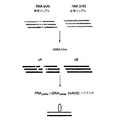
本発明を実施する別の方式に従えば、本発明の方法は、試験条件に由来するRNAを基準条件に由来するニ本鎖cDNAとハイブリッド形成させ、そして得られる二本鎖DNA領域を同定及び/又はクローニングすることを含む。この第2の変法は、更に特定的には、Rループ型構造(図5参照)に由来する、RNAと二本鎖cDNAとのヘテロ三本鎖構造の形成に基づいている。この変法は、本質的にメッセンジャーRNA、又はメッセンジャーRNAの逆転写により、即ち、ポリTプライマーの存在下に産生されたcDNAを使用することによっても好ましく実施される。やはりこの変異態様においは、特定の態様は、本発明に従う2つの核酸集団を発生させる、2つのハイブリッド形成を平行して行うことを含む。この変法では、選択的スプライシングイベントに特異的な所望の領域は、対を形成していないRNA領域ではなくて、その代わりに相同性RNA配列により追い出されなかった二本鎖DNAである(図5参照)。 According to another mode of carrying out the present invention, the method of the present invention comprises hybridizing RNA derived from test conditions with double stranded cDNA derived from reference conditions and identifying and obtaining the resulting double stranded DNA region. And / or cloning. This second variant is more particularly based on the formation of a hetero triple stranded structure of RNA and double stranded cDNA derived from an R-loop structure (see FIG. 5). This variant is also preferably carried out essentially by messenger RNA, or by reverse transcription of messenger RNA, ie by using cDNA produced in the presence of poly-T primers. Again, in this variant embodiment, a particular embodiment involves performing two hybridizations in parallel, generating two nucleic acid populations according to the invention. In this variant, the desired region specific for the alternative splicing event is not an unpaired RNA region, but instead is a double-stranded DNA that was not driven out by the homologous RNA sequence (Fig. 5).
本発明の他の変法では、2つの試料間で発生する定性的な遺伝的差異(例えば、選択的スプライシングイベンド)を検出するための方法は、第1生物学的試料に由来する二本鎖cDNAを第2生物学的試料に由来するcDNA(二本鎖又は好ましくは一本鎖)とハイブリッド形成させることを含む(図6)。 In another variation of the invention, the method for detecting a qualitative genetic difference (eg, alternative splicing event) occurring between two samples is a duplex derived from a first biological sample. Hybridizing the cDNA with cDNA (double stranded or preferably single stranded) from a second biological sample (Figure 6).
前記した変法と異なり、この変法は、DNA/RNAヘテロ二本鎖又はヘテロ三本鎖構造を使用しないで、その代わりにDNA/DNA相同性ホモ二本鎖を使用する。この変法は、それがオルタナティブイントロン及びエキソンを示すのみならず、同じ核酸ライブラリー内でエキソン又はイントロンの欠失により形成された特異的連結部(specific junctions)も示すという点で有利である。更に、このようなライブラリーにおける配列はオルタナティブイントロン及びエキソンのフランキング配列に関する情報を与える。 Unlike the variant described above, this variant does not use DNA / RNA heteroduplex or heterotriple structures, but instead uses DNA / DNA homologous homoduplexes. This variant is advantageous in that it not only shows alternative introns and exons, but also specific junctions formed by exon or intron deletions within the same nucleic acid library. In addition, sequences in such libraries provide information on alternative intron and exon flanking sequences.
第1の態様に従えば、この方法は、一本鎖cDNAの第1複合集団を二本鎖cDNAの第2複合集団とハイブリッド形成させることを含む。この態様は、基準条件と比較して生理学的試験条件において発生するスプライシングイベントに特徴的な核酸集団を発生することを可能とする(図1A、変法#3、図6A、図26)。後に示されるとおり、この集団は、核酸のクローニング及び特徴付け、診断、スクリーニング、治療及び抗体産生又は全タンパク質又はタンパク質断片の合成のために使用することができる。この集団は、後に示される種々の使用分野に使用されうるライブラリーを発生させるため及び標識されたプローブを発生させるために使用することもできる(図1D)。
According to a first aspect, the method comprises hybridizing a first complex population of single stranded cDNA with a second complex population of double stranded cDNA. This aspect makes it possible to generate nucleic acid populations characteristic of splicing events occurring in physiological test conditions compared to reference conditions (FIG. 1A,
他の態様に従えば、この方法は、一本鎖cDNAの第1集団を一本鎖cDNAの第2集団とハイブリッド形成させることを含む。この態様では、試験試料及び基準試料の両方は一本鎖cDNAの形態にある。この態様は、基準試料からの二本鎖cDNAの再アニーリングを回避し、かくして、一方の鎖が試験試料から生じそして他方が基準試料から生じるDNA/DNAホモ二本鎖のみが形成されうる。形成されるハイブリッドは、診断、スクリーニング、治療及び抗体産生又は全タンパク質又はタンパク質断片の合成において使用することができる、2つの試料間で発生する差異スプライシングイベントを表す核酸のクローニング及び特徴付けを可能とする(図1D)。 According to another aspect, the method includes hybridizing a first population of single stranded cDNA with a second population of single stranded cDNA. In this embodiment, both the test sample and the reference sample are in the form of single stranded cDNA. This embodiment avoids re-annealing of double stranded cDNA from the reference sample, so that only DNA / DNA homoduplexes can be formed where one strand originates from the test sample and the other originates from the reference sample. The hybrids formed allow the cloning and characterization of nucleic acids representing differential splicing events that occur between two samples that can be used in diagnostics, screening, therapy and antibody production or synthesis of whole proteins or protein fragments. (FIG. 1D).
特定の態様では、本発明に従う核酸を同定及び/又はクローニングする方法は、
(a)試験条件に由来する複数の異なる一本鎖cDNAを含む核酸集団を、基準条件に由来する複数の異なる二本鎖cDNAを含む核酸集団とハイブリッド形成させ、
(b)段階(a)で形成されたハイブリッドから、対を形成していないDNA領域を同定及び/又はクローニングする、
ことを含む。
In a particular embodiment, the method for identifying and / or cloning a nucleic acid according to the invention comprises:
(A) hybridizing a nucleic acid population comprising a plurality of different single stranded cDNAs derived from test conditions with a nucleic acid population comprising a plurality of different double stranded cDNAs derived from reference conditions;
(B) identifying and / or cloning unpaired DNA regions from the hybrid formed in step (a);
Including that.
他の特定の態様では、本発明に従う核酸を同定及び/又はクローニングするための方法は、
(a)試験条件に由来する複数の異なる一本鎖cDNAを含む核酸集団を、基準条件に由来する複数の異なる一本鎖cDNAを含む核酸集団とハイブリッド形成させ、そして
(b)段階(a)で形成されたハイブリッドから、対を形成していないDNA領域を同定及び/又はクローニングする、
ことを含む。
In another particular embodiment, the method for identifying and / or cloning a nucleic acid according to the invention comprises
(A) hybridizing a nucleic acid population comprising a plurality of different single stranded cDNAs derived from test conditions with a nucleic acid population comprising a plurality of different single stranded cDNAs derived from reference conditions; and (b) step (a) Identifying and / or cloning unpaired DNA regions from the hybrid formed in
Including that.
特定の別の実施方式では、本発明の方法は、下記の段階:
(a)生理学的条件A(rA)の生物学的試料からRNAを得(rA)、
(b)生理学的条件B(rB)の同じ生物学的試料からRNAを得(rB)、
(c)段階(a)で与えられたrARNAからcDNA(cAcDNA)を及び段階Bで与えられたrBRNAからcDNA(cBcDNA)を標識された(例えばビオチニル化された)ポリTプライマーによって調製し、
(d)cBcDNAから二本鎖cDNAを調製してdcBcDNAを産生し、
(e)cAcDNAの一部をdcBcDNAの一部とハイブリッド形成させ(例えば、液相で)(dcB/cAcDNAホモ二本鎖を発生させ)、
(f)段階(e)で得られたホモ二本鎖内の対を形成していないDNA領域を同定及び/又はクローニングする、
ことを含む。
In certain other implementations, the method of the invention comprises the following steps:
(A) obtaining RNA from a biological sample at physiological condition A (rA) (rA);
(B) obtaining RNA from the same biological sample at physiological condition B (rB) (rB),
(C) preparing cDNA (cAcDNA) from the rARNA given in step (a) and cDNA (cBcDNA) from the rBRNA given in step B with labeled (eg biotinylated) poly T primers;
(D) preparing double stranded cDNA from cB cDNA to produce dcB cDNA;
(E) hybridizing a portion of cA cDNA with a portion of dcB cDNA (eg, in liquid phase) (generating a dcB / cA cDNA homoduplex);
(F) identifying and / or cloning unpaired DNA regions in the homoduplex obtained in step (e),
Including that.
他の特定の態様では、本発明に従う核酸を同定及び/又はクローニングする方法は、
(a)1つ又はそれより多くの選ばれた遺伝子又はRNAに由来する一本鎖cDNAを含む核酸集団を、生物学的試料に由来する複数の異なる一本鎖又は二本鎖cDNAを含む核酸集団とハイブリッド形成させ、
(b)段階(a)で形成されたハイブリッドから、対を形成していないDNA領域を同定及び/又はクローニングする、
ことを含む。
In another particular embodiment, the method for identifying and / or cloning a nucleic acid according to the invention comprises:
(A) a nucleic acid population comprising a plurality of different single-stranded or double-stranded cDNAs derived from a biological sample from a nucleic acid population comprising single-stranded cDNAs derived from one or more selected genes or RNAs Hybridize with the population,
(B) identifying and / or cloning unpaired DNA regions from the hybrid formed in step (a);
Including that.
特定の別の実施方式では、本発明の方法は、下記の段階:
(a)生理学的条件Aの生物学的試料からRNAを得(rA)、
(b)段階(a)のrAからcDNAを、標識された(例えば、ビオチニル化された)ポリTプライマーによって調製し(cAcDNA)、
(c)場合によりcAcDNAから二本鎖cDNAを調製してdcAcDNAを産生させ、
(d)段階(b)又は(c)の該cAcDNA又はdcAcDNAを1つ又はそれより多くの選ばれた遺伝子又はRNAに由来する一本鎖cDNAとハイブリッド形成させ(例えば、液相で)、そして
(e)段階(e)で得られたハイブリッド以内の対を形成していないDNA領域を同定及び/又はクローニングする、
ことを含む。
In certain other implementations, the method of the invention comprises the following steps:
(A) obtaining RNA from a biological sample of physiological condition A (rA),
(B) preparing a cDNA from the rA of step (a) with a labeled (eg biotinylated) poly T primer (cA cDNA);
(C) optionally preparing double stranded cDNA from cA cDNA to produce dcA cDNA;
(D) hybridizing (eg in liquid phase) the cA cDNA or dcA cDNA of step (b) or (c) with a single stranded cDNA derived from one or more selected genes or RNA; (E) identifying and / or cloning unpaired DNA regions within the hybrid obtained in step (e),
Including that.
この最後の態様に従えば、特定の病態生理学的状況において発生するいかなる選ばれた遺伝子又はRNAの生物学的に関係のあるスプライシング形態を同定することが可能である。特に、与えられた遺伝子のss−cDNA配列を産生しそして上記の方法を行うことにより、特定の組織又は条件において発生する与えられた遺伝子のスプライシング形態の存在、性質及び/又は配列を決定することが可能である。このようにして同定された対を形成していない領域は、該特異的組織又は条件における該遺伝子の生物学的に関係のあるスプライシング形態に相当するであろう。選ばれた遺伝子又はRNAは、関心のあるいかなる遺伝子又は遺伝子のファミリーであってもよく、例えば、ホルモン、サイトカイン、成長因子、腫瘍抑制剤、受容体、イオンチャンネル、転写因子、栄養因子、凝血因子(clotting factors)、リポタンパク質等の遺伝子又は遺伝子のファミリーであることができる。それらは、哺乳動物起源のもの又はいかなる他の起源のもの、例えば、植物、ウイルス等の起源のものであってもよい。特定の態様では、選ばれた遺伝子は、受容体、例えばGタンパク質結合受容体(G-Protein-Coupled Receptor)のすべて又は一部を含む核酸分子である。 According to this last aspect, it is possible to identify biologically relevant splicing forms of any selected gene or RNA that occur in a particular pathophysiological situation. In particular, determining the presence, nature, and / or sequence of a spliced form of a given gene that occurs in a particular tissue or condition by producing a ss-cDNA sequence of the given gene and performing the method described above Is possible. The unpaired regions thus identified will correspond to biologically relevant splicing forms of the gene in the specific tissue or condition. The selected gene or RNA may be any gene or family of genes of interest, such as hormones, cytokines, growth factors, tumor suppressors, receptors, ion channels, transcription factors, trophic factors, clotting factors (Clotting factors), genes such as lipoproteins or gene families. They may be of mammalian origin or any other origin, for example from plants, viruses etc. In a particular embodiment, the selected gene is a nucleic acid molecule comprising all or part of a receptor, such as a G-Protein-Coupled Receptor.
調査中の両試料(即ち、病態生理学的条件)について、細胞質ゾルポリA+RNAは、当該技術分野で知られているこれまでに記載された技術により抽出されうる。これらのRNAは、上記したとおり固有のRNアーゼH活性のある又はない逆転写酵素の作用によりcDNAに転換される。次いで、これらの一本鎖cDNAの1つは、ランダムヘキサマーによるプライミング及び当業者により知られている技術に従って二本鎖cDNAに転換される。ゆえに、調査中の条件の1つについて、一本鎖cDNA(「ドライバー」と呼ばれる)がありそして他方の条件について二本鎖cDNA(「テスター」と呼ばれる)がある。これらのcDNAは加熱により変性され、次いでドライバーがテスターに対して過剰であるように混合される。この過剰は1〜50倍、有利には10倍の間で選ばれる。2つの病態生理学的条件で出発して行われる与えられた実験では、ドライバーを発生する条件の選択は任意でありそして収集されたデータの性質に影響を与えてはいけない。実際問題として、上記したアプローチの場合におけると同じく、2つのmRNA集団間で発生する定性的な差を同定するためのストラテジーは、共通のメッセンジャーに存在するこれらの差をクローニングすることに基づいている:このストラテジーは、調査中の条件の1つにおいて独特の配列又は過剰の配列に相当する一本鎖の代わりに二本鎖内に存在する配列をクローニングすることに基づいている。cDNAの混合物を沈殿させ、次いでホルムアミド(例えば、80%)を含有する溶液中にとり取り込まれる(taken up)。ハイブリッド形成は16時間〜48時間、有利には24時間行われる。 For both samples under investigation (ie, pathophysiological conditions), cytosolic poly A + RNA can be extracted by previously described techniques known in the art. These RNAs are converted to cDNA by the action of reverse transcriptase with or without intrinsic RNase H activity as described above. One of these single stranded cDNAs is then converted to double stranded cDNA according to priming with random hexamers and techniques known by those skilled in the art. Thus, for one of the conditions under investigation there is a single stranded cDNA (referred to as “driver”) and for the other condition there is a double stranded cDNA (referred to as “tester”). These cDNAs are denatured by heating and then mixed so that the driver is in excess of the tester. This excess is chosen between 1 and 50 times, preferably 10 times. In a given experiment conducted starting with two pathophysiological conditions, the choice of conditions that generate the driver is arbitrary and should not affect the nature of the collected data. In practice, as in the approach described above, the strategy for identifying qualitative differences that occur between two mRNA populations is based on cloning these differences present in a common messenger. This strategy is based on cloning sequences present in duplexes instead of single strands that correspond to unique or excess sequences in one of the conditions under investigation. The cDNA mixture is precipitated and then taken up in a solution containing formamide (eg, 80%). Hybridization is carried out for 16 to 48 hours, preferably 24 hours.
特定の態様では、cDNAの1つの集団は試料に由来する一本鎖cDNA集団であり、該集団は、ビオチニル化プライマー(例えば、ビオチニル化オリゴdTプライマー)の存在下に逆転写酵素により得られ、かくしてビオチニル化一本鎖cDNAの発生をもたらす。 In certain embodiments, one population of cDNA is a single stranded cDNA population derived from a sample, the population obtained by reverse transcriptase in the presence of a biotinylated primer (eg, a biotinylated oligo dT primer); This leads to the generation of biotinylated single stranded cDNA.
特定の態様では、一本鎖cDNA集団と二本鎖cDNA集団とのハイブリッド形成は、95℃でのDNAsの熱変性、続いて相補的配列のハイブリッド形成のために適当なイオン及び温度条件下のインキュベーションにより行われる。このハイブリッド形成から4つの主な分子種:即ち、
−3′−ビオチニル化されている、第1の試料からの一本鎖DNA、
−再アニーリングされた、第2の試料からの二本鎖DNA、
−第2の試料からの変性された一本鎖DNA、及び
−3′−ビオチニル化されている第1の試料からの一本鎖DNAと第2の試料からの変性された一本鎖DNAとのハイブリッド形成により形成されたDNA/DNAホモ二本鎖、が得られる。これらのホモ二本鎖は、2つの試料を区別する遺伝子の差異スプライシングに相当する、一本鎖DNAループの形態の対を形成していない領域(1つ又は複数)を含有する。
In certain embodiments, hybridization of single-stranded and double-stranded cDNA populations is performed under conditions of suitable ions and temperature for heat denaturation of DNAs at 95 ° C. followed by hybridization of complementary sequences. Performed by incubation. From this hybridization, there are four main molecular species:
Single-stranded DNA from the first sample, which is -3'-biotinylated,
-Reannealed double stranded DNA from the second sample,
-Denatured single-stranded DNA from the second sample, and single-stranded DNA from the first sample that is -3'-biotinylated and denatured single-stranded DNA from the second sample; DNA / DNA homoduplex formed by hybridization is obtained. These homoduplexes contain non-paired region (s) in the form of single stranded DNA loops, corresponding to differential splicing of the genes that distinguish the two samples.
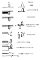
後にD節に開示されるように、これらのスプライシングイベント(スプライシングされた形態及びスプライシングされていない形態)に相当する配列を単離し、そして特異的核酸プローブをデザインするのに使用することができる。ハイブリッド形成産生物を沈殿させ、次いで二本鎖DNAのための4塩基認識部位(4-base recognition site)を有する制限エンドヌクレアーゼの作用に付す。ゆえに、このような制限酵素は、ハイブリッド形成期間中に形成された二本鎖cDNAを平均して256塩基毎に開裂するであろう。この酵素は、有利には付着末端を発生するように選ばれる。このような酵素としては、Sau3AI、HpaII、TaqI及びMseIの如き制限酵素が例示される。ゆえに、これらの酵素により消化された二本鎖断片は、開裂された制限部位を使用するクローニングストラテジーに利用可能である。このような断片には2つのタイプがあり:即ち、その2つの鎖が完全に相補的である完全にハイブリッド形成された断片、及び部分的にハイブリッド形成された断片、即ち、二本鎖領域により隣接された(flanked)一本鎖ループを含む断片(図6A)である。少数であるこれらの後者の断片は関心のある情報を含む。完全にハイブリッド形成された断片、これらはcDNA長の大部分から誘導されるので多数である、からそれらを分離するために、ゲル又はいかなる他の適当なマトリックスも使用される。これらの方法は、特に一本鎖DNAループを含有するDNA断片の、電気泳動又はゲルろ過期間中の、より遅い移動を利用する。この方法においては、所望の情報を含む該少数断片は、両集団における同じDNA領域に相当する多数断片から分取的に(preparatively)分離されうる。定性的な差に結び付いたポジティブ及びネガティブフィンガープリントを、同じ集団から単離することを可能とするこの変法は、RNA/DNAヘテロ二本鎖構造体で実施することもできる。この点で、すべての配列が対を形成している相同性ヘテロ二本鎖に比べて、RNAの一部が対を形成していないRNA/DNA二本鎖のより遅い移動の例は、実施例で記載されたgrb2/grb33モデルで説明される(特に図8、レーン2及び3参照)。
As disclosed later in Section D, sequences corresponding to these splicing events (spliced and unspliced forms) can be isolated and used to design specific nucleic acid probes. The hybridization product is precipitated and then subjected to the action of a restriction endonuclease with a 4-base recognition site for double-stranded DNA. Thus, such restriction enzymes will cleave on average every 256 bases of double stranded cDNA formed during the hybridization period. This enzyme is advantageously chosen to generate sticky ends. Examples of such enzymes include restriction enzymes such as Sau3AI, HpaII, TaqI and MseI. Thus, double stranded fragments digested by these enzymes are available for cloning strategies using cleaved restriction sites. There are two types of such fragments: a fully hybridized fragment whose two strands are fully complementary, and a partially hybridized fragment, ie a double stranded region. FIG. 6A is a fragment containing a flanked single stranded loop (FIG. 6A). A few of these latter pieces contain information of interest. Gels or any other suitable matrix are used to separate them from fully hybridized fragments, which are numerous because they are derived from the majority of the cDNA length. These methods take advantage of the slower movement of DNA fragments, particularly containing single-stranded DNA loops, during electrophoresis or gel filtration. In this method, the minority fragment containing the desired information can be preparatively separated from a larger number of fragments corresponding to the same DNA region in both populations. This variation, which allows positive and negative fingerprints linked to qualitative differences to be isolated from the same population, can also be performed on RNA / DNA heteroduplex structures. In this regard, an example of the slower movement of a non-paired RNA / DNA duplex compared to a homologous heteroduplex in which all sequences are paired is performed Illustrated in the grb2 / grb33 model described in the examples (see especially FIG. 8,
D−同定及び/又はクローニング
ハイブリッド形成により発生させた核酸集団から出発して、定性的な差(例えば、差異選択的スプライシングイベント)を特徴付ける領域は、当業者により知られているいかなる技術によっても同定することができる。
Starting from a nucleic acid population generated by D-identification and / or cloning hybridization, regions characterizing qualitative differences (eg, differentially alternative splicing events) are identified by any technique known by those skilled in the art. can do.
D1.RNA/DNAヘテロ二本鎖で出発する同定及び/又はクローニング
かくして、RNA/DNAヘテロ二本鎖の場合に(この方法の第1の変法)、これらの領域は、図3に示されたとおり、対を形成していないRNA領域(RNAループ)として本質的に現れる。ゆえにこれらの領域は、ヘテロ二本鎖及び一本鎖核酸(DNA、RNA)(過剰の未反応の核酸)を分離し、二本鎖RNA(ヘテロ二本鎖構造に参加した部分)を選択的に消化しそして最後に得られる一本鎖RNAを一本鎖DNAから分離することにより同定及びクローニングされうる。
D1. Identification and / or cloning starting with RNA / DNA heteroduplexes Thus, in the case of RNA / DNA heteroduplexes (first variant of this method), these regions are as shown in FIG. , Appearing essentially as unpaired RNA regions (RNA loops). Therefore, these regions separate heteroduplex and single stranded nucleic acids (DNA, RNA) (excess unreacted nucleic acid) and selectively double stranded RNA (parts participating in heteroduplex structure). And finally the resulting single stranded RNA can be identified and cloned by separating it from single stranded DNA.
この点で、図3に示された第1アプローチに従えば、対を形成していないRNA領域は、RNA/DNAヘテロ二本鎖に参加したRNAドメインを選択的に消化することができる酵素によるヘテロ二本鎖の処理によって同定される。このような活性を有する酵素は先行技術から知られておりそして商業的に入手可能である。それは、RNアーゼH、例えば、特に組換え技術によりE.coliに由来する商業的に入手可能なRNアーゼHを挙げることができる(Promega カタログ番号 M4281; Life Technology カタログ番号 18021)。かくしてこの第1処理は対を形成していない一本鎖RNA領域と一本鎖cDNAを含む混合物を発生させる。RNAは、当該技術分野で知られたいかなる技術によっても、特にcDNAを産生するのに使用されるこれらのプライマーの標識化に基づいてcDNAから分離することができる(上記参照)。これらのRNAは標的、関心のある遺伝子産生物を同定するため又はいかなる他の用途のための材料のソースとしても使用することができる。これらのRNAは、後に記載するとおり、同じくcDNAに転換されることができ、次いでベクターにクローニングされうる。 In this regard, according to the first approach shown in FIG. 3, unpaired RNA regions are due to enzymes capable of selectively digesting RNA domains that participate in RNA / DNA heteroduplexes. Identified by heteroduplex processing. Enzymes having such activity are known from the prior art and are commercially available. It is RNase H, for example E. coli by recombinant technology in particular. Mention may be made of the commercially available RNase H derived from E. coli (Promega catalog number M4281; Life Technology catalog number 18021). Thus, this first treatment generates a mixture comprising unpaired single stranded RNA regions and single stranded cDNA. RNA can be separated from cDNA by any technique known in the art, particularly based on the labeling of these primers used to produce cDNA (see above). These RNAs can be used as a source of material for identifying targets, gene products of interest, or for any other application. These RNAs can also be converted to cDNA and then cloned into a vector as described below.
この点で、RNAのクローニングは種々の方法で行うことができる。1つの方法は、各RNA端部に適合性プライマーの存在下に逆転写反応のための鋳型として作用するオリゴヌクレオチドを挿入することである。プライマーは、当業者により知られている技術に従って酵素、例えば、ファージT4由来のそして供与体分子の5′リン酸基と受容体分子の3′ヒドロキシル基間の分子間ホスホジエステル結合形成を触媒するRNAリガーゼによって付加することができる。このようなRNAリガーゼは商業的に入手可能である(例えば、Life Rechnologies-GIBCO BRL カタログ番号18003)。このようにして得られたcDNAを、次いで、図3に示されたとおり、適当なプライマーを使用して慣用の技術(例えばPCR)により増幅させることができる。この技術は短いRNA分子(1000塩基より小さい)をクローニングするのに特に適している。 In this regard, RNA cloning can be performed in various ways. One method is to insert an oligonucleotide that acts as a template for the reverse transcription reaction in the presence of a compatible primer at each RNA end. The primer catalyzes the formation of an intermolecular phosphodiester bond from an enzyme such as phage T4 and between the 5 'phosphate group of the donor molecule and the 3' hydroxyl group of the acceptor molecule according to techniques known by those skilled in the art. It can be added by RNA ligase. Such RNA ligases are commercially available (eg Life Rechnologies-GIBCO BRL catalog number 18003). The cDNA thus obtained can then be amplified by conventional techniques (eg PCR) using appropriate primers as shown in FIG. This technique is particularly suitable for cloning short RNA molecules (less than 1000 bases).
特異的なRNA領域をクローニング及び/又は同定するための他のアプローチは、例えは、RNAに沿ってランダムに転写を開始するランダムプライマーを使用して、RNアーゼHの如き二本鎖RNAに対して特異的に作用する酵素の消化物に対して行われる逆転写反応を含む。次いでこのようにして得られたcDNAを慣用の分子生物学技術に従って、例えば、T4ファージDNAリガーゼ(商業的に入手可能;例えば、Life Technologies-GIBCO BRL カタログ番号18003)によってcDNA端部にオリゴヌクレオチドを付加することにより形成されるプライマーを使用するPCRにより増幅される。この第2の技術は、図4と実施例に説明されている。この技術は、特に長いRNAに適合し、そして後に全イニシャル配列(entire initial sequence)を構築するのに十分な配列データの部分を与える。 Other approaches for cloning and / or identifying specific RNA regions include, for example, using a random primer that initiates transcription randomly along the RNA, against a double-stranded RNA such as RNase H. A reverse transcription reaction performed on digests of enzymes that act specifically. The cDNA thus obtained is then subjected to oligonucleotides at the ends of the cDNA according to conventional molecular biology techniques, for example by T4 phage DNA ligase (commercially available; eg, Life Technologies-GIBCO BRL catalog number 18003). Amplified by PCR using primers formed by the addition. This second technique is illustrated in FIG. 4 and the example. This technique is particularly suited to long RNAs and provides a portion of sequence data sufficient to later build an entire initial sequence.
特異的RNA領域をクローニング及び/又は同定するための更なるアプローチは、同じくランダムプライマーを使用する逆転写反応に基づいている(図4)。しかしながら、この変法に従えば、使用されるプライマーは少なくとも部分的にセミランダムプライマー、即ち、
−ランダム(縮重した(degenerated))領域、
−規定された程度の制限を有する最小プライミング領域、及び
−安定化領域、
を含むオリゴヌクレオチドである。
A further approach for cloning and / or identifying specific RNA regions is also based on reverse transcription reactions using random primers (FIG. 4). However, according to this variant, the primers used are at least partly semi-random primers, i.e.
-Random (degenerated) regions,
A minimum priming region with a specified degree of limitation; and a stabilization region,
Is an oligonucleotide.
好ましくは、これらは、5′→3′方向に、
−8〜24個の規定されたヌクレオチド、好ましくは10〜18個のヌクレオチドを含む安定化領域、
この安定化領域は、それ自体、本発明のセミランダムプライマーによって行われる初期増幅に由来する断片を再増幅するのに使用されるオリゴヌクレオチドの配列に相当することができる。更に、安定化領域は、制限酵素に対応する1つ又はそれより多くの部位、好ましくは非パリンドロームの部位の配列を含むことができる。これは、例えば、このようにして増幅された断片のクローニングを簡単化することを可能とする。安定化領域の特定の例は配列GAGAAGCGTTAT(配列番号1の残基1〜12)により与えられる;
−3〜8ヌクレオチド、更に特定的には5〜7個のヌクレオチドを有するランダム領域、及び
−オリゴヌクレオチドが平均して少なくとも約60塩基対毎に、好ましくは約250塩基対毎にハイブリッド形成するように規定された最小プライミング領域、
更に好ましくは、プライミング領域は、2〜4個の規定されたヌクレオチド、好ましくは3又は4個の、例えばAGGX、式中Xは4つの塩基A、C、G又はTの1つである、の如きヌクレオチドを含む。このようなプライミング領域の存在はオリゴヌクレオチドに平均して約256塩基対毎にハイブリッド形成する能力を与える;
を含むオリゴヌクレオチドである。
Preferably, these are in the 5 ′ → 3 ′ direction,
A stabilization region comprising 8-24 defined nucleotides, preferably 10-18 nucleotides,
This stabilization region can itself correspond to the sequence of the oligonucleotide used to re-amplify the fragment derived from the initial amplification performed by the semi-random primer of the present invention. Furthermore, the stabilization region may comprise a sequence of one or more sites corresponding to restriction enzymes, preferably non-palindromic sites. This makes it possible, for example, to simplify the cloning of the fragment thus amplified. A specific example of a stabilization region is given by the sequence GAGAAGCGTTAT (residues 1-12 of SEQ ID NO: 1);
-A random region having 3-8 nucleotides, more particularly 5-7 nucleotides; and-the oligonucleotide hybridizes on average at least about every 60 base pairs, preferably about every 250 base pairs Minimum priming area, as defined in
More preferably, the priming region is 2 to 4 defined nucleotides, preferably 3 or 4, eg AGGX, wherein X is one of 4 bases A, C, G or T. Such nucleotides. The presence of such a priming region gives the oligonucleotide the ability to hybridize on average about every 256 base pairs;
Is an oligonucleotide.
特に好ましい方式では、オリゴヌクレオチドは、式:
![]()
式中、不変の塩基はPCR実験において自己塩基対形成によるバツクグラウンドを最小にするように配置されており(ordered)、そしてNは4つの塩基が示された位置においてランダム様式で存在することができることを示し、そしてXは4種の塩基A、C、G又はTの1つである、
を有する。このようなオリゴヌクレオチドは同じく本発明の目的を構成する。
In a particularly preferred manner, the oligonucleotide has the formula:
![]()
Where invariant bases are ordered to minimize background due to self-base pairing in PCR experiments, and N may be present in a random fashion at the indicated positions. And X is one of four bases A, C, G or T.
Have Such oligonucleotides also constitute an object of the present invention.
この点で、クローニングされるべきRNAに対するプライミングイベントを増加させるように、反応は、
の如きオリゴヌクレオチド、各オリゴヌクレオチド集団(A、B、C、D)は単独で又は他との組み合わせにおいて使用することができる、により平行して行うことができる。
In this respect, so as to increase the priming event for the RNA to be cloned,
The oligonucleotides, each oligonucleotide population (A, B, C, D) can be used alone or in combination with the other.
逆転写反応の後、cDNAはオリゴヌクレオチドA又はB又はC又はDを使用するPCRにより増幅される。 After the reverse transcription reaction, the cDNA is amplified by PCR using oligonucleotides A or B or C or D.
上記に示されたとおり、所望のオリゴヌクレオチド集団の複雑性及び特異性に依存して、縮重した位置(degenerated positions)の数は3〜8、好ましくは5〜7の範囲にあることができる。3より少ないと、ハイブリッド形成は限定されそして8より大きいとオリゴヌクレオチド集団は特異的バンドの良好な増幅を確保するのにはあまりにも複雑すぎる。 As indicated above, depending on the complexity and specificity of the desired oligonucleotide population, the number of degenerated positions can range from 3 to 8, preferably from 5 to 7. . Below 3 the hybridization is limited and above 8 the oligonucleotide population is too complex to ensure good amplification of specific bands.
更に、これらのオリゴヌクレオチドの普遍の3′端部(制限されたプライミング領域)の長さは修正することもできる:4個の不変の塩基を有する上記したプライマーは平均して256塩基対断片の増幅を可能とするが、3個の不変の塩基を有するプライマーはより短い断片(平均して64塩基対)の増幅を可能とする。本発明の好ましい第1の態様では、プライミング領域が4個の不変の塩基を含むオリゴヌクレオチドを使用する。本発明の他の好ましい態様では、3個の不変の塩基のプライミング領域を有するオリゴヌクレオチドを使用する。実際、エキソンは137塩基の平均サイズを有するので、それらは、このようなオリゴヌクレオチドにより有利に増幅される。この点で、例えば、配列番号2,3、及び4を有するオリゴヌクレオチドも参照されたい。 In addition, the length of the universal 3 'end (restricted priming region) of these oligonucleotides can be modified: the primers described above with 4 invariant bases on average are 256 base pair fragments. While allowing amplification, primers with 3 invariant bases allow amplification of shorter fragments (on average 64 base pairs). In a preferred first aspect of the invention, an oligonucleotide is used in which the priming region contains 4 invariant bases. In another preferred embodiment of the invention, an oligonucleotide having a priming region of 3 invariant bases is used. In fact, since exons have an average size of 137 bases, they are advantageously amplified by such oligonucleotides. In this regard, see also, for example, oligonucleotides having SEQ ID NOs: 2, 3, and 4.
最後に、一般に、RNAの同定及び/又はクローニング段階は、できるだけ多くの情報を発生するようにPCRとクローニングの種々の方法に基づく。 Finally, in general, RNA identification and / or cloning steps are based on various methods of PCR and cloning to generate as much information as possible.
D2.ヘテロ二本鎖で出発する同定及び/又はクローニング
ヘテロ二本鎖構造の場合に(本方法の別の変法)、定性的に異なる領域(挿入、欠失、差異スプライシング)は本質的に、図5に示されるとおり、二本鎖DNA領域の形態で現れる。かくして、このような領域はRNAを消化することができる酵素の如き適当な酵素の存在下にそれらを処理し、次いで一本鎖DNAを消化することができる酵素により処理することによって同定及びクローニングすることができる。かくして、核酸は直接二本鎖DNAの形態で得られ、そして例えば、ベクターpMos−Blue(Amerisham, RPN5110)の如きいかなる適当なベクターにもクローニングされうる。この方法は、ヌクレアーゼ活性を有するように修飾された、所定の配列のRNA又はオリゴヌクレオチドを使用する従来述べられた方法(Landigraf et al., (1994), Biochemistry, 33: 10607-10615)とは区別されるべきである。
D2. Identification and / or cloning starting with heteroduplexes In the case of heteroduplex structures (another variant of the method), qualitatively different regions (insertions, deletions, differential splicing) are essentially As shown in FIG. 5, it appears in the form of a double-stranded DNA region. Thus, such regions are identified and cloned by treating them in the presence of a suitable enzyme, such as an enzyme capable of digesting RNA, and then treating the single-stranded DNA with an enzyme capable of digesting. be able to. Thus, the nucleic acid is obtained directly in the form of double stranded DNA and can be cloned into any suitable vector such as, for example, the vector pMos-Blue (Amerisham, RPN5110). This method is different from the previously described method (Landigraf et al., (1994), Biochemistry, 33: 10607-10615) using RNA or oligonucleotide of a predetermined sequence modified to have nuclease activity. It should be distinguished.
D3.DNA/DNAホモ二本鎖で開始する同定及び/又はクローニング(図6)
この態様では、関心のある配列(差異スプライシング)は、図6に示されたとおり、本質的に対を形成しないDNA領域の形態で現れる。このような領域は、適当な酵素の使用及び核酸精製段階を含む、本願に開示された種々の技術に従って同定及びクローニングされうる。
D3. Identification and / or cloning starting with DNA / DNA homoduplex (Figure 6)
In this embodiment, the sequence of interest (differential splicing) appears in the form of DNA regions that are essentially unpaired, as shown in FIG. Such regions can be identified and cloned according to various techniques disclosed herein, including the use of appropriate enzymes and nucleic acid purification steps.
特定の好ましい態様では、対を形成しない領域を含む核酸の集団は、
−二本鎖DNAに特異的な制限酵素により形成されたハイブリッドを消化し、
−対を形成していない領域を含む制限断片を単離し、そして
−単離された断片を増幅する、
ことにより同定又はクローニングされる。
In certain preferred embodiments, the population of nucleic acids comprising unpaired regions is
Digesting a hybrid formed by a restriction enzyme specific for double-stranded DNA;
Isolating restriction fragments containing unpaired regions, and amplifying the isolated fragments,
Identified or cloned.
ハイブリッドの消化の前に、分離段階を行って汚染しているハイブリッド(例えば、同じ試料からの2つのDNA鎖間で形成された)を除去することができる。この分離は、ハイブリッド形成の前に1つの試料中のcDNAを標識しそしてハイブリッド形成の後標識されなかったcDNAを除去することにより有利に行われる。特定の態様では、1つの試料に由来するcDNAはビオチニル化され、そして分離段階は、ハイブリッド形成産生物をストレプトアビジンでコーティンされた支持体、例えば、ビーズ(例えば、磁性ビーズ)と接触させることを含む。他の標識、例えば、親和性結合による選択的分離を可能とするアフイニティー対のいかなる他方のパートナーも使用することができることは理解されるべきである。アフイニティー精製されると、2つの分子種が得られる:関心のあるホモ二本鎖及び第1の試料からの出発標識化一本鎖cDNA(starting labeled single-stranded cDNA)。 Prior to hybrid digestion, a separation step can be performed to remove contaminating hybrids (eg, formed between two DNA strands from the same sample). This separation is advantageously performed by labeling the cDNA in one sample prior to hybridization and removing unlabeled cDNA after hybridization. In a particular embodiment, the cDNA from one sample is biotinylated and the separation step comprises contacting the hybridization product with a streptavidin-coated support, eg, a bead (eg, a magnetic bead). Including. It should be understood that other labels, such as any other partner of an affinity pair that allows selective separation by affinity binding, can be used. When affinity purified, two molecular species are obtained: the homoduplex of interest and the starting labeled single-stranded cDNA from the first sample.
関心のある対を形成していない領域を単離するために、産生物を二本鎖DNAsに特異的な制限酵素を使用して酵素消化に付す。したがって、ホモ二本鎖のみが消化されそして第1の試料からのいかなる汚染性の標識された一本鎖cDNAも元のままである(intact)。 To isolate the unpaired region of interest, the product is subjected to enzymatic digestion using a restriction enzyme specific for double stranded DNAs. Thus, only the homoduplex is digested and any contaminating labeled single stranded cDNA from the first sample is intact.
酵素は、好ましくは、小さなds制限断片を発生するように、ds−DNAsを頻繁に切断する酵素から選ばれる。好ましい態様では、制限酵素は4塩基開裂部位を認識する。このような開裂部位は平均して約250塩基毎に存在する。更なる好ましい態様では、付着末端を形成する。このような酵素の例は、例えば、Sau3A1、HpaII、TaqI、MseIを含む。 The enzyme is preferably selected from enzymes that frequently cleave ds-DNAs so as to generate small ds restriction fragments. In a preferred embodiment, the restriction enzyme recognizes a 4-base cleavage site. Such cleavage sites are present on average about every 250 bases. In a further preferred embodiment, a sticky end is formed. Examples of such enzymes include, for example, Sau3A1, HpaII, TaqI, MseI.
この処理の結果として、混合物は3つのタイプの分子種:
−その2つの鎖が完全に相補的である完全にハイブリッド形成したds断片、 −部分的にハイブリッド形成したds断片、即ち、二本鎖領域により隣接された(flanked)された1つ又はそれより多くの対を形成していない領域(即ち、一本鎖ループ)(図6A)。少数であるこれらのds断片は関心のある情報を含む。及び
−第1の試料からの標識された、消化されなかったsscDNA、
を含む。
As a result of this treatment, the mixture becomes three types of molecular species:
A fully hybridized ds fragment whose two strands are fully complementary, a partially hybridized ds fragment, ie one or more flanked by a double stranded region Many unpaired regions (ie single stranded loops) (FIG. 6A). A few of these ds fragments contain the information of interest. And-a labeled, undigested sscDNA from the first sample,
including.
これらの種を分離しそして部分的にハイブリッド形成されたds断片を単離するために、混合物をゲル又はいかなる他の適当なマトリックスにもよる分離方法に付すことができる。これらの方法は、特に一本鎖DNAループを含有するDNA断片の電気泳動又はゲルろ過期間中のより遅い移動を利用する。この方法において、所望の情報を含む少量断片は両集団における同一のDNA領域に相当する多量断片から分取的に(preparatively)分離されうる。 In order to separate these species and isolate the partially hybridized ds fragment, the mixture can be subjected to a separation method with a gel or any other suitable matrix. These methods take advantage of the slower migration, particularly during electrophoresis or gel filtration, of DNA fragments containing single stranded DNA loops. In this way, small fragments containing the desired information can be preparatively separated from large fragments corresponding to the same DNA region in both populations.
最も好ましい態様では、部分的にハイブリッド形成されたds断片は、第1の試料から生じる標識された、対を形成していないsscDNAを除去するために、混合物を上記したとおりのストレプトアビジンでコーティングされた支持体で最初に処理することにより単離する。次いで、対を形成していない領域を含むds断片を単離するために、混合物を標識された縮重したオリゴヌクレオチド(degenerated oligonuclotides)と接触させることができる(オリゴヌクレオチド捕捉)。これらの縮重したオリゴヌクレオチドは、すべての可能な配列の組み合わせを表し、かくしていかなるss配列ともハイブリッド形成することができる。これらの縮重したオリゴヌクレオチドは、更に好ましくは、長さが10〜30ヌクレオチド、更に好ましくは約24ヌクレオチドを含む。それらを特異的ハイブリッド形成が起こることを可能とする条件下に混合物と接触させ、それにより対を形成していない領域を含むds断片と特異的に反応させる。このようなハイブリッド形成は標識を使用する分離により該ds断片の捕捉及び単離を可能とする。例えば、標識はビオチンであることができそして対を形成していない領域を含むds断片はストレプトアビジンでコーティングされた支持体(例えば、磁性ビーズ)との接触により単離することができる。 In the most preferred embodiment, the partially hybridized ds fragment is coated with streptavidin as described above to remove labeled, unpaired sscDNA resulting from the first sample. Isolation by first treating with a support. The mixture can then be contacted with labeled degenerated oligonuclotides (oligonucleotide capture) to isolate ds fragments containing unpaired regions. These degenerate oligonucleotides represent all possible sequence combinations and can thus be hybridized to any ss sequence. These degenerate oligonucleotides more preferably comprise 10-30 nucleotides in length, more preferably about 24 nucleotides. They are contacted with the mixture under conditions that allow specific hybridization to occur, thereby specifically reacting with the ds fragment containing the unpaired region. Such hybridization allows capture and isolation of the ds fragment by separation using a label. For example, the label can be biotin and ds fragments containing unpaired regions can be isolated by contact with a streptavidin-coated support (eg, magnetic beads).
次いで対を形成していない領域を含むds断片は、媒体のイオン強度を低下することにより標識されたオリゴヌクレオチドから分離させる。 The ds fragment containing the unpaired region is then separated from the labeled oligonucleotide by reducing the ionic strength of the medium.
単離された断片を、次いで、それらの端部の各々で、アダプター又はリンカー、それらの端部の1つに開裂された制限部位を有する、にライゲーションする。この段階は、当業者に知られている技術に従って、例えば、ファージT4DNAリガーゼによるライゲーションにより行うことができる。このようにして導入された制限部位はcDNA断片の部位と適合性であるように選ばれる。導入されたリンカーは、酵素的増幅(PCR)のためのプライマーを発生させることを可能とする、既知の配列の二本鎖cDNA配列である。 The isolated fragments are then ligated at each of their ends to an adapter or linker, with a restriction site cleaved at one of those ends. This step can be performed according to techniques known to those skilled in the art, for example, by ligation with phage T4 DNA ligase. The restriction sites thus introduced are chosen to be compatible with the site of the cDNA fragment. The introduced linker is a double stranded cDNA sequence of known sequence that allows generation of primers for enzymatic amplification (PCR).
次の段階で、各々同定されるべき定性的な差を有する二本鎖を増幅する。そのために、リンカーと付加した二本鎖cDNAの熱変性の後、これらのcDNA端部の各々を特異的プライミング配列に共有結合させる。適当な特異的プライマーによるPCRに続いて、2つの種類の二本鎖cDNA:即ち、2つの病態生理学的条件を区別する定性的な差に特異的な配列を含有する断片及びこれらのスプライシングイベントのネガティブフィンガープリントを含む断片が得られる。これらの断片をクローニングすると、各スプライシングイベントについてポジティブ及びネガティブフィンガープリントが存在する選択的スプライシングライブラリーを発生する。ゆえに、このライブラリーは、オルタナテイブエキソン及びイントロンへのアクセスのみならず、これらのスプライシングされた配列の切除により形成された特異的連結部へのアクセスも与える。同じライブラリーにおいて、この差異遺伝子情報は、無差別に2つの病態生理学的条件から誘導されうる。更に、同定されたスプライシングイベントの差異な性質をチェックするようにそしてそれらが特異的に引き出される条件を決定するように、ライブラリーにおけるクローンを全mRNA集団の各々に由来するプローブとハイブリッド形成させることができる。 The next step is to amplify the duplexes that each have a qualitative difference to be identified. To that end, after thermal denaturation of the double-stranded cDNA added with the linker, each of these cDNA ends is covalently linked to a specific priming sequence. Following PCR with appropriate specific primers, two types of double-stranded cDNA: fragments containing sequences specific for qualitative differences that distinguish the two pathophysiological conditions and of these splicing events Fragments containing a negative fingerprint are obtained. Cloning these fragments generates an alternative splicing library in which a positive and negative fingerprint exists for each splicing event. Thus, this library provides not only access to alternative exons and introns, but also to specific junctions formed by excision of these spliced sequences. In the same library, this differential gene information can be derived indiscriminately from two pathophysiological conditions. In addition, the clones in the library are hybridized with probes from each of the total mRNA population so as to check the differential nature of the identified splicing events and to determine the conditions under which they are specifically derived. Can do.
次いで、この方法は、増幅された断片の配列決定、配列をデータベースに記憶させること、配列をデータベースにおいて分析してスプライスドメイン及び相当する連結領域を同定し、該スプライスドメイン又は連結領域に特異的なオリゴヌクレオチドを合成し及び/又は支持体上に該オリゴヌクレオチドを付着させることを更に含むことができる。これらの種々の段階は、cDNAの産生からスプライスオリゴヌクレオチドの付着までコンピューター支援又はコンピューター操作させることができる。 The method then sequencees the amplified fragment, stores the sequence in a database, analyzes the sequence in the database to identify the splice domain and the corresponding ligation region, and is specific for the splice domain or ligation region. It may further comprise synthesizing the oligonucleotide and / or attaching the oligonucleotide on a support. These various steps can be computer assisted or computer operated, from cDNA production to splice oligonucleotide attachment.
そのように同定された定性的な差に由来するcDNA断片は2つの主な使用:
−適当なベクターにクローニングして調査中の2つの病態生理学的条件間で発生する定性的な差を表すライブラリーを構築すること、
−差異スプライシングイベントの同定を可能とするDNAライブラリーをスクリーニングするためのプローブとしての使用、
を有する。
CDNA fragments derived from qualitative differences so identified have two main uses:
-Cloning into an appropriate vector to construct a library representing qualitative differences that occur between the two pathophysiological conditions under investigation;
Use as a probe to screen a DNA library allowing the identification of differential splicing events,
Have
本発明で使用されるベクターは、特にプラスミド、コスミド、ファージ、YAC、HAC等であることができる。かくして、これらの核酸はそのまま貯蔵することができ、又は複製のために使用されるクローニングベクターと適合性の微生物に導入することができ及び/又は培養物の形態で貯蔵することができる。 The vector used in the present invention can be in particular a plasmid, cosmid, phage, YAC, HAC and the like. Thus, these nucleic acids can be stored as is, or introduced into microorganisms compatible with the cloning vector used for replication and / or stored in the form of a culture.
各試料について本明細書に記載された方法を行うために必要な期間は、一般に2か月より少なく、特に6週間より少ない。更に、これらの種々の方法は、全体の時間の長さを減少させそして多数の試料の処理を簡単にするように自動化することができる。 The period required to perform the methods described herein for each sample is generally less than 2 months, especially less than 6 weeks. Furthermore, these various methods can be automated to reduce the overall length of time and simplify the processing of multiple samples.
この点で、本発明の他の目的は、本発明の方法により同定及び/又はクローニングされた核酸に関する。既に述べたとおり、これらの核酸はRNA又はcDNAであることができる。更に一般的には、2つの生理学的条件に特有の選択的スプライシングに相当する核酸を本質的に含む核酸組成物に関する。更に特定的には、これらの核酸は生物学的試験試料において同定されそして基準条件下の同じ生理学的試料に存在しない選択的スプライシングに相当する。本発明は、同じく、このようにしてクローニングされた核酸の、治療的又は診断生成物として又は後に記載するとおり活性分子を同定するためのスクリーニング手段としての使用に関する。 In this regard, another object of the present invention relates to nucleic acids identified and / or cloned by the method of the present invention. As already mentioned, these nucleic acids can be RNA or cDNA. More generally, it relates to a nucleic acid composition essentially comprising a nucleic acid corresponding to alternative splicing characteristic of two physiological conditions. More specifically, these nucleic acids are identified in biological test samples and represent alternative splicing that is not present in the same physiological sample under reference conditions. The invention also relates to the use of the nucleic acid thus cloned as a therapeutic or diagnostic product or as a screening tool for identifying active molecules as described later.
ゆえに上記に開示された種々の方法はすべて2つの病態生理学的条件間の差異スプライシングされた遺伝子情報を表すcDNA配列のクローニングをもたらす。かくしてこれらの方法の1つに由来するクローンの全体の組は関心のある2つの条件間に発生する定性的な差を表すライブラリーを構築することを可能とする。 Thus, the various methods disclosed above all result in the cloning of cDNA sequences representing differentially spliced genetic information between the two pathophysiological conditions. Thus, the entire set of clones derived from one of these methods makes it possible to construct a library that represents the qualitative differences that occur between the two conditions of interest.
E−定性的ライブラリーの発生
この点で、本発明は更に生物学的試料の与えられた生理学的状態を表す核酸ライブラリーを作成する方法を指向する。この方法は、有利には、該生理学的状態の遺伝子発現(例えば、選択的スプライシング)の定性的マーカーを表すが基準状態においては存在しない核酸をクローニングして、調査されるべき2つの状態間で発生する定性的な差に特異的なライブラリーを発生させることを含む。
E-Generation of Qualitative Library In this regard, the present invention is further directed to a method of creating a nucleic acid library that represents a given physiological state of a biological sample. This method advantageously clones nucleic acids that represent qualitative markers of gene expression (eg, alternative splicing) of the physiological state but are not present in the reference state, between the two states to be investigated. Generating a library specific to the qualitative differences that occur.
これらのライブラリーは、プラスミド又はファージベクターに挿入されたcDNAにより構築される。このようなライブラリーはニトロセルロースフィルター又は当業者により知られているいかなる他の支持体、例えばチップ又はバイオチップ上に付着させることができる。 These libraries are constructed by cDNA inserted into a plasmid or phage vector. Such a library can be deposited on a nitrocellulose filter or any other support known by those skilled in the art, such as a chip or biochip.
定性的差異スクリーニングの特徴の1つ又は独創的特徴の1つは、この技術が、1つではなくて、有利には、2つの与えられた条件間で発生する定性的な差の全体の組を表す2つの差異ライブラリー:ライブラリー対(図1D参照)をもたらすことである。 One of the features of qualitative difference screening or one of the original features is that the technique is not one but advantageously the entire set of qualitative differences that occur between two given conditions. Resulting in two difference libraries representing library pairs (see FIG. 1D).
かくして、本発明は、好ましくは、第1の生物学的試料に由来するRNAを第2の生物学的試料に由来するcDNAとハイブリッド形成することにより得られうるいかなる核酸組成物又はライブラリーにも関する。更に好ましくは、本発明のライブラリー又は組成物は、2つの生物学的試料間の発現の定性的な差を表す核酸を含みそして(i)第1の生物学的試料に由来するRNAと第2の生物学的試料に由来するcDNAとの少なくとも1つのハイブリッド形成段階、(ii)発現の定性的な差を表すこれらの核酸を選び、そして場合により(iii)該核酸をクローニングすることを含む方法により発生される。 Thus, the present invention is preferably for any nucleic acid composition or library obtainable by hybridizing RNA from a first biological sample with cDNA from a second biological sample. Related. More preferably, the library or composition of the invention comprises a nucleic acid that represents a qualitative difference in expression between two biological samples and (i) a RNA and a first derived from the first biological sample. At least one hybridization step with cDNA from two biological samples, (ii) selecting those nucleic acids that represent a qualitative difference in expression, and optionally (iii) cloning the nucleic acids Generated by the method.
更に、このようなライブラリーが構築されと、得られるライブラリーの特異性を改良するために、クローン選択の段階を進行させることが可能である。実際に、観察されるあるミスマッチは単に定性的な差(例えば、差異選択的スプライシング)のみによるのではなくて、例えば、逆転写欠陥から生じうるということがありうる。このようなイベントは一般に有意ではないけれども、核酸クローニングの前にそれらを防止するか又はそれらの発生率を減少させることが好ましい。これを達成するために、ライブラリークローンを調査されるべき両生理学的条件において発生するcDNA集団とハイブリッド形成させることができる(上記段階c)。非差異方式で両集団とハイブリッド形成するクローンは、非特異的であると考えられそして場合により捨てられるか又は第2プライオリティとして処理されるであろう(実際、試験試料における新しいアイソフォームの出現は、基準試料に存在する最初のアイソフォームがこの試験試料から消失したことを必ずしも示さない)。いずれかの集団の1つのみとハイブリッド形成するか又は集団の1つと優先的にハイブリッド形成するクローンは特異的であると考えられそして濃縮又は精製されたライブラリーを構築するのに優先して選ばれうる。 Furthermore, once such a library has been constructed, it is possible to proceed with a clonal selection step in order to improve the specificity of the resulting library. Indeed, it is possible that some mismatches that are observed are not simply due to qualitative differences (eg, differential alternative splicing), but may result from, for example, reverse transcription defects. Although such events are generally not significant, it is preferable to prevent them or reduce their incidence prior to nucleic acid cloning. To achieve this, library clones can be hybridized with cDNA populations that develop in both physiological conditions to be investigated (step c above). Clones that hybridize with both populations in a non-differential manner will be considered non-specific and will either be discarded or treated as a second priority (in fact, the appearance of a new isoform in the test sample is , Does not necessarily indicate that the first isoform present in the reference sample has disappeared from the test sample). Clones that hybridize with only one of any population or preferentially hybridize with one of the population are considered specific and are chosen in preference to construct enriched or purified libraries Can be.
精製段階は、ハイブリッド形成すること又及び統計的に関係がある数(statistically relevant number)の病理試料に由来するプローブによってクローンの同一性をチェックすることにより行うこともできる。 The purification step can also be performed by hybridization and by checking the identity of the clone with a probe derived from a statistically relevant number of pathological samples.
ゆえに、本願は、同じく、生理学的条件の典型的な選択的スプライシングに特異的な核酸を含むいかなる核酸ライブラリーも指向する。これらのライブラリーは有利には、選択的スプライシングの特異的RNA領域に相当する一般に二本鎖のcDNAを含む。このようなライブラリーは、一般にクローニングベクター内に組み込まれた核酸、又は該核酸を含有する細胞培養物からなることができる。 Thus, the present application is also directed to any nucleic acid library containing nucleic acids specific for alternative splicing typical of physiological conditions. These libraries advantageously comprise a generally double stranded cDNA corresponding to a specific RNA region of alternative splicing. Such a library can generally consist of nucleic acids incorporated into a cloning vector or a cell culture containing the nucleic acids.
最初のRNAの選択は、得られるライブラリーの特徴を部分的に決定する:
−両条件A及びBのRNAは当業者に知られている技術に従って単離されたmRNA又は全成熟RNA(total mature RNAs)である。かくしてライブラリーはいわゆる制限された定性的差異スクリーニングライブラリーである。何故ならば、それらは両病態生理学的条件の成熟RNAを特徴付ける定性的な差に制限されるからである。
−両条件の一方のRNAはmRNA又は成熟した全RNAであるが、これに対して
他方の条件のRNAは、細胞核から当業者に知られている技術に従って単離された、スプライシングによりプロセッシングされていないプレメッセンジャーRNAである。この状況においては、得られるライブラリーはいわゆる複合差異スクリーニングライブラリーである。何故ならば、成熟RNA間の差に制限されるのではなくて、すべてのイントロンを含む、他方には欠けている与えられた条件におけるスプライシングされた転写物の全体の組を含むからである。
−最後に、RNAは単一の病態生理学的条件から生じることができ、そしてこの場合に差異スクリーニングは同じ試料からの成熟RNA及びプレメッセンジャーRNAを含む。このような場合に、得られるライブラリーは自己由来の定性的差異スクリーニングライブラリーである。このようなライブラリーの有用性は、それらが与えられた条件において転写されたイントロンの全範囲をもっぱら含むという点にある。それらが異なる条件の成熟RNAに由来するプローブとハイブリッド形成するかどうかは、調査中の条件がイントロンを固持すると共にそれらの容易な同定を与えることにより特徴付けられるならば、迅速に確かめることを可能とする。
Initial RNA selection partially determines the characteristics of the resulting library:
-Both conditions A and B RNA are mRNA or total mature RNAs isolated according to techniques known to those skilled in the art. The library is thus a so-called limited qualitative difference screening library. This is because they are limited to qualitative differences that characterize mature RNA in both pathophysiological conditions.
-One RNA in both conditions is mRNA or mature total RNA, whereas the other RNA is isolated from the cell nucleus according to techniques known to those skilled in the art and processed by splicing. There is no pre-messenger RNA. In this situation, the resulting library is a so-called composite difference screening library. Because it is not limited to differences between mature RNAs, it includes the entire set of spliced transcripts in a given condition that contains all introns and the other lacks.
-Finally, RNA can arise from a single pathophysiological condition, and in this case the difference screen includes mature RNA and pre-messenger RNA from the same sample. In such a case, the resulting library is an autologous qualitative difference screening library. The utility of such libraries is that they contain exclusively the entire range of introns transcribed in a given condition. Whether they hybridize with probes derived from mature RNA under different conditions can be quickly ascertained if the conditions under investigation are characterized by retaining introns and giving them easy identification And
一般的にいえば、ライブラリーは、クローニングされた核酸により形質転換された細胞培養物の、固体培地上(特に、寒天培地上)へのスプレッディングにより発生される。形質転換は、当業者に知られているいかなる技術(トランスフェクション、リン酸カルシウム沈殿、エレクトロポレーション、バクテリオファージによる感染等)によっても行われる。細胞培養物は、一般にバクテリア培養物、例えばE.coliである。それは、真核細胞培養物、特に下等真核細胞(例えば酵母)であることもできる。このスプレッディング段階は皿又はいかなる他の適当な支持体上でも無菌条件で行うことができる。更に、寒天培地上の広がった培養物は例えば冷凍した状態で(グリセロール又はいかなる他の適当な作用物質中でも)貯蔵することができる。当然、これらのライブラリーは、「デュプリケート(duplicate)」、即ち、後に更に完全に記載されるよく知られた技術に従って作られたコピーを産生するために使用することができる。更に、このようなライブラリーは、一般に、増幅されたライブラリー、即ち、各クローンを増幅された状態で含むライブラリーを作成するのに使用される。増幅されたライブラリーは、下記の如く作成される:即ち、広がった培養物から出発して、すべての細胞クローンを回収し、そしていかなる適合性の培地でも使用して冷凍された状態で又は冷所に貯蔵するためにパッケージする。この増幅されたライブラリーは、有利にはE.coliバクテリア培養物から作成されそして無菌条件で4℃で貯蔵される。この増幅されたライブラリーは、種々の用途のために種々の支持体上にこのようなクローンを含有するいかなるその後に作成されるライブラリーの作成及び制限のない複製も可能とする。このようなライブラリーは、更に、関心のあるいかなるクローンの単離及び特徴付けも可能とする。本発明のライブラリーを構成する各クローンは、実際に生理学的条件の特徴的エレメントであり、そしてそれゆえマーカー、抗体産生、診断、遺伝子導入治療等のためのサーチの如き種々の調査のための特に興味のある標的を構成する。これらの種々の用途は以下に更に詳細に論議される。ライブラリーは、一般に、適当な支持体(例えばペトリ皿)上の寒天培地に培養物をスプレッディングすることにより上記したようにして作成される。寒天培地を使用することの利点は各コロニーを分離しそして別個に認識することができるということである。この培養物から出発して、同じデュプリケートを、当該技術分野で知られている技術に従ういかなる適当な支持体上でもレプリカ平板培養(replica-plating)により簡単に実質的量において作成することができる。かくして、デュプリケートは、フィルター、その上に細胞付着が可能である膜(ナイロン、ニトロセルロース等)によって得られうる。次いでフィルターは、核酸を変化させないいかなるパッケージング培地中にも、乾燥された状態で例えば4℃でそのまま貯蔵されうる。フィルターは、同じく細胞、タンパク質等を捨てそしてこのような成分のみを核酸として保持することのような方式で処理することができる。これらの処理法は、特に、プロテアーゼ、洗剤等の使用を含むことができる。処理されたフィルターは、いかなる装置においても又は核酸のために許容できるいかなる条件下にでも同じく貯蔵することができる。 Generally speaking, a library is generated by spreading a cell culture transformed with a cloned nucleic acid onto a solid medium (especially on an agar medium). Transformation is performed by any technique known to those skilled in the art (transfection, calcium phosphate precipitation, electroporation, infection with bacteriophage, etc.). Cell cultures are generally bacterial cultures such as E. coli. E. coli. It can also be a eukaryotic cell culture, in particular a lower eukaryotic cell (eg yeast). This spreading step can be performed under aseptic conditions on a dish or any other suitable support. Furthermore, the spread culture on the agar medium can be stored, for example, frozen (in glycerol or any other suitable agent). Of course, these libraries can be used to produce “duplicate”, ie, copies made according to well-known techniques described more fully later. In addition, such libraries are generally used to create an amplified library, ie, a library that contains each clone in an amplified state. An amplified library is made as follows: starting from an expanded culture, all cell clones are recovered and frozen in any compatible medium or refrigerated. Package for storage. This amplified library is advantageously E. coli. made from E. coli bacterial cultures and stored at 4 ° C. under aseptic conditions. This amplified library allows for the creation and unlimited replication of any subsequently generated library containing such clones on various supports for various applications. Such a library further allows the isolation and characterization of any clone of interest. Each clone making up the library of the present invention is actually a characteristic element of physiological conditions, and therefore for various investigations such as searching for markers, antibody production, diagnosis, gene transfer therapy, etc. Constitutes a target of particular interest. These various uses are discussed in more detail below. Libraries are generally created as described above by spreading cultures on agar media on a suitable support (eg, a Petri dish). The advantage of using an agar medium is that each colony can be isolated and recognized separately. Starting from this culture, the same duplicate can easily be made in substantial quantities by replica-plating on any suitable support according to techniques known in the art. Thus, duplicates can be obtained by filters and membranes (nylon, nitrocellulose, etc.) on which cell attachment is possible. The filter can then be stored as is, eg, at 4 ° C., in a dry state in any packaging medium that does not alter the nucleic acid. Filters can also be processed in such a way as to discard cells, proteins, etc. and retain only such components as nucleic acids. These treatment methods can include the use of proteases, detergents and the like, among others. The treated filter can also be stored in any device or under any conditions acceptable for nucleic acids.
核酸ライブラリーは、同じくバイオチップ又はいかなる他の適当な装置へのトランスファーにより核酸から直接作成することができる。 A nucleic acid library can also be made directly from nucleic acids by transfer to a biochip or any other suitable device.
本発明は、同じく2つの生理学的条件を区別する選択的スプライシングイベントの特異的なオリゴヌクレオチドを含むいかなるライブラリーも指向する。これらは、有利には、5〜100mer、好ましくは、50mer未満、例えば、25merの範囲の一本鎖オリゴヌクレオチドである。 The present invention is directed to any library that contains specific oligonucleotides of alternative splicing events that also distinguish between two physiological conditions. These are advantageously single-stranded oligonucleotides ranging from 5 to 100 mer, preferably less than 50 mer, for example 25 mer.
これらのオリゴヌクレオチドは、与えられた条件又はタイプの生理学的条件を表す選択的スプライシングに特異的である。かくして、このようなオリゴヌクレオチドは、例えば、試験核酸集団及び基準核酸集団に特徴的な選択的スプライシングイベントを表すオリゴヌクレオチドであることができる。これらのオリゴヌクレオチドは、好ましくは、例えば、特異的イントロン又はエキソンから好ましくは調査中の2つの状況の1つにおいて発現された配列から誘導されうるか又はそれらはエキソン又はイントロンの保持又は欠失により形成された連結部に相当することができる。 These oligonucleotides are specific for alternative splicing that represents a given condition or type of physiological condition. Thus, such oligonucleotides can be, for example, oligonucleotides that represent alternative splicing events characteristic of a test nucleic acid population and a reference nucleic acid population. These oligonucleotides can preferably be derived, for example, from specific introns or exons, preferably from sequences expressed in one of the two situations under investigation, or they are formed by retention or deletion of exons or introns. It can correspond to the connected part.
ある選択的スプライシングイベントがアポトーシス条件において観察されることが文献に報告されている。これは、例えば、Bclx、Bax、Fas又はGrb2遺伝子内のスプライシングについて特に当てはまる。文献及び/又はデータベースにおいて入手可能な公表されたデータ又は配列を参照することにより、スプライシングされた形態又はスプライシングされていない形態に特異的なオリゴヌクレオチドを発生させることが可能である。これらのオリゴヌクレオチドは、例えば、下記のストラテジーに従って発生させることができる:即ち、
(a)アポトーシス条件に特徴的なタンパク質又はスプライシングイベント及びスプライシングされたドメインの配列を同定すること。この同定法は公表されたデータ又はデータベースにおいて入手可能な配列の編集(compilation)に基づくことができる。
(b)このドメインの1つ又はそれより多くの領域に相当する1つ又はそれより多くのオリゴヌクレオチドを人工的に合成すること。これはゆえにハイブリッド形成により試験試料のRNAにおけるスプライシングされていない形態の同定を可能とする。
(c)スプライシングされたドメインにより分離された2つのドメイン間の連結領域に相当する1種又はそれより多くのオリゴヌクレオチドを人工的に合成すること。これらのオリゴヌクレオチドはそれゆえハイブリッド形成により試験試料のRNAにおけるスプライシングされた形態の同定を可能とする。
(d)アポトーシス条件に特徴的な他のタンパク質又はスプライシングイベントに関して上記した段階(a)〜(c)を繰り返すこと。
(e)上記に同定されたメッセンジャーのアポトーシス形態に特異的な1種又は複数種のオリゴヌクレオチドを第1の適当な支持体上に移しそして非アポトーシス形態に特異的な1種又はそれより多くのオリゴヌクレオチドを他の適当な支持体上に移すこと。
It has been reported in the literature that certain alternative splicing events are observed in apoptotic conditions. This is especially true for splicing within, for example, the Bclx, Bax, Fas or Grb2 genes. By referencing published data or sequences available in the literature and / or databases, it is possible to generate oligonucleotides that are specific for spliced or unspliced forms. These oligonucleotides can be generated, for example, according to the following strategy:
(A) identifying protein or splicing events and spliced domain sequences characteristic of apoptotic conditions. This identification method can be based on published data or compilation of sequences available in the database.
(B) artificially synthesizing one or more oligonucleotides corresponding to one or more regions of this domain. This therefore allows the identification of unspliced forms in the RNA of the test sample by hybridization.
(C) artificially synthesizing one or more oligonucleotides corresponding to the junction region between two domains separated by a spliced domain. These oligonucleotides therefore allow the identification of spliced forms in the RNA of the test sample by hybridization.
(D) Repeat steps (a)-(c) above for other proteins or splicing events characteristic of apoptotic conditions.
(E) transferring one or more oligonucleotides specific for the apoptotic form of the messenger identified above onto a first suitable support and one or more specific for the non-apoptotic form Transfer the oligonucleotide onto another suitable support.
このようにして得られた2つの支持体は、細胞又は試験試料の生理学的状態及び特にそれらのアポトーシス状態を、このような細胞又は試料に由来する核酸調製物のハイブリッド形成により評価するために使用することができる。 The two supports thus obtained are used for assessing the physiological state of cells or test samples and in particular their apoptotic state by hybridization of nucleic acid preparations derived from such cells or samples. can do.
異なる病態生理学的状態(神経変性、毒性、増殖等)に特異的なオリゴヌクレオチドを使用して、他の同様なライブラリーを発生させることができ、かくして用途の範囲を広げることができる。 Oligonucleotides specific for different pathophysiological conditions (neurodegeneration, toxicity, proliferation, etc.) can be used to generate other similar libraries, thus expanding the range of applications.
オルタナティブイントロン又はエキソンライブラリーは、個々の生物、組織又は細胞培養物のゲノムに関する情報が記録されているデータベースをシステマティックに分析することにより編集されたコンピューター化されたデータベースシステムの形態にあることもてきる。このような場合に、このようなバーチャルなデータベースの精巧化により得られたデータは、2つの病態生理学的条件を平行して試験するのに役立つオリゴヌクレオチドプライマーを発生させるのに使用することができる。 An alternative intron or exon library may be in the form of a computerized database system compiled by systematically analyzing a database that records information about the genome of an individual organism, tissue or cell culture. The In such cases, the data obtained by elaboration of such a virtual database can be used to generate oligonucleotide primers that are useful for testing two pathophysiological conditions in parallel. .
コンピューター化されたデータベースは、与えられたクラスのタンパク質を表す又は特定の配列に特異的な多様なヌクレオチドプローブを誘導するのに更に使用することができる。次いで、これらのプローブは、種々のオルタナティブイントロン及びエキソンクローニング技術に由来するクローンライブラリー上に付着させて、これらの分子ライブラリーの複雑度を認識しそして与えられたクラスのタンパク質又は与えられた規定された配列が、2つの異なる病態生理学的状態を比較するとき、差異的にスプライシングされるかどうかを迅速に決定することができる。 Computerized databases can further be used to derive a variety of nucleotide probes representing a given class of proteins or specific for a particular sequence. These probes are then attached onto clonal libraries derived from various alternative introns and exon cloning techniques to recognize the complexity of these molecular libraries and to a given class of proteins or a given definition. It can be quickly determined if the sequence being sequenced is differentially spliced when comparing two different pathophysiological states.
本発明に従う更なる核酸組成物又はライブラリーは、本発明の方法(DATAS)に従って同定された配列から発生させたアンチセンスライブラリーである。このタイプのライブラリーを発生させるために、このような配列はクローニングされて、DATASのために使用されたメッセンジャーRNAに対してアンチセンスオリエンテーション(antisense orientation)に相当するRNA断片として発現される。これはいわゆるアンチセンスライブラリーをもたらす。このアプローチは、クローニングされた断片のオリエンテーションを可能とするクローニング変異体を優先的に利用する。このようなアンチセンスライブラリーの有用性は、それが細胞系のトランスフェクションを可能としそして形態的であろうが酵素的であろうが又はレポーター遺伝子又は選択剤に対する抵抗を与える遺伝子の使用により示されるすべての表現型の変化(phenotypic alterations)の監視を可能とするということである。アンチセンス発現ベクターの導入の後の表現型変化の分析は、一般に、いわゆる安定なクローン、即ち、発現ベクター及び宿主ゲノムの協同した複製を可能とするクローンの選択の後行われる。この協同は、細胞ゲノムへの発現ベクターの組込みにより可能とされるか又は発現ベクターがエピソームであるとき選択的圧力により可能とされる。このような選択的圧力は、トランスフェクションされた細胞培養物を、発現ベクターにより担われた遺伝子の産物が細胞内で発現されるときにのみ脱毒性化されうる毒性剤で処理することにより加えられる。これは宿主及び導入遺伝子複製間の同期化をもたらす。有利には、与えられた細胞内で50〜100ベクターコピーの発現を可能とするエプスタインバーウイルスに由来するエピソームベクターが使用される(Deiss et al., (1996), EMBO J., 15: 3861-3870; Kissil et al., (1995), J.Biol.Chem, 270: 27932-27936)。 Further nucleic acid compositions or libraries according to the invention are antisense libraries generated from sequences identified according to the method of the invention (DATAS). In order to generate this type of library, such sequences are cloned and expressed as RNA fragments that correspond to the antisense orientation relative to the messenger RNA used for DATAS. This results in a so-called antisense library. This approach preferentially utilizes cloning variants that allow for the orientation of the cloned fragment. The usefulness of such antisense libraries is demonstrated by the use of genes that allow transfection of cell lines and whether morphological or enzymatic, or confer resistance to reporter genes or selective agents. It is possible to monitor all phenotypic alterations. Analysis of phenotypic changes after introduction of an antisense expression vector is generally performed after selection of so-called stable clones, ie clones that allow cooperative replication of the expression vector and the host genome. This cooperation is enabled by the integration of the expression vector into the cell genome or by selective pressure when the expression vector is episomal. Such selective pressure is applied by treating the transfected cell culture with a toxic agent that can be detoxified only when the product of the gene carried by the expression vector is expressed in the cell. It is done. This results in synchronization between host and transgene replication. Advantageously, an episomal vector derived from Epstein Barr virus that allows the expression of 50-100 vector copies in a given cell is used (Deiss et al., (1996), EMBO J., 15: 3861 -3870; Kissil et al., (1995), J. Biol. Chem, 270: 27932-27936).
これらのアンチセンスライブラリーが含有するDATAS配列に関係するこれらのアンチセンスライブラリーの利点は、その発現が抑制されて選ばれた表現型を産生する遺伝子の同定を可能とするのみならず、この遺伝子のどのスプライシングアイソフォームが影響を受けたかの同定も可能とするということである。アンチセンス断片が与えられたエキソンを標的とする場合に、タンパク質ドメイン及び従ってこのドメインが関与する機能は観察された表現型に反対の作用をすることはそれから推定できる。この点で、DATASとアンチセンスアプローチとのカップリングは機能的ゲノミクスに対する近道を表す。 The advantages of these antisense libraries related to the DATAS sequences they contain not only allows the identification of genes whose expression is repressed to produce the selected phenotype, but this It also makes it possible to identify which splicing isoform of a gene was affected. It can then be deduced that when an antisense fragment targets a given exon, the protein domain and thus the function involved in this domain has the opposite effect on the observed phenotype. In this regard, the coupling between DATAS and the antisense approach represents a shortcut to functional genomics.
本発明は、上記したスプライスオリゴヌクレオチドをデサインするための配列情報を得るための顕著な利点を与える。特に、本発明は、ポジティブ及びネガティブイベント(即ち、スプライスされたドメイン及びスプライスされていないドメイン)の両方を得ることを可能とする。かくして本発明は、2つの病態生理学的状態又は与えられた状況を区別するスプライシングによりリクルートされたエキソン−エキソン及びエキソン−イントロン連結部を特徴付ける配列のすべてに対するアクセスを与えるライブラリーの作成を可能とする。 The present invention provides significant advantages for obtaining sequence information for designing the above-described splice oligonucleotides. In particular, the invention makes it possible to obtain both positive and negative events (ie spliced and unspliced domains). Thus, the present invention allows the creation of a library that gives access to all of the sequences that characterize the exon-exon and exon-intron junctions recruited by splicing to distinguish between two pathophysiological states or a given situation. .
F−DNAチップ
本発明は、更に上記した核酸組成物又はライブラリーを含むいかなる支持体材料(膜、フィルター、バイオチップ、チップ等)も指向する。これは更に特定的には細胞ライブラリー又は核酸ライブラリーであることができる。本発明は、本発明に従ういくつかのライブラリーを含むいかなるキット又は支持体にも関する。特に、基準生理学的条件に対する試験生理学的条件の定性的特徴を表すライブラリーと、コントロールとして、試験生理学的条件に対する基準生理学的条件の特徴を表すライブラリー(「ライブラリー対」」を平行して使用することが有利でありうる。かくして、本発明に従う有利なキットは、2つの生理学的条件に属する2つの差異定性的ライブラリー(differential qualitative libraries)(「ライブラリー対」)を含む。1つの特定の態様に従えば、本発明に従うキットは、例えば、異なる生理学的状態又は異なる生物学的試料に相当する上記したいくつかのライブラリー対を含む。キットは、例えば、共通の支持体上に順次に配列されたこれらの異なるライブラリー対を含むことができる。
F-DNA chip The present invention is further directed to any support material (membrane, filter, biochip, chip, etc.) containing the nucleic acid composition or library described above. This can more particularly be a cell library or a nucleic acid library. The present invention relates to any kit or support comprising several libraries according to the present invention. In particular, a library representing the qualitative characteristics of the test physiological condition relative to the reference physiological condition and a library representing the characteristics of the reference physiological condition relative to the test physiological condition ("library pair") in parallel as a control. Thus, an advantageous kit according to the present invention comprises two differential qualitative libraries (“library pairs”) belonging to two physiological conditions. According to a particular embodiment, a kit according to the invention comprises several library pairs as described above, eg corresponding to different physiological conditions or different biological samples, for example on a common support. These different library pairs arranged in sequence can be included.
上記したこの発明の特定の態様は、スプライスオリゴヌクレオチドアレイ、即ち、エキソンとイントロンを識別することができるオリゴヌクレオチドでコーティングされた支持体材料である。オリゴヌクレオチドはエキソン又はイントロン配列及び/又はエキソン−エキソン又はイントロン−エキソン(いかなるオリエンテーションにおいても)連結領域に対して特異的であることができる。かくして本発明の特定の目的は、支持体材料に固定化された複数のオリゴヌクレオチドを含む産生物であって、(i)該オリゴヌクレオチドは遺伝子又はRNAのエキソン−エキソン又はエキソン−イントロン連結領域に対して相補的及び特異的な配列を含み、(ii)該オリゴヌクレオチドは5〜100ヌクレオチドの長さを有し、そして(iii)該産生物は同じ遺伝子又はRNAの異なるエキソン−エキソン又はエキソン−イントロン連結領域に対して相補的及び特異的な少なくとも2組のオリゴヌクレオチドを含み、
該産生物はハイブリッド形成が起こることを可能とする条件下に核酸を含有する試料と接触させると、該試料中の連結領域の存在又は不存在の決定を可能とする、産生物も含む。
A particular aspect of the invention described above is a splice oligonucleotide array, ie, a support material coated with an oligonucleotide capable of distinguishing exons and introns. Oligonucleotides can be specific for exon or intron sequences and / or exon-exon or intron-exon (in any orientation) junction region. Thus, a particular object of the present invention is a product comprising a plurality of oligonucleotides immobilized on a support material, wherein (i) the oligonucleotides are in the exon-exon or exon-intron junction region of a gene or RNA. And (ii) the oligonucleotide has a length of 5 to 100 nucleotides, and (iii) the product is a different exon-exon or exon of the same gene or RNA. Comprising at least two sets of oligonucleotides complementary and specific to the intron linking region;
The product also includes products that, when contacted with a sample containing nucleic acid under conditions that allow hybridization to occur, allow the determination of the presence or absence of a linking region in the sample.
上記したとおり、支持体上の核酸は好ましくは配置されており、即ち、支持体の既知の分離した区域又は「セル」に位置している。2〜1000組の異なるオリゴヌクレオチド又はそれより多くを含む、支持体に結合した複数の(組の)オリゴヌクレオチドがあることができる。それらは、支持体材料の表面に高い又は低い密度で付着させることができる。オリゴヌクレオチドは、好ましくは、予め決定された幾何学的配列において支持体の表面に付着される。特に、支持体上の特定の「セル」の形状、サイズ及び位置は標準化して、自動的評価を可能とするか又は容易にすることができる。従って、オリゴヌクレオチドの各組は、担体材料の表面の規定された位置を有する「セル」に対応する。セルの数は状況に依存して少数〜数百まで変わることができる。 As mentioned above, the nucleic acids on the support are preferably arranged, i.e. located in known discrete areas or "cells" of the support. There can be multiple (sets) of oligonucleotides attached to the support, including 2 to 1000 different oligonucleotides or more. They can be deposited at high or low density on the surface of the support material. The oligonucleotide is preferably attached to the surface of the support in a predetermined geometric arrangement. In particular, the shape, size and position of a particular “cell” on the support can be standardized to allow or facilitate automatic evaluation. Thus, each set of oligonucleotides corresponds to a “cell” having a defined location on the surface of the carrier material. The number of cells can vary from a few to a few hundred depending on the situation.
試料における連結領域の存在又は不存在の決定のための産生物の有効性を増加させるために、下記の特徴の少なくとも1つを有するオリゴヌクレオチドを使用することが特に好ましい。
−長さが10〜60ヌクレオチド、更に好ましくは長さが10〜50ヌクレオチド、なお更に好ましくは長さが10〜40ヌクレオチドであるオリゴヌクレオチド。オリゴヌクレオチド配列は、有利には標的スプライスドメイン又はスプライス連結部に集中するが、オルタナティブ配置(alternative configuration)も使用することができる。最も好ましい態様では、オリゴヌクレオチドは長さが18〜30ヌクレオチド、更に特定的には長さが約24ヌクレオチドを含有しそして本質的に標的スプライスドメインに中心を置く(即ち、オリゴヌクレオチド配列の少なくとも40%、好ましくは少なくとも45%が標的スプライス連結部の各部位から延びている)。特定の態様では、オリゴヌクレオチドはスプライス連結部に完全に中心を置いた24mer(即ち、オリゴの配列の12ヌクレオチドがスプライス連結部の各側にハイブリッド形成する)。
−25〜65%、好ましくは30〜60%のGC含有率を有するオリゴヌクレオチド。GC含有率はオリゴヌクレオチドの長さに依存して当業者により調節されうる。40merでは、30%〜60%のGC含有率を有することが好ましい。24merでは、40%〜60%のGC含有率を有することが好ましい。
−60〜80℃の溶融温度を有するオリゴヌクレオチド。溶融温度は、オリゴヌクレオチドの長さに依存して当業者により調節されうる。例えば、40merでは、65〜75℃の溶融温度を有することが好ましい。24merでは、65〜70℃の溶融温度を有することが好ましい。
−ヘアピン傾向及び/又は自己ニ量体化傾向を本質的に欠いているオリゴヌクレオチド。
In order to increase the effectiveness of the product for the determination of the presence or absence of a linking region in a sample, it is particularly preferred to use an oligonucleotide having at least one of the following characteristics:
An oligonucleotide having a length of 10-60 nucleotides, more preferably a length of 10-50 nucleotides, even more preferably a length of 10-40 nucleotides. Oligonucleotide sequences are advantageously concentrated in the target splice domain or splice junction, although alternative configurations can also be used. In the most preferred embodiment, the oligonucleotide contains 18-30 nucleotides in length, more particularly about 24 nucleotides in length and is essentially centered in the target splice domain (ie, at least 40 of the oligonucleotide sequence). %, Preferably at least 45% extend from each site of the target splice junction). In a particular embodiment, the oligonucleotide is a 24mer fully centered at the splice junction (ie, 12 nucleotides of the oligo sequence hybridize to each side of the splice junction).
An oligonucleotide having a GC content of −25 to 65%, preferably 30 to 60%. The GC content can be adjusted by one skilled in the art depending on the length of the oligonucleotide. In 40mer, it is preferable to have a GC content of 30% to 60%. 24 mer preferably has a GC content of 40% to 60%.
-An oligonucleotide having a melting temperature of 60-80 ° C. The melting temperature can be adjusted by one skilled in the art depending on the length of the oligonucleotide. For example, 40 mer preferably has a melting temperature of 65 to 75 ° C. 24 mer preferably has a melting temperature of 65-70 ° C.
An oligonucleotide essentially lacking a tendency to hairpin and / or self-dimerization.
上記した1つの単一産生物においては、お互いに対して均質なオリゴヌクレオチド、即ち、上記したとおりの同様な特徴を有するオリゴヌクレオチドを使用することが好ましい。 In one single product as described above, it is preferred to use oligonucleotides that are homogeneous with respect to each other, ie, oligonucleotides having similar characteristics as described above.
本発明の更なる目的は、核酸のアレイを作成する方法であって、
a)第1の試料に由来する複数の異なるcDNAを第2の試料に由来する複数の異なるcDNAとハイブリッド形成させ、該生物学的試料の少なくとも1つにおけるcDNAの組成又は配列は少なくとも部分的に未知であり、
b)a)で形成されたハイブリッドから、対を形成していない領域を含む核酸の集団を同定又はクローニングし、該クローニング又は同定された核酸は該試料間で差異的にスプライシングされる遺伝子の部分に相当する対を形成していない領域を含み、
c)b)でクローニング又は同定された核酸に特異的な核酸プローブ、好ましくはオリゴヌクレオチドプローブを合成し、そして
d)支持体上に該核酸を付着させて核酸のアレイを作成する、
ことを含む方法である。
A further object of the present invention is a method of making an array of nucleic acids comprising:
a) hybridizing a plurality of different cDNAs from a first sample with a plurality of different cDNAs from a second sample, wherein the composition or sequence of the cDNA in at least one of the biological samples is at least partially Unknown,
b) identifying or cloning a population of nucleic acids comprising unpaired regions from the hybrid formed in a), wherein the cloned or identified nucleic acid is the portion of the gene that is differentially spliced between the samples Including a region not forming a pair corresponding to
c) synthesizing a nucleic acid probe, preferably an oligonucleotide probe, specific to the nucleic acid cloned or identified in b), and d) attaching the nucleic acid on a support to create an array of nucleic acids.
It is a method including.
本発明は、スプライスオリゴヌクレオチドのアレイを作成する方法であって、
−スプライシングされた形態及びスプライシングされていない形態の1つ又は複数の遺伝子の配列を含む核酸配列のライブラリーを提供し、
−該形態の該遺伝子におけるスプライシングにより創られた連結部の配列を決定し、該連結部は該形態の該遺伝子に特異的であり、
−該連結部配列に相補的及び特異的なオリゴヌクレオチドを合成し、該オリゴヌクレオチドは10〜100ヌクレオチド、好ましくは10〜60ヌクレオチドの長さを有し、そして
−支持体上に該オリゴヌクレオチドを付着させてスプライスオリゴヌクレオチドのアレイを作成する、
ことを含む方法に関する。
The present invention is a method of making an array of splice oligonucleotides comprising:
-Providing a library of nucleic acid sequences comprising the sequence of one or more genes in spliced and unspliced forms;
Determining the sequence of the junction created by splicing in the form of the gene, the junction being specific for the gene of the form;
-Synthesizing oligonucleotides complementary and specific to the junction sequence, the oligonucleotides having a length of 10-100 nucleotides, preferably 10-60 nucleotides, and-the oligonucleotides on a support To create an array of splice oligonucleotides,
Relates to a method comprising:
配列のライブラリーは、上記した方法により作成することができる。連結部の配列は当該技術分野で知られている種々の方法により決定することができる。典型的には、ライブラリーにおける配列を互いに比較して相補的部分を同定する。このような相補的部分は連結領域を規定する欠失した又は挿入された配列も同定する。このような連結領域に特異的なオリゴヌクレオチドは当該技術分野で知られた技術を使用して、典型的には化学的合成によりデザイン及び合成することができる。有利には、オリゴヌクレオチドは上記した特徴の少なくとも1つを示す。支持体上のこれらのオリゴの付着は、多様な技術により達成することができる(活性化された支持体との直接の結合、スペーサー基による間接の結合、化学的カップリング、非共有結合又は共有結合カップリング、電気的カップリング等)。担体材料上にポリヌクレオチドを固定化するための種々の方法が当該技術分野において、例えば、GB2,197,720、FR2,726,286及びWO97/18226に記載されている。これらの特許は参照により本明細書に組み込まれる。受動的吸着による固定化は、例えば、Inouye(J.Clin.Microbiol.28(1990)1469)に記載されている。共有結合又は紫外線による固定化は、例えばMorrissey et al.(Mol.Cell.Probe 3(1989)189)に記載されている。上記したとおり、支持体は固体又は半固体であることができそしてガラス、ポリマー、プラスチック、シリカ、金属、ゲル、ポリスチレン、テフロン(登録商標)又はナイロン又は例えばEP373203及びWO90/15070に記載されたいかなる他の材料も含むことができる。担体材料の典型的な例は3Dリンク活性化スライド(3D-link activated slides)(Motorola)を含む。核酸は好ましくは支持体の表面に規則的に位置している(ordered)。それらの密度は当業者により調節されうる。 A library of sequences can be created by the method described above. The sequence of the linking portion can be determined by various methods known in the art. Typically, sequences in a library are compared with each other to identify complementary portions. Such complementary portions also identify deleted or inserted sequences that define the linking region. Oligonucleotides specific for such linking regions can be designed and synthesized, typically by chemical synthesis, using techniques known in the art. Advantageously, the oligonucleotide exhibits at least one of the characteristics described above. The attachment of these oligos on the support can be achieved by a variety of techniques (direct binding to the activated support, indirect binding by spacer groups, chemical coupling, non-covalent or covalent). Coupling coupling, electrical coupling, etc.). Various methods for immobilizing polynucleotides on support materials are described in the art, for example, GB 2,197,720, FR 2,726,286 and WO 97/18226. These patents are incorporated herein by reference. Immobilization by passive adsorption is described, for example, in Inouye (J. Clin. Microbiol. 28 (1990) 1469). Covalent binding or UV immobilization is described, for example, in Morrissey et al. (Mol. Cell. Probe 3 (1989) 189). As mentioned above, the support can be solid or semi-solid and can be glass, polymer, plastic, silica, metal, gel, polystyrene, Teflon or nylon or any of those described in eg EP373203 and WO90 / 15070 Other materials can also be included. Typical examples of carrier materials include 3D-link activated slides (Motorola). The nucleic acids are preferably ordered on the surface of the support. Their density can be adjusted by those skilled in the art.
上記方法の特定の態様では、オリゴヌクレオチド合成及び付着は、例えば光リソグラフィー又は圧電方法を使用して、同時に、即ち、チップ上のオリゴヌクレオチドのin situ合成により達成される。in situ合成方法の例はUS50,510,270及びUS5,700,637に開示されている。これらの特許は参照により本明細書に組み込まれる。 In a particular aspect of the above method, oligonucleotide synthesis and attachment is accomplished simultaneously, i.e., by in situ synthesis of oligonucleotides on the chip, using, for example, photolithography or piezoelectric methods. Examples of in situ synthesis methods are disclosed in US50,510,270 and US5,700,637. These patents are incorporated herein by reference.
上記の方法は、有利にはコンピューター支援又はコンピューター操作される。特に、オリゴヌクレオチドのデザインは、種々のソフトウエア、例えば、ArrayDesigner2、Featurama及びPrimerFinderにより操作されうる。支持体上のオリゴヌクレオチドのスポッティングはロボット装置、例えば、MicroGridll (BioRobotics)により操作されうる。 The above method is advantageously computer assisted or computer operated. In particular, oligonucleotide design can be manipulated by various software, eg, ArrayDesigner2, Featurama and PrimerFinder. The spotting of oligonucleotides on the support can be manipulated by a robotic device, for example MicroGridll (BioRobotics).
G−プローブの発生
2つの病態生理学的状態間に発生する定性的な差を表す本発明に従うcDNA組成物の他の使用は、そのプローブを誘導することからなる。このようなプローブは実際に使用して2つの病態生理学的条件間の差異スプライシングイベントをスクリーニングすることができる。
Generation of G-probes Another use of a cDNA composition according to the present invention that represents a qualitative difference occurring between two pathophysiological states consists of inducing the probe. Such probes can actually be used to screen for differential splicing events between two pathophysiological conditions.
これらのプローブ(図1D参照)は、当該技術分野で知られた慣用の技術に従って核酸ライブラリー又は集団を標識することにより調製することができる。かくして、標識化は、好ましくは、酵素的、放射性、蛍光、免疫学的手段等により行うことができる。標識化は好ましくは放射性又は蛍光性である。このタイプの標識化は、例えば、標識されたヌクレオチドを核酸集団(合成の後又は合成期間中)に導入して、慣用の方法によるそれらの可視化を可能とすることにより達成されうる。 These probes (see FIG. 1D) can be prepared by labeling a nucleic acid library or population according to conventional techniques known in the art. Thus, labeling can preferably be performed by enzymatic, radioactive, fluorescent, immunological means and the like. The labeling is preferably radioactive or fluorescent. This type of labeling can be achieved, for example, by introducing labeled nucleotides into the nucleic acid population (after or during synthesis) to allow their visualization by conventional methods.
ゆえに、1つの用途は、慣用のゲノムライブラリーをスクリーニングすることである。このようなライブラリーは、ベクターがファージ又はコスミドから誘導されるかどうかに依存して、10kb〜40kbのDNA断片を含むことができる。かくして、DATASにより発生されたプローブとハイブリッド形成しそして2つの条件間で発生する差異スプライシングイベントを表すクローンの数は、かくして選択的スプライシングにより影響を受けた遺伝子の数を、それらが調査されている一方又は他方の条件で発現されているかどうかに従って、ほぼ反映する。 Thus, one application is to screen a conventional genomic library. Such libraries can contain 10 kb to 40 kb DNA fragments, depending on whether the vector is derived from a phage or cosmid. Thus, the number of clones that hybridize with the probe generated by DATAS and represent the differential splicing event that occurs between the two conditions is thus investigated for the number of genes affected by alternative splicing. It almost reflects according to whether it is expressed in one or the other condition.
好ましくは、本発明のプローブは、スプライシングイベントを同定するのに適合したゲノムDNAライブラリー(一般にヒト起源の)をスクリーニングするのに使用される。このようなゲノムライブラリーは、好ましくは、統計的に単一の差異スプライシング可能なエレメントのみ、即ち、単一のエキソン又は単一のエキソンのみを生じさせるように、制限されたサイズのDNA断片(一般にベクターにクローニングされた)からなる。ゆえに、ゲノムDNAライブラリーは、ゲノムDNAを4塩基により制限された認識部位を有する酵素で消化し、かくして制御された消化により1kbの平均サイズを有するDNA断片を得る可能性を与えることにより作成される。このような断片は、高等真核生物ゲノムを表すDNAライブラリーを構成するのに107クローンの発生を必要とする。このようなライブラリーは同じく本願の目的である。ついでこのライブラリーを定性的差異スクリーニングに由来するプローブとハイブリッド形成させる。実際、調査されそして2つの病態生理学的条件A及びBを比較する各実験では、2つのプローブ(プローブ対)が得られる。1つのプローブは条件Aの特徴的なスプライシングイベントに富んでおりそして他方のプローブはBの特徴的スプライシングマーカーに富んでいる。いずれかのプローブの1つと優先的にハイブリッド形成するゲノムライブラリーにおけるクローンは対応する病態生理学的条件において優先的にスプライシングされる配列を有する。 Preferably, the probes of the invention are used to screen a genomic DNA library (generally of human origin) that is adapted to identify splicing events. Such a genomic library preferably has a limited size DNA fragment (such as a single exon or only a single exon), so as to yield only a single statistically differentially splicable element. Generally cloned into a vector). Thus, a genomic DNA library is created by digesting genomic DNA with an enzyme having a recognition site limited by 4 bases, thus giving the possibility of obtaining DNA fragments with an average size of 1 kb by controlled digestion. The Such fragments require the generation of 10 7 clones to construct a DNA library representing the higher eukaryotic genome. Such a library is also an object of the present application. This library is then hybridized with probes derived from qualitative difference screening. In fact, in each experiment investigated and comparing two pathophysiological conditions A and B, two probes (probe pairs) are obtained. One probe is rich in the characteristic A splicing event and the other probe is rich in the B characteristic splicing marker. Clones in the genomic library that preferentially hybridize with one of either probe have sequences that are preferentially spliced in the corresponding pathophysiological conditions.
本発明の方法は、かくして遺伝子発現における定性的な差のシステマチックな同定を与える。これらの方法は、例えば、毒物学、薬理学又は更に薬理ゲノミックスの分野において関心のある分子の同定及び/又はクローニングに関係する多くの用途を有する。 The method of the invention thus provides a systematic identification of qualitative differences in gene expression. These methods have many applications related to the identification and / or cloning of molecules of interest, for example in the field of toxicology, pharmacology or even pharmacogenomics.
H−用途
ゆえに、本発明は、更に、治療又は診断的価値のある分子を同定するための前記した方法、核酸又はライブラリーの使用に関する。本発明は更に特定的には病理学において変化させられるタンパク質又はタンパク質ドメインを同定するための上記した方法、核酸又はライブラリーの使用に関する。
Because of H-use , the present invention further relates to the use of the methods, nucleic acids or libraries described above to identify molecules of therapeutic or diagnostic value. The invention further relates to the use of the above-described methods, nucleic acids or libraries for identifying proteins or protein domains that are altered in pathology.
これらの技術の主要な強みの1つは、実際、メッセンジャー内の、結果として対応するタンパク質内の与えられた障害において影響を受ける機能的ドメインの同定である。これは、病理学的状態の進展及び持続における与えられたドメインの重要性を評価することを可能とする。病理学的障害の影響を与えられたタンパク質ドメインに制限することの直接の利点は、該タンパク質ドメインが治療目的の小さな分子をスクリーニングするための関係のある標的と考えることができるということにある。この情報は更に遺伝子治療により送達されうる治療的に活性なポリペプチドをデザインするためのキーを構成する:即ち、このようなポリペプチドは特に本明細書に記載された技術により同定されるドメインに対する中和抗体に由来する一本鎖抗体であることができる。 One of the major strengths of these techniques is in fact the identification of functional domains that are affected in a given disorder within the messenger and consequently within the corresponding protein. This makes it possible to assess the importance of a given domain in the development and persistence of pathological conditions. The direct advantage of limiting the pathological disorder to an affected protein domain is that the protein domain can be considered as a relevant target for screening small molecules for therapeutic purposes. This information further constitutes a key for designing therapeutically active polypeptides that can be delivered by gene therapy: ie, such polypeptides are specifically directed to domains identified by the techniques described herein. It can be a single chain antibody derived from a neutralizing antibody.
更に特定的には、本発明に従う方法は、
−オルタナティブエキソン(alternative exons)に由来するコード配列であることができ、
−2つの病態生理学的状態間の差異スプライシングされたイントロンにより生じた非コード配列に相当することができる、
分子を提供する。これらの2つの点から種々の情報を得ることができる。
More particularly, the method according to the invention comprises:
The coding sequence can be derived from alternative exons,
-Difference between the two pathophysiological states can correspond to non-coding sequences generated by spliced introns,
Provide molecules. Various information can be obtained from these two points.
2つの病態生理学的状態を区別するエキソンの選択的スプライシングは、特定のタンパク質の1つ又はそれより多くの機能をモジュレートする(より正確な用語では、抑制又は回復する)ことができる遺伝子発現の調節機構を反映する。ゆえに、構造的及び機能的ドメインの大部分(SH2、SH3、PTB、PDZ及び種々の酵素の触媒ドメイン)はいくつかの連続的エキソン(contiguous exons)によりコードされているので、2つの形状(configurations)を考慮することができる:
i)ドメインは病理的条件においてトランケーションされる(Zhu, Q. et al., (1994), J.Exp.Med., 180(2): 461-470);これは、このようなドメインが関与するシグナル伝達経路(signaling pathways)は治療目的で回復されなければならないということを示す。
ii)ドメインは病理的障害の過程で保持されるが、これに対してそれらは健康な状態では存在しない;これらのドメインはこのようなドメインにより媒介されるシグナル伝達に拮抗することを意図した低分子量化合物に対する標的をスクリーニングすると考えられうる。
Alternative splicing of exons that distinguishes between two pathophysiological states of gene expression that can modulate (in more precise terms, repress or restore) one or more functions of a particular protein Reflects the regulation mechanism. Thus, since most of the structural and functional domains (SH2, SH3, PTB, PDZ and the catalytic domains of various enzymes) are encoded by several contiguous exons, the two configurations ) Can be considered:
i) Domains are truncated in pathological conditions (Zhu, Q. et al., (1994), J. Exp. Med., 180 (2): 461-470); this involves such domains Indicates that signaling pathways must be restored for therapeutic purposes.
ii) Domains are retained in the course of pathological disorders, whereas they do not exist in a healthy state; these domains are low intent to antagonize signaling mediated by such domains It can be thought of as screening targets for molecular weight compounds.
差異スプライシングされた配列は、コード配列の5′又は3′に位置した非コード領域に相当するか又は2つのコードエキソン間に存在するイントロンに相当することができる。非コード領域においては、これらの差異スプライシングは、メッセンジャー安定性又は翻訳性の変更(modification)を反映することができる(Bloom, T.J. and Beavo, J.A., (1995), Proc.Natl.Acad.Sci.USA, 93(24): 14188-14192; Ambartsumian, N.et al., (1995), Gene, 159(1): 125-130)。これらの現象の調査はこのような情報に基づいて行われるべきであり、そして相当するタンパク質をその蓄積又は消失を考慮して候補標的とみなす(qualify)かも知れない。コード配列におけるイントロンの保持は、リーディングフレーム内に停止コドンを導入することによりネイティブタンパク質のトランケーションをもたらす(Varesco., L., et al., (1994), Hum.Genet., 93(3): 281-286; Canton, H., et al., (1996), Mol.Pharmacol., 50(4), 799-807; Ion, A., et al., (1996), Am.J.Hum.Genet., 58(6): 1185-1191)。このような停止コドンが読みとられる前に、一般に多数の追加のコドンの翻訳が起こり、それにより選択的スプライシングのタンパク質マーカーとして挙動する特定の配列が翻訳されたタンパク質に付加される。これらの追加のアミノ酸は、病理学的条件に固有の選択的形態に特異的な抗体を産生させるのに使用することができる。これらの抗体は次いで診断ツールとして使用することができる。トランケーションされたタンパク質は、性質の変更(change)又は変化(alteration)すら受ける。かくして、酵素はそれらの触媒ドメイン又は調節ドメインをあいまいにする(loose)ことができ、不活性になるか又は構成的に活性化されうる。アダプターは、それらのシグナル伝達カスケードの種々のパートナーを連結する能力を失うことがある(Watanabe, K.et al., (1995), J.Biol.Chem., 270(23): 13733-13739)。受容体のスプライシング産生物は、それらの対応するリガンドに結合する能力を失った受容体の形成をもたらすことがあり(Nakajima, et.al., (1996), life Sci., 58(9): 761-768)そしてそれらの細胞外ドメインの放出により受容体の可溶性形態を発生させることもできる(Cheng J., (1994), Science, 263(5154): 1759-1762)。この場合に、診断試験は、種々の生理学的流体中の与えられたリガンドに結合する受容体の循環する可溶性形態の存在に基づいてデザインされうる。 The differentially spliced sequence can correspond to a non-coding region located 5 'or 3' of the coding sequence or can correspond to an intron present between two coding exons. In the non-coding region, these differential splicing can reflect messenger stability or translational modifications (Bloom, TJ and Beavo, JA, (1995), Proc. Natl. Acad. Sci. USA, 93 (24): 14188-14192; Ambartsumian, N. et al., (1995), Gene, 159 (1): 125-130). The investigation of these phenomena should be based on such information, and the corresponding protein may be qualified as a candidate target considering its accumulation or disappearance. Intron retention in the coding sequence results in truncation of the native protein by introducing a stop codon in the reading frame (Varesco., L., et al., (1994), Hum. Genet., 93 (3): 281-286; Canton, H., et al., (1996), Mol. Pharmacol., 50 (4), 799-807; Ion, A., et al., (1996), Am. J. Hum. Genet., 58 (6): 1185-1191). Before such a stop codon is read, translation of a number of additional codons generally occurs, thereby adding a specific sequence that acts as a protein marker for alternative splicing to the translated protein. These additional amino acids can be used to produce antibodies specific for the selective forms inherent in the pathological conditions. These antibodies can then be used as diagnostic tools. Truncated proteins are subject to property changes or even alterations. Thus, enzymes can loosen their catalytic or regulatory domains and become inactive or constitutively activated. Adapters may lose the ability to link various partners of their signaling cascade (Watanabe, K. et al., (1995), J. Biol. Chem., 270 (23): 13733-13739) . Receptor splicing products may lead to the formation of receptors that have lost the ability to bind their corresponding ligands (Nakajima, et.al., (1996), life Sci., 58 (9): 761-768) and the release of their extracellular domains can also generate soluble forms of the receptor (Cheng J., (1994), Science, 263 (5154): 1759-1762). In this case, diagnostic tests can be designed based on the presence of circulating soluble forms of the receptor that bind to a given ligand in various physiological fluids.
本発明は、更に特定的には、病理学に関与するタンパク質に対して特異的な抗原性ドメインを同定するための前記した方法、核酸又はライブラリーの使用に関する。本発明は病理学的状態を診断するための上記した核酸、タンパク質又はペプチドの使用にも関する。 The present invention more particularly relates to the use of a method, nucleic acid or library as described above for identifying antigenic domains specific for proteins involved in pathology. The invention also relates to the use of a nucleic acid, protein or peptide as described above for diagnosing a pathological condition.
本発明は、病理に関与するタンパク質又はタンパク質ドメインを同定及び/又は製造するための方法であって、
(a)病理試料のメッセンジャーRNAを健康な試料のcDNAとハイブリッド形成させるか又はその逆、又は両方を平行して行うこと、
(b)形成されたハイブリッド内で、健康な状態に対して病理状態に特異的な定性的な差(対を形成しない(RNA)又は対を形成した(二本鎖DNA))に相当する領域を同定すること、
(c)段階(b)で同定された1つ又はそれより多くの領域に相当するタンパク質又はタンパク質ドメインを同定及び/又は産生させること、
を含む方法にも関する。
The present invention is a method for identifying and / or producing a protein or protein domain involved in pathology comprising:
(A) Hybridizing the messenger RNA of the pathological sample with the cDNA of a healthy sample or vice versa,
(B) A region corresponding to a qualitative difference specific to a pathological state with respect to a healthy state (not forming a pair (RNA) or forming a pair (double-stranded DNA)) in the formed hybrid Identifying
(C) identifying and / or producing a protein or protein domain corresponding to one or more of the regions identified in step (b);
It also relates to a method comprising
そのように同定された領域は、一般に、差異スプライシングに相当するが、それらは例えば挿入(1つ又は複数)又は欠失(1つ又は複数)の如き他の遺伝子変化に相当することもできる。 The regions so identified generally correspond to differential splicing, but they can also correspond to other genetic changes such as insertion (s) or deletion (s).
タンパク質(1つ又は複数)又はタンパク質ドメインを単離し、配列決定しそして治療又は診断用途において、特に他の抗体産生のために使用することができる。 The protein (s) or protein domains can be isolated, sequenced and used in therapeutic or diagnostic applications, particularly for the production of other antibodies.
この点をよりよく説明するために、本発明の定性的差異スクリーニングはがん抑制遺伝子を有利に同定することを可能とする。実際、例は、抑制遺伝子が腫瘍の進行中に不活性化される1つの方法は選択的形態(alternative forms)のスプライシングのモジュレーションによる不活性化であることを実施例は示すことができる。 In order to better illustrate this point, the qualitative difference screening of the present invention allows advantageously identifying tumor suppressor genes. Indeed, the examples can show that one way in which suppressor genes are inactivated during tumor progression is inactivation by modulation of alternative forms of splicing.
このゆえに、小細胞肺癌において、RBファミリーに属するタンパク質p130(網膜芽細胞腫タンパク質)の遺伝子は、コンセンサススプライシング部位で突然変異させられる。この突然変異は、エキソン2の除去及び未成熟停止コドン(premature stop codon)の存在によるタンパク質の合成の欠如を生じさせる。この観察は、腫瘍形成におけるRBファミリーメンバーの重要性を強調するその種の最初のものであった。同様に、ある種の非小細胞肺癌において、タンパク質p161NK4A、サイクリン依存性キナーゼcdk4及びcdk6の阻害剤であるタンパク質、の遺伝子は、供与スプライシング部位(donor splicing site)で突然変異される。この突然変異は、短い半減期を有するトランケーションされたタンパク質の産生をもたらし、これはRBの不活性なリン酸化形態の蓄積をもたらす。更に、WT1、Wilmのがん抑制遺伝子は、選択的スプライシングにより発生したいくつかのメッセンジャーRNAに転写される。乳癌では、種々の変異体の相対的割合は健康な組織に比べて変更されており(modified)、それにより腫瘍の進行におけるWT1の種々の機能的ドメインの重要性を理解することへの診断ツール又は手がかりを与える。細胞形質転換期間中の種々のメッセンジャーRNA形態及びタンパク質アイソフォーム間の割合に影響を与える同じ変化プロセスがニューロフイブリンNF1の場合にも見出される。更に、スプライシング現象のモジュレーションが腫瘍進行のマーカーとして挙動するという概念はHDM2の例によりさらに支持され、その場合5つの選択的スプライシングイベントが卵巣及び膵臓癌において検出され、その発現は腫瘍進展の段階に依存して増加する。更に、頭部及び頚部癌では、p53が不活性化される機構の1つはコンセンサススプライシング部位での突然変異を伴う。
Therefore, in small cell lung cancer, the gene for protein p130 (retinoblastoma protein) belonging to the RB family is mutated at the consensus splicing site. This mutation results in the absence of protein synthesis due to the removal of
これらの少数の例は、与えられた腫瘍と隣接した健康な組織とを識別する選択的スプライシングパターンのためのシステマチックスクリーニングに基づく本発明の方法の利益を説明する。このようにして得られた結果は、既知のがん抑制遺伝子の特徴付けのみならず、定性的差異スクリーニング方法の独創的且つシステマチックな面に鑑み、新しいがん抑制遺伝子に影響を与えそうな腫瘍に対して特異的な新規な選択的スプライシングの同定も可能とする。 These few examples illustrate the benefits of the method of the present invention based on systematic screening for alternative splicing patterns that discriminate between a given tumor and adjacent healthy tissue. The results obtained in this way are likely to affect new tumor suppressor genes in view of not only the characterization of known tumor suppressor genes but also the original and systematic aspects of qualitative difference screening methods. It also allows the identification of novel alternative splicing specific for tumors.
ゆえに、本発明は、前記した、がん抑制遺伝子又はこれらのがん抑制遺伝子内の遺伝子変化(例えば、スプライシングイベント)を同定及び/又はクローニングすることを更に指向する。この方法は、有利には、下記の段階:
(a)腫瘍試料のメッセンジャーRNAを健康な試料のcDNAとハイブリッド形成させるか又はその逆、又は両方を平行して行うこと、
(b)形成されたハイブリッド内で、健康な試料に対する腫瘍試料に特異的な領域を同定すること、
(c)段階(b)で同定された1つ又はそれより多くの領域に相当するタンパク質又はタンパク質ドメインを同定すること、
を含むことができる。
Therefore, the present invention is further directed to identifying and / or cloning the above described tumor suppressor genes or genetic changes (eg, splicing events) within these tumor suppressor genes. This method advantageously comprises the following steps:
(A) hybridizing tumor sample messenger RNA with healthy sample cDNA, or vice versa, in parallel;
(B) identifying a region specific to the tumor sample relative to a healthy sample within the formed hybrid;
(C) identifying a protein or protein domain corresponding to one or more regions identified in step (b);
Can be included.
次いで、同定されたタンパク質又はタンパク質ドメインのがん抑制性を、種々の既知のモデルで試験することができる。次いで、これらのタンパク質又はそれらのネイティブ形態(健康な組織で観察されるスプライシングパターンを示す)を、種々の治療的又は診断用途に、特に抗腫瘍遺伝子治療のために使用することができる。 The tumor suppressive properties of the identified protein or protein domain can then be tested in various known models. These proteins or their native forms (which exhibit a splicing pattern observed in healthy tissue) can then be used for various therapeutic or diagnostic applications, particularly for anti-tumor gene therapy.
ゆえに、本願は、本技術を具体化する種々の観点のみならず、研究において得られる情報の利用、低分子量の化合物についてのスクリーニングアッセイの開発及び遺伝子治療又は診断ツールの開発にも関する。 Thus, this application relates not only to the various aspects that embody this technology, but also to the use of information obtained in research, the development of screening assays for low molecular weight compounds and the development of gene therapy or diagnostic tools.
これに関して、本発明は更に、遺伝毒物学における上記した方法、核酸又はライブラリーの使用、即ち、試験化合物の毒性を予言することに関する。 In this regard, the present invention further relates to the use of the above-described methods, nucleic acids or libraries in genetic toxicology, ie to predict the toxicity of a test compound.
毒性作用物質による細胞又は組織の処理期間中に開始される遺伝子プログラムは、主としてアポトーシスプロセス又はプログラムされた細胞死と顕著に相関している。このようなアポトーシス機構を調節することにおいて選択的スプライシングプロセスの重要性は文献に記載されている。しかしながら、今日までに述べられた単一の遺伝子工学技術は、2つの与えられた病態生理学的条件に特有の選択的スプライシングによる配列変化の完全なスクリーニング及び単離を可能としない。本発明により開発された定性的差異スプライシングスクリーニング方法は、cDNAライブラリー内の2つの条件間で発生するすべてのスプライシング差を集めることを可能とする。標準毒性化合物で処理された又は処理されていない組織(又は細胞培養物)RNA配列(例えばメッセンジャーRNA)を比較することは、調べられるべき毒性作用を特徴付ける遺伝子発現の定性的な差を含むcDNAライブラリーの発生を可能とする。次いでこれらのcDNAライブラリーを、毒性について評価されるべき化学品で処理した同じ組織又は細胞から生じるRNAに由来するプローブとハイブリッド形成させることができる。これらのプローブの与えられた標準毒性条件に特異的な遺伝子配列とハイブリッド形成する相対的能力は、化合物の毒性を決定することを可能とする。更に、毒性作用物質により誘発された定性的差異ライブラリーの発生及び利用のためのDATASの使用に加えて、本発明の一部は、あるメッセンジャーRNAのスプライシングにおける調節欠陥が、当業者に知られている細胞毒性及びアポトーシス試験で決定されたIC50より低い用量で、ある毒性作用物質により誘発されうることを証明することからもなる。このような調節欠陥(又は脱調節(deregulations)をマーカーとして使用して分子(化学的又は遺伝子)の毒性及び/又は効能を評価することができる。 Genetic programs initiated during the treatment of cells or tissues with toxic agents correlate significantly with primarily apoptotic processes or programmed cell death. The importance of alternative splicing processes in regulating such apoptotic mechanisms has been described in the literature. However, the single genetic engineering techniques described to date do not allow for complete screening and isolation of sequence changes by alternative splicing specific to two given pathophysiological conditions. The qualitative difference splicing screening method developed according to the present invention makes it possible to collect all the splicing differences that occur between two conditions in a cDNA library. Comparing tissue (or cell culture) RNA sequences (eg messenger RNA) treated or not treated with standard toxic compounds is a cDNA live that contains qualitative differences in gene expression that characterize the toxic effects to be examined. The rally can be generated. These cDNA libraries can then be hybridized with probes derived from RNA originating from the same tissue or cell treated with the chemical to be evaluated for toxicity. The relative ability of these probes to hybridize with gene sequences specific for a given standard toxicity condition makes it possible to determine the toxicity of the compound. Furthermore, in addition to the use of DATAS for the generation and utilization of qualitative difference libraries induced by toxic agents, some of the present invention is known to those skilled in the art for regulatory defects in splicing certain messenger RNAs. It also consists of demonstrating that it can be induced by certain toxic agents at doses lower than the IC50 determined in the cytotoxicity and apoptosis tests. Such regulatory defects (or deregulations) can be used as markers to assess the toxicity and / or efficacy of molecules (chemical or genetic).
ゆえに、本発明は、また、生物学的試料に対して化合物により誘発されるスプライシング形態及び/又はパターンの検出に基づく、化合物の毒性及び/又は治療潜在力を検出又は監視するためのいかなる方法にも関する。本発明は更に、マーカーとしてのスプライシング形態及び/又はパターンのいかなる変更(modification)も使用して分子の毒性及び/又は効能を評価することに関する。 Thus, the present invention also relates to any method for detecting or monitoring a compound's toxicity and / or therapeutic potential based on the detection of splicing forms and / or patterns induced by the compound on a biological sample. Also related. The invention further relates to assessing the toxicity and / or efficacy of a molecule using any modification of the splicing form and / or pattern as a marker.
毒性評価又は監視は更に特定的には2つのアプローチに従って行うことができる:
第1のアプローチに従えば、一方では処理に付されなかった基準組織又は細胞培養物と他方ではその毒性が評価されるべき産生物により処理されたものとの間で、定性的差異スクリーニングを行うことができる。この産生物により特定的に誘発された定性的な差を表すクローンの分析は、次いでアポトーシスの如き毒性反応に関与するcDNAに密接に関係するイベントのこれらのクローン内の最終的検出を与える。
Toxicity assessment or monitoring can be performed more specifically according to two approaches:
According to the first approach, a qualitative difference screen is performed between a reference tissue or cell culture that has not been subjected to treatment on the one hand and one that has been treated with a product whose toxicity is to be evaluated on the other hand. be able to. Analysis of clones that represent qualitative differences specifically induced by this product then gives final detection within those clones of events closely related to cDNAs involved in toxic reactions such as apoptosis.
このようなマーカーは監視される。何故ならば、それらはが問題の産生物の用量及び問題の産生物による処理の期間の関数として生じ、それによりその毒物学的プロフィルを確立することができるからである。 Such markers are monitored. This is because they occur as a function of the dose of the product in question and the duration of treatment with the product in question, thereby establishing its toxicological profile.
ゆえに、本願は、その毒性が測定されるべき化合物によりモデル生物学的システムにおいて誘発された毒性マーカーを、上記した方法に従う定性的差異スクリーニングによって同定するための方法も指向する。これについて、本発明は、特に、試験毒性化合物による処理を受けたか又は受けていない試料のcDNA及びRNA間の定性的差異ライブラリーを作成し、そして処理後の試料の性質に特異的な毒性マーカーを調べることを含む、与えられた生物学的試料の毒性状態に特異的な核酸を同定及び/又はクローニングするための方法に関する。 Thus, the present application is also directed to a method for identifying toxic markers induced in a model biological system by a compound whose toxicity is to be measured by qualitative difference screening according to the method described above. In this regard, the present invention specifically creates a library of qualitative differences between cDNA and RNA of a sample that has been or has not been treated with a test toxic compound, and a toxicity marker that is specific to the nature of the sample after treatment. A method for identifying and / or cloning nucleic acids specific for the toxic state of a given biological sample.
第2のアプローチに従えば、与えられた基準組織又は細胞モデルについて投与量及び処理期間の関数として毒性プロフィルを完全に表すアバクス(abacus)が種々の種類の毒性産生物について作成される。各アバクスドットについて、定性的な遺伝的差異を表すcDNAライブラリーを発生させることができる。後者は定性的差異ライブラリーを表す、即ち、それらはアバクスダイアグラムにおいて選ばれたドットからの遺伝子情報及びコントロール組織又は細胞モデルにおける対応するドットからの遺伝子情報を抽出することにより得られる。実施例に記載したとおり、定性的差異スクリーニングは、1つの条件に由来するmRNAを他の条件に由来するcDNAとハイブリッド形成させることに基づいている。上記したとおり、定性的差異スクリーニングは、プレメッセンジャー種を含有する全RNA又は核RNAを使用して行うこともできる。 According to the second approach, abacus, which fully represents the toxicity profile as a function of dose and duration of treatment for a given reference tissue or cell model, is created for various types of toxic products. For each abax dot, a cDNA library representing qualitative genetic differences can be generated. The latter represents a qualitative difference library, i.e., they are obtained by extracting genetic information from the selected dots in the abax diagram and corresponding information in the control tissue or cell model. As described in the Examples, qualitative difference screening is based on the hybridization of mRNA from one condition with cDNA from another condition. As described above, qualitative difference screening can also be performed using total RNA or nuclear RNA containing pre-messenger species.
この点で、本発明は、与えられた生物学的試料に対する試験化合物の毒性を決定又は評価するための方法であって、
−健康な状態からの該生物学的試料及び基準毒性化合物による該試料の処理から生じる毒性の種々の段階における該生物学的試料のcDNA及びRNA間の差異ライブラリーを、
−該試験化合物により処理された生物学的試料の核酸調製物とハイブリッド形成させ、そして
−種々のライブラリーとのハイブリッド形成の程度を決定することにより試験化合物の毒性を評価する、
ことを含む方法に関する。
In this regard, the present invention is a method for determining or assessing the toxicity of a test compound against a given biological sample comprising:
A library of differences between cDNA and RNA of the biological sample at various stages of toxicity resulting from treatment of the sample with the biological sample from a healthy state and a reference toxic compound;
-Hybridizing with a nucleic acid preparation of a biological sample treated with the test compound; and
-Assessing the toxicity of the test compound by determining the degree of hybridization with the various libraries;
Relates to a method comprising:
この方法に従えば、
−条件A(試験)からのRNAと条件B(基準)からのcDNA(rA/cB)
−条件B(基準)からのRNAと条件A(試験)からのcDNA(rB/cA)
間の、各条件(化合物投与量及び/又はインキュベーション時間)について2つのクロスハイブリッド形成を進行させることが有利である。
If you follow this method,
-RNA from condition A (test) and cDNA from condition B (reference) (rA / cB)
-RNA from condition B (reference) and cDNA from condition A (test) (rB / cA)
In between, it is advantageous to allow two cross-hybridizations to proceed for each condition (compound dosage and / or incubation time).
かくして、各アバクスドットにおける各基準毒性条件は、2つの定性的差異スクリーニングライブラリーに対応する。このようなライブラリーの1つは、定性的な差、即ち、特に正常な基準条件に特異的な選択的スプライシングイベントのフルコレクションであり、これに対して他方のライブラリーは毒性状況に特異的なスプライシングイベントのフルコレクションである。これらのライブラリーは固体支持体材料、例えば、ナイロン又はニトロセルロースフィルター又は有利にはチップ上でレプリカ平板培養される(replica-plated)。可変長(考慮されるスプライシングイベントに従って)のcDNA断片から最初に形成されたこれらのライブラリーは、予め単離された配列に由来するオリゴヌクレオチドを使用することにより最適化されうる。 Thus, each baseline toxicity condition at each abax dot corresponds to two qualitative difference screening libraries. One such library is a full collection of qualitative differences, ie, alternative splicing events that are specific specifically to normal reference conditions, whereas the other library is specific to a toxic situation. A full collection of splicing events. These libraries are replica-plated on solid support materials such as nylon or nitrocellulose filters or preferably chips. These libraries initially formed from variable length (according to the splicing event considered) cDNA fragments can be optimized by using oligonucleotides derived from previously isolated sequences.
化合物が医薬開発のための候補者である場合には、これは毒性アバクスダイアグラムに記録されたのと同じ組織又は細胞モデルで試験することができる。次いで分子プローブを、関心のある化合物で処理された生物学的試料から抽出されたmRNAから合成することができる。次いでこれらのプローブを、rA/cB及びrB/cAライブラリーを有するフィルター上でハイブリッド形成させる。例えば、rA/cBライブラリーは正常な条件に特異的な配列を含有することができそしてrB/cAライブラリーは毒性条件に特異的な選択的スプライシングされた種を含有することができる。次いで化合物の無害又は毒性は、試験化合物により処理された基準組織又は細胞モデルに属するmRNA由来のプローブのハイブリッド形成プロフィルを検査することにより容易に評価される:
−rA/cBライブラリーとの有効なハイブリッド形成及びrB/cAライブラリーにおけるシグナルがないことは、化合物が調べているモデルにおける毒性を持たないことを証明する
−プローブとrB/cAライブラリークローンとのポジティブハイブリッド形成は、試験化合物で誘発された毒性の証拠である。
If the compound is a candidate for drug development, it can be tested in the same tissue or cell model recorded on the toxicity abax diagram. Molecular probes can then be synthesized from mRNA extracted from biological samples treated with the compound of interest. These probes are then hybridized on filters with rA / cB and rB / cA libraries. For example, an rA / cB library can contain sequences specific for normal conditions and an rB / cA library can contain alternatively spliced species specific for toxic conditions. The harmlessness or toxicity of the compound is then easily assessed by examining the hybridization profile of probes derived from mRNA belonging to a reference tissue or cell model treated with the test compound:
-Efficient hybridization with the rA / cB library and the absence of signal in the rB / cA library demonstrates that the compound has no toxicity in the model being investigated.
-Positive hybridization between the probe and the rB / cA library clone is evidence of toxicity induced by the test compound.
このようなライブラリーに関係する実際の用途は、エタノール、カンプトテシン又はPMAの如き毒性作用物質による処理の後の肝細胞培養モデル、例えばHepG2系、腎上皮細胞、例えば、Hk−2系又は内皮細胞、例えばECV304系により与えられうる。 Practical applications related to such libraries include hepatocyte culture models after treatment with toxic agents such as ethanol, camptothecin or PMA, such as HepG2 systems, renal epithelial cells such as Hk-2 systems or endothelial cells Can be provided by the ECV304 system, for example.
好ましい例は、毒性作用物質又は刺激剤による処理を受けているか又は受けていない皮膚培養物モデルの化粧品試験における使用により与えられうる。 A preferred example may be given by the use in skin testing of skin culture models with or without treatment with toxic agents or stimulants.
ゆえに、本願の更なる目的は、文献に開示されたアバクスチャートに従う広いクラスの毒性作用物質を表す化合物により処理された基準器官、組織又は細胞培養物から造られた差異スクリーニングライブラリー(cDNAとRNA間の)である。本発明は更に当業者に知られたフィルター又は支持体材料(ニトロセルロース、ナイロン...)上へのこれらのライブラリーのスプレッディングを包含する。有利には、これらの支持体材料は、このゆえに遺伝毒性チップを規定するチップであることができる。本発明は更に種々の毒性作用物質の作用の基礎をなす機構を理解するためにこれらのライブラリーを構成する種々のクローンに関する配列決定データの利用可能性(potential exploitation)に関するものであり、そしてその毒性が決定されるべき化合物又は医薬品により処理された細胞又は組織に由来するプローブとのハイブリッド形成におけるこれらのライブラリーの使用に関する。有利には、本発明は、種々の毒性条件下に処理された皮膚細胞から作成された、上記したタイプの如き核酸ライブラリーに関する。本発明は更にこれらの個々の皮膚差異ライブラリーを含むキットに関する。 Therefore, a further object of the present application is to provide a differential screening library (cDNA and cDNA) made from reference organs, tissues or cell cultures treated with compounds representing a broad class of toxic agents according to the Avax chart disclosed in the literature. Between RNA). The present invention further encompasses the spreading of these libraries on filters or support materials (nitrocellulose, nylon ...) known to those skilled in the art. Advantageously, these support materials can therefore be chips that define a genotoxic chip. The invention further relates to the potential exploitation of sequencing data for the various clones that make up these libraries to understand the mechanism underlying the action of various toxic agents, and It relates to the use of these libraries in hybridization with probes derived from cells or tissues treated with a compound or pharmaceutical agent whose toxicity is to be determined. Advantageously, the present invention relates to a nucleic acid library of the type described above made from skin cells treated under various toxic conditions. The invention further relates to a kit comprising these individual skin difference libraries.
本発明は更に、前記した方法、核酸又はライブラリーを使用して、試験化合物の治療的有効性を評価する(予測する)こと又は高めること(遺伝薬理学)を指向する。 The present invention is further directed to assessing (predicting) or enhancing (genopharmacology) the therapeutic efficacy of a test compound using the methods, nucleic acids or libraries described above.
この特定の使用では、基礎となる原理は、前記した原理にきわめて似ている。基準差異ライブラリーは、器官のコントロール細胞培養物からのcDNA及びRNA及び病理学的モデルを刺激するそのカウンターパート(counterparts)からのcDNA及びRNA間で確立される。次いで産生物の治療的有効性を、病理モデルの特異的な遺伝子発現の定性的変動に拮抗するその潜在力を監視することにより評価することができる。これは、基準ライブラリーと病理モデルに由来するプローブのハイブリッド形成プロフィルの変化により証明される:即ち、処理がない場合には、プローブは疾患の特異的マーカーを含有するライブラリーとのみハイブリッド形成する。有効な産生物による処理の後、プローブは、それが病理モデルから誘導されるとしても、健康なモデル同等物のマーカーを有する他のライブラリーと優先的にハイブリッド形成する。 In this particular use, the underlying principle is very similar to that described above. A reference difference library is established between cDNA and RNA from organ control cell cultures and cDNA and RNA from its counterparts that stimulate pathological models. The therapeutic efficacy of the product can then be assessed by monitoring its potential to antagonize qualitative variations in specific gene expression in the pathological model. This is evidenced by a change in the hybridization profile of the probe from the reference library and the pathological model: ie, in the absence of treatment, the probe hybridizes only with the library containing the disease specific marker. . After treatment with a valid product, the probe preferentially hybridizes with other libraries with markers of healthy model equivalents, even if it is derived from a pathological model.
この点で、モデルは、更に、与えられた生物学的試料に対する試験化合物の治療的有効性を決定又は評価する方法であって、
−健康な状態及び病理的状態(種々の進展状態の)における該生物学的試料からのcDNA及びRNA間の差異ライブラリーを、
−該試験化合物により処理される生物学的試料に由来する核酸の調製物とハイブリッド形成させ、そして
−該ディファレントライブラリーとのハイブリッド形成の程度を決定することにより試験化合物の治療的潜在力を評価する、
ことを含む方法を指向する。
In this regard, the model is further a method for determining or evaluating the therapeutic efficacy of a test compound against a given biological sample, comprising:
A library of differences between cDNA and RNA from the biological sample in healthy and pathological conditions (of various developmental states)
-Hybridizing with a preparation of nucleic acid from a biological sample treated with the test compound; and-assessing the therapeutic potential of the test compound by determining the extent of hybridization with the differential library. To
Orient a method that involves that.
このような用途は、標準栄養因子により拮抗される神経変性のある面を刺激するアポトーシスモデルにより例示される。かくして、NGFの存在下に神経突起に分化するPC12褐色細胞腫系に由来する細胞は、この成長因子の除去によりアポトーシスに入る。このアポトーシスプロセスは、多くのプログラムされた細胞死マーカーの発現により伴われ、そのいくつかは選択的スプライシングにより調節されそしてIGF1によりダウンレギュレーションされる。定性的差異スクリーニングに由来する2つのライブラリーは、一方では、NGF除去に続くアポトーシスのプロセスにおける分化したPC12細胞のmRNA抽出物から発生されそして他方ではIGF−1を補充することによりアポトーシスを受けるのを阻止された分化したPC12細胞から発生される。これらのライブラリーに、アポトーシスのプロセスにおける分化したPC12に由来するmRNAから調製されたプローブをハイブリッド形成させることができ、そしてその生存は試験されるべき神経保護産生物による処理により高められる。かくして、定性的特徴を逆にするための試験化合物の有効性は、該プローブの、より良好な生存率を有する細胞を表すこれらの特異的ライブラリークローンに選択的にハイブリッド形成する能力を監視することにより認識されうる。この試験は次いでこのような化合物の誘導体の有効性又は神経保護化合物のいかなる他の新規なファミリーの有効性も試験しそしてその薬理学的プロフィルを改良するのに使用することができる。 Such applications are exemplified by an apoptosis model that stimulates certain aspects of neurodegeneration that are antagonized by standard trophic factors. Thus, cells derived from the PC12 pheochromocytoma lineage that differentiate into neurites in the presence of NGF enter apoptosis by removal of this growth factor. This apoptotic process is accompanied by the expression of many programmed cell death markers, some of which are regulated by alternative splicing and downregulated by IGF1. Two libraries derived from a qualitative difference screen are generated on the one hand from mRNA extracts of differentiated PC12 cells in the process of apoptosis following NGF removal and on the other hand undergo apoptosis by supplementing with IGF-1. It is generated from differentiated PC12 cells that are blocked. These libraries can be hybridized with probes prepared from mRNA derived from differentiated PC12 in the process of apoptosis and its survival is enhanced by treatment with the neuroprotective product to be tested. Thus, the effectiveness of a test compound to reverse qualitative characteristics monitors the ability of the probe to selectively hybridize to these specific library clones representing cells with better viability Can be recognized. This test can then be used to test the efficacy of derivatives of such compounds or the efficacy of any other new family of neuroprotective compounds and improve their pharmacological profile.
特定の態様では、本発明の方法は、健康な神経細胞及びこの神経変性モデル細胞に由来する本発明に従う差異ライブラリーとのハイブリッド形成を行うことにより神経保護試験化合物の有効性を評価することを可能とする。 In a particular embodiment, the method of the invention comprises evaluating the efficacy of a neuroprotective test compound by hybridizing with healthy neurons and a difference library according to the invention derived from this neurodegenerative model cell. Make it possible.
他の態様では、腫瘍及び健康な細胞試料から確立された差異ライブラリーを使用して抗腫瘍化合物を試験することに興味がある。
既に注目したとおり、本発明の方法は、化合物の種々の誘導体の、健康な試料を表すライブラリーのハイブリッド形成プロフィルと同様なハイブリッド形成プロフィルを誘発する能力を試験することによって、化合物の性質を改良するのに使用することができる。
In other embodiments, it is of interest to test anti-tumor compounds using a difference library established from tumor and healthy cell samples.
As already noted, the method of the present invention improves the properties of compounds by testing the ability of various derivatives of compounds to elicit hybridization profiles similar to those of libraries representing healthy samples. Can be used to do.
本発明は更に薬理ゲノミクスにおいて前記した方法、核酸又はライブラリーの使用、即ち、試験化合物又は処理に対する患者の応答を評価する(予言する)ことを指向する。 The present invention is further directed to the use of the methods, nucleic acids or libraries described above in pharmacogenomics, ie to assess (predict) the patient's response to a test compound or treatment.
薬理ゲノミクスは、どの処理が与えられた病理に対して合理的に成功するかを決定する観点で患者の遺伝子プロフィルを確立することを目標とする。本発明に記載された技術は、この点で与えられた処理に応答する病理的な条件と処理に対して応答しないか又は不十分に応答する他の条件間で発生する定性的な差を表すcDNAライブラリーを確立することを可能とし、かくして種々の治療ストラテジーを適格とすることができる。一旦これらの標準ライブラリーが確立されると、それらは患者のメッセンジャーRNAから調製されたプローブとハイブリッド形成させることができる。ハイブリッド形成の結果は、どの患者が応答性又は非応答性条件に相当するハイブリッド形成プロフィルを有するかを決定すること、かくして患者管理における処理選択を精密にすることを可能とする。 Pharmacogenomics aims to establish a patient's genetic profile in terms of determining which treatment is reasonably successful for a given pathology. The technique described in this invention represents a qualitative difference that occurs between a pathological condition that responds to a treatment given in this respect and other conditions that do not respond or respond poorly to the treatment. It is possible to establish a cDNA library and thus qualify for different treatment strategies. Once these standard libraries are established, they can be hybridized with probes prepared from patient messenger RNA. The result of hybridization allows to determine which patients have a hybridization profile corresponding to responsive or non-responsive conditions and thus refine the treatment selection in patient management.
本願では、その目的は、一方では、成功しそうな最も適切な処理方式を患者の経歴に依存して示唆し、そして他方では、これらの患者が最も利益を得られそうな与えられた処理方式を登録することである。他の用途と同様に、2つの定性的差異スクリーニングライブラリーを作成する:1つは与えられた処理に応答することが知られている病理モデル又は試料に基づいており、そして他方は治療よに不十分に応答性であるか又は応答しない更なる病理モデル又は試料に基づいている。これらの2つのライブラリーを、次いで個々の患者の生検組織から抽出されたmRNAに由来するプローブとハイブリッド形成させる。このようなプローブが1つの特定の条件に特異的な選択的にスプライシングされた形態と優先的にハイブリッド形成するどうかに依存して、患者は、最初にモデルを規定するのに役立った標準処理に応答性の対象及び非応答性対象に分けられうる。 In this application, the objective is to suggest, on the one hand, the most appropriate treatment strategy that is likely to be successful, depending on the patient's history, and on the other hand, the given treatment strategy that these patients are most likely to benefit from. Is to register. As with other applications, create two qualitative difference screening libraries: one is based on a pathological model or sample known to respond to a given treatment, and the other is therapeutic. Based on additional pathological models or samples that are poorly responsive or unresponsive. These two libraries are then hybridized with probes derived from mRNA extracted from individual patient biopsy tissues. Depending on whether such a probe preferentially hybridizes to an alternatively spliced form specific to one particular condition, the patient will have a standard treatment that initially helped define the model. It can be divided into responsive objects and non-responsive objects.
この点で、本発明は試験化合物又は処置に対する患者の応答を決定又は評価するための方法であって、
−該化合物/処理に応答性の生物学的試料からのcDNA及びRNAと該化合物/処理に不十分に応答性であるか又は非応答性である生物学的試料からのcDNA及びRNA間の差異ライブラリーを、
−患者の病理的生物学的試料に由来する核酸調製物とハイブリッド形成させ、そして
種々のライブラリーとのハイブリッド形成の程度を決定することにより患者の応答性を評価する、
ことを含む方法も指向する。
In this regard, the present invention is a method for determining or evaluating a patient's response to a test compound or treatment comprising:
Differences between cDNA and RNA from a biological sample responsive to the compound / treatment and cDNA and RNA from a biological sample that is poorly responsive or non-responsive to the compound / treatment Library
-Assessing patient responsiveness by hybridizing with nucleic acid preparations from patient pathological biological samples and determining the extent of hybridization with various libraries;
It is also directed to a method involving
薬理ゲノミクスにおける定性的差異スクリーニングの有用性の好ましい例は、同じ組織学的起源の2つの腫瘍であって、その1つは抗腫瘍性化合物で処理されると(例えば、遺伝子治療により野生型p53タンパク質をコードするcDNAの導入)退行を示し、他方はこのような処理に対して非応答性である2つの腫瘍間の定性的差異スクリーニングにより説明される。これらの2つの条件間の定性的差異ライブラリーを構築することに由来する第1の利益は、これらのライブラリーを構成するクローンを分析することによって、第1モデルでは観察されるが第2では存在しない退行の期間中どの分子機構が引き出されるかを決定することができることである。 A preferred example of the usefulness of qualitative difference screening in pharmacogenomics is two tumors of the same histological origin, one of which is treated with antitumor compounds (eg, wild type p53 by gene therapy). Introduction of cDNA encoding the protein) is demonstrated by screening qualitative differences between two tumors that show regression and the other is non-responsive to such treatment. The first benefit from building libraries of qualitative differences between these two conditions is observed in the first model, but in the second, by analyzing the clones that make up these libraries It is possible to determine which molecular mechanisms are drawn during periods of non-existent regression.
次いで、これらのライブラリーに由来するcDNAを有するフィルターの使用又はいかなる他の支持体材料の使用も、該処理に対するその応答が予測されるべき腫瘍生検のmRNAに由来するプローブとのハイブリッド形成を行うことを可能とする。結果を見て患者に最適化された処理方式を割り当てることが可能である。 The use of filters with cDNAs derived from these libraries or any other support material would then cause hybridization with probes derived from tumor biopsy mRNAs whose response to the treatment is to be predicted. Make it possible to do. Based on the results, it is possible to assign an optimized treatment method to the patient.
この方法の1つの特定の例は、p53がん抑制遺伝子治療に対する腫瘍応答を決定することからなる。あるタンパク質及びある腫瘍はこの型の処置に多かれ少なかれ応答することが実際に報告されている(Rothet al., (1995)Nature Medicine, 2: 958)。ゆえに、処置を最適化するため及び受けている臨床試験における患者の登録に関する最善の選択をするために、どのタイプの腫瘍及び/又はどの患者が野生型p53遺伝子治療に感受性であるかを決定することができることが必須である。有利には、本発明の方法は、p53応答性細胞及び非応答性細胞の定性的特徴に特異的なライブラリーを提供することによりこの方法を簡単にすることを可能とする。p53に感受性又は抵抗性の細胞モデルの例は、例えば、Sabbatini et al.により記載されており(Genes Dev., (1995), 9: 2184)又はRoemer et al.により記載されている(Oncogene, (1996), 12: 2069)。患者の生検試料に由来するプローブとこれらのライブラリーのハイブリッド形成は、患者応答性の評価をより容易にするであろう。更に、特異的ライブラリーはp53応答性に関与する核酸の同定を可能とするであろう。 One particular example of this method consists of determining a tumor response to p53 tumor suppressor gene therapy. Certain proteins and certain tumors have actually been reported to respond more or less to this type of treatment (Rothet al., (1995) Nature Medicine, 2: 958). Therefore, to determine the type of tumor and / or which patient is susceptible to wild-type p53 gene therapy in order to optimize treatment and make the best choice regarding patient enrollment in the clinical trials undergoing It is essential to be able to. Advantageously, the method of the invention makes it possible to simplify this method by providing a library specific for the qualitative characteristics of p53 responsive and non-responsive cells. Examples of cell models sensitive or resistant to p53 are described, for example, by Sabbatini et al. (Genes Dev., (1995), 9: 2184) or by Roemer et al. (Oncogene, (1996), 12: 2069). Hybridization of these libraries with probes derived from patient biopsy samples will make it easier to assess patient responsiveness. In addition, specific libraries will allow the identification of nucleic acids involved in p53 responsiveness.
ゆえに、本願は、少なくとも1種の薬理学的作用物質に対する応答性が変わる病理試料又は病理モデルからの差異スクリーニングライブラリーの確立も指向する。これらのライブラリーは上記したとおりの制限されたライブラリー、複合ライブラリー又は自己由来のライブラリー(autologous libraries)であることができる。それは、当業者に知られたフィルター又は支持体材料(ニトロセルロース、ナイロン)上のこれらのライブラリーのスプレッディングにも関する。有利な方式では、これらの支持体材料はかくして薬理ゲノムチップを規定するチップであることができる。更に本発明は、種々の処理に異なって応答する病理試料をもたらした機構を解明する観点でこのようなライブラリーを形成する種々のクローンの配列決定データの利用可能性(potential exploitation)に関し、そしてこれらのライブラリーを規定するのに最初に使用された標準処理に対する応答を予言することが望まれる病理的条件から生じる生検組織に由来するプローブとのハイブリッド形成を行うためのこのようなライブラリーの使用に関する。 Thus, the present application is also directed to the establishment of a difference screening library from pathological samples or pathological models that alter responsiveness to at least one pharmacological agent. These libraries can be restricted libraries, composite libraries or autologous libraries as described above. It also relates to the spreading of these libraries on filters or support materials (nitrocellulose, nylon) known to those skilled in the art. In an advantageous manner, these support materials can thus be chips that define a pharmacogenomic chip. The present invention further relates to the potential exploitation of the sequencing data of the various clones forming such a library in terms of elucidating the mechanism that resulted in pathological samples responding differently to various treatments, and Such libraries for hybridization with probes derived from biopsy tissue resulting from pathological conditions where it is desired to predict response to the standard treatment originally used to define these libraries About the use of.
かくして、本発明は、スプライシング形態及び/又はパターンの変動が、薬理ゲノムマーカーのソース、即ち、患者の処理に応答する能力又は患者が処理に応答する方式を決定するためのマーカーのソースを表すことを記載する。したがって、この点で、本発明は、更に薬理ゲノムマーカー(pharmacogenomic markers)のソースとして選択的スプライシングにより発生したアイソフォームにおける個々の間の変動性の使用を指向する(スプライソソーム分析)。本発明は、薬理ゲノムマーカーのソースとして処理により誘発されたスプライシング修飾(splicing modifications)の使用にも関する。かくして、上記に説明されたとおり、本発明のDATAS法は2つの生物学的試料間で発生する定性的な差を表す核酸を発生させることを可能とする。このような核酸又はその誘導体(プローブ、プライマー、相補的酸等)は、対象の処理に応答する能力及び方式又は与えられた処理/病理に対する対象の素質等を証明するという観点で対象のスプライソソームを分析するのに使用することができる。 Thus, the present invention represents that variation in splicing morphology and / or patterns represents a source of pharmacogenomic markers, ie, a marker source for determining the ability of a patient to respond to treatment or the manner in which the patient responds to treatment. Is described. Thus, in this regard, the present invention is further directed to the use of inter-individual variability in isoforms generated by alternative splicing as a source of pharmacogenomic markers (spliceosome analysis). The invention also relates to the use of treatment-induced splicing modifications as a source of pharmacogenomic markers. Thus, as explained above, the DATAS method of the present invention makes it possible to generate nucleic acids that represent qualitative differences that occur between two biological samples. Such nucleic acids or derivatives thereof (probes, primers, complementary acids, etc.) can be used to demonstrate the ability and mode of response to a subject's treatment or the subject's identity for a given treatment / pathology, etc. Can be used to analyze.
これらの種々の一般的例は、遺伝毒性、遺伝薬理学及び薬理ゲノミクスの研究及び潜在的診断又は治療標的に関する調査における定性的差異スクリーニングライブラリーの有用性を説明する。このようなライブラリーは、2つの病態生理学的状況間で発生する定性的な差をクローニングすることから誘導される。これらの定性的な差を表すcDNAの他の使用は、上記した特徴を有するゲノムDNAライブラリーをスクリーニングするようにデザインされたプローブを発生させることであるので、このようなアプローチは、遺伝毒性、遺伝薬理学及び薬理ゲノミクスのいかなる研究のためにも実施することができ、そして遺伝子同定のために実施することもできる。例えば、遺伝毒性の研究では、単一イントロン又は単一エキソンへのそれらのインサート(inserts)のサイズにより統計的に制限されたゲノムクローンは、基準細胞又は組織試料と基準毒性化合物により処理された同じ細胞又は組織間の定性的差異分析に由来するDATASプローブとのそれらのハイブリッド形成に従ってフィルター上に配列される。種々のクラスの毒性を表すこのようなクローンが選ばれると、それらは次いで、その毒性が評価されるべき化合物により処理された同じ細胞集団又は同じ組織試料の全メッセンジャーRNAに由来するプローブとハイブリッド形成させることができる。 These various general examples illustrate the usefulness of qualitative difference screening libraries in studies of genotoxicity, genetic pharmacology and pharmacogenomics and in investigations regarding potential diagnostic or therapeutic targets. Such libraries are derived from cloning qualitative differences that occur between two pathophysiological situations. Since another use of cDNA representing these qualitative differences is to generate probes designed to screen genomic DNA libraries with the characteristics described above, such an approach is genotoxic, It can be performed for any study of genetic pharmacology and pharmacogenomics and can also be performed for gene identification. For example, in genotoxicity studies, genomic clones that are statistically limited by the size of their inserts into single introns or single exons are the same treated with reference cells or tissue samples and reference toxic compounds. Arranged on the filter according to their hybridization with DATAS probes derived from qualitative difference analysis between cells or tissues. When such clones representing different classes of toxicity are selected, they then hybridize with probes derived from total messenger RNA of the same cell population or tissue sample treated with the compound whose toxicity is to be assessed. Can be made.
本発明の他の利点及び実際の用途は限定としてではなく説明の目的で与えられる下記の実施例から明らかになるであろう。本発明の使用分野は図7に示される。 Other advantages and practical applications of the invention will become apparent from the following examples, given by way of illustration and not by way of limitation. The field of use of the present invention is shown in FIG.
実施例
1.一本鎖cDNAを使用するRNAにおける選択的スプライシング及び他の定性的修飾の差異クローニング
一方は正常であり(mN)そして他方は病理起源(mP)である2つの条件に相当するメッセンジャーRNAを生検試料又は培養された細胞から単離する。これらのメッセンジャーRNAを逆転写(RT)により相補的なDNAs(cN)及び(cP)に転化する。次いでmN/cP及びcN/mPハイブリッドを液相において調製する(cN/mPの形成をもたらすいずれかの場合の1つを説明する図2の略図参照)。
Example 1. Differential cloning of alternative splicing and other qualitative modifications in RNA using single stranded cDNA Isolate from sample or cultured cells. These messenger RNAs are converted to complementary DNAs (cN) and (cP) by reverse transcription (RT). The mN / cP and cN / mP hybrids are then prepared in the liquid phase (see the schematic in FIG. 2 describing one of the cases that leads to the formation of cN / mP).
これらのハイブリッドは、連続的に熱サイクリングに付されたフェノールエマルション(PERT技術又はPhenol Emulsion DNA Reassociation Technique)において有利に調製される(Miller, R., D. and Riblet, R., (1995), Nucleic Acids Research, 23(12): 2339-2340)。典型的には、このハイブリッド形成は、水性相(120mMリン酸ナトリウム緩衝液、2.5M NaCl、10mMEDTA)及び水性相の8%に相当しそして二回蒸留されたフェノールから形成された有機相から形成されたエマルション中で0.1〜1μgのポリA+RNAと0.1〜2μgの相補的DNAを使用して行われる。 These hybrids are advantageously prepared in a phenol emulsion (PERT technology or Phenol Emulsion DNA Reassociation Technique) that has been subjected to continuous thermal cycling (Miller, R., D. and Riblet, R., (1995), Nucleic Acids Research, 23 (12): 2339-2340). Typically, this hybridization is equivalent to 8% of the aqueous phase (120 mM sodium phosphate buffer, 2.5 M NaCl, 10 mM EDTA) and the organic phase formed from double distilled phenol. Performed in the formed emulsion using 0.1-1 μg poly A + RNA and 0.1-2 μg complementary DNA.
ヘテロ二本鎖を得るために他の方法も有利に使用される:逆転写反応の後、新しく合成されたcDNAを排除クロマトグラフィーによりビオチニル化オリゴdTプライマーから分離する。このcDNA0.1〜2μgを0.3M酢酸ナトリウム及び2容積のエタノールの存在下に0.1〜1μgのポリA+RNAと共沈させる。これらの共沈した核酸を、80%ホルムアミド、40mMPIPES(ピペラジンビス(2−エタンスルホン酸))pH6.4、0.4MNaCl及び1mMEDTAからなるハイブリッド形成緩衝液30lμl中に取り込ませる。 Other methods are also advantageously used to obtain heteroduplexes: after the reverse transcription reaction, the newly synthesized cDNA is separated from the biotinylated oligo dT primer by exclusion chromatography. 0.1-2 μg of this cDNA is co-precipitated with 0.1-1 μg of poly A + RNA in the presence of 0.3 M sodium acetate and 2 volumes of ethanol. These co-precipitated nucleic acids are incorporated into 30 l μl of hybridization buffer consisting of 80% formamide, 40 mM PIPES (piperazine bis (2-ethanesulfonic acid)) pH 6.4, 0.4 M NaCl and 1 mM EDTA.
溶液中の核酸を85℃で10分間熱変性し、次いで40℃で少なくとも16時間〜48時間ハイブリッド形成を行う。 The nucleic acid in solution is heat denatured at 85 ° C. for 10 minutes and then hybridized at 40 ° C. for at least 16 hours to 48 hours.
ホルムアミドハイブリッド形成方法の利点は、それがcDNA及びRNA鎖対形成のためのより高度に選択的な条件を与えるということである。 An advantage of the formamide hybridization method is that it provides more highly selective conditions for cDNA and RNA strand pairing.
これらの2つのハイブリッド形成技術の結果として、RNA/DNAへテロ二本鎖が得られ、その塩基対形成の程度はRTの全体のcDNA(entire cDNA)を合成する能力に依存する。観察される他の一本鎖構造は、調べている2つの病態生理学的状態を区別する選択的スプライシングに相当するRNA(及びDNA)領域である。 As a result of these two hybridization techniques, RNA / DNA heteroduplexes are obtained, the degree of base pairing of which depends on the ability to synthesize the entire RT cDNA. Another single-stranded structure observed is the RNA (and DNA) region that represents alternative splicing that distinguishes between the two pathophysiological states being investigated.
次いで、この方法は、このようなスプライスループにより生じる遺伝情報を特徴付けることを目的とする。 The method then aims to characterize the genetic information generated by such splice loops.
このために、ストレプトアビジンコーテッドビーズによるcDNA(ビオチニル化オリゴdTによりプライミングされた)の捕捉によりヘテロ二本鎖を精製する。有利には、これらのビーズは、磁性を有するビーズであり、これは、ヘテロ二本鎖を、ヘテロ二本鎖構造に参加していないRNAから磁性分離器の作用により分離することを可能とする。このようなビーズ及びこのような分離器は商業的に入手可能である。 For this, the heteroduplex is purified by capture of cDNA (primed by biotinylated oligo dT) with streptavidin-coated beads. Advantageously, these beads are magnetic beads, which allow heteroduplexes to be separated from RNA that does not participate in the heteroduplex structure by the action of a magnetic separator. . Such beads and such separators are commercially available.
この方法のこの段階で、ヘテロ二本鎖及びRNAとのハイブリッド形成に参加しなかったcDNAを単離する。この物質を次いで、cDNAとハイブリッド形成したRNAの領域を選択的に加水分解するRNアーゼHの作用に付す。この加水分解の生成物は一方ではcDNAでありそして、他方では、不完全な逆転写反応の結果としてスプライスループ又はハイブリッド形成しなかった領域に相当するRNA断片である。このRNA断片は、上記した同じ実験方法に従う磁性分離、及び汚染性RNアーゼ活性のないDNアーゼによる消化によりDNAから分離される。 At this stage of the method, cDNA that did not participate in heteroduplex and RNA hybridization is isolated. This material is then subjected to the action of RNase H which selectively hydrolyzes the region of RNA hybridized with the cDNA. The product of this hydrolysis is on the one hand cDNA and on the other hand RNA fragments corresponding to regions that did not splice loop or hybridize as a result of incomplete reverse transcription. This RNA fragment is separated from the DNA by magnetic separation according to the same experimental method described above and digestion with DNase without contaminating RNase activity.
1.1Grb2遺伝子のスプライシング変異体に関するDATAS方法の確認
このアプローチの実行可能性は、一方ではGrb2のコード領域に相当するRNA、及びGrb3.3のコード領域に相補的な一本鎖cDNAを使用するin vitroシステムにおいて証明された。Grb2遺伝子は、651塩基対のオープンリーディングフレームを有する。Grb33は、選択的スプライシングにより発生されそしてGrb2のSH2機能的ドメインにおける121塩基対の欠失を含むgrb2のアイソフォームである(Fath et al., (1994), Science 264; 971-4)。Grb2及びGrb33RNAは、RiboMaxキット(Promega)によってT7プロモーターにより駆動されるGrb2又はGrb33コード配列を有するプラスミドから当業者により知られている方法により合成される。産生物の分析は、合成が均質であることを示す(図8A)。可視化の目的で、Grb2RNAは、RiboProbeキット(Promega)によってin vitro転写期間中標識された塩基の組込みにより放射能標識もされた。Grb2及びGrb33cDNAは、SuperscriptIIキット(Life Technologies)及び、Grb2配列の相補体(complement)(618-639)に相当するGrb2及びGrb33に共通のビオチニル化オリゴヌクレオチドプライマーを使用して、上で得られた合成RNA生成物からの逆転写により合成された。RNA及びcDNAを供給者の教示(Promega, Life Technologies)に従って処理し、排除カラム(RNアーゼのないセファデックスG25又はG50、5Prime、3Prime)で精製しそして分光法により定量した。
1.1 Validation of the DATAS method for splicing variants of the Grb2 gene The feasibility of this approach, on the one hand, uses RNA corresponding to the coding region of Grb2, and single-stranded cDNA complementary to the coding region of Grb3.3 Proven in an in vitro system. The Grb2 gene has an open reading frame of 651 base pairs. Grb33 is an isoform of grb2 that is generated by alternative splicing and contains a 121 base pair deletion in the SH2 functional domain of Grb2 (Fath et al., (1994), Science 264; 971-4). Grb2 and Grb33 RNA are synthesized by methods known by those skilled in the art from plasmids having Grb2 or Grb33 coding sequences driven by the T7 promoter by the RiboMax kit (Promega). Analysis of the product shows that the synthesis is homogeneous (FIG. 8A). For visualization purposes, Grb2 RNA was also radiolabeled by the incorporation of labeled bases during the in vitro transcription period by the RiboProbe kit (Promega). Grb2 and Grb33 cDNA were obtained above using the Superscript II kit (Life Technologies) and biotinylated oligonucleotide primers common to Grb2 and Grb33 corresponding to the complement of the Grb2 sequence (618-639) Synthesized by reverse transcription from a synthetic RNA product. RNA and cDNA were processed according to the supplier's instructions (Promega, Life Technologies), purified on an exclusion column (Sephadex G25 or G50 without RNase, 5 Prime, 3 Prime) and quantified by spectroscopy.
DATASの第1段階は、80%ホルムアミド、40mMPIPES(pH6.4)、0.4MNaCl、1mMEDTAを含有するハイブリッド形成緩衝液30μl中で、標識されたGrb2RNA10ngを、
1.ビオチニル化grb33cDNA100ng、
2.ビオチニル化grb2cDNA100ng、
3.水
と懸濁させて一緒にすることにより行った。核酸を85℃で10分間加熱することにより変性し、その後40℃で16時間ハイブリッド形成を行う。ストレプトアビジンビーズ上に捕捉した後、試料を前記したとおりRNアーゼHで処理する。
The first stage of DATAS consists of 10 ng of labeled Grb2 RNA in 30 μl of hybridization buffer containing 80% formamide, 40 mM PIPES (pH 6.4), 0.4 M NaCl, 1 mM EDTA.
1. 100 ng of biotinylated grb33 cDNA,
2. 100 ng of biotinylated grb2 cDNA,
3. This was done by suspending together with water. The nucleic acid is denatured by heating at 85 ° C. for 10 minutes, followed by hybridization at 40 ° C. for 16 hours. After capture on streptavidin beads, the sample is treated with RNase H as described above.
これらの段階は、6%アクリルアミドゲルでの電気泳動、続いて標識されたgrb2RNAに由来する種の定性及び定量を可能とするInstant Imager (Packard Instruments)によるゲルの処理により分析される(図8B)。かくして、レーン2, 3及び4は、grb2/grb33及びgrb2/grb2二本鎖が定量的に形成されることを示す。grb2/grb33複合体の移動はgrb2RNAの移動より相対的に遅く(レーン2)、grb2/grb2複合体の移動はより速い(レーン3)。レーン5, 6及び7は、grb2/grb33及びgrb2/grb2複合体の80%はビーズにより捕捉され、これに対してビオチニル化されていないgrb2RNAだけがビーズ上澄液において見いだされたことを示す、ストレプトアビジンビーズにより保持されなかった試料に相当する。RNアーゼHによる処理は、ブロモフェノールブルー(BPB)より速く移動する遊離ヌクレオチドに加えて、キシレンシアノールブルー(XC(図において矢印により示された)より遅く移動する種、及び特に、grb2/grb2 複合体及びgrb2RNAに相当するレーン9及び10に対してgrb2/grb33複合体に相当するレーン8における種を放出する。レーン11、12及び13は、試料を排除カラムに通して遊離ヌクレオチドを除去した後のレーン8、9及び10に相当する。レーン8及び11に観察される移動は、grb33からgrb2を区別する121ヌクレオチドの欠失に相当するRNA分子について予測される移動である。
These steps are analyzed by electrophoresis on a 6% acrylamide gel followed by treatment of the gel with an Instant Imager (Packard Instruments) that allows qualitative and quantitative analysis of species derived from labeled grb2 RNA (FIG. 8B). . Thus,
この結果は、2つのスプライシングアイソフォームに由来する2つの配列間で二本鎖の形成により発生したRNAループを得ることが可能であることを明らかに示す。 This result clearly shows that it is possible to obtain an RNA loop generated by the formation of a duplex between two sequences derived from two splicing isoforms.
1.2.健康な状態及び毒性状態における肝細胞の定性的ライブラリーを発生させるためのDATAS方法の適用
より複雑な状況を検査した。分子の毒性を予言するためのツールとしてDATAS技術の適用の範囲内で、ヒト肝細胞細胞系HepG2を0.1Mエタノールで18時間処理した。処理を受けた細胞又は受けなかった細胞からRNAを抽出した。上記したDATAS変法(ビオチニル化sscDNAの調製、液相でのクロスハイブリッド形成、磁場を加えて種を分離すること、RNアーゼH消化)を、基準条件(又は条件A)における未処理細胞及び試験条件(又は条件B)における処理された細胞で行った(図9)。抽出されたRNAは放射能標識されていないので、RNアーゼH消化により発生したRNAを、T4ポリヌクレオチドキナーゼ及びガンマ−P32ATPによってRNA5′リン酸を標識されたリン酸で置換するための交換反応を行うことにより可視化した。次いで、これらの標識された産生物をアクリルアミド/尿素ゲルにローディングしそしてInstant Imager(Packaed Instruments)を使用する露光により分析した。次いで、A/B及びB/Aハイブリッド形成に由来する複合シグネーチャーを、ゲルにおいてより遅く移動しそして大きい核酸配列に相当する第1の群のシグナル及び25〜500ヌクレオチド間の移動する第2の群のシグナルで可視化することができた。これらのシグネーチャーは条件A/Aでははるかに低い強度であり、これは、エタノールがA/B及びB/Aシグナルの存在として明らかに示されたRNAスプライシングイベントのリプログラミングを誘発することができることを示唆する。
1.2. A more complex situation was examined than the application of the DATAS method to generate a qualitative library of hepatocytes in a healthy and toxic state . Within the scope of the application of DATAS technology as a tool to predict molecular toxicity, the human hepatocyte cell line HepG2 was treated with 0.1 M ethanol for 18 hours. RNA was extracted from cells that received or did not receive treatment. The above-mentioned DATAS modification (preparation of biotinylated sscDNA, cross-hybridization in liquid phase, separation of species by applying a magnetic field, RNase H digestion), untreated cells and test under standard conditions (or condition A) Performed on the treated cells in condition (or condition B) (FIG. 9). Since the extracted RNA is not radiolabeled, replacement of the RNA generated by RNase H digestion with T4 polynucleotide kinase and gamma-P 32 ATP to replace the
1.3..同定された核酸からのライブラリーのクローニング及び作成
次いで、RNアーゼHの作用に対して抵抗性のこれらのDNA断片をクローニングするためにいくつかの実験的な選択が考えられうる:
A.第1のアプローチはこのようなループを単離及びクローニングすることからなる(図3)。
このアプローチに従えば、当該技術分野で知られた条件に従ってRNAリガーゼにより各端部へのオリゴヌクレオチドのライゲーションを進める。次いでこれらのオリゴヌクレオチドをプライマーとして使用してRT PCRを行う。PCR産生物をクローニングしそして関心のある2つの病態生理学的条件に相当する全相補的DNAプローブでスクリーニングする。いずれかのプローブの1つと優先的にハイブリッド形成するこれらのクローンのみがスプライスループを含有し、スプライスループは次いで配列決定され及び/又はライブラリーを発生させるのに使用される。
1.3. Cloning and generation of libraries from identified nucleic acids Several experimental choices can then be considered to clone these DNA fragments resistant to the action of RNase H:
A. The first approach consists of isolating and cloning such loops (Figure 3).
Following this approach, the ligation of the oligonucleotide to each end is proceeded by RNA ligase according to conditions known in the art. RT PCR is then performed using these oligonucleotides as primers. PCR products are cloned and screened with fully complementary DNA probes corresponding to the two pathophysiological conditions of interest. Only those clones that preferentially hybridize with one of either probe contain a splice loop, which is then sequenced and / or used to generate a library.
B.第2のアプローチ(図4)は、少なくとも部分的にランダムプライマーにより開始された、RNアーゼH消化によりヘテロ二本鎖構造から開放された一本鎖RNAに対する逆転写反応を行うことからなる。かくして、これらはランダム3′及び5′配列を有するプライマー、ランダム3′端部及び規定された5′配列を有するプライマー、又はまだセミランダムオリゴヌクレオチド、即ち、縮重(degeneration)の領域及び規定された領域を含むプライマーであることができる。 B. The second approach (Figure 4) consists of performing a reverse transcription reaction on single stranded RNA released from the heteroduplex structure by RNase H digestion, at least partially initiated by random primers. Thus, these are primers with random 3 'and 5' sequences, primers with random 3 'ends and defined 5' sequences, or still semi-random oligonucleotides, ie regions of degeneration and defined. A primer containing a region.
ゆえに、このストラテジーに従えば、プライマーを一本鎖RNAに沿ってどこででも又はセミランダムプライマーの選択により決定される各一連の塩基においてハイブリッド形成させることができる。次いで上記したオリゴヌクレオチドに相当するプライマーを使用してPCRを行って、スプライスループ誘導配列(splice loop-derived sequences)を得る。 Thus, following this strategy, primers can be hybridized along single stranded RNA anywhere or at each series of bases determined by the choice of semi-random primers. PCR is then performed using primers corresponding to the oligonucleotides described above to obtain splice loop-derived sequences.
図10(レーン1〜12)は、いくつかのDATAS実験で得られそして下記のセミランダムオリゴヌクレオチド:
の使用にカップリングしたPCR断片のアクリルアミドゲル分析を示す。
FIG. 10 (lanes 1-12) is obtained from several DATAS experiments and includes the following semi-random oligonucleotides:
Shows acrylamide gel analysis of PCR fragments coupled to the use of
これらの結果を同じオリゴヌクレオチドを使用してしかし鋳型として全cDNAにより得られたシグナル(レーン13)の複雑性と比較すると、DATASは定性的な差に相当する情報をフィルターする(プロフィル化する)ことを可能とすることが証明される。 When comparing these results to the complexity of the signal obtained using the same oligonucleotide but with the whole cDNA as template (lane 13), DATAS filters (profiles) information corresponding to qualitative differences. It proves to be possible.
この変法を、上記したプロトコル(実施例1.1)に従うgrb2RNA/grb33一本鎖cDNA二本鎖のRNアーゼH消化により発生したgrb2RNAドメインに相当するイベントをクローニングするのに使用した。そうするために、モデル
![]()
(式中、Nは上記の如く定義され、W、X及びYは各々定義された不変の塩基を表し、そしてZは定義された塩基又は3′−OH基を示す、配列番号3)から選ばれそしてgrb2欠失における断片を増幅するように選ばれた配列:
![]()
を有するオリゴヌクレオチドを使用してPCR断片の発生を可能とし、該PCR断片は、クローニング及び配列決定の後、実際grb2の欠失したドメイン(grb2 deleted domain)(grb2における194−281)から誘導されることが示された。
This variation was used to clone events corresponding to the grb2 RNA domain generated by RNase H digestion of the grb2RNA / grb33 single stranded cDNA duplex following the protocol described above (Example 1.1). Model to do so
![]()
(Wherein N is defined as above, W, X and Y each represent a defined invariant base, and Z represents a defined base or a 3′-OH group, SEQ ID NO: 3). And a sequence chosen to amplify a fragment in the grb2 deletion:
![]()
Which enables the generation of PCR fragments that, after cloning and sequencing, are actually derived from the grb2 deleted domain (194-281 in grb2) Rukoto has been shown.
ゆえに、これらの2つのアプローチは、プローブとして使用することができる、試験されるべき両条件において差異スプライシングを表す核酸組成物の産生を可能とするか、又は定性的差異cDNAライブラリーを構築することを可能とする。DATAS技術の定性的な差を表すプロフィル化されたcDNAライブラリーを発生する能力を下記の実施例1.4で更に説明する。 Thus, these two approaches allow the production of nucleic acid compositions that represent differential splicing in both conditions to be tested, which can be used as probes, or construct qualitative differential cDNA libraries. Is possible. The ability to generate profiled cDNA libraries representing qualitative differences in DATAS technology is further illustrated in Example 1.4 below.
1.4.ヒト内皮細胞を表すプロフィル化ライブラリーの作成
この実施例はヒト内皮細胞系(ECV304)を使用して行われた。遺伝子発現の定性的分析は、一方では、増殖している細胞から、そして他方では、アノイキス(anoikis)(接着支持体を除去することにより誘発されるアポトーシス)のプロセスにおける細胞から抽出されたcystolicRNAを使用することにより達成された。
1.4. Generation of a Profiling Library Representing Human Endothelial Cells This example was performed using a human endothelial cell line (ECV304). Qualitative analysis of gene expression is based on the extraction of cystic RNA extracted from cells in the process of proliferating cells on the one hand and on the other hand in the process of anoikis (apoptosis induced by removing the adhesion support). Achieved by using.
ECV細胞をEarle塩(Life Sciences)を補充された199培地中で増殖させた。アノイキスは、ポリHEMA処理された培養皿(polyHEMA-treated culture dishes)上での4時間の通過により誘発された。RNA調製のために、細胞をNonidetP−40を含有する緩衝液中に溶解させた。次いで、核酸を遠心により除去する。次いで細胞質溶液をQuiagen社の教示に従ってRneasyシリカマトリックスにRNAを特異的に固定するように調節した。洗浄の後、全RNAをDEPC処理された水中に溶離する。Dynabeadsオリゴ(dT)25磁性ビーズ(Dynal)での分離により全RNAからメッセンジャーRNAを調製する。ビーズを固定緩衝液中に懸濁させた後、全RNAを室温で5分間インキュベーションする。磁性分離及び洗浄の後、ビーズを溶離緩衝液中に取り込みそして65℃でインキュベーションしてメッセンジャーRNAを放出する。 ECV cells were grown in 199 medium supplemented with Earle salt (Life Sciences). Anoikis was induced by passage for 4 hours on polyHEMA-treated culture dishes. For RNA preparation, cells were lysed in a buffer containing Nonidet P-40. The nucleic acid is then removed by centrifugation. The cytoplasmic solution was then adjusted to specifically fix the RNA to the Rneasy silica matrix according to the instructions of Quiagen. After washing, total RNA is eluted in DEPC-treated water. Messenger RNA is prepared from total RNA by separation on Dynabeads oligo (dT) 25 magnetic beads (Dynal). After suspending the beads in fixation buffer, total RNA is incubated for 5 minutes at room temperature. After magnetic separation and washing, the beads are taken up in elution buffer and incubated at 65 ° C. to release messenger RNA.
SuperScriptII又はThermoScript逆転写酵素(Life Technologies)及びオリゴ(dT)プライマーによってメッセンジャーRNAから第1DNA鎖を合成する。RNアーゼH消化の後、セファデックスG50(5Prime−3Prime)カラムに通すことにより遊離ヌクレオチドを除去する。 フェノール/エタノール抽出及びエタノール沈殿の後、試料を紫外線吸光度により定量する。 A first DNA strand is synthesized from the messenger RNA with SuperScript II or ThermoScript reverse transcriptase (Life Technologies) and oligo (dT) primers. After RNase H digestion, free nucleotides are removed by passing through a Sephadex G50 (5 Prime-3 Prime) column. After phenol / ethanol extraction and ethanol precipitation, the sample is quantified by UV absorbance.
必要な量のRNA及びcDNA(この場合には各々200ng)をプールしそしてエタノール沈殿させる。試料を脱イオンされたホルムアミド(特記しないかぎり、80%(V/V))を補充されたハイブリッド形成緩衝液(40mMHepes(pH7.2)、400mMNaCl,1mMEDTA)中に30μlの容積において取り込む。70℃で5分変性した後、試料を40℃で一夜インキュベーションする。
The required amount of RNA and cDNA (in this
ストレプトアビジンビーズ(Dynal)を次いで固定化緩衝液(2X=10mMトリスHCl(pH7.5)、2MNaCl、1mMEDTA)中で洗浄し次いで再調整する(reconditioned)。ハイブリッド形成試料を水で200μlの容積に希釈し、ビーズ200μlに調節しそして30℃で60分間インキュベーションする。ビーズの磁性捕捉及び洗浄の後、ビーズをRNアーゼH緩衝液150μl中に懸濁させ、次いで37℃で20分間インキュベーションする。磁性捕捉の後、ハイブリッド形成しなかった領域を上澄液中に放出し、上澄液をDエヌアーゼで処理し、次いで酸性フェノール/クロロホルムで抽出しそしてエタノール沈殿させる。少量の核酸のエタノール沈殿を市販のポリマーSeeDNA(Amersham Pharmacia Biotech)を使用して行い、非常に希薄な溶液(ng/ml範囲の)からの核酸の定量的回収を可能とする。 Streptavidin beads (Dynal) are then washed and reconditioned in immobilization buffer (2X = 10 mM Tris HCl (pH 7.5), 2 M NaCl, 1 mM EDTA). The hybridization sample is diluted with water to a volume of 200 μl, adjusted to 200 μl of beads and incubated at 30 ° C. for 60 minutes. After magnetic capture and washing of the beads, the beads are suspended in 150 μl of RNase H buffer and then incubated at 37 ° C. for 20 minutes. After magnetic capture, the non-hybridized area is released into the supernatant, the supernatant is treated with D-enase, then extracted with acidic phenol / chloroform and ethanol precipitated. Ethanol precipitation of small amounts of nucleic acids is performed using the commercially available polymer SeeDNA (Amersham Pharmacia Biotech), allowing quantitative recovery of nucleic acids from very dilute solutions (in the ng / ml range).
RNアーゼH消化に由来するRNA試料からのcDNAの合成を、ランダムヘキサヌクレオチド及びSuperscriptII逆転写酵素によって行う。次いでRNAをRNアーゼH及びRNアーゼT1の混合物で消化する。プライマー、組み込まれなかったヌクレオチド及び酵素をGlassMAX SpinカートリッジによってcDNAから分離する。スプライスループに相当するcDNAを次いで本発明において前記したセミランダムオリゴヌクレオチドを使用するPCRに付す。この場合に、選ばれたオリゴヌクレオチドは下記のとおりである:
![]()
![]()
PCR反応は、Taqポリメラーゼを使用して30サイクル行う:
・最初の変性:94℃で1分
・94℃で30秒
・55℃で30秒
・72℃で30秒
・最終伸長:72℃で5分
PCR産生物をTaqポリメラーゼの活性に由来する断片のクローニングを簡単にするように、3′端部にフローティングT(floating T)を有するpGEM−Tベクター(Promega)にクローニングする。コンピテントJM109バクテリア(Promega)における形質転換の後、得られるコロニーをニトロセルロースフィルターに移し、そして一方では増殖している細胞からの及び他方ではアノイキスにおける全cDNAに関して行われたPCRの産生物に由来するプローブと優先的にハイブリッド形成させる。同じオリゴヌクレオチド
![]()
をこれらのPCR反応のために使用する。第1の実験態様では、アポトーシスにおける細胞からのプローブと優先的にハイブリッド形成する34のクローン及び増殖している細胞からのプローブとハイブリッド形成する13クローンを単離した。
The PCR reaction is performed for 30 cycles using Taq polymerase:
First denaturation: 94 ° C. for 1 min. 94 ° C. for 30 sec. 55 ° C. for 30 sec. 72 ° C. for 30 sec. Final extension: 72 ° C. for 5 min. To simplify cloning, cloning into pGEM-T vector (Promega) with floating T at the 3 'end. After transformation in competent JM109 bacteria (Promega), the resulting colonies are transferred to nitrocellulose filters and derived from the products of PCR performed on the whole cDNA on the one hand from growing cells and on the other hand in anoikis Preferentially hybridize with the probe. Same oligonucleotide
![]()
Are used for these PCR reactions. In the first experimental embodiment, 34 clones that preferentially hybridize with probes from cells in apoptosis and 13 clones that hybridize with probes from proliferating cells were isolated.
これらの13クローンのうち、3クローンはSHCタンパク質のSH2ドメインに由来する同じcDNA断片を含有する。 Of these 13 clones, 3 clones contain the same cDNA fragment derived from the SH2 domain of the SHC protein.
この断片は下記の配列:
を有する。
This fragment has the following sequence:
Have
SHC SH2ドメインに隣接する(flanking)PCRプライマー
の使用は、アノイキスにおけるECV細胞で特異的に観察されるSHC SH2ドメイン欠失の特徴付けを可能とした。このプライマー対によって、もとのままのSH2ドメインを含有する382塩基対cDNA断片に相当する一つの増幅産生物が指数関数的に増殖しているECV細胞からのRNAから得られる。PCRをアノイキスにおける細胞からのRNAで行う場合には、更なる287塩基対の断片が観察される。この追加の断片はSCHメッセンジャーに由来するが欠失を有するメッセンジャーRNA由来のものである。
PCR primer flanking the SHC SH2 domain
Use of SHC SH2 domain deletion specifically observed in ECV cells in anoikis. With this primer pair, one amplified product corresponding to a 382 base pair cDNA fragment containing the original SH2 domain is obtained from RNA from exponentially growing ECV cells. If PCR is performed with RNA from cells in anoikis, an additional 287 base pair fragment is observed. This additional fragment is from a SCH messenger but from a messenger RNA with a deletion.
この欠失は下記の配列:
を有する。
This deletion has the following sequence:
Have
この欠失は、SHCタンパク質の52kDa及び46kDa形態をコードするメッセンジャーオープンリーディングフレームの塩基1198〜1293に相当する(Pelicci, G.et al., (1992), Cell, 70: 93-104)。 This deletion corresponds to bases 1198 to 1293 of the messenger open reading frame encoding the 52 kDa and 46 kDa forms of the SHC protein (Pelicci, G. et al., (1992), Cell, 70: 93-104).
文献と共にSH2ドメインに関する構造データは、このような欠失がホスホチロシンに対するアフイニティーの損失をもたらすことを示す。何故ならば、それはリン酸化チロシンとの相互作用に関与するアミノ酸を含むからである(Waksman, G.et al., (1992), Nature, 358: 646-653)。SHCタンパク質は、それらのSH2及びPTBドメイン(ホスホチロシン結合ドメイン)を介して種々のパートナーを連結するアダプターであるので、この欠失は我々が△SHCと呼ぶSHCのネイティブな負の優性形態(native negative dominant form)を発生させる。遺伝子が配列決定されているタンパク質のSH2ドメインは2つのエキソンに担われているので、DATASにより同定される欠失はSHC遺伝子のオルタナティブエキソン(alternative eon)に相当するようである。 Structural data on the SH2 domain along with the literature indicates that such a deletion results in a loss of affinity for phosphotyrosine. Because it contains amino acids involved in interaction with phosphorylated tyrosine (Waksman, G. et al., (1992), Nature, 358: 646-653). Since SHC proteins are adapters that link various partners through their SH2 and PTB domains (phosphotyrosine binding domains), this deletion is the native negative dominant form of SHC we call ΔSHC. generate a dominant form). Since the SH2 domain of the protein whose gene is sequenced is carried by two exons, the deletion identified by DATAS appears to correspond to an alternative eon of the SHC gene.
△SHCのタンパク質及び核酸配列は図17に示される(配列番号9及び10)。 ΔSHC protein and nucleic acid sequences are shown in FIG. 17 (SEQ ID NOs: 9 and 10).
SHC SH2ドメインは、細胞増殖及び生存力に関与する多数のシグナルの伝達に関与しているので、△SHC配列の検査は、SHCタンパク質に対するその負の優性な性質及びその種々の細胞シグナルを妨害する能力を予言することを可能とする。 Since the SHC SH2 domain is involved in the transmission of a number of signals involved in cell proliferation and viability, examination of the ΔSHC sequence interferes with its negative dominant nature for SHC proteins and its various cellular signals It is possible to predict the ability.
本発明は、SHCのこの新規なスプライシングされた形態、このスプライシングに相当するタンパク質ドメイン、生物学的試料におけるその検出を可能とするいかなる抗体又は核酸プローブ、及び例えば、診断又は治療目的のそれらの使用にも関する。 The present invention relates to this novel spliced form of SHC, the protein domain corresponding to this splicing, any antibody or nucleic acid probe that allows its detection in biological samples and their use eg for diagnostic or therapeutic purposes Also related.
本発明は、特に塩基1198〜1293に相当する少なくとも1つの欠失、更に特定的には、配列番号8の配列の欠失を含むいかなるSHC変異体にも関する。本発明は、更に特定的には、配列番号10の配列によりコードされた配列番号9の配列を有する△SHC変異体に関する。 The invention particularly relates to any SHC variant comprising at least one deletion corresponding to bases 1198 to 1293, more particularly a deletion of the sequence SEQ ID NO: 8. The present invention more particularly relates to a ΔSHC variant having the sequence of SEQ ID NO: 9 encoded by the sequence of SEQ ID NO: 10.
ゆえに、本発明は、上記△SHC変異体を同定するため及び/又は生物学的試料におけるSHC/△SHC比のいかなる変化も同定するための、いかなる核酸プローブ、オリゴヌクレオチド又は抗体にも関する。これは特に配列番号8の配列のすべて又は一部に相補的なプローブ又はオリゴヌクレオチド、あるいはこの配列によりコードされたタンパク質ドメインに対する抗体であることができる。このようなプローブ、オリゴヌクレオチド又は抗体は、生物学的試料におけるスプライシングされていない形態(例えば、SHC)の存在を検出することを可能とする。 The present invention therefore relates to any nucleic acid probe, oligonucleotide or antibody for identifying the ΔSHC variant and / or for identifying any change in the SHC / ΔSHC ratio in a biological sample. This can in particular be a probe or oligonucleotide complementary to all or part of the sequence of SEQ ID NO: 8, or an antibody against the protein domain encoded by this sequence. Such probes, oligonucleotides or antibodies make it possible to detect the presence of an unspliced form (eg SHC) in a biological sample.
これらの物質は、更にスプライシングされた形態(例えば△SHC)、即ち、例えばスプライシングから生じる連結領域(配列番号10の配列においてヌクレオチド1198のまわりに位置した)に相当する、に特異的なプローブ、オリゴヌクレオチド及び/又は抗体と平行して使用することができる。 These substances are further spliced forms (eg, ΔSHC), ie, probes, oligos specific to the ligation region resulting from, for example, splicing (located around nucleotide 1198 in the sequence of SEQ ID NO: 10). It can be used in parallel with nucleotides and / or antibodies.
このような物質は、免疫抑制に関する疾患(がん、免疫抑制治療、AIDS等)の診断のために使用することができる。 Such substances can be used for diagnosis of diseases related to immunosuppression (cancer, immunosuppressive treatment, AIDS, etc.).
本発明は、i)SHCタンパク質におけるスプライシングされたドメイン(特に、例えば、自己免疫疾患又は移植片拒絶及びがんにおける免疫寛容の状態を誘発するために)又はii)△SHCタンパク質により得られた追加された機能をブロックすることに基づく分子のいかなるスクリーニング法にも関する。 The invention relates to i) spliced domains in SHC proteins (especially to induce autoimmune disease or graft rejection and immune tolerance status in cancer) or ii) additional gains obtained by ΔSHC proteins It relates to any screening method for molecules based on blocking their function.
本発明は更に、△SHCの治療的使用及び特に、例えば、SHCタンパク質ハイパーリン酸化が証明されうるがん性細胞又はがん(ex vivo又はin vivo)の処理を指向する。ゆえに、この点で、本発明は△SHCをコードする配列を含むいかなるベクターにも関し、特にウイルスベクターに関する。このベクターは、好ましくは、哺乳動物、特にヒト起源のがん性細胞又は増殖している細胞、例えば平滑筋細胞、内皮細胞(再発狭窄症)、繊維芽細胞(繊維症)をトランスフェクションすることができる。ウイルスベクターは特に、アデルウイルス、レトロウイルス、AAV、ヘルペスベクター等により例示されうる。 The present invention is further directed to the therapeutic use of ΔSHC and in particular to the treatment of cancerous cells or cancers (ex vivo or in vivo) where eg SHC protein hyperphosphorylation can be demonstrated. Thus, in this respect, the present invention relates to any vector comprising a sequence encoding ΔSHC, and in particular to a viral vector. This vector is preferably transfected with cancerous cells or proliferating cells of mammalian, especially human origin, such as smooth muscle cells, endothelial cells (recurrent stenosis), fibroblasts (fibrosis). Can do. Viral vectors can be exemplified by adeleviruses, retroviruses, AAV, herpes vectors and the like.
2.二本鎖cDNAを使用するRNAの選択的スプライシング及び他の定性的修飾の差異クローニング(図5)
標準分子生物学法により正常な(mN)条件及び病理(mP)条件に相当するメッセンジャーRNAを産生させ、そして対応する二本鎖相補的DNAs(dsN及びdsP)を産生させる。次いで70%ホルムアミドを含有する溶液中でmNをdsPと及びmPをdsNとハイブリッド形成させることによりRループ構造を得る。条件NとP間の差異スプライシングされた核酸ドメインは二本鎖DNAの形態で残っているであろう。次いで追い出された(displaced)一本鎖DNAsをグリオキサールで処理してホルムアミドの除去時のRNA鎖の更なる追い出しを回避する。ホルムアミド及びグリオキサールの除去及びRNアーゼHによる処理の後、ミツバチ型構造が得られ、対を形成していない一本鎖DNAsはミツバチの羽を表し、そして関心のある対を形成した二本鎖ドメインはミツバチの身体を思い起こさせる。一本鎖DNAを特異的に消化する酵素、例えば、ヌクレアーゼS1又はヤエナリヌクレアーゼ(mung bean nuclease)の使用は、二本鎖形態で残ったDNAの単離を可能とし、これを次にクローニングしそして配列決定する。この第2の技術は、関心のあるドメインのRNAフィンガープリントを生じさせる第1の方法と比べて、関心のあるドメインの二本鎖DNAフィンガープリントの直接の形成を可能とする。
2. Differential splicing of RNA using double-stranded cDNA and differential cloning of other qualitative modifications (Figure 5 )
Standard molecular biology methods produce messenger RNA corresponding to normal (mN) and pathological (mP) conditions and the corresponding double-stranded complementary DNAs (dsN and dsP). The R loop structure is then obtained by hybridizing mN with dsP and mP with dsN in a solution containing 70% formamide. The differentially spliced nucleic acid domain between conditions N and P will remain in the form of double stranded DNA. The displaced single stranded DNAs are then treated with glyoxal to avoid further displacement of the RNA strand upon removal of formamide. After removal of formamide and glyoxal and treatment with RNase H, honey bee-type structures were obtained, unpaired single-stranded DNAs represented bee wings and the pair of double-stranded domains of interest Reminds the body of a bee. The use of an enzyme that specifically digests single-stranded DNA, such as nuclease S1 or mung bean nuclease, allows the isolation of the remaining DNA in double-stranded form, which is then cloned. Then sequence. This second technique allows the direct formation of a double stranded DNA fingerprint of the domain of interest compared to the first method of generating an RNA fingerprint of the domain of interest.
このアプローチは、上記したgrb2/grb33モデルに関して行われた。grb2二本鎖DNAは、grb2の配列(1〜22)及びgrb2の相補的配列(618〜639)に相当する2つのヌクレオチドプライマーを使用してgrb2一本鎖cDNAのPCR増幅により産生された。このPCR断片をアガロースゲルで精製し、アフイニティーカラム(JetQuick, Genomed)で洗浄しそして分光法により定量した。同時に、grb2及びgrb33リーディングフレームに相当する2つの合成RNAを、RiboMaxキット(Promega)によってT7プロモーターの制御下にgrb2又はgrb33cDNAを有するプラスミドベクターから産生させた。RNAを供給者により教示されたとおりに精製しそして排除カラム(Sephadex G50、5prime−3prime)で洗浄した。二本鎖grb2DNA(1〜639)600ngを、
下記の緩衝液:100mMPIPES(pH7.2)、35mMNaCl、10mMEDTA、70%脱イオン化ホルムアミド(Sigma)、中で3つの別々の反応において、
1.grb33RNA3μg
2.grb2RNA3μg
3.水
と一緒にした。
This approach was performed with respect to the grb2 / grb33 model described above. The grb2 double stranded DNA was produced by PCR amplification of the grb2 single stranded cDNA using two nucleotide primers corresponding to the grb2 sequence (1-22) and the grb2 complementary sequence (618-639). The PCR fragment was purified on an agarose gel, washed with an affinity column (JetQuick, Genomed) and quantified by spectroscopy. At the same time, two synthetic RNAs corresponding to the grb2 and grb33 reading frames were produced from a plasmid vector with grb2 or grb33 cDNA under the control of the T7 promoter by the RiboMax kit (Promega). The RNA was purified as taught by the supplier and washed with an exclusion column (Sephadex G50, 5 prime-3 prime). 600 ng of double stranded grb2 DNA (1-639)
In three separate reactions in the following buffers: 100 mM PIPES (pH 7.2), 35 mM NaCl, 10 mM EDTA, 70% deionized formamide (Sigma),
1. grb33RNA 3μg
2. 3 g of grb2RNA
3. With water.
試料を56℃に加熱し、次いで10分毎に−0.2℃ずつ44℃に冷却した。次いでそれらを4℃で貯蔵した。アガロースゲルの分析は、コントロールレーン3(図11A)と比較してレーン1及び2の変化した移動パターンを示し、これは新しい複合体が形成されたことを示す。次いで試料を脱イオン化グリオキサール(Sigma)(5%v/v又は1M)で12℃で2時間処理した。次いで複合体をエタノール(0.1MNaCl、エタノール2容積)で沈殿させ、70%エタノールで洗浄し、乾燥し、次いで水中に再懸濁させる。次にそれらをRNアーゼH(Life Technology)により処理し、次いで一本鎖DNAに特異的な酵素により処理する。ヌクレアーゼS1及びヤエナリヌクレアーゼはこのような性質を有しそして商業的に入手可能である(Life Technologies, Amersham)。このような消化物(酵素を供給された緩衝液中で5分間インキュベーション)をアガロースゲル上で分析した(図11B)。有意な消化産生物が反応1に由来する複合体(grb2/grb33)からのみ得られた(図11B、レーン7及び10)。これらの消化はヤエナリヌクレアーゼ(レーン10)の場合よりヌクレアーゼS1(レーン7)によるのがより完全であると思われる。かくして、100塩基対よりわずかに大きいサイズに相当するバンド(レーン7で矢印により示された)を精製し、pMos−Blueベクター(Amersham)にクローニングしそして配列決定した。この断片はgrb33において欠失しているgrb2の120塩基対ドメインに相当する。
The sample was heated to 56 ° C. and then cooled to 44 ° C. by −0.2 ° C. every 10 minutes. They were then stored at 4 ° C. Analysis of the agarose gel showed an altered migration pattern in
このアプローチは、当業者により知られた方法に従って産生された全メッセンジャーRNA集団及び全二本鎖cDNA集団で出発して今や実行することができる。基準条件に相当するRNAを試験条件に由来する二本鎖cDNAとハイブリッド形成させそしてその逆をした。上記したプロトコルの適用の後、消化物をアガロースゲル上にローディングして50〜300塩基対の範囲のサイズに相当するバンドを単離及び精製する。
このようなバンドを次いでベクター(pMos-Blue, Amersham)にクローニングして定性的差異イベントに富んだインサートのライブラリーを発生させる。
This approach can now be performed starting with a total messenger RNA population and a full double stranded cDNA population produced according to methods known by those skilled in the art. RNA corresponding to the reference conditions was hybridized with double stranded cDNA derived from the test conditions and vice versa. Following application of the protocol described above, the digest is loaded onto an agarose gel to isolate and purify bands corresponding to sizes ranging from 50 to 300 base pairs.
Such bands are then cloned into a vector (pMos-Blue, Amersham) to generate a library of inserts enriched in qualitative difference events.
3.定性的差異スクリーニングに由来するライブラリーの構築
上記した2つの実施例は、2つの与えられた病理条件間で発生する差異スプライシングされた配列のすべて又は一部を表すcDNAのクローニングをもたらす。これらのcDNAは、このようなcDNAのプラスミド又はファージベクターへの挿入によりライブラリーの構築を可能とする。これらのライブラリーは、ニトロセルロースフィルター又は当該技術分野で知られたいかなる他の支持体材料にも付着させることができる、例えば、チツプ又はバイオチップ又は膜上に付着させることができる。前記したライブラリーを光から離して冷所に貯蔵することができる。これらのライブラリーは、慣用の技術により支持体材料上に付着及び固定されると、化合物により処理して、プラスミド又はファージの複製を可能とした宿主バクテリアを除去することができる。これらのライブラリーは、有利には、クローニングされたcDNAに相当するcDNA断片からなるが、選択的スプライシングイベントに由来するこれらの配列のみをフィルター上に付着させるようにPCRにより作成されうる。
3. Construction of a library derived from qualitative difference screening The two examples described above result in the cloning of cDNA representing all or part of the differentially spliced sequences that occur between two given pathological conditions. These cDNAs allow library construction by insertion of such cDNAs into plasmids or phage vectors. These libraries can be attached to a nitrocellulose filter or any other support material known in the art, for example, on a chip or biochip or membrane. The library described above can be stored in a cool place away from light. Once these libraries are attached and immobilized on a support material by conventional techniques, they can be treated with compounds to remove host bacteria that have allowed plasmid or phage replication. These libraries are advantageously composed of cDNA fragments corresponding to the cloned cDNA, but can be generated by PCR so that only those sequences derived from alternative splicing events are deposited on the filter.
定性的差異スクリーニングの特徴の1つ及び独創的な特徴の1つは、この方法は有利には、2つの与えられた条件間で発生する定性的な差の全体のアレイを表す1つのみならず2つの差異ライブラリー(「ライブラリー対」)をもたらすということである。特に、本発明の差異スプライシングライブラリーの1つは、基準生理学的条件と比べて試験生理学的条件の独特の定性的マーカーを表し、他方のライブラリーは、試験生理学的条件に対して基準生理学的条件の独特の定性的マーカーを表す。このライブラリーのカップルは、ライブラリー対または「差異スプライシングライブラリー」とも呼ばれる。 One of the features of the qualitative difference screening and one of the original features is that if the method advantageously represents only one representing the entire array of qualitative differences that occur between two given conditions. Result in two difference libraries ("library pair"). In particular, one of the differential splicing libraries of the present invention represents a unique qualitative marker of the test physiological condition compared to the reference physiological condition, while the other library is a reference physiological condition relative to the test physiological condition. Represents a unique qualitative marker of the condition. This couple of libraries is also referred to as a library pair or “differential splicing library”.
定性的差異スクリーニングの利益の1つは、次の節に記載のとおり、化合物の毒性を評価することを可能とするということであるので、この技術の実行の良好な実施例は、一方では、未処理HepG2細胞、そして他方ではエタノール処理された細胞、 に特異的な配列に相当するcDNAクローンを得るためのDATASの使用である。後者の細胞は、細胞毒性の兆候及び1Mエタノールに18時間さらすことから出発するヌクレオソーム間の断片化(internucleosomal fragmentation)を介したDNA分解を示す。エタノール毒性の初期マーカーを得るために、メッセンジャーRNAを、未処理細胞及び0.1Mエタノールで18時間処理された細胞から調製する。一本鎖cDNA及びRNアーゼHを使用するDATAS変法の実行の後、得られるクローニングされたcDNAをPCRにより増幅し、アガロースゲル上で電気泳動させ、次いで当業者により知られた技術にしたがってナイロンフィルターに移す。一方では未処理状態の特異的定性的な差に特異的なクローン及び他方ではエタノール処理された細胞に特異的な配列に特異的なクローンの各組について、2つの同じフィルターデュプリケート(duplicate)を作成する。かくして、クローンの各組のフィンガープリントを、一方では未処理細胞に特異的なプローブとハイブリッド形成させそして他方では0.1Mエタノールで18時間処理された細胞に特異的なプローブとハイブリッド形成させる。 One of the benefits of qualitative difference screening is that it makes it possible to assess the toxicity of a compound, as described in the next section, so a good example of the implementation of this technique, on the other hand, Use of DATAS to obtain cDNA clones corresponding to sequences specific to untreated HepG2 cells and, on the other hand, ethanol-treated cells. The latter cells show signs of cytotoxicity and DNA degradation via internucleosomal fragmentation starting from exposure to 1 M ethanol for 18 hours. To obtain an initial marker of ethanol toxicity, messenger RNA is prepared from untreated cells and cells treated with 0.1 M ethanol for 18 hours. After performing the DATAS variant using single stranded cDNA and RNase H, the resulting cloned cDNA is amplified by PCR, electrophoresed on an agarose gel, and then nylon according to techniques known by those skilled in the art. Move to filter. Create two identical filter duplicates for each set of clones, one specific for specific qualitative differences in the untreated state and the other specific for sequences specific for ethanol-treated cells. To do. Thus, each set of clone fingerprints is hybridized on the one hand with a probe specific for untreated cells and on the other hand with a probe specific for cells treated with 0.1 M ethanol for 18 hours.
図12において得られそして示された差異ハイブリッド形成プロフィルは、DATAS技術により与えられたサブトラクションの品質を認識することを可能とする。かくして、未処理細胞(NT)からのmRNAと処理された細胞(Tr)からのcDNAとのハイブリッド形成から誘導されそして未処理条件に特異的な定性的な差に相当するであろうクローンは、未処理細胞の全メッセンジャーRNA集団を表すプローブと優先的にハイブリッド形成する。反対に、RNA(Tr)/cDNA(NT)ヘテロ二本鎖に対するRNアーゼHの作用に抵抗性の産物に由来するクローンは、処理された細胞からの全メッセンジャーRNAに由来するプローブと優先的にハイブリッド形成する。 The difference hybridization profile obtained and shown in FIG. 12 makes it possible to recognize the quality of subtraction provided by the DATAS technique. Thus clones derived from hybridization of mRNA from untreated cells (NT) and cDNA from treated cells (Tr) and which would represent a qualitative difference specific to untreated conditions are: Hybridizes preferentially with probes representing the total messenger RNA population of untreated cells. Conversely, clones derived from products resistant to the action of RNase H on RNA (Tr) / cDNA (NT) heteroduplexes preferentially with probes derived from total messenger RNA from treated cells. Hybridize.
一方では処理された条件に特異的なクローンと、他方では未処理条件に特異的なクローンの2組は、2つの異なる細胞状態に特徴的な定性的差異ライブラリーの例を表す。 Two sets of clones specific for treated conditions on the one hand and clones specific for untreated conditions on the other represent examples of qualitative difference libraries characteristic of two different cellular states.
4.定性的差異ライブラリーの使用及び利益
本発明の差異スプライシングライブラリーの可能性のある用途を特に図13〜15に示す。かくして、これらのライブラリーは以下のために有用である:
4.1,化合物の毒性の評価(図13)
この実施例では、基準条件はAと名づけられそして毒性条件はBと名づけられる。毒性アバクスチャートは、種々の期間の時間、種々の濃度の基準毒性化合物の存在下に条件Aを処理することにより得られる。毒性アバクスチャートの種々のドットについて、定性的差異ライブラリー、即ち、この例では制限されたライブラリーrA/cB及びrB/cAを構築する(ライブラリー対)。ライブラリー対を有利には支持体上に付着させる。次いで支持体を種々の用量の試験化合物で処理された元の生物学的試料に由来するプローブとハイブリッド形成させる:産生物X、Y及びZ。試験産生物の毒性潜在力を決定するためにハイブリッド形成反応を展開する(developed):この例では、産生物Zは高度に毒性でありそして産生物Yは中間的なプロフィルを示す。毒性アバクスチャートを構築することの実行可能性はエタノール処理及びHepG2が関与する定性的差異スクリーニングライブラリーの構築に関して前記した例で明白に説明される。
4). Uses and Benefits of Qualitative Difference Library Potential uses of the difference splicing library of the present invention are specifically illustrated in FIGS. Thus, these libraries are useful for:
4.1 Evaluation of compound toxicity (Figure 13)
In this example, the reference condition is named A and the toxic condition is named B. Toxicity Abax charts are obtained by treating Condition A in the presence of various concentrations of the reference toxic compound for various periods of time. For the various dots of the toxic abax chart, a qualitative difference library is constructed, ie, the restricted libraries rA / cB and rB / cA in this example (library pair). The library pair is advantageously deposited on a support. The support is then hybridized with probes from the original biological sample treated with various doses of test compound: products X, Y and Z. A hybridization reaction is developed to determine the toxicity potential of the test product: In this example, product Z is highly toxic and product Y exhibits an intermediate profile. The feasibility of constructing a toxic abax chart is clearly illustrated in the example described above with respect to the construction of a qualitative difference screening library involving ethanol treatment and HepG2.
4.2.医薬組成物の効能の評価(図14):
この実施例では、本発明に従う制限されたライブラリー対は、病理モデルB及び健康なモデルA(又は基準活性産生物で処理された病理モデル)で出発して構築される。差異ライブラリーrA/cB及びrB/cAを場合により支持体上に付着させる。このライブラリー対は両条件間で発生するスプライシングにおける差を完全に表す。このライブラリー対は試験化合物の効能を評価することを可能とし、即ち、病理型プロフィル(rB/cA)から出発して「健康様("healthy-like")」プロフィル(rC/cB)を発生させるその能力を決定することを可能とする。この実施例では、ライブラリー対を、試験化合物により処理されているか又はされていない条件A及びBから調製されたプローブとハイブリッド形成させる。得られうるハイブリッド形成プロフィルは図14に示される。この用途の実行可能性は健康な条件と毒性条件に特徴的な定性的差異ライブラリーの前記した構築の実行可能性と同一である。毒性条件は、病理条件により置き換えられ、そして試験化合物の多少優先的に基準条件又は病理条件とハイブリッド形成するプローブを産生する能力が評価される。
4.2. Evaluation of efficacy of pharmaceutical composition (FIG. 14):
In this example, a limited library pair according to the present invention is constructed starting with pathological model B and healthy model A (or pathological model treated with a reference activity product). Difference libraries rA / cB and rB / cA are optionally deposited on the support. This library pair fully represents the difference in splicing that occurs between both conditions. This library pair makes it possible to evaluate the efficacy of a test compound, ie generate a “healthy-like” profile (rC / cB) starting from a pathological profile (rB / cA) It is possible to determine its ability to be. In this example, a library pair is hybridized with a probe prepared from conditions A and B that are or are not treated with a test compound. The resulting hybridization profile is shown in FIG. The feasibility of this application is identical to the feasibility of the above construction of a qualitative difference library characteristic for healthy and toxic conditions. Toxic conditions are replaced by pathological conditions, and the ability of the test compound to produce probes that hybridize somewhat preferentially to reference or pathological conditions is assessed.
4.3.処理に対する病理試料の応答を予言すること(図15)
この実施例では、本発明に従う制限されたライブラリー対は、2つの病理モデル、その1つは与えられた産生物(例えば、野生型p53遺伝子)による処理に応答性であり:条件A;他方は非応答性である:条件B、で出発して構築される。このライブラリー対(rA/cB;rB/cA)を支持体上に付着させる。
4.3. Predicting the response of pathological samples to treatment (Figure 15)
In this example, a limited library pair according to the present invention is responsive to treatment with two pathological models, one of which is a given product (eg, wild-type p53 gene): Condition A; Is non-responsive: constructed starting from condition B. This library pair (rA / cB; rB / cA) is deposited on a support.
次いでこのライブラリー対を使用して同じ産生物に対する病理試験試料の感受性を決定する。その目的で、このライブラリー対を、基準処理に対する応答を評価することが望まれる患者の生検組織に由来するプローブとハイブリッド形成させる。応答性生検試料及び非応答性生検試料のハイブリッド形成プロフィルを図15に示す。 This library pair is then used to determine the sensitivity of the pathological test sample to the same product. To that end, this library pair is hybridized with a probe derived from a biopsy tissue of a patient where it is desired to evaluate the response to the reference treatment. The hybridization profiles of responsive and non-responsive biopsy samples are shown in FIG.
4.4.みなし子受容体(orphan receptors)に対するリガンドの同定
膜又は核内受容体のそれらのリガンドによる活性化は、あるRNAのスプライシングにおける調節欠陥を特異的に誘発することができる。本発明のDATAS法によるこれらのイベントの同定は、受容体、特にみなし子受容体に対する天然又は合成リガンドを調べるのに使用することができる、受容体活性化を監視するためのツール(マーカー、ライブラリー、キット等)を提供する。この用途に従えば、調節欠陥と関連したマーカーを同定しそして支持体上に付着させる。種々の組成物及び/又は試験化合物により処理された又は処理されていない、調査下の受容体を(過剰)発現する全細胞RNAを抽出しそして支持体とのハイブリッド形成反応におけるプローブとして使用する。支持体上に付着されたマーカーのいくらか又はすべてとのハイブリッド形成の検出は、関心のある受容体が活性化されることそしてそれゆえ対応する組成物/化合物が該受容体のリガンドを構成し又は含有することを示す。
4.4. Identification of ligands for orphan receptors Activation of membranes or nuclear receptors by those ligands can specifically induce regulatory defects in splicing of certain RNAs. The identification of these events by the DATAS method of the present invention is a tool for monitoring receptor activation (marker, live, which can be used to investigate natural or synthetic ligands for receptors, particularly phantom receptors. Rally, kit, etc.). According to this application, markers associated with the regulatory defects are identified and deposited on the support. Total cellular RNA expressing (over) expressing the receptor under investigation, treated or not with various compositions and / or test compounds, is extracted and used as a probe in a hybridization reaction with a support. Detection of hybridization with some or all of the markers attached on the support is such that the receptor of interest is activated and therefore the corresponding composition / compound constitutes a ligand for the receptor or It shows that it contains.
4.5.治療的関心のある標的の同定
これは、そのスプライシングが病理又は病理モデルにおいて変化させられる遺伝子を同定すること、及び更に特定的には、修飾されたエキソン又はイントロンを同定することにより達成される。このアプローチは、例えば、増殖、分化又はアポトーシスの現象に関与する病理又はいかなる病態生理学的プロセスにおいても変化させられる機能的ドメインをコードする配列を決定することを可能とするであろう。
4.5. Identification of targets of therapeutic interest This is accomplished by identifying genes whose splicing is altered in the pathology or pathological model, and more particularly by identifying modified exons or introns. This approach would make it possible, for example, to determine sequences that encode functional domains that are altered in pathologies or any pathophysiological processes involved in proliferation, differentiation or apoptosis phenomena.
差異スプライシングされた遺伝子を同定するための定性的差異スクリーニングの利益の例は、野生型p53発現の誘導を介するアポトーシス誘導のモデルへのDATASの適用により与えられる。この細胞モデルは、誘導性p53がん抑制遺伝子発現システムをトランスフェクションすることにより確立された。p53で誘導されたアポトーシスと特異的に関連する定性的な差を同定するために、誘導された細胞及び誘導されなかった細胞に由来するメッセンジャーRNAで出発してDATASを 実施した。これらの実験では、200ngのpoly+RNA及び200ngのcDNAを使用してヘテロ二本鎖を形成させた。各クロスハイブリッド形成から約100クローンが得られた。もとの条件からの全メッセンジャーRNAを表すプローブとこれらのバクテリアクローン、したがってそれらが含有するcDNA断片のハイブリッド形成は、細胞死をもたらす効力のあるp53誘導の期間中に特異的に発現された配列の同定を可能とした(図16)。 An example of the benefits of qualitative difference screening to identify differentially spliced genes is given by the application of DATAS to a model of apoptosis induction via induction of wild-type p53 expression. This cell model was established by transfecting the inducible p53 tumor suppressor gene expression system. To identify qualitative differences specifically associated with p53-induced apoptosis, DATAS was performed starting with messenger RNA from induced and non-induced cells. In these experiments, 200 ng poly + RNA and 200 ng cDNA were used to form heteroduplexes. Approximately 100 clones were obtained from each cross-hybridization. Hybridization of probes representing total messenger RNA from the original conditions and these bacterial clones, and thus the cDNA fragments they contain, is a specifically expressed sequence during potent p53 induction leading to cell death. Could be identified (FIG. 16).
これらの断片は、存在するメッセージの品質を変えるエキソン又はイントロン配列に由来し、そしてそれらが参加するか又はそれらが妨害する機能的ドメインを、細胞死を誘発又は抑制するための処理の標的として適格とする(qualify)。 These fragments are derived from exon or intron sequences that alter the quality of the message present and qualify functional domains they participate in or interfere with as targets for processing to induce or suppress cell death. (Qualify).
このようなアプローチは、非アポトーシス条件とアポトーシス条件間のすべての差異スプライシングイベントを含むライブラリー対の構築ももたらす。このライブラリー対は、他の病態生理学的条件又は与えられた処理に由来するプローブのハイブリッド形成する能力を試験するのに使用することができる。このようなハイブリッド形成の結果は、アポトーシスに向かう試験条件の遺伝子発現プログラムの潜在的コミットメントに関する指示を与えるであろう。 Such an approach also results in the construction of a library pair that includes all the differential splicing events between non-apoptotic and apoptotic conditions. This pair of libraries can be used to test the ability of a probe from other pathophysiological conditions or a given treatment to hybridize. The result of such hybridization will provide an indication as to the potential commitment of the gene expression program under test conditions towards apoptosis.
上記説明から明らかなとおり、本発明は更に:
−本願に記載の方法により同定されそしてそれらが病理条件に特徴的であることを特徴とする配列を認識するいかなる核酸プローブ、いかなるヌクレオチド、いかなる抗体にも関し、
−スクリーニングアッセイを考案することにより治療目的の有機分子の調査のための本明細書に開示された技術を適用することに由来する情報の使用であって、有機分子は健康な条件と病理学的条件間で発生する差異スプライシングされたドメインを標的とすることを特徴とするか、又はそれらは差異スプライシングの結果としてタンパク質により獲得された機能の抑制に基づいていることを特徴とする使用に関し、
−遺伝子治療用途のための本願に記載された方法に由来する情報の利用に関し、
−遺伝子治療により送達されるcDNAの使用であって、該cDNAが規定された細胞シグナル伝達経路のアンタゴニスト又はアゴニストとして挙動する、cDNAの使用に関し、
.調査の目的での診断手段又は試薬の商業的産生、
.治療用途のための分子、ポリペプチド、核酸の発生又は調査、
を目的とするオルタナティブエキソン又はイントロンの分子ライブラリーのいかなる構築又はいかなる使用にも関し、
−オルタナティブエキソン又はイントロンのアレイを含有するすべてのコンピューター化されたバーチャルライブラリーであって、該ライブラリーは2つの異なる病態生理学的条件を区別する選択的スプライシング形態を特徴付けるために核酸プローブ又はオリゴヌクレオチドのデザインを可能とすることを特徴とするライブラリーのいかなる構築又はいかなる使用にも関し、
−本発明の方法により同定又はクローニングされた選択的スプライシング産物を妨害することができるポリペプチド、センスもしくはアンチセンス核酸又は化合物を含むいかなる医薬組成物又は診断組成物にも関し、
−病理条件に固有の選択的スプライシングイベントと対照的に正常な条件を表すスプライシングパターンを回復することができるポリペプチド、センスもしくはアンチセンス核酸又は化合物を含むいかなる医薬組成物又は診断組成物にも関する。
As is apparent from the above description, the present invention further includes:
-For any nucleic acid probe, any nucleotide, any antibody that recognizes sequences characterized by the methods described herein and characterized by their being characteristic of pathological conditions;
-The use of information derived from applying the techniques disclosed herein for the investigation of organic molecules for therapeutic purposes by devising screening assays, wherein the organic molecules are healthy conditions and pathological For use characterized by targeting differentially spliced domains that occur between conditions, or based on the suppression of functions acquired by proteins as a result of differential splicing,
-The use of information derived from the methods described herein for gene therapy applications,
-The use of cDNA delivered by gene therapy, wherein the cDNA behaves as an antagonist or agonist of a defined cell signaling pathway;
. Commercial production of diagnostic tools or reagents for research purposes,
. Generation or investigation of molecules, polypeptides, nucleic acids for therapeutic use,
For any construction or any use of a molecular library of alternative exons or introns aimed at
All computerized virtual libraries containing an array of alternative exons or introns, the library being a nucleic acid probe or oligonucleotide to characterize alternative splicing forms that distinguish two different pathophysiological conditions For any construction or use of a library characterized by enabling the design of
-For any pharmaceutical or diagnostic composition comprising a polypeptide, sense or antisense nucleic acid or compound capable of interfering with alternative splicing products identified or cloned by the method of the invention,
-Relates to any pharmaceutical or diagnostic composition comprising a polypeptide, sense or antisense nucleic acid or compound capable of restoring a splicing pattern representing a normal condition in contrast to an alternative splicing event specific to the pathological condition .
5.毒性化合物によるRNAスプライシング機構の脱調節
この実施例は、差異スプライシング形態及び/又はプロフィルを化合物の毒性及び/又は効能を監視及び/又は決定するためのマーカーとして使用することができることを示す。
5. Deregulation of RNA splicing mechanisms by toxic compounds This example shows that differential splicing forms and / or profiles can be used as markers for monitoring and / or determining compound toxicity and / or efficacy.
RNAスプライシング調節欠陥に対する毒性化合物の効果を下記のとおり試験した。HepG2肝細胞を種々の用量の3つの毒性化合物(エタノール、カンプトテシン、PMA(ホルボール12−ミリステート13−アセテート)で処理した。2つの細胞毒性試験(トリパンブルー、MTT)を種々の時点で行った:エタノールでは4時間及び18時間;カンプトテシンでは4時間及び18時間;PMAでは18時間及び40時間。 The effect of toxic compounds on RNA splicing regulation defects was tested as follows. HepG2 hepatocytes were treated with various doses of three toxic compounds (ethanol, camptothecin, PMA (phorbol 12-myristate 13-acetate). Two cytotoxicity studies (trypan blue, MTT) were performed at various time points. : 4 and 18 hours for ethanol; 4 and 18 hours for camptothecin; 18 and 40 hours for PMA.
トリパンブルーは、生きている細胞により取り込まれうる染料である。顕微鏡下の[青]及び[白]細胞の簡単な計数は、処理後の生存細胞の百分率又は生存百分率を与える。実験ポイントは三回反復で(in triplicate)決定される。 Trypan blue is a dye that can be taken up by living cells. Simple counting of [blue] and [white] cells under the microscope gives the percentage of viable cells or the percentage of viable after treatment. The experimental point is determined in triplicate.
MTT試験は、生きている細胞が可溶性テトラゾリウム塩(MTT)を不溶性ホルマザン沈殿に転化する能力を測定する比色試験である。これらの暗青色ホルマザン結晶を溶解させそしてそれらの濃度を550nmにおける吸光度を測定することにより決定することができる。かくして24ウエルの皿に150,000個の細胞を一夜播種し、次いで毒性化合物で細胞を処理した後、MTT(Sigma)50μlを加える(PBS中の5mg/mlの濃度で)。ホルマザン結晶形成反応をCO2インキュベーター中で5時間行う(37℃、5%CO2、95%湿度)。可溶化溶液(イソプロパノール−トリトンX−100(10%)中の0.1NHCl)500μlの添加の後、結晶を攪拌しながら溶解しそしてそれらの吸光度を550〜660nmで測定する。決定は、適当なコントロール(生存率、細胞死、ブランク)を使用して3回反復してなされる。 The MTT test is a colorimetric test that measures the ability of living cells to convert soluble tetrazolium salt (MTT) into an insoluble formazan precipitate. These dark blue formazan crystals can be dissolved and their concentration can be determined by measuring the absorbance at 550 nm. Thus, 150,000 cells are seeded overnight in 24-well dishes and then treated with toxic compounds, followed by the addition of 50 μl of MTT (Sigma) (at a concentration of 5 mg / ml in PBS). The formazan crystal formation reaction is carried out in a CO2 incubator for 5 hours (37 ° C., 5% CO2, 95% humidity). After the addition of 500 μl of solubilizing solution (0.1 N HCl in isopropanol-Triton X-100 (10%)), the crystals are dissolved with stirring and their absorbance is measured at 550-660 nm. The determination is made in triplicate using appropriate controls (viability, cell death, blank).
アポトーシス又はプログラムされた細胞死の試験も抗ヒストン抗体及びELISAによりDNA断片化を測定することによって行った。RocheからのCell Death ELISA Plusを使用した。 Apoptosis or programmed cell death studies were also performed by measuring DNA fragmentation with anti-histone antibodies and ELISA. Cell Death ELISA Plus from Roche was used.
これらの3つの試験の結果(図18 A、B、C)は、下記の濃度:
・エタノール:0.1M
・カンプトテシン:1μg/ml
・PMA:50ng/ml
は、測定されたIC50値より十分に低かったことを示す。
The results of these three tests (FIGS. 18A, B, C) are as follows:
・ Ethanol: 0.1M
・ Camptothecin: 1μg / ml
・ PMA: 50ng / ml
Indicates well below the measured IC50 value.
かくして、HepG2細胞を、これらの3つの濃度のこれらの3種の化合物で、エタノール及びカンプトテシンの場合には4時間そしてPMAの場合には18時間処理した。メッセンジャーRNAをRneasyキット(Quiagen)で精製された全RNAから出発してDynal−Oligo−(dT)ビーズで精製した。Superscript逆転写酵素(Life Technologies)及びプライマーとしてランダムヘキサマーを使用してこれらのメッセンジャーRNAからcDNAを合成した。 Thus, HepG2 cells were treated with these three concentrations of these three compounds for 4 hours in the case of ethanol and camptothecin and 18 hours in the case of PMA. Messenger RNA was purified with Dynal-Oligo- (dT) beads starting from total RNA purified with Rneasy kit (Quiagen). CDNA was synthesized from these messenger RNAs using Superscript reverse transcriptase (Life Technologies) and random hexamers as primers.
これらの最初の鎖は、下記のオリゴヌクレオチドプライマーによるPCR増幅反応(94℃1分、55℃1分、72℃1分、30サイクル)のための鋳型として役立った:
MACH−α:
これらのプライマーはMACH−αの種々の記載されたアイソフォーム(1、2及び3、それぞれ595、550及び343塩基対を増幅する)に共通の領域に相当する。MACH−α(Caspase−8)は、プログラムされた細胞死に関与するプロテアーゼである(Boldin et al., (1996), Cell, 85: 803-815)。
BCL−X:
これらのプライマーは、bcl−Xの種々の記載されたアイソフォーム(bcl−XI、bcl−Xs、BCL−Xβ)に共通の領域に相当し(Boise et al., (1993), Cell 74: 597-608; U72398(Genbank)そしてこれらの3つのアイソフォームのための単一の204塩基対断片を増幅するであろう。
FASR:
これらのプライマーは、あるFASRアイソフォームに共通の領域に相当し、そして野生型形態FasRでは478塩基対断片を、アイソフォーム△8では452塩基対及びアイソフォーム△TMでは415塩基対を増幅するであろう。
These first strands served as templates for PCR amplification reactions (94 ° C. 1 min, 55 ° C. 1 min, 72 ° C. 1 min, 30 cycles) with the following oligonucleotide primers:
MACH-α:
These primers correspond to regions common to the various described isoforms of MACH-α (1, 2 and 3, amplifying 595, 550 and 343 base pairs, respectively). MACH-α (Caspase-8) is a protease involved in programmed cell death (Boldin et al., (1996), Cell, 85: 803-815).
BCL-X:
These primers correspond to regions common to the various described isoforms of bcl-X (bcl-XI, bcl-Xs, BCL-Xβ) (Boise et al., (1993), Cell 74: 597 -608; U72398 (Genbank) and a single 204 base pair fragment for these three isoforms will be amplified.
FASR:
These primers represent a region common to certain FASR isoforms and amplify a 478 base pair fragment in the wild-type form FasR, 452 base pairs in isoform Δ8 and 415 base pairs in isoform ΔTM. I will.
図19に示された結果は、
・カンプトテシンはアイソフォームMACH−α1の発現の減少及びアイソフォームMACH−α3の増加を誘導する
・カンプトテシンは、新規なbcl−Xアイソフォームの出現(200塩基対付近のダブレットにおける上部バンド)を誘導する
・カンプトテシンは、Fas△TMに相当することができるより短いアイソフォームの発現により置き換えられた、野生型形態のfas受容体の減少を誘導する
・エタノ−ルはより短いアイソフォームにより置き換えられるbcl−xの消失を誘導する
・エタノールはより短いアイソフォームを犠牲にしてfas受容体の長い野生型形態の増加を誘導する、
ことを示す。
The result shown in FIG.
• Camptothecin induces a decrease in expression of isoform MACH-α1 and an increase in isoform MACH-α3 • Camptothecin induces the appearance of a new bcl-X isoform (upper band in doublets near 200 base pairs) • Camptothecin induces a decrease in the wild-type form of the fas receptor, replaced by expression of a shorter isoform that can correspond to FasΔTM • Ethanol is replaced by a shorter isoform induces the disappearance of x. Ethanol induces an increase in the long wild-type form of the fas receptor at the expense of shorter isoforms,
It shows that.
これらの結果は、低い濃度の毒性化合物による処理はあるRNAの選択的スプライシングにおける調節欠陥を誘導することができ、そしてこれを特異的な方式で誘導することができることを証明する。転写後のレベルにおけるこれらの調節欠陥の同定、特にDATAS技術の適用による同定は、かくして、分子の毒性を予言するためのツールを構成する。 These results demonstrate that treatment with low concentrations of toxic compounds can induce a regulatory defect in alternative splicing of certain RNAs and can induce this in a specific manner. Identification of these regulatory deficiencies at the post-transcriptional level, particularly by application of DATAS technology, thus constitutes a tool for predicting the toxicity of molecules.
6.スプライスオリゴヌクレオチドアレイ
特定の遺伝子から生じるRNAアイソフォームは、それらのスプライス連結部配列の点で異なる。本発明は、これらの配列差を利用して連結部オリゴヌクレオチドプライマーを使用することにより特定のアイソフォームの発現を分析することを今や提唱する。これらのプライマーは成熟メッセンジャーRNAのスプライス連結部を横切って特異的にハイブリッド形成するようにデザインされ、そしてそれ故アイソフォーム特異的である。このようなプライマーは、汚染ゲノムDNAにハイブリッド形成しない、従って実験の再現性を増加させる、という追加の利点を与える。
6). Splice Oligonucleotide Arrays RNA isoforms that arise from specific genes differ in their splice junction sequences. The present invention now proposes to take advantage of these sequence differences to analyze the expression of specific isoforms by using linking oligonucleotide primers. These primers are designed to hybridize specifically across the splice junction of mature messenger RNA and are therefore isoform specific. Such primers offer the additional advantage of not hybridizing to contaminating genomic DNA, thus increasing the reproducibility of the experiment.
DATAS技術により同定された選択的にスプライシングされた遺伝子がスプライシング連結部分析のために選ばれた。図20に示された5つの位置に関係するこれらの遺伝子の各々についてオリゴヌクレオチドプローブを発生させた(3つの連結部プライマー及び2つのエキソンプライマー)。 An alternatively spliced gene identified by the DATAS technique was chosen for splicing junction analysis. Oligonucleotide probes were generated for each of these genes related to the five positions shown in FIG. 20 (three junction primers and two exon primers).
exon1オリゴヌクレオチドは野生型及びより短いアイソフォームの両方を監視するであろう。
exon2オリゴヌクレオチドは野生型アイソフォームのみを監視するであろう。
jct 1−2オリゴヌクレオチドは野生型アイソフォームのみを監視するであろう。
jct 2−3オリゴヌクレオチドは野生型アイソフォームのみを監視するであろう。
jct 1−3オリゴヌクレオチドは短いアイソフォームのみを監視するであろう。
The exon1 oligonucleotide will monitor both wild type and shorter isoforms.
The exon2 oligonucleotide will monitor only the wild type isoform.
The jct 1-2 oligonucleotide will monitor only the wild type isoform.
The jct 2-3 oligonucleotide will monitor only the wild type isoform.
The jct 1-3 oligonucleotide will monitor only the short isoform.
スプライシングアイソフォームの存在は、試料中の連結部1−3オリゴヌクレオチドとのハイブリッドの存在を検出することにより、又は更に好ましくは、1つの生物学的試料内の野生型(長い)アイソフォームと短いアイソフォームとの比を測定することにより決定することができる。このような測定は、中立(neutral)(例えば、もし哺乳動物アイソフォームが監視されるならば、非哺乳動物)複合RNA混合物中にスパイクされた(spiked)合成RNAを使用してこれらのオリゴヌクレオチドの各々のハイブリッド形成有効性を決定することにより行うことができる。次いで正規化係数を使用して、
(野生型/短い)=exon2/Jct1−3=Jct1−2/Jct1−3
=Jct2−3/Jct1−3
を監視することができる。
The presence of the splicing isoform is detected by detecting the presence of a hybrid with the linkage 1-3 oligonucleotide in the sample or, more preferably, short with the wild type (long) isoform in one biological sample. It can be determined by measuring the ratio to the isoform. Such measurements are made using synthetic RNAs spiked into neutral (eg, non-mammalian if mammalian isoforms are monitored) complex RNA mixtures. By determining the hybridization effectiveness of each. Then using the normalization factor,
(Wild type / short) = exon2 / Jct1-3 = Jct1-2 / Jct1-3
= Jct2-3 / Jct1-3
Can be monitored.
例えば、2つの生物学的試料A及びB間の野生型と短いアイソフォームとの比の変化は:
(野生型/短い)A/(野生型/短い)B=[(野生型)A/(野生型)B]×[(短い)B/(短い)A]
により計算することができる。
[(野生型)A/(野生型)B]は、エキソン2(共通エキソン)、Jct1−2又はjct2−3オリゴヌクレオチドにより得られる結果を使用することにより測定することができる。
[(短い)B/(短い)A]は、Jct1−3オリゴヌクレオチドにより得られた結果を使用して測定することができる。
For example, the change in the ratio of wild type to short isoform between two biological samples A and B is:
(Wild type / short) A / (wild type / short) B = [(wild type) A / (wild type) B ] × [(short) B / (short) A ]
Can be calculated.
[(Wild type) A / (wild type) B ] can be measured by using the results obtained with exon 2 (common exon), Jct1-2 or jct2-3 oligonucleotide.
[(Short) B / (Short) A ] can be measured using the results obtained with the Jct1-3 oligonucleotide.
これらのプライマーの各々を3つの異なる長さ(24、30及び40塩基)として発生させた。これらのプライマーを3D−link(登録商標)(Motorola)活性化スライド上に配置してマイクロアレイ分析のための三次元マトリックスを創る。確認の目的で、これらの分析は、3つの選ばれた遺伝子の各アイソフォームに相当するin vitro転写されたRNAを使用して行うことができる。 Each of these primers was generated as three different lengths (24, 30 and 40 bases). These primers are placed on 3D-link® (Motorola) activated slides to create a three-dimensional matrix for microarray analysis. For confirmation purposes, these analyzes can be performed using in vitro transcribed RNA corresponding to each isoform of the three selected genes.
低酸素症関連モデルから単離された3つのDATASクローンは課役のmRNAに相当する:
・GenbankリファレンスAF161460.1:ホモサピエンスHSPC111mRNA
・RefseqリファレンスNM_031370.1:ホモサピエンスヘテロ核リボ核タンパク質D(Homo sapiens heterogenous nuclear ribonucleoprotein D)
・RefseqリファレンスNM_016127:ホモサピエンス仮想的タンパク質(hypothetical protein)MGC8721。
Three DATAS clones isolated from a hypoxia-related model represent the role mRNA:
Genbank reference AF161460.1: Homo sapiens HSPC111 mRNA
Refseq reference NM_031370.1: Homo sapiens heterogenous nuclear ribonucleoprotein D
Refseq reference NM — 016127: homosapiens hypothetical protein MGC8721.
各遺伝子について、プライマーオリゴヌクレオチドの対を同定されたDATAS断片のまわりにデザインした。PCR増幅は、野生型の長いアイソフォーム及びエキソン配列に相当するいくらかの核酸配列を欠失しているより短いアイソフォームを発生させるであろう。 For each gene, a primer oligonucleotide pair was designed around the identified DATAS fragment. PCR amplification will generate shorter isoforms that lack some nucleic acid sequence corresponding to the wild-type long isoform and exon sequences.
これらのプライマーオリゴヌクレオチドは、配列番号17及び18(AF161460.1について)、配列番号19及び20(NM_031370.1について)、配列番号21及び22(NM_016127について)である。 These primer oligonucleotides are SEQ ID NOs: 17 and 18 (for AF161460.1), SEQ ID NOs: 19 and 20 (for NM_031370.1), and SEQ ID NOs: 21 and 22 (for NM_016127).
野生型形態及びより短い形態は同定されておりそして、
に相当する。
Wild-type and shorter forms have been identified and
It corresponds to.
6.1.オリゴヌクレオチドデザイン
我々は、検出を改良するために、発生されるべきすべてのオリゴヌクレオチドのための共通の熱力学的プロフィルを作成することを決定した。ゆえに、我々は、均一な溶融温度を選びそして一定の長さを有するオリゴヌクレオチドをデザインした。我々は、24マー、30マー及び40マーを評価することを決定した。また、標的スプライス連結部(target splice junction)に対するオリゴヌクレオチドの種々の位置を、中心のあるオリゴヌクレオチドから非対称オリゴヌクレオチドまで考慮した。オリゴヌクレオチドのデザインは、高いスループット特徴を与えるソフトウエアー、例えば、Array Designer2又はFeaturamaにより支援することができる。この実施例では、Primer Finderを使用しそして下記の基準が定義されそして適用された:
−24マー及び30マーでは40〜60%の%GC及び40マーでは30%〜60%の%GC 、
−溶融温度:24マー及び30マーでは65℃〜70℃及び40マープライマーでは65〜75℃、
−プライマー濃度:50nM
−塩濃度:50mM。
有意なヘアピン傾向及び自己二量体化傾向を有するプライマーは隠れている(hidden)。
6.1. Oligonucleotide design We decided to create a common thermodynamic profile for all oligonucleotides to be generated to improve detection. Therefore, we chose a uniform melting temperature and designed an oligonucleotide with a certain length. We decided to evaluate 24 mer, 30 mer and 40 mer. Also, the various positions of the oligonucleotides relative to the target splice junction were considered from the central oligonucleotide to the asymmetric oligonucleotide. Oligonucleotide design can be supported by software that provides high throughput characteristics, such as Array Designer2 or Featurama. In this example, Primer Finder was used and the following criteria were defined and applied:
-40-60%% GC for -24 mer and 30 mer and 30%-60%% GC for 40 mer,
-Melting temperature: 65 to 70 ° C for 24 and 30 mer and 65 to 75 ° C for 40 mer primer
Primer concentration: 50 nM
-Salt concentration: 50 mM.
Primers with significant hairpin and self-dimerization tendencies are hidden.
配列番号29〜79のオリゴヌクレオチドをデザインしそして合成した。それらは25μMの濃度で1Xプリンティングバッファーに取り込ませた。BioroboticsからのMicroGridIIスポッターを使用して製造者の教示(Motorola)に従ってスライドを調製してスプライスオリゴアレイを作成した。 The oligonucleotides of SEQ ID NOs: 29-79 were designed and synthesized. They were incorporated into 1X printing buffer at a concentration of 25 μM. Slides were prepared using a MicroGrid II spotter from Biorobotics according to the manufacturer's instructions (Motorola) to create a splice oligo array.
6.2.50/50比で混合された合成プローブとのハイブリッド形成
これらのバイオチップを、短いアイソフォーム及び野生型アイソフォームの50/50混合物とハイブリッド形成させた(全体で6)。プローブ調製及びハイブリッド形成条件を下記に詳述する。
6. Hybridization with synthetic probes mixed in a 2.50 / 50 ratio These biochips were hybridized with a 50/50 mixture of short and wild type isoforms (total 6). Probe preparation and hybridization conditions are detailed below.
試験核酸を95℃で3分間変性し、そして遠心により冷却した。試験核酸をスライド(3D−Link(登録商標)、Motorola)上に置きそしてカバースリップを注意深く所定の位置に置いた。ハイブリッド形成温度は、与えられた長さを有するオリゴヌクレオチドのすべてについて均一である溶融温度より15℃低いと規定された。 Test nucleic acids were denatured at 95 ° C. for 3 minutes and cooled by centrifugation. The test nucleic acid was placed on a slide (3D-Link®, Motorola) and the coverslip was carefully placed in place. The hybridization temperature was defined as 15 ° C. below the melting temperature, which is uniform for all oligonucleotides with a given length.
これらのハイブリッド形成温度は:
・50℃(24マー)
・55℃(30マー)
・60℃及び50℃(40マー)
である。
These hybridization temperatures are:
・ 50 ℃ (24mer)
・ 55 ℃ (30mer)
・ 60 ℃ and 50 ℃ (40mer)
It is.
ハイブリッド形成あたり下記の条件を使用した:
・断片化されたcRNAプローブ20ng
・ハイブリッド形成緩衝液(5×SSC/0.1%SDS)、14μlとするのに十分な量
・1.5μl鮭精子DNA1μg/μl。
The following conditions were used per hybridization:
-20 ng of fragmented cRNA probe
• Hybridization buffer (5 × SSC / 0.1% SDS), sufficient to make 14 μl • 1.5
加湿されたハイブリッド形成チャンバ中で8〜16時間インキュベーションを行った。スライドを、ハイブリッド形成のために使用された温度で2×SSC/0.1%SDSの低ストリンジェンシー溶液で洗浄した。室温で0.2×SSC及び0.1×SSC緩衝液を使用する追加の洗浄によりストリンジェンシーを増加させた。次いでスライドをスピン乾燥しそしてScan Array4000(Packard Instruments)及びScanArrayソフトウエアを使用して走査した。次いでスポット当たりの蛍光強度をQuant Arrayにより決定した。 Incubations were performed for 8-16 hours in a humidified hybridization chamber. Slides were washed with 2 × SSC / 0.1% SDS low stringency solution at the temperature used for hybridization. Stringency was increased by additional washes using 0.2x SSC and 0.1x SSC buffer at room temperature. The slides were then spin dried and scanned using Scan Array 4000 (Packard Instruments) and ScanArray software. The fluorescence intensity per spot was then determined by Quant Array.
スライドの分析は、ブロッキング緩衝液及びハイブリッド形成条件及びハイブリッド形成後の条件の適切な選択を示す低いバックグラウンド強度値を示す。更に、スポットは、ガラススライドの品質、プリントされるべき標的の適当な濃度及び使用されるプリンティング緩衝液により均一でありそして同じ形態である。予想されるとおり、赤いスポットは共通のエキソン、スキップドエキソン及び連結部1−2及び2−3に特異的なオリゴについて得られ、そして緑色のスポットは共通のエキソン及び連結部1−3に特異的なオリゴについて得られた。 Analysis of the slides shows a low background intensity value indicating proper selection of blocking buffer and hybridization conditions and post-hybridization conditions. Furthermore, the spots are uniform and in the same form depending on the quality of the glass slide, the appropriate concentration of the target to be printed and the printing buffer used. As expected, red spots are obtained for common exons, skipped exons and oligos specific to junctions 1-2 and 2-3, and green spots are specific to common exons and junctions 1-3. For a typical oligo.
両方の像を重ね合わせると、スポットは共通のエキソンに特異的なオリゴではオレンジで現れ、これは等量のCy5短形態cRNA及びCy3長形態cRNAのハイブリッド形成を示す。 When both images are superimposed, the spots appear orange in the common exon specific oligo, indicating hybridization of equal amounts of Cy5 short and Cy3 long form cRNA.
スプライス連結部に中心のあるオリゴヌクレオチドで最善の結果が得られた。しかしながら、他の可能性も考慮されそして再現性のある結果を生じた。観察から、ハイブリッド形成は連結部に中心のあるオリゴ(12/12)で高度に特異的でありそして特異性はオリゴが連結部におけるより高度に非対称的な位置に向かうと減少することが明らかである。しかしながら、僅かな非対称性はハイブリッド形成の品質に影響を与えない[(NM016127jct1−2(13/11)、jct1−2bis(12/12)、jct1−3(13/11)、jct1−3bis(12/12))。 Best results were obtained with the oligonucleotide centered at the splice junction. However, other possibilities were considered and produced reproducible results. From the observation it is clear that hybridization is highly specific with the oligo at the center of the junction (12/12) and specificity decreases as the oligo goes to a more highly asymmetric position at the junction. is there. However, slight asymmetry does not affect the quality of hybridization [(NM016127jct1-2 (13/11), jct1-2bis (12/12), jct1-3 (13/11), jct1-3bis (12 / 12)).
同様な結果が30マー及び40マーを使用して得られたが、最善の特異性は30マーにより達成される。 Similar results were obtained using 30 and 40 mers, but the best specificity is achieved with 30 mers.
6.3.種々の比で混合された合成プローブとのハイブリッド形成
本方法が長いアイソフォーム対短いアイソフォームの比を決定することを可能とすることを確証するために、これらの比を0、20、40、60、80,100%に調節した。次いでこれらのプローブをスライドにハイブリッド形成させた。長いアイソフォーム(Cyanine3)500ng及び短いアイソフォーム500ng(500ng)を調製した。両方の試料を断片化しそして脱塩した。次いでプローブをハイブリッド形成緩衝液中で10ng/μlの濃度に希釈しそして断片化した長い形態及び短い形態を下表に記載の如く一緒にプールした。
各試料4μlをハイブリッド形成緩衝液(5×SSC/0.1%SDS)で15μlとしそして鮭の精子DNA(1μg/μl)1.5μlを加えて後変性した(95℃で2分)。次いで試料をスライド(24マーオリゴアレイ)上に加えそしてカバースリップを注意深く置いた。加湿されたハイブリッド形成チャンバ中で50℃で16時間にわたりインキュベーションを行った。スライドを2×SSC/0.1%SDS50℃の低ストリンジェンシー溶液で洗浄した。次いで室温で0.2×SSC及び0.1×SSC緩衝液を使用する追加の洗浄によりストリンジェンシーを増加させた。次いでスライドをスピン乾燥しそしてScan Array4000(Packard Instruments)及びScanArrayソフトウエアを使用して走査した。次いでスポット当たりの蛍光強度をQuantArrayにより決定した。
4 μl of each sample was made 15 μl with hybridization buffer (5 × SSC / 0.1% SDS) and post denatured (2 min at 95 ° C.) by adding 1.5 μl of spider sperm DNA (1 μg / μl). The sample was then added onto the slide (24-mer oligo array) and a coverslip was carefully placed. Incubation was performed at 50 ° C. for 16 hours in a humidified hybridization chamber. Slides were washed with 2 × SSC / 0.1
野生型と短いアイソフォームとの共通のエキソンに相当する強度の比は、
(強度Cy5−バックグラウンド強度Cy5)/(強度Cy3−バックグラウンド強度Cy3)
に従って計算した。
図21Aは、3つの試験された遺伝子について計算された比は予想値と良く一致していることを示す。
The intensity ratio corresponding to a common exon between the wild type and the short isoform is
(Intensity Cy5-background intensity Cy5) / (Intensity Cy3-background intensity Cy3)
Calculated according to
FIG. 21A shows that the ratios calculated for the three tested genes are in good agreement with the expected values.
比の変化を連結部1−3に関しても監視した。図21Bに示されたとおり、長いアイソフォームの量は増加すると共に蛍光強度は連結部1−3に関して減少する(AF16140のための2つの連結部オリゴヌクレオチドに関する図21B参照)。 The change in ratio was also monitored for connection 1-3. As shown in FIG. 21B, the amount of long isoform increases and the fluorescence intensity decreases for linkage 1-3 (see FIG. 21B for the two linkage oligonucleotides for AF16140).
これらの結果は、長いアイソフォーム対短いアイソフォームの比を決定することができることを確証する。 These results confirm that the ratio of long to short isoforms can be determined.
6.4.複合RNA集団とのハイブリッド形成
この実施例では、我々はcRNAの複合混合物にスパイクされた標識されたアイソフォームをハイブリッド形成させることができることを証明しそして蛍光強度の有意な値を検出するのに必要なプローブの量として感受性を評価した。
6.4. Hybridization with complex RNA populations In this example, we demonstrate that it is possible to hybridize spiked labeled isoforms to complex mixtures of cRNA and are necessary to detect significant values of fluorescence intensity Sensitivity was evaluated as the amount of probe.
減少していく量の6つのアイソフォームの全量(20ng、5ng、1.25ng、0.32ng、0.16ng、0.08ng、0.04ng)をショウジョウバエ(Drosophila)RNA300ngにスパイクした。得られる断片化アイソフォームプローブを20ng/μl、1ng/μl、及び0.1ng/μlの濃度とした。ショウジョウバエの全RNAを線状増幅に付しそしてcRNAを1μg/μlの最終濃度にした。 Decreasing amounts of all six isoforms (20 ng, 5 ng, 1.25 ng, 0.32 ng, 0.16 ng, 0.08 ng, 0.04 ng) were spiked into 300 ng of Drosophila RNA. The resulting fragmented isoform probes were at concentrations of 20 ng / μl, 1 ng / μl, and 0.1 ng / μl. Drosophila total RNA was subjected to linear amplification and cRNA was brought to a final concentration of 1 μg / μl.
下記の表はハイブリッド形成されるべき試料の組成物を記載する。
試料を95℃で3分間変性しそして遠心により冷却した。試料をガラススライド上に加えそしてカバースリップを置いた。加湿されたハイブリッド形成チャンバ中で50℃で8〜16時間にわたりインキュベーションを行った。スライドを2×SSC/0.1%SDS50℃の低ストリンジェンシー溶液で洗浄した。室温で0.2×SSC及び0.1×SSC緩衝液を使用する追加の洗浄によりストリンジェンシーは増加する。次いでスライドをスピン乾燥しそしてScan Array 4000(Packard Instruments)及びScan Arrayソフトウエアを使用して走査する。次いでスポット当たりの蛍光強度をQuant Arrayにより決定する。
Samples were denatured at 95 ° C. for 3 minutes and cooled by centrifugation. The sample was added on a glass slide and a coverslip was placed. Incubations were performed at 50 ° C. for 8-16 hours in a humidified hybridization chamber. Slides were washed with 2 × SSC / 0.1
図22はNM_016127について得られた像を示す(同様な結果が他の2つの遺伝子で得られた)。検出可能な最も低い量はショウジョウバエの断片化された標識されたcRNAにスパイクされた断片化された標識されたアイソフォーム0.16ngのあたりである。この結果は、全ショウジョウバエ(Drosophila)RNA3000ngを使用する場合にも観察された。 FIG. 22 shows the images obtained for NM — 016127 (similar results were obtained with the other two genes). The lowest amount detectable is around 0.16 ng of a fragmented labeled isoform spiked into a Drosophila fragmented labeled cRNA. This result was also observed when 3000 ng of total Drosophila RNA was used.
図23及び24は、長いアイソフォームと短いアイソフォームとの50/50の比は全量で0.16ngに材料の量を減少させるときでも依然として計算することができることを更に証明する。 FIGS. 23 and 24 further demonstrate that the 50/50 ratio of long and short isoforms can still be calculated when reducing the amount of material to 0.16 ng in total.
要するに、感受性の調査は、全RNA3000ngにおけるアイソフォーム当たり26pg(6で割った0.16ng)までの蛍光強度の検出をもたらす(即ち、0.001%で存在するmRNAの検出又は総計107個の細胞について細胞当たり3コピーのmRNAの検出)。 In short, a sensitivity study results in detection of fluorescence intensity up to 26 pg per isoform (0.16 ng divided by 6) in 3000 ng of total RNA (ie, detection of mRNA present at 0.001% or a total of 10 7 Detection of 3 copies of mRNA per cell for cells).
6.5.複合ヒトRNA集団とのハイブリッド形成
ヘパトーマ細胞系HepG2に由来するRNA3mgをスプライスオリゴヌクレオチドスライドとのハイブリッド形成のためのプローブとして使用した。
6.5. Hybridization with a complex
像及び蛍光値はNM_016127及びNM_031370について図25に示される。Jct1−3又はJct1−3bis蛍光値は、NM_031370はその野生型カウンターパートと比較したとき有意水準で発現され、NM_016127についてそうではないことを示す。これらの結果は、ヒト試料からを含めて試料におけるスプライシング変動を検出するための本発明の方法及び生成物の特異性及び感受性を確証する。 Images and fluorescence values are shown in FIG. 25 for NM_016127 and NM_031370. Jct1-3 or Jct1-3bis fluorescence values indicate that NM_031370 is expressed at a significant level when compared to its wild-type counterpart and not for NM_016127. These results confirm the specificity and sensitivity of the methods and products of the present invention for detecting splicing variations in samples, including from human samples.
6.6前立腺腫瘍監視
スプライスイベントのより大きいパネルで更なる確認を行った。PSA(前立腺特異的抗原又はKallikren−3(KLK3)及びのKallikrein−2(KLK2)遺伝子に重点を置いた。オリゴヌクレオチドを発生させて野生型RNAの発現及び以前に特徴付けられたPSA及びKLK2のいくつかのスプライスイベント(patent application WO03/076610参照)の発現を監視した。
Further confirmation was made in a larger panel of 6.6 prostate tumor monitoring splice events. PSA (focused on prostate specific antigen or Kallikren-3 (KLK3) and Kallikrein-2 (KLK2) genes. Oligonucleotides were generated to express wild-type RNA and previously characterized PSA and KLK2 The expression of several splice events (see patent application WO 03/076610) was monitored.
PSA及びKLK2は同じ又は同様な長さの5エキソン及び4イントロンからなる同様な構造を有する極めて相同性の遺伝子である。157オリゴヌクレオチド(配列番号80〜236)が、この節で以前に述べた基準(簡単に言えば、24ヌクレオチドの長さ(7オリゴヌクレオチドはTm、GC百分率又は二次構造制限に適合するように25ヌクレオチド長さであるけれども)、65〜70℃のTm、40〜60%の%GC、自己二量体化及びヘアピン形成のないこと)に従ってデザインされた。スプライス連結部が標的化されるとき、オリゴヌクレオチドはそれらの中心に又はそれらの中心近くに連結部を位置づけるようにデザインされる。 PSA and KLK2 are highly homologous genes with similar structures consisting of 5 exons and 4 introns of the same or similar length. 157 oligonucleotides (SEQ ID NOs: 80-236) are 24 nucleotides in length (simply stated that 7 oligonucleotides meet Tm, GC percentages or secondary structure limitations as previously described in this section). Designed according to Tm of 65-70 ° C., 40-60%% GC, no self-dimerization and no hairpin formation). When splice junctions are targeted, the oligonucleotides are designed to position the junction at or near their center.
これらのオリゴヌクレオチドは、
−PSAの5つのエキソン(配列番号80〜85)及びKLK2の5つのエキソン(配列番号175〜180)、
−PSAの4つのイントロン(配列番号98〜101)及びKLK2の4つのイントロン(配列番号185〜188)、
−PSAの4つのエキソン−エキソン連結部(配列番号86〜89)及びKLK2の4つのエキソン−エキソン連結部(配列番号181〜184)、
−PSAの8つのエキソン−イントロン連結部(配列番号90〜97)及びKLK2の8つのエキソン−イントロン連結部(配列番号189〜195)、
を標的とする。
These oligonucleotides are
-5 exons of PSA (SEQ ID NO: 80-85) and 5 exons of KLK2 (SEQ ID NO: 175-180),
-4 introns of PSA (SEQ ID NO: 98-101) and 4 introns of KLK2 (SEQ ID NO: 185-188),
-4 exon-exon junctions of PSA (SEQ ID NO: 86-89) and 4 exon-exon junctions of KLK2 (SEQ ID NO: 181-184),
-8 exon-intron junctions of PSA (SEQ ID NO: 90-97) and 8 exon-intron junctions of KLK2 (SEQ ID NO: 189-195),
To target.
これらのオリゴヌクレオチドの残りはPSA内のスプライスイベントを特異的に監視するようにデザインされた。
これらのオリゴヌクレオチドの大部分はスプライスイベントを識別することができる。何故ならば、それらはスプライスイベントに特徴的な特異的連結部内にあるからである。
Most of these oligonucleotides can identify splice events. Because they are in the specific junction characteristic of splice events.
オリゴヌクレオチド(配列番号80〜236)は方形プレート(quadriplates)としてスポットされるべき25マイクロモルの濃度の1Xプリンティング緩衝液中に取り込まれた5′端部(C6−NH2)における修飾をともなって合成された。BioRoboticsからのMicroGridllスポッターを使用して製造者の教示に従ってスライド(codelink(登録商標)、Amersham)を調製してスプライスオリゴアレイ又はスプライスチップを作成した。Arabidopsis thaliana 光化学系IクロロフィルA/B結合タンパク質遺伝子に相当するコントロールオリゴヌクレオチドもチップ上に加えた。
Oligonucleotide (SEQ ID NO: 80 to 236) is accompanied by modifications in the square plate 5 'end was incorporated in
オリゴヌクレオチドの特異性を確認するために、野生型及び各アイソフォームに相当する個々の標識されたプローブとのハイブリッド形成を行った。これらのプローブは低酸素症関連モデルからの長いアイソフォーム及び短いアイソフォームについて前記した如くして調製する。55℃の温度で前記のようにハイブリッド形成を行った。スライドを加工しそして前記のように分析した。 In order to confirm the specificity of the oligonucleotides, hybridization with individual labeled probes corresponding to the wild type and each isoform was performed. These probes are prepared as described above for long and short isoforms from hypoxia-related models. Hybridization was performed as described above at a temperature of 55 ° C. Slides were processed and analyzed as described above.
Cy3及びCy5シグナルを区別することができる共焦点レーザースキャナー(Scan Array 4000、Packard Instrument)によりハイブリッド形成したアレイを読み取りそしてScan Arrayソフトウエア(Packard Instrument)を使用して各チャンネルについて別々のTIFF像を作ることにより遺伝子発現を評価する。Quant Array(Packard Instrument)はこれらの像をインポートし、スポットを横切ってグリッドを置きそして生のスポット強度からローカルバックグラウンドを差し引くことができる。Quant Arrayは各ハイブリッド形成についてエクスポートテキストファイルを発生させる。 Read the hybridized array with a confocal laser scanner (Scan Array 4000, Packard Instrument) capable of distinguishing Cy3 and Cy5 signals and use Scan Array software (Packard Instrument) to generate separate TIFF images for each channel. Assess gene expression by making. The Quant Array (Packard Instrument) can import these images, place a grid across the spots, and subtract the local background from the raw spot intensity. Quant Array generates an export text file for each hybridization.
次にGeneTrafficソフトウエア(Iobion Informatics)を使用して、試料調製並びにマイクロアレイ実験のアノテーション(MIAME)、アドレスデータ分析に必要なプロトコルを追跡しそして広範な統計的分析を可能とする。 GeneTraffic software (Iobion Informatics) is then used to track sample preparation as well as microarray experiment annotation (MIAME), the protocols required for address data analysis and allow extensive statistical analysis.
バックグラウンドの2倍を超える蛍光シグナルを示すすべてのオリゴヌクレオチドを記録した。 All oligonucleotides showing a fluorescent signal greater than twice background were recorded.
図28は、野生型KLK2とハイブリッド形成したスライドの像及びポジティブオリゴヌクレオチド及びそれらのシグナル強度(バックグラウンドを超える倍比(fold)として)を示す分析を示す。23オリゴヌクレオチドがバックグラウンドの2倍を超えるシグナルを生じそしてすべて野生型KLK2とハイブリッド形成すると予測された。重要なことに、連結部と関連したアイソフォーム特異的識別性オリゴヌクレオチドはいかなるシグナルも生じなかった。 FIG. 28 shows an image showing images of slides hybridized with wild type KLK2 and positive oligonucleotides and their signal intensity (as fold above background). Twenty-three oligonucleotides were expected to give more than twice background signal and all hybridize with wild type KLK2. Importantly, the isoform-specific discriminating oligonucleotide associated with the junction did not produce any signal.
図29は、PSA−016アイソフォームとハイブリッド形成したスライドの像及びポジティブオリゴヌクレオチド及びそれらのシグナル強度(バックグラウンドを超える倍比(fold)として)を示す分析を示す。4つのオリゴヌクレオチドがバックグラウンドの2倍を超えるシグナルを生じそしてすべてPSA−016とハイブリッド形成すると予測された。重要なことであるが、識別性オリゴヌクレオチドPSA−016−ex1−ex2(配列番号122)はその対応するアイソフォームとハイブリッド形成する。他の3つのオリゴヌクレオチドはすべてPSA−016内に存在し、かくしてハイブリッド形成すると予測された(それらはPSA−016に対して独特ではないけれども)。 FIG. 29 shows an image of slides hybridized with PSA-016 isoform and analysis showing positive oligonucleotides and their signal intensity (as fold above background). Four oligonucleotides were expected to give a signal of more than twice background and all hybridize with PSA-016. Importantly, the discriminating oligonucleotide PSA-016-ex1-ex2 (SEQ ID NO: 122) hybridizes to its corresponding isoform. The other three oligonucleotides were all present within PSA-016 and were thus expected to hybridize (although they are not unique to PSA-016).
このようなアッセイは、スプライス変異体の発現を十分に監視するためのオリゴヌクレオチドのデザインの正当性を立証する。 Such an assay validates the design of the oligonucleotide to adequately monitor the expression of the splice variant.
次にPSA/KLK2スプライスチップを、前立腺がん患者からの正常な組織及びがん前立腺組織に由来する複合RNAから作られたプローブとハイブリッド形成させた。これらのRNAはGenomics Collaborative、USAから得られた。これらのRNAを発生させるために使用された組織は、公認の病理学者によりコントロールされた。腫瘍組織は腫瘍細胞を少なくも70%含有すると評価された。RNAはAgilent Bioanalyserでのゲル電気泳動を行うことにより品質制御されそしていかなる劣化の兆候も示さなかった。簡単に言えば、全RNA5ugを線状増幅(T7増幅法)のための出発物質として使用する。次に、得られるcRNAを50%DMSOの存在下にCy3又はCy5蛍光染料で間接標識した。標識されたプローブ(良性及び腫瘍試料)5〜6ugを化学的に断片化しそしてチップ上で共ハイブリッド形成させた。図30は一人の前立腺がん患者からの良性及び腫瘍RNAに由来するプローブと共ハイブリッド形成したPSA/KLK2スプライスチップの像を示す。 The PSA / KLK2 splice chip was then hybridized with a probe made from normal RNA from prostate cancer patients and composite RNA from cancer prostate tissue. These RNAs were obtained from Genomics Collaborative, USA. The tissue used to generate these RNAs was controlled by a certified pathologist. Tumor tissue was estimated to contain at least 70% tumor cells. The RNA was quality controlled by performing gel electrophoresis on an Agilent Bioanalyser and did not show any signs of degradation. Briefly, 5 ug of total RNA is used as starting material for linear amplification (T7 amplification method). The resulting cRNA was then indirectly labeled with Cy3 or Cy5 fluorescent dye in the presence of 50% DMSO. 5-6 ug of labeled probes (benign and tumor samples) were chemically fragmented and cohybridized on the chip. FIG. 30 shows an image of a PSA / KLK2 splice chip cohybridized with a benign and tumor RNA derived probe from a single prostate cancer patient.
3つの追加のマッチした対をハイブリッド形成させそして分析した。表Aは、4人の患者で得られた結果を示す。患者の少なくとも一人においてバックグラウンドの2倍より高いシグナルを示すオリゴヌクレオチドのみが選ばれた。106のオリゴヌクレオチドがこの基準に合格し、その中でも63は識別性オリゴヌクレオチド、即ち、連結部(野生型形態又はスプライス変異体からのいずれかの)を標的とするオリゴヌクレオチドであった。43のオリゴヌクレオチドは少なくとも一人の患者において正常な試料と腫瘍試料間の調節異常(dysregulations)を示した(1.5倍のアツプレギュレーション又はダウンレギュレーションの閾値)。これらの43のオリゴヌクレオチドの内の6つは少なくとも3人の患者について一致していた。 Three additional matched pairs were hybridized and analyzed. Table A shows the results obtained with 4 patients. Only oligonucleotides that showed a signal higher than twice the background in at least one patient were selected. 106 oligonucleotides passed this criterion, of which 63 were discriminating oligonucleotides, ie oligonucleotides targeting the junction (either from the wild-type form or from the splice variant). Forty-three oligonucleotides showed dysregulations between normal and tumor samples in at least one patient (1.5-fold upregulation or downregulation threshold). Six of these 43 oligonucleotides were consistent for at least 3 patients.
他の重要な分析は、野生型形態に対するスプライス変異体の相対的豊富さを評価することからなる。これは、野生型基準に対する各オリゴヌクレオチドの強度の比を測定することで評価することができる。表BはPSA及びKLK2のエキソン1及びエキソン4から取られた2つの異なる基準を使用しそして4人の患者からの良性組織で評価された結果を示す。両基準とも同じ結果を生じるが、該結果はアイソフォームの相対的発現において患者間で大きな変動性がある。各オリゴヌクレオチドは異なる効力でその標的にハイブリッド形成するので、野生型に対する各アイソフォームの絶対百分率を誘導することは困難である。しかしながら、4人の患者にわたって与えられたオリゴヌクレオチドについてこれらの比を比較することは可能である。次いで2倍より大きい差が少なくとも二人の患者において56のオリゴヌクレオチドで観察されうる。
Another important analysis consists of assessing the relative abundance of splice variants relative to the wild-type form. This can be assessed by measuring the ratio of the intensity of each oligonucleotide relative to the wild type standard. Table B shows the results of using two different criteria taken from
このタイプの分析は、大規模でのスプライス変異体の発現を監視するための実行可能性を確認する。PSA/KLK2チップは、特に連結部オリゴヌクレオチドはスプライス変異体に含まれるスプライス連結部に関する価値ある情報を与えるように特別にデサザインされうることを示す。PSA/KLK2レパートリーにおける腫瘍脱調製及び患者間の変動性の同定は、前立腺がんの検出、診断又は予後を改善するのに価値あることを証明することができる。 This type of analysis confirms the feasibility to monitor the expression of splice variants on a large scale. The PSA / KLK2 chip shows that in particular the junction oligonucleotide can be specially designed to give valuable information about the splice junction contained in the splice variant. Tumor depreparation in the PSA / KLK2 repertoire and identification of variability between patients can prove valuable in improving prostate cancer detection, diagnosis or prognosis.
7.スプライス連結部同定
7.1.この実施例は、ss−cDNAの第1集団及びds−cDNAの第2集団からのスプライスドメインの同定又はクローニングを説明する。この方法は、不均一RNA集団からなる複合生物学的試料を使用して行われた。更に特定的には、下記の2つのRNA試料を使用した:
−試料1:特にhnRNPA1を発現するEC293細胞に由来するRNA、及び
−試料2:ポナステロン(ponasterone)により誘導されると、hnRNPA1bと名付けられたhnRNPA1のスプライシング変異体を発現するプラスミドpIND−マウスA1bでトランスフェクションされたEC293細胞に由来するRNA。
7). Splice junction identification 7.1. This example illustrates the identification or cloning of splice domains from a first population of ss-cDNA and a second population of ds-cDNA. This method was performed using a complex biological sample consisting of a heterogeneous RNA population. More specifically, the following two RNA samples were used:
-Sample 1: RNA derived from EC293 cells, in particular expressing hnRNPA1, and-Sample 2: plasmid pIND-mouse A1b expressing a splicing variant of hnRNPA1 named hnRNPA1b when induced by ponasterone RNA derived from transfected EC293 cells.
かくして、これらの2つの複合RNA集団は、種々の遺伝子の種々のアイソフォーム、特に、hnRNPA1コードするRNAの2つのアイソフォーム、追加のエキソンを含むhnRNPA1bアイソフォーム、を含有する。 Thus, these two complex RNA populations contain different isoforms of different genes, in particular, two isoforms of RNA encoding hnRNPA1, an hnRNPA1b isoform containing additional exons.
該試料の各々からのmRNA1μgを逆転写反応において使用して、オリゴdTプライマーの存在下にss−cDNAを産生させた。これらの試料の1つの逆転写のために、ビオチニル化オリゴdTプライマーを使用して、標識されたss−cDNAの1つの集団を産生させた。次いで試料1のss−cDNAからds−cDNAを産生させた。これらの複合cDNA集団を、次いで1/5の割合のss−cDNAとds−cDNAを使用してハイブリッド形成させた。平行して、コントロール状況(試料1、「C」)に由来するビオチニル化ss−cDNA及び誘導された状況(試料2、「I」)に由来するds−cDNAを同じ割合で使用してハイブリッド形成を行った。cDNAをハイブリッド形成緩衝液(80%ホルムアミド、20%SDS)中に懸濁させ、熱変性し、そして40℃で一夜冷却することによりハイブリッド形成を行った。反応混合物中の標識された分子種をストレプトアビジンコーテッドビーズを使用して回収した。ハイブリッドをSau3AIにより消化した。対を形成していない領域を含む得られる断片をビオチニル化(半)ランダムオリゴヌクレオチド(N25又はN25GGC)とインキュベーションして、存在するすべての一本鎖配列とのハイブリッドの形成を引起し、それにより対を形成していない領域を含む断片を捕捉した。このようなハイブリッドをストレプトアビジンコーテッドビーズを使用して回収し、断片を溶離しそしてアダプターを各末端でライゲーションして、選ばれたds−cDNA断片のすべての両方の鎖の増幅反応のための鋳型配列を与えた。両鎖の増幅が行われるので、この方法は、RNAのスプライシングされた配列と対応するスプライシングされていない配列の特徴を示す核酸のライブラリーを発生させる。次いでこれらの断片をTAベクターにクローニングして、コンピュータソフトウエアを使用して配列決定及び/又は分析する。
1 μg of mRNA from each of the samples was used in a reverse transcription reaction to produce ss-cDNA in the presence of oligo dT primer. For reverse transcription of one of these samples, a biotinylated oligo dT primer was used to produce one population of labeled ss-cDNA. Next, ds-cDNA was produced from the ss-cDNA of
結果を図27に示す。これらの結果は、PCR増幅すると、スプライシングされたドメインの特徴を示す核酸の集団が得られることを示す。実際に、ハイブリッド形成産生物(I/C又はC/I)を分析するときスメアー(smear)が得られ、一方このようなスメアーはコントロール実験では現れない。更に、ハイブリッド形成産生物I/C及びC/IにおいてhnRNPA1及びA1bに特異的なプライマーによりこれらの核酸を増幅すると、特異的バンドが観察され、それによりこの方法は生物学的試料を識別する生物学的に関係のあるスプライシング(biologically relevant splicings)の選別を可能とすることを証明する。 The results are shown in FIG. These results indicate that PCR amplification yields a population of nucleic acids exhibiting spliced domain characteristics. In fact, smears are obtained when analyzing hybridization products (I / C or C / I), whereas such smears do not appear in control experiments. Furthermore, when these nucleic acids are amplified with primers specific for hnRNPA1 and A1b in the hybridization products I / C and C / I, a specific band is observed, whereby the method identifies a biological sample. Prove that it enables the selection of biologically relevant splicings.
7.2.この別の実施例は、ss−cDNAの第1集団及びds−cDNAの第2集団からのスプライスイベント及びスプライス連結部の同定又はクローニングを説明する。この方法を1つ又はそれより多くの特異的RNAに富んだ不均一RNA集団からなる複合生物学的試料を使用して行った。更に特定的には、下記の2つのRNA試料を使用した:
試料1:特にPSA(Prostate Specific Antigen又はKLK3)を発現しそして潜在的にいくらかのスプライスアイソフォームを発現するMDA−2B前立腺がん細胞からの増幅されたRNA、及び
試料2:明白にPSAを発現しそして潜在的にいくらかのスプライスアイソフォームを発現するLnCap前立腺がん細胞からの増幅されたRNA。
7.2. This alternative example illustrates the identification or cloning of splice events and splice junctions from a first population of ss-cDNA and a second population of ds-cDNA. This method was performed using a complex biological sample consisting of a heterogeneous RNA population enriched in one or more specific RNAs. More specifically, the following two RNA samples were used:
Sample 1: In particular, amplified RNA from MDA-2B prostate cancer cells that express PSA (Prostate Specific Antigen or KLK3) and potentially express some splice isoform, and Sample 2: expressly express PSA And potentially amplified RNA from LnCap prostate cancer cells that express some splice isoform.
該試料の各々からのmRNA1マイクログラムを逆転写反応において使用してPSA様cDNAに富んだss−cDNASを産生させた。このために、T7プロモーターテイルと関連したPSA遺伝子の第5エキソンにおける配列に相補的なofa配列からなるPSA特異的オリゴヌクレオチドプライマーを使用した:
![]()
次いで二本鎖cDNAをこれらのss−cDNAから産生させた。これらのds−cDNAは機能的T7プロモーターを示し、かくしてT7RNAポリメラーゼを使用して相補的RNA(cRNA)を産生させるのに役立つことができる。該試料の各々からのcRNAを逆転写反応において使用して、エキソン1の5′端部に位置したPSA特異的プライマー
![]()
の存在下にss−cDNAを産生させた。これらの試料の1つの逆転写のために、ビオチニル化プライマーを使用して標識されたss−cDNAの1つの集団を産生させた。次いでds−cDNAを試料2のss−cDNAから産生させた。これらの複合cDNA集団を、次いで1/5の割合のss−cDNAとds−cDNAを使用してハイブリッド形成させた。平行して、同じ割合で、試料1に由来するビオチニル化ss−cDNA「コントロール1」)及び試料2に由来するds−cDNA「コントロール2」のみを使用してハイブリッド形成を行った。cDNAをハイブリッド形成緩衝液(80%ホルムアミド、20%SDS)中に懸濁させ、熱変性し、そして40℃で一夜冷却することによりハイブリッド形成を行った。反応混合物中の標識された分子種をストレプトアビジンコーテッドビーズを使用して回収した。ハイブリッドをBssSI及びStyI、それぞれPSAのエキソン2及びエキソン5に位置した2つの制限酵素、により消化した。対を形成していない領域を含む得られる断片をビオチニル化(半)ランダムオリゴヌクレオチド(N25又はN25GGC)とインキュベーションして、存在するすべての一本鎖配列とのハイブリッドの形成を引起し、それにより対を形成していない領域を含む断片を捕捉した。このようなハイブリッドを、ストレプトアビジンコーテッドビーズを使用して回収し、断片を溶離しそしてアダプターを各末端でライゲーションして、選ばれたds−cDNA断片のすべての両方の鎖の増幅反応のための鋳型配列を与えた。ここで、アダプターは、BssSI及びStyI酵素の付着性末端に相当する(下記の2つのオリゴヌクレオチド:5′−GCATTAACCCTCACTAAAGGGAC−3′及び
から作成されたBssIアダプター;下記の2つのオリゴヌクレオチド:5′−GCATTAACCCTCACTAAAGGGAC−3′ 及び
から作成されたStyIアダプター。両鎖の増幅はアダプター配列に由来するプライマーにより行われるので、この方法は、RNAのスプライシングされた配列と対応するスプライシングされていない配列の両方に特徴的な核酸のライブラリーを発生させる。次いでこれらの断片をアガロース電気泳動でスメアーとして可視化し、一方2つのコントロールから生じた増幅はいかなる可視シグナルも生成しなかった。次いでそれらをTAベクターにクローニングして、コンピュータソフトウエアを使用して配列決定及び/又は分析する。この実施例のプロセスは図31に示される。
One microgram of mRNA from each of the samples was used in a reverse transcription reaction to produce ss-cDNAS enriched in PSA-like cDNA. For this, a PSA-specific oligonucleotide primer consisting of an ofa sequence complementary to the sequence in
![]()
Double stranded cDNA was then produced from these ss-cDNAs. These ds-cDNAs represent a functional T7 promoter and can thus serve to produce complementary RNA (cRNA) using T7 RNA polymerase. A PSA-specific primer located at the 5 'end of
![]()
Ss-cDNA was produced in the presence of. For the reverse transcription of one of these samples, one population of ss-cDNA labeled using a biotinylated primer was produced. Ds-cDNA was then produced from the ss-cDNA of
BssI adapter made from; two oligonucleotides: 5'-GCATTAACCCTCACTAAAGGGAC-3 'and
StyI adapter created from Since amplification of both strands is performed with primers derived from the adapter sequence, this method generates a library of nucleic acids characteristic of both the spliced sequence of RNA and the corresponding unspliced sequence. These fragments were then visualized as smears on agarose electrophoresis, while amplification resulting from the two controls did not produce any visible signal. They are then cloned into a TA vector and sequenced and / or analyzed using computer software. The process of this example is shown in FIG.
配列分析は、予想されるBssI及びStyI部位に2つのアダプター配列を示しそしてこれらの2つの部位の間にPSA遺伝子に由来する配列を示すクローニングされた断片を示した。野生型PSA配列に相当するクローンは回収されそして下記のイベントに相当するクローンも回収された:
・エキソン3の5′端部における129ヌクレオチドの欠失。このイベントは以前に記載された(Tanaka T.et al, Cancer Res.2000; 60(1): 56-9)。
・エキソン3内の119ヌクレオチドの欠失。このイベントは以前にGenbankに寄託されておりそしてその後公表された(AJ459782; Heuze Vourc'h N.et al, Eur J Biochem.2003, 270(4): 706-14
・エキソン3の3′端部の18ヌクレオチドの伸長部(イントロン3の5′端部の18ヌクレオチドに相当する)。このイベントはPCT patent application noWO03/076610に以前に記載されている。
Sequence analysis showed a cloned fragment showing two adapter sequences at the expected BssI and StyI sites and showing the sequence derived from the PSA gene between these two sites. Clones corresponding to the wild type PSA sequence were recovered and clones corresponding to the following events were also recovered:
A deletion of 129 nucleotides at the 5 'end of
A deletion of 119 nucleotides in
An extension of 18 nucleotides at the 3 ′ end of exon 3 (corresponding to 18 nucleotides at the 5 ′ end of intron 3). This event has been described previously in PCT patent application n o WO03 / 076610 .
コンセンサススプライスイベントに相当する新規なイベントも特徴付けられた:
・エキソン3の5′端部における新規な欠失。
・エキソン4及びエキソン3の大部分を包含する新規な欠失。
A new event corresponding to a consensus splice event was also characterized:
A new deletion at the 5 'end of
A novel deletion encompassing most of
要するに、初めて記載された2つを含む5つのPSAアイソフォームが、関心のある遺伝子(ここではPSA)に無関係の遍在するプライマーによるPCR増幅の後それらの配列の先見的知識なしに特徴付けられた。 In short, five PSA isoforms, including the first two described, are characterized without prior knowledge of their sequences after PCR amplification with ubiquitous primers unrelated to the gene of interest (here PSA). It was.
表A。4人の前立腺がん患者に由来するプローブとのPSA/KLK2スプライスチップのハイブリッド形成分析。強調されたオリゴヌクレオチドは連結部オリゴヌクレオチドに相当する。各患者について、正規化された強度が示される。少なくとも一人の患者においてバックグラウンドの2倍より高い蛍光シグナルを生じるオリゴヌクレオチドのみが選ばれた。ブランクは、シグナルがバックグラウンドの2倍より低いことを示す。1.5より高いアップ又はダウンレギュレーションの倍比変化(fold change)を示すオリゴヌクレオチドは黄色で強調されておりそして倍比変化の欄において青で強調される。 Table A. Hybridization analysis of PSA / KLK2 splice chip with probes from 4 prostate cancer patients. The highlighted oligonucleotide corresponds to the linking oligonucleotide. For each patient, the normalized intensity is shown. Only oligonucleotides that produced a fluorescent signal higher than twice background in at least one patient were selected. A blank indicates that the signal is less than twice the background. Oligonucleotides exhibiting an up or down-regulation fold change higher than 1.5 are highlighted in yellow and in blue in the fold change column.
表B。4人の前立腺がん患者に由来するプローブによるPSA/KLK2スプライ スチップの追跡分析。少なくとも一人の患者においてバックグラウンドの2倍より高い蛍光シグナルを生じるオリゴヌクレオチドのみ(良性組織のみ)が選ばれた。比は、各オリゴヌクレオチドのシグナル強度を、2つの基準、即ち、PSA及びKLK2についてのエキソン1又はエキソン4のシグナル強度で割ることにより計算される。ブランクはシグナルがその患者でバックグラウンドの2倍より低いことを示す。与えられたオリゴヌクレオチドについては、比は黄色で強調されそして、2つの差が少なくとも2倍差異なるとき青で強調された。
Table B. Follow-up analysis of PSA / KLK2 splice chip with probes from 4 prostate cancer patients. Only oligonucleotides (benign tissues only) that produced a fluorescent signal higher than twice background in at least one patient were selected. The ratio is calculated by dividing the signal intensity of each oligonucleotide by the signal intensity of
Claims (39)
a)第1の試料に由来する複数の異なるcDNAを第2の試料に由来する複数の異なるcDNAとハイブリッド形成させ、その際該生物学的試料の少なくとも1つにおけるcDNAの組成又は配列が少なくとも部分的に未知であり;そして
b)a)で形成されたハイブリッドから、対を形成していない領域を含む核酸の集団を同定又はクローニングし、その際該クローニング又は同定された核酸が該試料間で差異的にスプライシングされる遺伝子の部分に相当する対を形成していない領域を含む、
ことを含む方法。 A method for identifying or cloning a nucleic acid comprising a sequence corresponding to a portion of a gene that is differentially spliced between two biological samples containing the nucleic acid, the method comprising: The composition or sequence of the nucleic acid is at least partially unknown, and the method comprises:
a) hybridizing a plurality of different cDNAs from a first sample with a plurality of different cDNAs from a second sample, wherein at least a portion of the composition or sequence of the cDNA in at least one of the biological samples is B) identifying or cloning a population of nucleic acids comprising unpaired regions from the hybrid formed in a), wherein the cloned or identified nucleic acid is between the samples Including unpaired regions corresponding to portions of the differentially spliced gene,
A method involving that.
−対を形成していない領域を含む制限断片を単離すること、そして
−単離された断片を増幅すること、
により、対を形成していない領域を含む核酸の集団を同定又はクローニングする、請求項1に記載の方法。 Digesting the hybrid formed with a restriction enzyme specific for double-stranded DNA,
-Isolating restriction fragments containing unpaired regions; and-amplifying the isolated fragments;
The method of claim 1, wherein a population of nucleic acids comprising unpaired regions is identified or cloned.
b)a)で形成されたハイブリッドから、対を形成していない領域を含む核酸の集団を同定又はクローニングし、該クローニング又は同定された核酸が、該試料間で差異的にスプライシングされる遺伝子の部分に相当する対を形成していない領域を含み;
c)b)でクローニング又は同定された核酸に特異的な核酸プローブを合成し;そして
d)該核酸プローブを支持体上に付着させて核酸のアレイを作成する、
ことを含む、核酸のアレイを製造する方法。 a) hybridizing a plurality of different cDNAs from a first sample with a plurality of different cDNAs from a second sample, wherein the composition or sequence of the cDNA in at least one of the biological samples is at least partially Unknown to;
b) identifying or cloning a population of nucleic acids comprising unpaired regions from the hybrid formed in a), wherein the cloned or identified nucleic acid is differentially spliced between the samples Including regions that do not form pairs corresponding to portions;
c) synthesizing nucleic acid probes specific for the nucleic acids cloned or identified in b); and d) attaching the nucleic acid probes on a support to create an array of nucleic acids.
A method for producing an array of nucleic acids.
−該形態の該遺伝子における、スプライシングにより創られた連結部の配列を決定すること、ここで該連結部が該形態の該遺伝子に対して特異的である、
−該連結部配列に相補的な及び特異的なオリゴヌクレオチドを合成すること、ここで該オリゴヌクレオチドが10〜60ヌクレオチドの長さを有する、そして
−該オリゴヌクレオチドを支持体上に付着させてスプライスオリゴヌクレオチドのアレイを作成すること、
を含む、スプライスオリゴヌクレオチドのアレイを製造する方法。 -Providing a library of nucleic acid sequences comprising sequences of one or more genes in spliced and unspliced forms;
-Determining the sequence of the splice-created junction in the form of the gene, wherein the junction is specific for the form of the gene;
-Synthesizing oligonucleotides complementary and specific to the junction sequence, wherein the oligonucleotides have a length of 10-60 nucleotides, and-the oligonucleotide is spliced onto a support Creating an array of oligonucleotides;
A method for producing an array of splice oligonucleotides.
該製造物は、ハイブリッド形成が起こることを可能とする条件下で、核酸を含有する試料と接触させた場合に、該試料における該連結領域の存在又は不存在の決定を可能とする、
製造物。 A product comprising a plurality of oligonucleotides immobilized on a support material, wherein (i) the oligonucleotide is complementary and specific to an exon-exon or exon-intron linking region of a gene or RNA (Ii) the oligonucleotide has a length of 5 to 100 nucleotides, and (iii) the product is in a different exon-exon or exon-intron junction region of the same gene or RNA. At least two sets of oligonucleotides complementary and specific to
The product allows determination of the presence or absence of the linking region in the sample when contacted with a sample containing nucleic acid under conditions that allow hybridization to occur.
Product.
Applications Claiming Priority (2)
| Application Number | Priority Date | Filing Date | Title |
|---|---|---|---|
| US10/283,881 US20030165931A1 (en) | 1998-03-11 | 2002-10-30 | Qualitative differential screening |
| PCT/IB2003/005268 WO2004040018A2 (en) | 2002-10-30 | 2003-10-29 | Qualitative differential screening for the detection of rna splice sites |
Publications (2)
| Publication Number | Publication Date |
|---|---|
| JP2006504426A true JP2006504426A (en) | 2006-02-09 |
| JP2006504426A5 JP2006504426A5 (en) | 2010-03-18 |
Family
ID=32228803
Family Applications (1)
| Application Number | Title | Priority Date | Filing Date |
|---|---|---|---|
| JP2004547929A Pending JP2006504426A (en) | 2002-10-30 | 2003-10-29 | Qualitative difference screening for detection of RNA splice sites |
Country Status (6)
| Country | Link |
|---|---|
| US (2) | US20030165931A1 (en) |
| EP (1) | EP1556520A2 (en) |
| JP (1) | JP2006504426A (en) |
| AU (1) | AU2003276609B2 (en) |
| CA (1) | CA2504142A1 (en) |
| WO (1) | WO2004040018A2 (en) |
Families Citing this family (8)
| Publication number | Priority date | Publication date | Assignee | Title |
|---|---|---|---|---|
| US6881571B1 (en) * | 1998-03-11 | 2005-04-19 | Exonhit Therapeutics S.A. | Qualitative differential screening |
| FR2798673B1 (en) * | 1999-09-16 | 2004-05-28 | Exonhit Therapeutics Sa | METHODS AND COMPOSITIONS FOR DETECTION OF PATHOLOGICAL EVENTS |
| US6713257B2 (en) * | 2000-08-25 | 2004-03-30 | Rosetta Inpharmatics Llc | Gene discovery using microarrays |
| US7807447B1 (en) | 2000-08-25 | 2010-10-05 | Merck Sharp & Dohme Corp. | Compositions and methods for exon profiling |
| US20030170637A1 (en) * | 2002-03-06 | 2003-09-11 | Pirrung Michael C. | Method of analyzing mRNA splice variants |
| WO2005108608A1 (en) * | 2004-05-10 | 2005-11-17 | Kabushiki Kaisha Dnaform | Method for isolating nucleic acid isoforms |
| WO2010077288A2 (en) * | 2008-12-09 | 2010-07-08 | The Salk Institute For Biological Studies | Methods for identifying differences in alternative splicing between two rna samples |
| WO2020150666A1 (en) * | 2019-01-17 | 2020-07-23 | The Regents Of The University Of California | Methods for accurate and sensitive unveiling of rna splicing events and applications thereof |
Citations (1)
| Publication number | Priority date | Publication date | Assignee | Title |
|---|---|---|---|---|
| WO1999046403A1 (en) * | 1998-03-11 | 1999-09-16 | Exonhit Therapeutics S.A. | Qualitative differential screening |
Family Cites Families (20)
| Publication number | Priority date | Publication date | Assignee | Title |
|---|---|---|---|---|
| US4965188A (en) * | 1986-08-22 | 1990-10-23 | Cetus Corporation | Process for amplifying, detecting, and/or cloning nucleic acid sequences using a thermostable enzyme |
| US5985549A (en) * | 1985-10-22 | 1999-11-16 | University Of Massachusetts | Non-isotopic in-situ hybridization method for detection of nucleic acids |
| US4888278A (en) * | 1985-10-22 | 1989-12-19 | University Of Massachusetts Medical Center | In-situ hybridization to detect nucleic acid sequences in morphologically intact cells |
| US5057410A (en) * | 1988-08-05 | 1991-10-15 | Cetus Corporation | Chimeric messenger RNA detection methods |
| US6309822B1 (en) * | 1989-06-07 | 2001-10-30 | Affymetrix, Inc. | Method for comparing copy number of nucleic acid sequences |
| US5143854A (en) * | 1989-06-07 | 1992-09-01 | Affymax Technologies N.V. | Large scale photolithographic solid phase synthesis of polypeptides and receptor binding screening thereof |
| US5474796A (en) * | 1991-09-04 | 1995-12-12 | Protogene Laboratories, Inc. | Method and apparatus for conducting an array of chemical reactions on a support surface |
| US5674682A (en) * | 1992-10-29 | 1997-10-07 | Thomas Jefferson University | Nucleic acid primers for detecting micrometastasis of prostate cancer |
| US5837832A (en) * | 1993-06-25 | 1998-11-17 | Affymetrix, Inc. | Arrays of nucleic acid probes on biological chips |
| US5529925A (en) * | 1993-12-03 | 1996-06-25 | St. Jude Children's Research Hospital | Nucleic acid sequences and fusion proteins present in human t(2;5) lymphoma |
| US6974666B1 (en) * | 1994-10-21 | 2005-12-13 | Appymetric, Inc. | Methods of enzymatic discrimination enhancement and surface-bound double-stranded DNA |
| US6881571B1 (en) * | 1998-03-11 | 2005-04-19 | Exonhit Therapeutics S.A. | Qualitative differential screening |
| US6060240A (en) * | 1996-12-13 | 2000-05-09 | Arcaris, Inc. | Methods for measuring relative amounts of nucleic acids in a complex mixture and retrieval of specific sequences therefrom |
| US6849400B1 (en) * | 1997-07-23 | 2005-02-01 | Gen-Probe Incorporated | Methods for detecting and measuring spliced nucleic acids |
| JPH11262621A (en) * | 1998-03-17 | 1999-09-28 | Ebara Corp | Dehumidifying air conditioner |
| US6168920B1 (en) * | 1998-08-10 | 2001-01-02 | Incyte Genomics, Inc. | Extracellular adhesive proteins |
| US6160105A (en) * | 1998-10-13 | 2000-12-12 | Incyte Pharmaceuticals, Inc. | Monitoring toxicological responses |
| FR2798673B1 (en) * | 1999-09-16 | 2004-05-28 | Exonhit Therapeutics Sa | METHODS AND COMPOSITIONS FOR DETECTION OF PATHOLOGICAL EVENTS |
| CA2450577A1 (en) * | 2001-06-21 | 2003-01-03 | Diversa Corporation | Methods for the manufacture of pure single enantiomer compounds and for selecting enantioselective enzymes |
| US7833779B2 (en) * | 2001-07-25 | 2010-11-16 | Jivan Biologies Inc. | Methods and systems for polynucleotide detection |
-
2002
- 2002-10-30 US US10/283,881 patent/US20030165931A1/en not_active Abandoned
-
2003
- 2003-10-29 EP EP03809824A patent/EP1556520A2/en not_active Ceased
- 2003-10-29 WO PCT/IB2003/005268 patent/WO2004040018A2/en active Application Filing
- 2003-10-29 CA CA002504142A patent/CA2504142A1/en not_active Abandoned
- 2003-10-29 AU AU2003276609A patent/AU2003276609B2/en not_active Ceased
- 2003-10-29 JP JP2004547929A patent/JP2006504426A/en active Pending
-
2009
- 2009-12-17 US US12/641,194 patent/US20100144555A1/en not_active Abandoned
Patent Citations (2)
| Publication number | Priority date | Publication date | Assignee | Title |
|---|---|---|---|---|
| WO1999046403A1 (en) * | 1998-03-11 | 1999-09-16 | Exonhit Therapeutics S.A. | Qualitative differential screening |
| JP2002505887A (en) * | 1998-03-11 | 2002-02-26 | エグゾニ・テラピューティック・ソシエテ・アノニム | Differential screening |
Also Published As
| Publication number | Publication date |
|---|---|
| US20100144555A1 (en) | 2010-06-10 |
| CA2504142A1 (en) | 2004-05-13 |
| US20030165931A1 (en) | 2003-09-04 |
| AU2003276609A1 (en) | 2004-05-25 |
| EP1556520A2 (en) | 2005-07-27 |
| WO2004040018A3 (en) | 2004-08-19 |
| AU2003276609B2 (en) | 2008-05-22 |
| WO2004040018A2 (en) | 2004-05-13 |
Similar Documents
| Publication | Publication Date | Title |
|---|---|---|
| JP4527280B2 (en) | Differential screening | |
| US8003375B2 (en) | Qualitative differential screening | |
| Carulli et al. | High throughput analysis of differential gene expression | |
| US20100144555A1 (en) | Qualitative differential screening | |
| US20040106140A1 (en) | Methods of identification and isolation of polynucleotides containing nucleic aicd differences | |
| JP7232643B2 (en) | Deep sequencing profiling of tumors | |
| JP2003523183A (en) | Method for amplifying and detecting a plurality of polynucleotides on a solid support | |
| JP2003527867A (en) | Microarray-based analysis of polynucleotide sequence alterations | |
| CA2205017A1 (en) | Method for the detection of ras oncogenes, in particular the k-ras oncogene | |
| JP2017512071A (en) | Systems and methods for clonal replication and amplification of nucleic acid molecules for genomic and therapeutic applications | |
| US6461814B1 (en) | Method of identifying gene transcription patterns | |
| JP2003245072A (en) | Determination of signal transmission path | |
| JP2023547394A (en) | Nucleic acid detection method by oligohybridization and PCR-based amplification | |
| CN116829735A (en) | Method for detecting target nucleic acid sequence | |
| JP2004507206A (en) | Tissue-specific genes important for diagnosis | |
| US20060228714A1 (en) | Nucleic acid representations utilizing type IIB restriction endonuclease cleavage products | |
| US6509153B1 (en) | Genetic markers of toxicity preparation and uses | |
| CN114107446A (en) | Nucleic acid detection kit and detection method thereof | |
| JP2004187606A (en) | Method for identifying, analyzing and/or cloning nucleic acid isoform | |
| US6017739A (en) | Method and nucleic acid-concentratiing assay kit for concentrating mutant nucleic acid | |
| Christian et al. | Amplification of chromosomal DNA in situ | |
| WO2004007768A1 (en) | Method to analyze polymeric nucleic acid sequence variations | |
| WO2007011016A1 (en) | Method for production of single-stranded gene tags each having transcription initiation site | |
| EP1525326A1 (en) | Analysis of biological samples |
Legal Events
| Date | Code | Title | Description |
|---|---|---|---|
| A621 | Written request for application examination |
Free format text: JAPANESE INTERMEDIATE CODE: A621 Effective date: 20061027 |
|
| A131 | Notification of reasons for refusal |
Free format text: JAPANESE INTERMEDIATE CODE: A131 Effective date: 20090901 |
|
| A601 | Written request for extension of time |
Free format text: JAPANESE INTERMEDIATE CODE: A601 Effective date: 20091118 |
|
| A602 | Written permission of extension of time |
Free format text: JAPANESE INTERMEDIATE CODE: A602 Effective date: 20091126 |
|
| A601 | Written request for extension of time |
Free format text: JAPANESE INTERMEDIATE CODE: A601 Effective date: 20091211 |
|
| A602 | Written permission of extension of time |
Free format text: JAPANESE INTERMEDIATE CODE: A602 Effective date: 20091218 |
|
| A521 | Written amendment |
Free format text: JAPANESE INTERMEDIATE CODE: A523 Effective date: 20100128 |
|
| A524 | Written submission of copy of amendment under section 19 (pct) |
Free format text: JAPANESE INTERMEDIATE CODE: A524 Effective date: 20100128 |
|
| A131 | Notification of reasons for refusal |
Free format text: JAPANESE INTERMEDIATE CODE: A131 Effective date: 20100309 |
|
| A521 | Written amendment |
Free format text: JAPANESE INTERMEDIATE CODE: A523 Effective date: 20100608 |
|
| A02 | Decision of refusal |
Free format text: JAPANESE INTERMEDIATE CODE: A02 Effective date: 20101214 |