JP2005523053A - 対象物表面の折り畳まれた解剖学的部分の視覚化のための医用ビューイングシステム及び画像処理方法 - Google Patents
対象物表面の折り畳まれた解剖学的部分の視覚化のための医用ビューイングシステム及び画像処理方法 Download PDFInfo
- Publication number
- JP2005523053A JP2005523053A JP2003585014A JP2003585014A JP2005523053A JP 2005523053 A JP2005523053 A JP 2005523053A JP 2003585014 A JP2003585014 A JP 2003585014A JP 2003585014 A JP2003585014 A JP 2003585014A JP 2005523053 A JP2005523053 A JP 2005523053A
- Authority
- JP
- Japan
- Prior art keywords
- patch
- folded portion
- viewing system
- data
- medical
- Prior art date
- Legal status (The legal status is an assumption and is not a legal conclusion. Google has not performed a legal analysis and makes no representation as to the accuracy of the status listed.)
- Withdrawn
Links
Images


Classifications
-
- G—PHYSICS
- G06—COMPUTING OR CALCULATING; COUNTING
- G06T—IMAGE DATA PROCESSING OR GENERATION, IN GENERAL
- G06T7/00—Image analysis
- G06T7/0002—Inspection of images, e.g. flaw detection
- G06T7/0012—Biomedical image inspection
-
- G—PHYSICS
- G06—COMPUTING OR CALCULATING; COUNTING
- G06T—IMAGE DATA PROCESSING OR GENERATION, IN GENERAL
- G06T19/00—Manipulating 3D models or images for computer graphics
-
- G—PHYSICS
- G06—COMPUTING OR CALCULATING; COUNTING
- G06T—IMAGE DATA PROCESSING OR GENERATION, IN GENERAL
- G06T7/00—Image analysis
- G06T7/10—Segmentation; Edge detection
- G06T7/12—Edge-based segmentation
-
- G—PHYSICS
- G06—COMPUTING OR CALCULATING; COUNTING
- G06V—IMAGE OR VIDEO RECOGNITION OR UNDERSTANDING
- G06V10/00—Arrangements for image or video recognition or understanding
- G06V10/20—Image preprocessing
- G06V10/26—Segmentation of patterns in the image field; Cutting or merging of image elements to establish the pattern region, e.g. clustering-based techniques; Detection of occlusion
-
- G—PHYSICS
- G06—COMPUTING OR CALCULATING; COUNTING
- G06T—IMAGE DATA PROCESSING OR GENERATION, IN GENERAL
- G06T2207/00—Indexing scheme for image analysis or image enhancement
- G06T2207/10—Image acquisition modality
- G06T2207/10072—Tomographic images
- G06T2207/10081—Computed x-ray tomography [CT]
-
- G—PHYSICS
- G06—COMPUTING OR CALCULATING; COUNTING
- G06T—IMAGE DATA PROCESSING OR GENERATION, IN GENERAL
- G06T2207/00—Indexing scheme for image analysis or image enhancement
- G06T2207/30—Subject of image; Context of image processing
- G06T2207/30004—Biomedical image processing
- G06T2207/30028—Colon; Small intestine
-
- G—PHYSICS
- G06—COMPUTING OR CALCULATING; COUNTING
- G06T—IMAGE DATA PROCESSING OR GENERATION, IN GENERAL
- G06T2210/00—Indexing scheme for image generation or computer graphics
- G06T2210/41—Medical
-
- G—PHYSICS
- G06—COMPUTING OR CALCULATING; COUNTING
- G06V—IMAGE OR VIDEO RECOGNITION OR UNDERSTANDING
- G06V2201/00—Indexing scheme relating to image or video recognition or understanding
- G06V2201/03—Recognition of patterns in medical or anatomical images
Landscapes
- Engineering & Computer Science (AREA)
- Physics & Mathematics (AREA)
- General Physics & Mathematics (AREA)
- Theoretical Computer Science (AREA)
- Computer Vision & Pattern Recognition (AREA)
- Quality & Reliability (AREA)
- General Engineering & Computer Science (AREA)
- Medical Informatics (AREA)
- Nuclear Medicine, Radiotherapy & Molecular Imaging (AREA)
- Radiology & Medical Imaging (AREA)
- Health & Medical Sciences (AREA)
- Multimedia (AREA)
- Computer Graphics (AREA)
- Computer Hardware Design (AREA)
- General Health & Medical Sciences (AREA)
- Software Systems (AREA)
- Apparatus For Radiation Diagnosis (AREA)
- Measuring And Recording Apparatus For Diagnosis (AREA)
- Image Processing (AREA)
- Magnetic Resonance Imaging Apparatus (AREA)
- Image Analysis (AREA)
- Ultra Sonic Daignosis Equipment (AREA)
Applications Claiming Priority (2)
| Application Number | Priority Date | Filing Date | Title |
|---|---|---|---|
| EP02290949 | 2002-04-16 | ||
| PCT/IB2003/001383 WO2003088151A2 (en) | 2002-04-16 | 2003-04-04 | Medical viewing system and image processing method for visualisation of folded anatomical portions of object surfaces |
Publications (2)
| Publication Number | Publication Date |
|---|---|
| JP2005523053A true JP2005523053A (ja) | 2005-08-04 |
| JP2005523053A5 JP2005523053A5 (enExample) | 2006-05-25 |
Family
ID=29225735
Family Applications (1)
| Application Number | Title | Priority Date | Filing Date |
|---|---|---|---|
| JP2003585014A Withdrawn JP2005523053A (ja) | 2002-04-16 | 2003-04-04 | 対象物表面の折り畳まれた解剖学的部分の視覚化のための医用ビューイングシステム及び画像処理方法 |
Country Status (7)
| Country | Link |
|---|---|
| US (1) | US7286693B2 (enExample) |
| EP (1) | EP1500053B1 (enExample) |
| JP (1) | JP2005523053A (enExample) |
| AT (1) | ATE331995T1 (enExample) |
| AU (1) | AU2003214561A1 (enExample) |
| DE (1) | DE60306511T2 (enExample) |
| WO (1) | WO2003088151A2 (enExample) |
Cited By (2)
| Publication number | Priority date | Publication date | Assignee | Title |
|---|---|---|---|---|
| JP2010284218A (ja) * | 2009-06-09 | 2010-12-24 | Toshiba Corp | 超音波診断装置及び医用画像処理装置 |
| JP2011507584A (ja) * | 2007-12-20 | 2011-03-10 | コーニンクレッカ フィリップス エレクトロニクス エヌ ヴィ | 身体及び体輪郭の3次元再構成 |
Families Citing this family (18)
| Publication number | Priority date | Publication date | Assignee | Title |
|---|---|---|---|---|
| US7260250B2 (en) * | 2002-09-30 | 2007-08-21 | The United States Of America As Represented By The Secretary Of The Department Of Health And Human Services | Computer-aided classification of anomalies in anatomical structures |
| WO2005073921A2 (en) * | 2003-11-03 | 2005-08-11 | Bracco Imaging S.P.A. | System and methods for screening a luminal organ |
| US7831471B2 (en) * | 2004-06-08 | 2010-11-09 | Total Intellectual Property Protection Services, LLC | Virtual digital imaging and method of using the same in real estate |
| US20060079746A1 (en) * | 2004-10-11 | 2006-04-13 | Perret Florence M | Apparatus and method for analysis of tissue classes along tubular structures |
| WO2006053065A2 (en) * | 2004-11-08 | 2006-05-18 | The Board Of Trustees Of The Leland Stanford Junior University | Polyp identification through subtraction of models of medical images |
| JP4213135B2 (ja) * | 2005-04-22 | 2009-01-21 | ザイオソフト株式会社 | 展開画像投影方法、展開画像投影プログラム、展開画像投影装置 |
| US8249687B2 (en) * | 2005-06-02 | 2012-08-21 | Vital Images, Inc. | Systems and methods for virtual identification of polyps |
| US7957591B2 (en) * | 2005-10-07 | 2011-06-07 | Siemens Medical Solutions Usa, Inc. | Method and apparatus for performing segmentation based on partial differential equations |
| US8081180B2 (en) * | 2006-11-17 | 2011-12-20 | University Of Washington | Function-based representation of N-dimensional structures |
| US8401264B2 (en) | 2005-12-08 | 2013-03-19 | University Of Washington | Solid modeling based on volumetric scans |
| DE102006003179B4 (de) * | 2006-01-23 | 2009-02-26 | Siemens Ag | Verfahren und Vorrichtung zur virtuellen Endoskopie eines Hohlkanals |
| US20080008371A1 (en) * | 2006-06-13 | 2008-01-10 | Kevin Woods | Considerations when colon segmentation differs between CAD processing and visualization |
| NL1032602C2 (nl) * | 2006-09-29 | 2008-04-01 | Koninkl Philips Electronics Nv | Werkwijzen, systeem en computerprogrammaproduct voor het detecteren van een protrusie. |
| US20090010507A1 (en) * | 2007-07-02 | 2009-01-08 | Zheng Jason Geng | System and method for generating a 3d model of anatomical structure using a plurality of 2d images |
| DE102008049467B4 (de) * | 2008-09-29 | 2016-12-29 | Siemens Healthcare Gmbh | Verfahren und Vorrichtung zur Registrierung von tomographischen Volumendatensätzen des Darms |
| US20100189326A1 (en) * | 2009-01-29 | 2010-07-29 | Mcginnis Ryan | Computer-aided detection of folds in medical imagery of the colon |
| US8665268B2 (en) * | 2009-09-22 | 2014-03-04 | Siemens Aktiengesellschaft | Image data and annotation processing system |
| CN102883661B (zh) * | 2010-05-10 | 2015-06-03 | 株式会社日立医疗器械 | 图像处理装置以及图像处理方法 |
Family Cites Families (3)
| Publication number | Priority date | Publication date | Assignee | Title |
|---|---|---|---|---|
| US5891030A (en) * | 1997-01-24 | 1999-04-06 | Mayo Foundation For Medical Education And Research | System for two dimensional and three dimensional imaging of tubular structures in the human body |
| US7022131B1 (en) * | 1998-05-29 | 2006-04-04 | By-Pass Inc. | Methods and devices for vascular surgery |
| WO2000041134A1 (en) | 1999-01-04 | 2000-07-13 | Koninklijke Philips Electronics N.V. | Method, system and apparatus for processing an image representing a tubular structure and for constructing a path through said structure |
-
2003
- 2003-04-04 DE DE60306511T patent/DE60306511T2/de not_active Expired - Fee Related
- 2003-04-04 AU AU2003214561A patent/AU2003214561A1/en not_active Abandoned
- 2003-04-04 EP EP03710140A patent/EP1500053B1/en not_active Expired - Lifetime
- 2003-04-04 WO PCT/IB2003/001383 patent/WO2003088151A2/en not_active Ceased
- 2003-04-04 JP JP2003585014A patent/JP2005523053A/ja not_active Withdrawn
- 2003-04-04 AT AT03710140T patent/ATE331995T1/de not_active IP Right Cessation
- 2003-04-04 US US10/511,176 patent/US7286693B2/en not_active Expired - Fee Related
Cited By (3)
| Publication number | Priority date | Publication date | Assignee | Title |
|---|---|---|---|---|
| JP2011507584A (ja) * | 2007-12-20 | 2011-03-10 | コーニンクレッカ フィリップス エレクトロニクス エヌ ヴィ | 身体及び体輪郭の3次元再構成 |
| JP2010284218A (ja) * | 2009-06-09 | 2010-12-24 | Toshiba Corp | 超音波診断装置及び医用画像処理装置 |
| US9138202B2 (en) | 2009-06-09 | 2015-09-22 | Kabushiki Kaisha Toshiba | Ultrasonic diagnosis apparatus and medical image processing method |
Also Published As
| Publication number | Publication date |
|---|---|
| ATE331995T1 (de) | 2006-07-15 |
| US20050163356A1 (en) | 2005-07-28 |
| WO2003088151A2 (en) | 2003-10-23 |
| WO2003088151A3 (en) | 2004-06-03 |
| US7286693B2 (en) | 2007-10-23 |
| EP1500053A2 (en) | 2005-01-26 |
| DE60306511T2 (de) | 2007-07-05 |
| AU2003214561A1 (en) | 2003-10-27 |
| DE60306511D1 (de) | 2006-08-10 |
| EP1500053B1 (en) | 2006-06-28 |
Similar Documents
| Publication | Publication Date | Title |
|---|---|---|
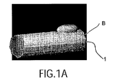
| JP2005523053A (ja) | 対象物表面の折り畳まれた解剖学的部分の視覚化のための医用ビューイングシステム及び画像処理方法 | |
| JP6080248B2 (ja) | 3次元画像表示装置および方法並びにプログラム | |
| US7304644B2 (en) | System and method for performing a virtual endoscopy | |
| JP5301197B2 (ja) | 断面画像表示装置および方法ならびにプログラム | |
| CN104885126B (zh) | 感兴趣组织的计算机辅助识别 | |
| CN116546916A (zh) | 用于虚拟胰腺造影管线的系统和方法 | |
| US9373181B2 (en) | System and method for enhanced viewing of rib metastasis | |
| JP4436838B2 (ja) | 局所的曲率分布パターンの分析方法 | |
| RU2419882C2 (ru) | Способ визуализации секущих плоскостей для изогнутых продолговатых структур | |
| US9123163B2 (en) | Medical image display apparatus, method and program | |
| JP2010075549A (ja) | 画像処理装置 | |
| US9767550B2 (en) | Method and device for analysing a region of interest in an object using x-rays | |
| JP2023553916A (ja) | ボリュメトリック画像データ内の物質境界を特定する方法 | |
| JP4686279B2 (ja) | 医用診断装置及び診断支援装置 | |
| WO2015111308A1 (ja) | 3次元医用画像表示制御装置およびその作動方法並びに3次元医用画像表示制御プログラム | |
| CN1707523A (zh) | 医学图像显示和处理方法、ct设备、工作站及程序产品 | |
| US20200118319A1 (en) | Visualization of anatomical cavities | |
| US20110285695A1 (en) | Pictorial Representation in Virtual Endoscopy | |
| CN108113749B (zh) | 距解剖腔的壁的距离的可视化 | |
| CN119998843A (zh) | 经由自动增强进行的高效肋骨平面可视化 | |
| Skalski et al. | 3D segmentation and visualisation of mediastinal structures adjacent to tracheobronchial tree from CT data | |
| JP2022135986A (ja) | 管腔臓器の視覚化に関するプログラム及び方法 |
Legal Events
| Date | Code | Title | Description |
|---|---|---|---|
| A521 | Request for written amendment filed |
Free format text: JAPANESE INTERMEDIATE CODE: A523 Effective date: 20060331 |
|
| A621 | Written request for application examination |
Free format text: JAPANESE INTERMEDIATE CODE: A621 Effective date: 20060331 |
|
| A761 | Written withdrawal of application |
Free format text: JAPANESE INTERMEDIATE CODE: A761 Effective date: 20071212 |