JP2005510323A - Confocal microscopy using multispectral encoding and systems and apparatus for spectroscopy-encoded confocal microscopy - Google Patents
Confocal microscopy using multispectral encoding and systems and apparatus for spectroscopy-encoded confocal microscopy Download PDFInfo
- Publication number
- JP2005510323A JP2005510323A JP2003548016A JP2003548016A JP2005510323A JP 2005510323 A JP2005510323 A JP 2005510323A JP 2003548016 A JP2003548016 A JP 2003548016A JP 2003548016 A JP2003548016 A JP 2003548016A JP 2005510323 A JP2005510323 A JP 2005510323A
- Authority
- JP
- Japan
- Prior art keywords
- light
- tissue
- spectrum
- confocal
- probe
- Prior art date
- Legal status (The legal status is an assumption and is not a legal conclusion. Google has not performed a legal analysis and makes no representation as to the accuracy of the status listed.)
- Pending
Links
- 238000004624 confocal microscopy Methods 0.000 title abstract description 24
- 239000000523 sample Substances 0.000 claims abstract description 107
- 238000001228 spectrum Methods 0.000 claims abstract description 56
- 239000013307 optical fiber Substances 0.000 claims abstract description 23
- 238000003384 imaging method Methods 0.000 claims description 60
- 238000000034 method Methods 0.000 claims description 40
- 230000003287 optical effect Effects 0.000 claims description 22
- 239000000463 material Substances 0.000 claims description 20
- 238000001514 detection method Methods 0.000 claims description 18
- 238000001356 surgical procedure Methods 0.000 claims description 14
- 238000005286 illumination Methods 0.000 claims description 12
- 230000007246 mechanism Effects 0.000 claims description 12
- 239000000835 fiber Substances 0.000 claims description 11
- 239000006185 dispersion Substances 0.000 claims description 10
- 230000033001 locomotion Effects 0.000 claims description 8
- 210000003754 fetus Anatomy 0.000 claims description 5
- 210000001685 thyroid gland Anatomy 0.000 claims description 5
- 229910052710 silicon Inorganic materials 0.000 claims description 4
- 239000010703 silicon Substances 0.000 claims description 4
- 210000001835 viscera Anatomy 0.000 claims description 3
- 238000010226 confocal imaging Methods 0.000 claims description 2
- 210000000746 body region Anatomy 0.000 claims 11
- 238000006243 chemical reaction Methods 0.000 claims 1
- 230000002452 interceptive effect Effects 0.000 claims 1
- 230000003595 spectral effect Effects 0.000 abstract description 9
- 238000001839 endoscopy Methods 0.000 abstract 1
- 238000001115 scanning electrochemical microscopy Methods 0.000 description 26
- 210000001519 tissue Anatomy 0.000 description 14
- 238000013461 design Methods 0.000 description 12
- 238000010586 diagram Methods 0.000 description 11
- 238000001727 in vivo Methods 0.000 description 6
- 206010028980 Neoplasm Diseases 0.000 description 5
- 230000000849 parathyroid Effects 0.000 description 5
- 230000008901 benefit Effects 0.000 description 4
- 238000012986 modification Methods 0.000 description 4
- 230000004048 modification Effects 0.000 description 4
- 238000002310 reflectometry Methods 0.000 description 4
- 210000003484 anatomy Anatomy 0.000 description 3
- 230000015572 biosynthetic process Effects 0.000 description 3
- 201000011510 cancer Diseases 0.000 description 3
- 238000005305 interferometry Methods 0.000 description 3
- 238000002271 resection Methods 0.000 description 3
- XUMBMVFBXHLACL-UHFFFAOYSA-N Melanin Chemical compound O=C1C(=O)C(C2=CNC3=C(C(C(=O)C4=C32)=O)C)=C2C4=CNC2=C1C XUMBMVFBXHLACL-UHFFFAOYSA-N 0.000 description 2
- 230000005540 biological transmission Effects 0.000 description 2
- 238000003745 diagnosis Methods 0.000 description 2
- 238000002059 diagnostic imaging Methods 0.000 description 2
- 210000000056 organ Anatomy 0.000 description 2
- 230000008569 process Effects 0.000 description 2
- 238000012216 screening Methods 0.000 description 2
- 238000002834 transmittance Methods 0.000 description 2
- 238000001574 biopsy Methods 0.000 description 1
- 230000001427 coherent effect Effects 0.000 description 1
- 230000001419 dependent effect Effects 0.000 description 1
- 238000012631 diagnostic technique Methods 0.000 description 1
- 201000010099 disease Diseases 0.000 description 1
- 208000037265 diseases, disorders, signs and symptoms Diseases 0.000 description 1
- 239000000428 dust Substances 0.000 description 1
- 238000005516 engineering process Methods 0.000 description 1
- 210000000981 epithelium Anatomy 0.000 description 1
- 238000009615 fourier-transform spectroscopy Methods 0.000 description 1
- 210000003128 head Anatomy 0.000 description 1
- 230000007774 longterm Effects 0.000 description 1
- 210000001165 lymph node Anatomy 0.000 description 1
- 238000002595 magnetic resonance imaging Methods 0.000 description 1
- 238000005259 measurement Methods 0.000 description 1
- 238000000691 measurement method Methods 0.000 description 1
- 238000000386 microscopy Methods 0.000 description 1
- 239000000203 mixture Substances 0.000 description 1
- 210000000214 mouth Anatomy 0.000 description 1
- 210000003205 muscle Anatomy 0.000 description 1
- 238000000399 optical microscopy Methods 0.000 description 1
- RGCLLPNLLBQHPF-HJWRWDBZSA-N phosphamidon Chemical compound CCN(CC)C(=O)C(\Cl)=C(/C)OP(=O)(OC)OC RGCLLPNLLBQHPF-HJWRWDBZSA-N 0.000 description 1
- 230000000704 physical effect Effects 0.000 description 1
- 230000010287 polarization Effects 0.000 description 1
- 238000012545 processing Methods 0.000 description 1
- 230000002035 prolonged effect Effects 0.000 description 1
- 229910052761 rare earth metal Inorganic materials 0.000 description 1
- 238000011160 research Methods 0.000 description 1
- 230000035945 sensitivity Effects 0.000 description 1
- 238000000926 separation method Methods 0.000 description 1
- 239000007787 solid Substances 0.000 description 1
- 125000006850 spacer group Chemical group 0.000 description 1
- 238000004611 spectroscopical analysis Methods 0.000 description 1
- 238000003325 tomography Methods 0.000 description 1
- 230000009466 transformation Effects 0.000 description 1
- 238000013519 translation Methods 0.000 description 1
- 238000002604 ultrasonography Methods 0.000 description 1
Images


Classifications
-
- A—HUMAN NECESSITIES
- A61—MEDICAL OR VETERINARY SCIENCE; HYGIENE
- A61B—DIAGNOSIS; SURGERY; IDENTIFICATION
- A61B1/00—Instruments for performing medical examinations of the interior of cavities or tubes of the body by visual or photographical inspection, e.g. endoscopes; Illuminating arrangements therefor
- A61B1/00064—Constructional details of the endoscope body
- A61B1/00071—Insertion part of the endoscope body
- A61B1/0008—Insertion part of the endoscope body characterised by distal tip features
- A61B1/00096—Optical elements
-
- A—HUMAN NECESSITIES
- A61—MEDICAL OR VETERINARY SCIENCE; HYGIENE
- A61B—DIAGNOSIS; SURGERY; IDENTIFICATION
- A61B1/00—Instruments for performing medical examinations of the interior of cavities or tubes of the body by visual or photographical inspection, e.g. endoscopes; Illuminating arrangements therefor
- A61B1/00163—Optical arrangements
- A61B1/00172—Optical arrangements with means for scanning
-
- A—HUMAN NECESSITIES
- A61—MEDICAL OR VETERINARY SCIENCE; HYGIENE
- A61B—DIAGNOSIS; SURGERY; IDENTIFICATION
- A61B5/00—Measuring for diagnostic purposes; Identification of persons
- A61B5/0059—Measuring for diagnostic purposes; Identification of persons using light, e.g. diagnosis by transillumination, diascopy, fluorescence
- A61B5/0062—Arrangements for scanning
- A61B5/0068—Confocal scanning
-
- A—HUMAN NECESSITIES
- A61—MEDICAL OR VETERINARY SCIENCE; HYGIENE
- A61B—DIAGNOSIS; SURGERY; IDENTIFICATION
- A61B5/00—Measuring for diagnostic purposes; Identification of persons
- A61B5/0059—Measuring for diagnostic purposes; Identification of persons using light, e.g. diagnosis by transillumination, diascopy, fluorescence
- A61B5/0075—Measuring for diagnostic purposes; Identification of persons using light, e.g. diagnosis by transillumination, diascopy, fluorescence by spectroscopy, i.e. measuring spectra, e.g. Raman spectroscopy, infrared absorption spectroscopy
-
- A—HUMAN NECESSITIES
- A61—MEDICAL OR VETERINARY SCIENCE; HYGIENE
- A61B—DIAGNOSIS; SURGERY; IDENTIFICATION
- A61B5/00—Measuring for diagnostic purposes; Identification of persons
- A61B5/0059—Measuring for diagnostic purposes; Identification of persons using light, e.g. diagnosis by transillumination, diascopy, fluorescence
- A61B5/0082—Measuring for diagnostic purposes; Identification of persons using light, e.g. diagnosis by transillumination, diascopy, fluorescence adapted for particular medical purposes
- A61B5/0084—Measuring for diagnostic purposes; Identification of persons using light, e.g. diagnosis by transillumination, diascopy, fluorescence adapted for particular medical purposes for introduction into the body, e.g. by catheters
-
- A—HUMAN NECESSITIES
- A61—MEDICAL OR VETERINARY SCIENCE; HYGIENE
- A61B—DIAGNOSIS; SURGERY; IDENTIFICATION
- A61B5/00—Measuring for diagnostic purposes; Identification of persons
- A61B5/41—Detecting, measuring or recording for evaluating the immune or lymphatic systems
- A61B5/414—Evaluating particular organs or parts of the immune or lymphatic systems
- A61B5/415—Evaluating particular organs or parts of the immune or lymphatic systems the glands, e.g. tonsils, adenoids or thymus
-
- A—HUMAN NECESSITIES
- A61—MEDICAL OR VETERINARY SCIENCE; HYGIENE
- A61B—DIAGNOSIS; SURGERY; IDENTIFICATION
- A61B5/00—Measuring for diagnostic purposes; Identification of persons
- A61B5/41—Detecting, measuring or recording for evaluating the immune or lymphatic systems
- A61B5/414—Evaluating particular organs or parts of the immune or lymphatic systems
- A61B5/418—Evaluating particular organs or parts of the immune or lymphatic systems lymph vessels, ducts or nodes
-
- A—HUMAN NECESSITIES
- A61—MEDICAL OR VETERINARY SCIENCE; HYGIENE
- A61B—DIAGNOSIS; SURGERY; IDENTIFICATION
- A61B5/00—Measuring for diagnostic purposes; Identification of persons
- A61B5/68—Arrangements of detecting, measuring or recording means, e.g. sensors, in relation to patient
- A61B5/6846—Arrangements of detecting, measuring or recording means, e.g. sensors, in relation to patient specially adapted to be brought in contact with an internal body part, i.e. invasive
- A61B5/6847—Arrangements of detecting, measuring or recording means, e.g. sensors, in relation to patient specially adapted to be brought in contact with an internal body part, i.e. invasive mounted on an invasive device
- A61B5/6852—Catheters
-
- G—PHYSICS
- G01—MEASURING; TESTING
- G01J—MEASUREMENT OF INTENSITY, VELOCITY, SPECTRAL CONTENT, POLARISATION, PHASE OR PULSE CHARACTERISTICS OF INFRARED, VISIBLE OR ULTRAVIOLET LIGHT; COLORIMETRY; RADIATION PYROMETRY
- G01J3/00—Spectrometry; Spectrophotometry; Monochromators; Measuring colours
- G01J3/02—Details
- G01J3/0205—Optical elements not provided otherwise, e.g. optical manifolds, diffusers, windows
- G01J3/0208—Optical elements not provided otherwise, e.g. optical manifolds, diffusers, windows using focussing or collimating elements, e.g. lenses or mirrors; performing aberration correction
-
- G—PHYSICS
- G01—MEASURING; TESTING
- G01J—MEASUREMENT OF INTENSITY, VELOCITY, SPECTRAL CONTENT, POLARISATION, PHASE OR PULSE CHARACTERISTICS OF INFRARED, VISIBLE OR ULTRAVIOLET LIGHT; COLORIMETRY; RADIATION PYROMETRY
- G01J3/00—Spectrometry; Spectrophotometry; Monochromators; Measuring colours
- G01J3/02—Details
- G01J3/0205—Optical elements not provided otherwise, e.g. optical manifolds, diffusers, windows
- G01J3/0218—Optical elements not provided otherwise, e.g. optical manifolds, diffusers, windows using optical fibers
-
- G—PHYSICS
- G01—MEASURING; TESTING
- G01J—MEASUREMENT OF INTENSITY, VELOCITY, SPECTRAL CONTENT, POLARISATION, PHASE OR PULSE CHARACTERISTICS OF INFRARED, VISIBLE OR ULTRAVIOLET LIGHT; COLORIMETRY; RADIATION PYROMETRY
- G01J3/00—Spectrometry; Spectrophotometry; Monochromators; Measuring colours
- G01J3/02—Details
- G01J3/0291—Housings; Spectrometer accessories; Spatial arrangement of elements, e.g. folded path arrangements
-
- G—PHYSICS
- G01—MEASURING; TESTING
- G01J—MEASUREMENT OF INTENSITY, VELOCITY, SPECTRAL CONTENT, POLARISATION, PHASE OR PULSE CHARACTERISTICS OF INFRARED, VISIBLE OR ULTRAVIOLET LIGHT; COLORIMETRY; RADIATION PYROMETRY
- G01J3/00—Spectrometry; Spectrophotometry; Monochromators; Measuring colours
- G01J3/02—Details
- G01J3/06—Scanning arrangements arrangements for order-selection
-
- G—PHYSICS
- G01—MEASURING; TESTING
- G01J—MEASUREMENT OF INTENSITY, VELOCITY, SPECTRAL CONTENT, POLARISATION, PHASE OR PULSE CHARACTERISTICS OF INFRARED, VISIBLE OR ULTRAVIOLET LIGHT; COLORIMETRY; RADIATION PYROMETRY
- G01J3/00—Spectrometry; Spectrophotometry; Monochromators; Measuring colours
- G01J3/12—Generating the spectrum; Monochromators
- G01J3/14—Generating the spectrum; Monochromators using refracting elements, e.g. prisms
-
- G—PHYSICS
- G01—MEASURING; TESTING
- G01J—MEASUREMENT OF INTENSITY, VELOCITY, SPECTRAL CONTENT, POLARISATION, PHASE OR PULSE CHARACTERISTICS OF INFRARED, VISIBLE OR ULTRAVIOLET LIGHT; COLORIMETRY; RADIATION PYROMETRY
- G01J3/00—Spectrometry; Spectrophotometry; Monochromators; Measuring colours
- G01J3/12—Generating the spectrum; Monochromators
- G01J3/18—Generating the spectrum; Monochromators using diffraction elements, e.g. grating
-
- G—PHYSICS
- G01—MEASURING; TESTING
- G01J—MEASUREMENT OF INTENSITY, VELOCITY, SPECTRAL CONTENT, POLARISATION, PHASE OR PULSE CHARACTERISTICS OF INFRARED, VISIBLE OR ULTRAVIOLET LIGHT; COLORIMETRY; RADIATION PYROMETRY
- G01J3/00—Spectrometry; Spectrophotometry; Monochromators; Measuring colours
- G01J3/28—Investigating the spectrum
-
- G—PHYSICS
- G02—OPTICS
- G02B—OPTICAL ELEMENTS, SYSTEMS OR APPARATUS
- G02B21/00—Microscopes
- G02B21/0004—Microscopes specially adapted for specific applications
- G02B21/002—Scanning microscopes
- G02B21/0024—Confocal scanning microscopes (CSOMs) or confocal "macroscopes"; Accessories which are not restricted to use with CSOMs, e.g. sample holders
- G02B21/0028—Confocal scanning microscopes (CSOMs) or confocal "macroscopes"; Accessories which are not restricted to use with CSOMs, e.g. sample holders specially adapted for specific applications, e.g. for endoscopes, ophthalmoscopes, attachments to conventional microscopes
-
- G—PHYSICS
- G02—OPTICS
- G02B—OPTICAL ELEMENTS, SYSTEMS OR APPARATUS
- G02B21/00—Microscopes
- G02B21/0004—Microscopes specially adapted for specific applications
- G02B21/002—Scanning microscopes
- G02B21/0024—Confocal scanning microscopes (CSOMs) or confocal "macroscopes"; Accessories which are not restricted to use with CSOMs, e.g. sample holders
- G02B21/0052—Optical details of the image generation
- G02B21/0056—Optical details of the image generation based on optical coherence, e.g. phase-contrast arrangements, interference arrangements
-
- G—PHYSICS
- G02—OPTICS
- G02B—OPTICAL ELEMENTS, SYSTEMS OR APPARATUS
- G02B21/00—Microscopes
- G02B21/0004—Microscopes specially adapted for specific applications
- G02B21/002—Scanning microscopes
- G02B21/0024—Confocal scanning microscopes (CSOMs) or confocal "macroscopes"; Accessories which are not restricted to use with CSOMs, e.g. sample holders
- G02B21/0052—Optical details of the image generation
- G02B21/0064—Optical details of the image generation multi-spectral or wavelength-selective arrangements, e.g. wavelength fan-out, chromatic profiling
-
- G—PHYSICS
- G02—OPTICS
- G02B—OPTICAL ELEMENTS, SYSTEMS OR APPARATUS
- G02B21/00—Microscopes
- G02B21/0004—Microscopes specially adapted for specific applications
- G02B21/002—Scanning microscopes
- G02B21/0024—Confocal scanning microscopes (CSOMs) or confocal "macroscopes"; Accessories which are not restricted to use with CSOMs, e.g. sample holders
- G02B21/0052—Optical details of the image generation
- G02B21/0076—Optical details of the image generation arrangements using fluorescence or luminescence
-
- G—PHYSICS
- G02—OPTICS
- G02B—OPTICAL ELEMENTS, SYSTEMS OR APPARATUS
- G02B23/00—Telescopes, e.g. binoculars; Periscopes; Instruments for viewing the inside of hollow bodies; Viewfinders; Optical aiming or sighting devices
- G02B23/24—Instruments or systems for viewing the inside of hollow bodies, e.g. fibrescopes
- G02B23/2407—Optical details
- G02B23/2453—Optical details of the proximal end
-
- G—PHYSICS
- G01—MEASURING; TESTING
- G01J—MEASUREMENT OF INTENSITY, VELOCITY, SPECTRAL CONTENT, POLARISATION, PHASE OR PULSE CHARACTERISTICS OF INFRARED, VISIBLE OR ULTRAVIOLET LIGHT; COLORIMETRY; RADIATION PYROMETRY
- G01J3/00—Spectrometry; Spectrophotometry; Monochromators; Measuring colours
- G01J3/02—Details
- G01J3/06—Scanning arrangements arrangements for order-selection
- G01J2003/062—Scanning arrangements arrangements for order-selection motor-driven
-
- G—PHYSICS
- G01—MEASURING; TESTING
- G01J—MEASUREMENT OF INTENSITY, VELOCITY, SPECTRAL CONTENT, POLARISATION, PHASE OR PULSE CHARACTERISTICS OF INFRARED, VISIBLE OR ULTRAVIOLET LIGHT; COLORIMETRY; RADIATION PYROMETRY
- G01J3/00—Spectrometry; Spectrophotometry; Monochromators; Measuring colours
- G01J3/02—Details
- G01J3/06—Scanning arrangements arrangements for order-selection
- G01J2003/064—Use of other elements for scan, e.g. mirror, fixed grating
- G01J2003/065—Use of fibre scan for spectral scan
-
- G—PHYSICS
- G01—MEASURING; TESTING
- G01J—MEASUREMENT OF INTENSITY, VELOCITY, SPECTRAL CONTENT, POLARISATION, PHASE OR PULSE CHARACTERISTICS OF INFRARED, VISIBLE OR ULTRAVIOLET LIGHT; COLORIMETRY; RADIATION PYROMETRY
- G01J3/00—Spectrometry; Spectrophotometry; Monochromators; Measuring colours
- G01J3/02—Details
- G01J3/06—Scanning arrangements arrangements for order-selection
- G01J2003/069—Complex motion, e.g. rotation of grating and correcting translation
Landscapes
- Physics & Mathematics (AREA)
- Health & Medical Sciences (AREA)
- Life Sciences & Earth Sciences (AREA)
- Spectroscopy & Molecular Physics (AREA)
- General Physics & Mathematics (AREA)
- Surgery (AREA)
- General Health & Medical Sciences (AREA)
- Engineering & Computer Science (AREA)
- Biophysics (AREA)
- Heart & Thoracic Surgery (AREA)
- Medical Informatics (AREA)
- Molecular Biology (AREA)
- Pathology (AREA)
- Animal Behavior & Ethology (AREA)
- Biomedical Technology (AREA)
- Public Health (AREA)
- Veterinary Medicine (AREA)
- Optics & Photonics (AREA)
- Analytical Chemistry (AREA)
- Radiology & Medical Imaging (AREA)
- Chemical & Material Sciences (AREA)
- Nuclear Medicine, Radiotherapy & Molecular Imaging (AREA)
- Vascular Medicine (AREA)
- Immunology (AREA)
- Endocrinology (AREA)
- Astronomy & Astrophysics (AREA)
- Ophthalmology & Optometry (AREA)
- Microscoopes, Condenser (AREA)
- Investigating Or Analysing Materials By Optical Means (AREA)
- Instruments For Viewing The Inside Of Hollow Bodies (AREA)
- Endoscopes (AREA)
Abstract
光ファイバ(9)の末端に接続された可撓性プローブによる内視鏡法に特に有用な走査型共焦点顕微鏡法システム及び装置。プローブは、対象物のある領域にわたって1次元に延びるスペクトル成分を有する多重スペクトル光のビームを送出し、別の次元での走査のために動かされる、回折格子(12)及びレンズ(14)を有する。上記領域の画像を提供するために、反射共焦点スペクトルが測定される。 Scanning confocal microscopy system and apparatus particularly useful for endoscopy with a flexible probe connected to the end of an optical fiber (9). The probe has a diffraction grating (12) and a lens (14) that emits a beam of multispectral light having a spectral component that extends in one dimension over a region of the object and is moved for scanning in another dimension. . The reflection confocal spectrum is measured to provide an image of the region.
Description
本出願は、1999年2月26日出願の国際特許出願第PCT/US99/04356号の米国内出願である、2000年8月24日出願の米国特許出願第09/622971号の一部継続出願であり、1998年2月26日に出願された米国仮特許出願第60/076041号の優先権を主張するものである。上記出願の明細書の内容は本明細書に参照として含まれる。 This application is a continuation-in-part of US patent application Ser. No. 09/622971 filed Aug. 24, 2000, which is a US application of International Patent Application No. PCT / US99 / 04356 filed Feb. 26, 1999. And claims the priority of US Provisional Patent Application No. 60/076041, filed February 26,1998. The contents of the specification of the above application are included herein by reference.
本発明は生物組織標本切片の検査または画像化のための共焦点顕微鏡法のためのシステム(方法及び装置)に関し、特に、多重スペクトル照明光を用い、多重スペクトル光を処理する、そのようなシステムに関する。 The present invention relates to systems (methods and apparatus) for confocal microscopy for examination or imaging of biological tissue specimen sections, and in particular, such systems using multispectral illumination light and processing multispectral light. About.
医用画像化技術は、患者の巨視的な解剖学的構造に関する不可欠な情報を医師に提供するため、この20年にわたり進歩してきた。X線撮影法、磁気共鳴画像化法、コンピュータ連動断層撮影法及び超音波法のような画像化法は、100μmから1mmの範囲の分解能で人体の大規模構造の非侵襲的検査を可能にする。 Medical imaging technology has progressed over the last 20 years to provide physicians with essential information about the patient's macroscopic anatomy. Imaging methods such as X-ray imaging, magnetic resonance imaging, computer-linked tomography and ultrasound enable non-invasive examination of large-scale structures of the human body with resolutions ranging from 100 μm to 1 mm. .
しかし、ガンの初期段階の検出のような疾病過程の多くでは、適切な診断のためにさらに高い分解能が必要である。さらに、ガンのスクリーニング及び腫瘍縁の外科的検出のような臨床的処置にはさらに分解能の高い診断用画像化方法が必要である。 However, many disease processes, such as early detection of cancer, require higher resolution for proper diagnosis. Furthermore, clinical procedures such as cancer screening and surgical detection of tumor margins require higher resolution diagnostic imaging methods.
上記及びその他の臨床問題に現場で対処するためには、標準的な組織病理学に近い分解能をもつ非侵襲性画像化技術が用いる必要がある。その可能性を秘めた有望な非侵襲性画像化様式は、反射率共焦点顕微鏡法として知られる光学顕微鏡法の形態のものである。 To address these and other clinical issues in the field, non-invasive imaging techniques with a resolution close to standard histopathology need to be used. A promising non-invasive imaging modality with that potential is in the form of optical microscopy known as reflectance confocal microscopy.
現在、高速走査型共焦点顕微鏡法は、アクセス可能な皮膚及び眼の表面に限られている。その理由は、唯一の信頼できる光走査法が自由空間で実施されなければならないからである。さらに、そのような光走査器の大きさのため、内視鏡またはカテーテルなど、小型のプローブでの使用が妨げられている。高速走査機構を小型化し、人体の全表面、婦人科用途、プローブを用いる用途、及び内部器官系を含めるために、共焦点顕微鏡法の医用用途の数を増やすことが、本発明の特徴である。 Currently, fast scanning confocal microscopy is limited to accessible skin and eye surfaces. This is because the only reliable optical scanning method must be performed in free space. Furthermore, the size of such an optical scanner prevents its use with small probes such as endoscopes or catheters. It is a feature of the present invention to increase the number of confocal microscopy medical applications to miniaturize the high speed scanning mechanism and to include the entire surface of the human body, gynecological applications, probe applications, and internal organ systems. .
多重スペクトル光を共焦点顕微鏡法で使用することが提案されたが、検査下にある人体の垂直方向に間隔を置いた各領域の画像化に対して提案されたにすぎない。1990年10月25日に発行された、ビー・ピカード(B. Picard)の米国特許第4965441号の明細書を参照されたい。回折格子を用いて干渉計で分解される多重スペクトル光を得て分光法画像を得るための干渉計が、1996年10月15日に発行されたエイ・クヌッタル(A. Knuttal)の米国特許第5565986号の明細書に開示されている。多重スペクトル光を得る色分離回折格子を有するレンズが、1997年2月4日に発行された米国特許第5600486号の明細書に開示されている。そのような多重スペクトル案は小型で可撓性のプローブを用いる高分解能画像化には有効ではない。本発明にしたがう共焦点顕微鏡システムは小型化して、小型プローブに組み込むことができる。さらに、単線の光ファイバによる光伝送を可能にすることにより、プローブをカテーテルまたは内視鏡に容易に組み込むこともできる。すなわち、本発明にしたがう共焦点顕微鏡により、人体のアクセス可能な表面の全ての画像化が可能になり、共焦点顕微鏡法の生物医学用途が一桁多くなる。 Although it has been proposed to use multispectral light in confocal microscopy, it has only been proposed for imaging vertically spaced areas of the human body under examination. See B. Picard U.S. Pat. No. 4,965,441 issued Oct. 25, 1990. An interferometer for obtaining a spectroscopic image by obtaining multispectral light resolved by an interferometer using a diffraction grating is disclosed in US Patent No. A. Knuttal, issued Oct. 15, 1996. This is disclosed in the specification of No. 5565986. A lens having a color separation diffraction grating that obtains multispectral light is disclosed in US Pat. No. 5,600,066 issued on Feb. 4, 1997. Such a multispectral scheme is not effective for high resolution imaging using small, flexible probes. The confocal microscope system according to the present invention can be miniaturized and incorporated into a small probe. Further, the probe can be easily incorporated into a catheter or endoscope by allowing light transmission through a single optical fiber. That is, the confocal microscope according to the present invention enables the imaging of all accessible surfaces of the human body, increasing the biomedical use of confocal microscopy by an order of magnitude.
簡単に説明すれば、本発明を具現化している共焦点顕微鏡法システムは、プローブを挿入することができる人体の注目する領域を、1つの次元に沿って延びる共焦点スペクトルで照明する。プローブの光学系または(光ファイバとすることができる)可撓性導光部材のプローブへの取付けにより可能となるプローブの物理的運動により、さらに1つまたは2つの次元に沿うスペクトルの走査が可能になり、よって前記領域の2次元または3次元の画像化が提供される。反射された共焦点スペクトルは、好ましくは干渉計法により実施することができるヘテロダイン検波機構により、分光法を用いて検出またはデコードすることができる。 Briefly described, a confocal microscopy system embodying the present invention illuminates a region of interest on a human body into which a probe can be inserted with a confocal spectrum extending along one dimension. The probe's physical movement, enabled by the probe's optics or the flexible light guide (which can be an optical fiber) attached to the probe, allows scanning of the spectrum along one or two additional dimensions. Thus, two-dimensional or three-dimensional imaging of the region is provided. The reflected confocal spectrum can be detected or decoded using spectroscopy, preferably by a heterodyne detection mechanism that can be implemented by interferometry.
以下の文献が参照として本明細書に含まれる。 The following documents are included herein by reference:
ピー・コーカフ(P. Corcuff)及びジェイ・エル・レベック(J. L. Leveque),「タンデム走査型顕微鏡によるヒトの皮膚の生体内視(In vivo vision of the human skin with the tandem scanning microscope」,Dermatology,1993年,第186巻,p.50−54,
エム・ラヤディヤクシャ(M. Rajadhyaksha)等,「ヒトの皮膚の生体内共焦点走査型レーザ顕微鏡法:メラニンが強く対抗する(In vivo confocal scanning laser microscopy of human skin: Melanin provides strong contest)」,J. Invest. Derm.,1995年,第104巻,p.946,
ビー・アール・マスターズ(B. R. Masters)編,「眼科学における非侵襲性診断法(Noninvasive diagnostic techniques in ophthalmology)」,(米国ニューヨーク),スプリンガー−バーラグ(Springer-Verlag),1990年に収録されている、アール・エイチ・ウエッブ(R. H. Webb),「走査型レーザ検眼鏡(Scanning Laser opthalmoscope)」,及び
ジー・ジェイ・ターネイ(G. J. Tearney)、アール・エイチ・ウエッブ及びビー・イー・ブーマ(B. E. Bouma),「スペクトルコード化共焦点顕微鏡法(Spectrally encoded confocal microscopy)」,Optics Letters,1998年,第23巻,第15号,p.1152−1154。
P. Corcuff and JL Leveque, “In vivo vision of the human skin with the tandem scanning microscope”, Dermatology, 1993 Year, 186, p. 50-54,
M. Rajadhyaksha et al., “In vivo confocal scanning laser microscopy of human skin: Melanin provides strong contest”, J. Invest. Derm., 1995, 104, 946,
Recorded in BR Masters, “Noninvasive diagnostic techniques in ophthalmology” (New York, USA), Springer-Verlag, 1990 , RH Webb, "Scanning Laser opthalmoscope", and GJ Tearney, RH Web and BE Bouma "Spectally encoded confocal microscopy", Optics Letters, 1998, Vol. 23, No. 15, pp. 1152-1154.
生体内のアクセス可能な上皮組織のほとんどを画像化するためには、3つの重要な要件が満たされなければならない。第1に、集束ビームが標本にかけて走査されなければならない。第2に、画像アクイジション時間が運動のアーティファクトを防止するに十分に短くなければならない。最後に、内視鏡またはカテーテルに組み込むに十分に装置が小さくなければならない。生体内共焦点画像化システムに対する高速ビーム走査要件に対処するために、タンデム走査型及びレーザ走査型の共焦点顕微鏡法のような技法が開発された。しかし、これらの方法では、容易には小型化できない大型機械装置の使用により高速走査が得られている。この結果、これらの技法の効用は基本的に皮膚科学及び眼科学の分野に限定されている。有望な新しい光ファイバを使用する技法である、スペクトルコード化共焦点顕微鏡法(“SECM”)が最近示された。この技法により、カテーテルまたは内視鏡のような小型プローブによる反射率共焦点顕微鏡法の実施が可能になる。SECMは試料から反射された1次元空間情報をコード化するために波長分割多重化(“WDM”)を用いる。高速走査軸は、それぞれの位置が相異なる光波長で表されている一連の焦点で置き換えられる。空間位置の関数としての反射強度は反射光のスペクトルを測定することにより決定される(図8)。2次元画像はプローブの低速機械運動により波長コード化軸を走査することによりつくられる。すなわち、本発明を具現化している内視鏡装置は、標準的な内視鏡と一体化されるかまたは独立型の装置として、様々な組織及び器官のSECM画像化を可能にする。 In order to image most of the accessible epithelial tissue in vivo, three important requirements must be met. First, the focused beam must be scanned across the specimen. Second, the image acquisition time must be short enough to prevent motion artifacts. Finally, the device must be small enough to be incorporated into an endoscope or catheter. Techniques such as tandem scanning and laser scanning confocal microscopy have been developed to address the high speed beam scanning requirements for in vivo confocal imaging systems. However, in these methods, high-speed scanning is obtained by using a large machine that cannot be easily downsized. As a result, the utility of these techniques is basically limited to the fields of dermatology and ophthalmology. Spectral coded confocal microscopy (“SECM”), a technique that uses a promising new optical fiber, has recently been shown. This technique allows reflectance confocal microscopy to be performed with a small probe such as a catheter or endoscope. SECM uses wavelength division multiplexing ("WDM") to encode one-dimensional spatial information reflected from a sample. The fast scan axis is replaced with a series of focal points where each position is represented by a different light wavelength. The reflection intensity as a function of spatial position is determined by measuring the spectrum of the reflected light (FIG. 8). A two-dimensional image is created by scanning the wavelength encoding axis by the slow mechanical movement of the probe. That is, an endoscopic device embodying the present invention enables SECM imaging of various tissues and organs, either integrated with a standard endoscope or as a stand-alone device.
本発明の一実施形態にしたがえば、生体内内視鏡共焦点顕微鏡法の実施が可能である装置が提供される。そのような装置はおそらく、内部器官系において非侵襲性非細胞診断画像化を実施するためのツールを医師に提供できよう。そのような様式は、ガンのスクリーニングまたは生検ガイダンス及び手術中の腫瘍またはその他の組織の識別を含む、様々な臨床用途を可能にする能力においてかなり長期にわたる強い影響を有するであろう。本発明を具現化している装置は生体内内視鏡共焦点顕微鏡法画像化を可能にでき、おそらく、注目する発症組織の診断を可能にするであろう。複雑さが増すにもかかわらず、そのような装置はそのような装置でなければアクセス不能な組織へのアクセスを提供でき、したがって、診断ツールとしての共焦点顕微鏡法の価値を相当に高めることができるであろう。 According to one embodiment of the present invention, an apparatus is provided that is capable of performing endoscopic confocal microscopy in vivo. Such a device could possibly provide a doctor with a tool for performing non-invasive non-cytodiagnostic imaging in the internal organ system. Such a modality will have a fairly long-term impact on the ability to enable a variety of clinical applications, including cancer screening or biopsy guidance and identification of tumors or other tissues during surgery. An apparatus embodying the present invention can enable in-vivo endoscopic confocal microscopy imaging and possibly allow for the diagnosis of the onset tissue of interest. Despite the increased complexity, such devices can provide access to tissues that would otherwise be inaccessible, thus significantly increasing the value of confocal microscopy as a diagnostic tool. It will be possible.
本発明は添付図面によりさらに明白になるであろう。 The invention will become more apparent from the accompanying drawings.
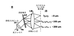
ここで図を参照すれば、共焦点顕微鏡法のための多重スペクトルコード化は、顕微鏡への入力として広帯域幅光源10を用いる。顕微鏡のプローブ8において、光ファイバ9を介して与えられる光源のスペクトルが回折格子12により分散され、対物レンズ14により試料16上に集束される。レンズ9aは、図1に示されるように、光ファイバからの光をコリメートするために、光ファイバ9と回折格子12の間に配されることが好ましいが、レンズ9aは省略することもできる。それぞれの波長に対する光点は試料上の別々の位置xに集束される(図1)。横断方向の位置の関数としての反射率が、プローブ8から戻される、試料16からの反射共焦点スペクトルを測定することによって求められる。
Referring now to the figure, multispectral coding for confocal microscopy uses a
波長の数すなわち分解し得る点の数は、

により決定され、ここで、λは中心波長、∂λはスペクトル帯域幅、Nは多色入力ビーム10で照明される回折格子12の線数、mは回折次数である。光源の総帯域幅がΔλであれば、分解可能な点の数nは、
で定められる。 Determined by
中心波長が800nm,帯域幅が25nm,入力光点直径が5nm,回折格子線数が1800本/mm,及び回折次数が1の入力光源に対し、スペクトルコード化共焦点システムによりn=281の点を分解することができる(図2)。本例に用いたパラメータは普通の廉価な光コンポーネントで得ることができる。この点数は単に入力光点直径を大きくするかまたは光源の帯域幅を広げることにより、増加させることができる。光点直径を大きくするとプローブ直径が大きくなる。光源の帯域幅の拡張は、より広帯域の超発光ダイオード、希土類元素ドープファイバ超蛍光源または固体モードロックレーザを用いることにより達成できよう。 For an input light source having a center wavelength of 800 nm, a bandwidth of 25 nm, an input light spot diameter of 5 nm, a diffraction grating line number of 1800 lines / mm, and a diffraction order of 1, n = 281 by the spectrally encoded confocal system. Can be decomposed (FIG. 2). The parameters used in this example can be obtained with ordinary inexpensive optical components. This score can be increased by simply increasing the input light spot diameter or increasing the bandwidth of the light source. Increasing the light spot diameter increases the probe diameter. Increasing the bandwidth of the light source could be achieved by using a wider band superluminescent diode, a rare earth element doped fiber superfluorescent source or a solid mode-locked laser.
次に多重スペクトルプロセスを考察する。初めに、直接スペクトル測定を考察する。軸に垂直な方向の位置の関数としての試料16からの反射率は試料アーム18からの反射共焦点スペクトルを測定することにより求められる。スペクトルは、マイケルソン干渉計20の試料アームにプローブ8を組み込み(図3)、干渉計の出力ポート19において高分解能分光計21を通過させた光を検出することにより高効率で測定できる。すなわち、測定される波長のそれぞれは試料上の別々の位置xに対応する(図3)。従来の実時間共焦点顕微鏡法に優る本方法の利点は、高品質分光計で十分に得られる範囲の上記パラメータに対してほぼ0.1nmのスペクトル分解能をもって分光計21によりプローブ8の外部で高速軸走査(〜15kHz)を実施できることである。
Now consider the multispectral process. First, consider direct spectral measurements. The reflectance from the
ヘテロダイン検波の使用により高感度を達成できる。ミラー24をもつ変調器23によるなどして(図3)、基準アーム22が変調されていれば、試料アーム18からの光と基準アーム22からの光の干渉も変調されるであろう。
High sensitivity can be achieved by using heterodyne detection. If the
次いで、基準アーム変調周波数に対する検出器26のロックイン検出によって高い信号対雑音比を達成できる。
A high signal to noise ratio can then be achieved by
別のスペクトル測定法は、干渉分光法またはフーリエ変換分光法である。これは、基準アーム22に直線平行移動ミラー28を挿入し、試料アーム18及び基準アーム22のそれぞれから反射された光の干渉による干渉分光計からの相互相関出力30を測定することにより達成できる(図4)。このタイプの分光法検出の利点は、直接検出法より高いスペクトル分解能、高い戻り光の利用効率及び移動ミラー28のドップラーシフトによる基準アーム22の固有変調を達成できる能力、及び試料16から反射率データ及び位相データのいずれをも抽出できる能力などである。試料から位相データを抽出できる能力は、試料の分子組成を明らかにするためにも、標本試料16の反射率とは別の画像コントラスト源をさらに提供するためにも有用な、軸に垂直な方向の位置xの関数としての屈折率の検出を可能にすることができる。最後に、干渉計法検出には、可干渉性ゲーティングにより共焦点信号から高次の多重散乱を排除できる能力がある。
Another spectral measurement method is interferometry or Fourier transform spectroscopy. This can be achieved by inserting a linear translation mirror 28 into the
最後に画像形成を考察する。横断方向の位置xの多重スペクトルコード化により一次元ラスター走査の実施が可能になる。画像を得るためには別の軸の走査が行われなければならず、これは通常、より低速である。y軸のそのような低速走査を達成する方法には、y方向での光ファイバ9の移動(図5B)、または前方走査構成における光ファイバ軸の周りのプローブ8全体の回転(図5C)、または側方照射構成における光ファイバ軸の周りのプローブ8全体の回転(図5D)がある。z軸に沿う光ファイバ9または対物レンズ14の走査により断面画像をつくることができる(図6)。最後に、光ファイバ9(または回折格子12と対物レンズ14の間の別のレンズ36)を対物レンズの像面に対して前後に走査することによりズームモードをつくることができる(図7)。y軸またはz軸に沿う直線運動及び回転のいずれも圧電変換器の使用により小型プローブで容易に達成することができる。図5Aに示されるように、試料の微視的区画の画像を表す信号を、(図3に関して説明したような)分光計または(図4に関して説明したような)フーリエ変換により分光検出器32からコンピュータ34で受け取ることができ、コンピュータに接続されたディスプレイ上に画像を表示できる。
Finally, consider image formation. Multi-spectral coding of the transverse position x makes it possible to perform a one-dimensional raster scan. To obtain an image, another axis scan must be performed, which is usually slower. Methods for achieving such slow scanning of the y axis include movement of the
前述したように、スペクトルコード化共焦点顕微鏡法(“SECM”)により、カテーテルまたは内視鏡のような小型プローブを介した反射率共焦点顕微鏡法の実施が可能になる。SECMは、試料から反射される1次元空間情報をコード化するために波長分割多重化(“WDM”)を用いる。それぞれの位置が光の相異なる波長で表される一連の集束点で高速走査軸が置き換えられる。反射光のスペクトルを測定することにより空間位置の関数としての反射強度が求められる(図8)。プローブの低速の機械的運動によって波長コード化軸を走査することにより2次元画像がつくられる。すなわち、本発明を具現化している内視鏡法装置により、標準的な内視鏡と一体化されるかまたはスタンドアローン装置として、様々な組織及び器官のSECM画像化が可能になる。 As previously mentioned, spectrally encoded confocal microscopy (“SECM”) allows for the implementation of reflectivity confocal microscopy through a small probe such as a catheter or endoscope. SECM uses wavelength division multiplexing (“WDM”) to encode one-dimensional spatial information reflected from a sample. The fast scan axis is replaced by a series of focusing points, each position represented by a different wavelength of light. By measuring the spectrum of the reflected light, the reflection intensity as a function of spatial position is determined (FIG. 8). A two-dimensional image is created by scanning the wavelength encoding axis by the slow mechanical movement of the probe. That is, an endoscopic device embodying the present invention enables SECM imaging of various tissues and organs, either as a standard endoscope or as a stand-alone device.
図8は、本発明の一実施形態にしたがうSECMプローブ38の基本的な光学特性及びコンポーネントを示す。SECMプローブ38は、図1に示されるプローブ8の要素と同様であり、そのような要素については同じ参照数字で表されている要素を備える。それらの要素の説明は図1に関して上で既に与えられており、ここで繰り返すことはしない。図8に示されるように、対物レンズユニット14は、回折格子12により分散されたファイバ9からの光源スペクトルを試料16における結像面上に集束させるための、1つまたはそれより多くの(例えば2つの)レンズを備えることができる。結像面は、試料16の任意の表面上、任意の部位内等に合焦させ得ることに注意されたい。回折格子12により分散され、対物レンズユニット14によって結像面上に集束させられる光源スペクトルの範囲(λ−nからλ0を経てλ+nまで)は、SECMプローブ38の視野(“FOV”)を形成できる。この範囲は、縦方向(すなわち図6に示されるように“z軸”に沿う方向)、縦方向を実質的に横断する方向、上記2つの方向の間の任意の方向等を含む、任意の方向に延びることができる第1の次元(すなわち、第1の次元に沿うベクトルは前記任意の方向を指す)上に集束させることができる。例えば、第1の次元は、軸とは異なる(すなわち、図6に示される“z軸”上にはない)任意の方向に延びる非軸方向次元とすることができる。第1の次元のような次元は、当然、互いに逆に双方向に延び得ることに注意されたい。前記範囲は、第1の次元に沿う直線上、曲線上、円周上、楕円周上、あるいは任意の点列上に集束させることができる。集束範囲は、結像面を形成するために、(“低速軸”と称されることもある)第2の次元に沿う別の方向、すなわち第1の次元に沿うベクトルの方向とは異なる方向(例えば、第1の次元に沿うベクトルに垂直な方向等)に、走査することができる。スペクトルは、結像面を形成するために、別の方向、すなわち第1の方向に沿うベクトルの方向とは異なる方向に延びる軸の周りを走査することもできる。
FIG. 8 illustrates the basic optical properties and components of the SECM probe 38 according to one embodiment of the present invention. The SECM probe 38 is similar to the elements of the
図9Aは、本発明の一実施形態にしたがう、前方画像化SECMプローブ/カテーテル40の構成の例を、基本機構とともに示す。ここに示されるように、前方画像化SECMプローブ/カテーテル40は、光学系及び制御系ハウジング45、回転子/アクチュエータ50、近端で(例えば、図3,4及び5Aに示されるような)SECMシステムに接続され、遠端で光を集束し、光の向きを変える(図9A)光ファイバ素子(例えばファイバ9)を入れることができる内コア55を備えることができる。プローブ8または38の光コンポーネントと同様の光コンポーネント(または“遠端光学系”)を、内コア55内の光学系及び制御系ハウジング45に封入することができる。したがって、上述したように、内コア55(したがってその中の光コンポーネント)を回転または平行移動させるか、あるいはビームを偏向させることにより2次元画像化を達成することができる。本発明の一実施形態にしたがう前方画像化SECMプローブ/カテーテル40のために特別に設計された光コンポーネントの例及びその特性を、図10を参照しながら以下でさらに詳細に説明する。内コアは、誘導ワイヤ70を遠端光学系への電気的または機械的/圧気式接続と一緒に入れることができるシース65内に封入することができる。光コンポーネントを水分、塵埃等から保護するために、透明窓72を設けることもできる。図9Bは、本発明の一実施形態にしたがう、側方画像化SECMプローブ/カテーテル42の構成の例を、基本機構とともに示す。図9Aに示されるように、側方画像化SECMプローブ/カテーテル42は、前方画像化SECMプローブの要素と同様の要素を備えることができる。それらの要素をここで繰り返して説明はしない。しかし、側方画像化SECMプローブ/カテーテル42の(試料16における)結像面はプローブ/カテーテル42の軸に対して角度をもたせることができ、一方、前方画像化SECMプローブ/カテーテル40の結像面は光学系及び制御系ハウジング45の遠端から広がり得ることに注意されたい。すなわち、画像化されるべき試料16のタイプ(例えば、囲繞構造)に応じて、プローブ/カテーテル40及び/またはプローブ/カテーテル42を用いることができる。図11を参照して以下に説明するように、側方画像化SECMプローブ/カテーテル42の光学系及び制御系ハウジング45の光コンポーネントは、プローブの軸に対して任意の角度で結像面を合焦させるように調節することができる。
FIG. 9A shows an example of the configuration of a forward imaging SECM probe /
プローブ/カテーテル40(図9A)の前方画像化構成では、本質的にビームを偏向させ得る回折格子(例えば、内コア55内の光学系及び制御系ハウジング45に封入され得る、回折格子12)が存在する中でビーム経路をプローブ/カテーテル40の軸に合わせることが課題となりうる。ビーム経路合わせは、GRISMとしても知られる回折格子−プリズム対75(図10)を用いて達成され得る。図10に示されるように、GRISM75は、(屈折率がnpであることを特徴とする材料でつくられ、φで定められる傾角面を有する)プリズム76,回折格子77,及び屈折率がそれぞれn1及びn2であることを特徴とする材料78及び79を備えることができる。本用途の場合、透過モードGRISM75(図10)が好ましい。ブレーズド格子及びバイナリ格子を用いることができるが、好ましい実施形態は傾角プリズム面に固着されたホログラフィック格子を備えるものであることが研究により示された。ホログラフィック格子は、ディクソン格子等を含む任意のタイプとすることができる。図10に示される距離f1及びf2は、相互間の、及び/または1つまたはそれより多くのGRISM75の特性値/パラメータ(例えば、設計及び材料によりあらかじめ定めることができる、np,φ,n1,n2及びGRISM75の寸法等)による、あらかじめ定められた関係を有することができる。本発明の一実施形態にしたがえば、GRISM75のプリズムは、シリコン、その他の高屈折率材料等でつくることができる。高屈折率材料を用いる場合は、透過率を高め、有害な背面反射を回避するため、屈折率界面の全てに適切な反射防止膜を用いることができる。
In the forward imaging configuration of probe / catheter 40 (FIG. 9A), there is a diffraction grating that can essentially deflect the beam (eg,
すなわち、GRISM75を含む、図10に示される光コンポーネントは、図9Aに示される前方画像化プローブ/カテーテル40の内コア55内の光学系及び制御系ハウジング45に封入される光コンポーネントとすることができる。大偏向角を可能にするためには、高屈折率材料が必要となり得る。表1は、様々な波長において可能な設計のいくつかについての設計パラメータのリストを示す。設計の選択肢は、視野、分解能並びに回折格子及びプリズムの入手可能性の間の許容トレードオフであり、そのような許容トレードオフに依存する。
設計パラメータは上表1に挙げられた値の±5%の範囲内とし得ることに注意されたい。 Note that the design parameters can be within a range of ± 5% of the values listed in Table 1 above.
可能な別の設計により、様々な角度でのSECM画像化が可能になる。これは、細い内腔を画像化する場合、あるいは口腔表面のような複雑であるかまたは平らではない表面を画像化する場合に好ましい設計であり得る。図11に示されるように、反射型プリズムまたは反射型GRISM80(簡単のため、以降はGRISM80と称する)を、装置(例えば、図9Bのプローブ/カテーテル42)の画像化角の完全制御を可能にするように用いることができる。図11に示される距離f1及びf2は、相互間の、及び/または1つまたはそれより多くのGRISM80の特性値/パラメータ(例えば、設計及び材料によりあらかじめ定めることができる、GRISM80の反射角,寸法等)による、あらかじめ定められた関係を有することができる。本発明の一実施形態にしたがえば、GRISM80は、シリコン、その他の高屈折率材料等でつくることができる。高屈折率材料を用いる場合は、透過率を高め、有害な背面反射を回避するために屈折率界面全部に適切な反射防止膜が必要である。
Another possible design allows SECM imaging at various angles. This may be a preferred design when imaging thin lumens or when imaging complex or uneven surfaces such as the oral cavity surface. As shown in FIG. 11, a reflective prism or reflective GRISM 80 (hereinafter referred to as GRISM 80 for simplicity) allows complete control of the imaging angle of the device (eg, probe /
2次元画像化を達成するため、様々な方法で低速軸を走査できる。可能な方法の1つは、円形区画(例えば、図12Aに示されるような前方画像化プローブ40の場合)あるいは円筒形区画(例えば、図13Aに示されるような側方画像化プローブ42の場合)を画像化するため、例えば回転子50により、プローブの内コア55を回転させることである。(例えば、図13Bに示されるようにアクチュエータ50を用いて内コア55を滑動させることにより)直線的に平行移動するようにプローブを構成して、プローブの軸に平行な平面から画像を得ることも可能である。別の動作モードは、限定的ではないが、圧電素子、電気光学素子、音響光学素子、機械素子、電磁気素子、または圧気素子85を含む機械的または光学的技法を用いるビーム偏向とすることができる(図12B)。
The slow axis can be scanned in various ways to achieve two-dimensional imaging. One possible method is the case of a circular section (eg in the case of a
検査下にある組織内の様々な層を画像化できるようにするために、プローブの焦平面を調節することもできる。前方画像化カテーテル(40)では、これは、画像化窓の前面または背後におかれた弾性スペーサに可変圧力を印加することによるか(図14A)、外装シース及び画像化窓72に対して内コアを直線的に平行移動させることによるか(図14B)、またはねじ溝付外装シース及び画像化窓72に対して同じくねじ溝付の内部光学系集成体とともに内コアを回転させることにより(図14C)、行うことができる。側方画像化プローブ(42)の場合は、機械式、圧気式または圧電式とすることができる並進器95(図15A)、あるいはプローブ外部のバルーン100(図15B)により、焦平面を調節することができる。
The focal plane of the probe can also be adjusted to allow the various layers in the tissue under examination to be imaged. In the anterior imaging catheter (40), this is by applying variable pressure to a resilient spacer placed in front of or behind the imaging window (FIG. 14A), or inward relative to the sheath and
前方画像化用途に十分に適していると思われる別のGRISM構造は、対称二重プリズム構造1600(図16)である。図16に示されるように、二重プリズムGRISM1600はプリズム1605及び1610並びに回折格子1615を有することができる。本発明の一実施形態にしたがえば、プリズム1605は屈折率がnpであることを特徴とする材料でつくることができ、φで定められる傾角面を有することができる。回折格子1615は屈折率がngであることを特徴とする材料でつくることができる。回折格子1615はホログラフィック格子とすることができる。二重プリズムGRISM1600は、プリズム1610を屈折率がnpであることを特徴とする材料でつくることもでき、φで定められる傾角面を有することもできるから、対称とすることができる。これにより、回折格子1615に出入りするビームを同じ角度(リトロー角)にすることが可能となり、よって、本構造はいずれの偏光においても極めて有効となる。二重プリズムGRISM1600の1つまたはそれより多くの特性値/パラメータ、例えば、np,ng,φ等は、用途の要求にしたがってあらかじめ定めることができる。二重プリズムGRISM1600に接する屈折率n空気の空気は、異なる屈折率nを有する材料で置き換えることができる。あらかじめ定められた屈折率nを有する材料により、回折格子1615をプリズム1605及び1610から隔てることもできる。異なるプリズム材料(例えば、シリコン、その他の高屈折率材料等)の選択、したがって異なるプリズム角の選択により、装置要件を満たすための、出力ビーム広がり(Δθ)の大幅な合せ込みが可能になる。分散を最大化しながらビームクリッピング及び分散光学素子の総長を最小限に抑えるためには、高屈折率材料がプリズムに必要となり得る。表2は、様々な波長において可能な設計のいくつかについての設計パラメータのリストを示す。本構成の枢要な利点は、前方へのビーム伝搬を維持しながら高いスペクトル分散を達成し得る能力である。
設計パラメータは上表2に挙げられた値の±5%の範囲内とし得ることに注意されたい。好ましい実施形態が以下の表3に挙げられるパラメータを含み得ることに、さらに注意されたい。
好ましい設計パラメータは上表3に挙げられた値の±5%の範囲内とし得ることに、さらにまた、注意されたい。 It is further noted that preferred design parameters can be in the range of ± 5% of the values listed in Table 3 above.
前方画像化のための別の構成はプリズムの連接である(図17)。この構成は回折格子構成に比較すると分散能力が低いという問題があるが、比較的簡素であり、非常に小さい寸法につくることができる。 Another configuration for forward imaging is prism concatenation (FIG. 17). Although this configuration has a problem that the dispersion capability is low compared to the diffraction grating configuration, it is relatively simple and can be made to a very small size.
低速(y)軸走査は、ファイバ9及びコリメータ9aを併せて傾けることにより実施することもできる。これは、ファイバ/コリメータ集成体を保持するコーン105を、プッシュプル(図18A及び図19A)及び偏心レバー回転(図18B及び図19B)などの数多くの方法により軸回転させることで達成できる。これらの方式のいずれも、ファイバ/コリメータ集成体と直列の変換器により実施でき、したがって装置の全体直径を大きくすることはない。
Low speed (y) axis scanning can also be performed by tilting the
(ファイバ/コリメータ、ファイバまたは対物レンズさえも)走査するための別の方法は、円筒形圧電二重層を用いることによる方法である。このバイモルフは、電圧が印加されると直径が拡大し、よって自由縁に取り付けられた物体を高効率で走査するという特性を有する(図20A,20B及び20C)。この構成の利点には単純性及び高トルク能力があり得るが、この構成には高電圧が必要となり得るし、小直径に対しては、今日入手できる圧電材料の物理特性により拡大量が限定され得る。 Another way to scan (fiber / collimator, fiber or even objective lens) is by using a cylindrical piezoelectric bilayer. This bimorph has the property of expanding in diameter when a voltage is applied, thus scanning an object attached to the free edge with high efficiency (FIGS. 20A, 20B and 20C). The advantages of this configuration can be simplicity and high torque capability, but this configuration can require high voltages, and for small diameters, the physical properties of piezoelectric materials available today limit the amount of expansion. obtain.
上述した本発明のシステム、方法、装置及び技法は、手術中の組織識別に用いることができる。本発明の特徴を具現化しているプローブ(8,40または42)を用いれば、外科医が手術中に組織のタイプに関する情報を得ることもでき、よって、手術の実施に必要な時間を短縮でき、手術の結果を改善することができる。手術中に外科医が未知のタイプの組織に遭遇した場合に、時間を節約することができる。一例は、上皮小体摘出及び甲状腺摘出中の上皮小体の識別である。このタイプの手術においては、(小さく、解剖学的位置が一定していない)上皮小体の識別は困難であり、別の組織が誤って上皮小体であると考えられることが多く、この結果、(甲状腺摘出手術における)上皮小体の不慮の切除または損傷あるいは(上皮小体摘出手術における)筋肉、脂肪またはリンパ節の切除がおこり、切開処置の遅滞により手術時間が長くなる結果となる。上記の例では、上皮小体を識別し、患者の組織の誤った外科切除を回避するために、(本発明の特徴を具現化しているSECM装置のような)手動プローブを用いることができよう。この問題は他の外科手術にも存在するが、頭部及び頸部の手術においてはその解剖学的領域における複雑な解剖学的構造により、特に重大である。本発明の特徴を具現化している装置は、甲状組織、胎児組織等を含む、いかなるタイプの組織の識別にも用いることができる。さらに、全ての外科手術に関して、本発明にしたがうシステムにより提供される能力より、まさしく、より多くの情報が切開前に外科医に提供されるので、手術時間を短縮できる。 The systems, methods, devices and techniques of the present invention described above can be used for tissue identification during surgery. With the probe (8, 40 or 42) embodying features of the present invention, the surgeon can also obtain information regarding the type of tissue during the operation, thus reducing the time required to perform the operation, The result of surgery can be improved. Time can be saved if the surgeon encounters an unknown type of tissue during surgery. An example is the identification of parathyroid bodies during parathyroidectomy and thyroidectomy. In this type of surgery, it is difficult to identify parathyroid bodies (small and anatomical positions are constant), and it is often considered that another tissue is mistakenly a parathyroid body, , Accidental resection or damage of the parathyroid body (in thyroidectomy surgery) or muscle, fat or lymph node resection (in parathyroidectomy surgery), resulting in prolonged operation time due to delay of the incision procedure. In the above example, a manual probe (such as a SECM device embodying features of the present invention) could be used to identify parathyroid bodies and avoid accidental surgical resection of patient tissue. . While this problem exists in other surgical procedures, it is particularly serious in head and neck surgery due to the complex anatomy in the anatomical region. Devices embodying features of the present invention can be used to identify any type of tissue, including thyroid tissue, fetal tissue, and the like. Furthermore, for all surgeries, the operating time can be shortened because more information is provided to the surgeon prior to the incision than the capability provided by the system according to the present invention.
上述の説明から、(a)可撓性カテーテルまたは内視鏡を介した、光ファイバ使用のコンパクトな共焦点顕微鏡法を可能にすることができ、(b)プローブ外部の高速走査を行い、(c)位相情報を取り出すことができ、(d)光源の帯域幅及び回折格子上のビーム直径に比例する分解可能点数を提供する、共焦点顕微鏡法システムを本発明が提供することは明らかであろう。当業者には、疑いなく、本明細書に説明される共焦点顕微鏡法システム及び本発明にしたがうプローブ/カテーテルの変形及び改変が思い浮かぶであろう。したがって、上述の説明は説明としてとらえられるべきであり、限定の意味でとらえられるべきではない。すなわち、本発明の好ましい実施形態及びそれらの改変を本明細書で詳細に説明したが、本発明はそれらの実施形態及び改変に限定されず、その他の改変及び変形が添付される特許請求の範囲により定められる本発明の精神及び範囲を逸脱することなく当業者により実施され得ることを理解すべきである。 From the above description, (a) a compact confocal microscopy using an optical fiber can be enabled via a flexible catheter or endoscope, (b) fast scanning outside the probe, ( It is clear that the present invention provides a confocal microscopy system that can retrieve c) phase information and (d) provide a resolvable score proportional to the bandwidth of the light source and the beam diameter on the grating. Let's go. Those skilled in the art will undoubtedly think of the confocal microscopy system described herein and the variations and modifications of the probe / catheter according to the present invention. Accordingly, the above description should be taken as illustrative and not in a limiting sense. In other words, the preferred embodiments of the present invention and modifications thereof have been described in detail in the present specification, but the present invention is not limited to these embodiments and modifications, and other modifications and variations are appended. It should be understood that it can be practiced by those skilled in the art without departing from the spirit and scope of the invention as defined by.
8 SECMプローブ
9 光ファイバ
9a レンズ
10 多色入力ビーム
12 回折格子
14 対物レンズ
16 試料
8
Claims (74)
光の発生源、
前記光の共焦点スペクトルをつくる手段、
第1の次元を定める方向で前記共焦点スペクトルを前記組織に集束させて前記組織から戻される光を受け取る手段、ここで、前記共焦点スペクトルをつくるための前記手段は、前記集束させる手段により集束させられたときに、前記組織における前記第1の次元とは異なる第2の次元に沿って延びる共焦点スペクトルを与えることができる。及び、
前記組織を表す画像を提供するために、前記戻される光のスペクトルにしたがって前記戻される光を検出する手段、
を備えることを特徴とするシステム。 In a system for confocal imaging of tissues,
The source of light,
Means for creating a confocal spectrum of the light;
Means for focusing the confocal spectrum on the tissue in a direction defining a first dimension and receiving light returned from the tissue, wherein the means for creating the confocal spectrum is focused by the means for focusing When done, a confocal spectrum can be provided that extends along a second dimension different from the first dimension in the tissue. as well as,
Means for detecting the returned light in accordance with the spectrum of the returned light to provide an image representative of the tissue;
A system comprising:
多色光の光源を提供する工程、
回折素子を用いて前記光の共焦点スペクトルをつくる工程、
前記多色光及び前記回折素子の特性にしたがってコード化された前記組織の、実質的に軸に垂直な直線上の複数の位置に沿って前記組織に前記共焦点スペクトルを集束させる工程、
前記組織から戻される光を受け取る工程、及び、
前記戻される光を分光法により検出して、前記組織に集束させられた前記共焦点スペクトルの前記コード化された位置にしたがって前記組織の区画の画像をつくる工程、
を含むことを特徴とする方法。 In a method for imaging tissue with a confocal method,
Providing a multicolor light source;
Creating a confocal spectrum of the light using a diffractive element;
Focusing the confocal spectrum on the tissue along a plurality of positions on a straight line substantially perpendicular to the axis of the tissue encoded according to the characteristics of the polychromatic light and the diffractive element;
Receiving light returned from the tissue; and
Detecting the returned light spectroscopically to produce an image of a section of the tissue according to the encoded position of the confocal spectrum focused on the tissue;
A method comprising the steps of:
第1の次元に沿って1つまたはそれより多くの波長をもつ照明光を提供できる回折素子、及び、
前記第1の次元とは異なる第2の次元に沿う方向で前記組織に前記照明光を集束させるレンズ、
を備え、前記レンズが前記1つまたはそれより多くの波長にしたがう前記組織の1つまたはそれより多くの位置を表す前記組織から戻される照明光を受け取ることを特徴とするシステム。 In a system for imaging tissue,
A diffractive element capable of providing illumination light having one or more wavelengths along a first dimension; and
A lens that focuses the illumination light onto the tissue in a direction along a second dimension different from the first dimension;
And wherein the lens receives illumination light returned from the tissue representing one or more positions of the tissue according to the one or more wavelengths.
軸方向ではない一次元に沿って延びる光の共焦点スペクトルで前記人体領域を照明する手段、及び、
別の次元に沿って前記スペクトルを移動させて、反射された前記光の共焦点スペクトルを測定することにより、前記人体領域の画像を得る手段、
を備えることを特徴とする装置。 In an apparatus that can be moved to a body region of interest for use with a confocal microscope system, the apparatus comprises:
Means for illuminating the human body region with a confocal spectrum of light extending along a non-axial direction, and
Means for obtaining an image of the human body region by moving the spectrum along another dimension and measuring a confocal spectrum of the reflected light;
A device comprising:
光源からの光を受け取るための入力、
前記光の共焦点スペクトルをつくるための前記入力に結合された光分散ユニット、
軸とは異なる次元に沿って前記組織に前記共焦点スペクトルを集束させるために使用できる集束ユニット、及び、
前記組織を表す画像を提供するために前記組織から戻される光のスペクトルにしたがって前記戻される光を検出するために使用できる光検出ユニット、
を備えることを特徴とする装置。 In an apparatus for imaging tissue with a confocal method,
Input to receive light from the light source,
A light dispersion unit coupled to the input for creating a confocal spectrum of the light;
A focusing unit that can be used to focus the confocal spectrum onto the tissue along a dimension different from the axis; and
A light detection unit that can be used to detect the returned light according to a spectrum of light returned from the tissue to provide an image representative of the tissue;
A device comprising:
光を発生するために使用できる光源、
前記光の共焦点スペクトルをつくるための前記光源に結合された光分散ユニット、
軸とは異なる次元に沿って前記組織に前記共焦点スペクトルを集束させるために使用できる集束ユニット、及び、
前記組織を表す画像を提供するために前記組織から戻される光のスペクトルにしたがって前記戻される光を検出するために使用できる光検出ユニット、
を備えることを特徴とするシステム。 In a system for imaging tissue with a confocal method,
A light source that can be used to generate light,
A light dispersion unit coupled to the light source for creating a confocal spectrum of the light;
A focusing unit that can be used to focus the confocal spectrum onto the tissue along a dimension different from the axis; and
A light detection unit that can be used to detect the returned light according to a spectrum of light returned from the tissue to provide an image representative of the tissue;
A system comprising:
第1の面及び第2の面を有し、前記第2の面が前記第1の面に対して第1のあらかじめ定められた角度をなしている第1のプリズム、
第3の面及び第4の面を有し、前記第3の面が前記第1のプリズムの前記第1の面に相対し、前記第4の面が前記第3の面に対して第2のあらかじめ定められた角度をなしている第2のプリズム、及び、
前記第1のプリズムの前記第1の面と前記第2のプリズムの前記第3の面の間に配された回折格子、
を備え、
光が、前記第2のプリズムの前記第4の面を前記光の共焦点スペクトルが出る角度とほぼ同じ角度で、前記第1のプリズムの前記第2の面に入る、
ことを特徴とする光偏向ユニット。 In an optical deflection unit for use in an optical device,
A first prism having a first surface and a second surface, wherein the second surface forms a first predetermined angle with respect to the first surface;
The third surface has a third surface and a fourth surface, the third surface is opposite to the first surface of the first prism, and the fourth surface is second with respect to the third surface. A second prism having a predetermined angle, and
A diffraction grating disposed between the first surface of the first prism and the third surface of the second prism;
With
Light enters the second surface of the first prism at approximately the same angle that the confocal spectrum of the light exits the fourth surface of the second prism;
An optical deflection unit characterized by that.
軸方向ではない一次元に沿って延びる光の共焦点スペクトルで前記人体領域を照明するために使用できる照明ユニット、及び、
前記照明ユニットに結合された画像検出ユニット、
を備え、前記画像検出ユニットが、前記スペクトルを別の次元に沿って移動させて、反射される前記光の共焦点スペクトルを測定することにより、前記人体領域の画像を得るように動作可能であることを特徴とする装置。 In an apparatus that can be moved to a region of the human body of interest for use with a confocal microscope system, the apparatus comprises:
An illumination unit that can be used to illuminate the body region with a confocal spectrum of light extending along a non-axial direction, and
An image detection unit coupled to the illumination unit;
The image detection unit is operable to obtain an image of the human body region by moving the spectrum along another dimension and measuring a confocal spectrum of the reflected light. A device characterized by that.
Applications Claiming Priority (2)
| Application Number | Priority Date | Filing Date | Title |
|---|---|---|---|
| US09/999,182 US6831781B2 (en) | 1998-02-26 | 2001-11-29 | Confocal microscopy with multi-spectral encoding and system and apparatus for spectroscopically encoded confocal microscopy |
| PCT/US2002/038335 WO2003046636A1 (en) | 2001-11-29 | 2002-11-27 | Confocal microscopy with multi-spectral encoding and system and apparatus for spectroscopically encoded confocal microscopy |
Related Child Applications (1)
| Application Number | Title | Priority Date | Filing Date |
|---|---|---|---|
| JP2009294737A Division JP2010107519A (en) | 2001-11-29 | 2009-12-25 | Confocal microscopy with multi-spectral encoding and system and apparatus for spectroscopically encoded confocal microscopy |
Publications (2)
| Publication Number | Publication Date |
|---|---|
| JP2005510323A true JP2005510323A (en) | 2005-04-21 |
| JP2005510323A5 JP2005510323A5 (en) | 2006-01-19 |
Family
ID=25545995
Family Applications (4)
| Application Number | Title | Priority Date | Filing Date |
|---|---|---|---|
| JP2003548016A Pending JP2005510323A (en) | 2001-11-29 | 2002-11-27 | Confocal microscopy using multispectral encoding and systems and apparatus for spectroscopy-encoded confocal microscopy |
| JP2009294737A Pending JP2010107519A (en) | 2001-11-29 | 2009-12-25 | Confocal microscopy with multi-spectral encoding and system and apparatus for spectroscopically encoded confocal microscopy |
| JP2012231709A Pending JP2013056165A (en) | 2001-11-29 | 2012-10-19 | Confocal microscopy with multi-spectral encoding and system and apparatus for spectroscopically encoded confocal microscopy |
| JP2015027425A Expired - Fee Related JP6098953B2 (en) | 2001-11-29 | 2015-02-16 | Confocal microscopy using multispectral encoding and systems and apparatus for spectroscopy-encoded confocal microscopy |
Family Applications After (3)
| Application Number | Title | Priority Date | Filing Date |
|---|---|---|---|
| JP2009294737A Pending JP2010107519A (en) | 2001-11-29 | 2009-12-25 | Confocal microscopy with multi-spectral encoding and system and apparatus for spectroscopically encoded confocal microscopy |
| JP2012231709A Pending JP2013056165A (en) | 2001-11-29 | 2012-10-19 | Confocal microscopy with multi-spectral encoding and system and apparatus for spectroscopically encoded confocal microscopy |
| JP2015027425A Expired - Fee Related JP6098953B2 (en) | 2001-11-29 | 2015-02-16 | Confocal microscopy using multispectral encoding and systems and apparatus for spectroscopy-encoded confocal microscopy |
Country Status (5)
| Country | Link |
|---|---|
| US (1) | US6831781B2 (en) |
| EP (1) | EP1461654A4 (en) |
| JP (4) | JP2005510323A (en) |
| AU (1) | AU2002351184A1 (en) |
| WO (1) | WO2003046636A1 (en) |
Cited By (30)
| Publication number | Priority date | Publication date | Assignee | Title |
|---|---|---|---|---|
| JP2008541891A (en) * | 2005-05-31 | 2008-11-27 | ダブリュ・オー・エム・ワールド・オブ・メディスン・アー・ゲー | Method and apparatus for visually characterizing tissue |
| JP2012515576A (en) * | 2009-01-20 | 2012-07-12 | ザ ジェネラル ホスピタル コーポレイション | Endoscopic biopsy device, system, and method |
| JP2013507189A (en) * | 2009-10-06 | 2013-03-04 | ザ ジェネラル ホスピタル コーポレーション | Apparatus and method for imaging specific cells, including eosinophils |
| JP2014064935A (en) * | 2008-07-14 | 2014-04-17 | General Hospital Corp | Apparatus and method for color endoscope examination |
| JP2014139822A (en) * | 2008-07-24 | 2014-07-31 | Regents Of The Univ Of California:The | Apparatus and method for dispersive fourier-transform imaging |
| US9629528B2 (en) | 2012-03-30 | 2017-04-25 | The General Hospital Corporation | Imaging system, method and distal attachment for multidirectional field of view endoscopy |
| US9642531B2 (en) | 2010-03-05 | 2017-05-09 | The General Hospital Corporation | Systems, methods and computer-accessible medium which provide microscopic images of at least one anatomical structure at a particular resolution |
| USRE46412E1 (en) | 2006-02-24 | 2017-05-23 | The General Hospital Corporation | Methods and systems for performing angle-resolved Fourier-domain optical coherence tomography |
| US9664615B2 (en) | 2004-07-02 | 2017-05-30 | The General Hospital Corporation | Imaging system and related techniques |
| US9733460B2 (en) | 2014-01-08 | 2017-08-15 | The General Hospital Corporation | Method and apparatus for microscopic imaging |
| US9763623B2 (en) | 2004-08-24 | 2017-09-19 | The General Hospital Corporation | Method and apparatus for imaging of vessel segments |
| US9784681B2 (en) | 2013-05-13 | 2017-10-10 | The General Hospital Corporation | System and method for efficient detection of the phase and amplitude of a periodic modulation associated with self-interfering fluorescence |
| US9795301B2 (en) | 2010-05-25 | 2017-10-24 | The General Hospital Corporation | Apparatus, systems, methods and computer-accessible medium for spectral analysis of optical coherence tomography images |
| US9951269B2 (en) | 2010-05-03 | 2018-04-24 | The General Hospital Corporation | Apparatus, method and system for generating optical radiation from biological gain media |
| US9968261B2 (en) | 2013-01-28 | 2018-05-15 | The General Hospital Corporation | Apparatus and method for providing diffuse spectroscopy co-registered with optical frequency domain imaging |
| US9968245B2 (en) | 2006-10-19 | 2018-05-15 | The General Hospital Corporation | Apparatus and method for obtaining and providing imaging information associated with at least one portion of a sample, and effecting such portion(s) |
| US10058250B2 (en) | 2013-07-26 | 2018-08-28 | The General Hospital Corporation | System, apparatus and method for utilizing optical dispersion for fourier-domain optical coherence tomography |
| US10117576B2 (en) | 2013-07-19 | 2018-11-06 | The General Hospital Corporation | System, method and computer accessible medium for determining eye motion by imaging retina and providing feedback for acquisition of signals from the retina |
| US10228556B2 (en) | 2014-04-04 | 2019-03-12 | The General Hospital Corporation | Apparatus and method for controlling propagation and/or transmission of electromagnetic radiation in flexible waveguide(s) |
| US10285568B2 (en) | 2010-06-03 | 2019-05-14 | The General Hospital Corporation | Apparatus and method for devices for imaging structures in or at one or more luminal organs |
| US10426548B2 (en) | 2006-02-01 | 2019-10-01 | The General Hosppital Corporation | Methods and systems for providing electromagnetic radiation to at least one portion of a sample using conformal laser therapy procedures |
| US10478072B2 (en) | 2013-03-15 | 2019-11-19 | The General Hospital Corporation | Methods and system for characterizing an object |
| US10736494B2 (en) | 2014-01-31 | 2020-08-11 | The General Hospital Corporation | System and method for facilitating manual and/or automatic volumetric imaging with real-time tension or force feedback using a tethered imaging device |
| US10893806B2 (en) | 2013-01-29 | 2021-01-19 | The General Hospital Corporation | Apparatus, systems and methods for providing information regarding the aortic valve |
| US10912462B2 (en) | 2014-07-25 | 2021-02-09 | The General Hospital Corporation | Apparatus, devices and methods for in vivo imaging and diagnosis |
| US10939825B2 (en) | 2010-05-25 | 2021-03-09 | The General Hospital Corporation | Systems, devices, methods, apparatus and computer-accessible media for providing optical imaging of structures and compositions |
| US11179028B2 (en) | 2013-02-01 | 2021-11-23 | The General Hospital Corporation | Objective lens arrangement for confocal endomicroscopy |
| US11452433B2 (en) | 2013-07-19 | 2022-09-27 | The General Hospital Corporation | Imaging apparatus and method which utilizes multidirectional field of view endoscopy |
| US11490826B2 (en) | 2009-07-14 | 2022-11-08 | The General Hospital Corporation | Apparatus, systems and methods for measuring flow and pressure within a vessel |
| US11490797B2 (en) | 2012-05-21 | 2022-11-08 | The General Hospital Corporation | Apparatus, device and method for capsule microscopy |
Families Citing this family (131)
| Publication number | Priority date | Publication date | Assignee | Title |
|---|---|---|---|---|
| AU2002230842A1 (en) | 2000-10-30 | 2002-05-15 | The General Hospital Corporation | Optical methods and systems for tissue analysis |
| US9295391B1 (en) | 2000-11-10 | 2016-03-29 | The General Hospital Corporation | Spectrally encoded miniature endoscopic imaging probe |
| US9897538B2 (en) | 2001-04-30 | 2018-02-20 | The General Hospital Corporation | Method and apparatus for improving image clarity and sensitivity in optical coherence tomography using dynamic feedback to control focal properties and coherence gating |
| GB2408797B (en) | 2001-05-01 | 2006-09-20 | Gen Hospital Corp | Method and apparatus for determination of atherosclerotic plaque type by measurement of tissue optical properties |
| JP2005530128A (en) * | 2002-01-11 | 2005-10-06 | ザ・ジェネラル・ホスピタル・コーポレイション | Apparatus for OCT imaging using axial line focus to improve resolution and depth regions |
| US7355716B2 (en) | 2002-01-24 | 2008-04-08 | The General Hospital Corporation | Apparatus and method for ranging and noise reduction of low coherence interferometry LCI and optical coherence tomography OCT signals by parallel detection of spectral bands |
| ATE338301T1 (en) | 2002-04-15 | 2006-09-15 | Epos Technologies Ltd | METHOD AND SYSTEM FOR COLLECTING POSITIONAL DATA |
| US7643153B2 (en) | 2003-01-24 | 2010-01-05 | The General Hospital Corporation | Apparatus and method for ranging and noise reduction of low coherence interferometry LCI and optical coherence tomography OCT signals by parallel detection of spectral bands |
| WO2004066824A2 (en) | 2003-01-24 | 2004-08-12 | The General Hospital Corporation | System and method for identifying tissue using low-coherence interferometry |
| JP4338412B2 (en) * | 2003-02-24 | 2009-10-07 | Hoya株式会社 | Confocal probe and confocal microscope |
| CA2519937C (en) | 2003-03-31 | 2012-11-20 | Guillermo J. Tearney | Speckle reduction in optical coherence tomography by path length encoded angular compounding |
| JP2007526620A (en) | 2003-06-06 | 2007-09-13 | ザ・ジェネラル・ホスピタル・コーポレイション | Wavelength tuning source device and method |
| US7030383B2 (en) * | 2003-08-04 | 2006-04-18 | Cadent Ltd. | Speckle reduction method and apparatus |
| EP2270448B1 (en) | 2003-10-27 | 2020-03-18 | The General Hospital Corporation | Method and apparatus for performing optical imaging using frequency-domain interferometry |
| FR2864438B1 (en) * | 2003-12-31 | 2006-11-17 | Mauna Kea Technologies | MINIATURE INTEGRATED SCANNING OPTICAL HEAD FOR REALIZING A HOMOGENEOUS CONFOCAL IMAGE, AND CONFOCAL IMAGING SYSTEM USING THE SAME |
| EP1759268A2 (en) | 2004-05-17 | 2007-03-07 | Epos Technologies Limited | Acoustic robust synchronization signaling for acoustic positioning system |
| KR101239250B1 (en) | 2004-05-29 | 2013-03-05 | 더 제너럴 하스피탈 코포레이션 | Process, system and software arrangement for a chromatic dispersion compensation using reflective layers in optical coherence tomography (oct) imaging |
| WO2005121862A1 (en) * | 2004-06-14 | 2005-12-22 | Olympus Corporation | Optical scanning microscope observing device |
| US8081316B2 (en) | 2004-08-06 | 2011-12-20 | The General Hospital Corporation | Process, system and software arrangement for determining at least one location in a sample using an optical coherence tomography |
| WO2006024014A2 (en) | 2004-08-24 | 2006-03-02 | The General Hospital Corporation | Process, system and software arrangement for measuring a mechanical strain and elastic properties of a sample |
| US7365859B2 (en) | 2004-09-10 | 2008-04-29 | The General Hospital Corporation | System and method for optical coherence imaging |
| KR101257100B1 (en) | 2004-09-29 | 2013-04-22 | 더 제너럴 하스피탈 코포레이션 | System and Method for Optical Coherence Imaging |
| WO2006035443A2 (en) | 2004-09-29 | 2006-04-06 | Tel Hashomer Medical Research Infrastructure And Services Ltd. | Monitoring of convection enhanced drug delivery |
| WO2006058049A1 (en) | 2004-11-24 | 2006-06-01 | The General Hospital Corporation | Common-path interferometer for endoscopic oct |
| EP1816949A1 (en) * | 2004-11-29 | 2007-08-15 | The General Hospital Corporation | Arrangements, devices, endoscopes, catheters and methods for performing optical imaging by simultaneously illuminating and detecting multiple points on a sample |
| CN100516841C (en) * | 2004-12-09 | 2009-07-22 | 暨南大学 | Method for producing light field or wide field fluorescence light section |
| US7367944B2 (en) | 2004-12-13 | 2008-05-06 | Tel Hashomer Medical Research Infrastructure And Services Ltd. | Method and system for monitoring ablation of tissues |
| WO2006100682A2 (en) | 2005-03-23 | 2006-09-28 | Epos Technologies Limited | Method and system for digital pen assembly |
| EP1875436B1 (en) | 2005-04-28 | 2009-12-09 | The General Hospital Corporation | Evaluation of image features of an anatomical structure in optical coherence tomography images |
| JP2008542758A (en) * | 2005-05-31 | 2008-11-27 | ザ ジェネラル ホスピタル コーポレイション | System, method and apparatus capable of using spectrally encoded heterodyne interferometry for imaging |
| WO2006130802A2 (en) | 2005-06-01 | 2006-12-07 | The General Hospital Corporation | Apparatus, method and system for performing phase-resolved optical frequency domain imaging |
| US7909817B2 (en) * | 2005-06-08 | 2011-03-22 | Innovaquartz, Inc. (AMS Research Corporation) | Lateral laser fiber for high average power and peak pulse energy |
| GB0515758D0 (en) * | 2005-07-30 | 2005-09-07 | Univ Hospital Of North Staffor | Improvements in and relating to optical coherence tomography |
| WO2007019574A2 (en) | 2005-08-09 | 2007-02-15 | The General Hospital Corporation | Apparatus, methods and storage medium for performing polarization-based quadrature demodulation in optical coherence tomography |
| PL1937137T3 (en) * | 2005-09-29 | 2022-11-21 | General Hospital Corporation | Method and apparatus for optical imaging via spectral encoding |
| US7889348B2 (en) | 2005-10-14 | 2011-02-15 | The General Hospital Corporation | Arrangements and methods for facilitating photoluminescence imaging |
| JP5680826B2 (en) * | 2006-01-10 | 2015-03-04 | ザ ジェネラル ホスピタル コーポレイション | Data generation system using endoscopic technology for encoding one or more spectra |
| US9087368B2 (en) | 2006-01-19 | 2015-07-21 | The General Hospital Corporation | Methods and systems for optical imaging or epithelial luminal organs by beam scanning thereof |
| WO2007084903A2 (en) | 2006-01-19 | 2007-07-26 | The General Hospital Corporation | Apparatus for obtaining information for a structure using spectrally-encoded endoscopy techniques and method for producing one or more optical arrangements |
| EP2659852A3 (en) | 2006-02-01 | 2014-01-15 | The General Hospital Corporation | Apparatus for applying a plurality of electro-magnetic radiations to a sample |
| WO2007092911A2 (en) | 2006-02-08 | 2007-08-16 | The General Hospital Corporation | Methods, arrangements and systems for obtaining information associated with an anatomical sample using optical microscopy |
| CN101466298B (en) | 2006-04-05 | 2011-08-31 | 通用医疗公司 | Methods arrangements and systems for polarization-sensitive optical frequency domain imaging of a sample |
| EP2015669A2 (en) | 2006-05-10 | 2009-01-21 | The General Hospital Corporation | Processes, arrangements and systems for providing frequency domain imaging of a sample |
| US7782464B2 (en) | 2006-05-12 | 2010-08-24 | The General Hospital Corporation | Processes, arrangements and systems for providing a fiber layer thickness map based on optical coherence tomography images |
| WO2008024948A2 (en) | 2006-08-25 | 2008-02-28 | The General Hospital Corporation | Apparatus and methods for enhancing optical coherence tomography imaging using volumetric filtering techniques |
| US7911621B2 (en) | 2007-01-19 | 2011-03-22 | The General Hospital Corporation | Apparatus and method for controlling ranging depth in optical frequency domain imaging |
| JP2010517080A (en) | 2007-01-19 | 2010-05-20 | ザ ジェネラル ホスピタル コーポレイション | Rotating disk reflection for fast wavelength scanning of dispersive broadband light |
| KR101639420B1 (en) | 2007-03-14 | 2016-07-22 | 퀄컴 테크놀로지스, 인크. | Mems microphone |
| EP2132840A2 (en) | 2007-03-23 | 2009-12-16 | The General Hospital Corporation | Methods, arrangements and apparatus for utlizing a wavelength-swept laser using angular scanning and dispersion procedures |
| US10534129B2 (en) | 2007-03-30 | 2020-01-14 | The General Hospital Corporation | System and method providing intracoronary laser speckle imaging for the detection of vulnerable plaque |
| WO2008131082A1 (en) | 2007-04-17 | 2008-10-30 | The General Hospital Corporation | Apparatus and methods for measuring vibrations using spectrally-encoded endoscopy techniques |
| US8115919B2 (en) | 2007-05-04 | 2012-02-14 | The General Hospital Corporation | Methods, arrangements and systems for obtaining information associated with a sample using optical microscopy |
| WO2009013745A1 (en) * | 2007-07-23 | 2009-01-29 | Ramot At Tel Aviv University Ltd. | Photocatalytic hydrogen production and polypeptides capable of same |
| EP2173254A2 (en) | 2007-07-31 | 2010-04-14 | The General Hospital Corporation | Systems and methods for providing beam scan patterns for high speed doppler optical frequency domain imaging |
| JP5536650B2 (en) | 2007-08-31 | 2014-07-02 | ザ ジェネラル ホスピタル コーポレイション | System and method for self-interfering fluorescence microscopy and associated computer-accessible media |
| CN101918811B (en) | 2007-10-25 | 2013-07-31 | 圣路易斯华盛顿大学 | Confocal photoacoustic microscopy with optical lateral resolution |
| WO2009059034A1 (en) | 2007-10-30 | 2009-05-07 | The General Hospital Corporation | System and method for cladding mode detection |
| US11123047B2 (en) | 2008-01-28 | 2021-09-21 | The General Hospital Corporation | Hybrid systems and methods for multi-modal acquisition of intravascular imaging data and counteracting the effects of signal absorption in blood |
| US9332942B2 (en) | 2008-01-28 | 2016-05-10 | The General Hospital Corporation | Systems, processes and computer-accessible medium for providing hybrid flourescence and optical coherence tomography imaging |
| US7898656B2 (en) | 2008-04-30 | 2011-03-01 | The General Hospital Corporation | Apparatus and method for cross axis parallel spectroscopy |
| WO2009137701A2 (en) | 2008-05-07 | 2009-11-12 | The General Hospital Corporation | System, method and computer-accessible medium for tracking vessel motion during three-dimensional coronary artery microscopy |
| US8861910B2 (en) | 2008-06-20 | 2014-10-14 | The General Hospital Corporation | Fused fiber optic coupler arrangement and method for use thereof |
| EP3289972B1 (en) | 2008-07-30 | 2023-09-06 | Vanderbilt University | Process and system for intra-operative use of fluorescence and applications of same |
| US8440952B2 (en) * | 2008-11-18 | 2013-05-14 | The Regents Of The University Of California | Methods for optical amplified imaging using a two-dimensional spectral brush |
| EP2359121A4 (en) | 2008-12-10 | 2013-08-14 | Gen Hospital Corp | Systems, apparatus and methods for extending imaging depth range of optical coherence tomography through optical sub-sampling |
| WO2010080991A2 (en) | 2009-01-09 | 2010-07-15 | Washington University In St. Louis | Miniaturized photoacoustic imaging apparatus including a rotatable reflector |
| WO2010085775A2 (en) | 2009-01-26 | 2010-07-29 | The General Hospital Corporation | System, method and computer-accessible medium for providing wide-field superresolution microscopy |
| US9178330B2 (en) | 2009-02-04 | 2015-11-03 | The General Hospital Corporation | Apparatus and method for utilization of a high-speed optical wavelength tuning source |
| WO2010105197A2 (en) | 2009-03-12 | 2010-09-16 | The General Hospital Corporation | Non-contact optical system, computer-accessible medium and method for measuring at least one mechanical property of tissue using coherent speckle techniques(s) |
| MX2012003156A (en) * | 2009-09-14 | 2012-09-28 | Sloan Kettering Inst Cancer | Apparatus, system and method for providing laser steering and focusing for incision, excision and ablation of tissue in minimally-invasive surgery. |
| WO2011136857A2 (en) | 2010-01-06 | 2011-11-03 | Trustees Of Boston University | Correlation confocal microscope |
| WO2011127428A2 (en) | 2010-04-09 | 2011-10-13 | Washington University | Quantification of optical absorption coefficients using acoustic spectra in photoacoustic tomography |
| US20120022338A1 (en) | 2010-05-28 | 2012-01-26 | The General Hospital Corporation | Apparatus, systems, methods and computer-accessible medium for analyzing information regarding cardiovascular diseases and functions |
| JP5541972B2 (en) | 2010-06-09 | 2014-07-09 | オリンパス株式会社 | Scanning confocal microscope |
| WO2012058381A2 (en) | 2010-10-27 | 2012-05-03 | The General Hospital Corporation | Apparatus, systems and methods for measuring blood pressure within at least one vessel |
| US9063074B2 (en) * | 2010-12-30 | 2015-06-23 | Empire Technology Development Llc | Analyzing tissue morphology in three dimensions |
| US8997572B2 (en) | 2011-02-11 | 2015-04-07 | Washington University | Multi-focus optical-resolution photoacoustic microscopy with ultrasonic array detection |
| WO2012149175A1 (en) | 2011-04-29 | 2012-11-01 | The General Hospital Corporation | Means for determining depth-resolved physical and/or optical properties of scattering media |
| WO2013013049A1 (en) | 2011-07-19 | 2013-01-24 | The General Hospital Corporation | Systems, methods, apparatus and computer-accessible-medium for providing polarization-mode dispersion compensation in optical coherence tomography |
| US10241028B2 (en) | 2011-08-25 | 2019-03-26 | The General Hospital Corporation | Methods, systems, arrangements and computer-accessible medium for providing micro-optical coherence tomography procedures |
| EP2769491A4 (en) | 2011-10-18 | 2015-07-22 | Gen Hospital Corp | Apparatus and methods for producing and/or providing recirculating optical delay(s) |
| US20140336990A1 (en) * | 2011-11-24 | 2014-11-13 | National University Corporation Tokyo University Of Agriculture And Technology | Measurement device and measurement method |
| EP2888616A4 (en) | 2012-08-22 | 2016-04-27 | Gen Hospital Corp | System, method, and computer-accessible medium for fabrication minature endoscope using soft lithography |
| WO2014063005A1 (en) | 2012-10-18 | 2014-04-24 | Washington University | Transcranialphotoacoustic/thermoacoustic tomography brain imaging informed by adjunct image data |
| US9562860B1 (en) * | 2013-06-19 | 2017-02-07 | Theranos, Inc. | Methods and devices for sample analysis |
| WO2015077355A1 (en) | 2013-11-19 | 2015-05-28 | Washington University | Systems and methods of grueneisen-relaxation photoacoustic microscopy and photoacoustic wavefront shaping |
| TWI533834B (en) * | 2013-12-10 | 2016-05-21 | Ying-Jie Su | Magnetic manipulation of the surgical lighting device and a hand with a lighting function Assisted system |
| JP6655019B2 (en) * | 2014-01-31 | 2020-02-26 | ザ ジェネラル ホスピタル コーポレイション | Probes and spectrum coding probes |
| EP2932889A3 (en) | 2014-01-31 | 2016-01-27 | The General Hospital Corporation | Apparatus for performing multidimensional velocity measurements using amplitude and phase in optical interferometry |
| US10288868B2 (en) | 2014-01-31 | 2019-05-14 | Canon U.S.A., Inc. | Optical probe, light intensity detection, imaging method and system |
| US10130259B2 (en) | 2014-02-05 | 2018-11-20 | British Columbia Cancer Agency Branch | Systems for optical imaging of biological tissues |
| KR101476820B1 (en) * | 2014-04-07 | 2014-12-29 | 주식회사 썸텍 | 3D video microscope |
| US10959712B2 (en) * | 2015-03-10 | 2021-03-30 | The General Hospital Corporation | Apparatus, method and computer-accessible medium for obtaining tissue sample |
| WO2017002535A1 (en) * | 2015-06-29 | 2017-01-05 | 国立大学法人徳島大学 | Measuring device |
| US10194065B2 (en) | 2015-08-05 | 2019-01-29 | Canon U.S.A., Inc. | Endoscope probes and systems, and methods for use therewith |
| US10966597B2 (en) | 2015-08-05 | 2021-04-06 | Canon U.S.A., Inc. | Forward and angle view endoscope |
| US11131860B1 (en) * | 2015-09-14 | 2021-09-28 | Wavefront Research, Inc. | Wide spatial field optical systems |
| US10584954B2 (en) | 2015-09-16 | 2020-03-10 | The General Hospital Corporation | Apparatus and methods for mirror tunnel imaging device and for providing pseudobessel beams in a miniaturized optical system for imaging |
| US9784619B2 (en) * | 2015-10-13 | 2017-10-10 | Beijing Information Science & Technology University | Fiber grating demodulation system for enhancing spectral resolution of detector array |
| US9869588B2 (en) * | 2015-10-13 | 2018-01-16 | Beijing Information Science & Technology University | Fiber grating demodulation system for enhancing spectral resolution by finely shifting slit |
| US9869587B2 (en) * | 2015-10-14 | 2018-01-16 | Bejing Information Science & Technology University | Fiber grating demodulation system for enhancing spectral resolution by finely shifting linear array detector |
| US9784618B2 (en) * | 2015-10-14 | 2017-10-10 | Beijing Information Science & Technology University | Fiber grating demodulation system for enhancing spectral resolution by finely rotating light splitting grating |
| JP6570427B2 (en) * | 2015-11-06 | 2019-09-04 | 浜松ホトニクス株式会社 | Image acquisition apparatus, image acquisition method, and spatial light modulation unit |
| JP6448055B2 (en) | 2015-11-09 | 2019-01-09 | 富士フイルム株式会社 | Ultrasound endoscope and method for manufacturing ultrasound endoscope |
| JP6649044B2 (en) * | 2015-11-10 | 2020-02-19 | 富士フイルム株式会社 | Ultrasonic endoscope and method of manufacturing ultrasonic endoscope |
| US10451479B2 (en) * | 2015-11-11 | 2019-10-22 | The Curators Of The University Of Missouri | Multichannel ultra-sensitive optical spectroscopic detection |
| US9869854B2 (en) | 2015-12-16 | 2018-01-16 | Canon U.S.A, Inc. | Endoscopic system |
| WO2017117203A1 (en) | 2015-12-28 | 2017-07-06 | Canon U.S.A., Inc. | Optical probe, light intensity detection, imaging method and system |
| JP7158283B2 (en) | 2016-02-12 | 2022-10-21 | ザ ジェネラル ホスピタル コーポレイション | High-speed, long-depth range imaging apparatus and method using optical coherence tomography |
| WO2017139657A1 (en) | 2016-02-12 | 2017-08-17 | Canon U.S.A., Inc. | Simple monolithic optical element for forward-viewing spectrally encoded endoscopy |
| US10321810B2 (en) | 2016-06-13 | 2019-06-18 | Canon U.S.A., Inc. | Spectrally encoded endoscopic probe having a fixed fiber |
| EP3482155B1 (en) | 2016-07-05 | 2023-09-06 | The General Hospital Corporation | Common-path optical waveguide probe and method of manufacturing |
| US10401610B2 (en) | 2016-07-15 | 2019-09-03 | Canon Usa, Inc. | Spectrally encoded probe with multiple diffraction orders |
| JP2019527576A (en) | 2016-07-15 | 2019-10-03 | キヤノン ユーエスエイ, インコーポレイテッドCanon U.S.A., Inc | Spectral encoding probe |
| WO2018057924A1 (en) | 2016-09-23 | 2018-03-29 | Canon U.S.A. Inc. | Spectrally encoded endoscopy apparatus and methods |
| US10898068B2 (en) | 2016-11-01 | 2021-01-26 | Canon U.S.A., Inc. | Multi-bandwidth spectrally encoded endoscope |
| JP2018094395A (en) | 2016-11-03 | 2018-06-21 | キヤノン ユーエスエイ, インコーポレイテッドCanon U.S.A., Inc | Diagnostic spectrally encoded endoscopy apparatuses and systems, and methods for use with the same |
| US10222607B2 (en) | 2016-12-14 | 2019-03-05 | Canon U.S.A., Inc. | Three-dimensional endoscope |
| WO2018116290A1 (en) * | 2016-12-22 | 2018-06-28 | ContinUse Biometrics Ltd. | Vibration sensing system with wavelength encoding |
| EP3602003A1 (en) | 2017-03-31 | 2020-02-05 | Life Technologies Corporation | Apparatuses, systems and methods for imaging flow cytometry |
| WO2018209046A1 (en) | 2017-05-10 | 2018-11-15 | Washington University | Snapshot photoacoustic photography using an ergodic relay |
| US10825152B2 (en) | 2017-09-14 | 2020-11-03 | Canon U.S.A., Inc. | Distortion measurement and correction for spectrally encoded endoscopy |
| EP3722787B1 (en) * | 2017-12-07 | 2024-03-27 | Yokogawa Electric Corporation | Spectroscopic analyzer |
| US11530979B2 (en) | 2018-08-14 | 2022-12-20 | California Institute Of Technology | Multifocal photoacoustic microscopy through an ergodic relay |
| WO2020051246A1 (en) | 2018-09-04 | 2020-03-12 | California Institute Of Technology | Enhanced-resolution infrared photoacoustic microscopy and spectroscopy |
| EP3637167A1 (en) * | 2018-10-11 | 2020-04-15 | Baden-Württemberg Stiftung gGmbH | Area scanning confocal microscopy (ascm) |
| US11369280B2 (en) | 2019-03-01 | 2022-06-28 | California Institute Of Technology | Velocity-matched ultrasonic tagging in photoacoustic flowgraphy |
| WO2020198402A1 (en) * | 2019-03-25 | 2020-10-01 | Mks Technology (D/B/A Snowy Range Instruments) | Multi-dispersive spectrometer |
| WO2021041429A1 (en) | 2019-08-27 | 2021-03-04 | The General Hospital Corporation | System and method for high-resolution, high-speed capsule endomicroscopy |
| US11986269B2 (en) | 2019-11-05 | 2024-05-21 | California Institute Of Technology | Spatiotemporal antialiasing in photoacoustic computed tomography |
| JP2024532078A (en) | 2021-08-16 | 2024-09-05 | スペクトラウェーブ, インコーポレイテッド | Multi-modality rotating optical system and method of use thereof |
Family Cites Families (29)
| Publication number | Priority date | Publication date | Assignee | Title |
|---|---|---|---|---|
| FR2626383B1 (en) | 1988-01-27 | 1991-10-25 | Commissariat Energie Atomique | EXTENDED FIELD SCAN AND DEPTH CONFOCAL OPTICAL MICROSCOPY AND DEVICES FOR CARRYING OUT THE METHOD |
| GB9015793D0 (en) | 1990-07-18 | 1990-09-05 | Medical Res Council | Confocal scanning optical microscope |
| US5127730A (en) | 1990-08-10 | 1992-07-07 | Regents Of The University Of Minnesota | Multi-color laser scanning confocal imaging system |
| US6111645A (en) | 1991-04-29 | 2000-08-29 | Massachusetts Institute Of Technology | Grating based phase control optical delay line |
| US5248876A (en) | 1992-04-21 | 1993-09-28 | International Business Machines Corporation | Tandem linear scanning confocal imaging system with focal volumes at different heights |
| US5698397A (en) | 1995-06-07 | 1997-12-16 | Sri International | Up-converting reporters for biological and other assays using laser excitation techniques |
| EP0601873B1 (en) * | 1992-12-11 | 1999-07-28 | Raytheon Company | Achromatic and apochromatic prism element employing prisms and gratings |
| DE69323702T2 (en) * | 1992-12-18 | 1999-07-22 | Raytheon Co., El Segundo, Calif. | A combination of a diffraction grating and a prism |
| US5450203A (en) | 1993-12-22 | 1995-09-12 | Electroglas, Inc. | Method and apparatus for determining an objects position, topography and for imaging |
| DE4411017C2 (en) * | 1994-03-30 | 1995-06-08 | Alexander Dr Knuettel | Optical stationary spectroscopic imaging in strongly scattering objects through special light focusing and signal detection of light of different wavelengths |
| US5459325A (en) | 1994-07-19 | 1995-10-17 | Molecular Dynamics, Inc. | High-speed fluorescence scanner |
| US5600486A (en) | 1995-01-30 | 1997-02-04 | Lockheed Missiles And Space Company, Inc. | Color separation microlens |
| US5526338A (en) | 1995-03-10 | 1996-06-11 | Yeda Research & Development Co. Ltd. | Method and apparatus for storage and retrieval with multilayer optical disks |
| US5785651A (en) | 1995-06-07 | 1998-07-28 | Keravision, Inc. | Distance measuring confocal microscope |
| US5742419A (en) * | 1995-11-07 | 1998-04-21 | The Board Of Trustees Of The Leland Stanford Junior Universtiy | Miniature scanning confocal microscope |
| DE19640495C2 (en) * | 1996-10-01 | 1999-12-16 | Leica Microsystems | Device for confocal surface measurement |
| EP0971626A1 (en) * | 1997-03-06 | 2000-01-19 | Massachusetts Institute Of Technology | Instrument for optically scanning of living tissue |
| US5887009A (en) * | 1997-05-22 | 1999-03-23 | Optical Biopsy Technologies, Inc. | Confocal optical scanning system employing a fiber laser |
| US6341036B1 (en) * | 1998-02-26 | 2002-01-22 | The General Hospital Corporation | Confocal microscopy with multi-spectral encoding |
| RU2148378C1 (en) * | 1998-03-06 | 2000-05-10 | Геликонов Валентин Михайлович | Device for performing optic coherent tomography, optic fiber scanning device and method for diagnosing biological tissue in vivo |
| US6166373A (en) | 1998-07-21 | 2000-12-26 | The Institute For Technology Development | Focal plane scanner with reciprocating spatial window |
| FR2783330B1 (en) * | 1998-09-15 | 2002-06-14 | Assist Publ Hopitaux De Paris | DEVICE FOR OBSERVING THE INTERIOR OF A BODY PRODUCING AN IMPROVED OBSERVATION QUALITY |
| JP2000121961A (en) * | 1998-10-13 | 2000-04-28 | Olympus Optical Co Ltd | Confocal optical scanning probe system |
| JP2000292703A (en) * | 1999-04-02 | 2000-10-20 | Olympus Optical Co Ltd | Confocal optical device |
| JP2001174744A (en) * | 1999-10-06 | 2001-06-29 | Olympus Optical Co Ltd | Optical scanning probe device |
| JP2001149305A (en) * | 1999-11-29 | 2001-06-05 | Olympus Optical Co Ltd | Optical scanning probe device |
| JP2001327460A (en) * | 2000-05-18 | 2001-11-27 | Olympus Optical Co Ltd | Endoscope device |
| JP4460117B2 (en) | 2000-06-29 | 2010-05-12 | 独立行政法人理化学研究所 | Grism |
| DE10331906B4 (en) * | 2003-07-15 | 2005-06-16 | Leica Microsystems Heidelberg Gmbh | Light source with a microstructured optical element and microscope with light source |
-
2001
- 2001-11-29 US US09/999,182 patent/US6831781B2/en not_active Expired - Lifetime
-
2002
- 2002-11-27 AU AU2002351184A patent/AU2002351184A1/en not_active Abandoned
- 2002-11-27 EP EP02786831.4A patent/EP1461654A4/en not_active Withdrawn
- 2002-11-27 JP JP2003548016A patent/JP2005510323A/en active Pending
- 2002-11-27 WO PCT/US2002/038335 patent/WO2003046636A1/en active Application Filing
-
2009
- 2009-12-25 JP JP2009294737A patent/JP2010107519A/en active Pending
-
2012
- 2012-10-19 JP JP2012231709A patent/JP2013056165A/en active Pending
-
2015
- 2015-02-16 JP JP2015027425A patent/JP6098953B2/en not_active Expired - Fee Related
Cited By (35)
| Publication number | Priority date | Publication date | Assignee | Title |
|---|---|---|---|---|
| US9664615B2 (en) | 2004-07-02 | 2017-05-30 | The General Hospital Corporation | Imaging system and related techniques |
| US9763623B2 (en) | 2004-08-24 | 2017-09-19 | The General Hospital Corporation | Method and apparatus for imaging of vessel segments |
| JP2008541891A (en) * | 2005-05-31 | 2008-11-27 | ダブリュ・オー・エム・ワールド・オブ・メディスン・アー・ゲー | Method and apparatus for visually characterizing tissue |
| US10426548B2 (en) | 2006-02-01 | 2019-10-01 | The General Hosppital Corporation | Methods and systems for providing electromagnetic radiation to at least one portion of a sample using conformal laser therapy procedures |
| USRE46412E1 (en) | 2006-02-24 | 2017-05-23 | The General Hospital Corporation | Methods and systems for performing angle-resolved Fourier-domain optical coherence tomography |
| US9968245B2 (en) | 2006-10-19 | 2018-05-15 | The General Hospital Corporation | Apparatus and method for obtaining and providing imaging information associated with at least one portion of a sample, and effecting such portion(s) |
| JP2014064935A (en) * | 2008-07-14 | 2014-04-17 | General Hospital Corp | Apparatus and method for color endoscope examination |
| US10835110B2 (en) | 2008-07-14 | 2020-11-17 | The General Hospital Corporation | Apparatus and method for facilitating at least partial overlap of dispersed ration on at least one sample |
| JP2014139822A (en) * | 2008-07-24 | 2014-07-31 | Regents Of The Univ Of California:The | Apparatus and method for dispersive fourier-transform imaging |
| JP2014097401A (en) * | 2009-01-20 | 2014-05-29 | General Hospital Corp | Endoscopic biopsy apparatus, system and method |
| US9615748B2 (en) | 2009-01-20 | 2017-04-11 | The General Hospital Corporation | Endoscopic biopsy apparatus, system and method |
| JP2012515576A (en) * | 2009-01-20 | 2012-07-12 | ザ ジェネラル ホスピタル コーポレイション | Endoscopic biopsy device, system, and method |
| US11490826B2 (en) | 2009-07-14 | 2022-11-08 | The General Hospital Corporation | Apparatus, systems and methods for measuring flow and pressure within a vessel |
| JP2016027903A (en) * | 2009-10-06 | 2016-02-25 | ザ ジェネラル ホスピタル コーポレイション | Apparatus and method for imaging specific cell such as eosinophil |
| JP2013507189A (en) * | 2009-10-06 | 2013-03-04 | ザ ジェネラル ホスピタル コーポレーション | Apparatus and method for imaging specific cells, including eosinophils |
| US9642531B2 (en) | 2010-03-05 | 2017-05-09 | The General Hospital Corporation | Systems, methods and computer-accessible medium which provide microscopic images of at least one anatomical structure at a particular resolution |
| US10463254B2 (en) | 2010-03-05 | 2019-11-05 | The General Hospital Corporation | Light tunnel and lens which provide extended focal depth of at least one anatomical structure at a particular resolution |
| US9951269B2 (en) | 2010-05-03 | 2018-04-24 | The General Hospital Corporation | Apparatus, method and system for generating optical radiation from biological gain media |
| US9795301B2 (en) | 2010-05-25 | 2017-10-24 | The General Hospital Corporation | Apparatus, systems, methods and computer-accessible medium for spectral analysis of optical coherence tomography images |
| US10939825B2 (en) | 2010-05-25 | 2021-03-09 | The General Hospital Corporation | Systems, devices, methods, apparatus and computer-accessible media for providing optical imaging of structures and compositions |
| US10285568B2 (en) | 2010-06-03 | 2019-05-14 | The General Hospital Corporation | Apparatus and method for devices for imaging structures in or at one or more luminal organs |
| US9629528B2 (en) | 2012-03-30 | 2017-04-25 | The General Hospital Corporation | Imaging system, method and distal attachment for multidirectional field of view endoscopy |
| US11490797B2 (en) | 2012-05-21 | 2022-11-08 | The General Hospital Corporation | Apparatus, device and method for capsule microscopy |
| US9968261B2 (en) | 2013-01-28 | 2018-05-15 | The General Hospital Corporation | Apparatus and method for providing diffuse spectroscopy co-registered with optical frequency domain imaging |
| US10893806B2 (en) | 2013-01-29 | 2021-01-19 | The General Hospital Corporation | Apparatus, systems and methods for providing information regarding the aortic valve |
| US11179028B2 (en) | 2013-02-01 | 2021-11-23 | The General Hospital Corporation | Objective lens arrangement for confocal endomicroscopy |
| US10478072B2 (en) | 2013-03-15 | 2019-11-19 | The General Hospital Corporation | Methods and system for characterizing an object |
| US9784681B2 (en) | 2013-05-13 | 2017-10-10 | The General Hospital Corporation | System and method for efficient detection of the phase and amplitude of a periodic modulation associated with self-interfering fluorescence |
| US10117576B2 (en) | 2013-07-19 | 2018-11-06 | The General Hospital Corporation | System, method and computer accessible medium for determining eye motion by imaging retina and providing feedback for acquisition of signals from the retina |
| US11452433B2 (en) | 2013-07-19 | 2022-09-27 | The General Hospital Corporation | Imaging apparatus and method which utilizes multidirectional field of view endoscopy |
| US10058250B2 (en) | 2013-07-26 | 2018-08-28 | The General Hospital Corporation | System, apparatus and method for utilizing optical dispersion for fourier-domain optical coherence tomography |
| US9733460B2 (en) | 2014-01-08 | 2017-08-15 | The General Hospital Corporation | Method and apparatus for microscopic imaging |
| US10736494B2 (en) | 2014-01-31 | 2020-08-11 | The General Hospital Corporation | System and method for facilitating manual and/or automatic volumetric imaging with real-time tension or force feedback using a tethered imaging device |
| US10228556B2 (en) | 2014-04-04 | 2019-03-12 | The General Hospital Corporation | Apparatus and method for controlling propagation and/or transmission of electromagnetic radiation in flexible waveguide(s) |
| US10912462B2 (en) | 2014-07-25 | 2021-02-09 | The General Hospital Corporation | Apparatus, devices and methods for in vivo imaging and diagnosis |
Also Published As
| Publication number | Publication date |
|---|---|
| WO2003046636A9 (en) | 2004-02-26 |
| WO2003046636A1 (en) | 2003-06-05 |
| US20020122246A1 (en) | 2002-09-05 |
| AU2002351184A1 (en) | 2003-06-10 |
| JP2010107519A (en) | 2010-05-13 |
| EP1461654A4 (en) | 2013-10-02 |
| US6831781B2 (en) | 2004-12-14 |
| JP2015157073A (en) | 2015-09-03 |
| JP6098953B2 (en) | 2017-03-22 |
| EP1461654A1 (en) | 2004-09-29 |
| JP2013056165A (en) | 2013-03-28 |
Similar Documents
| Publication | Publication Date | Title |
|---|---|---|
| JP6098953B2 (en) | Confocal microscopy using multispectral encoding and systems and apparatus for spectroscopy-encoded confocal microscopy | |
| AU758078B2 (en) | Confocal microscopy with multi-spectral encoding | |
| JP5371433B2 (en) | Optical imaging method and apparatus by spectral coding | |
| US9295391B1 (en) | Spectrally encoded miniature endoscopic imaging probe | |
| JP5844792B2 (en) | Endoscopic biopsy device, system, and method | |
| US7242833B2 (en) | Method and apparatus for high resolution coherent optical imaging | |
| US20150116726A1 (en) | Arrangements, devices, endoscopes, catheters and methods for performing optical imaging by simultaneously illuminating and detecting multiple points on a sample | |
| JP4755934B2 (en) | Tunable laser device and optical tomographic imaging apparatus | |
| Pitris et al. | A Novel GRISM-Based Probe for Spectrally Encoded Confocal Microscopy | |
| Bouma et al. | Optical coherence tomography for upper gastrointestinal tract diagnosis |
Legal Events
| Date | Code | Title | Description |
|---|---|---|---|
| A521 | Written amendment |
Free format text: JAPANESE INTERMEDIATE CODE: A523 Effective date: 20051128 |
|
| A621 | Written request for application examination |
Free format text: JAPANESE INTERMEDIATE CODE: A621 Effective date: 20051128 |
|
| A131 | Notification of reasons for refusal |
Free format text: JAPANESE INTERMEDIATE CODE: A131 Effective date: 20081118 |
|
| A601 | Written request for extension of time |
Free format text: JAPANESE INTERMEDIATE CODE: A601 Effective date: 20090218 |
|
| A602 | Written permission of extension of time |
Free format text: JAPANESE INTERMEDIATE CODE: A602 Effective date: 20090225 |
|
| A521 | Written amendment |
Free format text: JAPANESE INTERMEDIATE CODE: A523 Effective date: 20090518 |
|
| A02 | Decision of refusal |
Free format text: JAPANESE INTERMEDIATE CODE: A02 Effective date: 20090825 |
|
| A521 | Written amendment |
Free format text: JAPANESE INTERMEDIATE CODE: A523 Effective date: 20091225 |
|
| A911 | Transfer to examiner for re-examination before appeal (zenchi) |
Free format text: JAPANESE INTERMEDIATE CODE: A911 Effective date: 20100209 |
|
| A912 | Re-examination (zenchi) completed and case transferred to appeal board |
Free format text: JAPANESE INTERMEDIATE CODE: A912 Effective date: 20100305 |