JP2005297435A - Multilayer film, composite material, implant, and method for producing composite material - Google Patents
Multilayer film, composite material, implant, and method for producing composite material Download PDFInfo
- Publication number
- JP2005297435A JP2005297435A JP2004119023A JP2004119023A JP2005297435A JP 2005297435 A JP2005297435 A JP 2005297435A JP 2004119023 A JP2004119023 A JP 2004119023A JP 2004119023 A JP2004119023 A JP 2004119023A JP 2005297435 A JP2005297435 A JP 2005297435A
- Authority
- JP
- Japan
- Prior art keywords
- oxide
- group
- ammonium
- multilayer film
- composite material
- Prior art date
- Legal status (The legal status is an assumption and is not a legal conclusion. Google has not performed a legal analysis and makes no representation as to the accuracy of the status listed.)
- Withdrawn
Links
Images

Landscapes
- Materials For Medical Uses (AREA)
- Application Of Or Painting With Fluid Materials (AREA)
- Laminated Bodies (AREA)
Abstract
【課題】 基材からの影響を隔絶するとともに、基材の接着強度を高め、生体活性に優れ、アパタイト結晶の核形成を誘起しうる表面組成と構造を有する複合材料を提供する。
【解決手段】 複数の金属酸化物から構成された金属酸化物下地層と、該下地層を被覆する少なくとも1種類の金属酸化物から構成された被覆層とからなることを特徴とする多層膜及び基材上に該多層膜を有する複合材料。本発明の複合材料は、一般式:AxMeFyで示されるフルオロ金属錯化合物の少なくとも2種類と、フッ素捕捉剤とを含む下地層形成水溶液中に基材を浸漬することによって基材表面に下地層を形成し、6フッ化チタンアンモニウムとフッ素捕捉剤を含む被覆層形成水溶液又は6フッ化チタンアンモニウムとフッ素捕捉剤とともに、その他のフルオロ金属錯化合物の少なくとも1種類を含む被覆層形成水溶液中に浸漬することによって被覆層を形成することによって製造することができる。
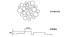
【選択図】 図1PROBLEM TO BE SOLVED: To provide a composite material having a surface composition and structure capable of isolating the influence from a base material, increasing the adhesive strength of the base material, being excellent in bioactivity and inducing nucleation of apatite crystals.
A multilayer film comprising: a metal oxide underlayer composed of a plurality of metal oxides; and a coating layer composed of at least one metal oxide covering the underlayer; A composite material having the multilayer film on a substrate. The composite material of the present invention has an underlayer on the surface of the substrate by immersing the substrate in an underlayer-forming aqueous solution containing at least two kinds of fluorometal complex compounds represented by the general formula: AxMeFy and a fluorine scavenger. Formed and immersed in an aqueous solution for forming a coating layer containing ammonium hexafluoride and a fluorine scavenger, or in an aqueous solution for forming a coating layer containing at least one other fluoro metal complex compound together with ammonium hexafluoride and a fluorine scavenger. It can manufacture by forming a coating layer by this.
[Selection] Figure 1
Description
本発明は、基材に対して高い密着性を有し、優れた生体親和性を付与しうる多層膜、該多層膜を基材上に有し、高い生体活性を発現しうる複合材料及びその製造方法に関する。 The present invention relates to a multilayer film having high adhesion to a base material and capable of imparting excellent biocompatibility, a composite material having the multilayer film on the base material and capable of expressing high bioactivity, and its It relates to a manufacturing method.
一般に生体内に代替材料を埋め込んで生体親和性を獲得しようとする試みは、古くから取り組まれてきた課題である。従来から考えられてきた概念は、生体と代替材料とのやり取りが全くない耐蝕性のものが理想的と考えられ、実際一部の金属材料(例えば、チタン合金Ti6Al4V)が硬質組織代替材料として臨床的に成功を収めてきた。つまり生体が材料に及ぼす影響、逆に材料が生体に及ぼす影響が全くない、互いに独立して存在しうる相互不可侵の金属材料が有望と考えられてきた。
しかし、近年の一連の研究では、いわゆる生体親和性は相互不可侵の要件だけでは十分ではなく、本来の代替材料(人工骨、人工関節)の表面に、生体と人工材料の橋渡しとなるヒドロキシアパタイトの核形成を誘起し、爾後アパタイト表面に生体が取り入れ易い環境を形成することが重要なことが判明した。
例えば、チタン金属では、生体組織との間に10〜50nmの厚さの表面反応場としての均質層が形成され、表面が酸化されてこれら非晶質酸化チタンの一部が生体に取り込まれ再構成されながら、次第に生体親和性を高めていくことが確認されている(非特許文献1)。
詳述すれば、チタン金属表面に形成される酸化物非晶質物質が、生体に一部取り込まれ再構成される過程で表面に多数のTi−OHを形成する。これらの官能基の存在が周囲の体液中のアパタイト成分を濃集させ、増加したアパタイト成分はアパタイトの核形成を誘起し、これらの核は周囲からカルシウムイオンとリン酸イオンを取り込んでアパタイトの成長を促進させる。これらのプロセスを経由することによってチタン金属表面に緻密で均一な骨類似構造を有するアパタイトの層が形成される。
近年、ペースメーカー、人工関節、人工骨、人工歯根、経皮端子、人工血管、血流センサー、各種カテーテルなど、生体組み込み型の機器,器具が実用化されるに伴い、むしろこれらの生体親和性がよりいっそう問題となっている。現在一般に考えられている生体材料が生体内で活性を有するということは、その表面にアパタイト層を形成し、‘骨’となって生体に親和することであるが、チタン金属以外の材料としては、現在CaO−SiO2系ガラスが検討されている。この材料は、生体内でその表面に多数のSi−OH基を形成し、これが周囲のアパタイト成分の濃度を増大させアパタイトの核形成を誘起することで生体活性を発現する。
これら人工材料が生体内でその表面にアパタイトを形成する条件は、表面にアパタイトの核形成を誘起する官能基(−OH)を有することであるとされている。Si−OHの他にもTi−OH、Zr−OH、Ta−OHもアパタイトの核形成を誘起し得ることが確認されている(非特許文献2)。
In general, attempts to acquire biocompatibility by embedding an alternative material in a living body are problems that have been addressed for a long time. The conventional concept is considered to be ideally corrosion-resistant with no interaction between the living body and the substitute material. In fact, some metal materials (for example, titanium alloy Ti6Al4V) are clinically used as hard tissue substitute materials. Has been successful. In other words, it has been considered promising to be a mutually inviolable metal material that can exist independently of each other, and has no influence on the material by the living body, and on the contrary, the influence of the material on the living body.
However, in a series of recent studies, so-called biocompatibility is not sufficient for the mutual inviolability requirement, and hydroxyapatite that serves as a bridge between the living body and the artificial material on the surface of the original substitute material (artificial bone, artificial joint) It has been found that it is important to induce an nucleation of the body and to form an environment in which the living body can easily be taken into the apatite surface after sputum.
For example, in the case of titanium metal, a homogeneous layer as a surface reaction field having a thickness of 10 to 50 nm is formed between living body tissues, the surface is oxidized, and a part of these amorphous titanium oxides is taken into the living body and regenerated. While being configured, it has been confirmed that the biocompatibility is gradually increased (Non-patent Document 1).
More specifically, the oxide amorphous material formed on the titanium metal surface forms a large number of Ti-OH on the surface in the process of being partially taken up and reconstituted in the living body. The presence of these functional groups concentrates the apatite component in the surrounding body fluid, and the increased apatite component induces apatite nucleation, and these nuclei take in calcium ions and phosphate ions from the surroundings to grow apatite. To promote. By passing through these processes, an apatite layer having a dense and uniform bone-like structure is formed on the titanium metal surface.
In recent years, as bio-incorporated devices and instruments such as pacemakers, artificial joints, artificial bones, artificial tooth roots, percutaneous terminals, artificial blood vessels, blood flow sensors, and various catheters are put into practical use, their biocompatibility is rather It is even more problematic. The fact that biomaterials that are currently considered to have activity in vivo means that an apatite layer is formed on the surface and becomes 'bone' and is compatible with the living body, but as materials other than titanium metal, Currently, CaO—SiO 2 glass is being studied. This material forms a large number of Si—OH groups on the surface thereof in vivo, and this increases the concentration of surrounding apatite components and induces apatite nucleation to express biological activity.
The condition for these artificial materials to form apatite on the surface thereof in vivo is considered to have a functional group (—OH) that induces nucleation of apatite on the surface. It has been confirmed that Ti—OH, Zr—OH, and Ta—OH can induce nucleation of apatite in addition to Si—OH (Non-patent Document 2).
そこで、人工骨や人工関節の基材がポリエチレンやポリメチルメタクリレート(PMMA)のような高温に弱いプラスチック素材である場合に、どのようにしてこのような表面官能基構造を導入するか、またいかにして表面にアパタイト結晶の核形成を誘起するかが、本来生体活性のないプラスチック素材を生体活性材料にするための大きな課題であったと言える。
従って、ポリエチレンやPMMAのようなプラスチック素材に生体親和性を付与するためには、ヒドロキシアパタイトが表面に成長し易いように表面処理を施す必要がある。
実際、既にポリエチレンテレフタレート(PET)表面を処理し生体親和性を高めた実例が存在する。まず、PETの極細繊維の織物を、酸素ガス中でグロー放電処理し、その表面に多数のCOOHなどの極性基を形成しておく。これを予め擬似体液に浸漬したCaOとSiO2を主成分とするガラス粒子の上に置くと、ガラスから溶出した珪酸イオンが効率よくPET表面に付着し,アパタイトの核形成を誘起する。このようにしてアパタイト核をプランニングしたPETを別の擬似体液中に浸漬すると、それらの核が擬似体液からカルシウムイオンとリン酸イオンを取り込んで成長し個々のPET繊維の表面に緻密で均一なアパタイト層を形成する(非特許文献3)。
また、シランカップリング法に基づきTiO2 膜を、PET、エチレンビニルアルコール共重合体(EVOH)、ポリアミド6、ポリアミド66等に成膜形成し、これらプラスチックに生体活性を付与した例がある(非特許文献4)。
しかし、これらの例はいずれもプロセスが複雑で膜形成に時間がかかり、かつ生体材料にとって重要な要件である膜の機械的強度が十分とはいえない点が問題であった。
Therefore, in order to impart biocompatibility to a plastic material such as polyethylene or PMMA, it is necessary to perform a surface treatment so that hydroxyapatite can easily grow on the surface.
In fact, there are already examples of treating the surface of polyethylene terephthalate (PET) to increase biocompatibility. First, a PET ultrafine fiber fabric is subjected to glow discharge treatment in oxygen gas to form a large number of polar groups such as COOH on the surface. When this is placed on glass particles mainly composed of CaO and SiO 2 previously immersed in a simulated body fluid, silicate ions eluted from the glass are efficiently attached to the PET surface and induce nucleation of apatite. When the PET in which the apatite nuclei are planned in this way is immersed in another simulated body fluid, the nuclei take in calcium ions and phosphate ions from the simulated body fluid and grow to form a dense and uniform apatite on the surface of each PET fiber. A layer is formed (Non-patent Document 3).
In addition, there is an example in which a TiO 2 film is formed on PET, ethylene vinyl alcohol copolymer (EVOH), polyamide 6, polyamide 66, and the like based on the silane coupling method, and bioactivity is imparted to these plastics (non-contained). Patent Document 4).
However, all of these examples have problems in that the process is complicated and it takes time to form a film, and the mechanical strength of the film, which is an important requirement for biomaterials, cannot be said to be sufficient.
本発明は、上記従来技術における問題点を解決し、プラスチック等の生体活性のない基材を用いて、その基材上に密着性及び機械的強度の高い膜を形成することにより、生体環境内でアパタイト形成を誘起し、生体活性を発現でき、人工骨や人工関節材料などのインプラントとして有用な複合材料を提供するとともに、その効率のよい製造方法を提供することを目的とする。 The present invention solves the above-described problems in the prior art, and forms a film having high adhesion and mechanical strength on the base material using a base material having no bioactivity, such as plastic. An object of the present invention is to provide a composite material that can induce apatite formation and express biological activity and is useful as an implant such as an artificial bone or artificial joint material, and an efficient manufacturing method thereof.
本発明は、プラスチック等の基材に生体親和性を付与するために、表面に水酸基を有する非晶質或いは結晶質の酸化物膜を高付着力で形成し、アパタイト結晶の核形成を誘起するとともに、アパタイトの成分における過飽和状態が増大するような組成及び構造(組織)を調整して、これらの形成反応を円滑に進行させれば、上記の目的を達成しうることを見出し、完成したものである。 The present invention induces nucleation of apatite crystals by forming an amorphous or crystalline oxide film having a hydroxyl group on the surface with high adhesion to impart biocompatibility to a substrate such as plastic. In addition, by adjusting the composition and structure (structure) so that the supersaturation state in the apatite component increases, it was found that the above-mentioned purpose could be achieved if these formation reactions proceed smoothly. It is.
すなわち、本発明は、複数の金属酸化物から構成された金属酸化物下地層と、該下地層を被覆する少なくとも1種類の金属酸化物から構成された被覆層とからなることを特徴とする多層膜を提供するものである。
上記の下地層を構成する金属酸化物は、周期表の4族金属、5族金属、13族金属及び14族金属からなる群から選択された少なくとも2種の金属の酸化物であることが好ましい。
本発明の多層膜において、下地層の構成主要成分である金属酸化物は、酸化ケイ素、酸化チタン及び酸化ジルコニウムから選ばれた2種又は3種であるのが好ましい。
また、被覆層の構成主要成分は、酸化チタンであり、酸化チタンの一部の代わりに酸化ケイ素、酸化ジルコニウム、酸化ニオブ、酸化スズ、酸化タンタル、酸化ガリウム、酸化ハフニウム、酸化ゲルマニウム及びアルミナから選ばれる少なくとも1種類を含んでいてもよい。
本発明は、さらに基材上に前記多層膜を有することを特徴とする複合材料に関する。
なお、本明細書において、構成主要成分とは、構成成分のうち60%〜100%を占める成分を言うものとする。
That is, the present invention comprises a multilayer comprising a metal oxide underlayer composed of a plurality of metal oxides and a coating layer composed of at least one kind of metal oxide covering the underlayer. A membrane is provided.
The metal oxide constituting the underlayer is preferably an oxide of at least two metals selected from the group consisting of Group 4 metal, Group 5 metal, Group 13 metal and Group 14 metal of the Periodic Table. .
In the multilayer film of the present invention, it is preferable that the metal oxide which is the main constituent component of the underlayer is two or three selected from silicon oxide, titanium oxide and zirconium oxide.
The main component of the coating layer is titanium oxide, which is selected from silicon oxide, zirconium oxide, niobium oxide, tin oxide, tantalum oxide, gallium oxide, hafnium oxide, germanium oxide, and alumina instead of a part of titanium oxide. It may contain at least one kind.
The present invention further relates to a composite material comprising the multilayer film on a substrate.
In addition, in this specification, a component main component shall mean the component which occupies 60%-100% among component components.
本発明の複合材料の製造方法は、基材表面に、複数の金属酸化物から構成された金属酸化物下地層と、該下地層を被覆する少なくとも1種類の金属酸化物から構成された被覆層とからなる多層膜を形成することを特徴とする。
本発明の複合材料の製造方法は、下記の一般式(I):
AxMeFy ・・・(I)
[式中、AはNH4 、Na、K又はHを表わし、Meは周期表の4族金属、5族金属、13族金属及び14族金属からなる群から選択した金属の酸化物を表わし、x及びyは化合物の電気的中性を満たす数である]で示されるフルオロ金属錯化合物の少なくとも2種類と、フッ素捕捉剤とを含む下地層形成水溶液中に基材を浸漬することによって基材表面に下地層を形成し、6フッ化チタンアンモニウムとフッ素捕捉剤とを含む被覆層形成水溶液又は6フッ化チタンアンモニウムとフッ素捕捉剤とともに、6フッ化ケイ素アンモニウム、6フッ化ジルコニウムアンモニウム、7フッ化ニオブアンモニウム、6フッ化スズアンモニウム、7フッ化タンタルアンモニウム、6フッ化ガリウムアンモニウム、6フッ化ハフニウムアンモニウム、6フッ化ゲルマニウムアンモニウム及び6フッ化アルミニウムアンモニウムからなる群から選ばれる少なくとも1種類のフルオロ金属錯化合物を含む被覆層形成水溶液中に浸漬することによって被覆層を形成するのが好ましい。
The method for producing a composite material according to the present invention includes a metal oxide underlayer composed of a plurality of metal oxides on a substrate surface, and a coating layer composed of at least one metal oxide covering the underlayer. The multilayer film which consists of these is formed.
The method for producing the composite material of the present invention comprises the following general formula (I):
A x MeF y (I)
Wherein A represents NH 4 , Na, K or H, Me represents an oxide of a metal selected from the group consisting of Group 4 metals, Group 5 metals, Group 13 metals and Group 14 metals of the Periodic Table; x and y are numbers satisfying the electrical neutrality of the compound] by immersing the substrate in an underlayer-forming aqueous solution containing at least two types of fluorometal complex compounds represented by A base layer is formed on the surface, and a coating layer forming aqueous solution containing ammonium hexafluoride titanium and a fluorine scavenger, or ammonium hexafluoride, ammonium hexafluoride ammonium, fluorine fluoride together with ammonium hexafluoride and a fluorine scavenger. Niobium ammonium fluoride, tin hexafluoride ammonium, tantalum ammonium fluoride, gallium ammonium hexafluoride, hafnium ammonium hexafluoride, Preferably, a coating layer is formed by dipping into the coating layer forming solution containing at least one metal fluoro complex compound selected from the group consisting of germanium ammonium and 6 aluminum fluoride ammonium.
本発明によれば、好適な下地層を設けたことにより、基材からの影響を隔絶するとともに、基材と被覆層との接着強度を高め、また被覆層の生体活性を向上させることができ、アパタイト結晶の核形成を誘起するような表面組成と構造を有する複合材料を提供することができる。
したがって、本発明によれば、プラスチック等の生体活性を有しない基材を用いても高い生体親和性を有し、生体活性に極めて優れた複合材料を提供することができる。また、本発明の方法によれば、低温(室温)環境下で、かつ複雑な形状を有する基材に対しても高い生体活性を有する多層膜を高付着力で形成することができ、生体活性に優れた複合材料を簡単な操作で効率よく製造することができる。
さらに、本発明の複合材料を、インプラントとして生体内に埋入するか又はリン酸カルシウム系化合物形成溶液に浸漬すると、リン酸カルシウム系化合物をその表面に形成することができる。
According to the present invention, by providing a suitable underlayer, it is possible to isolate the influence from the substrate, increase the adhesive strength between the substrate and the coating layer, and improve the bioactivity of the coating layer. It is possible to provide a composite material having a surface composition and structure that induces nucleation of apatite crystals.
Therefore, according to the present invention, it is possible to provide a composite material having high biocompatibility and extremely excellent bioactivity even when a base material having no bioactivity such as plastic is used. Further, according to the method of the present invention, a multilayer film having high bioactivity can be formed with high adhesive force even on a substrate having a complicated shape in a low temperature (room temperature) environment. It is possible to efficiently produce an excellent composite material with a simple operation.
Furthermore, when the composite material of the present invention is implanted in a living body as an implant or immersed in a calcium phosphate compound forming solution, a calcium phosphate compound can be formed on the surface thereof.
本発明の多層膜は、前記のように、複数の金属酸化物から構成された金属酸化物下地層と、該下地層を被覆する少なくとも1種類の金属酸化物から構成された被覆層とからなる。
上記の下地層を構成する金属酸化物は、周期表の4族金属、5族金属、13族金属及び14族金属からなる群から選択される少なくとも2種の金属の酸化物であることが好ましい。
下地層は、主として、酸化ケイ素、酸化チタン及び酸化ジルコニウムから選ばれた2種又は3種から構成するのが好ましく、特に、SiO2 0〜1.0:TiO2 0〜0.6:ZrO2 0〜1.0のモル比範囲内で含むのが好ましく、SiO2 0.1〜0.3:TiO2 0.03〜0.2:ZrO2 0.5〜0.9のモル比範囲内で含むのがより好ましい。
また、下地層の構成としては、主要成分が、SiO2 0〜1.0:TiO2 0〜0.6:ZrO2 0〜1.0のモル比範囲内で存在し、かつ酸化ジルコニウムの代わりに、或いは酸化ジルコニウムとともに、酸化ニオブ、酸化スズ、酸化タンタル、酸化ガリウム、酸化ハフニウム、酸化ゲルマニウム及びアルミナから選ばれる少なくとも1種類を含む構成も好適である。
本発明の多層膜において、下地層は、0.05〜0.5μmの層厚であるのが好ましく、0.02〜0.1μmの層厚であるのがより好ましい。
As described above, the multilayer film of the present invention comprises a metal oxide base layer composed of a plurality of metal oxides and a coating layer composed of at least one kind of metal oxide covering the base layer. .
The metal oxide constituting the underlayer is preferably an oxide of at least two metals selected from the group consisting of Group 4 metal, Group 5 metal, Group 13 metal and Group 14 metal of the Periodic Table. .
The underlayer is preferably composed mainly of two or three kinds selected from silicon oxide, titanium oxide and zirconium oxide, and particularly
As the structure of the underlying layer, the main component, SiO 2 0~1.0: TiO 2 0~0.6 : present in a molar ratio range of
In the multilayer film of the present invention, the underlayer preferably has a layer thickness of 0.05 to 0.5 μm, and more preferably 0.02 to 0.1 μm.
本発明の多層膜において、被覆層の構成主要成分は酸化チタンであり、酸化チタンの一部の代わりに酸化ケイ素、酸化ジルコニウム、酸化ニオブ、酸化スズ、酸化タンタル、酸化ガリウム、酸化ハフニウム、酸化ゲルマニウム及びアルミナから選ばれる少なくとも1種類を含んでいてもよい。詳述すれば、被覆層の構成成分は、酸化チタン60〜100%、酸化ケイ素、酸化ジルコニウム、酸化ニオブ、酸化スズ、酸化タンタル、酸化ガリウム、酸化ハフニウム、酸化ゲルマニウム及びアルミナから選ばれる少なくとも1種類40%未満から成る。
被覆層の層厚は、0.1〜1.0μmであるのが好ましく、0.2〜0.5μmであるのがより好ましい。
In the multilayer film of the present invention, the main component of the coating layer is titanium oxide, and instead of a part of titanium oxide, silicon oxide, zirconium oxide, niobium oxide, tin oxide, tantalum oxide, gallium oxide, hafnium oxide, germanium oxide And at least one selected from alumina. Specifically, the constituent component of the coating layer is at least one selected from titanium oxide 60 to 100%, silicon oxide, zirconium oxide, niobium oxide, tin oxide, tantalum oxide, gallium oxide, hafnium oxide, germanium oxide, and alumina. Consists of less than 40%.
The thickness of the coating layer is preferably 0.1 to 1.0 μm, and more preferably 0.2 to 0.5 μm.
本発明の複合材料は、上記のような多層膜を基材上に設けたものである。
ここで基材としては、広範な材料から選択することができ、例えば、合成高分子化合物、天然高分子化合物、生体高分子化合物、プラスチック等を含む高分子化合物、金属、半導体、ガラス、セラミックス、カーボン材料、ダイヤモンド、カルサイト等の塩、無機高分子化合物、貝殻、水晶等の天然物などを挙げることができる。特に高分子樹脂材料からなるものが好適である。具体的には、ポリエチレン、アクリル樹脂(PMMA)やこれらにアクリル系、シリコン系ハードコート、メラミン系ハードコートなどのハードコートを施したものでもよい。
これらの基材の形状は、特に制限されるものはなく、板状、繊維状、編物状、多孔質、粒状、その他複雑形状などいかなるものでもよい。
また、これらの基材は、基本的に平坦なものでもよいが、金属酸化物膜やその上にアパタイト膜が形成されやすくするため或いはこれらと基材との間の密着強度を向上させるため、凹凸状に表面が荒れた状態のものでもよい。
The composite material of the present invention is obtained by providing the above multilayer film on a substrate.
Here, the substrate can be selected from a wide range of materials, for example, synthetic polymer compounds, natural polymer compounds, biopolymer compounds, polymer compounds including plastics, metals, semiconductors, glass, ceramics, Examples thereof include carbon materials, salts such as diamond and calcite, inorganic polymer compounds, natural products such as shells and crystals. Particularly preferred is a polymer resin material. Specifically, polyethylene, acrylic resin (PMMA), or those obtained by applying a hard coat such as an acrylic, silicon hard coat, or melamine hard coat may be used.
The shape of these base materials is not particularly limited, and may be any shape such as a plate shape, a fiber shape, a knitted shape, a porous shape, a granular shape, and other complicated shapes.
In addition, these substrates may be basically flat, in order to facilitate the formation of a metal oxide film and an apatite film thereon or to improve the adhesion strength between these and the substrate, The thing with the surface roughened in unevenness may be used.
次に、本発明の複合材料の製造方法について好ましい実施態様を説明する。
基材は、紫外線照射、プラズマ照射、酸化雰囲気曝露又は酸若しくは塩基中への浸漬により粗面化した後、下地層を形成するのが好ましい。例えば、プラスチック基材に直に酸化物膜を成膜する場合、表面が撥水状態になっていることが多く、表面処理(プラズマ照射、薬品処理)を施すことによってプラスチック表面を活性化し、親水状態にした後に、下地層形成水溶液中に浸漬して成膜を実施するのが好ましい。
Next, a preferred embodiment of the method for producing the composite material of the present invention will be described.
The substrate is preferably roughened by ultraviolet irradiation, plasma irradiation, oxidizing atmosphere exposure, or immersion in an acid or base, and then a base layer is formed. For example, when an oxide film is formed directly on a plastic substrate, the surface is often in a water-repellent state, and surface treatment (plasma irradiation, chemical treatment) is performed to activate the plastic surface and make it hydrophilic. It is preferable to form the film by immersing it in an underlayer-forming aqueous solution after the state has been reached.
本発明において、下地層の形成は、下記の一般式(I):
AxMeFy ・・・(I)
[式中、AはNH4 、Na、K又はHを表わし、Meは周期表の4族金属、5族金属、13族金属及び14族金属からなる群から選択した金属の酸化物を表わし、x及びyは化合物の電気的中性を満たす数である]で示されるフルオロ金属錯化合物の少なくとも2種類と、フッ素捕捉剤とを含む下地層形成水溶液中に基材を浸漬することによって実施するのが好ましい。
上記一般式(I)で示されるフルオロ金属錯化合物の具体例としては、6フッ化チタンアンモニウム:(NH4)2TiF6、6フッ化珪素アンモニウム:(NH4)2SiF6、6フッ化ジルコニウムアンモニウム:(NH4)2ZrF6、7フッ化タンタルアンモニウム:(NH4)2TaF7 、7フッ化ニオブアンモニウム:(NH4)2NbF7、6フッ化スズアンモニウム:(NH4)2SnF6、6フッ化ガリウムアンモニウム:(NH4)2GaF6 、6フッ化ハフニウムアンモニウム:(NH4)2HfF6、6フッ化ゲルマニウムアンモニウム:(NH4)2GeF6、6フッ化アルミニウムアンモニウム:(NH4)3AlF6などがある。
上記のようなフルオロ金属錯化合物とフッ素捕捉剤を含む水溶液中に、或いは前者水溶液にフッ素捕捉剤を直接添加混合し、この水溶液に基材を浸漬することによって化学反応が開始され成膜が進行する。
In the present invention, the underlayer is formed by the following general formula (I):
A x MeF y ... (I)
Wherein A represents NH 4 , Na, K or H, Me represents an oxide of a metal selected from the group consisting of Group 4 metals, Group 5 metals, Group 13 metals and Group 14 metals of the Periodic Table; x and y are numbers satisfying the electrical neutrality of the compound], and is carried out by immersing the substrate in an underlayer-forming aqueous solution containing at least two types of fluorometal complex compounds represented by Is preferred.
Specific examples of the fluorometal complex compound represented by the general formula (I) include ammonium hexafluoride: (NH 4 ) 2 TiF 6 , ammonium hexafluoride: (NH 4 ) 2 SiF 6 , hexafluoride. Zirconium ammonium: (NH 4 ) 2 ZrF 6 , tantalum ammonium 7 fluoride: (NH 4 ) 2 TaF 7 , ammonium niobium 7 fluoride: (NH 4 ) 2 NbF 7 , ammonium hexafluoride: (NH 4 ) 2 SnF 6 , gallium ammonium hexafluoride: (NH 4 ) 2 GaF 6 , ammonium hafnium hexafluoride: (NH 4 ) 2 HfF 6 , germanium ammonium hexafluoride: (NH 4 ) 2 GeF 6 , aluminum ammonium hexafluoride : (NH 4 ) 3 AlF 6 and the like.
In the aqueous solution containing the fluoro metal complex compound and the fluorine scavenger as described above, or by adding the fluorine scavenger directly to the former aqueous solution and immersing the substrate in this aqueous solution, the chemical reaction starts and the film formation proceeds. To do.
本発明において、使用するフルオロ金属錯化合物は、対応する金属酸化物にフッ化水素酸水溶液を作用させ、さらにアンモニア水によって中和することによって調製することもできる。具体的には、酸化チタン、酸化ケイ素、酸化ジルコニウム、酸化タンタル、酸化ニオブ、酸化スズ、酸化ガリウム、酸化ハフニウム、酸化ガリウム、酸化ゲルマニウム、酸化アルミニウムよりなる群から選ばれる酸化物にフッ化水素酸水溶液を作用させ、更にアンモニア水によって中和すればよい。こうして得た水溶液にホウ素化合物を混合した下地層形成水溶液に、基材を浸漬することによっても上記と同様の成膜反応が開始される。この際アンモニア水の添加量は、水溶液のpHを変化させ、作成する基材種にあわせるために調整することができる。 In the present invention, the fluorometal complex compound to be used can also be prepared by allowing a hydrofluoric acid aqueous solution to act on the corresponding metal oxide and further neutralizing with ammonia water. Specifically, hydrofluoric acid is selected from the group consisting of titanium oxide, silicon oxide, zirconium oxide, tantalum oxide, niobium oxide, tin oxide, gallium oxide, hafnium oxide, gallium oxide, germanium oxide, and aluminum oxide. An aqueous solution is allowed to act, and further neutralized with aqueous ammonia. The film formation reaction similar to the above is also started by immersing the base material in an underlayer-forming aqueous solution in which a boron compound is mixed with the aqueous solution thus obtained. At this time, the amount of ammonia water added can be adjusted in order to change the pH of the aqueous solution and match the type of substrate to be prepared.
一般には特定のフルオロ金属錯化合物水溶液中に、ホウ素化合物からなるフッ素捕捉剤を添加すると、ホウ素とフッ素が反応して安定なフルオロホウ素錯化合物を形成し、そのため溶液中のフッ化物イオン濃度が減少し、これを補うためフルオロ金属錯化合物が加水分解して、金属酸化物となって析出し始める。pHが7以下での析出にあたっては、数〜数十ナノメートルの球状粒子として表面にOH基を有した状態で、緻密に配列を繰り返しながら積層する。これらのような球状粒子の積層の結果、被膜表面にはこれらによる特異な表面構造が形成される。このような析出プロセスを用いて、複合酸化物下地層を形成する。
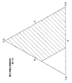
下地層の場合、基本的に6フッ化チタンアンモニウム、6フッ化珪素アンモニウム及び6フッ化ジルコニウムアンモニウムを適当な比率(図3参照)で混合したものにフッ素捕捉剤を添加することによって、複合酸化物下地層の組成範囲をモル比で、SiO2 0〜1.0:TiO2 0〜0.6:ZrO2 0〜1.0の範囲内で形成する。ここで、酸化ジルコニウムの代わりに、その一部が酸化ニオブ、酸化スズ、酸化タンタル、酸化ガリウム、酸化ハフニウム、酸化ゲルマニウム及びアルミナであってもよい。
In general, when a fluorine scavenger consisting of a boron compound is added to a specific aqueous solution of a fluorometal complex compound, boron and fluorine react to form a stable fluoroboron complex compound, which reduces the fluoride ion concentration in the solution. In order to compensate for this, the fluoro metal complex compound hydrolyzes and begins to precipitate as a metal oxide. When depositing at a pH of 7 or less, the particles are stacked as spherical particles of several to several tens of nanometers with OH groups on the surface while repeating the arrangement precisely. As a result of laminating spherical particles such as these, a unique surface structure is formed on the coating surface. A complex oxide underlayer is formed using such a deposition process.
In the case of an underlayer, basically a composite oxidation is achieved by adding a fluorine scavenger to a mixture of titanium ammonium fluoride, ammonium ammonium fluoride and ammonium ammonium fluoride fluoride in an appropriate ratio (see FIG. 3). The composition range of the material underlayer is formed in a molar ratio of
下地層形成水溶液に添加し、成膜反応を開始させる均一系フッ素捕捉剤は、フッ素と反応して安定なフルオロ金属錯化合物及び/又はフッ化物を形成することにより、金属酸化物を析出させるようにフッ素イオンの平衡を移動させるものである。例えば、オルトホウ酸,メタホウ酸等のホウ酸の他、酸化ホウ素などのホウ素化合物、塩化アルミニウムなどが例示される。これらのフッ素捕捉剤のうちホウ素化合物が特に好適である。このような捕捉剤は、通常水溶液の形で用いられるが、粉末の形で添加して系中に溶解させてもよい。このような捕捉剤の添加は、一回に或いは数回に分けて間歇的に行ってもよく、制御された供給速度、例えば一定の速度で連続的に行ってもよい。 A homogeneous fluorine scavenger that is added to the underlayer-forming aqueous solution and starts the film-forming reaction reacts with fluorine to form a stable fluorometal complex compound and / or fluoride, thereby precipitating the metal oxide. This moves the equilibrium of fluorine ions. For example, in addition to boric acid such as orthoboric acid and metaboric acid, boron compounds such as boron oxide, aluminum chloride and the like are exemplified. Of these fluorine scavengers, boron compounds are particularly suitable. Such a scavenger is usually used in the form of an aqueous solution, but it may be added in the form of a powder and dissolved in the system. Such addition of the scavenger may be performed intermittently in one time or divided into several times, or may be performed continuously at a controlled supply rate, for example, at a constant rate.
不均一系フッ素捕捉剤としては、アルミニウム、チタン、鉄、ニッケル、マグネシウム、銅、亜鉛などの金属、ガラスなどのセラミックス、及びケイ素、酸化カルシウム、酸化ホウ素、酸化アルミニウム、二酸化ケイ素、酸化マグネシウムなどの化合物が例示される。このような固形物を水溶液に添加又は挿入すると固形物近傍のF−(フッ素イオン)が消費されて、その濃度が減少するので、その部分の化学平衡がシフトして金属酸化物が析出する。このような固形物を用いると、その添加又は挿入する方法と反応条件により、水溶液に浸漬した基材表面の全体に金属酸化物を析出することもできるし、その析出を選択された一部、即ち該固形物の存在する近傍に限定することも可能である。あるいは、均一系と不均一系のフッ素捕捉剤とを併用することにより、基材表面の析出物薄膜を部分的に厚くすることもできる。
以上のような成膜プロセスからなる本発明によれば、基材上にフォトリソグラフィなどの方法でレジスト膜を形成し、金属酸化物膜が形成される部位を制限することもできる。
Heterogeneous fluorine scavengers include metals such as aluminum, titanium, iron, nickel, magnesium, copper, zinc, ceramics such as glass, and silicon, calcium oxide, boron oxide, aluminum oxide, silicon dioxide, magnesium oxide, etc. Examples are compounds. When such a solid is added to or inserted into an aqueous solution, F- (fluorine ions) in the vicinity of the solid is consumed and its concentration decreases, so that the chemical equilibrium of that portion shifts and a metal oxide is deposited. When such a solid is used, the metal oxide can be deposited on the entire surface of the substrate immersed in the aqueous solution, depending on the method of addition or insertion and the reaction conditions. That is, it can be limited to the vicinity where the solid is present. Alternatively, the precipitate thin film on the surface of the substrate can be partially thickened by using a homogeneous and heterogeneous fluorine scavenger in combination.
According to the present invention comprising the film formation process as described above, a resist film can be formed on a base material by a method such as photolithography to limit the portion where the metal oxide film is formed.
こうして形成した下地層の上に被覆層を設ける。被覆層は、主として酸化チタンより構成され、この他に酸化ケイ素、酸化ジルコニウム、酸化ニオブ、酸化スズ、酸化タンタル、酸化ガリウム、酸化ハフニウム、酸化ゲルマニウム、アルミナ等の少なくとも一種類含有してもよい。
被覆層の形成は、6フッ化チタンアンモニウムとフッ素捕捉剤を含む被覆層形成水溶液又は6フッ化チタンアンモニウムとフッ素捕捉剤とともに、6フッ化ケイ素アンモニウム、6フッ化ジルコニウムアンモニウム、7フッ化ニオブアンモニウム、6フッ化スズアンモニウム、7フッ化タンタルアンモニウム、6フッ化ガリウムアンモニウム、6フッ化ハフニウムアンモニウム、6フッ化ゲルマニウムアンモニウム及び6フッ化アルミニウムアンモニウムからなる群から選ばれる少なくとも1種類のフルオロ金属錯化合物を含む被覆層形成水溶液中に下地層を有する基材を浸漬することによって実施するのが好ましい。
A coating layer is provided on the base layer thus formed. The coating layer is mainly composed of titanium oxide, and may further contain at least one kind of silicon oxide, zirconium oxide, niobium oxide, tin oxide, tantalum oxide, gallium oxide, hafnium oxide, germanium oxide, alumina and the like.
The coating layer is formed by using a coating layer forming aqueous solution containing ammonium hexafluoride and a fluorine scavenger, or ammonium hexafluoride, ammonium hexafluoride, and ammonium niobium fluoride together with ammonium hexafluoride and a fluorine scavenger. At least one fluorometal complex compound selected from the group consisting of stannic ammonium hexafluoride, tantalum ammonium hexafluoride, gallium ammonium hexafluoride, hafnium ammonium hexafluoride, germanium ammonium hexafluoride and ammonium ammonium fluoride fluoride It is preferable to carry out by immersing a base material having an underlayer in a coating layer forming aqueous solution containing.
このような被覆層の表面では、金属酸化物が水又は水溶液中で水酸基を形成するのが好ましく、被覆層の表面は、平均径1〜300nmの大きさの金属酸化物粒子の集合体から構成される多孔組織からなるのが好ましい。特に、複合酸化物から構成すると、より生体親和性更には生体活性を向上させることができる。 On the surface of such a coating layer, the metal oxide preferably forms a hydroxyl group in water or an aqueous solution, and the surface of the coating layer is composed of an aggregate of metal oxide particles having an average diameter of 1 to 300 nm. It is preferable to consist of a porous structure. In particular, when the composite oxide is used, biocompatibility and further bioactivity can be improved.
下地層の形成後及び被覆層の形成後に、40〜150℃で1〜20時間加熱してもよい。これは、多層膜を熟成させ、より付着力ある強固な膜とするため効果がある。 You may heat at 40-150 degreeC for 1 to 20 hours after formation of a base layer and formation of a coating layer. This is effective for aging the multilayer film to obtain a strong film having more adhesive force.
なお、下地層及び/又は被覆層の形成を、ゾル・ゲル法、溶射法、電極法、真空蒸着法或いはスパッタリング法を用いて行うこともできる。 In addition, formation of a base layer and / or a coating layer can also be performed using a sol-gel method, a thermal spraying method, an electrode method, a vacuum evaporation method, or a sputtering method.
なお、生体親和性は相互不可侵の要件だけでは十分ではなく、人工材料(人工骨)の表面に、生体と人工材料の橋渡しとなるリン酸カルシウム系化合物、とりわけヒドロキシアパタイト類の核形成を誘起し、爾後ヒドロキシアパタイト類層を表面に形成することが重要である。なお、リン酸カルシウム系化合物とは、第一リン酸カルシウム(Ca(H2PO4)2)、第二リン酸カルシウム(CaHPO4)、第三リン酸カルシウム(Ca3(PO4)2)、リン酸四カルシウム(Ca4(PO4)2O)、リン酸八カルシウム(Ca8H2(PO4)6)、ヒドロキシアパタイト類を含むアパタイト類、アモルファスリン酸カルシウムなどを含み結晶水をもつものも含む。またヒドロキシアパタイトとは、化学式Ca10(PO4)6(OH)2で表される化合物をいう。ヒドロキシアパタイト類とは、ヒドロキシアパタイトまたはその構成元素が置換及び/または欠損しているものを包含していう。ヒドロキシアパタイト類は、例えばヒドロキシアパタイトを構成する元素或いは基の一部が、Na、Kなどの周期表の1族の元素、Mgなどの周期表の2族の元素、Znなどの周期表の12族の元素、F、Clなどの周期表の17族の元素、CO3 2-、HPO4 2-、SO4 2-などの基で置換されていてよい。さらに希土類によって置換されてもよい。 In addition, the biocompatibility is not enough for the requirement of mutual invasion, but on the surface of the artificial material (artificial bone), induces nucleation of calcium phosphate compounds, especially hydroxyapatites, which bridge the living body and the artificial material, It is important to form a hydroxyapatite layer on the surface. Note that the calcium phosphate-based compounds, monocalcium phosphate (Ca (H 2 PO 4) 2), dibasic calcium phosphate (CaHPO 4), tribasic calcium phosphate (Ca 3 (PO 4) 2 ), tetracalcium phosphate (Ca 4 (PO 4 ) 2 O), octacalcium phosphate (Ca 8 H 2 (PO 4 ) 6 ), apatites including hydroxyapatites, amorphous calcium phosphate, and the like having crystal water. Hydroxyapatite refers to a compound represented by the chemical formula Ca 10 (PO 4 ) 6 (OH) 2 . Hydroxyapatites include those in which hydroxyapatite or its constituent elements are substituted and / or missing. Hydroxyapatites are, for example, elements of the hydroxyapatite or a part of the group are elements of Group 1 of the periodic table such as Na and K, elements of Group 2 of the periodic table such as Mg, and 12 of the periodic table such as Zn. It may be substituted with a group element, a group 17 element of the periodic table such as F or Cl, a group such as CO 3 2− , HPO 4 2− , SO 4 2− . Furthermore, it may be replaced by a rare earth.
複合材料が、その表面に水酸基を有していると、生体内に埋入されても、あるいはリン酸カルシウム系化合物形成溶液に浸漬されても、リン酸カルシウム系化合物をその表面に効果的に形成することが可能であり、その結果生体親和性さらには生体活性が非常に優れるようになる。
一般に先に述べた析出粒子から構成される凹凸の表面構造は、特に凹部ではOH‐イオンが特に濃集し易い環境にあるため、これら水酸イオン濃度の増加によってアパタイト結晶の核形成が起こり易く、爾後のヒドロキシアパタイト形成を容易にする。
When the composite material has a hydroxyl group on its surface, it can effectively form a calcium phosphate compound on its surface even if it is embedded in a living body or immersed in a calcium phosphate compound forming solution. As a result, the biocompatibility and the bioactivity become very excellent.
In general, the surface structure of irregularities composed of the precipitated particles described above is in an environment where OH-ions are particularly likely to concentrate, particularly in the recesses. , Facilitates the formation of hydroxyapatite after sowing.
我々は、これらアパタイトの形成を容易ならしめる表面構造を原子間力顕微鏡(AFM,SPA500)によって入念に観察し、その模式図を図1に示す。その結果これらの表面構造は、基本的に径1〜300nmからなる金属酸化物微粒子からなる多孔構造からなることが判明した。またこれらの粒子が集合して、平均径10〜50nm,高さ1〜30nmからなる島状の粒子集合体を構成しているのが判った。これらの集合組織が、アパタイトの形成反応を助長する働きがあると考えられる。ここで平均径は、AFM像における位相像をもとに算出し、また平均高さ(深さ)は、断面像の計測結果から算出した。 We carefully observed the surface structure that facilitates the formation of these apatites with an atomic force microscope (AFM, SPA500), and a schematic diagram is shown in FIG. As a result, it has been found that these surface structures are basically porous structures made of metal oxide fine particles having a diameter of 1 to 300 nm. It was also found that these particles aggregated to form an island-shaped particle aggregate having an average diameter of 10 to 50 nm and a height of 1 to 30 nm. These textures are considered to have a function of promoting the apatite formation reaction. Here, the average diameter was calculated based on the phase image in the AFM image, and the average height (depth) was calculated from the measurement result of the cross-sectional image.
一方、これらの表面構造の他にアパタイト結晶の核を形成、成長させるための条件としては、アパタイトと複合材料の間の素材上の親和性が重要である。このためには、素材間の親和性を高める工夫が必要となる。この素材間の親和性を実現するために、被覆層形成水溶液に水溶性カルシウム塩を10-5〜1モル/リットル、好ましくは10-4〜10-1モル/リットルの量で溶解させ、これにフッ素捕捉剤を添加して金属酸化物を析出させることによって金属酸化物中にカルシウムイオンを混入させるのが好ましい。水溶性カルシウム塩としては、例えば、硝酸カルシウム、塩化カルシウムなどが挙げられる。
これによってアパタイト形成を大きく促進させることが可能となった。これは金属酸化物膜表面に析出成長させるにあたって、その原料となるカルシウムを複合材料の被覆層中に混入させておくと、アパタイト形成においてより親和性の高い環境を提供する働きがあるためである。
これらの事実は実験によってカルシウムの混入がある場合とない場合とで比較し、前者の場合では明らかにアパタイトの形成が顕著であることを確かめることができた。
On the other hand, in addition to these surface structures, as a condition for forming and growing nuclei of apatite crystals, affinity on the material between the apatite and the composite material is important. For this purpose, a device for increasing the affinity between materials is required. In order to realize the affinity between the materials, a water-soluble calcium salt is dissolved in the coating layer forming aqueous solution in an amount of 10 −5 to 1 mol / liter, preferably 10 −4 to 10 −1 mol / liter. It is preferable to add calcium ions to the metal oxide by adding a fluorine scavenger to the metal oxide and precipitating the metal oxide. Examples of the water-soluble calcium salt include calcium nitrate and calcium chloride.
This makes it possible to greatly promote apatite formation. This is because, in the case of precipitation growth on the surface of the metal oxide film, if calcium as a raw material is mixed in the coating layer of the composite material, it has a function of providing a higher affinity environment in apatite formation. .
These facts were compared by experiment with and without calcium contamination, and it was confirmed that the formation of apatite was clearly remarkable in the former case.
本発明の複合材料は、そのまま体内に埋入することができるが、予めリン酸カルシウム系化合物形成溶液または擬似体液に浸漬し、表面にリン酸カルシウム系化合物の析出層を形成することもできる。
本発明において当該材料の生体活性(生体親和性)の発現を確認する手段として用いられるリン酸カルシウム系化合物形成溶液或いは擬似体液は、カルシウムイオン(Ca2+)を0.02〜25mM、リン酸水素イオン(HPO4 2-)を0.01〜10mM含有し、pHが6〜8であることが好ましい。より好ましくはカルシウムイオンを0.2〜20mM、リン酸水素イオンを0.1〜8mM含み、pHは6.8〜7.6である。更に好ましくは、カルシウムイオンを1.2〜5mM、リン酸水素イオンを0.5〜2mM含み、pHは7.2〜7.5である。
Although the composite material of the present invention can be embedded in the body as it is, it can be preliminarily immersed in a calcium phosphate compound forming solution or a simulated body fluid to form a precipitated layer of the calcium phosphate compound on the surface.
In the present invention, the calcium phosphate compound forming solution or simulated body fluid used as a means for confirming the expression of the bioactivity (biocompatibility) of the material is 0.02 to 25 mM calcium ion (Ca 2+ ) and hydrogen phosphate ion. It is preferable that 0.01 to 10 mM of (HPO 4 2− ) is contained and the pH is 6 to 8. More preferably, the calcium ion is 0.2 to 20 mM, the hydrogen phosphate ion is 0.1 to 8 mM, and the pH is 6.8 to 7.6. More preferably, the calcium ion is 1.2 to 5 mM, the hydrogen phosphate ion is 0.5 to 2 mM, and the pH is 7.2 to 7.5.
リン酸カルシウム系化合物形成溶液の調製には、リン酸二水素カリウム・三水和物及び塩化カルシウムを用いることが好ましい。リン酸カルシウム系化合物形成溶液のpHは、適切な緩衝液、例えばNH2C(CH2OH)3を用い、更に塩酸のような酸を加えて調製することが好ましい。
生体適合性に優れたリン酸カルシウム系化合物を形成させるためには、カルシウムイオンとリン酸水素イオンに加えて、例えば、塩化ナトリウム、炭酸水素ナトリウム、塩化カリウム、塩化マグネシウム・六水和物、硫酸ナトリウムなどをさらに含むことが好ましい。この場合、ナトリウムイオン(Na+)を1.4〜1420mM、カリウムイオン(K+)を0.05〜50mM、マグネシウムイオン(Mg2+)を0.01〜15mM、塩素イオン(Cl-)を1.4〜1500mM、炭酸水素イオン(HCO3 -)を0.04〜45mM、硫酸イオン(SO4 2-)を5.0×10⁻³mM〜5mM含有していてもよい。好ましくは、ナトリウムイオンを14〜1140mM、カリウムイオンを0.5〜40mM、マグネシウムイオンを0.1〜12mM、塩素イオンを14.5〜1200mM、炭酸水素イオンを0.4〜36mM、硫酸イオンを0.05〜4mM含む。より好ましくはナトリウムイオンを70〜290mM、カリウムイオンを2.5〜10mM、マグネシウムイオンを0.7〜3.0mM、塩素イオンを70〜300mM、炭酸水素イオンを2.0〜9.0mM、硫酸イオンを0.2〜1.0mM含有していてもよい。
For the preparation of the calcium phosphate compound forming solution, potassium dihydrogen phosphate trihydrate and calcium chloride are preferably used. The pH of the calcium phosphate compound forming solution is preferably prepared by using an appropriate buffer solution such as NH 2 C (CH 2 OH) 3 and further adding an acid such as hydrochloric acid.
In order to form calcium phosphate compounds with excellent biocompatibility, in addition to calcium ions and hydrogen phosphate ions, for example, sodium chloride, sodium bicarbonate, potassium chloride, magnesium chloride hexahydrate, sodium sulfate, etc. It is preferable that it is further included. In this case, sodium ion (Na + ) is 1.4 to 1420 mM, potassium ion (K + ) is 0.05 to 50 mM, magnesium ion (Mg 2+ ) is 0.01 to 15 mM, and chlorine ion (Cl − ) is It may contain 1.4 to 1500 mM, hydrogen carbonate ion (HCO 3 − ) 0.04 to 45 mM, and sulfate ion (SO 4 2− ) 5.0 × 10 −3 mM to 5 mM. Preferably, sodium ion is 14 to 1140 mM, potassium ion is 0.5 to 40 mM, magnesium ion is 0.1 to 12 mM, chlorine ion is 14.5 to 1200 mM, bicarbonate ion is 0.4 to 36 mM, and sulfate ion is Contains 0.05-4 mM. More preferably, sodium ion is 70 to 290 mM, potassium ion is 2.5 to 10 mM, magnesium ion is 0.7 to 3.0 mM, chlorine ion is 70 to 300 mM, bicarbonate ion is 2.0 to 9.0 mM, sulfuric acid It may contain 0.2-1.0 mM of ions.
本明細書で使用される擬似体液とは、「リン酸カルシウム系化合物形成溶液」の概念に包含され、無機成分の組成が、人体の血漿中の無機成分に類似し、ナトリウムイオンを142.0mM、カリウムイオンを5.0mM、マグネシウムイオンを1.5mM、カルシウムイオンを2.5mM、塩素イオンを148.8mM、炭酸水素イオンを4.2mM、リン酸水素イオンを4.2mM、リン酸水素イオンを1.0mM、硫酸イオンを0.5mM含み、トリスヒドロキシメチルアミノメタンと塩酸を用いてpHを7.25〜7.4に調製したものである。 The simulated body fluid used in the present specification is included in the concept of “calcium phosphate-based compound forming solution”, the composition of the inorganic component is similar to that of the inorganic component in human plasma, sodium ion is 142.0 mM, potassium Ion 5.0 mM, Magnesium ion 1.5 mM, Calcium ion 2.5 mM, Chlorine ion 148.8 mM, Hydrogen carbonate ion 4.2 mM, Hydrogen phosphate ion 4.2 mM, Hydrogen phosphate ion 1 0.0mM, 0.5mM sulfate ion, and pH adjusted to 7.25 to 7.4 using trishydroxymethylaminomethane and hydrochloric acid.
本発明が従来技術にない特徴的な点は、第一に体液中でヒドロキシアパタイトの形成を促すような表面に官能基(水酸基)を有する金属酸化物を,低温(室温)環境下でかつ複雑形状を有する対象物に、まず金属酸化物複合膜を下地としその上に生体活性酸化チタン膜を形成する方式で、酸化チタンの基材に対する付着強度を著しく高めることが可能となった点である。これによって従来では生体代替材料として顧みられなかったポリエチレンやアクリルといったようなプラスチック基材表面に高付着力の下で生体親和性を持たせることが可能となった。
また第二に体液中でヒドロキシアパタイトが比較的容易に形成される複合材料の表面形状を提示したことである。更に第三に、一般には特定の製法で作成された水酸基を有する酸化物では生体親和性が確認されているが、これらの酸化物を複数種複合させたものでは、相乗的効果によって生体活性を高める傾向にある点を見出したことである。
そこで表面形状制御及び金属酸化物の複合化という観点から、以下により具体的に詳しく述べる。
A characteristic feature of the present invention that is not found in the prior art is that a metal oxide having a functional group (hydroxyl group) on the surface that promotes the formation of hydroxyapatite in body fluids is complex in a low temperature (room temperature) environment. First, it is possible to remarkably increase the adhesion strength of titanium oxide to a base material by forming a bioactive titanium oxide film on a metal oxide composite film as a base on an object having a shape. . As a result, it has become possible to impart biocompatibility to the surface of a plastic substrate such as polyethylene or acrylic, which has not been considered as a biosubstitute material, under high adhesion.
Secondly, the surface shape of the composite material in which hydroxyapatite is relatively easily formed in the body fluid is presented. Third, in general, an oxide having a hydroxyl group prepared by a specific production method has been confirmed to have biocompatibility. However, a composite of these oxides has a biological activity due to a synergistic effect. I found a tendency to increase.
In view of the control of the surface shape and the compounding of the metal oxide, it will be described in more detail below.
(1)表面形状制御
本発明では金属酸化物の析出反応を進行させるために、まずフルオロ金属錯化合物塩(例えばフッ化金属アンモニウム塩)を溶解した水溶液に、金属からフッ素を取り去るためにフッ素捕捉剤として酸化ホウ素等を添加する。これによってホウ素とフッ素が反応して安定なフルオロホウ素錯化合物を形成し、そのため溶液中のフッ化物イオンの濃度が減少し、これを補うためフルオロ金属錯化合物が加水分解して、基材表面に金属酸化物粒子として析出を開始する。この際水溶液中での析出反応は、ほぼ均質核形成プロセスに類似し、数ナノ〜数百ナノメートルサイズの粒子となって析出する。これらの球状粒子はアモルファス状態からなることもあり、多結晶集合体や前者との混合状態或いは単結晶からなることもある。ここで単結晶粒子は、基材表面に対して表面自由エネルギーの高い結晶方位で接合し、結晶配向性を示す場合もある。
このようなナノサイズの析出粒子は、基材表面に付着析出し特異な表面構造を形成する。形成粒子のサイズが均一である場合は,基材表面にナノ粒子が整然とかつ緻密に配列しながら膜形成が進行する。一層が形成されると更にその上に第二層が形成され始め、ところどころに島状析出物が形成される。しかし、一般的には形成粒子のサイズが不均一であるため、積層が進行するにつれて粒子配列の不整が生じ形成膜表面に凹凸が生じたり膜の各所に空隙がつながって生じるホローが形成されたりする。
これらの表面構造を機能発現にとって適正なものとするためには、水溶液中の溶解した試薬の濃度や、析出温度及び析出時間等を適当な範囲に制御しておくことが望ましい。
(1) Surface shape control In the present invention, in order to advance the precipitation reaction of metal oxide, first, fluorine capture is performed to remove fluorine from the metal in an aqueous solution in which a fluoro metal complex compound salt (for example, a metal fluoride ammonium salt) is dissolved. Boron oxide or the like is added as an agent. As a result, boron and fluorine react to form a stable fluoroboron complex compound, thereby reducing the concentration of fluoride ions in the solution. To compensate for this, the fluorometal complex compound is hydrolyzed to form a substrate surface. Precipitation begins as metal oxide particles. At this time, the precipitation reaction in an aqueous solution is almost similar to a homogeneous nucleation process, and precipitates as particles having a size of several nanometers to several hundred nanometers. These spherical particles may be in an amorphous state, or may be in a mixed state with a polycrystalline aggregate or the former, or a single crystal. Here, the single crystal particles may be bonded to the base material surface in a crystal orientation with high surface free energy and exhibit crystal orientation.
Such nano-sized precipitated particles adhere to and deposit on the surface of the substrate to form a unique surface structure. When the size of the formed particles is uniform, film formation proceeds while the nanoparticles are regularly and densely arranged on the surface of the substrate. When one layer is formed, a second layer is further formed thereon, and island-like precipitates are formed in some places. However, since the size of the formed particles is generally non-uniform, irregularity of the particle arrangement occurs due to the progress of lamination, resulting in irregularities on the surface of the formed film or hollows formed by connecting voids in various parts of the film. To do.
In order to make these surface structures suitable for function expression, it is desirable to control the concentration of the dissolved reagent in the aqueous solution, the deposition temperature, the deposition time, and the like within an appropriate range.
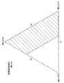
図2は、例えば、ある特定組成の酸化物膜作成(図4参照)のために、ある試薬調合比(図3)で析出させたときの析出温度と膜厚及び表面構造の関係を模式的に示す概念図である。なお析出時間は15時間とした。図3及び図4には、本発明において好ましい組成比範囲を斜線を付して示す。
温度領域を大まかに30℃未満、30〜40℃及び40℃を超える範囲にその析出物の粒子集合状態から分割すると、30℃未満での析出では数ナノメートルレベルの析出粒子の集合によって基材表面を整然と覆い、一層が形成されるとさらにその上に島状粒子集合体を形成しながら析出が進行していく。したがって膜は緻密で表面の凹凸も少ない。30〜40℃の温度領域になると数ナノメートル〜数十ナノメートルと粒子分布に幅が生じ、サイズ的に混在した粒子より構成されるようになる。これによって表面構造に凹凸が生じるようになる。本発明の目的とするリン酸カルシウム系化合物の核形成が誘起され易い構造とは、即ち、適度のOH基の密度分布からなり、リン酸カルシウム系化合物成分の過飽和状態が増大しやすく、かつ核形成のサイトを提供するような表面構造に相当する。さらに40℃を超えると、数十ナノメートル〜数百ナノメートルサイズの粒子が出現し、形成された膜に亀裂が入り易くなる。粒子径が大きくなることによって可視光波長領域の光を散乱するため膜が白濁して見える。表面構造は、顕微鏡レベルでラフになり,多孔質体に近い状態になる。
FIG. 2 schematically shows, for example, the relationship between the deposition temperature, film thickness, and surface structure when deposited at a certain reagent preparation ratio (FIG. 3) in order to create an oxide film having a specific composition (see FIG. 4). It is a conceptual diagram shown in FIG. The deposition time was 15 hours. In FIGS. 3 and 4, preferred composition ratio ranges in the present invention are indicated by hatching.
When the temperature region is roughly divided into a range of less than 30 ° C., 30-40 ° C., and more than 40 ° C. from the particle aggregation state of the precipitate, the precipitation at a temperature of less than 30 ° C. is caused by the aggregation of precipitated particles of several nanometers. When the surface is covered orderly and a single layer is formed, precipitation proceeds while further forming island-like particle aggregates thereon. Therefore, the film is dense and there are few surface irregularities. In the temperature range of 30 to 40 ° C., the particle distribution has a width of several nanometers to several tens of nanometers, and is composed of particles mixed in size. This causes irregularities in the surface structure. The structure in which nucleation of the calcium phosphate compound targeted by the present invention is likely to be induced means that it has a moderate density distribution of OH groups, the supersaturated state of the calcium phosphate compound component is likely to increase, and nucleation sites are formed. Corresponds to the surface structure as provided. When the temperature exceeds 40 ° C., particles having a size of several tens of nanometers to several hundreds of nanometers appear, and the formed film is easily cracked. As the particle size increases, light in the visible light wavelength region is scattered, and the film appears cloudy. The surface structure becomes rough at the microscopic level and becomes close to a porous body.
以上、3領域の表面構造のアウトラインを比較したが、30℃未満及び30〜40℃領域が膜の付着性、機械的強度といった観点から好ましい。特に、30〜40℃領域で形成された膜の表面構造は、リン酸カルシウム系化合物が形成され易い。すなわち生体活性を有する表面構造である。またこれらの製造上のパラメーターを有効にコントロールすることによって最適な表面構造を再現性良く形成することが可能となる。
なお40℃を超えて形成された金属酸化物膜であっても本発明の目的を著しく損なうものではなく、そのまま使用に供することができる。
また上記のような温度は、酸化物組成、金属種、フッ素捕捉剤の濃度、原料水溶液濃度等に応じて適宜変化するが、いずれにせよ、100℃以下の温度であれば、本発明の目的とする金属酸化物を形成することができる。
As described above, the outlines of the surface structures of the three regions were compared. However, the regions of less than 30 ° C. and the region of 30 to 40 ° C. are preferable from the viewpoints of film adhesion and mechanical strength. In particular, the surface structure of the film formed in the 30 to 40 ° C. region is likely to form a calcium phosphate compound. That is, the surface structure has bioactivity. Further, by effectively controlling these manufacturing parameters, it is possible to form an optimum surface structure with good reproducibility.
Even a metal oxide film formed at a temperature exceeding 40 ° C. does not significantly impair the object of the present invention, and can be used as it is.
The temperature as described above is appropriately changed according to the oxide composition, the metal species, the concentration of the fluorine scavenger, the concentration of the raw material aqueous solution, and the like. A metal oxide can be formed.
(2)複合金属酸化物膜の生成
本発明における膜形成のための水溶液中での反応は下記のように表現される。
例えば、フルオロ金属アンモニウム錯体を純水中に溶解させると、下記の反応式(1)により溶解する。
(2) Formation of Composite Metal Oxide Film A reaction in an aqueous solution for film formation in the present invention is expressed as follows.
For example, when a fluoro metal ammonium complex is dissolved in pure water, it is dissolved by the following reaction formula (1).
(化1)
(NH4)XMeFy + H2O =xNH4 + + MeFy x- + H2O (1)
(Chemical formula 1)
(NH 4 ) X MeF y + H 2 O = xNH 4 + + MeF y x- + H 2 O (1)
この状態に、フッ素捕捉剤として酸化ホウ素を添加すると、下記の反応式(3)により、水溶液中のフッ素が減少するため、下記の反応式(2)の化学平衡がフッ素を生成する右方向にシフトし、金属酸化物が生成し、基材表面に析出する。 In this state, when boron oxide is added as a fluorine scavenger, fluorine in the aqueous solution is reduced according to the following reaction formula (3). Therefore, the chemical equilibrium of the following reaction formula (2) is directed to the right to generate fluorine. The metal oxide is generated and deposited on the surface of the substrate.
(化2)
MeFy x- + (y-x)/2H2O = MeO(y-x)/2↓+ yF― + (y-x)H+ (2)
B2O3 + 8F― + 6H+ =2BF4― + 3H2O (3)
(Chemical formula 2)
MeF y x- + (yx) / 2H 2 O = MeO (yx) / 2 ↓ + yF― + (yx) H + (2)
B 2 O 3 + 8F- + 6H + = 2BF 4 - + 3H 2 O (3)
本発明を生体内材料(インプラント)に応用する場合は、膜付着力、膜の機械的耐性及び生体活性強度を膜組成によってコントロールしなければならない。一方、特に本製造方法によれば金属酸化物を数種類複合化することで、一種類の酸化物の場合よりも生体活性が向上することが期待できる。
例えば酸化チタン、酸化ジルコニウム及び二酸化ケイ素はそれぞれ特殊な製法によればそれぞれ生体活性を示すが、これらを複合化することによりこれらの相乗効果により、生体活性の高さが大幅に増強されることを本発明者は見出し、そして更に検討した結果、これら酸化物複合体の析出条件を特定し、試薬調合組成比と生成された膜組成、膜特性との関連性について検討し、生体適合性が強く現れる組成領域を特定できた。
一般に、(NH4)2SiF6―(NH4)2TiF6―(NH4)2ZrF6―B2O3―H2O系では、上述の式において、Me=Si,Ti,Zr
のそれぞれにおいて反応式が相互作用しながら進行し、それぞれの酸化物或いは複合酸化物は酸化物複合体として共析する。
即ち同時に表現すると、
When the present invention is applied to a biomaterial (implant), the film adhesion, the mechanical resistance of the film and the bioactive strength must be controlled by the film composition. On the other hand, in particular, according to the present production method, it can be expected that the bioactivity is improved by combining several kinds of metal oxides as compared with the case of one kind of oxide.
For example, titanium oxide, zirconium oxide, and silicon dioxide each show bioactivity according to a special production method. By combining these, the high bioactivity is greatly enhanced by their synergistic effect. As a result of finding and further studying the present inventor, the present inventors have identified the conditions for depositing these oxide complexes, examined the relationship between the composition ratio of the reagent preparation, the generated film composition, and the film characteristics, and has strong biocompatibility. The composition region that appears was able to be identified.
In general, (NH 4 ) 2 SiF 6- (NH 4 ) 2 TiF 6- (NH 4 ) 2 ZrF 6 --B 2 O 3 --H 2 O system, Me = Si, Ti, Zr
In each of the above, the reaction formula proceeds while interacting, and each oxide or composite oxide co-deposits as an oxide composite.
In other words,
(化3)
(NH4)2(Si,Ti,Zr)F6 + H2O = 2NH4 + + (Si,Ti,Zr)F6 2- + H2O
(Si,Ti,Zr)F6 2- + 2H2O = (Si,Ti,Zr)O2 ↓ + 6F- + 4H+
B2O3 + 8F― + 6H+ =2BF4― + 3H2O
(Chemical formula 3)
(NH 4 ) 2 (Si, Ti, Zr) F 6 + H 2 O = 2NH 4 + + (Si, Ti, Zr) F 6 2- + H 2 O
(Si, Ti, Zr) F 6 2- + 2H 2 O = (Si, Ti, Zr) O 2 ↓ + 6F - + 4H +
B 2 O 3 + 8F- + 6H + = 2BF 4 - + 3H 2 O
これらの共析物の化学組成は、反応前の試薬調整比と必ずしも一致するとは限らない。
具体的な膜組成は,上記したように、SiO2 0〜1.0:TiO2 0〜0.6:ZrO2 0〜1.0のモル比範囲内にあり、これらの酸化ジルコニウムの一部は、酸化ニオブ、酸化スズ、酸化タンタル、酸化ガリウム、酸化ハフニウム及びアルミナ等で置換されてもよい。
原料として6フッ化珪素アンモニウム,6フッ化チタンアンモニウム、6フッ化ジルコニウムアンモニウム(それぞれASF,ATF,AZFと略記)を用いたとき特に生体活性強度が強く発現し、機械的化学的耐性が良好な複合成分比を図3に、そして図4に対応析出酸化物複合膜の組成比の領域を示している。
なおSi,Ti,Zr系酸化物複合体が形成されれば、図3の最適領域は適宜変更することができる。
The chemical composition of these eutectoids does not necessarily coincide with the reagent adjustment ratio before the reaction.
Specific film composition, as described above, SiO 2 0~1.0: TiO 2 0~0.6 : is in the molar ratio range of
When using ammonium hexafluoride, ammonium hexafluoride and zirconium ammonium hexafluoride (abbreviated as ASF, ATF, AZF, respectively) as raw materials, bioactive strength is particularly strong and mechanical and chemical resistance is good. FIG. 3 shows the composite component ratio and FIG. 4 shows the region of the composition ratio of the corresponding deposited oxide composite film.
If the Si, Ti, Zr-based oxide composite is formed, the optimum region in FIG. 3 can be changed as appropriate.
以上のように形成された金属酸化物下地層と基材との接着強度は、通常5MP以上であり、非常に基材との接着性に優れており、このため使用時生体内で膜が脱落することもない。 The adhesive strength between the metal oxide underlayer formed as described above and the base material is usually 5 MP or more, and the adhesive strength to the base material is very excellent. I don't have to.
以下、本発明を実施例により説明するが、本発明はこれらの実施例に何ら限定されるものではない。
なお、下記の実施例で測定する基材と多層膜との接着強度は、基材の表と裏にステンレス治具を接着剤を用いて接着し、上下に引っ張り力を印加したときの破断応力で示す。
EXAMPLES Hereinafter, although an Example demonstrates this invention, this invention is not limited to these Examples at all.
In addition, the adhesive strength between the base material and the multilayer film measured in the following examples is the breaking stress when a stainless steel jig is bonded to the front and back of the base material using an adhesive and a tensile force is applied vertically. It shows with.
ポリメチルメタクリレート(PMMA)製骨形状モデル(サイズ120×30×30mm)を用意した。このサンプルの表面をプラズマ照射装置(ヤマト科学製PB1000S)によって、200Wで4分間プラズマ照射を行い、表面処理を行った。
その後純水中に6フッ化珪素アンモニウム、6フッ化チタンアンモニウム及び6フッ化ジルコニウムアンモニウムをそれぞれ30g、8g、2g加え、更に酸化ホウ素30gを加えた水溶液を攪拌しながら純水を加えて1リットルに調製し完全に溶解させた。この水溶液の温度を40℃に保ちながら、モデルサンプルをプラズマ照射後ただちにこの水溶液に浸漬した。
20時間浸漬後サンプルを処理液から取り出し純水によって洗浄し、乾燥させた。
その後、別の容器に6フッ化チタンアンモニウム2g及び硝酸カルシウム0.5g入れ、純水中に溶解させ、更に1リットルになるまで純水を加え40℃としたものを用意しておき、酸化ホウ素15gを添加混合溶解させ、これに先に洗浄乾燥させたサンプルを6時間浸漬放置した。放置後、サンプルを取り出して純水にて洗浄乾燥させ、サンプル上に2層膜を形成した。同様にPMMA基板(60×60×1mm)をモデルと同じ条件下で処理し、同じく2層膜を形成した。これによって測定した形成膜の基板に対する接着強度は8.0MPであった。
その後、サンプルの生体親和性を確認するために、浸漬するための擬似体液を調製した。
A bone shape model (size 120 × 30 × 30 mm) made of polymethyl methacrylate (PMMA) was prepared. The surface of this sample was subjected to surface treatment by plasma irradiation at 200 W for 4 minutes using a plasma irradiation apparatus (PB1000S manufactured by Yamato Kagaku).
Then, 30 g, 8 g and 2 g of ammonium hexafluoride, ammonium hexafluoride and zirconium ammonium hexafluoride were added to pure water, respectively, and 1 liter of pure water was added while stirring an aqueous solution containing 30 g of boron oxide. And completely dissolved. While maintaining the temperature of this aqueous solution at 40 ° C., the model sample was immersed in this aqueous solution immediately after plasma irradiation.
After immersion for 20 hours, the sample was taken out of the treatment solution, washed with pure water, and dried.
Then, 2 g of ammonium hexafluoride and 0.5 g of calcium nitrate were put in another container, dissolved in pure water, and further prepared with pure water added to 40 ° C. until it became 1 liter. Boron oxide 15 g was added, mixed and dissolved, and the sample previously washed and dried was immersed in the sample for 6 hours. After standing, the sample was taken out, washed with pure water and dried to form a two-layer film on the sample. Similarly, a PMMA substrate (60 × 60 × 1 mm) was treated under the same conditions as the model, and a two-layer film was formed in the same manner. The adhesion strength of the formed film thus measured to the substrate was 8.0 MP.
Thereafter, in order to confirm the biocompatibility of the sample, a simulated body fluid for immersion was prepared.
−擬似体液−
まず蒸留水700mlにNaClを7.996g、NaHCO3を0.350g、KClを0.224g、K2HPO4・3H2Oを0.228g、MgCl2/6H2Oを0.306g、1M塩酸を35ml、CaCl2を0.278g、Na2SO4を0.072g、トリスヒドロキシメチルアミノメタンを6.058g溶解させ、36.5℃でpHが7.4となるように、1M塩酸で調製し、その後蒸留水を加えて溶液全体を1リットルとした。
-Simulated body fluid-
First, 7.996 g of NaCl, 0.350 g of NaHCO 3 , 0.224 g of KCl, 0.228 g of K 2 HPO 4 .3H 2 O, 0.306 g of MgCl 2 / 6H 2 O, 1M hydrochloric acid in 700 ml of distilled water 35 ml, CaCl 2 0.278 g, Na 2 SO 4 0.072 g, and trishydroxymethylaminomethane 6.058 g are dissolved and prepared with 1M hydrochloric acid so that the pH is 7.4 at 36.5 ° C. Then, distilled water was added to make the whole solution 1 liter.
上記の擬似体液中に、先に2層膜を形成したモデルサンプルを浸漬し、2週間保持した。その後モデルを擬似体液中から取り出して表面に形成された薄膜のX線回折測定を実施した。浸漬一週間後にヒドロキシアパタイト類に帰属される回折ピークが観察され、二週間後にそのピーク強度が増大したことから、モデル表面におけるヒドロキシアパタイト類の生成が確認された。 The model sample in which the two-layer film was previously formed was immersed in the above simulated body fluid and held for 2 weeks. Thereafter, the model was taken out from the simulated body fluid and X-ray diffraction measurement was performed on the thin film formed on the surface. A diffraction peak attributed to hydroxyapatites was observed after one week of immersion, and the peak intensity increased after two weeks, confirming the formation of hydroxyapatites on the model surface.
ポリエチレン製骨形状モデル(サイズ120×30×30mm)を用意した。
この表面にプラズマ照射装置(ヤマト科学製PB1000S)によって200W、4分プラズマ照射を行い、表面処理を行なった。
その後純水中に6フッ化珪素アンモニウム、6フッ化チタンアンモニウム及び6フッ化ジルコニウムアンモニウムをそれぞれ30g、8g、2g加え,更に酸化ホウ素30g加えた水溶液を攪拌しながら純水を加えて1リットルに調製し、完全に溶解させた。この水溶液を40℃に保ちながら、モデルサンプルをこの水溶液に浸漬した。20時間浸漬後サンプルを処理液から取り出し純水によって洗浄し、乾燥させた。
その後、別の容器に6フッ化チタンアンモニウム3.5g及び硝酸カルシウム0.5gを純水中に加え完全に溶解後、酸化ホウ素30gを添加しながら純水を加えて1リットルに調製し、完全に溶解した水溶液中に、上記サンプルを再度浸漬した。6時間後これを取り出して洗浄後乾燥させ、モデルサンプル上に2層膜を形成した。同様にポリエチレン基板(10×10×1mm)をモデルと同一条件で処理し、同じく2層膜を形成した。これによって測定した形成膜の基板に対する接着強度は、12MPであった。
実施例1と同じ擬似体液中に、2層膜を形成したモデルサンプルを浸漬し、10日間ほど保持した。その後モデルを擬似体液中から取り出して表面に形成された薄膜のX線回折測定を実施した。浸漬後3〜4日でヒドロキシアパタイト類に帰属される回折ピークが観察され、1週間後にそのピーク強度が増大したことからモデル表面におけるヒドロキシアパタイト類の生成が確認された。
A polyethylene bone shape model (size 120 × 30 × 30 mm) was prepared.
The surface was subjected to surface treatment by 200 W plasma irradiation for 4 minutes using a plasma irradiation apparatus (PB1000S manufactured by Yamato Kagaku).
Then, 30 g, 8 g and 2 g of ammonium hexafluoride, ammonium hexafluoride and zirconium ammonium hexafluoride were added to pure water, respectively, and pure water was added to 1 liter while stirring the aqueous solution containing 30 g of boron oxide. Prepared and completely dissolved. The model sample was immersed in this aqueous solution while keeping this aqueous solution at 40 ° C. After immersion for 20 hours, the sample was taken out of the treatment solution, washed with pure water, and dried.
Then, after adding 3.5 g of titanium hexafluoride and 0.5 g of calcium nitrate to pure water in a separate container and completely dissolving, add pure water while adding 30 g of boron oxide to prepare 1 liter. The sample was immersed again in the aqueous solution dissolved in the solution. After 6 hours, this was taken out, washed and dried to form a two-layer film on the model sample. Similarly, a polyethylene substrate (10 × 10 × 1 mm) was treated under the same conditions as the model to form a two-layer film. The adhesion strength of the formed film to the substrate measured in this way was 12 MP.
The model sample in which the two-layer film was formed was immersed in the same simulated body fluid as in Example 1, and held for about 10 days. Thereafter, the model was taken out from the simulated body fluid and X-ray diffraction measurement was performed on the thin film formed on the surface. A diffraction peak attributed to hydroxyapatites was observed 3 to 4 days after immersion, and the peak intensity increased after 1 week, confirming the formation of hydroxyapatites on the model surface.
ポリメチルメタクリレート(PMMA)製骨形状モデル(サイズ120×30×30mm)にアクリル系ハードコートを施したものを用意した。このサンプルの表面に親水性を持たせるために、8モル/リットルの濃度の水酸化ナトリウム水溶液を80℃に保ち、このサンプルを45分間浸漬した。
その後純水中に6フッ化珪素アンモニウム、6フッ化チタンアンモニウム及び6フッ化ジルコニウムアンモニウムをそれぞれ30g、8g、2g加え,更に酸化ホウ素30gを加えた水溶液を攪拌しながら純水を加えて1リットルに調製し、完全に溶解させた。この水溶液を40℃に保ちながら、モデルサンプルをこの水溶液に浸漬した。20時間浸漬後サンプルを処理液から取り出し純水によって洗浄し、乾燥させた。
その後、別の容器に6フッ化チタンアンモニウム3.5g及び硝酸カルシウム0.5gを純水中に加え完全に溶解後、酸化ホウ素30gを添加しながら純水を加えて1リットルに調製し、完全に溶解した水溶液中に、上記サンプルを再度浸漬した。6時間後これを取り出して洗浄後、70℃で2時間乾燥させ、モデルサンプル上に2層膜を形成した。同様にPMMA基板(アクリル系ハードコート付基板)(10×10×1mm)をモデルと同じ条件下で処理し、同じく2層膜を形成した。これによって測定した形成膜の基板に対する接着強度は、5.5MPであった。
実施例1と同じ擬似体液中に、2層膜を形成したモデルサンプルを浸漬し、10日間ほど保持した。その後モデルを擬似体液中から取り出して表面に形成された薄膜のX線回折測定を実施した。浸漬後3〜4日でヒドロキシアパタイト類に帰属される回折ピークが観察され、1週間後にそのピーク強度が増大したことからモデル表面におけるヒドロキシアパタイト類の生成が確認された。
A bone shape model (size 120 × 30 × 30 mm) made of polymethyl methacrylate (PMMA) was prepared by applying an acrylic hard coat. In order to give hydrophilicity to the surface of this sample, an aqueous 8 mol / liter sodium hydroxide solution was kept at 80 ° C., and this sample was immersed for 45 minutes.
Then, 30 g, 8 g, and 2 g of ammonium hexafluoride, ammonium hexafluoride, and ammonium hexafluoride were added to pure water, respectively, and 1 liter of pure water was added while stirring the aqueous solution containing 30 g of boron oxide. And dissolved completely. The model sample was immersed in this aqueous solution while keeping this aqueous solution at 40 ° C. After immersion for 20 hours, the sample was taken out of the treatment solution, washed with pure water, and dried.
Then, after adding 3.5 g of titanium hexafluoride and 0.5 g of calcium nitrate to pure water in a separate container and completely dissolving, add pure water while adding 30 g of boron oxide to prepare 1 liter. The sample was immersed again in the aqueous solution dissolved in the solution. After 6 hours, this was taken out, washed, and dried at 70 ° C. for 2 hours to form a two-layer film on the model sample. Similarly, a PMMA substrate (substrate with acrylic hard coat) (10 × 10 × 1 mm) was treated under the same conditions as the model to form a two-layer film. The adhesion strength of the formed film to the substrate measured in this way was 5.5 MP.
The model sample in which the two-layer film was formed was immersed in the same simulated body fluid as in Example 1, and held for about 10 days. Thereafter, the model was taken out from the simulated body fluid and X-ray diffraction measurement was performed on the thin film formed on the surface. A diffraction peak attributed to hydroxyapatites was observed 3 to 4 days after immersion, and the peak intensity increased after 1 week, confirming the formation of hydroxyapatites on the model surface.
基材としてポリエチレン製平板(100×100×1mm)を用いた以外は、実施例2と同様の操作を行った。2層膜形成後の基材を実施例1と同じ擬似体液中に浸漬したところ、1週間後にヒドロキシアパタイト類に帰属されるX線回折ピークが観察され、2週間後にそのピーク強度が増大したことから、基材表面におけるヒドロキシアパタイト類の生成が確認された。 The same operation as in Example 2 was performed except that a flat plate made of polyethylene (100 × 100 × 1 mm) was used as the substrate. When the base material after forming the two-layer film was immersed in the same simulated body fluid as in Example 1, an X-ray diffraction peak attributed to hydroxyapatites was observed after 1 week, and the peak intensity increased after 2 weeks. From this, it was confirmed that hydroxyapatites were formed on the substrate surface.
基材としてポリエチレン製織布(100×100×0.5mm)を用いた以外は、実施例2と同様の操作を行った。2層膜形成後の基材を実施例1と同じ擬似体液中に浸漬したところ、1週間後にヒドロキシアパタイト類に帰属されるX線回折ピークが観察され、2週間後にそのピーク強度が増大したことから、基材表面におけるヒドロキシアパタイト類の生成が確認された。 The same operation as in Example 2 was performed except that a polyethylene woven fabric (100 × 100 × 0.5 mm) was used as the substrate. When the base material after forming the two-layer film was immersed in the same simulated body fluid as in Example 1, an X-ray diffraction peak attributed to hydroxyapatites was observed after 1 week, and the peak intensity increased after 2 weeks. From this, it was confirmed that hydroxyapatites were formed on the substrate surface.
基材としてウレタンフォーム(100×100×1mm)を用いた以外は、実施例2と同様の操作を行った。2層膜形成後の基材を実施例1と同じ擬似体液中に浸漬したところ、1週間後にヒドロキシアパタイト類に帰属されるX線回折ピークが観察され、2週間後にそのピーク強度が増大したことから、基材表面におけるヒドロキシアパタイト類の生成が確認された。 The same operation as in Example 2 was performed except that urethane foam (100 × 100 × 1 mm) was used as the substrate. When the base material after forming the two-layer film was immersed in the same simulated body fluid as in Example 1, an X-ray diffraction peak attributed to hydroxyapatites was observed after 1 week, and the peak intensity increased after 2 weeks. From this, it was confirmed that hydroxyapatites were formed on the substrate surface.
基材としてチタン合金(Ti-6Al-4V)製平板(100×100×1mm)を用い、更にプラズマ照射を行わなかった以外は、実施例2と同様の操作を行った。2層膜形成後の基材を実施例1と同じ擬似体液中に浸漬したところ、1週間後にヒドロキシアパタイト類に帰属されるX線回折ピークが観察され、2週間後にそのピーク強度が増大したことから、基材表面におけるヒドロキシアパタイト類の生成が確認された。 The same operation as in Example 2 was performed except that a flat plate (100 × 100 × 1 mm) made of a titanium alloy (Ti-6Al-4V) was used as a base material and plasma irradiation was not performed. When the base material after forming the two-layer film was immersed in the same simulated body fluid as in Example 1, an X-ray diffraction peak attributed to hydroxyapatites was observed after 1 week, and the peak intensity increased after 2 weeks. From this, it was confirmed that hydroxyapatites were formed on the substrate surface.
基材としてアルミナ製平板(100×100×1mm)を用い、実施例2と同様の操作を行った。2層膜形成後の基材を実施例1と同じ擬似体液中に浸漬したところ、1週間後にヒドロキシアパタイト類に帰属されるX線回折ピークが観察され、2週間後にそのピーク強度が増大したことから、基材表面におけるヒドロキシアパタイト類の生成が確認された。 The same operation as in Example 2 was performed using an alumina flat plate (100 × 100 × 1 mm) as a substrate. When the base material after forming the two-layer film was immersed in the same simulated body fluid as in Example 1, an X-ray diffraction peak attributed to hydroxyapatites was observed after 1 week, and the peak intensity increased after 2 weeks. From this, it was confirmed that hydroxyapatites were formed on the substrate surface.
ポリエチレン製骨形状モデル(サイズ120×30×30mm)を用意した。
この表面にプラズマ照射装置(ヤマト科学製PB1000S)によって200Wで4分プラズマ照射を行い、表面処理を行なった。
その後純水中に6フッ化珪素アンモニウム、6フッ化チタンアンモニウムをそれぞれ19.1g、5.6g加え、更に酸化ホウ素30g加えた水溶液を攪拌しながら純水を加えて1リットルに調製し、完全に溶解させた。この水溶液を40℃に保ちながら、モデルサンプルをこの水溶液に浸漬した。20時間浸漬後サンプルを処理液から取り出し純水によって洗浄し乾燥させた。その後、別の容器に6フッ化チタンアンモニウム3.5g及び硝酸カルシウム0.5gを純水中に加え完全に溶解後、酸化ホウ素30gを添加しながら純水を加えて1リットルに調製し、完全に溶解した水溶液中に、上記サンプルを再度浸漬した。6時間後これを取り出して洗浄後乾燥させ、モデルサンプル上に2層膜を形成した。
一方ポリエチレン基板(10×10×1mm)を上記と全く同様に処理して、形成された膜の基板に対する接着強度を測定したところ8MPであった。
実施例1と同じ擬似体液中に、2層膜を形成したモデルサンプルを浸漬し、10日ほど保持した。その後モデルを擬似体液中から取り出して表面に形成された薄膜のX線回折測定を実施した。浸漬後3〜4日でヒドロキシアパタイト類に帰属される回折ピークが観察され、1週間後にそのピーク強度が増大したことからモデル表面におけるヒドロキシアパタイト類の生成が確認された。
A polyethylene bone shape model (size 120 × 30 × 30 mm) was prepared.
This surface was subjected to surface treatment by plasma irradiation at 200 W for 4 minutes with a plasma irradiation apparatus (PB1000S manufactured by Yamato Kagaku).
Thereafter, 19.1 g and 5.6 g of silicon hexafluoride ammonium and titanium hexafluoride ammonium were added to pure water, respectively, and pure water was added to the liter while stirring the aqueous solution containing 30 g of boron oxide. Dissolved in. The model sample was immersed in this aqueous solution while keeping this aqueous solution at 40 ° C. After immersion for 20 hours, the sample was taken out of the treatment solution, washed with pure water and dried. Then, after adding 3.5 g of titanium hexafluoride and 0.5 g of calcium nitrate to pure water in a separate container and completely dissolving, add pure water while adding 30 g of boron oxide to prepare 1 liter. The sample was immersed again in the aqueous solution dissolved in the solution. After 6 hours, this was taken out, washed and dried to form a two-layer film on the model sample.
On the other hand, a polyethylene substrate (10 × 10 × 1 mm) was treated in exactly the same manner as described above, and the adhesion strength of the formed film to the substrate was measured.
A model sample in which a two-layer film was formed was immersed in the same simulated body fluid as Example 1, and held for about 10 days. Thereafter, the model was taken out from the simulated body fluid and X-ray diffraction measurement was performed on the thin film formed on the surface. A diffraction peak attributed to hydroxyapatites was observed 3 to 4 days after immersion, and the peak intensity increased after 1 week, confirming the formation of hydroxyapatites on the model surface.
ポリエチレン製骨形状モデル(120×30×30mm)を用意し、下地層形成用の処理液の調製組成を6フッ化珪素アンモニウム、6フッ化チタンアンモニウムそれぞれ、17.4g、0.39gとした以外は実施例9と全く同一条件で処理し、2層膜を形成した。
同様にポリエチレン基板(10×10×1mm)を同じ条件下で処理し、同じく2層膜を形成した。これによって測定した形成膜の基板に対する接着強度は、7.5MPであった。
実施例1と同じ擬似体液中に、2層膜を形成したモデルサンプルを浸漬し、10日ほど保持した。その後モデルを擬似体液中から取り出して表面に形成された薄膜のX線回折測定を実施した。浸漬後3〜4日でヒドロキシアパタイト類に帰属される回折ピークが観察され、1週間後にそのピーク強度が増大したことからモデル表面におけるヒドロキシアパタイト類の生成が確認された。
A polyethylene bone shape model (120 × 30 × 30 mm) was prepared, and the preparation composition of the treatment liquid for forming the underlayer was changed to 17.4 g and 0.39 g of ammonium hexafluoride and titanium ammonium fluoride, respectively. Were processed under exactly the same conditions as in Example 9 to form a two-layer film.
Similarly, a polyethylene substrate (10 × 10 × 1 mm) was treated under the same conditions to form a two-layer film. The adhesion strength of the formed film to the substrate measured by this was 7.5 MP.
A model sample in which a two-layer film was formed was immersed in the same simulated body fluid as Example 1, and held for about 10 days. Thereafter, the model was taken out from the simulated body fluid and X-ray diffraction measurement was performed on the thin film formed on the surface. A diffraction peak attributed to hydroxyapatites was observed 3 to 4 days after immersion, and the peak intensity increased after 1 week, confirming the formation of hydroxyapatites on the model surface.
ポリエチレン製骨形状モデル(120×30×30mm)を用意し、下地層形成用の処理液の調製組成を6フッ化珪素アンモニウム、6フッ化ジルコニウムアンモニウムそれぞれ、14.4g、4.57gとした以外は実施例9と全く同一条件で処理し、2層膜を形成した。
同様にポリエチレン基板(10×10×1mm)を同じ条件で処理し、同じく2層膜を形成した。これによって測定した形成膜の基板に対する接着強度は、6MPであった。
実施例1と同じ擬似体液中に,2層膜を形成したモデルサンプルを浸漬し、10日ほど保持した。その後モデルを擬似体液中から取り出して表面に形成された薄膜のX線回折測定を実施した。浸漬後3〜4日でヒドロキシアパタイト類のX線回折ピークが観察され、1週間後にそのピーク強度が増大したことからモデル表面におけるヒドロキシアパタイト類の生成が確認された。
A polyethylene bone shape model (120 × 30 × 30 mm) was prepared, and the preparation composition of the treatment liquid for forming the underlayer was changed to 14.4 g and 4.57 g of ammonium hexafluoride and zirconium ammonium hexafluoride, respectively. Were processed under exactly the same conditions as in Example 9 to form a two-layer film.
Similarly, a polyethylene substrate (10 × 10 × 1 mm) was treated under the same conditions to form a two-layer film. The adhesion strength of the formed film to the substrate measured in this way was 6 MP.
In the same simulated body fluid as in Example 1, the model sample with the two-layer film formed was immersed and held for about 10 days. Thereafter, the model was taken out from the simulated body fluid and X-ray diffraction measurement was performed on the thin film formed on the surface. X-ray diffraction peaks of hydroxyapatites were observed 3 to 4 days after immersion, and the peak intensity increased after 1 week, confirming the formation of hydroxyapatites on the model surface.
ポリエチレン製骨形状モデル(120×30×30mm)を用意し、下地層形成用の処理液の調整組成を6フッ化チタンアンモニウム、6フッ化ジルコニウムアンモニウムそれぞれ、0.39g、11.7gとした以外は実施例9と全く同一条件で処理し、2層膜を形成した。
同様にポリエチレン基板(10×10×1mm)を同じ条件で処理し、同じく2層膜を形成した。これによって測定した形成膜の基板に対する接着強度は、5.5MPであった。
実施例1と同じ擬似体液中に、2層膜を形成したモデルサンプルを浸漬し、10日ほど保持した。その後モデルを擬似体液中から取り出して表面に形成された薄膜のX線回折測定を実施した。浸漬後3〜4日でヒドロキシアパタイト類のX線回折ピークが観察され、1週間後にそのピーク強度が増大したことからモデル表面におけるヒドロキシアパタイト類の生成が確認された。
A polyethylene bone shape model (120 × 30 × 30 mm) was prepared, and the adjustment composition of the treatment liquid for forming the underlayer was set to 0.39 g and 11.7 g of ammonium hexafluoride and zirconium ammonium hexafluoride, respectively. Were processed under exactly the same conditions as in Example 9 to form a two-layer film.
Similarly, a polyethylene substrate (10 × 10 × 1 mm) was treated under the same conditions to form a two-layer film. The adhesion strength of the formed film to the substrate measured in this way was 5.5 MP.
A model sample in which a two-layer film was formed was immersed in the same simulated body fluid as Example 1, and held for about 10 days. Thereafter, the model was taken out from the simulated body fluid and X-ray diffraction measurement was performed on the thin film formed on the surface. X-ray diffraction peaks of hydroxyapatites were observed 3 to 4 days after immersion, and the peak intensity increased after 1 week, confirming the formation of hydroxyapatites on the model surface.
ポリエチレン製骨形状モデル(120×30×30mm)を用意し、下地層形成用の処理液の調整組成を6フッ化チタンアンモニウム、6フッ化ジルコニウムアンモニウムそれぞれ、2.5g、9.0gとした以外は実施例9と全く同一条件で処理し、2層膜を形成した。
同様に、ポリエチレン基板(10×10×1mm)を同じ条件で処理し、同じく2層膜を形成した。これによって形成した2層膜の基板に対する接着強度は、6.8MPであった。
実施例1と同じ擬似体液中に、2層膜を形成したモデルサンプルを浸漬し、10日ほど保持した。その後モデルを擬似体液中から取り出して表面に形成された薄膜のX線回折測定を実施した。浸漬後3〜4日でヒドロキシアパタイトに帰属される回折ピークが観察され、1週間後にそのピーク強度が増大したことからモデル表面におけるヒドロキシアパタイト類の生成が確認された。
A polyethylene bone shape model (120 x 30 x 30 mm) was prepared, and the adjustment composition of the treatment liquid for forming the underlayer was 2.5 g and 9.0 g of ammonium hexafluoride and zirconium ammonium hexafluoride, respectively. Were processed under exactly the same conditions as in Example 9 to form a two-layer film.
Similarly, a polyethylene substrate (10 × 10 × 1 mm) was treated under the same conditions to form a two-layer film. The adhesive strength of the two-layer film formed thereby to the substrate was 6.8 MP.
A model sample in which a two-layer film was formed was immersed in the same simulated body fluid as Example 1, and held for about 10 days. Thereafter, the model was taken out from the simulated body fluid and X-ray diffraction measurement was performed on the thin film formed on the surface. A diffraction peak attributed to hydroxyapatite was observed 3 to 4 days after immersion, and the peak intensity increased after 1 week, confirming the formation of hydroxyapatite on the model surface.
ポリエチレン製骨形状モデル(120×30×30mm)を用意し、下地層形成用の処理液の調製組成を、6フッ化珪素アンモニウム、6フッ化チタンアンモニウム、6フッ化ジルコニウムアンモニウムそれぞれ、5.8g、13.4g、7.8gとした以外は実施例9と全く同一条件で処理し、2層膜を形成した。
同様にポリエチレン基板(10×10×1mm)を同一条件で処理し、同じく2層膜を形成した。こうして形成した2層膜の基板に対する接着強度は、10MPであった。
実施例1と同じ擬似体液中に、2層膜を形成したモデルサンプルを浸漬し、10日ほど保持した。その後モデルを擬似体液中から取り出して表面に形成された薄膜のX線回折測定を実施した。浸漬後3〜4日でヒドロキシアパタイト類に帰属される回折ピークが観察され、1週間後にそのピーク強度が増大したことからモデル表面におけるヒドロキシアパタイト類の生成が確認された。
A polyethylene bone shape model (120 × 30 × 30 mm) is prepared, and the preparation composition of the treatment liquid for forming the underlayer is 5.8 g of ammonium hexafluoride, ammonium hexafluoride, zirconium ammonium hexafluoride, respectively. , 13.4 g, and 7.8 g except for the same conditions as in Example 9 to form a two-layer film.
Similarly, a polyethylene substrate (10 × 10 × 1 mm) was treated under the same conditions to form a two-layer film. The adhesive strength of the two-layer film thus formed to the substrate was 10 MP.
A model sample in which a two-layer film was formed was immersed in the same simulated body fluid as Example 1, and held for about 10 days. Thereafter, the model was taken out from the simulated body fluid and X-ray diffraction measurement was performed on the thin film formed on the surface. A diffraction peak attributed to hydroxyapatites was observed 3 to 4 days after immersion, and the peak intensity increased after 1 week, confirming the formation of hydroxyapatites on the model surface.
ポリエチレン製骨形状モデル(サイズ120×30×30mm)を用意した。この表面にプラズマ照射装置(ヤマト科学製PB1000S)によって200Wで4分間プラズマ照射を行い、表面を前処理した。
その後純水中に6フッ化珪素アンモニウム、6フッ化チタンアンモニウム、6フッ化ジルコニウムアンモニウム、7フッ化ニオブアンモニウム、6フッ化スズアンモニウムをそれぞれ、30g、8g、1g、0.7g、0.4g加え、更に酸化ホウ素30gを加えた水溶液を攪拌しながら純水を加えて1リットルに調製し完全に溶解させた。この水溶液を40℃に保ちながら、モデルサンプルをプラズマ照射後直ちにこの水溶液に浸漬した。
20時間浸漬後サンプルを処理液から取り出し純水によって洗浄し、乾燥させた。その後、別の容器に6フッ化チタンアンモニウム3.5g及び硝酸カルシウム0.5gを純水中に加え完全に溶解後、酸化ホウ素30gを添加しながら純水を加えて1リットルに調製し、完全に溶解した水溶液中に上記サンプルを再度浸漬した。6時間後これを取り出して洗浄後乾燥させ、モデルサンプル上に2層膜を形成した。
一方ポリエチレン基板(10×10×1mm)を上記と全く同様に処理して、形成された膜の基板に対する接着強度を測定したところ10MPであった。
実施例1と同じ擬似体液中に、2層膜を形成したモデルサンプルを浸漬し10日ほど保持した。その後モデルを擬似体液中から取り出して表面に形成された薄膜のX線回折測定を実施した。浸漬後3〜4日でヒドロキシアパタイト類に帰属される回折ピークが観察され、1週間後にそのピーク強度が増大したことからモデル表面におけるヒドロキシアパタイト類の生成が確認された。
A polyethylene bone shape model (size 120 × 30 × 30 mm) was prepared. The surface was pretreated by performing plasma irradiation at 200 W for 4 minutes with a plasma irradiation apparatus (PB1000S manufactured by Yamato Kagaku).
Thereafter, 30 g, 8 g, 1 g, 0.7 g, and 0.4 g of ammonium hexafluoride, ammonium hexafluoride, ammonium ammonium fluoride, zirconium hexafluoride, ammonium niobium fluoride, and tin ammonium hexafluoride in pure water, respectively. In addition, pure water was added while stirring an aqueous solution to which 30 g of boron oxide was added, and the solution was completely dissolved by dissolving it in 1 liter. While maintaining this aqueous solution at 40 ° C., the model sample was immersed in this aqueous solution immediately after plasma irradiation.
After immersion for 20 hours, the sample was taken out of the treatment solution, washed with pure water, and dried. Then, after adding 3.5 g of titanium hexafluoride and 0.5 g of calcium nitrate to pure water in a separate container and completely dissolving, add pure water while adding 30 g of boron oxide to prepare 1 liter. The sample was immersed again in the aqueous solution dissolved in the solution. After 6 hours, this was taken out, washed and dried to form a two-layer film on the model sample.
On the other hand, a polyethylene substrate (10 × 10 × 1 mm) was treated in exactly the same manner as described above, and the adhesion strength of the formed film to the substrate was measured to be 10 MP.
In the same simulated body fluid as Example 1, the model sample in which the two-layer film was formed was immersed and held for about 10 days. Thereafter, the model was taken out from the simulated body fluid and X-ray diffraction measurement was performed on the thin film formed on the surface. A diffraction peak attributed to hydroxyapatites was observed 3 to 4 days after immersion, and the peak intensity increased after 1 week, confirming the formation of hydroxyapatites on the model surface.
ポリエチレン製骨形状モデル(サイズ120×30×30mm)を用意した。
この表面にプラズマ照射装置(ヤマト科学製PB1000S)によって200Wで4分間プラズマ照射を行い、表面処理を行なった。
その後純水中に6フッ化珪素アンモニウム、6フッ化チタンアンモニウム及び6フッ化ジルコニウムアンモニウムをそれぞれ30g、8g、2g加え、更に酸化ホウ素30gを加えた水溶液を攪拌しながら純水を加えて1リットルに調製し、完全に溶解させた。この水溶液を40℃に保ちながら、モデルサンプルをこの水溶液に浸漬した。20時間浸漬後サンプルを処理液から取り出し純水によって洗浄し、乾燥させた。
その後別の容器に6フッ化チタンアンモニウム、6フッ化ジルコニウムアンモニウム、7フッ化タンタルアンモニウムをそれぞれ3.5g、0.5g、0.8g及び硝酸カルシウム0.5gを純水中に加え完全に溶解後、酸化ホウ素30gを添加しながら純水を加えて1リットルに調製し、完全に溶解した水溶液中に、上記サンプルを再度浸漬した。6時間後これを取り出して洗浄後乾燥させ、モデルサンプル上に2層膜を形成した。同様にポリエチレン基板(10×10×1mm)をモデルと同一条件で処理し、同じく2層膜を形成した。これによって測定した形成膜の基板に対する接着強度は、7.5MPであった。
実施例1と同じ擬似体液中に、2層膜を形成したモデルサンプルを浸漬し、10日間ほど保持した。その後モデルを擬似体液中から取り出して表面に形成された薄膜のX線回折測定を実施した。浸漬後3〜4日でヒドロキシアパタイト類に帰属される回折ピークが観察され、1週間後にそのピーク強度が増大したことからモデル表面におけるヒドロキシアパタイト類の生成が確認された。
A polyethylene bone shape model (size 120 × 30 × 30 mm) was prepared.
This surface was subjected to surface treatment by plasma irradiation at 200 W for 4 minutes with a plasma irradiation apparatus (PB1000S manufactured by Yamato Kagaku).
Then, 30 g, 8 g and 2 g of ammonium hexafluoride, ammonium hexafluoride and zirconium ammonium hexafluoride were added to pure water, respectively, and 1 liter of pure water was added while stirring an aqueous solution containing 30 g of boron oxide. And dissolved completely. The model sample was immersed in this aqueous solution while keeping this aqueous solution at 40 ° C. After immersion for 20 hours, the sample was taken out of the treatment solution, washed with pure water, and dried.
Then, 3.5g, 0.5g, 0.8g of ammonium hexafluoride, ammonium hexafluoride, ammonium hexafluoride and 0.5g of calcium nitrate were added to pure water and dissolved completely in another container. Thereafter, pure water was added while adding 30 g of boron oxide to prepare 1 liter, and the sample was immersed again in a completely dissolved aqueous solution. After 6 hours, this was taken out, washed and dried to form a two-layer film on the model sample. Similarly, a polyethylene substrate (10 × 10 × 1 mm) was treated under the same conditions as the model to form a two-layer film. The adhesion strength of the formed film to the substrate measured by this was 7.5 MP.
The model sample in which the two-layer film was formed was immersed in the same simulated body fluid as in Example 1, and held for about 10 days. Thereafter, the model was taken out from the simulated body fluid and X-ray diffraction measurement was performed on the thin film formed on the surface. A diffraction peak attributed to hydroxyapatites was observed 3 to 4 days after immersion, and the peak intensity increased after 1 week, confirming the formation of hydroxyapatites on the model surface.
(被覆層の接着強度)
基材としてポリエチレン製平板(10.0mm×10.0mm×1.0mm)を用意した。この表面にプラズマ照射装置(ヤマト科学製PB1000S)によって200Wで4分間プラズマ照射を行い、表面処理を行なった。
その後純水中に6フッ化珪素アンモニウム、6フッ化チタンアンモニウム及び6フッ化ジルコニウムアンモニウムをそれぞれ30g、8g、2g加え、更に酸化ホウ素30g加えた水溶液を攪拌しながら純水を加えて1リットルに調製し、完全に溶解させた。この水溶液を40℃に保ちながら、基材をこの水溶液に浸漬した。20時間浸漬後サンプルを処理液から取り出し純水によって洗浄し、乾燥させた。
その後、別の容器に6フッ化チタンアンモニウム3.5g及び硝酸カルシウム0.5gを純水中に加え完全に溶解後、酸化ホウ素30gを添加しながら純水を加えて1リットルに調製し、完全に溶解した水溶液中に、上記基材を再度浸漬した。6時間後これを取り出して洗浄後乾燥させ、基材上に被覆層を形成した。
基材の表と裏に実施例1と同様にステンレス治具を接着剤を用いて接着し、上下に引っ張り力を印加したときの破断応力を測定した。このときの破断応力は、5サンプルの平均で13.09MPであった。破断面を観察すると、下地層と基材の間で破断した部分と、下地層と被覆層の間で破断した部分が混在していた。
(Adhesive strength of coating layer)
A polyethylene flat plate (10.0 mm × 10.0 mm × 1.0 mm) was prepared as a substrate. This surface was subjected to surface treatment by plasma irradiation at 200 W for 4 minutes with a plasma irradiation apparatus (PB1000S manufactured by Yamato Kagaku).
Then, 30 g, 8 g and 2 g of ammonium hexafluoride, ammonium hexafluoride and zirconium ammonium hexafluoride were added to pure water, respectively, and 30 g of boron oxide was added to the liter to add 1 liter of pure water while stirring. Prepared and completely dissolved. The substrate was immersed in this aqueous solution while keeping this aqueous solution at 40 ° C. After immersion for 20 hours, the sample was taken out of the treatment solution, washed with pure water, and dried.
Then, after adding 3.5 g of titanium hexafluoride and 0.5 g of calcium nitrate to pure water in a separate container and completely dissolving, add pure water while adding 30 g of boron oxide to prepare 1 liter. The base material was dipped again in the aqueous solution dissolved in. After 6 hours, this was taken out, washed and dried to form a coating layer on the substrate.
A stainless steel jig was bonded to the front and back of the base material using an adhesive in the same manner as in Example 1, and the breaking stress when a tensile force was applied up and down was measured. The breaking stress at this time was 13.09 MP on the average of 5 samples. When the fracture surface was observed, a portion that was broken between the base layer and the substrate and a portion that was broken between the base layer and the coating layer were mixed.
実施例17と同様にポリエチレン製平板基材に被覆層を形成した後、実施例1と同じ擬似体液中に多層膜を形成した基材を浸漬し、14日間保持した。その後基材を擬似体液中から取り出し、純水によって洗浄し、乾燥させた。
実施例1と同様の方法で、破断応力を測定した。破断応力は、10サンプルの平均で6.70MP、最大で10.41MPであった。破断面を観察すると、アパタイト内で破断していた。
After forming a coating layer on a flat plate substrate made of polyethylene in the same manner as in Example 17, the substrate on which the multilayer film was formed was immersed in the same simulated body fluid as in Example 1, and held for 14 days. Thereafter, the substrate was taken out from the simulated body fluid, washed with pure water, and dried.
The breaking stress was measured by the same method as in Example 1. The breaking stress was 6.70 MP on average for 10 samples and 10.41 MP at the maximum. When the fracture surface was observed, it was broken in the apatite.
基材としてポリメチルメタクリレート製平板(アクリル系ハードコート付)を用いたほかは実施例18と同様にして、破断応力を測定した。破断応力は、4サンプルの平均で5.60MP、最大で5.89MPであった。破断面を観察すると、アパタイト内で破断していた。 The breaking stress was measured in the same manner as in Example 18 except that a polymethyl methacrylate flat plate (with an acrylic hard coat) was used as the substrate. The breaking stress was 5.60 MP on the average of 4 samples and 5.89 MP at the maximum. When the fracture surface was observed, it was broken in the apatite.
(比較例1)
ポリエチレン製骨状モデル(120×30×30mm)を用意し、下地層形成用の処理液の調製組成を、6フッ化珪素アンモニウム、6フッ化チタンアンモニウム、6フッ化ジルコニウムアンモニウムそれぞれ、4.8g、9.0g、6.5gとした以外は、実施例9と全く同一の条件で処理し、2層膜を形成した。しかし形成した下地層は、従来のように透明ではなく白濁半透明状態であった。
同様にポリエチレン基板(10×10×1mm)を同一条件で処理し、同じく2層膜を形成した。これによって測定した形成膜の基板に対する付着強度は、4.0MP弱で、必要とされる5.0MPを下回った。この結果、この2層膜は、生体材料としての要件を欠くものであった。
(Comparative Example 1)
A polyethylene bone model (120 × 30 × 30 mm) was prepared, and the preparation composition of the treatment liquid for forming the underlayer was 4.8 g of ammonium hexafluoride, ammonium hexafluoride, zirconium ammonium hexafluoride, respectively. , 9.0 g, and 6.5 g except that the treatment was performed under the same conditions as in Example 9 to form a two-layer film. However, the formed underlayer was not transparent as in the prior art, but was cloudy and translucent.
Similarly, a polyethylene substrate (10 × 10 × 1 mm) was treated under the same conditions to form a two-layer film. The adhesion strength of the formed film measured on this with respect to the substrate was a little less than 4.0 MP, which was lower than the required 5.0 MP. As a result, this two-layer film lacked the requirement as a biomaterial.
(試験例1)
(下地層の接着強度)
基材としてポリメチルメタクリレート製平板(アクリル系ハードコート付)(10.0mm×10.0mm×1.0mm)を用意した。この基材の表面をプラズマ照射装置(ヤマト科学製PB1000S)によって、200Wで4分間プラズマ照射を行い、表面処理を行った。
その後純水中に6フッ化珪素アンモニウム、6フッ化チタンアンモニウム及び6フッ化ジルコニウムアンモニウムをそれぞれ30g、8g、2g加え、更に酸化ホウ素30gを加えた水溶液を攪拌しながら純水を加えて1リットルに調製し完全に溶解させた。この水溶液の温度を40℃に保ちながら、基材をプラズマ照射後ただちにこの水溶液に浸漬した。
20時間浸漬後サンプルを処理液から取り出し純水によって洗浄し、乾燥させた。
下地層を形成した基材の表と裏にステンレス治具(接着面の大きさ:10.0mm×10.0mm)を接着剤を用いて接着し、上下に引っ張り力を印加したときの破断応力を測定した。破断応力は、4サンプルの平均で10.64MP、最大で11.41MPであった。
(Test Example 1)
(Underlayer adhesive strength)
A flat plate made of polymethyl methacrylate (with acrylic hard coat) (10.0 mm × 10.0 mm × 1.0 mm) was prepared as a substrate. The surface of the base material was subjected to surface treatment by plasma irradiation at 200 W for 4 minutes with a plasma irradiation apparatus (PB1000S manufactured by Yamato Kagaku).
Then, 30 g, 8 g and 2 g of ammonium hexafluoride, ammonium hexafluoride and zirconium ammonium hexafluoride were added to pure water, respectively, and 1 liter of pure water was added while stirring an aqueous solution containing 30 g of boron oxide. And completely dissolved. While maintaining the temperature of this aqueous solution at 40 ° C., the substrate was immersed in this aqueous solution immediately after plasma irradiation.
After immersion for 20 hours, the sample was taken out of the treatment solution, washed with pure water, and dried.
Stainless steel jig (adhesive surface size: 10.0 mm × 10.0 mm) is bonded to the front and back of the base material on which the underlayer is formed using an adhesive, and the tensile stress is applied when tensile force is applied up and down. Was measured. The breaking stress was 10.64 MP on average for the four samples and 11.41 MP at the maximum.
このような本発明に係る複合材料は、下記のような広範な応用が可能である。
1.人工骨、人工歯根、医療用ならびに歯科用インプラント材、人工血管、人工臓器、骨接合材
2.医療器具、経皮端子等の生体用端子
3.ハイブリッド生体材料
4.バイオセンサー、チップデバイス(プロティンチップ、DHAチップ)、
化学反応マイクロチップ、化学実験プラント、ドラッグデリバリー、マイクロマシーン、ナノマシーン
5.細胞(特にSE細胞)培養の型、スキャホールド(アパタイトだけで作るよりも丈夫)
6.微細マシーン
7.マイクロアレイとマイクロTAS
Such a composite material according to the present invention can be widely applied as follows.
1. 1. Artificial bone, artificial tooth root, medical and dental implant material, artificial blood vessel, artificial organ, osteosynthesis material 2. Medical terminals, biomedical terminals such as percutaneous terminals Hybrid biomaterial 4. Biosensor, chip device (protein chip, DHA chip),
4. Chemical reaction microchip, chemical experimental plant, drug delivery, micromachine, nanomachine Cell culture type (especially SE cells), scaffold (stronger than making only apatite)
6). 6. Fine machine Microarray and microTAS
Claims (24)
AxMeFy
[式中、AはNH4、Na、K又はHを表わし、Meは周期表の4族金属、5族金属、13族金属及び14族金属からなる群から選択した金属の酸化物を表わし、x及びyは化合物の電気的中性を満たす数である]で示されるフルオロ金属錯化合物の少なくとも2種類と、フッ素捕捉剤とを含む下地層形成水溶液中に基材を浸漬することによって基材表面に下地層を形成し、6フッ化チタンアンモニウムとフッ素捕捉剤とを含む被覆層形成水溶液又は6フッ化チタンアンモニウムとフッ素捕捉剤とともに、6フッ化ケイ素アンモニウム、6フッ化ジルコニウムアンモニウム、7フッ化ニオブアンモニウム、6フッ化スズアンモニウム、7フッ化タンタルアンモニウム、6フッ化ガリウムアンモニウム、6フッ化ハフニウムアンモニウム、6フッ化ゲルマニウムアンモニウム及び6フッ化アルミニウムアンモニウムからなる群から選ばれる少なくとも1種類のフルオロ金属錯化合物を含む被覆層形成水溶液中に浸漬することによって被覆層を形成する請求項17記載の複合材料の製造方法。 The following general formula
A x MeF y
Wherein A represents NH 4 , Na, K or H, Me represents an oxide of a metal selected from the group consisting of Group 4 metals, Group 5 metals, Group 13 metals and Group 14 metals of the Periodic Table; x and y are numbers satisfying the electrical neutrality of the compound] by immersing the substrate in an underlayer-forming aqueous solution containing at least two types of fluorometal complex compounds represented by A base layer is formed on the surface, and a coating layer forming aqueous solution containing ammonium hexafluoride titanium and a fluorine scavenger, or ammonium hexafluoride, ammonium hexafluoride ammonium, fluorine fluoride together with ammonium hexafluoride and a fluorine scavenger. Niobium ammonium fluoride, tin hexafluoride ammonium, tantalum ammonium fluoride, gallium ammonium hexafluoride, hafnium ammonium hexafluoride, 18. The method for producing a composite material according to claim 17, wherein the coating layer is formed by dipping in an aqueous solution for forming a coating layer containing at least one fluorometal complex compound selected from the group consisting of ammonium germanium fluoride and ammonium ammonium fluoride. .
The method for producing a composite material according to claim 17, wherein the underlayer and / or the coating layer are formed using a sol-gel method, a thermal spraying method, an electrode method, a vacuum deposition method or a sputtering method.
Priority Applications (1)
| Application Number | Priority Date | Filing Date | Title |
|---|---|---|---|
| JP2004119023A JP2005297435A (en) | 2004-04-14 | 2004-04-14 | Multilayer film, composite material, implant, and method for producing composite material |
Applications Claiming Priority (1)
| Application Number | Priority Date | Filing Date | Title |
|---|---|---|---|
| JP2004119023A JP2005297435A (en) | 2004-04-14 | 2004-04-14 | Multilayer film, composite material, implant, and method for producing composite material |
Publications (1)
| Publication Number | Publication Date |
|---|---|
| JP2005297435A true JP2005297435A (en) | 2005-10-27 |
Family
ID=35329562
Family Applications (1)
| Application Number | Title | Priority Date | Filing Date |
|---|---|---|---|
| JP2004119023A Withdrawn JP2005297435A (en) | 2004-04-14 | 2004-04-14 | Multilayer film, composite material, implant, and method for producing composite material |
Country Status (1)
| Country | Link |
|---|---|
| JP (1) | JP2005297435A (en) |
Cited By (10)
| Publication number | Priority date | Publication date | Assignee | Title |
|---|---|---|---|---|
| JP2007303944A (en) * | 2006-05-10 | 2007-11-22 | Osaka Univ | Hard tissue evaluation method |
| WO2008029612A1 (en) * | 2006-09-08 | 2008-03-13 | Japan Medical Materials Corporation | Bioimplant |
| JP2008061897A (en) * | 2006-09-08 | 2008-03-21 | Saga Univ | Biological implant |
| JP2008073098A (en) * | 2006-09-19 | 2008-04-03 | Saga Univ | Biological implant |
| JPWO2013129292A1 (en) * | 2012-02-29 | 2015-07-30 | 石原産業株式会社 | Bone cement composition |
| CN106400087A (en) * | 2016-09-08 | 2017-02-15 | 西安交通大学 | Method of preparing titanium implant having bioelectricity activity by carrying out water-vapor treatment on tin-bearing microarc oxidation coating |
| US10610614B2 (en) | 2006-09-08 | 2020-04-07 | Kyocera Corporation | Bioimplant with evanescent coating film |
| US11278642B2 (en) | 2006-09-08 | 2022-03-22 | Takao Hotokebuchi | Bioimplant with evanescent coating film |
| US12226550B2 (en) | 2012-02-03 | 2025-02-18 | Saga University | Method of manufacturing a bioimplant |
| WO2026023475A1 (en) * | 2024-07-22 | 2026-01-29 | 日本化学工業株式会社 | Base material particles, base material particle production method, conductive particles, and conductive material |
-
2004
- 2004-04-14 JP JP2004119023A patent/JP2005297435A/en not_active Withdrawn
Cited By (12)
| Publication number | Priority date | Publication date | Assignee | Title |
|---|---|---|---|---|
| JP2007303944A (en) * | 2006-05-10 | 2007-11-22 | Osaka Univ | Hard tissue evaluation method |
| WO2008029612A1 (en) * | 2006-09-08 | 2008-03-13 | Japan Medical Materials Corporation | Bioimplant |
| JP2008061897A (en) * | 2006-09-08 | 2008-03-21 | Saga Univ | Biological implant |
| US10004604B2 (en) | 2006-09-08 | 2018-06-26 | Kyocera Corporation | Bioimplant for artifical joint with evanescent coating film |
| US10610614B2 (en) | 2006-09-08 | 2020-04-07 | Kyocera Corporation | Bioimplant with evanescent coating film |
| US11278642B2 (en) | 2006-09-08 | 2022-03-22 | Takao Hotokebuchi | Bioimplant with evanescent coating film |
| US11998659B2 (en) | 2006-09-08 | 2024-06-04 | Kyocera Corporation | Bioimplant with evanescent coating film |
| JP2008073098A (en) * | 2006-09-19 | 2008-04-03 | Saga Univ | Biological implant |
| US12226550B2 (en) | 2012-02-03 | 2025-02-18 | Saga University | Method of manufacturing a bioimplant |
| JPWO2013129292A1 (en) * | 2012-02-29 | 2015-07-30 | 石原産業株式会社 | Bone cement composition |
| CN106400087A (en) * | 2016-09-08 | 2017-02-15 | 西安交通大学 | Method of preparing titanium implant having bioelectricity activity by carrying out water-vapor treatment on tin-bearing microarc oxidation coating |
| WO2026023475A1 (en) * | 2024-07-22 | 2026-01-29 | 日本化学工業株式会社 | Base material particles, base material particle production method, conductive particles, and conductive material |
Similar Documents
| Publication | Publication Date | Title |
|---|---|---|
| Heimann | Plasma-sprayed hydroxylapatite coatings as biocompatible intermediaries between inorganic implant surfaces and living tissue | |
| Kizuki et al. | Preparation of bioactive Ti metal surface enriched with calcium ions by chemical treatment | |
| Karamian et al. | Surface characteristics and bioactivity of a novel natural HA/zircon nanocomposite coated on dental implants | |
| EP2595669B1 (en) | Implant having a multilayered coating and a process for preparing thereof | |
| Li et al. | Na-doped hydroxyapatite coating on carbon/carbon composites: Preparation, in vitro bioactivity and biocompatibility | |
| US20060216494A1 (en) | Organic-inorganic nanocomposite coatings for implant materials and methods of preparation thereof | |
| SE0900560A1 (en) | Ion-substituted hydroxyapatite coatings | |
| Qadir et al. | Surface characterization and biocompatibility of hydroxyapatite coating on anodized TiO2 nanotubes via PVD magnetron sputtering | |
| Oshida | Hydroxyapatite: synthesis and applications | |
| JP3461818B2 (en) | Hard tissue repair material and method for producing the same | |
| JP2005297435A (en) | Multilayer film, composite material, implant, and method for producing composite material | |
| KR20230012649A (en) | The method of making apatite powder using laser | |
| Xia et al. | Biomimetic hydroxyapatite deposition on titanium oxide surfaces for biomedical application | |
| JP2021513451A (en) | Zirconium and Titanium Phosphate Coatings for Implants and Other Substrate | |
| Li et al. | Chemical surface modification of metallic biomaterials | |
| JP2004123484A (en) | Metal oxide film and its use | |
| Xiao et al. | Low-temperature fabrication of titania layer on 3D-printed 316L stainless steel for enhancing biocompatibility | |
| Yamaguchi et al. | Chemical and heat treatments for inducing bone-bonding ability of Ti-6Al-4V pedicle screw | |
| Vranceanu et al. | Pulsed electrochemical deposition of Ag doped hydroxyapatite bioactive coatings on Ti6Al4V for medical purposes | |
| Tas | Electroless deposition of brushite (CaHPO4· 2H2O) crystals on Ti–6Al–4V at room temperature | |
| Ranga et al. | In-vitro study of sol gel synthesized bioactive glass ceramics for anti-microbial properties | |
| Suresh Kumar | Hydroxyapatite coatings for biodegradable magnesium alloys: Recent advances, strategies, challenges, and future prospects | |
| Reiner et al. | Biomimetic calcium phosphate growth over differently shaped Ti substrates: modeling the effect of surface curvature | |
| Doroshenko et al. | Electrodeposition of Calcium Phosphate Coatings in Polyvinyl Alcohol | |
| Yabutsuka et al. | Changes in surface condition during fabrication process of bioactive apatite nuclei incorporated peek |
Legal Events
| Date | Code | Title | Description |
|---|---|---|---|
| A621 | Written request for application examination |
Free format text: JAPANESE INTERMEDIATE CODE: A621 Effective date: 20070413 |
|
| A977 | Report on retrieval |
Free format text: JAPANESE INTERMEDIATE CODE: A971007 Effective date: 20091023 |
|
| A131 | Notification of reasons for refusal |
Free format text: JAPANESE INTERMEDIATE CODE: A131 Effective date: 20091104 |
|
| A601 | Written request for extension of time |
Free format text: JAPANESE INTERMEDIATE CODE: A601 Effective date: 20100104 |
|
| A602 | Written permission of extension of time |
Free format text: JAPANESE INTERMEDIATE CODE: A602 Effective date: 20100107 |
|
| A761 | Written withdrawal of application |
Free format text: JAPANESE INTERMEDIATE CODE: A761 Effective date: 20100119 |