JP2004242599A - Cd8+ cytotoxic t-lymphocyte epitope peptide and application thereof - Google Patents
Cd8+ cytotoxic t-lymphocyte epitope peptide and application thereof Download PDFInfo
- Publication number
- JP2004242599A JP2004242599A JP2003036969A JP2003036969A JP2004242599A JP 2004242599 A JP2004242599 A JP 2004242599A JP 2003036969 A JP2003036969 A JP 2003036969A JP 2003036969 A JP2003036969 A JP 2003036969A JP 2004242599 A JP2004242599 A JP 2004242599A
- Authority
- JP
- Japan
- Prior art keywords
- hcmv
- cells
- ctl
- peptide
- cytotoxic
- Prior art date
- Legal status (The legal status is an assumption and is not a legal conclusion. Google has not performed a legal analysis and makes no representation as to the accuracy of the status listed.)
- Pending
Links
Images
Landscapes
- Measuring Or Testing Involving Enzymes Or Micro-Organisms (AREA)
- Medicines That Contain Protein Lipid Enzymes And Other Medicines (AREA)
- Medicines Containing Antibodies Or Antigens For Use As Internal Diagnostic Agents (AREA)
- Medicines Containing Material From Animals Or Micro-Organisms (AREA)
- Peptides Or Proteins (AREA)
Abstract
Description
【0001】
【発明の属する技術分野】
本発明は、ヒトサイトメガロウイルス(human cytomegalovirus、以下、HCMVと称する)に特異的なCD8+細胞傷害性Tリンパ球(cytotoxic T lymphocyte、以下、CTLと称する)エピトープペプチド、該ペプチドを用いたHCMVの感染を治療又は予防するワクチン、HCMVに対する受動免疫療法剤、及びHCMVに特異的なCD8+CTLの定量方法に関する。
【0002】
【従来の技術】
HCMVは大部分の健常な成人が潜在的に保有するウイルスであるが、がん患者、骨髄移植を行った患者、エイズ患者などにおいて免疫力が低下していると活動化する。活動化し、増殖したHCMVは致死的な間質性肺炎のほか、網膜炎、肝炎などを引き起す。このため、HCMVを安全にかつ有効に制御する新しい方法が強く望まれている。
【0003】
HCMV感染細胞の活動を制御している主な免疫担当細胞はCD8+CTLである。CD8+CTLはHCMV感染細胞を発見するとそれを破壊する能力を持っているため、その機能を有効に活性化すれば、HCMV関連疾患の新しい診断と治療法の開発につながる可能性が高い。
【0004】
CTLがウイルス感染細胞を認識する際、以下のような特徴がある。
(1) CTLはウイルス粒子そのものを認識することはできない。
(2) CTLはウイルスタンパク質中の特定部位にある8〜10個のアミノ酸からなるペプチド(以下、エピトープペプチドと称する)を認識して感染細胞を破壊する。
(3) この特定の8〜10個のアミノ酸は、ウイルス感染細胞表面にあるヒト白血球抗原(human leucocyte antigen、以下、HLAと称する)に結合してCTLに提示される。
(4) HLAは人種間、個人間によって異なり、HLAが異なると同じウイルスでもエピトープペプチドが異なる。
【0005】
以上の(1)から(4)で述べたことから明らかなように、HCMVに特異的なエピトープペプチドとなり得るアミノ酸を過不足なく決定することは、これらのウイルス感染症に対して有効な免疫があるか否かの診断、さらには、免疫治療法を施す際の必須の事項となり得る。
【0006】
この問題を解決するために、すでに白人に多いHLA分子や、日本人に多いHLA−A24分子上に乗ってT細胞に提示されるペプチドとそれらの利用が報告されている。例えば、HCMVタンパク質のアミノ酸配列について、HLA−A24、A26、B61の各結合モチーフを有する8〜10個のアミノ酸よりなるエピトープペプチドを検索し得る、インターネット上に公開されているBioInformatics & Molecular Analysis Section(BIMAS)のHLA Peptide Binding Predictions(http://bimas.dcrt.nih.gov/molbio/hla#bind/index.html.)によって照合し、HLA−A24、A26、B61の各結合モチーフを有する8〜10個のアミノ酸よりなる抗原エピトープ候補ペプチドを多数スクリーニングし、エピトープ候補ペプチドから同定されたHCMVに特異的なCD8+CTLエピトープペプチドが報告されている(例えば、特許文献1参照)。
【0007】
しかしながら、HLA−A24を持たない日本人は全日本人の45%に達し、日本人の多くが保有する上記以外のHLA分子によって提示されるHCMVに特異的なCD8+CTLエピトープペプチドの報告は極めて少ない現状にあり、HCMVの感染を治療又は予防するのに有用な、さらなるHLA型に対応したエピトープペプチドの同定、開発が求められている。
【0008】
【特許文献】特開2002−255997号公報
【0009】
【発明が解決しようとする課題】
本発明は、HCMVに特異的なCD8+CTLエピトープペプチド、特に、HLA−A*0207、A*1101、B*1501、B*4001、B*4002、B*4006、B*4403、B*5101、Cw*0102、Cw*0401、Cw*0801、Cw*1202又はCw*1502型に提示され得るエピトープペプチド、HCMVの感染を治療又は予防するワクチン、HCMVに対する受動免疫療法剤、及びHCMVに特異的なCD8+CTLの定量方法を提供することを目的とする。
【0010】
【課題を解決するための手段】
本発明者らは、上記課題を解決するため鋭意研究を行った結果、抗原性が強くCD8+CTLの標的となり、HCMVの潜在感染中に発現するリン酸化タンパク質であるHCMV−pp65遺伝子の断片を活性化B細胞、樹状細胞、マクロファージ等の抗原提示細胞内で発現させて、該遺伝子のどの部分(領域)がCTLと反応するペプチドを有しているのかを探索し、HLA−A*0207、A*1101、B*1501、B*4001、B*4002、B*4006、B*4403、B*5101、Cw*0102、Cw*0401、Cw*0801、Cw*1202又はCw*1502型に提示され、かつCD8+CTLに認識され得るエピトープペプチドを複数見い出し、本発明を完成するに至った。
【0011】
すなわち、本発明は以下の(1)〜(8)を提供する。
(1) 配列番号1〜配列番号12からなる群から選択される少なくとも1つのアミノ酸配列を含む、ヒトサイトメガロウイルスに特異的なCD8+細胞傷害性Tリンパ球エピトープペプチド。
(2) (1)に記載のペプチドを有効成分として含む、ヒトサイトメガロウイルスの感染を治療又は予防するためのワクチン。
(3) (1)に記載のペプチドをパルスした抗原提示細胞を有効成分として含む、ヒトサイトメガロウイルスの感染を治療又は予防するためのワクチン。
(4) (1)に記載のペプチド又は該ペプチドをパルスした抗原提示細胞により末梢血リンパ球を刺激して得られるCD8+細胞傷害性Tリンパ球を含む、ヒトサイトメガロウイルスに対する受動免疫療法剤。
(5) (1)に記載のペプチドから調製した主要組織適合抗原複合体及び/又は主要組織適合抗原複合体−テトラマーと末梢血リンパ球とを反応させ、該主要組織適合抗原複合体及び/又は主要組織適合抗原複合体−テトラマーにCD8+細胞傷害性Tリンパ球が結合した結合体を形成させ、該結合体から単離して得られるCD8+細胞傷害性Tリンパ球を含む、ヒトサイトメガロウイルスに対する受動免疫療法剤。
(6) (1)に記載のペプチドから調製した主要組織適合抗原複合体−標識磁気ビーズと末梢血リンパ球とを反応させ、主要組織適合抗原複合体−標識磁気ビーズにCD8+細胞傷害性Tリンパ球が結合した結合体を形成させ、該結合体から単離して得られるCD8+細胞傷害性Tリンパ球を含む、ヒトサイトメガロウイルスに対する受動免疫療法剤。
(7) (1)に記載のペプチドで末梢血を刺激し、該ウイルスに特異的なCD8+細胞傷害性Tリンパ球を得、該CD8+細胞傷害性Tリンパ球が産生するサイトカイン及び/又はケモカインを測定することを特徴とする、ヒトサイトメガロウイルスに特異的なCD8+細胞傷害性Tリンパ球の定量方法。
(8) (1)に記載のペプチドから主要組織適合性複合体及び/又は主要組織適合性複合体−テトラマーを調製し、該主要組織適合性複合体及び/又は主要組織適合性複合体−テトラマーと末梢血とを反応させる、該末梢血中のヒトサイトメガロウイルスに特異的なCD8+細胞傷害性Tリンパ球の定量方法。
【0012】
【発明の実施の形態】
本発明についてさらに詳細に説明する。
【0013】
1.エピトープペプチド
本発明でいうペプチドは、生理活性を有し、隣接するアミノ酸残基のα−アミノ基とカルボキシル基間のペプチド結合により相互に結合した線状のアミノ酸の分子鎖を意味する。ペプチドは特定長のものを意味するものではなく、種々の長さであり得る。また、無電荷又は塩の形態であってもよく、場合によっては、グリコシル化、アミド化、ホスホリル化、カルボキシル化、リン酸化等により修飾されていてもよい。さらには、本発明のエピトープの生理活性及び免疫活性を実質的に改変せず、投与した場合に有害な活性を有するものでない限り、1個又は数個(例えば、1〜10個)のアミノ酸の挿入、付加、置換等が生じたペプチドも本発明に含まれる。例えば、ペプチドのN末端又はC末端に付加的アミノ酸配列が介在するものも含まれる。また、本発明のペプチドは、糖類、ポリエチレングリコール、脂質等が付加された複合体、放射性同位元素等による誘導体、あるいは重合体等の形態として用いることができる。
【0014】
本発明のCD8+CTLとは、ヒトリンパ球上に存在する表面抗原分子の一つであるCD8を発現しているCD8+細胞傷害性Tリンパ球を意味する。
【0015】
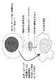
また、HCMVに特異的なCD8+CTLエピトープペプチドとは、CD8+CTLに認識され得るHCMVタンパク質中の特定部位にある8〜10個のアミノ酸配列からなるペプチドであって、CD8+CTLの抗原受容体と免疫的に結合するHCMVの構造部分であり、HCMV感染細胞を直接に傷害し、ウイルスを排除し得るCD8+CTLの細胞性免疫機構を活性化する抗原基を意味する。図1にその機構を示す。
【0016】
HCMVに特異的なCD8+CTLエピトープペプチド、特に、HLA−A*0207、A*1101、B*1501、B*4001、B*4002、B*4006、B*4403、B*5101、Cw*0102、Cw*0401、Cw*0801、Cw*1202又はCw*1502型に提示され得るエピトープペプチドは、CD8+CTLの標的となるHCMVタンパク質の遺伝子を抗原提示細胞(例えば、pp65遺伝子をコードするレトロウイルスベクター感染細胞)内で共発現させ、該HCMV−pp65遺伝子がどのHLAによって提示され、CTLを刺激し得るのかを確認し、さらにHCMV−pp65遺伝子のどの領域がCTLを刺激し得るエピトープペプチドとなり得るのかを検索することによって同定、取得することができる。
【0017】
抗原提示細胞として利用し得る細胞としては、活性化B細胞、樹状細胞、マクロファージ等が挙げられるが、特に活性化B細胞の使用が好ましい。なお、本明細書において抗原提示細胞とは、エピトープペプチドが結合し得るHLAをその表面上に発現する細胞の中でCD8+CTL刺激能を有するものを意味する。
【0018】
また、本明細書において、抗原提示細胞内で発現させるHCMVタンパク質とは、抗原性が強くCD8+CTLの標的となり、HCMVの潜在感染細胞内で発現するものであれば特に限定されないが、特に、抗原性の強さの点からHCMV−pp65タンパク質が好ましい。このタンパク質及び遺伝子(HCMV−pp65遺伝子)については、文献に詳細が報告されており(Ruger B, Klages S, Walla B, et al. Primary structure and transcription of the genes coding for the two virion phosphoproteins pp65 and pp71 of human cytomegalovirus. J. Virol. 1987;61:446−453)、それらを参考にすることができる。
【0019】
HCMV−pp65遺伝子、並びにHLA−A*0207、A*1101、B*1501、B*4001、B*4002、B*4006、B*4403、B*5101、Cw*0102、Cw*0401、Cw*0801、Cw*1202及びCw*1502型の各種HLA cDNAは、例えば、Akatsukaらの文献及びKondoらの文献(Akatsuka Y, Goldberg TA, Kondo E, Martin EG, Obata Y, Morishima Y, Takahashi T, Hansen JA. Efficient cloning and expression of HLA class I cDNA in human B−lymphoblastoid cell lines. Tissue Antigens. 2002;59:502−511、及びKondo E, Topp MS, Kiem HP, Obata Y, Morishima Y, Kuzushima K, Tanimoto M, Harada M, Takahashi T, Akatsuka Y. Efficient generation of antigen−specific cytotoxic T cells using retrovirally transduced CD40−activated B cells. J Immunol. 2002;169:2164−2171)に基づいて構築したプラスミドpcDNA3−pp65及びpcDNA3.1を用いることができる。
【0020】
HCMV−pp65遺伝子及び各種HLA cDNAの抗原提示細胞への共導入の方法については、Budker等の文献(Budker, V. et al. pH−sensitive, cationic liposomes: a new synthetic virus−like vector. Nature Biotechnology 1996;14: 760−764)に基づいて実施することができる。
【0021】
また、HCMV−pp65遺伝子がいずれの型のHLAに提示され、CTLを刺激し得るのかについては、エリスポットアッセイ(enzyme−linked immunospot)で検討することができる。エリスポットアッセイは、文献(Kondo Eら(前述)、及びGeginat G, Schenk S, Skoberne M, Goebel W, Hof H. A novel approach of direct ex vivo epitope mapping identifies dominant and subdominant CD4 and CD8 T cell epitopes from Listeria monocytogenes. J Immunol. 2001;166:1877−1884)に基づいて実施することができる。
【0022】
次いで、エリスポットアッセイの結果に基づき、以下のようにして、特定のHLA型に提示されるHCMV−pp65タンパク質の部位(領域)、すなわちエピトープペプチドを同定することができる。
【0023】
HCMV−pp65遺伝子を、例えば、前述のプラスミドpcDNA3−pp65を制限酵素ApaI及びBamHIで切断し、Erase−a−Baseシステム(Promega、Madison、WI)でエキソヌクレアーゼIII処理して段階的な3’欠失体を得る。この欠失体をE.coliへ結合し、形質転換させて欠失突然変異体を得、該変異体より得られた各プラスミドクローンを配列決定し、選択することができる。
【0024】
得られたHCMV−pp65遺伝子断片と前述のpcDNA3.1を、抗原提示細胞、例えば293T細胞等にリポフェクション等の常法に基づいて共導入し、続いてCTLを刺激してエリスポットアッセイを行い、HCMV−pp65タンパク質のどの断片(領域)にCTLが反応するペプチドが存在するのかを検索することによって、HLAに提示されCD8+CTLが認識し得るエピトープペプチドを同定し、取得することができる。
【0025】
一方、別法として、HCMVタンパク質のアミノ酸配列について、各種HLA型(例えば、HLA−A*0207、A*1101、B*1501、B*4001、B*4002、B*4006、B*4403、B*5101、Cw*0102、Cw*0401型等)の結合モチーフを有する8〜10個のアミノ酸よりなるエピトープペプチドを検索し得る照合媒体、例えば、インターネット上に公開されているBioInformatics & Molecular Analysis Section (BIMAS)のHLA Peptide Binding Predictions (http://bimas.dcrt.nih.gov/molbio/hla#bind/index.html.)によって照合し、エピトープ候補ペプチドをスクリーニングすることもできる。得られたエピトープ候補ペプチドについては、例えば、以下に示すような方法のいずれかによりHCMVに特異的なCD8+CTLエピトープペプチドであるか否かを決定することができる。
【0026】
(1)エピトープペプチド決定方法1
10%ウシ胎児血清含有IMDM培地にて培養中の293T細胞に、エピトープ候補ペプチド配列を含むさまざまな程度に3’端を欠失したpp65遺伝子又は8〜10アミノ酸からなるペプチド配列をコードする遺伝子を含む遺伝子をHLA遺伝子と共に導入しておく。約48時間後、10%ウシ胎児血清含有RPMI1640培地にpp65遺伝子を導入した活性化B細胞で反復刺激して作成しておいた細胞傷害性リンパ球を約5×10³/mlの細胞濃度で浮遊し、上記で準備した293T細胞に添加し、炭酸ガス恒温槽にて37℃で1晩培養する。次いで、エリスポットアッセイ(Kuzushima K他著、The Journal of Clinical Investigation, 104巻:163−171頁,1999年等で報告されている)を実施してエピトープペプチドを決定する。
【0027】
(2)エピトープペプチド決定方法2
10%ウシ胎児血清含有RPMI1640培地にpp65遺伝子を導入した活性化B細胞で反復刺激して作成しておいた細胞傷害性リンパ球を約5×10³/mlの細胞濃度で浮遊し、これにエピトープ候補ペプチドの中の任意の1種を1μg/mlの濃度で処理した自己LCL(Lymphoblastoid cell line)加える。炭酸ガス恒温槽にて37℃で1晩培養する。これらのエピトープ候補ペプチドが刺激するか否かについては、エリスポットアッセイ等で判定する。
【0028】
本発明の配列番号1〜12のアミノ酸配列を有するペプチドは、上記のような方法における検討の結果、HLA−A*0207、A*1101、B*1501、B*4001、B*4002、B*4006、B*4403、B*5101、Cw*0102、Cw*0401、Cw*0801、Cw*1202又はCw*1502型に提示され得るHCMVに特異的なCD8+CTLエピトープペプチドとして確認されたものである。
【0029】
本発明の配列番号1〜12に示されるエピトープペプチドは、従来の各種のペプチド合成方法によって調製され得る。例えば、固相ペプチド合成法等の有機化学的合成法、あるいは、ペプチドをコードするDNAを調製し、組換えDNA技術を用いて調製することも可能である。また、市販の化学合成装置(例えば、アプライドバイオシステムズ社の ペプチド合成装置)による合成も可能である。
【0030】
2.ワクチン
本発明のCD8+CTLエピトープペプチドは、能動免疫ペプチドワクチン療法においてワクチンとして用いることができる。すなわち、本発明のCD8+CTLエピトープペプチドを含んでなるワクチンを患者に投与し、HCMVに特異的なCD8+CTLを体内で増殖させ、疾患に対する予防及び治療に役立てることができる。使用するエピトープペプチドは1種のみの使用であっても、あるいはワクチンの使用目的に応じて2種以上のペプチドを組み合わせ、混合して使用することもできる。
【0031】
また、抗原提示細胞(例えば、樹状細胞、活性化B細胞、マクロファージ等)に本発明のCD8+CTLエピトープペプチドをパルスしたもの(以下、CD8+CTLエピトープペプチドパルス細胞と称する)を含むワクチンも使用することができる。ここで、パルスするとは、適当な培養液中で、抗原提示細胞と該ペプチドを30分から1時間混合することを意味する。
【0032】
本発明のCD8+CTLエピトープペプチド、又はCD8+CTLエピトープペプチドパルス細胞を含んでなるワクチンは、当分野において公知の方法を用いて調製することができる。例えば、かかるワクチンとしては、本発明のCD8+CTLエピトープペプチド又はCD8+CTLエピトープペプチドパルス細胞を有効成分として含有する注射剤又は固形剤等がある。エピトープペプチドは、中性又は塩の形態で処方することができ、例えば、薬学上許容され得る塩としては、塩酸、リン酸などの無機塩、又は、酢酸、酒石酸などの有機酸が挙げられる。また、本発明のエピトープペプチド又はエピトープペプチドパルス細胞は、製薬上許容され、該ペプチド又は該細胞の活性と相容性を有する賦形剤、例えば、水、食塩水、デキストロース、エタノール、グリセロール、DMSO、及びその他のアジュバント等、又はこれらの組み合わせと混合して用いることができる。さらに、必要に応じて、アルブミン、湿潤剤、乳化剤等の補助剤を添加してもよい。
【0033】
本発明のワクチンは、非経口投与及び経口投与により投与することができるが、一般には非経口投与が好ましい。非経口投与としては皮下注射、筋肉内注射、静脈内注射等の注射剤、座薬等がある。また、経口投与としては、スターチ、マンニトール、ラクトース、ステアリン酸マグネシウム、セルロース等の賦形剤との混合物として調製することができる。
【0034】
本発明のワクチンは、治療上有効な量で投与する。投与される量は、治療対象、免疫系に依存し、必要とする投与量は臨床医の判断により決定される。通常、適当な投与量は、患者一人当たり、エピトープペプチドでは1 〜100mg、エピトープペプチドパルス細胞では106〜109個の含有量とする。また、投与間隔は、対象、目的により設定することができる。
【0035】
3.受動免疫療法剤
本発明のCD8+CTLエピトープペプチドは、HCMVに対する受動免疫免疫治療剤の調製に用いることができる。すなわち、1)末梢血リンパ球を該ペプチドで刺激して得られるCD8+CTL、2)末梢血リンパ球を該ペプチドから調製した主要組織適合抗原複合体(major histocompatibility complex、以下、MHCと称する)又はMHC−テトラマーと反応させ、MHC又はMHC−テトラマーにCD8+CTLが結合した結合体を形成させ、該結合体から単離して得られるCD8+CTL、又は、3)末梢血リンパ球を該ペプチドから調製したMHC−標識磁気ビーズと反応させ、MHC−標識磁気ビーズにCD8+CTLが結合した結合体を形成させ、該結合体から単離して得られるCD8+CTL、を含む、受動免疫免疫治療剤を得ることができる。
【0036】
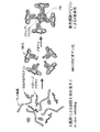
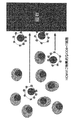
CD8+CTLエピトープペプチドを使用したMHC及びMHC−テトラマーは、例えば、以下のように調製することができる。タンパク質発現用の大腸菌から精製したHLA重鎖、β2ミクログロブリン及び本発明のCD8+CTLエピトープペプチドの複合体である、MHCをバッファー内で形成させる。組換えHLA重鎖タンパク質のC末端には予めビオチン結合部位を付加しておき、MHC形成後この部位にビオチンを付加する。市販の標識色素ストレプトアビジンとビオチン化MHCをモル比1:4で混合することによってMHC−テトラマーを作製する。図2にMHC−テトラマーの調製方法を示し、図3にはMHC−テトラマーとHCMVに特異的なCTLとの結合を示した。なお、各ステップにおいて、ゲル濾過によるタンパク質精製を行うのが好ましい。
【0037】
受動免疫療法剤に含まれるHCMVに特異的なCD8+CTLは、以下のような調製方法によって得ることができる。
(1)CD8+CTL調製方法1
リンパ球を末梢血等から分離し、これを適当な濃度のMHC又はMHC−テトラマーと37℃、15分間反応させる。MHC又はMHC−テトラマーと結合したHCMVに特異的なCD8+CTLは標識色素により染色されるので、フローサイトメーター、顕微鏡などを用いて染色されたCD8+CTLのみを単離する。あるいは、予め無菌プレートなどに固相化したMHC及び/又はMHC−テトラマーに反応させることもできる。プレートに固相化されたMHC及び/又はMHC−テトラマーに結合したHCMV特異的CD8+CTLを単離するためには、結合せずに浮遊している他の細胞を洗い流した後に、プレート上に残った抗原特異的CD8+CTLだけを新しい培養液に懸濁する。このようにして単離されたHCMVに特異的なCD8+CTLは、抗CD3抗体、PHA、IL−2等のT細胞刺激薬剤で刺激増殖させ、受動免疫療法に必要な細胞数を確保する。
【0038】
(2)CD8+CTL調製方法2
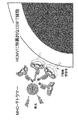
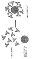
前述のようにペプチドから調製したビオチン化MHCをストレプトアビジン標識磁気ビーズと結合させ、結合体(以下、MHC−磁気ビーズと称する)を作製する。MHC−磁気ビーズを図4に示す。リンパ球を末梢血等から分離し、適当な濃度の前記MHC−磁気ビーズをリンパ球:ビーズ比、1:10で反応させる。MHC−磁気ビーズと結合したHCMVに特異的なCD8+CTLの入った試験管を磁場におくと、ビーズと結合したCD8+CTLは磁石のある側の試験管内壁に寄せられる。その機構を図5に示す。その後、それ以外の細胞を洗い流した後に、試験管を磁場からはずし、試験管内壁に残ったHCMVに特異的なCD8+CTLだけを新しい培養液に懸濁する。このように単離されたHCMVに特異的なCD8+CTLは、抗CD3抗体、PHA、IL−2等のT細胞刺激薬剤で刺激増殖させ、受動免疫療法に必要な細胞数を確保する。
【0039】
(3)CD8+CTL調製方法3
末梢血から分離したリンパ球を本発明のCD8+CTLエピトープペプチドで直接刺激するか、該ペプチドをパルスした抗原提示細胞で刺激する。刺激によって誘導されたCD8+CTLを炭酸ガス恒温槽にて37℃で7〜10日培養する。その後IL−2を添加し、CD8+CTLエピトープペプチドとIL−2、又は該抗原提示細胞とIL−2による刺激を週に1度くり返すことによって受動免疫療法に必要な細胞数のCD8+CTLを確保する。
【0040】
上記のようにして得られたHCMVに特異的なCD8+CTLはヒトアルブミン含有PBS等に懸濁させて、HCMVに対する受動免疫療法剤とすることができる。
【0041】
4.CD8+CTLの定量
HCMVに特異的なCD8+CTLが、ハイリスクの患者(何らかの原因により免疫能が低下した人、先天性免疫不全症、造血幹細胞移植又は固形臓器移植を受けて拒絶予防のために免疫抑制剤の投与を受けている患者、慢性ウイルス感染症患者、エイズ患者、高齢者、幼小児、妊婦等)の末梢血に存在するか否かを知ることは、抗ウイルス剤や免疫抑制剤の適正な使用を含め、これらの感染症管理の上で重要な情報である。HCMVに特異的なCD8+CTLの定量は、本発明のCD8+CTLエピトープペプチドを用いた以下の2つの方法によって行うことができる。
【0042】
第1の定量方法は、末梢血から分離されたリンパ球を本発明のCD8+CTLエピトープペプチドで刺激することによって誘導されるCD8+CTLが産生するインターフェロンガンマ(IFNγ)、インターロイキン等のサイトカイン及び/又はケモカインを定量する方法である。以下にIFNγを例にとり具体的に方法を示す。
【0043】
(1)サイトカイン定量による方法1(細胞内IFNγ産生細胞定量);
末梢血から分離したリンパ球を10%ウシ胎児血清含有RPMI1640培地に2×10⁶/mlの細胞濃度で浮遊させ、本発明のCD8+CTLエピトープペプチドを1μg/mlの濃度で加える。さらに細胞内蛋白輸送阻止剤であるBrefeldin A等を加え、炭酸ガス恒温槽にて37℃で5〜6時間培養する。培養後、細胞を固定、膜透過処理を行い、色素標識抗IFNγ抗体、抗CD8抗体と反応させる。フローサイトメーター等を用いて、CD8+リンパ球中のIFNγ陽性細胞%を定量する。
【0044】
(2)サイトカイン定量による方法2(エリスポットアッセイ);
96穴MultiScreen−HAプレート(Mi11ipore)を抗IFNγモノクローナル抗体で一晩、4℃でコーティングし各穴をPBSで洗浄した後、末梢血から分離したリンパ球を各穴にまく。エピトープペプチドを各穴に入れ37℃の5% CO2培養器にて20時間培養する。翌日、0.05%Tween‐20添加PBSでプレートを洗浄した後、抗IFNγウサギ血清、ペルオキシダーゼ標識抗ウサギIgGヤギ血清の順で各々90分ずつ室温で反応させる。さらに3−amino−9−ethylcarbasole (Sigma)と0.015%のH2O2を含む0.1M酢酸ナトリウムバッファー(pH 5.0)を各穴に入れ、室温で40分反応させる。IFNγスポットを可視化し、実体顕微鏡でカウントする。
【0045】
(3)サイトカイン定量による方法3(培養上清中に分泌されたIFNγを定量する方法);
末梢血から分離したリンパ球を10%ウシ胎児血清含有RPMI1640培地に2×10⁶/mlの細胞濃度で浮遊させ、本発明のCD8+CTLエピトープペプチドを1μg/mlの濃度で加える。炭酸ガス恒温槽にて37℃で24−48時間培養する。培養後、上清を回収し、その中に含まれるIFNγ濃度を市販のELISA キット(例えばENDOGEN社のHUMAN IFN gamma ELISA)を使用して定量する。
【0046】
第2の定量方法としては、本発明のCD8+CTLエピトープペプチドを使用して作製したMHC−テトラマーを用いて、末梢血中のHCMVに特異的なCD8+CTLを定量することができる。MHC−テトラマーの調製は前述のとおりである。定量は、例えば、以下のようにして実施することができる。末梢血などからリンパ球を分離し、適当な濃度のMHC−テトラマーと37℃、15分反応させる。該テトラマーと結合したCD8+CTLは標識色素により染色されるので、フローサイトメーター、顕微鏡等を用いてカウントする。
【0047】
【実施例】
以下に実施例を示し、本発明を具体的に説明するが、本発明はこれらに限定されるものではない。
【0048】
[実施例1] HCMVに特異的なCD8+CTLエピトープペプチドの同定
1.各種材料の調製
(1)血液の入手
CMV血清反応陽性のドナー11人およびCMV血清反応陰性のドナー8人から同意を得て末梢血の提供を受けた。血液中のCMV特異的IgGの存在については、酵素結合抗体免疫吸着アッセイでCMV血清陽性分析を行い、HLA型についてはHLA研究所(Kyoto, Japan)に依頼して確認した。また、ドナー末梢血単核細胞由来のCD8陽性分画及び陰性分画は、末梢血単核細胞をフィコール密度勾配で遠心分離により分離し、CD8 マイクロビーズ(Miltenyi Biotec, Bergisch Gladbach, Germany)を使用して単離した。表1に陽性ドナー及び陰性ドナーから得た末梢血由来Tリンパ球の各HLA型と抗CMV抗体の有無を示す。
【0049】
【表1】
2.HCMV−pp65遺伝子及び各種HLA遺伝子を導入した293T細胞又はLCLによるHCMVに特異的なCTLの誘導
(1)CD40活性化B細胞の調製及びLCLの樹立
CD40活性化B細胞は、文献(Kondo Eら(前述)、及びSchultze JL, Michalak S, Seamon MJ, Dranoff G, Jung K, Daley J, Delgado JC, Gribben JG, Nadler LM. CD40−activated human B cells: an alternative source of highly efficient antigen presenting cells to generate autologous antigen−specific T cells for adoptive immunotherapy, J Clin Invest. 1997;100:2757−2765)に基づいて、ドナー由来のCD8−除去末梢血単核細胞から調製した。
【0051】
すなわち、IL−4(4ng/mL; Ono Pharmaceutical, Ltd, Osaka, Japan)及びCsA (0.7μg/ml; Sandoz, Basel, Switzerland)を含む、プールしたヒト血清10%含有IMDM (Invitrogen, Tokyo, Japan)2mL中で、全CD8−除去末梢血単核細胞を、γ−照射(96Gy)したヒトCD40LをトランスフェクトしたNIH3T3細胞株 (t−CD40L:Dr. Gordon Freeman, Dana−Farber Cancer Institute, Boston, MA、の好意により提供された)上で培養した。3〜4日毎に増殖した細胞を新たに調製したt−CD40L細胞上に移し、CsA非含有の新鮮な培地を供給した。その結果、CD40活性化B細胞が得られた。
【0052】
(2)LCLの樹立
10%ウシ胎仔血清(FCS; Immuno−Biological Laboratories, Gunma, Japan)を含むRPMI 1640(Invitrogen)中で、CD40活性化B細胞をエプスタイン−バールウイルス(EBV)産生株(B95−8, ATCC, Rockville, MD)の上清で処理してLCLを樹立した。
【0053】
(3)HCMV−pp65遺伝子及び各種HLA遺伝子を導入した293T細胞又はLCLの調製
293T細胞およびLCL細胞への導入に利用するプラスミド、pcDNA3−pp65、pcDNA3−EGFP(enhanced green fluorescent protein)及びpcDNA3.1(HLAクラスI(HLA−A、HLA−B及びHLA−C)の各cDNAをコードするプラスミド)、は、文献(Akatsuka Y, Goldberg TA, Kondo E, Martin EG, Obata Y, Morishima Y, Takahashi T, Hansen JA. Efficient cloning and expression of HLA class I cDNA in human B−lymphoblastoid cell lines. Tissue Antigens. 2002;In press、及びKondo E, Topp MS, Kiem HP, Obata Y, Morishima Y, Kuzushima K, Tanimoto M, Harada M, Takahashi T, Akatsuka Y. Efficient generation of antigen−specific cytotoxic T cells using retrovirally transduced CD40−activated B cells. J Immunol. 2002;169:2164−2171)に基づいて構築した。
【0054】
各種プラスミドの293T細胞への導入は、文献(Budker, V. et al. pH−sensitive, cationic liposomes: a new synthetic virus−like vector. Nature Biotechnology 1996;14: 760−764)に従って実施した。
【0055】
まず、各種HLA cDNAとHCMV−pp65遺伝子をTransIT−293 (Mirus, Madison, WI)によって293T細胞に共導入して行い、細胞を得た。それらの細胞は、以下、「293T/pp65+各種HLA型」(例えば、293T/pp65+A*1101)で表す。また、HCMV−pp65遺伝子のみ導入した293T細胞(以下、「293T/pp65」と称する)、自己由来LCL、HCMV−pp65遺伝子のみ導入した自己由来(以下、「LCL/pp65」と称する)についても調製した。なおLCLへの遺伝子の導入は文献(Kondo Eら(前述))に詳細したように、pp65遺伝子をコードしたレトロウイルスベクターを感染させて行った。
また、導入率は、文献(Kondo Eら(前述))に基づいて評価した。
【0056】
3.HCMV−pp65遺伝子及び各種HLA遺伝子を導入した293T細胞又はLCLによるCTL細胞の刺激とエリスポットアッセイ(enzyme−linked immunospot)
103以上のCTLとともに、上記で調製した293T/pp65+各種HLA型細胞、293T/pp65細胞、LCL細胞、及びLCL/pp65細胞をそれぞれプレートした。4時間、その96ウェルプレート(3790; Costar)上のそれら細胞を完全培地200μL中でインキュベートした後、細胞を浮遊させ、そのすべてエリスポットプレートへ移し、16時間インキュベートした。
【0057】
エリスポットアッセイは、文献(Kondo Eら(前述)、及びGeginat G, Schenk S, Skoberne M, Goebel W, Hof H. A novel approach of direct ex vivo epitope mapping identifies dominant and subdominant CD4 and CD8 T cell epitopes from Listeria monocytogenes. J Immunol. 2001;166:1877−1884)に基づいて実施した。すなわち、MultiScreen−HA plate (MAHA S4510, Millipore)を抗−ヒトIFN−γモノクローナル抗体(M700A; Endogen, Woburn, MA)でコーティングし、エリスポットプレートとして使用した。
【0058】
スポットの検出には、ビオチン標識抗−ヒトIFN―γ抗体(M701B; 1 μg/ml, Endogen、およびストレプトアビジン−アルカリ性フォスファターゼ(Biosource International, Camarillo, CA)を使用した。IFNγスポットは、スキャナ(Canon, Tokyo, Japan)を用いてコンピューターで視覚化し、計測した。
【0059】
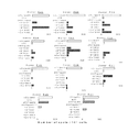
その結果を図6に示す。図中の各横軸は、CD8+CTLの103細胞についてのスポット数を表わす。
【0060】
ドナーP08の場合、ドナーP08から調製された多数のCD8+CTL細胞は、HLA−B*4006の遺伝子を含むpcDNA3.1プラスミドとpcDNA3−pp65プラスミドでトランスフェクトした293T細胞により刺激され、また、少数のCD8+CTL細胞がHLA−B*4403又はCw*0801の遺伝子を含む293T細胞によって刺激されたが、他のHLAクラスI遺伝子であるHLA−A*2402、A*3303又はCw*1403を含む293T細胞には刺激されなかった。このことは、ドナーP08由来のCD8+CTL細胞が、少なくとも3種類の異なるHLAクラスI対立遺伝子(HLA−B*4006、HLA−B*4403及びCw*0801)によって提示される複数のHCMV−pp65由来エピトープを認識し得ることを示唆している。
【0061】
なお、ドナーP06とP07の場合、CTL細胞がpp65遺伝子を単独導入した293T細胞を認識することが確認されているが、これは293T細胞がA*0201を内在的に発現することが知られていることによる。このため、ドナーP06及びP07においては、優性A*0201に提示されるエピトープ(配列番号21)を欠失した突然変異体pp65遺伝子(pcDNA3pp65ΔNLV)と各種HLA cDNAとを導入した293T細胞を使用した。
【0062】
4.各種HLA遺伝子により提示されたHCMV−pp65エピトープペプチドをコードする領域の同定
上記のHCMV−pp65に特異的なCTLCD8+によって認識されるエピトープペプチドを同定を以下のようにして行った。
(1) HCMV−pp65遺伝子断片の調製
Akatsukaらの文献及びKondoらの文献(前述)に基づいて作製したプラスミドpcDNA3−pp65をApaI及びBamHIで切断し、Erase−a−Baseシステム(Promega、Madison、WI)でエキソヌクレアーゼIII処理して段階的な3’欠失体を得た。この欠失体でE.coliを形質転換させて欠失突然変異体を含むプラスミドDNAを得た。該変異体より得られた各プラスミドクローンをHCMV−pp65遺伝子断片として配列決定し、選択した。
【0063】
以下の表2にHCMV−pp65遺伝子のC末端切断型フォームをコードする遺伝子断片の番号とその断片の遺伝子によってコードされるアミノ酸の長さを示す。
【0064】
【表2】
(2) HCMV−pp65遺伝子断片のミニ遺伝子の構築
HCMV−pp65遺伝子断片のミニ遺伝子の構築を行うために、CMVプロモーター(Pcmv)およびpcDNA3.1のBGHポリアデニル化シグナル(BGHpA)をPCR(KOD Plus; Toyobo, Japan)により増幅した。Pcmv用プライマーには、5’Pcmv: CTTAGGGTTAGGCGTTTTGC(配列番号13)及び3’Pcmv: NNCATGGTGGATCCGAGCTCGGTA(配列番号14)を用い、BGHpAのプライマーには、5’BGHpA: NNTAGCGCTCGAGTCTAGAGGG(配列番号15)及び3’BGHpA: GGTTCTTTCCGCCTCAGAAG(配列番号16)を用いた。
【0066】
さらに、上記プライマーとのオーバーラップ領域(下記5’GSP(配列番号17)の下線部分が上記3’Pcmv(配列番号14)の下線部分のオーバーラップ領域であり、下記3’GSP(配列番号18)の下線部分が上記5’BGHpA(配列番号15)の下線部分のオーバーラップ領域である)と(1)で作成したpp65の欠失変異体を用いてエピトープの存在が推定された領域(センス鎖をXで、アンチセンス鎖をYで、またそれぞれの塩基長をm、nで示す)に特異的な付加ヌクレオチドとを含むセンスプライマー及びアンチセンスプライマー(5’GSP: TCGGATCCACCATG (配列番号17) +Xm及び3’GSP: GACTCGAGCGCTA (配列番号18) +Yn)を設計し、全長のpp65をコードするプラスミドを鋳型としてPCRを行った。例えばpp65のアミノ酸配列のアミノ酸位置341番から349番を含むペプチド断片(配列番号10)をコードするミニ遺伝子を作るためには、Xm部分をCAGTACGATCCCGTGG(配列番号19)、Yn部をGAAGAGCGCAGCCACGG(配列番号20)とした。
【0067】
Pcmv−pp65ミニ遺伝子−BGHpAの順よりなるミニ遺伝子を発現させるためのカセットを作るために、各PCR産物を1μLずつ使用して鋳型とし、再度5’Pcmv: CTTAGGGTTAGGCGTTTTGC(配列番号13)および3’BGHpA: GGTTCTTTCCGCCTCAGAAG(配列番号16)を用いて3種類の断片をPCR法にて結合した。
【0068】
なお、本発明の実施例において、ミニ遺伝子法により推定されたエピトープペプチドが実際に認識されるかどうかを確認するための実験に用いた合成ペプチドはTorayリサーチセンター (Tokyo, Japan)に合成を依頼し、購入した。
【0069】
(3) 各種HCMV−pp65遺伝子断片を用いたCD+8CTL細胞の刺激によるエピトープペプチド領域の確認
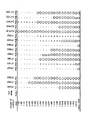
上記各種HCMV−pp65遺伝子3’欠失断片と各種HLA cDNA(HLA−A*0207、A*1101、B*1501、B*4001、B*4002、B*4006、B*4403、B*5101、Cw*0102、Cw*0401、Cw*0801、Cw*1202、Cw*1502型及び対照としてのHLA−A*2402、B*3501、B*5201)でトランスフェクトした293T細胞を用いて各ドナー由来のCTL細胞を刺激し、エリスポットアッセイによってCTL細胞の認識を確認した。その結果を図7に示す。なお、図中、縦軸にpp65遺伝子3’欠失断片の長さを示し、また「−」は、スポットがないことを示す。
【0070】
ドナーP11由来のCTL細胞は、HLA−A*2402 cDNAと、クローン#17又は該クローンより長いクローンでトランスフェクトした293T細胞を認識することができる。これは、アミノ酸残基336と365(各々、クローン#16および#17のC末端位置)の間に、A*2402で提示されるHCMV−pp65エピトープペプチドが部分的に位置していることを示している。この結果は、先に同定されたA*2402に提示されるエピトープペプチド(HCMV−pp65のアミノ酸配列のうち、341〜349番目のアミノ酸配列)と一致している(前述、特許文献1参照)。
【0071】
また、HLA−B*3501およびB*5201の結果についても、すでに報告されたエピトープペプチド、HCMV−pp65のアミノ酸配列のうち、123〜131番目のアミノ酸配列(Gavin MA, et al. Alkali hydrolysis of recombinant proteins allows for the rapid identification of class I MHC−restricted CTL epitopes.J Immunol 1993;151:3971−3980)およびHCMV−pp65のアミノ酸配列のうち、155〜163番目のアミノ酸配列(Bunde KF, et al. Cytomegalovirus (CMV) phosphoprotein 65 makes a large contribution to shaping the T cell repertoire in CMV−exposed individuals. J Infect Dis 2002;185:1709−1716)と各々一致していた。
【0072】
一方、その他のHLA型に提示されるHCMV−pp65エピトープペプチドについては未だ報告されていないので、未確定のHLAクラスIについて対応するHCMV−pp65エピトープペプチドの領域を調べた。
【0073】
その結果、各ドナー由来のCTL細胞によって認識された最短の長さのHCMV−pp65遺伝子断片が次のように決定された:A*0207(HCMV−pp65遺伝子断片番号#23、HCMV−pp65のアミノ酸配列の完全長)、A*1101(#22、HCMV−pp65のアミノ酸配列のうち、1〜513番目のアミノ酸配列)、B*4002(#13、HCMV−pp65のアミノ酸配列のうち、1〜283番目のアミノ酸配列)、B*4006(#23、HCMV−pp65のアミノ酸配列の完全長)、B*4403(#17、HCMV−pp65のアミノ酸配列のうち、1〜378番目のアミノ酸配列)、B*5101(#23、HCMV−pp65のアミノ酸配列の完全長)、Cw*0401(#16、HCMV−pp65のアミノ酸配列のうち、1〜355番目のアミノ酸配列)、Cw*0801(#8、HCMV−pp65のアミノ酸配列のうち、1〜211番目のアミノ酸配列)、Cw*1202(#14、HCMV−pp65のアミノ酸配列のうち、1〜310番目のアミノ酸配列)およびCw*1502(#8、HCMV−pp65のアミノ酸配列のうち、1〜400番目のアミノ酸配列)。
【0074】
Cw*1202では、完全長プラスミドをトランスフェクトした細胞が、それより短いプラスミドをトランスフェクトした細胞よりも弱い刺激でスポットを生じさせることが確認された(153/ウエルに対して371/ウエル)。この結果により、上記のような、P04、P07およびP10の場合になぜ少数のスポットだけが確認されるのかを説明できる。具体的には、HLA−Cw*1202に提示されるエピトープは、CTLによって顕著に認識されるが、完全長のpp65遺伝子に比べ、短いエピトープペプチドの方が効果的に刺激する。
【0075】
HLA−B*1501(ドナーP01において、最も有力にpp65エピトープを提示したHLA分子である)に関して、HCMV−pp65遺伝子断片#9(HCMV−pp65のアミノ酸配列のうち、1〜234番目のアミノ酸配列)はP01 CTL細胞によって認識された最短のクローンであった。これは、アミノ酸残基212〜234の領域内に、HLA−B*1501で提示されるエピトープペプチドのC末端が含まれていることを示唆するものである。
【0076】
B*4001の場合には、ドナーP03のCTL細胞はHCMV−pp65遺伝子断片#13(HCMV−pp65のアミノ酸配列のうち、1〜287番目のアミノ酸配列)又はそれより長いクローンを顕著に認識するとともにHCMV−pp65遺伝子断片#10(HCMV−pp65のアミノ酸配列のうち、1〜249番目のアミノ酸配列)、#11および#12についてもやや弱いながらも認識することができる(図7中、半丸の部分)。これは、ドナーP03のCTL細胞については、HLA−B*4001が提示するエピトープペプチドが2種類存在することを意味する。
【0077】
また、HLA−A*1101やB*4403などの場合においても、CTL細胞にわずかに認識される部分(図7中、小さな丸で示す部分)があり、優勢なエピトープ(大きな丸で示す部分)以外に、非常に弱く認識されている部分(小さな丸で示す部分)にさらなるエピトープペプチドの存在が考えられる。
【0078】
5.各HLA分子型によって提示されるHCMV−pp65エピトープペプチドのコンピューター・アルゴリズムによる同定
コンピューター・アルゴリズムによるエピトープ推定が多くの特定の腫瘍又はウイルス・ペプチドの同定に功を奏しているので、本試験においても結合モチーフを利用可能なHLA対立遺伝子に関してコンピューター・アルゴリズム、すなわち、上記エピトープペプチドを検索し得る照合媒体、例えば、インターネット上に公開されているBioInformatics & Molecular Analysis Section (BIMAS)のHLA Peptide Binding Predictions (http://bimas.dcrt.nih.gov/molbio/hla#bind/index.html.)を使用した。
【0079】
HLAクラスI対立遺伝子によって提示されるエピトープは一般に8〜10 merである。従って、図7で報告した領域のアミノ酸配列とそのN末端側の10アミノ酸配列をとともにBIMASウェブサイトにあるコンピューター・アルゴリズムにかけた。そのエピトープペプチドの推定結果を表3に示した。
【0080】
【表3】
HLA−A*0207の場合、BIMASウェブサイトにおいて、HLA−A*0207自体に対する推定は行えなかったが、HLA−A*0201に類似性を示すHLA−A*0207の結合モチーフは既に報告されている。したがって、HLA−A*0207のエピトープペプチドを推定するため、分析オプションのHLA分子としてHLA−A*0201を選択した。
【0082】
HLA−B*1501に関しては、トップ3のエピトープのスコアが類似している(6、4.4及び4)。したがって、別のアルゴリズム(University of Oklahoma Health Sciences Centerのウェブサイト)による推定を試みた。その結果、1つのエピトープだけが高いスコアを示し、そのスコアはBIMASウェブサイトによる3番目のスコアと一致した。
【0083】
各CTLにより推定されるエピトープの認識は、推定エピトープ又は無関係なpp65のペプチドをコードするミニ遺伝子をオーバーラッピングPCRによって作成し、ミニ遺伝子の導入細胞を使用して、エリスポットアッセイによって評価した。
【0084】
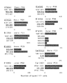
図8に示すように、HLA−B*5101以外の推定されたエピトープはすべて、適当なHLA遺伝子の存在下において各pp65特異的CTLによって十分に認識され、その特異性が確認された。
【0085】
ドナーP01由来のCTL細胞は、pp65のアミノ酸番号215〜223をコードするミニ遺伝子とHLA−B*1501でコトランスフェクトした293T細胞によって刺激されるが、それは、完全長pp65遺伝子で同様にコトランスフェクトした293T細胞による刺激に匹敵した。一方、pp65遺伝子のアミノ酸番号227〜235をコードするミニ遺伝子で同様にコトランスフェクトした293T細胞では刺激が生じなかった。この結果、HCMV−pp65のアミノ酸配列のうち、215〜223番目のアミノ酸配列(配列番号3)が、HLA−B*1501が提示するpp65遺伝子由来のエピトープペプチドの少なくとも1種であることが確認された。
【0086】
HLA−B*4001に関しては、ドナーP03由来のCTL細胞が表3に挙げられた2種類のミニ遺伝子(HCMV−pp65のアミノ酸配列のうち、232〜240番目のアミノ酸配列(配列番号4)および267〜275番目のアミノ酸配列(配列番号5))をともによく認識した。これは、クローン(図7)を使用したエリスポットアッセイの結果と類似していた。
【0087】
さらに、ドナーP02由来のCTL細胞は、HLA−B*4001中の2種類のミニ遺伝子をともに認識することができ(データは示さず)、両ミニ遺伝子がHLA−B*4001によって提示される優性pp65−エピトープペプチドであることが確認された。
【0088】
HLA−B*5101の場合、推定されたエピトープ(HCMV−pp65のアミノ酸配列のうち、547〜555番目のアミノ酸配列)をコードするミニ遺伝子又は2番めに高いスコア(HCMV−pp65のアミノ酸配列のうち、545〜553番目のアミノ酸配列)を有するミニ遺伝子は、ドナーP05由来のCTL細胞にあまり認識されなかった。このため、他の様々なミニ遺伝子についても検討したところ、9アミノ酸からなるペプチド(HCMV−pp65のアミノ酸配列のうち、545〜552番目のアミノ酸配列(配列番号8))をコードするミニ遺伝子がよく認識されることがわかった。この9アミノ酸からなるペプチドは、HLA−B*5101についてすでに報告された結合モチーフと一致しており、第2位置がアラニン又はプロリンで、C末端がイソロイシンである。
【0089】
これらの結果により、表4に示すように、HLA−A*0207、A*1101、B*1501、B*4001、B*4002、B*4006、B*4403、B*5101、Cw0102、Cw*0401、Cw*0801、Cw*1202及びCw*1502で各々提示される新規pp65由来エピトープペプチドが明らかになった。
【0090】
【表4】
6.コンピューター・アルゴリズムが利用できないHLA分子型によって提示されるHCMV−pp65エピトープペプチドの同定
HLA−C*0801、C*1202、C*1502に提示されるペプチドモチーフが十分判明していないため、これらのHLA型に対しては現在コンピューター・アルゴリズムが利用できない。従って上記と同様の方法で特定した図7に示す領域のアミノ酸配列をコードするミニ遺伝子(長さ8〜10)を、N末端側から1アミノ酸ずつずらしながら多数作成し、293T細胞にトランスフェクトして、各ドナー(P08、P09およびP02)由来のCTL細胞を刺激し、エリスポットアッセイによってCTL細胞の認識を確認した。その結果を図9に示す。 図9から明らかなように、HLA−C*0801およびC*1502によって提示されるpp65のエピトープペプチド配列は同一であり、 HCMV−pp65のアミノ酸配列のうち、198〜206番目のアミノ酸配列(配列番号11)であることが判明した。同様に、HLA−Cw*1202によって提示されるpp65のエピトープペプチド配列はHCMV−pp65のアミノ酸配列のうち、295〜302番目のアミノ酸配列(配列番号12)であることがわかった。
【0092】
HLA−C*0801、C*1202及びC*1502で各々提示される新規pp65由来エピトープペプチドを表5に示す。
【0093】
【表5】
[実施例2]ワクチン注射剤
DMSOに、配列番号1〜12のペプチドを最終濃度20mg/mlとなるように各々溶解し、ろ過滅菌した。得られたペプチド含有溶液を滅菌バイアル瓶に1mlずつ分注密栓し、ワクチン注射剤とした。
【0095】
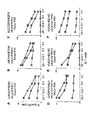
[実施例3]エピトープペプチドによるCTL株の誘導
CMV血清反応陽性のドナー3人(P01、P03およびP08)の末梢血単核細胞由来のCD8除去末梢血単核細胞から調製したCD40活性化B細胞に、上述のように作製し、上記で同定した5種のエピトープペプチド(配列番号4、5、6、10及び11)を合成して添加した。エピトープペプチドによって刺激されたCD40活性化B細胞を刺激細胞とし、自己のCD8陽性細胞と共培養した。刺激細胞による刺激を2回反復し、21日目に、pp65遺伝子を導入した自己LCL(LCL/pp65)、該ペプチドを添加したか或いは行わないEGFP遺伝子導入自己LCL(LCL/EGFP)を標的として、細胞傷害試験を行った。その結果を図9に示す。図中、■はLCL/pp65、○はLCL/EGFP、●は1μMの同起源ペプチドでパルスしたLCL/EGFPを示す。図9から明らかなように、P03ではHLA−B*4001によって提示される2つのペプチド(配列番号4、配列番号5)およびCw*0401によって提示されるペプチド(配列番号10)の計3種のエピトープペプチドのついて検討したところ、いずれのペプチドで刺激・誘導されたCTL株も該ペプチドを添加したLCL/EGFPのみならず、全長のpp65を発現しているLCL/pp65とも良好に傷害した。一方、該ペプチドを添加しなかった自己LCL/EGFPは傷害しなかった。同様に、P08ではHLA−B*4006で提示されるペプチド(配列番号6)、P01ではHLA−Cw*1502によって提示されるペプチド配列番号11)を用いてCTL株の誘導を行い、細胞傷害性試験を行ったところ、図9に示すように該ペプチドを添加したLCL/EGFPのみならず、全長のpp65を発現しているLCL/pp65とも良好に傷害した。以上の結果は、本発明で同定されたエピトープペプチドがT細胞を刺激しCTL株を誘導する生理活性を有することを示している。
【0096】
【発明の効果】
本発明により、HCMVに特異的なCD8+CTLエピトープペプチドを提供することができる。また、該エピトープペプチドを用いてHCMVの感染を管理、治療又は予防することができる。さらに、HCMVに特異的なCD8+CTLを定量することが可能である。
【0097】
【配列表】
【配列表フリーテキスト】
配列番号14:nはA又はC又はG又はTを表す(存在位置:1及び2)
配列番号15:nはA又はC又はG又はTを表す(存在位置:1及び2)
【図面の簡単な説明】
【図1】CD8+CTLによるウイルス感染細胞の認識機構を示す図である。
【図2】MHC−テトラマーの調製方法を示す図である。
【図3】MHC−テトラマーとHCMVに特異的なCTLの結合を示す図である。
【図4】MHC−磁気ビーズを示す図である。
【図5】MHC−磁気ビーズと結合したHCMVに特異的なCD8+CTLの磁場における反応を示す図である。
【図6】各HLA型とHCMV−pp65遺伝子断片を共導入した293T細胞を用いてCTLを刺激してエリスポットアッセイを行った結果を示す図である。
【図7】各ドナー由来のCTLによって認識された最短の長さのHCMV−pp65遺伝子断片を示す図である。
【図8】コンピューター・アルゴリズムを用いたHCMVに特異的なエピトープの同定結果を示す図である。
【図9】エピトープペプチドによるCTL株の誘導を示す図である。[0001]
BACKGROUND OF THE INVENTION
The present invention relates to CD8 specific for human cytomegalovirus (hereinafter referred to as HCMV).+Cytotoxic T lymphocyte (hereinafter referred to as CTL) epitope peptide, vaccine for treating or preventing HCMV infection using the peptide, passive immunotherapeutic agent for HCMV, and CD8 specific for HCMV+The present invention relates to a method for quantifying CTL.
[0002]
[Prior art]
HCMV is a virus potentially possessed by most healthy adults, but it activates when the immune system is reduced in cancer patients, bone marrow transplant patients, AIDS patients, and the like. Activated and proliferated HCMV causes fatal interstitial pneumonia, retinitis, hepatitis and the like. Therefore, a new method for controlling HCMV safely and effectively is strongly desired.
[0003]
The major immunocompetent cells that control the activity of HCMV-infected cells are CD8+CTL. CD8+Since CTLs have the ability to destroy HCMV-infected cells when discovered, effectively activating their functions is likely to lead to the development of new diagnoses and treatments for HCMV-related diseases.
[0004]
When CTL recognizes a virus-infected cell, it has the following characteristics.
(1) CTL cannot recognize virus particles themselves.
(2) CTL recognizes a peptide consisting of 8 to 10 amino acids at a specific site in a viral protein (hereinafter referred to as epitope peptide) and destroys infected cells.
(3) The specific 8 to 10 amino acids are presented to the CTL by binding to human leukocyte antigen (hereinafter referred to as HLA) on the surface of virus-infected cells.
(4) HLA differs among races and individuals, and epitope peptides differ for the same virus if HLA is different.
[0005]
As is clear from what has been described in (1) to (4) above, determining amino acids that can be epitope peptides specific for HCMV without excess or deficiency is an effective immunity against these viral infections. It can be an indispensable matter when diagnosing whether or not there is an immunotherapy.
[0006]
In order to solve this problem, it has already been reported that peptides present on T cells on HLA molecules often found in whites and HLA-A24 molecules often found in Japanese and their use. For example, with respect to the amino acid sequence of HCMV protein, BioInformatics & Molecular Analysis Section (on the Internet) that can search for an epitope peptide consisting of 8 to 10 amino acids having binding motifs of HLA-A24, A26, and B61. BIMAS) HLA Peptide Binding Predictions (http://bimas.dcrt.nih.gov/molbio/hla#bind/index.html.) And having binding motifs of HLA-A24, A26, and B61 Screening a large number of antigen epitope candidate peptides consisting of 10 amino acids, specific to HCMV identified from the epitope candidate peptides CD8+CTL epitope peptides have been reported (see, for example, Patent Document 1).
[0007]
However, 45% of all Japanese do not have HLA-A24, and CD8 specific for HCMV presented by HLA molecules other than the above possessed by many Japanese.+There are very few reports on CTL epitope peptides, and there is a need for the identification and development of epitope peptides corresponding to additional HLA types that are useful for treating or preventing HCMV infection.
[0008]
[Patent Document] Japanese Patent Application Laid-Open No. 2002-255997
[0009]
[Problems to be solved by the invention]
The present invention relates to CD8 specific for HCMV.+CTL epitope peptides, in particular HLA-A * 0207, A * 1101, B * 1501, B * 4001, B * 4002, B * 4006, B * 4403, B * 5101, Cw * 0102, Cw * 0401, Cw * Epitope peptide that can be presented in type 0801, Cw * 1202 or Cw * 1502, vaccine to treat or prevent HCMV infection, passive immunotherapy for HCMV, and CD8 specific for HCMV+It aims at providing the quantification method of CTL.
[0010]
[Means for Solving the Problems]
As a result of intensive studies to solve the above problems, the present inventors have strong antigenicity and CD8.+A fragment of the HCMV-pp65 gene that is a CTL target and is a phosphorylated protein expressed during HCMV latent infection is expressed in antigen-presenting cells such as activated B cells, dendritic cells, macrophages, etc. Search whether the part (region) has a peptide that reacts with CTL, HLA-A * 0207, A * 1101, B * 1501, B * 4001, B * 4002, B * 4006, B * 4403, Presented in B * 5101, Cw * 0102, Cw * 0401, Cw * 0801, Cw * 1202 or Cw * 1502 type and CD8+A plurality of epitope peptides that can be recognized by CTLs were found, and the present invention was completed.
[0011]
That is, the present invention provides the following (1) to (8).
(1) CD8 specific for human cytomegalovirus comprising at least one amino acid sequence selected from the group consisting of SEQ ID NO: 1 to SEQ ID NO: 12+Cytotoxic T lymphocyte epitope peptide.
(2) A vaccine for treating or preventing human cytomegalovirus infection comprising the peptide according to (1) as an active ingredient.
(3) A vaccine for treating or preventing human cytomegalovirus infection, comprising as an active ingredient antigen-presenting cells pulsed with the peptide according to (1).
(4) CD8 obtained by stimulating peripheral blood lymphocytes with the peptide according to (1) or antigen-presenting cells pulsed with the peptide+A passive immunotherapeutic agent for human cytomegalovirus comprising cytotoxic T lymphocytes.
(5) The major histocompatibility antigen complex and / or the major histocompatibility antigen complex-tetramer prepared from the peptide according to (1) is reacted with peripheral blood lymphocytes, and the major histocompatibility antigen complex and / or Major histocompatibility complex-tetramer with CD8+CD8 obtained by forming a conjugate in which cytotoxic T lymphocytes are bound and isolating from the conjugate+A passive immunotherapeutic agent for human cytomegalovirus comprising cytotoxic T lymphocytes.
(6) The major histocompatibility complex-labeled magnetic beads prepared from the peptide according to (1) are reacted with peripheral blood lymphocytes, and the major histocompatibility complex-labeled magnetic beads are reacted with CD8.+CD8 obtained by forming a conjugate in which cytotoxic T lymphocytes are bound and isolating from the conjugate+A passive immunotherapeutic agent for human cytomegalovirus comprising cytotoxic T lymphocytes.
(7) Stimulating peripheral blood with the peptide according to (1), and CD8 specific for the virus+Cytotoxic T lymphocytes were obtained and the CD8+CD8 specific for human cytomegalovirus, characterized by measuring cytokines and / or chemokines produced by cytotoxic T lymphocytes+A method for quantifying cytotoxic T lymphocytes.
(8) A major histocompatibility complex and / or major histocompatibility complex-tetramer is prepared from the peptide according to (1), and the major histocompatibility complex and / or major histocompatibility complex-tetramer is prepared. CD8 specific to human cytomegalovirus in the peripheral blood, which reacts with peripheral blood+A method for quantifying cytotoxic T lymphocytes.
[0012]
DETAILED DESCRIPTION OF THE INVENTION
The present invention will be described in further detail.
[0013]
1. Epitope peptide
The peptide referred to in the present invention means a molecular chain of linear amino acids having physiological activity and bound to each other by a peptide bond between an α-amino group and a carboxyl group of adjacent amino acid residues. Peptides are not meant to be of a specific length and can be of various lengths. Further, it may be in an uncharged or salt form, and may be modified by glycosylation, amidation, phosphorylation, carboxylation, phosphorylation or the like in some cases. Furthermore, one or several (for example, 1 to 10) amino acids, unless substantially altering the physiological activity and immune activity of the epitope of the present invention and having harmful activity when administered. Peptides in which insertion, addition, substitution or the like has occurred are also included in the present invention. For example, those having an additional amino acid sequence at the N-terminus or C-terminus of the peptide are also included. In addition, the peptide of the present invention can be used in the form of a complex to which saccharides, polyethylene glycol, lipids and the like are added, a derivative with a radioisotope, or a polymer.
[0014]
CD8 of the present invention+CTL is a CD8 expressing CD8, which is one of surface antigen molecules present on human lymphocytes.+Refers to cytotoxic T lymphocytes.
[0015]
Also, CD8 specific for HCMV+CTL epitope peptide is CD8+A peptide consisting of an 8 to 10 amino acid sequence at a specific site in an HCMV protein that can be recognized by CTL,+CD8 is a structural part of HCMV that immunologically binds to the antigen receptor of CTL and can directly damage HCMV-infected cells and eliminate viruses+It means an antigen group that activates the cellular immune mechanism of CTL. FIG. 1 shows the mechanism.
[0016]
CD8 specific for HCMV+CTL epitope peptides, in particular HLA-A * 0207, A * 1101, B * 1501, B * 4001, B * 4002, B * 4006, B * 4403, B * 5101, Cw * 0102, Cw * 0401, Cw * Epitope peptides that can be presented in the 0801, Cw * 1202 or Cw * 1502 type are CD8+The gene of the HCMV protein targeted for CTL can be co-expressed in an antigen-presenting cell (for example, a retrovirus vector-infected cell encoding the pp65 gene), and the HCMV-pp65 gene can be presented by any HLA to stimulate CTL And by searching which region of the HCMV-pp65 gene can be an epitope peptide capable of stimulating CTL, it can be identified and obtained.
[0017]
Examples of cells that can be used as antigen-presenting cells include activated B cells, dendritic cells, macrophages and the like, and the use of activated B cells is particularly preferred. In the present specification, the antigen-presenting cell means CD8 among cells expressing HLA on the surface thereof to which an epitope peptide can bind.+It means what has CTL stimulation ability.
[0018]
Moreover, in this specification, HCMV protein expressed in an antigen-presenting cell has a strong antigenicity and is CD8.+Although it will not specifically limit if it becomes a target of CTL and it expresses in the HCMV latent infection cell, HCMV-pp65 protein is especially preferable from the point of antigenic strength. Details of this protein and gene (HCMV-pp65 gene) have been reported in the literature (Ruger B, Klages S, Walla B, et al. Primary structure and transcription of the genes 71). of human cytomegalovirus. J. Virol. 1987; 61: 446-453), which can be referred to.
[0019]
HCMV-pp65 gene and HLA-A * 0207, A * 1101, B * 1501, B * 4001, B * 4002, B * 4006, B * 4403, B * 5101, Cw * 0102, Cw * 0401 and Cw * Various HLA cDNAs of the 0801, Cw * 1202 and Cw * 1502 types are described in, for example, the literature by Akatsuka et al. And the literature by Kondo et al. (Akatsuka Y, Goldberg TA, Kondo E, Martin EG, Obata Y, Morishik. JA Efficient cloning and expression of HLA class I cDNA in human B-lymphoblastoid cell lines. ue Antigens 2002; 59:. 502-511, and Kondo E, Topp MS, Kiem HP, Obata Y, Morishima Y, Kuzushima K, Tanimoto M, Harada M, Takahashi T, Akatsuka Y. Efficient generation of antigen-specific cytotoxic T The plasmids pcDNA3-pp65 and pcDNA3.1 constructed based on cells using retrotransduced CD40-activated B cells. J Immunol. 2002; 169: 2164-2171) can be used.
[0020]
For the method of co-introduction of the HCMV-pp65 gene and various HLA cDNAs into antigen-presenting cells, see Budker et al. (Budker, V. et al. PH-sensitive, categorical liposomes: anew synthetic virus-like molecules. 1996; 14: 760-764).
[0021]
In addition, which type of HLA the HCMV-pp65 gene can be presented to stimulate CTL can be examined by an enzyme-linked immunospot. ELISPOT assay, the literature (Kondo E, et al. (Supra), and Geginat G, Schenk S, Skoberne M, Goebel W, Hof H. A novel approach of direct ex vivo epitope mapping identifies dominant and subdominant CD4 and CD8 T cell epitopes from Listeria monocytogenes.J Immunol.2001; 166: 1877-1884).
[0022]
Then, based on the results of the Elispot assay, the site (region) of the HCMV-pp65 protein presented in a specific HLA type, that is, an epitope peptide can be identified as follows.
[0023]
For the HCMV-pp65 gene, for example, the plasmid pcDNA3-pp65 described above is cleaved with restriction enzymes ApaI and BamHI, and treated with exonuclease III in the Erase-a-Base system (Promega, Madison, WI) to give stepwise 3 ′ deletion. Get lost. This deletion is designated as E. coli. and then transformed to obtain a deletion mutant, and each plasmid clone obtained from the mutant can be sequenced and selected.
[0024]
The obtained HCMV-pp65 gene fragment and the aforementioned pcDNA3.1 were co-introduced into antigen-presenting cells, for example, 293T cells, based on a conventional method such as lipofection, followed by stimulating CTL and performing an eryspot assay, By searching which fragment (region) of the HCMV-pp65 protein contains a peptide that reacts with CTL, CD8+Epitope peptides that can be recognized by CTLs can be identified and obtained.
[0025]
On the other hand, as an alternative method, various HLA types (for example, HLA-A * 0207, A * 1101, B * 1501, B * 4001, B * 4002, B * 4006, B * 4403, B) are used for the amino acid sequence of the HCMV protein. * 5101, Cw * 0102, Cw * 0401 type, etc.) that can search for epitope peptides consisting of 8 to 10 amino acids having a binding motif such as BioInformatics & Molecular Analysis Section (for example, published on the Internet). BIMAS) HLA Peptide Predictions (http://bimas.dcrt.nih.gov/molbio/hla#bind/index.html.) It is also possible to screen for complementary peptide. With respect to the obtained epitope candidate peptide, for example, CD8 specific for HCMV can be obtained by any of the methods shown below.+Whether it is a CTL epitope peptide can be determined.
[0026]
(1) Epitope peptide determination method 1
A gene encoding a pp65 gene or a peptide sequence consisting of 8 to 10 amino acids lacking the 3 ′ end to various extents including the epitope candidate peptide sequence is added to 293T cells being cultured in IMDM medium containing 10% fetal bovine serum. The containing gene is introduced together with the HLA gene. About 48 hours later, about 5 × 10 5 cytotoxic lymphocytes prepared by repeated stimulation with activated B cells introduced with pp65 gene in RPMI 1640 medium containing 10% fetal bovine serum were prepared.3The cells are suspended at a cell concentration of / ml, added to the 293T cells prepared above, and cultured overnight at 37 ° C. in a carbon dioxide thermostat. Then, an EliSpot assay (reported in Kuzushima K et al., The Journal of Clinical Investigation, 104: 163-171, 1999, etc.) is performed to determine the epitope peptide.
[0027]
(2) Epitope peptide determination method 2
About 5 × 10 5 cytotoxic lymphocytes prepared by repeated stimulation with activated B cells introduced with pp65 gene in RPMI 1640 medium containing 10% fetal bovine serum3A self-LCL (Lymphoblast cell line) which is suspended at a cell concentration of / ml and treated with any one of the epitope candidate peptides at a concentration of 1 μg / ml is added thereto. Incubate overnight at 37 ° C. in a carbon dioxide oven. Whether these epitope candidate peptides stimulate or not is determined by an Elispot assay or the like.
[0028]
Peptides having the amino acid sequences of SEQ ID NOs: 1 to 12 of the present invention have been analyzed by the above method, and as a result, HLA-A * 0207, A * 1101, B * 1501, B * 4001, B * 4002, B * 4006, B * 4403, B * 5101, Cw * 0102, Cw * 0401, Cw * 0801, Cw * 1202 or Cw * 1502 type CD8 specific for HCMV+It has been confirmed as a CTL epitope peptide.
[0029]
The epitope peptides represented by SEQ ID NOs: 1 to 12 of the present invention can be prepared by various conventional peptide synthesis methods. For example, organic chemical synthesis methods such as solid phase peptide synthesis methods, or DNAs encoding peptides can be prepared and prepared using recombinant DNA technology. Furthermore, synthesis using a commercially available chemical synthesizer (for example, a peptide synthesizer manufactured by Applied Biosystems) is also possible.
[0030]
2. vaccine
CD8 of the present invention+CTL epitope peptides can be used as vaccines in active immune peptide vaccine therapy. That is, the CD8 of the present invention+A vaccine comprising a CTL epitope peptide is administered to a patient and CD8 specific for HCMV+CTLs can be propagated in the body to help prevent and treat diseases. The epitope peptide to be used can be used alone or in combination of two or more peptides depending on the intended use of the vaccine.
[0031]
In addition, CD8 of the present invention is applied to antigen-presenting cells (eg, dendritic cells, activated B cells, macrophages, etc.)+Pulsed with CTL epitope peptide (hereinafter CD8)+Vaccines containing CTL epitope peptide pulsed cells) can also be used. Here, pulsing means that the antigen-presenting cells and the peptide are mixed in an appropriate culture solution for 30 minutes to 1 hour.
[0032]
CD8 of the present invention+CTL epitope peptide or CD8+A vaccine comprising CTL epitope peptide pulsed cells can be prepared using methods known in the art. For example, such a vaccine includes CD8 of the present invention.+CTL epitope peptide or CD8+There are injections or solid preparations containing CTL epitope peptide pulse cells as active ingredients. The epitope peptide can be formulated in a neutral or salt form. Examples of pharmaceutically acceptable salts include inorganic salts such as hydrochloric acid and phosphoric acid, and organic acids such as acetic acid and tartaric acid. The epitope peptide or epitope peptide pulsed cell of the present invention is pharmaceutically acceptable and is compatible with the peptide or the activity of the cell, for example, water, saline, dextrose, ethanol, glycerol, DMSO. , And other adjuvants, or a combination thereof. Furthermore, you may add adjuvants, such as albumin, a wetting agent, and an emulsifier, as needed.
[0033]
The vaccine of the present invention can be administered by parenteral administration and oral administration, but parenteral administration is generally preferred. Parenteral administration includes injections such as subcutaneous injections, intramuscular injections, intravenous injections, and suppositories. For oral administration, it can be prepared as a mixture with excipients such as starch, mannitol, lactose, magnesium stearate, and cellulose.
[0034]
The vaccine of the present invention is administered in a therapeutically effective amount. The dose to be administered depends on the subject to be treated and the immune system, and the required dose is determined by the judgment of the clinician. In general, suitable doses are 1-100 mg for epitope peptides and 10 for epitope peptide pulsed cells per patient.6-109Individual content. In addition, the administration interval can be set according to the subject and purpose.
[0035]
3. Passive immunotherapy
CD8 of the present invention+CTL epitope peptides can be used in the preparation of a passive immunoimmunotherapy for HCMV. 1) CD8 obtained by stimulating peripheral blood lymphocytes with the peptide+CTL, 2) Reacting peripheral blood lymphocytes with a major histocompatibility complex (hereinafter referred to as MHC) prepared from the peptide or MHC-tetramer, and CD8 on MHC or MHC-tetramer+CD8 obtained by forming a CTL-bound conjugate and isolating it from the conjugate+CTL or 3) Peripheral blood lymphocytes are reacted with MHC-labeled magnetic beads prepared from the peptide, and CD8 is added to the MHC-labeled magnetic beads.+CD8 obtained by forming a CTL-bound conjugate and isolating it from the conjugate+A passive immune immunotherapeutic agent containing CTL can be obtained.
[0036]
CD8+MHC and MHC-tetramers using CTL epitope peptides can be prepared, for example, as follows. HLA heavy chain purified from E. coli for protein expression, β2 microglobulin and CD8 of the present invention+MHC, a complex of CTL epitope peptides, is formed in the buffer. A biotin binding site is added in advance to the C-terminus of the recombinant HLA heavy chain protein, and biotin is added to this site after MHC formation. An MHC-tetramer is prepared by mixing the commercially available labeling dye streptavidin and biotinylated MHC at a molar ratio of 1: 4. FIG. 2 shows a method for preparing MHC-tetramer, and FIG. 3 shows the binding of MHC-tetramer to CTL specific for HCMV. In each step, it is preferable to perform protein purification by gel filtration.
[0037]
CD8 specific for HCMV in passive immunotherapy+CTL can be obtained by the following preparation method.
(1) CD8+CTL preparation method 1
Lymphocytes are separated from peripheral blood or the like, and reacted with MHC or MHC-tetramer at an appropriate concentration at 37 ° C. for 15 minutes. CD8 specific for HCMV bound to MHC or MHC-tetramer+Since CTL is stained with a labeling dye, CD8 stained with a flow cytometer, a microscope or the like+Only CTL is isolated. Alternatively, it can be reacted with MHC and / or MHC-tetramer solid-phased in advance on a sterile plate or the like. HCMV-specific CD8 bound to MHC and / or MHC-tetramer immobilized on a plate+To isolate the CTL, the antigen-specific CD8 remaining on the plate after washing away other cells that were not bound and floating.+Suspend only CTL in fresh medium. CD8 specific for HCMV isolated in this way+CTL is stimulated and proliferated with a T cell stimulating drug such as anti-CD3 antibody, PHA, IL-2, and the like to secure the number of cells necessary for passive immunotherapy.
[0038]
(2) CD8+CTL preparation method 2
Biotinylated MHC prepared from peptides as described above is bound to streptavidin-labeled magnetic beads to produce a conjugate (hereinafter referred to as MHC-magnetic beads). MHC-magnetic beads are shown in FIG. Lymphocytes are separated from peripheral blood or the like, and the MHC-magnetic beads having an appropriate concentration are reacted at a lymphocyte: bead ratio of 1:10. CD8 specific for HCMV coupled to MHC-magnetic beads+When a test tube containing CTL was placed in a magnetic field, CD8 bound to the beads+CTL is brought to the inner wall of the test tube on the side where the magnet is located. The mechanism is shown in FIG. Then, after washing away other cells, the test tube was removed from the magnetic field, and the CD8 specific for HCMV remaining on the inner wall of the test tube was removed.+Suspend only CTL in fresh medium. CD8 specific for HCMV isolated in this way+CTL is stimulated and proliferated with a T cell stimulating drug such as anti-CD3 antibody, PHA, IL-2, and the like to secure the number of cells necessary for passive immunotherapy.
[0039]
(3) CD8+CTL preparation method 3
Lymphocytes isolated from peripheral blood were used as CD8 of the present invention.+Stimulate directly with CTL epitope peptide or with antigen-presenting cells pulsed with the peptide. CD8 induced by stimulation+CTL is cultured for 7 to 10 days at 37 ° C. in a carbon dioxide thermostat. IL-2 was then added and CD8+CD8 of the number of cells required for passive immunotherapy by repeating stimulation with CTL epitope peptide and IL-2 or the antigen-presenting cell and IL-2 once a week+CTL is secured.
[0040]
CD8 specific for HCMV obtained as described above+CTL can be suspended in human albumin-containing PBS or the like and used as a passive immunotherapy for HCMV.
[0041]
4). CD8+Quantification of CTL
CD8 specific for HCMV+CTL is a high-risk patient (a person whose immunity has been reduced due to any cause, a patient who has received congenital immunodeficiency, hematopoietic stem cell transplant or solid organ transplant and is receiving an immunosuppressant to prevent rejection, Knowing whether it is present in the peripheral blood of chronic viral infection patients, AIDS patients, the elderly, young children, pregnant women, etc.), including the proper use of antivirals and immunosuppressants This is important information for management. CD8 specific for HCMV+Quantification of CTL is determined by the CD8 of the present invention.+The following two methods using a CTL epitope peptide can be performed.
[0042]
In the first quantification method, lymphocytes isolated from peripheral blood are treated with CD8 of the present invention.+CD8 induced by stimulation with CTL epitope peptide+It is a method for quantifying cytokines such as interferon gamma (IFNγ) and interleukin and / or chemokines produced by CTL. A specific method will be described below using IFNγ as an example.
[0043]
(1) Method 1 (quantitative determination of intracellular IFNγ-producing cells) by cytokine quantification;
Lymphocytes isolated from peripheral blood are added 2 × 10 2 in RPMI 1640 medium containing 10% fetal bovine serum.6CD8 of the present invention suspended at a cell concentration of / ml+CTL epitope peptide is added at a concentration of 1 μg / ml. Furthermore, Brefeldin A etc. which are intracellular protein transport inhibitors are added, and it culture | cultivates at 37 degreeC for 5 to 6 hours in a carbon dioxide thermostat. After culturing, the cells are fixed, subjected to membrane permeabilization, and reacted with dye-labeled anti-IFNγ antibody and anti-CD8 antibody. CD8 using a flow cytometer+Quantify% IFNγ positive cells in lymphocytes.
[0044]
(2) Method 2 (erypot spot assay) by cytokine quantification;
A 96-well MultiScreen-HA plate (Mi11ipore) is coated with anti-IFNγ monoclonal antibody overnight at 4 ° C., and each well is washed with PBS. Then, lymphocytes separated from peripheral blood are plated into each well. Epitope peptide is put into each hole and 5% CO at 37 ° C2Incubate for 20 hours in an incubator. The next day, the plate is washed with PBS supplemented with 0.05% Tween-20, and then reacted with anti-IFNγ rabbit serum and peroxidase-labeled anti-rabbit IgG goat serum in that order for 90 minutes each at room temperature. In addition, 3-amino-9-ethylcarbasol (Sigma) and 0.015% H2O20.1M sodium acetate buffer (pH 5.0) containing is added to each well and allowed to react at room temperature for 40 minutes. IFNγ spots are visualized and counted with a stereomicroscope.
[0045]
(3) Method 3 by cytokine quantification (method of quantifying IFNγ secreted into the culture supernatant);
Lymphocytes isolated from peripheral blood are added 2 × 10 2 in RPMI 1640 medium containing 10% fetal bovine serum.6CD8 of the present invention suspended at a cell concentration of / ml+CTL epitope peptide is added at a concentration of 1 μg / ml. Incubate for 24-48 hours at 37 ° C. in a carbon dioxide oven. After the culture, the supernatant is collected, and the IFNγ concentration contained therein is quantified using a commercially available ELISA kit (for example, HUMAN IFN gamma ELISA manufactured by ENDOGEN).
[0046]
As a second quantification method, the CD8 of the present invention is used.+CD8 specific for HCMV in peripheral blood using MHC-tetramers generated using CTL epitope peptides+CTL can be quantified. The preparation of MHC-tetramer is as described above. The quantification can be performed, for example, as follows. Lymphocytes are separated from peripheral blood and reacted with an appropriate concentration of MHC-tetramer at 37 ° C. for 15 minutes. CD8 bound to the tetramer+Since CTL is stained with a labeling dye, it is counted using a flow cytometer, a microscope or the like.
[0047]
【Example】
EXAMPLES The present invention will be specifically described below with reference to examples, but the present invention is not limited to these examples.
[0048]
[Example 1] CD8 specific for HCMV+Identification of CTL epitope peptides
1. Preparation of various materials
(1) Obtaining blood
Peripheral blood was provided with consent from 11 CMV seropositive donors and 8 CMV seronegative donors. The presence of CMV-specific IgG in the blood was confirmed by a CMV seropositive analysis by enzyme-linked antibody immunosorbent assay, and the HLA type was confirmed by requesting the HLA laboratory (Kyoto, Japan). In addition, CD8 positive and negative fractions derived from donor peripheral blood mononuclear cells are obtained by separating peripheral blood mononuclear cells by centrifugation with a Ficoll density gradient and using CD8 microbeads (Miltenyi Biotec, Bergisch Gladbach, Germany). Isolated. Table 1 shows the presence or absence of each HLA type and anti-CMV antibody in peripheral blood-derived T lymphocytes obtained from positive and negative donors.
[0049]
[Table 1]
2. Induction of CTL specific for HCMV by 293T cells or LCL introduced with HCMV-pp65 gene and various HLA genes
(1) Preparation of CD40 activated B cells and establishment of LCL
CD40 activated B cells are described in the literature (Kondo E et al. (Supra), and Schultze JL, Michalak S, Seamon MJ, Dranoff G, Jung K, Daley J, Delgado JL, Gribben Jl, Gribben Jl, Gribben Jl. : Based on an alternative source of highly effective presenting cells to generate autologous antigenous-specific 19 in 65, and a specific T cells for ad hoc. Prepared from CD8-depleted peripheral blood mononuclear cells.
[0051]
That is, ILDM (4 ng / mL; Ono Pharmaceutical, Ltd, Osaka, Japan) and CsA (0.7 μg / ml; Sandoz, Basel, Switzerland) IMDM containing 10% pooled human serum (Invitrogen, Tokyo) Japan) NIH3T3 cell line (t-CD40L: Dr. Gordon Freeman, Dana-Farber Cancer Institute, Boston) transfected with whole CD8-depleted peripheral blood mononuclear cells in 2 mL of gamma-irradiated (96 Gy) human CD40L. , Provided by courtesy of MA). Cells grown every 3-4 days were transferred onto freshly prepared t-CD40L cells and fed fresh medium without CsA. As a result, CD40 activated B cells were obtained.
[0052]
(2) Establishment of LCL
CD40 activated B cells were transformed into Epstein-Barr virus (EBV) producing strain (B95-8, ATCC, Rockville) in RPMI 1640 (Invitrogen) containing 10% fetal bovine serum (FCS; Immuno-Biological Laboratories, Gunma, Japan). , MD) to establish LCL.
[0053]
(3) Preparation of 293T cells or LCL introduced with HCMV-pp65 gene and various HLA genes
Plasmids used for introduction into 293T cells and LCL cells, pcDNA3-pp65, pcDNA3-EGFP (enhanced green fluorescent protein) and pcDNA3.1 (HLA class I (HLA-A, HLA-B and HLA-C) cDNAs) The plasmid (encoding the plasmid) is from the literature (Akatsuka Y, Goldberg TA, Kondo E, Martin EG, Obata Y, Morishima Y, Takahashi T, Hansen JA, Efficient Cloning. Tissue Antig ns 2002;. In press, and Kondo E, Topp MS, Kiem HP, Obata Y, Morishima Y, Kuzushima K, Tanimoto M, Harada M, Takahashi T, Akatsuka Y. Efficient generation of antigen-specific cytotoxic T cells using retrovirally transduced CD40-activated B cells.J Immunol.2002; 169: 2164-2171).
[0054]
Introduction of various plasmids into 293T cells was performed according to the literature (Budker, V. et al. PH-sensitive, categorical liposomes: a new synthetic virus-like vector. Nature Biotechnology 1996-76: 14:76).
[0055]
First, various HLA cDNAs and HCMV-pp65 genes were co-introduced into 293T cells by TransIT-293 (Mirus, Madison, Wis.) To obtain cells. These cells are hereinafter referred to as “293T / pp65 + various HLA types” (for example, 293T / pp65 + A*1101). In addition, 293T cells into which only the HCMV-pp65 gene was introduced (hereinafter referred to as “293T / pp65”), autologous LCL, and autologous origin into which only the HCMV-pp65 gene was introduced (hereinafter referred to as “LCL / pp65”) were also prepared. did. The gene was introduced into the LCL by infecting a retrovirus vector encoding the pp65 gene as detailed in the literature (Kondo E et al. (Described above)).
The introduction rate was evaluated based on literature (Kondo E et al. (Described above)).
[0056]
3. Stimulation of CTL cells by 293T cells or LCL into which HCMV-pp65 gene and various HLA genes have been introduced and eryspot assay (enzyme-linked immunospot)
103Along with the above CTL, 293T / pp65 + various HLA type cells, 293T / pp65 cells, LCL cells, and LCL / pp65 cells prepared above were plated. After incubating the cells on the 96-well plate (3790; Costar) for 4 hours in 200 μL of complete medium, the cells were suspended, all transferred to an Elispot plate and incubated for 16 hours.
[0057]
ELISPOT assay, the literature (Kondo E, et al. (Supra), and Geginat G, Schenk S, Skoberne M, Goebel W, Hof H. A novel approach of direct ex vivo epitope mapping identifies dominant and subdominant CD4 and CD8 T cell epitopes from Listeria monocytogenes.J Immunol.2001; 166: 1877-1884). Specifically, MultiScreen-HA plate (MAHA S4510, Millipore) was coated with an anti-human IFN-γ monoclonal antibody (M700A; Endogen, Woburn, Mass.) And used as an Elispot plate.
[0058]
Biotin-labeled anti-human IFN-γ antibody (M701B; 1 μg / ml, Endogen, and streptavidin-alkaline phosphatase (Biosource International, Camarillo, Calif.) Was used for spot detection. , Tokyo, Japan) and visualized with a computer.
[0059]
The result is shown in FIG. Each horizontal axis in the figure represents CD8+CTL 103Represents the number of spots for a cell.
[0060]
In the case of donor P08, a number of CD8s prepared from donor P08+CTL cells were stimulated by 293T cells transfected with pcDNA3.1 and pcDNA3-pp65 plasmids containing the gene for HLA-B * 4006 and a small number of CD8+CTL cells were stimulated by 293T cells containing HLA-B * 4403 or Cw * 0801 genes, but 293T cells containing other HLA class I genes HLA-A * 2402, A * 3303 or Cw * 1403 Was not stimulated. This means that CD8 from donor P08+Suggests that CTL cells can recognize multiple HCMV-pp65 derived epitopes presented by at least three different HLA class I alleles (HLA-B * 4006, HLA-B * 4403 and Cw * 0801) ing.
[0061]
In the case of donors P06 and P07, it has been confirmed that CTL cells recognize 293T cells into which the pp65 gene has been introduced alone, which is known that 293T cells endogenously express A * 0201. Because it is. Therefore, in donors P06 and P07, 293T cells into which a mutant pp65 gene (pcDNA3pp65ΔNLV) lacking the epitope (SEQ ID NO: 21) presented in dominant A * 0201 and various HLA cDNAs were used were used.
[0062]
4). Identification of regions encoding HCMV-pp65 epitope peptides presented by various HLA genes
CTLCD8 specific to the above HCMV-pp65+The epitope peptide recognized by was identified as follows.
(1) Preparation of HCMV-pp65 gene fragment
A plasmid pcDNA3-pp65 prepared based on Akatsuka et al. And Kondo et al. (Described above) was cleaved with ApaI and BamHI and treated with exonuclease III in the Erase-a-Base system (Promega, Madison, WI). A typical 3 'deletion was obtained. In this deletion, E. coli was transformed to obtain plasmid DNA containing the deletion mutant. Each plasmid clone obtained from the mutant was sequenced and selected as an HCMV-pp65 gene fragment.
[0063]
Table 2 below shows the number of the gene fragment encoding the C-terminal truncated form of the HCMV-pp65 gene and the length of the amino acid encoded by the gene of that fragment.
[0064]
[Table 2]
(2) Construction of minigene of HCMV-pp65 gene fragment
In order to construct a minigene of the HCMV-pp65 gene fragment, the CMV promoter (Pcmv) and the BGH polyadenylation signal (BGHpA) of pcDNA3.1 were amplified by PCR (KOD Plus; Toyobo, Japan). Pcmv primers include 5 'Pcmv: CTTAGGGTTTAGCGGTTTGC (SEQ ID NO: 13) and 3' Pcmv: NNCATGGTGGATCCCGAGCTCGGTA (SEQ ID NO: 14) was used, and 5'BGHpA: NN was used as the primer for BGHpA.TAGCGCTCGAGTCTAGAGGG (SEQ ID NO: 15) and 3'BGHpA: GGTTCTTTCCGCCTCAGAAG (SEQ ID NO: 16) were used.
[0066]
Furthermore, the underlined part of the overlap region with the primer (5 ′ GSP (SEQ ID NO: 17) below is the overlap region of the underlined part of 3 ′ Pcmv (SEQ ID NO: 14), and the 3 ′ GSP (SEQ ID NO: 18) ) Underlined portion is the overlap region of the underlined portion of 5′BGHpA (SEQ ID NO: 15)) and the region where the presence of the epitope was estimated using the deletion mutant of pp65 prepared in (1) (sense A sense primer and an antisense primer (5 ′ GSP: 5 ′ GSP) containing additional nucleotides specific to the strand as X, the antisense strand as Y, and the respective base lengths as m and n)TCGGATCCCACCATG (SEQ ID NO: 17) +Xm and 3 'GSP:GACTCGAGCGCTA (SEQ ID NO: 18) +Yn) was designed and PCR was performed using a plasmid encoding full-length pp65 as a template. For example, to make a minigene encoding a peptide fragment (SEQ ID NO: 10) containing amino acid positions 341 to 349 of the amino acid sequence of pp65, the Xm portion is CAGTACGATCCCGTGG (SEQ ID NO: 19), and the Yn part is GAAGAGCGCAGCCCACGG (SEQ ID NO: 20).
[0067]
To make a cassette for expressing a minigene consisting of Pcmv-pp65 minigene-BGHpA in order, 1 μL of each PCR product was used as a template and again 5′Pcmv: CTTAGGGTTTAGCGGTTTGC (SEQ ID NO: 13) and 3 ′ BGHpA: Three types of fragments were ligated by PCR method using GGTTCTTTCCGCCCTCAGAAG (SEQ ID NO: 16).
[0068]
In the examples of the present invention, the synthetic peptide used in the experiment for confirming whether the epitope peptide estimated by the minigene method is actually recognized is requested to be synthesized by the Toray Research Center (Tokyo, Japan). And purchased.
[0069]
(3) CD using various HCMV-pp65 gene fragments+Confirmation of epitope peptide region by stimulation of 8CTL cells
Various HCMV-pp65 gene 3 ′ deletion fragments and various HLA cDNAs (HLA-A * 0207, A * 1101, B * 1501, B * 4001, B * 4002, B * 4006, B * 4403, B * 5101, Cw * 0102, Cw * 0401, Cw * 0801, Cw * 1202, Cw * 1502 and HLA-A * 2402, B * 3501, B * 5201) as controls and 293T cells transfected with each donor CTL cells were stimulated, and recognition of CTL cells was confirmed by an Elispot assay. The result is shown in FIG. In the figure, the vertical axis indicates the length of the pp65 gene 3 'deletion fragment, and "-" indicates no spot.
[0070]
CTL cells derived from donor P11 can recognize HLA-A * 2402 cDNA and 293T cells transfected with clone # 17 or a clone longer than the clone. This indicates that the HCMV-pp65 epitope peptide presented at A * 2402 is partially located between amino acid residues 336 and 365 (C-terminal positions of clones # 16 and # 17, respectively). ing. This result is consistent with the previously identified epitope peptide (amino acid sequence 341 to 349 of the amino acid sequence of HCMV-pp65) presented in A * 2402 previously identified (see Patent Document 1 above).
[0071]
In addition, regarding the results of HLA-B * 3501 and B * 5201, among the already reported epitope peptides, HCMV-pp65, the amino acid sequence at positions 123 to 131 (Gavin MA, et al. Alkaline hydration of recombinant) protein sequences for the rapid identification of classes I MHC-restricted CTL epitopes. J Immunol 1993; 151: 3971-3980, and the 155 to 163 th amino acid sequence of the amino acid sequence B (CMV) phosphoprotein 65 make a large contribution to shaping the T cell repertoire in CMV-exposed individuals. J Infect Dis 2002; 185: 1709-1716).
[0072]
On the other hand, since the HCMV-pp65 epitope peptides presented in other HLA types have not yet been reported, the corresponding HCMV-pp65 epitope peptide regions for uncertain HLA class I were examined.
[0073]
As a result, the shortest HCMV-pp65 gene fragment recognized by the CTL cells derived from each donor was determined as follows: A * 0207 (HCMV-pp65 gene fragment number # 23, HCMV-pp65 amino acids Full length of the sequence), A * 1101 (# 22, amino acid sequence 1 to 513 out of the amino acid sequence of HCMV-pp65), B * 4002 (# 13, 1-283 of the amino acid sequence of HCMV-pp65) The second amino acid sequence), B * 4006 (# 23, full length of the amino acid sequence of HCMV-pp65), B * 4403 (# 17, the amino acid sequence of 1 to 378 out of the amino acid sequence of HCMV-pp65), B * 5101 (# 23, full length amino acid sequence of HCMV-pp65), Cw * 0401 (# 16, HCMV-pp 5 of the amino acid sequence of amino acids 1-355), Cw * 0801 (# 8, amino acid sequence 1-211 of the amino acid sequence of HCMV-pp65), Cw * 1202 (# 14, HCMV- 1st to 310th amino acid sequence in the amino acid sequence of pp65) and Cw * 1502 (# 8, 1st to 400th amino acid sequence in the amino acid sequence of HCMV-pp65).
[0074]
Cw * 1202 confirmed that cells transfected with the full-length plasmid produced spots with weaker stimuli than cells transfected with the shorter plasmid (371 / well versus 153 / well). This result can explain why only a few spots are confirmed in the case of P04, P07, and P10 as described above. Specifically, the epitope presented in HLA-Cw * 1202 is significantly recognized by CTL, but shorter epitope peptides stimulate more effectively than the full-length pp65 gene.
[0075]
HCMV-pp65 gene fragment # 9 (amino acid sequence 1 to 234 out of the amino acid sequence of HCMV-pp65) with respect to HLA-B * 1501 (the HLA molecule that most potently presented the pp65 epitope in donor P01) Was the shortest clone recognized by P01 CTL cells. This suggests that the C-terminus of the epitope peptide presented by HLA-B * 1501 is included in the region of amino acid residues 212 to 234.
[0076]
In the case of B * 4001, the CTL cells of donor P03 markedly recognize HCMV-pp65 gene fragment # 13 (amino acid sequence 1 to 287 of the amino acid sequence of HCMV-pp65) or longer clones. HCMV-pp65 gene fragment # 10 (amino acid sequence 1 to 249 of the amino acid sequence of HCMV-pp65), # 11 and # 12 can also be recognized although they are somewhat weak (in FIG. portion). This means that there are two types of epitope peptides presented by HLA-B * 4001 for the CTL cells of donor P03.
[0077]
Further, even in the case of HLA-A * 1101 or B * 4403, there are portions that are slightly recognized by CTL cells (portions indicated by small circles in FIG. 7), and dominant epitopes (portions indicated by large circles). In addition to this, there is a possibility that a further epitope peptide exists in a portion that is recognized very weakly (portion indicated by a small circle).
[0078]
5). Computer algorithm identification of HCMV-pp65 epitope peptides presented by each HLA molecular type
Since epitope estimation by computer algorithm has been successful in identifying many specific tumors or viral peptides, the computer algorithm for the HLA alleles that can use binding motifs in this study, Matching media that can be searched, for example, HLA Peptide Binding Predictions (http: //bimas.dcrt.nib.mlbh/mlb/mol#.inh. .)It was used.
[0079]
The epitope presented by the HLA class I allele is generally 8-10 mer. Therefore, the amino acid sequence of the region reported in FIG. 7 and its 10 amino acid sequence on the N-terminal side were applied to the computer algorithm on the BIMAS website. The estimation results of the epitope peptide are shown in Table 3.
[0080]
[Table 3]
In the case of HLA-A * 0207, no estimation was made on HLA-A * 0207 itself on the BIMAS website, but a binding motif of HLA-A * 0207 showing similarity to HLA-A * 0201 has already been reported. Yes. Therefore, in order to estimate the epitope peptide of HLA-A * 0207, HLA-A * 0201 was selected as the analysis option HLA molecule.
[0082]
For HLA-B * 1501, the top 3 epitope scores are similar (6, 4.4 and 4). Therefore, an estimation using another algorithm (University of Oklahoma Health Sciences Center website) was attempted. As a result, only one epitope showed a high score, which was consistent with the third score by the BIMAS website.
[0083]
Recognition of the epitope predicted by each CTL was assessed by an Elispot assay using a minigene-introduced cell in which a minigene encoding a putative epitope or an irrelevant pp65 peptide was generated.
[0084]
As shown in FIG. 8, all the estimated epitopes other than HLA-B * 5101 were fully recognized by each pp65-specific CTL in the presence of the appropriate HLA gene, and its specificity was confirmed.
[0085]
CTL cells from donor P01 are stimulated by 293T cells co-transfected with HLA-B * 1501 with a minigene encoding amino acid numbers 215 to 223 of pp65, which is similarly cotrans with the full-length pp65 gene. Comparable to stimulation with the infected 293T cells. On the other hand, no stimulation occurred in 293T cells co-transfected in the same manner with the minigene encoding amino acid numbers 227 to 235 of the pp65 gene. As a result, it was confirmed that among amino acid sequences of HCMV-pp65, amino acids 215 to 223 (SEQ ID NO: 3) are at least one epitope peptide derived from the pp65 gene presented by HLA-B * 1501. It was.
[0086]
Regarding HLA-B * 4001, the CTL cells derived from donor P03 are the two kinds of minigenes listed in Table 3 (among HCMV-pp65 amino acid sequences, 232 to 240th amino acid sequence (SEQ ID NO: 4)) and 267. Both the ˜275th amino acid sequence (SEQ ID NO: 5)) were well recognized. This was similar to the results of the Elispot assay using the clone (Figure 7).
[0087]
Furthermore, donor P02-derived CTL cells can recognize both minigenes in HLA-B * 4001 (data not shown), and both minigenes are dominated by HLA-B * 4001. It was confirmed to be a pp65-epitope peptide.
[0088]
In the case of HLA-B * 5101, a minigene encoding the estimated epitope (the 547th to 555th amino acid sequence of the amino acid sequence of HCMV-pp65) or the second highest score (of the amino acid sequence of HCMV-pp65) Among them, the minigene having the amino acid sequence of the 545th to 553rd amino acids) was not recognized by CTL cells derived from the donor P05. For this reason, when other various minigenes were also examined, a minigene encoding a peptide consisting of 9 amino acids (amino acid sequence 545 to 552 (SEQ ID NO: 8) in the amino acid sequence of HCMV-pp65) was often found. I understood that it was recognized. This 9 amino acid peptide is consistent with the previously reported binding motif for HLA-B * 5101, where the second position is alanine or proline and the C-terminus is isoleucine.
[0089]
From these results, as shown in Table 4, HLA-A * 0207, A * 1101, B * 1501, B * 4001, B * 4002, B * 4006, B * 4403, B * 5101, Cw0102, Cw * Novel pp65-derived epitope peptides presented respectively at 0401, Cw * 0801, Cw * 1202 and Cw * 1502 were revealed.
[0090]
[Table 4]
6). Identification of HCMV-pp65 epitope peptides presented by HLA molecular types for which computer algorithms are not available
Since the peptide motifs presented in HLA-C * 0801, C * 1202, and C * 1502 are not well understood, no computer algorithm is currently available for these HLA types. Accordingly, a large number of minigenes (lengths 8 to 10) encoding the amino acid sequence of the region shown in FIG. 7 identified by the same method as described above are generated by shifting one amino acid from the N-terminal side, and transfected into 293T cells. CTL cells from each donor (P08, P09 and P02) were stimulated and the recognition of CTL cells was confirmed by an Elispot assay. The result is shown in FIG. As is clear from FIG. 9, the epitope peptide sequences of pp65 presented by HLA-C * 0801 and C * 1502 are the same, and the amino acid sequence of the 198th to 206th amino acids (SEQ ID NO: SEQ ID NO: HCMV-pp65). 11). Similarly, the pp65 epitope peptide sequence presented by HLA-Cw * 1202 was found to be the 295-302th amino acid sequence (SEQ ID NO: 12) of the HCMV-pp65 amino acid sequence.
[0092]
Table 5 shows novel pp65-derived epitope peptides presented by HLA-C * 0801, C * 1202 and C * 1502, respectively.
[0093]
[Table 5]
[Example 2] Vaccine injection
Each peptide of SEQ ID NOs: 1 to 12 was dissolved in DMSO to a final concentration of 20 mg / ml, and sterilized by filtration. The obtained peptide-containing solution was dispensed and sealed in 1 ml sterilized vials to obtain a vaccine injection.
[0095]
[Example 3] Induction of CTL strain by epitope peptide
CD40 activated B cells prepared from peripheral blood mononuclear cells derived from peripheral blood mononuclear cells of three CMV seropositive donors (P01, P03 and P08) were generated as described above and identified above. The five epitope peptides (SEQ ID NOs: 4, 5, 6, 10 and 11) were synthesized and added. CD40 activated B cells stimulated with the epitope peptide were used as stimulating cells and co-cultured with own CD8 positive cells. Stimulation with stimulating cells was repeated twice, and on day 21, targeting self-LCL introduced with pp65 gene (LCL / pp65) and EGFP gene-introduced self-LCL with or without the addition of the peptide (LCL / EGFP) A cytotoxicity test was performed. The result is shown in FIG. In the figure, ▪ indicates LCL / pp65, ○ indicates LCL / EGFP, and ● indicates LCL / EGFP pulsed with 1 μM of the cognate peptide. As is clear from FIG. 9, in P03, two peptides (SEQ ID NO: 4, SEQ ID NO: 5) presented by HLA-B * 4001 and a peptide presented by Cw * 0401 (SEQ ID NO: 10) in total of three types When the epitope peptide was examined, the CTL strain stimulated and induced with any peptide not only damaged LCL / EGFP added with the peptide but also LCL / pp65 expressing full-length pp65. On the other hand, autologous LCL / EGFP to which the peptide was not added was not damaged. Similarly, in P08, the CTL strain was induced using the peptide (SEQ ID NO: 6) presented by HLA-B * 4006 and the peptide SEQ ID NO: 11 presented by HLA-Cw * 1502 in P01. As a result of the test, as shown in FIG. 9, not only LCL / EGFP added with the peptide but also LCL / pp65 expressing full-length pp65 were damaged well. The above results indicate that the epitope peptide identified in the present invention has a physiological activity of stimulating T cells and inducing CTL lines.
[0096]
【The invention's effect】
In accordance with the present invention, CD8 specific for HCMV+CTL epitope peptides can be provided. The epitope peptide can be used to manage, treat or prevent HCMV infection. In addition, CD8 specific for HCMV+CTL can be quantified.
[0097]
[Sequence Listing]
[Sequence Listing Free Text]
Sequence number 14: n represents A or C or G or T (location: 1 and 2)
Sequence number 15: n represents A or C or G or T (location: 1 and 2)
[Brief description of the drawings]
FIG. 1 CD8+It is a figure which shows the recognition mechanism of the virus infected cell by CTL.
FIG. 2 shows a method for preparing MHC-tetramer.
FIG. 3 is a view showing binding of CTL specific to MHC-tetramer and HCMV.
FIG. 4 is a diagram showing MHC-magnetic beads.
FIG. 5: CD8 specific for HCMV bound to MHC-magnetic beads.+It is a figure which shows the reaction in the magnetic field of CTL.
FIG. 6 is a diagram showing the results of an Elispot assay performed by stimulating CTLs using 293T cells co-introduced with each HLA type and HCMV-pp65 gene fragment.
FIG. 7 shows the HCMV-pp65 gene fragment of the shortest length recognized by CTL derived from each donor.
FIG. 8 is a diagram showing identification results of epitopes specific for HCMV using a computer algorithm.
FIG. 9 shows the induction of CTL lines by epitope peptides.
Claims (8)
Priority Applications (1)
| Application Number | Priority Date | Filing Date | Title |
|---|---|---|---|
| JP2003036969A JP2004242599A (en) | 2003-02-14 | 2003-02-14 | Cd8+ cytotoxic t-lymphocyte epitope peptide and application thereof |
Applications Claiming Priority (1)
| Application Number | Priority Date | Filing Date | Title |
|---|---|---|---|
| JP2003036969A JP2004242599A (en) | 2003-02-14 | 2003-02-14 | Cd8+ cytotoxic t-lymphocyte epitope peptide and application thereof |
Publications (1)
| Publication Number | Publication Date |
|---|---|
| JP2004242599A true JP2004242599A (en) | 2004-09-02 |
Family
ID=33021914
Family Applications (1)
| Application Number | Title | Priority Date | Filing Date |
|---|---|---|---|
| JP2003036969A Pending JP2004242599A (en) | 2003-02-14 | 2003-02-14 | Cd8+ cytotoxic t-lymphocyte epitope peptide and application thereof |
Country Status (1)
| Country | Link |
|---|---|
| JP (1) | JP2004242599A (en) |
Cited By (4)
| Publication number | Priority date | Publication date | Assignee | Title |
|---|---|---|---|---|
| WO2018110374A1 (en) | 2016-12-14 | 2018-06-21 | 国立大学法人信州大学 | Chimeric antigen receptor gene-modified lymphocyte having cytocidal effect |
| CN114456235A (en) * | 2022-02-24 | 2022-05-10 | 中国海洋大学 | Paralichthys olivaceus T lymphocyte surface marker molecule CD8 alpha antibody and preparation method and application thereof |
| CN114729029A (en) * | 2021-12-23 | 2022-07-08 | 青岛华赛伯曼医学细胞生物有限公司 | Tumor antigen/MHC-I compound and preparation method and application thereof |
| EP4289944A2 (en) | 2015-10-08 | 2023-12-13 | National University Corporation Tokai National Higher Education and Research System | Method for preparing genetically-modified t cells which express chimeric antigen receptor |
-
2003
- 2003-02-14 JP JP2003036969A patent/JP2004242599A/en active Pending
Cited By (5)
| Publication number | Priority date | Publication date | Assignee | Title |
|---|---|---|---|---|
| EP4289944A2 (en) | 2015-10-08 | 2023-12-13 | National University Corporation Tokai National Higher Education and Research System | Method for preparing genetically-modified t cells which express chimeric antigen receptor |
| WO2018110374A1 (en) | 2016-12-14 | 2018-06-21 | 国立大学法人信州大学 | Chimeric antigen receptor gene-modified lymphocyte having cytocidal effect |
| CN114729029A (en) * | 2021-12-23 | 2022-07-08 | 青岛华赛伯曼医学细胞生物有限公司 | Tumor antigen/MHC-I compound and preparation method and application thereof |
| CN114456235A (en) * | 2022-02-24 | 2022-05-10 | 中国海洋大学 | Paralichthys olivaceus T lymphocyte surface marker molecule CD8 alpha antibody and preparation method and application thereof |
| CN114456235B (en) * | 2022-02-24 | 2023-05-16 | 中国海洋大学 | Paralichthys olivaceus T lymphocyte surface marker molecule CD8 alpha antibody, and preparation method and application thereof |
Similar Documents
| Publication | Publication Date | Title |
|---|---|---|
| Belz et al. | A previously unrecognized H-2Db-restricted peptide prominent in the primary influenza A virus-specific CD8+ T-cell response is much less apparent following secondary challenge | |
| JP4824389B2 (en) | Cytotoxic T cell epitope peptide that specifically attacks Epstein-Barr virus infected cells and uses thereof | |
| JP4574166B2 (en) | Novel human cytomegalovirus (HCMV) cytotoxic T cell epitopes, polyepitopes, compositions containing them, and their diagnostic, prophylactic and therapeutic uses | |
| JP6126804B2 (en) | Cloning method of T cell receptor | |
| Zhou et al. | Screening and identification of severe acute respiratory syndrome-associated coronavirus-specific CTL epitopes | |
| Kleihauer et al. | Ex vivo generation of human cytomegalovirus‐specific cytotoxic T cells by peptide‐pulsed dendritic cells | |
| JP4976294B2 (en) | Cytotoxic T cell epitope peptide and use thereof | |
| EP0400076B1 (en) | A synthetic antigen evoking anti-hiv response | |
| JP2021138721A (en) | Hiv pre-immunization and immunotherapy | |
| Holtappels et al. | Experimental preemptive immunotherapy of murine cytomegalovirus disease with CD8 T-cell lines specific for ppM83 and pM84, the two homologs of human cytomegalovirus tegument protein ppUL83 (pp65) | |
| JP2003528887A (en) | Human cytomegalovirus immuno-reactive peptide CTL epitope | |
| WO2016203577A1 (en) | Cytotoxic t-cell epitope peptide and use thereof | |
| CA3048643A1 (en) | Hiv immunotherapy with no pre-immunization step | |
| Konnai et al. | The influence of ovine MHC class II DRB1 alleles on immune response in bovine leukemia virus infection | |
| Fahrner et al. | The polarity and specificity of antiviral T lymphocyte responses determine susceptibility to SARS-CoV-2 infection in patients with cancer and healthy individuals | |
| JP2004242599A (en) | Cd8+ cytotoxic t-lymphocyte epitope peptide and application thereof | |
| JP2022514116A (en) | New cancer antigens and methods | |
| JP5133706B2 (en) | Hepatitis C virus-derived peptide | |
| WO2017086354A1 (en) | Hla-a11 restrictive cytotoxic t cell epitope peptide | |
| JP2009018990A (en) | Peptides originating in hepatitis c virus | |
| Sundaram et al. | Protective Efficacy of Multiepitope Human Leukocyte Antigen–A* 0201 Restricted Cytotoxic T-Lymphocyte Peptide Construct Against Challenge With Human T-Cell Lymphotropic Virus Type 1 Tax Recombinant Vaccinia Virus | |
| Materne et al. | Cytomegalovirus-specific T cell epitope recognition in congenital cytomegalovirus mother-infant pairs | |
| JP2002255997A (en) | Cd8+ cytotoxic t lymphocyte epitope peptide and its use | |
| JP2019110909A (en) | Epitope peptides of cytotoxic t lymphocyte and uses thereof | |
| JP2006188513A (en) | Cd8+ cell-injurious t-lymphocyte epitope peptide and application thereof |
Legal Events
| Date | Code | Title | Description |
|---|---|---|---|
| A131 | Notification of reasons for refusal |
Free format text: JAPANESE INTERMEDIATE CODE: A131 Effective date: 20060905 |
|
| A521 | Request for written amendment filed |
Free format text: JAPANESE INTERMEDIATE CODE: A523 Effective date: 20061106 |
|
| A131 | Notification of reasons for refusal |
Free format text: JAPANESE INTERMEDIATE CODE: A131 Effective date: 20070123 |
|
| A521 | Request for written amendment filed |
Free format text: JAPANESE INTERMEDIATE CODE: A523 Effective date: 20070326 |
|
| A02 | Decision of refusal |
Free format text: JAPANESE INTERMEDIATE CODE: A02 Effective date: 20071106 |