EP4575460A1 - Vorrichtung und verfahren zur charakterisierung biologischer zellen - Google Patents
Vorrichtung und verfahren zur charakterisierung biologischer zellen Download PDFInfo
- Publication number
- EP4575460A1 EP4575460A1 EP24221184.5A EP24221184A EP4575460A1 EP 4575460 A1 EP4575460 A1 EP 4575460A1 EP 24221184 A EP24221184 A EP 24221184A EP 4575460 A1 EP4575460 A1 EP 4575460A1
- Authority
- EP
- European Patent Office
- Prior art keywords
- zone
- cell
- biological cells
- biological
- candidate
- Prior art date
- Legal status (The legal status is an assumption and is not a legal conclusion. Google has not performed a legal analysis and makes no representation as to the accuracy of the status listed.)
- Pending
Links
Images


Classifications
-
- G—PHYSICS
- G01—MEASURING; TESTING
- G01N—INVESTIGATING OR ANALYSING MATERIALS BY DETERMINING THEIR CHEMICAL OR PHYSICAL PROPERTIES
- G01N15/00—Investigating characteristics of particles; Investigating permeability, pore-volume or surface-area of porous materials
- G01N15/10—Investigating individual particles
- G01N15/14—Optical investigation techniques, e.g. flow cytometry
- G01N15/1429—Signal processing
- G01N15/1433—Signal processing using image recognition
-
- G—PHYSICS
- G01—MEASURING; TESTING
- G01N—INVESTIGATING OR ANALYSING MATERIALS BY DETERMINING THEIR CHEMICAL OR PHYSICAL PROPERTIES
- G01N33/00—Investigating or analysing materials by specific methods not covered by groups G01N1/00 - G01N31/00
- G01N33/48—Biological material, e.g. blood, urine; Haemocytometers
- G01N33/50—Chemical analysis of biological material, e.g. blood, urine; Testing involving biospecific ligand binding methods; Immunological testing
- G01N33/53—Immunoassay; Biospecific binding assay; Materials therefor
- G01N33/569—Immunoassay; Biospecific binding assay; Materials therefor for microorganisms, e.g. protozoa, bacteria, viruses
- G01N33/56966—Animal cells
-
- B—PERFORMING OPERATIONS; TRANSPORTING
- B01—PHYSICAL OR CHEMICAL PROCESSES OR APPARATUS IN GENERAL
- B01L—CHEMICAL OR PHYSICAL LABORATORY APPARATUS FOR GENERAL USE
- B01L3/00—Containers or dishes for laboratory use, e.g. laboratory glassware; Droppers
- B01L3/50—Containers for the purpose of retaining a material to be analysed, e.g. test tubes
- B01L3/502—Containers for the purpose of retaining a material to be analysed, e.g. test tubes with fluid transport, e.g. in multi-compartment structures
- B01L3/5027—Containers for the purpose of retaining a material to be analysed, e.g. test tubes with fluid transport, e.g. in multi-compartment structures by integrated microfluidic structures, i.e. dimensions of channels and chambers are such that surface tension forces are important, e.g. lab-on-a-chip
- B01L3/502761—Containers for the purpose of retaining a material to be analysed, e.g. test tubes with fluid transport, e.g. in multi-compartment structures by integrated microfluidic structures, i.e. dimensions of channels and chambers are such that surface tension forces are important, e.g. lab-on-a-chip specially adapted for handling suspended solids or molecules independently from the bulk fluid flow, e.g. for trapping or sorting beads or physically stretching molecules
-
- C—CHEMISTRY; METALLURGY
- C12—BIOCHEMISTRY; BEER; SPIRITS; WINE; VINEGAR; MICROBIOLOGY; ENZYMOLOGY; MUTATION OR GENETIC ENGINEERING
- C12M—APPARATUS FOR ENZYMOLOGY OR MICROBIOLOGY; APPARATUS FOR CULTURING MICROORGANISMS FOR PRODUCING BIOMASS, FOR GROWING CELLS OR FOR OBTAINING FERMENTATION OR METABOLIC PRODUCTS, i.e. BIOREACTORS OR FERMENTERS
- C12M41/00—Means for regulation, monitoring, measurement or control, e.g. flow regulation
- C12M41/30—Means for regulation, monitoring, measurement or control, e.g. flow regulation of concentration
- C12M41/36—Means for regulation, monitoring, measurement or control, e.g. flow regulation of concentration of biomass, e.g. colony counters or by turbidity measurements
-
- G—PHYSICS
- G01—MEASURING; TESTING
- G01N—INVESTIGATING OR ANALYSING MATERIALS BY DETERMINING THEIR CHEMICAL OR PHYSICAL PROPERTIES
- G01N15/00—Investigating characteristics of particles; Investigating permeability, pore-volume or surface-area of porous materials
- G01N15/10—Investigating individual particles
- G01N15/14—Optical investigation techniques, e.g. flow cytometry
- G01N15/1484—Optical investigation techniques, e.g. flow cytometry microstructural devices
-
- G—PHYSICS
- G01—MEASURING; TESTING
- G01N—INVESTIGATING OR ANALYSING MATERIALS BY DETERMINING THEIR CHEMICAL OR PHYSICAL PROPERTIES
- G01N33/00—Investigating or analysing materials by specific methods not covered by groups G01N1/00 - G01N31/00
- G01N33/48—Biological material, e.g. blood, urine; Haemocytometers
- G01N33/50—Chemical analysis of biological material, e.g. blood, urine; Testing involving biospecific ligand binding methods; Immunological testing
- G01N33/53—Immunoassay; Biospecific binding assay; Materials therefor
- G01N33/543—Immunoassay; Biospecific binding assay; Materials therefor with an insoluble carrier for immobilising immunochemicals
- G01N33/54366—Apparatus specially adapted for solid-phase testing
- G01N33/54386—Analytical elements
-
- B—PERFORMING OPERATIONS; TRANSPORTING
- B01—PHYSICAL OR CHEMICAL PROCESSES OR APPARATUS IN GENERAL
- B01L—CHEMICAL OR PHYSICAL LABORATORY APPARATUS FOR GENERAL USE
- B01L2200/00—Solutions for specific problems relating to chemical or physical laboratory apparatus
- B01L2200/06—Fluid handling related problems
- B01L2200/0647—Handling flowable solids, e.g. microscopic beads, cells, particles
- B01L2200/0652—Sorting or classification of particles or molecules
-
- G—PHYSICS
- G01—MEASURING; TESTING
- G01N—INVESTIGATING OR ANALYSING MATERIALS BY DETERMINING THEIR CHEMICAL OR PHYSICAL PROPERTIES
- G01N15/00—Investigating characteristics of particles; Investigating permeability, pore-volume or surface-area of porous materials
- G01N15/10—Investigating individual particles
- G01N2015/1006—Investigating individual particles for cytology
-
- G—PHYSICS
- G01—MEASURING; TESTING
- G01N—INVESTIGATING OR ANALYSING MATERIALS BY DETERMINING THEIR CHEMICAL OR PHYSICAL PROPERTIES
- G01N15/00—Investigating characteristics of particles; Investigating permeability, pore-volume or surface-area of porous materials
- G01N15/10—Investigating individual particles
- G01N2015/1027—Determining speed or velocity of a particle
Definitions
- the present description relates to a device for characterizing biological cells and a method for characterizing biological cells.
- Characterization of biological cells is important for cell analysis and research. The characterization may be used to find similarities or differences between cells. It may further be used for cell sorting.
- Today, cells are commonly characterized by biomarkers on the surface of the cell membrane. This may be made by labeling the cells. For instance, biomarkers on the cells may be marked using antibodies connected to fluorescent molecules. However, the labeling may be invasive, and a further use of the cell may be difficult. Thus, there is a need for improvements of the field.
- An objective of the present description is to enable characterization of biological cells. It is a further objective of the present invention to enable label free characterization of biological cell, allowing for a high throughput characterization.
- a device for characterizing biological cells comprising:
- the device provides with an easy and robust characterization of biological cells.
- the device allows for a characterization of cell depending on several parameters.
- the device is configured to cause movement of biological cells to be affected by the CMM surface.
- a track of the biological cell is affected by modified movements caused by the CMM surface, such that the track may hold information of cell properties, i.e. characterization, of the biological cells.
- the characterization performed by the device may be used as a digital biomarker of the biological cells.
- the device may classify the cells into groups and the device thus gives information about the cells with a non-invasive technique.
- Cells may be characterized by the molecules they comprise, e.g. the molecules comprised in the cell membrane.
- Molecules comprised in the cell membrane may e.g. be antigens.
- a way of checking whether a cell comprises a certain cell membrane molecule may be to let the cell interact with a molecule that binds specifically to said cell membrane molecule.
- an antigen may correspond to one or more antibodies, where the antigen and antibody bind specifically to each other. Consequently, cells may be assigned into cell categories depending on which cell membrane molecules they comprise or which molecules the cells bind to.
- the antigen CD4 (cluster of differentiation 4) molecule in cell membranes is a glycoprotein which binds to anti-CD4 antibodies (CD4 positive cells).
- Cells that bind to anti-CD4 antibodies may form one cell category. Such cells may be characterized by their modified movements when they travel through a zone with a CMM surface coated by anti-CD4 antibodies. Analogously, cells that bind to anti-CD8 (cluster of differentiation 8) antibodies (CD8 positive cells) may form another cell category. Such cells may be characterized by their modified movements when they travel through a zone with a CMM surface coated by anti-CD8 antibodies. Thus, a cell category may be a category off cells which binds to a specific molecule such as an antibody.
- a CMM surface may be configured to modify the movement of the cell belonging to the associated cell category of the zone by modifying e.g. the speed, direction or path of cells comprised in the cell category.
- the at least one CMM surface of at least one zone of the flow channel may be configured to modify the movement of the cell belonging to the associated cell category of the zone by slowing down the speed of the cell movement. This may be made by the molecules binding and releasing to the cell belonging to the cell category such that the speed slows down.
- the at least one CMM surface of at least one zone of the flow channel may be configured to modify the movement of the cell belonging to the associated cell category of the zone by binding and releasing the cell belonging to the associated cell category of the zone.
- Such a movement may be termed a bind-release movement.
- the CMM surface may be configured not to bind a cell indefinitely. Binding and releasing the cell may temporarily halt the cell along its travel. Such a modified motion in the form of a modified speed may be detectable by the at least one detector.
- the cell may not necessarily bind and release at the same point of the CMM surface.
- the CMM surface may be configured to let a cell bind to the CMM surface, roll a distance along the CMM surface, and then release from the CMM surface in a bind-roll-release movement.
- the cell may e.g. roll along a surface in a direction different from the direction of the flow of liquid.
- the direction or path of a cell belonging to a category associated with the CMM surface may be different from the path of a cell belonging to a category not associated with the CMM surface.
- Such a modified motion in the form of a modified direction or path may be detectable by the at least one detector.
- zones with different CMM surfaces may enable modified movements of the cells belonging to multiple cell categories.
- the combination of information regarding the modified movements of cells in different zones may also enable complex analysis the cells.
- a biological cell may be subject to modified movements in several zones and different biological cells may be distinguished from each other based on in which combination of zones the biological cells are subject to modified movements.
- the at least one detector is configured to detect information representing the biological cells passing the at least one zone.
- the at least one detector may be configured to detect one modified movement of a cell in a zone, e.g. one bind and release event, a halt of motion, a change of speed, a change of direction, or a change of path, and thereby identify the cell as belonging to the cell category associated with the zone.
- the detector may be configured to detect a threshold number of modified movements of a cell in a zone, e.g. the cell displaying at least 2, at least 5, or at least 10 bind and release events within the zone, and thereby identify the cell as belonging to the cell category associated with the zone.
- the at least one detector may be configured to detect modified movements of two or more cells simultaneously, as they move across a CMM surface of the flow channel.
- the at least one detector may further be configured to detect information on shape and size of the biological cells.
- the at least one detector may be configured to acquire images of the biological cells in the flow channel.
- the at least one detector may be configured to acquire an image series representing a time-sequence of images of the biological cell. From the images, the processor may process information regarding the biological cells.
- the image acquired by the detector may be a 2D array of intensities, such as a hologram.
- the hologram may then include information of the area detected by the detector.
- the hologram may be used to reconstruct an image for the user to see.
- the processor may use the information in the hologram without reconstructing an image,
- the detector may comprise multiple image sensors.
- the multiple image sensors may image a larger area than a single image sensor could.
- Each image sensor may have its own light source.
- two or more image sensors may share a light source.
- the field of view of an image sensor may or may not overlap with the field of view of another image sensor.
- the processor divides the at least one zone of the flow channel into segments. It should be understood that it is not a physical division into segments, but the processor logically defines the segments of each of the zones for analysis of the tracks of the biological cells. Thus, the division of the zones into segments will not affect the biological cells passing through the zone but is rather only a computational way of making the analysis.
- the processor identifies candidate biological cells.
- the candidate biological cells are identified from the information detected by the at least one detector.
- the candidate biological cells may be biological cells belonging to the cell category of the zone.
- the candidate biological cells may be a biological cell not belonging to the cell category of the zone.
- the candidate biological cell may however also be non-cell.
- the non-cell could be a debris in the flow of liquid, a dead or injured cell, an artefact in the CMM surface of the zone or an imaging artefact interpreted by the detector as a cell.
- the processor of the device is configured to process information acquired by the detector such that it may decide whether a candidate biological cell is actually a biological cell of interest or not.
- Each candidate biological cell may be represented by a track that defines a path of a movement of the candidate biological cell through the at least one zone of the flow channel.
- the track may represent the position of a detected candidate biological cell in the flow channel over a time-sequence during which the candidate biological cell passes the zone.
- the detector acquires information on movement in space and time of the candidate biological cell and may provide such information to the processor.
- the track may represent a set of data points, where each data point represents a timepoint and the position of the candidate biological cell at least in two dimension but even in three dimensions.
- each track comprises information of x, optionally y, and optionally z, and t, where x, y and z are spatial coordinates and t is the timepoint.
- each candidate biological cell has a set of transit times defining its passing through the at least one zone.
- Each transit time represents a transit time of the candidate biological cell through a segment.
- the transit time is detected such that the set of transit times represents the time for the biological cell to pass the zone.
- the set of transit times are processed.
- the processing may be used to characterize the candidate biological cell by determining if the candidate biological cell is a biological cell belonging to an associated biological cell category of each respective zone.
- the candidate biological cell belongs to an associated biological cell category of the zone, there is a high probability that its movement is modified by the CMM surface. This may be detected by comparing for instance how long a candidate biological cell spends in the zone.
- the set of transit times for a cell associated with the biological cell category of the zone may include transit times through one or more segments that are longer than transit times of a cell not belonging to the biological cell category associated with the zone.
- the use of segments in the zone of the flow channel ensures that a plurality of transit times through respective segments of the zone are determined. This enables the processing of the set of transit times to identify variations in the transit times within the set, which is very useful for identifying the candidate biological cells that actually are biological cells belonging to the cell category associated with the zone. For instance, if only a single overall transit time through the entire zone would be used, the processor would have difficulty to distinguish between a biological cell belonging to the biological cell category associated with the zone from other cells or non-cells that may for some reason be passing through the zone at a general lower speed than expected.
- the processing of the transit times may reveal if the candidate biological cell has interacted with the CMM surface or not. It should be realized that the set of transit times may be processed in many different manners in order to determine the probability that the candidate biological cell is a biological cell belonging to an associated biological cell category of each respective zone.
- Each zone comprises a surface coated with molecules having an affinity and specificity to a cell category associated with the zone.
- the cell category may thus be purely defined as a group of cells that will be subject to modified movements. This group of cells may or may not have a correspondence with a conventionally used manner of defining cell categories. It should further be realized that a determination of a category of a biological cell into conventionally used categories may be based on movement of the cell through a plurality of zones having surfaces coated with different molecules and a combination of zones among the plurality of zones in which the cell is subject to specific modified movements.
- the order of the transit times through different segments may be useful. For instance, the order may reveal correlations between events where several candidate biological cells have a similar transit time through a specific segment of the zone. The order of the transit times may also reveal if there is for instance a sequence of 5 long transit times in consecutive segments indicating that the candidate biological cell has a lower speed while passing through a relatively long distance of the zone or if there are 5 separate long transit times through separate, non-consecutive segments.
- the transit times may be processed independently of their order such that only the lengths of the transit times within a set are relevant for the processing.
- the processing of the set of transit times may comprise determining at least one parameter representing a sequence of transit times within the set of transit times, and characterizing the candidate biological cell based on the at least one parameter.
- the set of transit times may be analytically processed.
- the transit times of each of the candidate biological cells passing the zone and tracked by the processor may be processed by investigating the distribution of the segment transit times. From the distribution, parameters such as mean transit time, standard deviation of all transit times of candidate biological cell, skewness of the distribution of all segment transit times, may be determined.
- the parameters from each of the candidate biological cells may be compared to each other. For instance, the skewness of the transit times may be plotted against the standard deviation of the transit times. This may result in clusters, such that the candidate biological cells belonging to the biological cell category associated with the zone may have similar skewness and standard deviation of the transit times. Further, the candidate biological cells not belonging to the biological cell category associated with the zone may have a similar skewness and standard deviation of the transit times. The skewness and the standard deviation of the candidate biological cells belonging to the biological cell category associated with the zone may differ from the skewness and standard deviation of the candidate biological cells not belonging to the biological cell category associated with the zone.
- skewness and standard deviation is only two examples of descriptive parameters which may be used to classify the candidate biological cells based on their transit time over the CMM surface.
- the determining at least one parameter representing a sequence of transit times may further comprise defining at least one subset of the at least one parameter and characterizing the candidate biological cell based on the at least one subset.
- the parameters may be compared and divided into subsets.
- the subset of the parameters may indicate candidate biological cells being similar and thus for instance belonging to the associated cell category of the zone.
- the processing of the set of transit times may be made by use of a machine learning model.
- the model may be a supervised machine learning model.
- the classification may be made with a high throughput and a large number of candidate biological cells may be characterized.
- the machine learning model may be trained to correctly classify candidate biological cells finding patterns for characterizing candidate biological cells based on the set of transit times which may not be evident through analytical analysis of the set of transit times.
- the characterization of the candidate biological cells may comprise a classification of the candidate biological cells.
- the classification may for instance classify the candidate biological cells into pre-known groups. For instance, the classification may determine if the candidate biological cell with a high probability has a certain marker on the cell surface, by analyzing its transfer through the zone.
- the processor may be configured to divide each zone into at least 100 segments.
- a large number of segments allows for a more precise segment time and even smaller differences can be measure compared to longer and fewer segments.
- a large number of segments also gives a large amount of data.
- the data processing may be more time consuming with a large amount of segments.
- a zone of 10 mm may be divided into approximately 400 segments.
- each segment represents approximately 25 ⁇ m of the zone. This gives a high resolution for the classification of the candidate biological cells.
- the number of segments may thus be 10, 100, 200, 300, 400, 500, 600, 700, 800, 900 or 1000 for each zone.
- the processor may be configured to divide each zone into only 2 segments. This may still provide a possibility of processing the set of transit times for correctly characterizing the candidate biological cell.
- the processor may be configured to divide the flow channel into segments outside of the at least one zone.
- the segments outside the at least one zone may give reference information of each candidate biological cell, such that each candidate biological cell has a control value for transit times.
- the flow channel may comprise a plurality of zones, each having an affinity and specificity to a separate cell category.
- the length of all segments of one zone has the same length.
- the zone is equally divided into segments.
- the transformation thus renders a virtual flow channel in which the tracks get a straight path.
- complicated movement patterns may be transformed into a more simple pattern allowing for a more simple analysis.
- both the length and the shape of the segments may be transformed into the virtual flow channel and get a more simple shape.
- the characterization may be based on the segments comprising the most relevant information, such that information on modified movements is not hidden among movements caused by other sources than the CMM surface.
- the detector may be an image sensor.
- the information detected by the at least one detector may be imaging data.
- the transit times may be calculated by measuring in the image data how far the candidate biological cell has moved during a specific time.
- Such a detector may enable a high cell throughput.
- a detector operating according to the principles of digital holographic imaging may have a large field of view since a lens may not be needed between the image sensor and the flow channel. Further, a detector operating according to the principles of digital holographic imaging may have a large depth of focus. Consequently, the detector may monitor a large area or a large volume, of the flow channel, for cells exhibiting modified movements. Monitoring a large area or volume simultaneously makes it possible to maintain a high flow rate and a high cell throughput.
- the non-scattered light from the light source may be passed along a common optical path with the light being scattered by the cells.
- the interference pattern may be formed between scattered and non-scattered light at the image sensor in a so-called in-line holography set-up.
- the scattered and non-scattered light may share the same light path.
- the non-scattered light may be passed along a separate reference light path, which is combined with the light having been scattered by the cells before reaching the image sensor. In such case, the light scattered by the cells may be either forward or backward scattered light.
- the interference pattern may be used for reconstructing images of the flow channel.
- reconstructed images may be provided to the processor as information representing the biological cells carried by the flow of liquid.
- the processor may be able to identify tracks in the time-sequence of interference patterns without the interference patterns being converted to reconstructed images.
- the detector comprises a light source, configured to illuminate an object in the flow channel, and an image sensor configured to detect an image series representing a time-sequence of interference patterns of the illuminated object
- the light source may be configured to emit at least partially coherent light.
- Coherent light may be advantageous as it may improve the interference visibility.
- a coherent light source may be a laser. However, it should be understood that also partially coherent light may provide an interference pattern with sufficient visibility.
- a partially coherent light source may e.g. be a light emitting diode which emits light directly onto the flow channel or through a pinhole onto the flow channel.
- a coherent light source may provide better interference visibility but be more expensive while a partially coherent light source may provide a worse interference visibility but be less expensive.
- the detector may use imaging with a lens to detect cells exhibiting modified movements.
- the detector may detect an image series representing a time-sequence of a cell in the flow channel instead of a time-sequence of interference patterns of the cell.
- the at least one detector may instead be an electrical sensor configured to detect an electrical signal that is affected by biological cells passing through the zone, such that the electrical signal may be used for sequentially determining a location of the candidate biological cells in the zone of the flow channel.
- the electrical sensor may be an array of electrical sensors.
- a method for characterizing biological cells comprising:
- This aspect may generally present the same or corresponding advantages as the former aspect.
- the processing of the transit times may be made by comparing the transit times between several biological cells passing through the segments of the zone. Thus, the transit times are used to determine what cells are interacting with the CMM surface and which are not interacting with the CMM surface. Interaction with the CMM surface generally gives longer transit times and thus the length of the transit times may be used to classify the biological cells.
- a computer program product comprising computer-readable instructions such that when executed on a processor, the computer program product will cause the processor to perform the method according to above.
- the computer program product may comprise a computer-readable medium on which the computer-readable instructions are stored.
- the computer program product may thus be provided as a non-transient computer program product stored on any tangible medium.
- the computer program product may be provided as a signal carrying the computer-readable instructions for allowing the computer program product to be loaded into a memory accessible to the processing unit.
- the CMM surfaces 14 of the zone 12 are coated by molecules having an affinity to biological cells 4' of a first category.
- the zone 12 may be considered to be associated with the first category.
- the CMM surfaces 14 of the first zone 12' are coated by molecules having an affinity to cells 4' of a first category.
- the first zone 12' may be considered to be associated with the first category.
- the CMM surfaces 14 of the second zone 12" are coated by molecules having an affinity to cells 4" of a second category.
- the second zone 12" may be considered to be associated with the second category.
- the molecules of the CMM surfaces 14 may be e.g. antibodies, aptamers, antibody fragments, peptides, molecularly imprinted polymers, receptors or lectins.
- the molecules of the CMM surface 14 of the first zone 12' may be anti-CD4 antibodies and the molecules of the CMM surface 14 of the second zone 12" may be anti-CD8 antibodies.
- a first cell category may be defined as biological cells 4' having a cell membrane comprising CD4 antigens ("CD4 positive cells") and a second cell category may be defined as biological cells 4" having a cell membrane comprising CD8 antigens ("CD8 positive cells").
- CMM surfaces may be provided on side walls or on the top surface of the flow channel instead or in addition to on the bottom part of the flow channel.
- the molecules of the CMM surfaces 14 may modify the speed of the movement of the cells 4.
- the modified movements may be expressed as cells 4 have a reduced speed if they interact with the CMM surfaces 14 of a zone 12.
- a biological cell 4' of the first category have a slower speed in the zone 12 compared to the speed of the biological cell 4" of the second category.
- one candidate biological cell 4', 4" may belong to more than one cell category.
- one candidate biological cell 4', 4" may belong to both the associated cell category with the first zone 12' and the second zone 12".
- the molecules may bind and release a cell 4 belonging to the associated cell category of the zone 12.
- an anti-CD4 antibody may bind to a cell 4 having a cell membrane comprising CD4 antigens, and subsequently release the cell.
- the speed of a biological cell 4' of the first category moving through the first zone 12' may be reduced such that it is possible to identify the cell as belonging to the first category.
- the movements of a biological cell 4" of the second category moving through the second zone 12" may change analogously.
- the biological cell When a biological cell belongs to a category associated with a zone the biological cell may, in that zone, exhibit e.g. a bind and release event, a halt of motion, a change of speed, a change of direction, or a change of path.
- a bind and release event e.g. a bind and release event, a halt of motion, a change of speed, a change of direction, or a change of path.
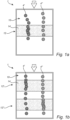
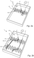
- Figs 2a and 2b illustrate a flow channel 10 of a device 1 for analyzing biological cells 4.
- the device 1 have many similarities to the device 1 described in Figs. 1a and 1b , thus only differences between the figures will be disclosed.
- the illustrated flow channel 10 comprises one zone 12.
- the flow 2 of liquid and the biological cells 4 passes the zone 12.
- the zone 12 comprises obstacles 16, in the figure obstacles 16 in the form of pillars 17.
- the illustrated flow channel 10 comprises two zones 12.
- the flow 2 of liquid and the biological cells 4 first passes a first zone 12' and then a second zone 12".
- Each zone 12 comprises obstacles 16, in the figure obstacles 16 in the form of pillars 17.
- the device 1 may be configured in the same way regardless of the device 1 having one or several zones 12. It should be understood that the device may have more than two zones.
- the side walls of the pillars 17 form lateral surfaces that obstruct the path of the biological cells 4.
- the biological cells 4 repeatedly hit the side walls of the pillars 17 during their travel through the flow channel 10.
- the side walls of the pillars 17 form cell movement modifying (CMM) surfaces 14.
- the CMM surfaces 14 comprise molecules having an affinity to a cell category.
- the CMM surfaces 14 of the zone 12 are coated by molecules having an affinity to biological cells 4' of a first category.
- the zone 12 may be considered to be associated with the first category.
- the CMM surfaces 14 of the first zone 12' are coated by molecules having an affinity to cells 4' of a first category.
- the first zone 12' may be considered to be associated with the first category.
- the CMM surfaces 14 of the second zone 12" are coated by molecules having an affinity to cells 4" of a second category.
- the second zone 12" may be considered to be associated with the second category.
- the molecules of the CMM surfaces 14 may modify the movement of the cells 4.
- the modified movements may be expressed as cells 4 taking a characteristic type of path if they interact with the CMM surfaces 14 of a zone 12.
- a biological cell 4' of the first category takes a more undulating path in the zone 12 compared to the path of the biological cell 4" of the second category.
- a biological cell 4' of the first category takes a more undulating path in the first zone 12' than in the second zone 12".
- a biological cell 4" of the second category takes a more undulating path in the second zone 12" than in the first zone 12'.
- the molecules may bind and release a cell 4 belonging to the associated cell category of the zone 12.
- an anti-CD4 antibody may bind to a cell 4 having a cell membrane comprising CD4 antigens, and subsequently release the cell.
- the movements of a biological cell 4' of the first category moving through the first zone 12' may change in a characteristic way that makes it possible to identify the cell as belonging to the first category.
- the movements of a biological cell 4" of the second category moving through the second zone 12" may change analogously.
- a biological cell belongs to a category associated with a zone the biological cell may, in that zone, exhibit e.g. a bind and release event, a halt of motion, a change of speed, a change of direction, or a change of path.
- Fig. 2a and 2b illustrates a cell 4' of the first category which, in the first zone 12', follows the curvature of the pillars 17 to an extent that indicates that the cell does not merely follow the flow 2 of liquid.
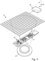
- Fig. 3 illustrates a device 1 comprising a detector 20, being an image sensor 24 and a light source 22.
- the illustrated light source 22 illuminates the zones 12 of the flow channel 10.
- the flow channel 10 of the device 1 in Fig. 3 is the flow channel 10 depicted in Fig. 2b .
- the illustrated light source 22 is configured to illuminate the cells 4 as they pass through the flow channel 10.
- the light source 22 may herein be a coherent light source 22, e.g. a laser, or a partially coherent light source 22, e.g. a light emitting diode or a light emitting diode with a pin hole or aperture.
- a coherent light source 22 e.g. a laser
- a partially coherent light source 22 e.g. a light emitting diode or a light emitting diode with a pin hole or aperture.
- the image sensor 24 may comprise a plurality of photo-sensitive elements configured to detect incident light.
- the image sensor 24 may herein be a CCD or CMOS camera.
- the image sensor 24 may acquire a time-sequence of image frames of the changing interference pattern as cells 4 pass the image sensor 24.
- the light source 22 and the image sensor 24 are arranged on opposite sides of the flow channel 10.
- the flow channel 10 is herein at least partially transparent such that the light may
- the detector 20 is configured to detect information representing the biological cells 4, carried by the flow of liquid 2, passing the at least one zone 12.
- the at least one detector 20 may be configured to detect information representing two or more biological cells 4 simultaneously, as they move across a CMM surface of the flow channel.
- the at least one detector 20 may be configured to detect information of at least one property of the biological cells 4.
- the property may be the size, shape or solidity of the biological cells 4.
- the at least one detector 20 may be an optical sensor or the device 1 may further comprise an optical sensor.
- the at least one detector 20 may be an electrical sensor or the device 1 may further comprise an electrical sensor.
- the device 1 further comprises a processor 26 which is configured to receive the information detected by the at least one detector 20.
- the processor 26 may e.g. perform a holographic reconstruction of two interference patterns detected by the detector 20 in the time-sequence of interference patterns. Said two reconstructions may depict e.g. a biological cell 4 in contact with a CMM surface 14.
- the processor 26 may not necessarily perform a full holographic reconstruction of two interference patterns in the time-sequence of interference patterns.
- the cell 4 may be detected as exhibiting a modified movement based on two, or more, interference patterns in the time-sequence of interference patterns or partial holographic reconstructions of the two, or more, interference patterns.
- the device 1 may be configured to detect modified movements of biological cells carried by the flow of liquid in a manner described in WO2022/241138 A1 , which is hereby incorporated by reference.
- the processor 26 is configured to process the information received from the at least one detector 20. The processing of the information will now be discussed in relation to Fig. 4a to c.
- the processor 26 may be a central processing unit (CPU), which may execute the instructions of one or more computer programs in order to process the information received from the at least one detector 20.
- CPU central processing unit
- the processing unit 120 may alternatively be implemented as firmware arranged e.g. in an embedded system, or as a specifically designed processing unit, such as an Application-Specific Integrated Circuit (ASIC) or a Field-Programmable Gate Array (FPGA), which may be configured to implement functionality for processing the information received from the at least one detector.
- ASIC Application-Specific Integrated Circuit
- FPGA Field-Programmable Gate Array
- the processing of the information comprises dividing each zone 12 of the flow channel 10 into segments 18, as illustrated in Fig 4a .
- the processor 26 logically defines the segments 18 of each of the zones 12 for analysis of the tracks 41', 41", 41′′′ of the biological cells 4', 4", 4′′′.
- the segmentation of the at least one zone 12 is a computational process and it will not affect the candidate biological cells 4', 4", 4′′′ passing the zone 12.
- the number of segments 18 may be 10, 100, 200, 300, 400, 500, 600, 700, 800, 900 or 1000 for each zone 12.
- Each zone 12 may be divided into segments 18 having a fixed and equal length 181.
- the length of the segments 18,181 may be 5, 10, 20, 25, 30, 35, 40, 45, 50, 100 or 1000 ⁇ m.
- a zone 12 of 10 mm may be divided into 400 segments 18, 181.
- each segment 18, 181 represents 25 ⁇ m of the zone 12.
- the processor 26 may further be configured to divide the flow channel 10 into segments 18 outside of the zone 12 as well.
- each zone 12 may, by the processor 26, be divided into segments having a fixed and equal transit time 18, 182 for candidate biological cells 4, 4', 4", 4′′′ whose movement is not modified by the CMM surface 14.
- Those segments 182 may for simplicity be called time segments 182.Thus, candidate biological cells having a high probability of not belonging to an associated biological cell category of the zone 12, 12', 12' may have a fixed an equal transit time through the time segments 182 The transit time of a biological cell 4, 4', 4", 4′′′ belonging to an associated biological cell category of the zone 12, 12', 12" has a high probability of having a transit time differing from the fixed and equal transit time.
- the number of time segments over the at least one zone may differ, as the transit time for candidate biological cells 4, 4', 4", 4′′′ whose movement is not modified by the CMM surface 14 over the time segments 182 are equal, but the total time to pass the at least one zone 12, 12', 12", 12′′′ differs depending on where the candidate biological cell moves 4, 4', 4", 4'". This is visualized in Fig 4b by two thicker paths.
- a zone 12 having a passing time of approximately 1 minute for candidate biological cells 4, 4', 4", 4′′′ not belonging to an associated biological cell category of the zone 12, 12', 12" may be divided into time segments 182 having a length of 10 seconds each. This could possibly give five, six, seven or any other number of time segments 182 of 10 seconds each.
- Dividing the zone 12, 12', 12" into time segments may be used to correct for abnormalities in the flow, such that candidate biological cells 4, 4', 4", 4′′′ may be affected by debris or any other anomality in the at least one zone 12, 12', 12", changing the transit times of the candidate biological cells 4, 4', 4", 4′′′ passing the spot of the anomality.
- dividing into time segments 182 may reveal that time segments 182 covers different length of the at least one zone 12, 12', 12", in other words that it may take different time for the candidate biological cells 4, 4', 4", 4′′′ to pass a certain distance of the at least one zone 12, 12', 12".
- the processor 26 may be configured to apply a transformation on the information detected by the detector and/or on identified tracks 41', 41", 41′′′ for mapping tracks of candidate biological cells 4, 4', 4", 4′′′ whose movement is not modified by the CMM surface 14, 14', 14" of the zone 12, 12', 12" onto straight paths of a virtual flow channel.
- the candidate biological cells 4, 4', 4", 4' may have undulating paths due to several reasons, as discussed above. It may be obstacles 16, such as pillars 17, debris or any other artefact of the at least one zone 12, 12', 12",
- obstacles 16 such as pillars 17, debris or any other artefact of the at least one zone 12, 12', 12"
- the processor 26 may be configured to normalize the transit times for each candidate biological cell 4, 4', 4", 4′′′ and for each segment 18. This normalization would allow for a simple way of analyzing flow channels 10 of different shapes, or for channels 10 having obstacles. It may further allow for a compensation for candidate biological cells 4, 4', 4", 4′′′ being stuck during the flow through the at least one zone 12, 12', 12". From this, the processor 26 may divide the zone into time segments 182 for the further analysis.
- the processor 26 may be configured to perform a weighted analysis of the transit times of each segment 18.
- the segments 18 may be then given different weights in the further analysis.
- the transit times of each segment 18 may be analyzed and based on that, decide if any segment 18 is of higher or lower importance. For instance, if the CMM surface 14 is covering the pillars or other obstacles, it may be of more interest to analyze the segments 18 covering such pillars. Likewise, knowing that a part of a zone 12, 12', 12", for instance before or after a pillar, may give rise to abnormal movements, may make it of less interest to analyze and therefore, that segment 18 may be given less weight in the analysis.
- the processor 26 may be configured to divide each zone 12, 12', 12" into individual segments 18 for each of the candidate biological cells 4, 4', 4", 4'", depending on the path taken by the biological cell.
- candidate biological cells 4, 4', 4", 4′′′ taking different paths though the at least one zone 12, 12', 12" may have passed a different number of segments 18 and may have a different number of transit times.
- candidate biological cells 4, 4', 4", 4′′′ taking completely different paths through the zone 12, 12', 12" may still be comparable, when taking into account the different paths.
- the processing of the information further comprises identifying candidate biological cells 4, as illustrated in Fig 4c where the candidate biological cells 4', 4" and 4′′′ are identified. These may be identified in information at a single point in time from the at least one detector 20.
- the candidate biological cells 4 are identified from the information detected by the at least one detector 20.
- the candidate biological cells 4 may be biological cells 4 belonging to the cell category of the zone 12. For instance, the candidate biological cell 4' of Fig. 2b belong to the associated cell category with the first zone 12', while the candidate biological cell 4" belongs to the associated cell category of the second zone 12".
- the candidate biological cells 4 may be a biological cell 4 not belonging to the cell category of the zone 12. For instance, the candidate biological cell 4' does not associate with the cell category of zone 12".
- the candidate biological cell 4 may however also be non-cell 4′′′.
- the non-cell 4′′′ could be a debris in the flow of liquid, a dead or injured cell, an artefact in the CMM surface of the zone or an imaging artefact interpreted by the detector as a cell.
- the processor 26 is configured to identify tracks 41', 41", 41′′′ from each of the candidate biological cells 4', 4", 4'". This is illustrated in Fig. 4d and 4e , illustrating how different candidate biological cells 4, 4', 4", 4" may move through the zone 12.
- the track 41' corresponds to a sequence of identified locations of the identified candidate biological cell 4' in sequential time points of the information from the at least one detector 20.
- the track 41" corresponds to a sequence of identified locations of the identified candidate biological cell 4".
- the track 41′′′ corresponds to a sequence of identified locations of the identified candidate biological cell 4′′′.
- each identified candidate biological cell 4', 4", 4′′′ has its own track 41', 41", 41′′′.
- the tracking monitors the position of the detected candidate biological cells 4', 4", 4′′′ in the flow channel 10 over a time-sequence during which the candidate biological cell 4', 4", 4′′′ passes the zone 12.
- the movements of the each of the candidate biological cell 4', 4", 4′′′ passing the zone 12 is represented by the tracks 41', 41", 41′′′.
- Each transit time represents the time it takes for one candidate biological cell 4', 4", 4′′′ to pass through the respective segment 18.
- the set of transit times represents the time it takes for the candidate biological cell 4', 4", 4′′′ to pass through the zone 12.
- a candidate biological cell 4', 4", 4′′′ belonging to the cell category associated with the zone 12 may have longer transit times than a candidate biological cell 4', 4", 4′′′ not belonging to the cell category associated to the zone 12.
- the candidate biological cell 4', 4", 4′′′ belonging to the cell category associated with the zone 12 interacts with the CMM surface 14 such that the passage of the candidate biological cells 4', 4", 4′′′ takes longer than for a candidate biological cell 4', 4", 4′′′ not interacting with the CMM surface 14.
- the set of transit times are processed to characterize the candidate biological cell 4', 4", 4′′′.
- the characterization may be used to determine if the candidate biological cell 4', 4", 4′′′ belongs to the biological cell category associated with the zone 12.
- the processing of the set of transit times may comprise determining at least one parameter representing a sequence of transit times within the set of transit times, and characterizing the candidate biological cell 4', 4", 4′′′ based on the at least one parameter.
- the processing may involve investigating the distribution of the segment transit times. From the distribution, parameters such as mean transit time, standard deviation of all transit times of candidate biological cell 4', 4", 4′′′, skewness of the distribution of all segment transit times may be determined.
- the parameters from each of the candidate biological cells 4', 4", 4′′′ may be compared to each other. For instance, the skewness of the transit times may be plotted against the standard deviation of the transit times. This may result in clusters, such that the candidate biological cells 4', 4", 4′′′ belonging to the biological cell category associated with the zone 12 may have similar skewness and standard deviation of the transit times. Further, the candidate biological cells 4', 4", 4′′′ not belonging to the biological cell category associated with the zone 12 may have a similar skewness and standard deviation of the transit times.
- the skewness and the standard deviation of the candidate biological cells 4', 4", 4′′′ belonging to the biological cell category associated with the zone 12 may differ from the skewness and standard deviation of the candidate biological cells 4', 4", 4′′′ not belonging to the biological cell category associated with the zone 12.
- skewness and standard deviation is only two examples of descriptive parameters which may be used to classify the candidate biological cells 4', 4", 4′′′ based on their transit time over the CMM surface 14.
- the processing of the set of transit times may be made by use of a machine learning model.
- the machine learning model may be based on supervised training.
- the machine learning model may be an artificial neural network.
- the machine learning model may thus be based on sets of transit times determined for known biological cells and/or other particles passing through the flow channel.
- the machine learning model may be based on training for a particular flow channel or based on general training of cells passing over CMM surfaces.
- Fig. 5 illustrates a flow chart of a method 100 for characterizing biological cells 4', 4", 4′′′.
- the method 100 comprises receiving 101 information detected by at least one detector 20 representing biological cells 4', 4", 4′′′, carried by a flow of liquid 2.
- the candidate biological cells 4', 4", 4′′′ are to be characterized when passing at least one zone 12 in a flow channel 10.
- Each zone 12 of the flow channel 10 is associated with a cell category.
- Each zone 12 of the flow channel 10 comprises at least one surface 14 coated with molecules having an affinity to the cell category associated with the zone.
- the at least one surface 14 of the zone 12 being configured to modify, by the molecules coating the at least one surface 14, a movement of a biological cell 4', 4", 4′′′ belonging to the associated cell category of the zone as the biological cell 4', 4", 4′′′ carried by the flow of liquid 2 passes the zone 12.
- the at least one surface 14 forms at least one biological cell movement modifying (CMM) surface.
- CMM biological cell movement modifying
- the method 100 further comprises processing 102 the received information.
- the processing 102 of the received information comprises dividing 102a each zone 12 of the flow channel 2 into segments 18.
- the segmentation of the at least one zone 12 is a computational process and it will not affect the candidate biological cells 4', 4", 4′′′ passing the zone 12.
- the processor 26 may further be configured to divide the flow channel 10 into segments 18 outside of the zone 12 as well.
- the processing 102 of the received information further comprises identifying 102b candidate biological cells 4', 4", 4′′′ in the flow channel 10. These may be identified in information at a single point in time from the at least one detector 20.
- the candidate biological cells 4 are identified from the information detected by the at least one detector 20.
- the processing 102 of the received information further comprises identifying 102c tracks 41', 41", 41′′′ from each of the candidate biological cells 4', 4", 4′′′.
- the tracking monitors the position of the detected candidate biological cells 4', 4", 4′′′ in the flow channel 10 over a time-sequence during which the candidate biological cell 4', 4", 4′′′ passes the zone 12.
- the movements of the candidate biological cell 4', 4", 4′′′ passing the zone 12 is represented by the tracks 41', 41", 41′′′.
- the processing 102 of the received information further comprises determining 102d a set of transit times.
- Each transit time in the set represents a transit time of the candidate biological cell 4', 4", 4′′′ through a segment 18.
- Each transit time represents the time it takes for one candidate biological cell 4', 4", 4′′′ to pass through the respective segment 18.
- the set of transit times represents the time it takes for the candidate biological cell 4', 4", 4′′′ to pass through the zone 12.
- the processing 102 of the received information further comprises processing 102e the set of transit times for characterizing the candidate biological cell 4', 4", 4′′′.
- the processing 102e may be made by determining whether the candidate biological cell 4', 4", 4′′′ is a biological cell 4', 4", 4′′′ belonging to an associated biological cell category of each respective zone 12.
- the processing 102e of the set of transit times may comprise determining at least one parameter representing a sequence of transit times within the set of transit times, and characterizing the candidate biological cell 4, 4', 4", 4′′′ based on the at least one parameter.
- the processing 102e may involve investigating the distribution of the segment transit times. From the distribution, parameters such as mean transit time, standard deviation of all transit times of candidate biological cell 4', 4", 4′′′, skewness of the distribution of all segment transit times may be determined.
- the parameters from each of the candidate biological cells 4', 4", 4′′′ may be compared to each other. For instance, the skewness of the transit times may be plotted against the standard deviation of the transit times. This may result in clusters, such that the candidate biological cells 4', 4", 4′′′ belonging to the biological cell category associated with the zone 12 may have similar skewness and standard deviation of the transit times. Further, the candidate biological cells 4', 4", 4′′′ not belonging to the biological cell category associated with the zone 12 may have a similar skewness and standard deviation of the transit times.
- skewness and standard deviation are only two examples of descriptive parameters which may be used to classify the candidate biological cells 4', 4", 4′′′ based on their transit time over the CMM surface 14.
- the processing 102e of the set of transit times may be made by use of a machine learning model.
Landscapes
- Health & Medical Sciences (AREA)
- Life Sciences & Earth Sciences (AREA)
- Chemical & Material Sciences (AREA)
- Engineering & Computer Science (AREA)
- Immunology (AREA)
- Biomedical Technology (AREA)
- General Health & Medical Sciences (AREA)
- Analytical Chemistry (AREA)
- Hematology (AREA)
- Biochemistry (AREA)
- Molecular Biology (AREA)
- Urology & Nephrology (AREA)
- Physics & Mathematics (AREA)
- General Physics & Mathematics (AREA)
- Pathology (AREA)
- Microbiology (AREA)
- Cell Biology (AREA)
- Biotechnology (AREA)
- Zoology (AREA)
- Organic Chemistry (AREA)
- Food Science & Technology (AREA)
- Bioinformatics & Cheminformatics (AREA)
- Dispersion Chemistry (AREA)
- Wood Science & Technology (AREA)
- Medicinal Chemistry (AREA)
- General Engineering & Computer Science (AREA)
- Genetics & Genomics (AREA)
- Tropical Medicine & Parasitology (AREA)
- Virology (AREA)
- Sustainable Development (AREA)
- Fluid Mechanics (AREA)
- Clinical Laboratory Science (AREA)
- Chemical Kinetics & Catalysis (AREA)
- Signal Processing (AREA)
- Apparatus Associated With Microorganisms And Enzymes (AREA)
Applications Claiming Priority (1)
| Application Number | Priority Date | Filing Date | Title |
|---|---|---|---|
| EP23219764 | 2023-12-22 |
Publications (1)
| Publication Number | Publication Date |
|---|---|
| EP4575460A1 true EP4575460A1 (de) | 2025-06-25 |
Family
ID=89321549
Family Applications (1)
| Application Number | Title | Priority Date | Filing Date |
|---|---|---|---|
| EP24221184.5A Pending EP4575460A1 (de) | 2023-12-22 | 2024-12-18 | Vorrichtung und verfahren zur charakterisierung biologischer zellen |
Country Status (2)
| Country | Link |
|---|---|
| US (1) | US20250208134A1 (de) |
| EP (1) | EP4575460A1 (de) |
Citations (1)
| Publication number | Priority date | Publication date | Assignee | Title |
|---|---|---|---|---|
| WO2022241138A1 (en) | 2021-05-13 | 2022-11-17 | Janssen Biotech, Inc. | Device and method for sorting biological cells |
-
2024
- 2024-12-18 EP EP24221184.5A patent/EP4575460A1/de active Pending
- 2024-12-19 US US18/987,869 patent/US20250208134A1/en active Pending
Patent Citations (1)
| Publication number | Priority date | Publication date | Assignee | Title |
|---|---|---|---|---|
| WO2022241138A1 (en) | 2021-05-13 | 2022-11-17 | Janssen Biotech, Inc. | Device and method for sorting biological cells |
Also Published As
| Publication number | Publication date |
|---|---|
| US20250208134A1 (en) | 2025-06-26 |
Similar Documents
| Publication | Publication Date | Title |
|---|---|---|
| US20220092773A1 (en) | Identifying the quality of the cell images acquired with digital holographic microscopy using convolutional neural networks | |
| US11113506B2 (en) | Method for providing an evaluation means for at least one optical application system of a microscope-based application technology | |
| Di Cataldo et al. | Computer-aided techniques for chromogenic immunohistochemistry: status and directions | |
| US20220254020A1 (en) | Deep Learning Method For Predicting Patient Response To A Therapy | |
| US20240226893A1 (en) | Device and Method for Sorting Biological Cells | |
| JP2013531787A5 (ja) | 粒子の運動度および/または細胞の分散を求めるためのホログラフィック変動顕微鏡装置、方法並びにプログラム | |
| US20240071057A1 (en) | Microscopy System and Method for Testing a Sensitivity of an Image Processing Model | |
| US20180231565A1 (en) | Methods for determining the risk of a systemic lupus erythematosus (sle) patient to develop neuropsychiatric syndromes | |
| CN113066064A (zh) | 基于人工智能的锥束ct图像生物结构识别及三维重建系统 | |
| KR102500220B1 (ko) | 3차원 굴절률 영상과 인공지능을 이용한 세포의 세부 분류 구분 방법 및 장치 | |
| Höfener et al. | Automated density-based counting of FISH amplification signals for HER2 status assessment | |
| EP4575460A1 (de) | Vorrichtung und verfahren zur charakterisierung biologischer zellen | |
| KR101657042B1 (ko) | 초해상도 단일 입자 추적 데이터의 실시간 분석 기술을 통한 막단백질 확산의 역학적 분석 | |
| ES2974024T3 (es) | Análisis de biomarcadores para datos multiplex de diagnóstico de alto rendimiento | |
| EP4575459A1 (de) | Vorrichtung und verfahren zur analyse biologischer zellen | |
| JP7445672B2 (ja) | ゲート領域推定プログラム、ゲート領域推定装置、学習モデルの生成方法 | |
| Georgantzoglou et al. | Neutrophil motion in numbers: How to analyse complex migration patterns | |
| Belyaev et al. | Automated characterisation of neutrophil activation phenotypes in ex vivo human Candida blood infections | |
| Tatarchenko et al. | Histogram-based deep learning for automotive radar | |
| JP7478248B2 (ja) | レンズレス顕微撮像システムとその方法、及び生化学物質検出システムとその方法 | |
| EP4685463A1 (de) | Vorrichtung und verfahren zur verfolgung biologischer zellen | |
| EP4685462A1 (de) | Vorrichtung und verfahren zum sortieren von biologischen zellen | |
| EP3279662A1 (de) | Lateral-flow-immuntestassay | |
| US20110058728A1 (en) | Device and Method for Automatic Detection of Dynamic Processes of Cells in Cell Samples | |
| US20250114793A1 (en) | System and Method for Label-Free Cell Identification and/or Classification and/or Selection |
Legal Events
| Date | Code | Title | Description |
|---|---|---|---|
| PUAI | Public reference made under article 153(3) epc to a published international application that has entered the european phase |
Free format text: ORIGINAL CODE: 0009012 |
|
| STAA | Information on the status of an ep patent application or granted ep patent |
Free format text: STATUS: THE APPLICATION HAS BEEN PUBLISHED |
|
| AK | Designated contracting states |
Kind code of ref document: A1 Designated state(s): AL AT BE BG CH CY CZ DE DK EE ES FI FR GB GR HR HU IE IS IT LI LT LU LV MC ME MK MT NL NO PL PT RO RS SE SI SK SM TR |
|
| STAA | Information on the status of an ep patent application or granted ep patent |
Free format text: STATUS: REQUEST FOR EXAMINATION WAS MADE |
|
| 17P | Request for examination filed |
Effective date: 20251209 |