EP4549564A2 - Schaumige viren und verfahren zur verwendung - Google Patents
Schaumige viren und verfahren zur verwendung Download PDFInfo
- Publication number
- EP4549564A2 EP4549564A2 EP25151397.4A EP25151397A EP4549564A2 EP 4549564 A2 EP4549564 A2 EP 4549564A2 EP 25151397 A EP25151397 A EP 25151397A EP 4549564 A2 EP4549564 A2 EP 4549564A2
- Authority
- EP
- European Patent Office
- Prior art keywords
- cells
- sfv
- pan1
- cancer
- cell
- Prior art date
- Legal status (The legal status is an assumption and is not a legal conclusion. Google has not performed a legal analysis and makes no representation as to the accuracy of the status listed.)
- Pending
Links
Images










Classifications
-
- A—HUMAN NECESSITIES
- A61—MEDICAL OR VETERINARY SCIENCE; HYGIENE
- A61K—PREPARATIONS FOR MEDICAL, DENTAL OR TOILETRY PURPOSES
- A61K35/00—Medicinal preparations containing materials or reaction products thereof with undetermined constitution
- A61K35/66—Microorganisms or materials therefrom
- A61K35/76—Viruses; Subviral particles; Bacteriophages
- A61K35/768—Oncolytic viruses not provided for in groups A61K35/761 - A61K35/766
-
- A—HUMAN NECESSITIES
- A61—MEDICAL OR VETERINARY SCIENCE; HYGIENE
- A61P—SPECIFIC THERAPEUTIC ACTIVITY OF CHEMICAL COMPOUNDS OR MEDICINAL PREPARATIONS
- A61P35/00—Antineoplastic agents
-
- C—CHEMISTRY; METALLURGY
- C12—BIOCHEMISTRY; BEER; SPIRITS; WINE; VINEGAR; MICROBIOLOGY; ENZYMOLOGY; MUTATION OR GENETIC ENGINEERING
- C12N—MICROORGANISMS OR ENZYMES; COMPOSITIONS THEREOF; PROPAGATING, PRESERVING, OR MAINTAINING MICROORGANISMS; MUTATION OR GENETIC ENGINEERING; CULTURE MEDIA
- C12N7/00—Viruses; Bacteriophages; Compositions thereof; Preparation or purification thereof
-
- C—CHEMISTRY; METALLURGY
- C12—BIOCHEMISTRY; BEER; SPIRITS; WINE; VINEGAR; MICROBIOLOGY; ENZYMOLOGY; MUTATION OR GENETIC ENGINEERING
- C12N—MICROORGANISMS OR ENZYMES; COMPOSITIONS THEREOF; PROPAGATING, PRESERVING, OR MAINTAINING MICROORGANISMS; MUTATION OR GENETIC ENGINEERING; CULTURE MEDIA
- C12N2740/00—Reverse transcribing RNA viruses
- C12N2740/00011—Details
- C12N2740/10011—Retroviridae
- C12N2740/17011—Spumavirus, e.g. chimpanzee foamy virus
- C12N2740/17021—Viruses as such, e.g. new isolates, mutants or their genomic sequences
-
- C—CHEMISTRY; METALLURGY
- C12—BIOCHEMISTRY; BEER; SPIRITS; WINE; VINEGAR; MICROBIOLOGY; ENZYMOLOGY; MUTATION OR GENETIC ENGINEERING
- C12N—MICROORGANISMS OR ENZYMES; COMPOSITIONS THEREOF; PROPAGATING, PRESERVING, OR MAINTAINING MICROORGANISMS; MUTATION OR GENETIC ENGINEERING; CULTURE MEDIA
- C12N2740/00—Reverse transcribing RNA viruses
- C12N2740/00011—Details
- C12N2740/10011—Retroviridae
- C12N2740/17011—Spumavirus, e.g. chimpanzee foamy virus
- C12N2740/17022—New viral proteins or individual genes, new structural or functional aspects of known viral proteins or genes
-
- C—CHEMISTRY; METALLURGY
- C12—BIOCHEMISTRY; BEER; SPIRITS; WINE; VINEGAR; MICROBIOLOGY; ENZYMOLOGY; MUTATION OR GENETIC ENGINEERING
- C12N—MICROORGANISMS OR ENZYMES; COMPOSITIONS THEREOF; PROPAGATING, PRESERVING, OR MAINTAINING MICROORGANISMS; MUTATION OR GENETIC ENGINEERING; CULTURE MEDIA
- C12N2740/00—Reverse transcribing RNA viruses
- C12N2740/00011—Details
- C12N2740/10011—Retroviridae
- C12N2740/17011—Spumavirus, e.g. chimpanzee foamy virus
- C12N2740/17032—Use of virus as therapeutic agent, other than vaccine, e.g. as cytolytic agent
Definitions
- This document relates to methods and materials for treating cancer.
- this document provides methods and materials for treating cancer using recombinant foamy viruses (e.g ., recombinant simian foamy viruses (SFVs)) as an oncolytic agent.
- recombinant foamy viruses e.g ., recombinant simian foamy viruses (SFVs)
- cancer remains a major public health issue in the United States with over 1.6 million new cases in 2017 alone ( National Cancer Institute, "Cancer Stat Facts: Cancer of Any Site,” seer.cancer.gov/statfacts/html/all.html ).
- Traditional therapies such as chemotherapeutics, radiation therapy and surgery, often fail, especially when cancer is advanced.
- cancer cells can eliminate or modify the components that are targeted by these therapies and effectively avoid being killed.
- it is critical to develop therapeutics that becoming resistant to is more difficult.
- Foamy viruses also known as Spumaviruses (e.g ., a virus in the genus Spumavirus ), are retroviruses that infect only dividing cells and induce formation of syncytia - large multinucleated cells. In contrast to its widespread prevalence in various mammalian species, FV infection has not been associated with any diseases in their natural hosts. The natural preference of FVs to infect dividing cells limits the possible off-target infections, whereas the strong cytopathic effect (CPE) caused by the virus can lead to cancer cell death. As described herein, FVs can be used as a retroviral platform for safe oncolytic virotherapy.
- Spumaviruses e.g ., a virus in the genus Spumavirus
- this document provides methods and materials for treating cancer.
- this document provides methods and materials for treating cancer using one or more recombinant FVs (e.g., one or more recombinant SFVs) as an oncolytic agent.
- one or more recombinant FVs can be used to reduce the number of cancer cells ( e.g., by infecting and killing cancer cells) in a mammal.
- one or more recombinant FVs can be used to stimulate anti-cancer immune responses in a mammal.
- Oncolytic virotherapy can provide an alternative approach to cancer treatment by utilizing selectively replicating viruses to destroy tumors, activate adaptive immune responses and ensure a life-long immunity against the tumors ( Russell et al., 2017 Molecular Therapy 25:1107-1116 ).
- recombinant SFVs that are replication competent and are generated by combining genome segments from chimpanzee SFV types 6 and 7 can be used as anticancer agents to reduce the number of cancer cells within a mammal ( e.g., a human).
- one aspect of this document features a chimeric SFV including a first genomic fragment of a first SFV strain, and a second genomic fragment of a second SFV strain.
- the first SFV strain can be a first chimpanzee SFV strain
- the second SFV strain can be a second chimpanzee SFV strain that is different from the first chimpanzee SFV strain.
- the first SFV strain and the second SFV strain can be serotypically distinct.
- the first SFV strain can be a chimpanzee PAN1 SFV strain
- the second SFV strain can be a chimpanzee PAN2 SFV strain.
- the first genomic fragment of the first SFV strain can include a 5' LTR, a nucleic acid encoding a gag polypeptide, and a 5' portion of a nucleic acid encoding pol polypeptide
- the second genomic fragment of the second SFV strain can include a 3' portion of a nucleic acid encoding a pol polypeptide, a nucleic acid encoding a env polypeptide, a nucleic acid encoding a tas polypeptide, a nucleic acid encoding a bel2 polypeptide, and a 3' LTR.
- the 5' portion of the nucleic acid encoding the pol polypeptide and the 3' portion of the nucleic acid encoding the pol polypeptide can be separated by a SfiI restriction site in the pol polypeptide.
- the chimeric SFV further also can include a transgene.
- the transgene can encode a detectable label.
- the detectable label can be a fluorophore selected from the group consisting of green fluorescent protein, mCherry, yellow fluorescent protein, cyan fluorescent protein, or Tomato.
- the transgene can encode a suicide polypeptide.
- the suicide polypeptide can be a thymidine kinase, an inducible Caspase 9, a sodium/iodide symporter, a viral polypeptide, or a nitroreductase.
- the transgene can encode a receptor.
- the receptor can be a chimeric antigen receptor (CAR).
- the CAR can target a cancer antigen.
- the chimeric SFV further also can include a promoter. The promoter can be substituted for the U3 region of a 3' LTR of the second genomic fragment of the second SFV strain.
- the promoter can be a cancer-specific promoter (e.g., a promoter from a cancer/testis antigen, a promoter from a carcinoembryonic antigen, a promoter from a melan-A antigen, a promoter from a prostate-specific antigen, or a promoter from a telomerase reverse transcriptase.
- a cancer-specific promoter e.g., a promoter from a cancer/testis antigen, a promoter from a carcinoembryonic antigen, a promoter from a melan-A antigen, a promoter from a prostate-specific antigen, or a promoter from a telomerase reverse transcriptase.
- this document features methods for treating a mammal having cancer.
- the methods can include, or consist essentially of, administering, to a mammal, a SFV having oncolytic anti-cancer activity, where the SFV includes a first genomic fragment of a first SFV strain and a second genomic fragment of a second SFV strain different from the first SFV strain.
- the mammal can be a human.
- the cancer can be a glioblastoma, a pancreatic adenocarcinoma, a cholangiocarcinoma, a mesothelioma, a melanoma, a prostate cancer, a breast cancer, an ovarian cancer, a liver cancer, or a colorectal cancer.
- the first SFV strain can be a chimpanzee SFV strain
- the second SFV strain can be a different chimpanzee SFV strain.
- the first SFV strain and the second SFV strain can be serotypically distinct.
- the first SFV strain can be a chimpanzee PAN1 SFV strain
- the second SFV strain can be a chimpanzee PAN2 SFV strain.
- the first genomic fragment of the first SFV strain can include a 5' LTR, a nucleic acid encoding a gag polypeptide, and a 5' portion of a nucleic acid encoding a pol polypeptide
- the second genomic fragment the said second SFV strain can include a 3' portion of a nucleic acid encoding a pol polypeptide, a nucleic acid encoding a env polypeptide, a nucleic acid encoding a tas polypeptide, a nucleic acid encoding a bel2 polypeptide, and a 3' LTR.
- the 5' portion of the nucleic acid encoding the pol polypeptide and the 3' portion of the nucleic acid encoding the pol polypeptide can be separated by a SfiI restriction site in the nucleic acid encoding the pol polypeptide.
- the SFV also can include a transgene encoding a suicide polypeptide.
- the suicide polypeptide can be a thymidine kinase, an inducible Caspase 9, a sodium/iodide symporter, a viral polypeptide, or a nitroreductase.
- the SFV also can include a promoter.
- the promoter can be substituted for the U3 region of a 3' LTR of the second genomic fragment of the second SFV strain.
- the promoter can be a cancer-specific promoter (e.g., a promoter from a cancer/testis antigen, a promoter from a carcinoembryonic antigen, a promoter from a melan-A antigen, a promoter from a prostate-specific antigen, or a promoter from a telomerase reverse transcriptase).
- This document provides methods and materials for treating cancer.
- this document provides methods and materials for treating cancer using one or more recombinant FVs as an oncolytic agent.
- this document provides recombinant FVs (e.g., recombinant SFVs) having oncolytic anti-cancer activity.
- this document provides methods for using one or more recombinant FVs provided herein to treat a mammal having, or at risk of having, cancer.
- one or more recombinant FVs can be administered to a mammal having, or at risk of having, cancer to reduce the number of cancer cells ( e.g., by infecting and killing cancer cells) in the mammal ( e.g., a human).
- one or more recombinant FVs can be administered to a mammal having, or at risk of having, cancer to stimulate anti-cancer immune responses in the mammal ( e.g ., a human).
- a recombinant FV described herein e.g., a recombinant FV having oncolytic anti-cancer activity
- a recombinant FV described herein e.g., a recombinant FV having oncolytic anti-cancer activity
- are non-pathogenic e.g ., to a mammal being treated as described herein.
- a recombinant FV described herein e.g., a recombinant FV having oncolytic anti-cancer activity
- can infect dividing cells e.g., can infect only dividing cells.
- a recombinant FV described herein e.g., a recombinant FV having oncolytic anti-cancer activity
- a recombinant FV described herein can bind to a cellular receptor (e.g., to facilitate viral entry to a cell).
- a recombinant FV described herein can bind to a heparan sulfate cellular receptor.
- Recombinant FVs described herein can include one or more nucleotide sequences that do not naturally occur in that FV (e.g., do no naturally occur in that FV prior to recombination). Nucleotide sequences that do not naturally occur in the FV can be from any appropriate source. In some cases, a nucleotide sequence that does not naturally occur in that FV can be from a non-viral organism. In some cases, a nucleotide sequence that does not naturally occur in that FV can be from a virus other than a FV.
- a nucleotide sequence that does not naturally occur in that FV can be from a FV obtained from a different species. In some cases, a nucleotide sequence that does not naturally occur in that FV can be from a different strain of FV ( e.g ., serotypically distinct strains). In some cases, a nucleotide sequence that does not naturally occur in that FV can be a synthetic nucleotide sequence.
- Recombinant FVs described herein can be derived from ( e.g., can include genomic elements such as nucleic acids encoding a polypeptide (or fragments thereof)) from any appropriate FV.
- FVs can be isolated from any appropriate species. For example, FVs can be isolated from non-human primates ( e.g. , monkeys such as Old World monkey species), humans, cats, cows, horses, or bats.
- a recombinant FV can include one or more nucleic acids encoding a polypeptide (or fragments thereof) from a simian FV (SFV).
- SFV simian FV
- simian species from which SFVs can be obtained include, without limitation, chimpanzee, gorilla, orangutan, African green monkey, baboon, macaque, marmoset, gibbon ape, cynomolgus monkey, and squirrel monkey.
- a recombinant FV described herein can include one or more nucleic acids encoding a polypeptide (or fragments thereof) and/or one or more viral elements from a SFV genome obtained from a chimpanzee (e.g ., a chimpanzee SFV such as a type 6 (PAN1) SFV strain and/or a type 7 (PAN2) SFV strain).
- a recombinant FV can include one or more nucleic acids encoding a polypeptide (or fragments thereof) and/or one or more viral elements from a FV be obtained from a human (e.g., a human foamy virus (HFV) and a prototype foamy virus (PFV)).
- a human e.g., a human foamy virus (HFV) and a prototype foamy virus (PFV)
- Nucleic acids that can be included in a FV genome include, for example, gag nucleic acid (e.g., nucleic acid encoding the group-specific antigen protein), pol nucleic acid (e.g., nucleic acid encoding the DNA polymerase), env nucleic acid (e.g., nucleic acid encoding the envelope protein), tas nucleic acid ( e.g., nucleic acid encoding the transactivator protein), and bel2 nucleic acid (e.g., nucleic acid encoding the Bet protein).
- gag nucleic acid e.g., nucleic acid encoding the group-specific antigen protein
- pol nucleic acid e.g., nucleic acid encoding the DNA polymerase
- env nucleic acid e.g., nucleic acid encoding the envelope protein
- tas nucleic acid e.g., nucleic acid encoding the transactivator protein
- Viral elements that can be included in a FV genome include, without limitation, a 5' long terminal repeat (LTR) and a 3' LTR, each of which can include a U3 region, a R region, and a U5 region.
- LTR 5' long terminal repeat
- 3' LTR 3' LTR
- An exemplary schematic FV genome is shown in Figure 1 .
- a recombinant FV e.g., SFV
- a recombinant FV having oncolytic anti-cancer activity can include a chimeric FV genome.
- a chimeric FV genome can include one or more nucleic acids encoding a polypeptide (or fragments thereof) and/or one or more viral elements from two or more ( e.g., two, three, four, five, or more) different FV genomes.
- a recombinant FV e.g., SFV
- a recombinant FV described herein can include one or more nucleic acids encoding a polypeptide (or fragments thereof) and/or one or more viral elements from two or more different strains of FV isolated from the same species.
- a recombinant SFV described herein can include one or more nucleic acids encoding a polypeptide (or fragments thereof) and/or one or more viral elements from a PAN1 strain of a chimpanzee SFV and one or more nucleic acids encoding a polypeptide (or fragments thereof) and/or one or more viral elements from a PAN2 strain of a chimpanzee SFV.
- a PAN1 strain of SFV can have a sequence set forth in, for example, National Center for Biotechnology Information (NCBI) Accession Nos: L25422 and X83297.
- a recombinant SFV having oncolytic anti-cancer activity can include the 5' LTR, the gag nucleic acid, and a portion ( e.g., a first portion such as the 5' portion) of the pol nucleic acidfrom a PAN1 SFV and a portion ( e.g., a second portion such as the 3' portion) of the pol nucleic acid, the env nucleic acid, the tas nucleic acid, the bel2 nucleic acid, and the 3' LTR from a PAN2 SFV.
- a recombinant SFV having oncolytic anti-cancer activity can include from the 5' LTR to the SfiI restriction site (localized in the pol nucleic acid) from a PAN1 SFV and from the SfiI restriction site (localized in the pol nucleic acid) to the 3' LTR from a PAN2 SFV (see, e.g., Figure 2B ).
- a recombinant FV e.g., SFV
- a recombinant FV e.g., SFV
- recombinant FVs having oncolytic anti-cancer activity can include one or more nucleic acids encoding a polypeptide (or fragments thereof) and/or one or more viral elements from two or more FVs isolated from different species ( e.g., one or more nucleic acids encoding a polypeptide (or fragments thereof) and/or one or more viral elements from a chimpanzee SFV and one or more nucleic acids encoding a polypeptide (or fragments thereof) and/or one or more viral elements from a human FV such as PVF).
- a recombinant FV e.g., SFV
- a recombinant FV having oncolytic anti-cancer activity can include a FV genome containing one or more modifications to one or more nucleic acids encoding a polypeptide (or fragments thereof) and/or one or more viral elements of the FV genome.
- the one or more modifications can be any appropriate modification. Examples of modifications that can be made to a nucleic acid encoding a polypeptide or to a viral element include, without limitation, deletions, insertions, and substitutions.
- a recombinant FV can include one or more genomic deletions.
- a recombinant FV can have a deletion ( e.g., a full deletion or a partial deletion) of the bel2 nucleic acid. In some cases, a recombinant FV can have a deletion ( e.g., a full deletion or a partial deletion) of the tas nucleic acid. In some cases, a recombinant FV can have a deletion ( e.g., a full deletion or a partial deletion) of the U3 region of the 3' LTR. For example, a recombinant FV can include one or more genomic insertions ( e.g ., insertion of one or more transgenes).
- a recombinant FV can include a transgene (e.g ., a nucleic acid encoding a suicide polypeptide).
- a recombinant FV can include a regulatory element (e.g ., promoter such as a cancer-specific promoter).
- a recombinant FV can include one or more genomic substitutions (e.g., a substitution of one or more nucleic acids encoding a polypeptide with one or more transgenes).
- a recombinant FV can have a U3 region of the 3' LTR substituted with a cancer-specific promoter.
- a recombinant FV can have bel2 nucleic acid replaced with a transgene (e.g ., a nucleic acid encoding a suicide polypeptide).
- a recombinant FV described herein e.g., a recombinant FV having oncolytic anti-cancer activity
- the transgene can be any appropriate transgene.
- a transgene can be a nucleotide sequence encoding a detectable label.
- detectable labels include, without limitation, fluorophores (e.g ., green fluorescent protein (GFP), mCherry, yellow fluorescent protein (YFP), cyan fluorescent protein (CFP), and Tomato), enzymes (e.g ., luciferase, CRISPR associated protein 9 (Cas9), Cre recombinase, restriction enzymes, convertases, thymidine kinases, and sodium/iodide symporters (NISs)), and antigens.
- fluorophores e.g ., green fluorescent protein (GFP), mCherry, yellow fluorescent protein (YFP), cyan fluorescent protein (CFP), and Tomato
- enzymes e.g ., luciferase, CRISPR associated protein 9 (Cas9), Cre recombinase, restriction enzymes, convertases, thymidine kinases, and sodium/iodide symporters (NISs)
- NISs
- a transgene can be a nucleotide sequence encoding a receptor (e.g., a chimeric antigen receptor (CAR)).
- a receptor e.g., a chimeric antigen receptor (CAR)
- a receptor such as a CAR, can target any appropriate antigen (e.g ., a cancer antigen).
- a transgene is a nucleotide sequence encoding a CAR
- the CAR can be any appropriate type of CAR.
- a CAR can be a first, second, third, or fourth generation CAR.
- a CAR can include an extracellular domain (e.g ., which can interact with a target molecule such as an antigen), a transmembrane domain, and an intracellular domain.
- an extracellular domain of a CAR can include an antigen recognition region that can bind the target antigen, a signal peptide, and, optionally, a spacer/linker.
- an extracellular domain of a CAR can include a single-chain variable fragment (scFv) including the light (VL) and heavy (VH) chains of an immunoglobin (e.g ., an immunoglobulin that can bind the target antigen).
- a transmembrane domain of a CAR can include a hydrophobic alpha helix that spans a cell membrane.
- a transmembrane domain of a CAR can be a CD28 transmembrane domain.
- a transmembrane domain of a CAR can be a CD3-zeta transmembrane domain.
- an intracellular domain of a CAR can include a cytoplasmic end of a receptor that can, when a target antigen is bound to the extracellular domain of the CAR, activate T cell signaling of a T cell presenting the CAR.
- An intracellular domain of a CAR can be a recombinant cytoplasmic domain.
- An intracellular domain of a CAR can be a chimeric cytoplasmic domain. Examples of domains that can be used as a cytoplasmic domain of an intracellular domain of a CAR include, without limitation, a CD3-zeta cytoplasmic domain, a CD28 domain, a 4-1BB (CD127) domain, and an OX40 domain.
- a CAR can be as described in, for example, International Patent Publication No. WO 2015/142675 (see, e.g., paragraph [0243], paragraph [0269], and Figure 59A), US Patent Publication No. 2019/085081 (see, e.g., paragraphs [0298] - [0308]), and US Patent Publication No. 2019/055299 (see, e.g., paragraphs [0061], [0064] - [0099]).
- a transgene can be a nucleotide sequence encoding another useful polypeptide.
- useful polypeptides include, without limitation, targeting polypeptides (e.g., ligands (e.g., natural ligands and artificial ligands) for cell surface receptors such as cytokines and hormones, single chain antibodies (e.g ., targeting cancer antigens such as HER2, CA125, CD19, CD30, CD33, CD123, FLT3, BCMA, CEA, melan-A antigen, and PSA), and other viral envelopes), transport polypeptides (e.g ., nuclear localization sequences (NLSs), mitochondorion targeting sequences, and lysosome targeting sequences), therapeutic polypeptides (e.g ., immunomodulatory factors such as chemokines and cytokines, antibodies such as antibodies blocking immune checkpoint molecules (e.g ., PD-1, PDL-1, and CTLA-4), genome editing
- targeting polypeptides e.
- the bel2 nucleic acid can be replaced with a transgene (e.g., a nucleotide sequence encoding GFP; see, e.g., Figure 2C ).
- a transgene e.g., a nucleotide sequence encoding GFP; see, e.g., Figure 2C .
- the bel2 nucleic acid can be replaced with a transgene (e.g., a nucleotide sequence encoding a suicide polypeptide; see, e.g., Figures 10A and 10B ).
- the U3 region of the 3' LTR can be replaced with a promoter (e.g ., a cancer-specific promoter such as a promoter from a tumor antigen or cancer marker such as cancer/testis (CT) antigens ( e.g ., NY-ESO1), CEA, melan-A antigen, and PSA, and a promoter from a telomerase reverse transcriptase (TERT) such as a human TERT (hTERT)); see, e.g., Figures 9A and 9B .
- a cancer-specific promoter such as a promoter from a tumor antigen or cancer marker such as cancer/testis (CT) antigens (e.g ., NY-ESO1), CEA, melan-A antigen, and PSA
- CT cancer/testis
- CEA e.g ., NY-ESO1
- CEA cancer/testis
- hTERT human TERT
- expression vectors containing a recombinant FV described herein e.g., a recombinant FV having oncolytic anti-cancer activity.
- Expression vectors can carry a recombinant FV described herein into another cell (e.g., a cancer cell), where it can be replicated and/or expressed.
- An expression vector also commonly referred to as an expression construct, is typically a plasmid or vector having an enhancer/promoter region controlling expression of a specific nucleotide sequence.
- the expression vector can use cellular protein synthesis machinery to produce the virus in the cell.
- expression vectors containing recombinant FVs described herein can be viral vectors.
- an expression vector containing a recombinant FV described herein can be a retroviral vector.
- expression vectors including a recombinant FV described herein also can be designed to allow insertion of one or more transgenes ( e.g ., at a multi-cloning site).
- expression vectors including a recombinant FV described herein also can include a nucleotide sequence encoding a detectable label.
- detectable labels include, without limitation, fluorophores (e.g ., GFP, mCherry, YFP, CFP, and Tomato), enzymes (e.g ., luciferase, Cas9, Cre recombinase, restriction enzymes, convertases, thymidine kinases, and NISs), and antigens.
- fluorophores e.g ., GFP, mCherry, YFP, CFP, and Tomato
- enzymes e.g ., luciferase, Cas9, Cre recombinase, restriction enzymes, convertases, thymidine kinases, and NISs
- antigens e.g luciferase, Cas9, Cre recombinase, restriction enzymes, convertases, thymidine kinases, and NISs
- antigens e.g luciferase, Cas9, Cre
- targeting polypeptides e.g., ligands (e.g., natural ligands and artificial ligands) for cell surface receptors such as cytokines and hormones, single chain antibodies ( e.g ., targeting cancer antigens such as HER2), and other viral envelopes), transport polypeptides ( e.g ., NLSs, mitochondorion targeting sequences, and lysosome targeting sequences), therapeutic polypeptides (e.g ., immunomodulatory factors such as chemokines and cytokines, antibodies such as antibodies blocking immune checkpoint molecules ( e.g ., PD-1, PDL-1, and CTLA-4), genome editing systems, viral polypeptides, NISs, and gene repair polypeptides), and cytotoxic polypeptides (e.g ., suicide polypeptides such as thymidine kinases, iCasp9, NISs, viral polypeptides), and cytotoxic polypeptides (e.g
- a recombinant FV provided herein can used for treating a mammal having, or at risk of having, cancer.
- methods for treating a mammal having, or at risk of having, cancer can include administering one or more recombinant FVs described herein to the mammal.
- methods for treating a mammal having, or at risk of having, cancer can include administering one or more expression vectors that encode a recombinant FV described herein or nucleic acid encoding a recombinant FV described herein to the mammal.
- one or more recombinant FVs described herein can be administered to a mammal to reduce the number of cancer cells in the mammal (e.g ., suppress and/or delay tumor growth) and/or to increase survival of the mammal.
- one or more recombinant FVs described herein can be administered to a mammal to induce syncytia formation of cancer cells within a mammal.
- one or more recombinant FVs described herein can be administered to a mammal to induce vacuolization of a cell of the mammal ( e.g ., of an infected cell of the mammal).
- a mammal e.g ., of an infected cell of the mammal
- one or more recombinant FVs described herein can be administered to a mammal to induce cell death in a cell of the mammal ( e.g ., in an infected cell of the mammal).
- any appropriate mammal having, or at risk of having, cancer can be treated as described herein.
- humans, non-human primates, monkeys, horses, bovine species, porcine species, dogs, cats, mice, and rats having cancer can be treated for cancer as described herein.
- a human having cancer can be treated.
- a mammal (e.g ., a human) treated as described herein is not a natural host of a FV used to generate a recombinant FV described herein (e.g., a recombinant FV having oncolytic anti-cancer activity).
- a human being treated with a recombinant SFV described herein can lack any pre-existing adaptive immunity to SFV.
- a mammal having any appropriate type of cancer can be treated as described herein (e.g., treated with one or more recombinant FVs described herein such as a recombinant FV having oncolytic anti-cancer activity).
- a cancer treated as described herein can include one or more solid tumors.
- a cancer treated as described herein can be a blood cancer.
- cancers examples include, without limitation, brain cancers (e.g., glioblastoma), pancreatic cancers (e.g., pancreatic adenocarcinoma), bile duct cancers ( e.g., cholangiocarcinoma), lung cancers ( e.g., mesothelioma), skin cancers ( e.g ., melanoma), prostate cancers, breast cancers, ovarian cancers, liver cancers, and colorectal cancers.
- a cancer treated as described herein can be a glioblastoma.
- a cancer treated as described herein can be a pancreatic adenocarcinoma.
- a cancer treated as described herein can be an ovarian cancer.
- methods described herein also can include identifying a mammal as having cancer.
- methods for identifying a mammal as having cancer include, without limitation, physical examination, laboratory tests (e.g., blood and/or urine), biopsy, imaging tests (e.g., X-ray, PET/CT, MRI, and/or ultrasound), nuclear medicine scans (e.g., bone scans), endoscopy, and/or genetic tests.
- a mammal can be administered or instructed to self-administer one or more recombinant FVs described herein (e.g., a recombinant FV having oncolytic anti-cancer activity) or a nucleic acid (e.g., an expression vector) encoding a recombinant FV provided herein.
- recombinant FVs described herein e.g., a recombinant FV having oncolytic anti-cancer activity
- a nucleic acid e.g., an expression vector
- One or more recombinant FVs described herein can be administered by any appropriate route, e.g., intravenous, intramuscular, subcutaneous, oral, intranasal, inhalation, transdermal, and parenteral, to a mammal.
- a mammal e.g., a human.
- One or more recombinant FVs described herein can be formulated into a composition (e.g ., a pharmaceutical composition) for administration to a mammal ( e.g ., a mammal having, or at risk of having, cancer).
- a composition e.g ., a pharmaceutical composition
- one or more recombinant FVs can be formulated into a pharmaceutically acceptable composition for administration to a mammal having, or at risk of having, cancer.
- one or more recombinant FVs can be formulated together with one or more pharmaceutically acceptable carriers (additives) and/or diluents.
- a pharmaceutical composition can be formulated for administration in solid or liquid form including, without limitation, sterile solutions, suspensions, sustained-release formulations, tablets, capsules, pills, powders, and granules.
- Pharmaceutically acceptable carriers, fillers, and vehicles that may be used in a pharmaceutical composition described herein include, without limitation, ion exchangers, alumina, aluminum stearate, lecithin, serum proteins, such as human serum albumin, buffer substances such as phosphates, glycine, sorbic acid, potassium sorbate, partial glyceride mixtures of saturated vegetable fatty acids, water, salts or electrolytes, such as protamine sulfate, disodium hydrogen phosphate, potassium hydrogen phosphate, sodium chloride, zinc salts, colloidal silica, magnesium trisilicate, polyvinyl pyrrolidone, cellulose-based substances, polyethylene glycol, sodium carboxymethylcellulose, polyacrylates, waxes, polyethylene-polyoxypropylene-block polymers,
- methods described herein also can include administering to a mammal (e.g., a mammal having cancer) one or more additional agents used to treat a cancer.
- the one or more additional agents used to treat a cancer can include any appropriate cancer treatment.
- a cancer treatment can include surgery.
- a cancer treatment can include radiation therapy.
- a cancer treatment can include administration of a pharmacotherapy such as a chemotherapy, hormone therapy, targeted therapy, and/or cytotoxic therapy.
- a mammal having cancer can be administered one or more recombinant FVs described herein (e.g ., recombinant FVs having oncolytic anti-cancer activity) and administered one or more additional agents used to treat a cancer.
- the additional agents used to treat a cancer can be administered at the same time or independently.
- one or more recombinant FVs described herein and one or more additional agents used to treat a cancer can be formulated together to form a single composition.
- one or more recombinant FVs described herein can be administered first, and the one or more additional agents used to treat a cancer administered second, or vice versa.
- the genome of the PAN1 virus was amplified from the 5' LTR to the unique restriction site SfiI localized in the pol gene, and the genome of PAN2 from the defective SfiI site (containing a point mutation in the sequence recognized by the restriction enzyme) to the 3' LTR; the total DNA from cells infected with PAN1 and cells infected with PAN2 were used as the templates for the PCR reactions. Both fragments, ⁇ 7 kb long each, were cloned into the expression vector pcDNA 3.1 (+), using NheI and NotI restriction sites.
- a segment of the PAN1/2 genome from the AflII restriction site in the tas gene to the 3' end of the genome was synthesized, where a portion of the bel2 open reading frame (ORF) (from the 3' end of the tas gene to the Poly Purine Tract) was replaced with the gene encoding GFP.
- ORF bel2 open reading frame
- the tas and gfp ORFs were separated by a self-cleaving T2A sequence, enabling the expression of the gfp gene.
- the synthesized segment was then inserted into the pcDNA3.1-PAN1/2c plasmid using the AflII and NotI restriction sites.
- the infectious clones were transfected into 293T cells. After two days BHK-21 cells were added to the culture. Two days later (the 4 th day post transfection) the co-cultured cells were passaged. Subsequently, the cells were passaged every 3 days until the infected BHK-21 cells started forming fusions. Then, the cells were transferred into a T-75 flask with fresh, uninfected BHK-21 cells and cultured until large numbers of syncytia appeared. The intracellular viral particles were then released from the cells by 2 cycles of freezing and thawing. Finally, the virus prep was filtered through a 0.45 ⁇ l syringe filter and stored in -80°C.
- an indicator cell line was constructed with a stably integrated lentivector carrying a mcherry or a gfp gene driven by the PAN1 promoter-enhancer elements from U3 of the 3' LTR. 10 5 of these indicator cells were then seeded in single wells in 24-well plates and infected with either 10 or 50 ⁇ l of unconcentrated virus prep. 48 hours post infection the cells were harvested for a flow cytometry analysis to determine the number of mCherry or GFP positive cells.
- PAN1 and PAN2 Two strains of SFVcpz were used: PAN1 and PAN2 ( Figure 1 ). Infectious clones were generated for PAN1 and for a chimeric virus between PAN1 and PAN2 viruses as described in the material and methods ( Figure 2 ). The chimeric virus was further modified by replacing a part of the bel2 orf with a gene encoding GFP. The viruses were rescued and the production of viral particles was confirmed by infecting A549 and BHK-21 cells with a sample of the media in which the virus-producer cells were cultured, containing SFV particles. 4 days later the cells were either fixed (for indirect fluorescence assay) or lysed (for western blot) and treated with baboon sera no. 2 or 4. Then stained with secondary antibody conjugated either with horseradish peroxidase or FITC ( Figure 3 ).
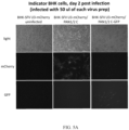
- Indicator BHK-21 cell lines with a reporter gene were created with a lentiviral vector to titrate the SFV viruses.
- BHK-21 cells Figure 4
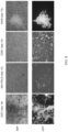
- indicator BHK-21-U3-mCherry Figure 5
- PAN1/2c When infected with PAN1/2c, regular BHK-21 cells showed no mCherry or GFP expression ( Figure 4 ).
- GFP expression was observed only when infected with the cells were infected with PAN1/2c-GFP ( Figure 4 ).
- Indicator BHK-21-U3-mCherry infected with PAN1/2c showed strong mCherry signal, similarly to cells infected with PAN1/2c-GFP, in which the red signal colocalized with the green signal ( Figure 5 ). All viruses rescued from the infectious clones and viruses obtained from ATCC were titrated using that cell line ( Figure 6 ). PAN2 and PAN1/2c showed the highest titers among all the viruses. Insertion of the GFP gene into the backbone of PAN1/2c resulted with an approximately 50 fold decrease in titers.
- Cancer-targeted SFVcpz are engineered by replacing the viral promoter with a cancer-specific promoter.
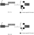
- the U3 region of the 3' LTR is replaced with a cancer-specific promoter as shown in Figure 9A .
- the nucleic acid encoding a tas polypeptide is also deleted as shown in Figure 9B .
- cancer-specific promoter in vitro by infecting relevant cells (e.g., human cancer cell lines and primary human cancer cells) and non-relevant cells (e.g., non-cancer cells).
- relevant cells e.g., human cancer cell lines and primary human cancer cells
- non-relevant cells e.g., non-cancer cells
- In vivo analyses include IV injection and biodistribution analyses between promoter-targeted and wildtype SFVcpz.
- Armed SFVcpz are engineered by replacing the viral promoter with a cancer-specific promoter.
- the nucleic acid encoding a bel2 polypeptide is replaced with a transgene encoding a suicide polypeptide as shown in Figure 10 .
- the 3' LTR is replaced with a cancer-specific promoter as shown in Figure 10B .
- Arming SFV with a transgene encoding a suicide polypeptide gene enhances tumoricidal effects of the SFV.
- Replacing U3 of the 3' LTR with a cancer-specific promoter may minimize unspecific SFV infection in non-cancer cells in vivo.
- Example 4 Replication Competent Simian Foamy Virus as an Oncolytic Virotherapy platform
- the supernatants of the infected cells were collected, filtered through a 0.45 ⁇ m filter and the titers of the progeny virions were measured using the indicator BHK-21-U3-mCherry cells.
- the progeny viruses were released by 2 cycles of freezing and thawing, the cell debris was concentrated by low-speed centrifugation and the supernatant was filtered through a 0.45 ⁇ m filter.
- the viral titers in the filtered supernatants were measured using indicator BHK-21-U3-mCherry cells.
- U251 cells were transduced with a lentiviral vector encoding the fluorophore mCherry driven by the SFV U3 promoter and a constitutively expressed puromycin resistance casette. After selection with puromycin, the surviving cells were grown and transduced with a second lentiviral vector, encoding luciferase driven by the SFV U3 promoter and a constitutively expressed neomycin resistance cassette. The transduced cells were selected using neomycin (G418).
- U251-U3-mCherry-luciferase cells U251-U3-mCherry-SFFV-luciferase cells (constitutively express luciferase from the spleen focus forming virus (SFFV) promoter) or wildtype U251 cells were seeded per well of a flat, transparent bottom, black 96-well plate.
- the cells were infected with PAN1/2 at MOIs 0, 0.2, 1 and 5.
- PAN1/2 spleen focus forming virus
- the media was aspirated off from each well, a 20 mg/ml stock of luciferase was diluted 100 fold in PBS and 100 ⁇ l of the diluted substrate was added to each well.
- mice Six-week old CB-17 SCID mice obtained from the vendor Envigo were injected subcutaneously in the right flank with 5*10 6 U251-U3-mCherry-luciferase cells. When tumors reached the volume of 0.3-0.5 cm 3 , they were directly injected with 2 doses of 1*10 6 IU of PAN1/2 or PFV in 100 ⁇ l PBS, or 2 doses of 100 ⁇ l PBS, or 4 doses of 5*10 5 IU of PAN1/2-GFP in 100 ⁇ l PBS. The mice were inspected and weighted twice a week.
- mice were followed for up to 96 days after the first injection unless they reached end-point conditions based on the tumor size (1.7 cm 3 ) or body scoring condition and were euthanized. Once a week, the mice were anesthetized and imaged with Xenogen IVIS-200 system after an intraperitoneal injection of luciferin (100 ⁇ l of 20 mg/ml luciferin stock). A total of 5 mice per group were sacrificed in 3 time points for the analysis of the spread of viruses in the tumor.
- mice half of their tumors were prepared for flow cytometry analysis by mincing, incubation with a solution containing type III collagenase (100 U/ml) and 3 mM CaCl 2 for 2 hours at 37°C on a rocking platform. Then the samples were passed through a cell strainer, concentrated by low-speed centrifugation and re-suspended in PBS containing 4% PFA. For the other half of the tumor, it was covered in OCT and frozen on dry ice.
- type III collagenase 100 U/ml
- CaCl 2 3 mM CaCl 2
- fragments were then prepared for immunohistochemistry by, briefly, sectioning, fixing in 4% PFA, incubating overnight at 4 °C with primary antibodies at 1:100 dilution: anti-mCherry (all tumors; chicken, polyclonal, Abcam) and anti-GFP (tumors infected with PAN1/2-GFP; rabbit, polyclonal, Abcam). Then, the sections were stained with anti-chicken and anti-rabbit secondary antibodies conjugated with fluorophores, respectively, Alexa 594 and Alexa 488 (1:2000 dilution). The sections were imaged using Zeiss LSM 510 Confocal Microscope and analyzed with the Zen software.
- TK Herpes Simplex Virus Thymidine Kinase
- the Herpes Simplex Virus Thymidine Kinase (TK) was amplified in a PCR reaction using primers with overhangs containing SacII (forward primer) and AgeI (reverse primer) restriction sites.
- the forward primer overhang also contained the sequence of the T2A self-cleaving peptide.
- the pcDNA3.1-PAN1/2-GFP plasmid was digested with SacII and BspEI restriction enzymes, and then ligated with the PCR product described above, digested with SacII and AgeI restriction enzymes. This gave rise to the construct pcDNA3.1-PAN1/2-TK.
- TK The expression of TK was validated by Western Blot, using a primary anti-TK rabbit antibody, and a secondary, anti-rabbit HRP-conjugated antibody (Santa Cruz Biotechnology).
- the indicator BHK-21-U3-mCherry cells we assessed the replication dynamics of the Foamy Viruses used in the study ( Fig. 11 ).
- the cell-free fraction of progeny virions of PAN1wt, PAN2 and PAN1/2 reached titers of approximately 10 6 IU/ml (Fig. 11bB).
- the titers of the cell free PAN1/2-GFP were approximately 5 fold lower than the titers of the parental PAN1/2 virus and PAN1clone had similar titers to PAN1/2-GFP ( Fig. 11B ).
- PFV reached the lowest titers of all the FVs ( ⁇ 1*10 4 IU/ml ( Fig. 11B )).
- the cell- associated fraction of progeny viruses is equal or slightly lower than the cell-free fraction ( Fig. 11B ).
- Newly produced PAN1/2 virus in the supernatant was detectable 36 hours post infection and peaked between 60 and 72 hours post infections, reaching 1.7*10 6 IU/ml.
- the release of the PAN1/2-GFP virus particles was slower, reaching titers of 10 5 IU/ml as late as 54 hours post infection, and peaking at ⁇ 6*10 5 IU/ml 108 hours post infection.
- the human glioblastoma U251 cell line was engineered to express mCherry and luciferase under the control of the FV promoter and enhancer elements in the U3 of the FV LTR (U251-U3-mCherry-luciferase). Luciferase catalyzes a reaction that produces bioluminescence after the addition of its substrate - Luciferin. Therefore, the FV replication in tumors formed by the U251-U3-mCherry-luciferase cells can be monitored non-invasively and quantitatively using the Xenogen imaging system.
- PAN1 / 2 efficiently replicates and persists in vivo in tumors without causing significant toxicity
- CB-17 SCID mice were subcutaneously implanted with indicator U251-U3-mCherry-luciferase cells.
- two doses of 1*10 6 IU of PAN1/2; 1*10 6 IU of PFV or 100 ⁇ l of PBS were injected intratumorally (IT).
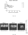
- the viruses did not cause significant toxicity, demonstrated by the healthy weights of the injected mice throughout the experiment ( Fig. 13A ) (the only cases of substantial weight lost are due to tumor burden).
- the replication of the FVs in the tumors was monitored weekly via Xenogen imaging ( Fig. 13B ). Bioluminescence was observed only in the PAN1/2 and PFV-injected mice, not in the PBS-injected mice ( Fig.
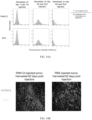
- mice bearing tumors injected with PAN1/2 and PFV were sacrificed at 3 different time points - days 7, 34 and 62 post 1 st virus injection. Their tumors were analyzed by flow cytometry to determine the percentage of mCherry + cells (i.e., the percentage of virus-infected cells) ( Fig. 14A ). On day 7, the percentage of mCherry+ cells was higher in the PAN1/2- than the PFV-injected tumor and it further increased over time. It also remained higher than at every subsequent time point ( Fig. 14A ). A PAN1/2-injected tumor and a PBS-injected tumor harvested 62 days post first injection were also sectioned, stained for mCherry and imaged with confocal microscopy ( Fig. 14B ). mCherry positive cells were easily detected in all sections of the PAN1/2 injected tumor, but not the PBS-injected tumor.
- PAN1/2 virus replicates very well in susceptible tumors in vivo, persists in those tumors for a prolonged period of time without causing significant toxicity in the infected mice.
- the virus can be used for gene delivery in vivo. It may also be superior to PFV due to faster replication and more efficient spread throughout the injected tumor.
- indicator U251-U3-mCherry-luciferase cells were injected into CB-17 SCID mice and upon tumor formation were intratumorally injected with 4 doses of 5*10 5 IU of PAN1/2-GFP.
- the virus was well tolerated by the injected mice, as indicated by their body weights ( Fig. 15A ).
- Bioluminescence on day 12 post 1 st injection (6 post last injection) was detectable for 4 out of 5 mice (the large size of the tumor of mouse #1 could have affected imaging) ( Fig. 15B ). The bioluminescence became stronger on day 19 and remained strong until the last time the mice were imaged (day 56, Fig. 15B ).
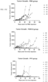
- Fig. 15C The replication of the virus did not seem to have affected the tumor growth substantially.
- mice On days 13, 39 and 66 after the first virus infection mice were sacrificed and their tumors were sectioned, stained for mCherry and GFP and imaged with confocal microscopy ( Fig. 15D ). All sections of the PAN1/2-GFP-injected tumors harvested on days 39 and 66 were positive for mCherry and GFP and those signals co-localized ( Fig. 15D ).
- a control, PBS-injected tumor was negative for both fluorophores. This demonstrated that PAN1/2 can be used for delivery of transgenes into tumors. As the virus spreads, more tumor cells will express the transgene. In an immune-deficient animal model, the expression of transgenes delivered by the virus is sustained for a prolonged period of time.
- SFV is sensitive to exogenous Interferon ⁇ , but does not induce Interferon ⁇ production in infected cells.
- SFV is sensitive to IFN ⁇ and whether a JAK-STAT pathway inhibitor Ruxolitinib can reverse this effect.
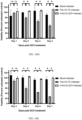
- Patient-derived human glioblastoma line GBM22 was pretreated with exogenous human IFN ⁇ or left untreated. 16 hours later we treated the cells with Ruxolitinib or DMSO control. 48 hours later, the cells were infected with 3*10 5 IU of PAN1/2-GFP. 6 days post infection, the cells were imaged ( Fig. 17B ) and the titers of newly produced PAN1/2-GFP virions in the supernatants were measured ( Fig. 17C ).
- the cells treated with IFN ⁇ +Ruxolitinib showed level of GFP expression similar to cells not treated with IFN ⁇ and treated with DMSO or Ruxolitinib ( Fig. 17b ).
- the PAN1/2-GFP titers in the supernatants of the cells treated with IFN ⁇ +Ruxolitinib did not significantly differ from the PAN1/2-GFP titers in the supernatants of the cells not treated with IFN ⁇ and treated with DMSO or Ruxolitinib ( Fig. 17C ).
- the cells treated with DMSO and IFN ⁇ showed significantly lower level of GFP expression than any of the combinations above ( Fig.
- Example 5 T cells expressing CARs (CAR-T cells)
- CAR-T cells are engineered by replacing the viral promoter of a SFV with nucleic acid encoding a CAR and infecting T cells with the SFV expressing the CAR.
- the nucleic acid encoding a bel2 polypeptide is replaced with a transgene encoding a CAR.
Landscapes
- Health & Medical Sciences (AREA)
- Life Sciences & Earth Sciences (AREA)
- Chemical & Material Sciences (AREA)
- Medicinal Chemistry (AREA)
- Organic Chemistry (AREA)
- Virology (AREA)
- General Health & Medical Sciences (AREA)
- Pharmacology & Pharmacy (AREA)
- Genetics & Genomics (AREA)
- Zoology (AREA)
- Animal Behavior & Ethology (AREA)
- Microbiology (AREA)
- Public Health (AREA)
- Veterinary Medicine (AREA)
- Wood Science & Technology (AREA)
- Engineering & Computer Science (AREA)
- Bioinformatics & Cheminformatics (AREA)
- Immunology (AREA)
- Oncology (AREA)
- Biomedical Technology (AREA)
- Mycology (AREA)
- Epidemiology (AREA)
- Biochemistry (AREA)
- General Engineering & Computer Science (AREA)
- Biotechnology (AREA)
- Chemical Kinetics & Catalysis (AREA)
- General Chemical & Material Sciences (AREA)
- Nuclear Medicine, Radiotherapy & Molecular Imaging (AREA)
- Medicines Containing Material From Animals Or Micro-Organisms (AREA)
- Medicines That Contain Protein Lipid Enzymes And Other Medicines (AREA)
- Pharmaceuticals Containing Other Organic And Inorganic Compounds (AREA)
- Micro-Organisms Or Cultivation Processes Thereof (AREA)
Applications Claiming Priority (3)
| Application Number | Priority Date | Filing Date | Title |
|---|---|---|---|
| US201862663637P | 2018-04-27 | 2018-04-27 | |
| PCT/US2019/027353 WO2019209557A1 (en) | 2018-04-27 | 2019-04-12 | Foamy viruses and methods of use |
| EP19793742.8A EP3784278B1 (de) | 2018-04-27 | 2019-04-12 | Schaumige viren und verwendungsverfahren |
Related Parent Applications (1)
| Application Number | Title | Priority Date | Filing Date |
|---|---|---|---|
| EP19793742.8A Division EP3784278B1 (de) | 2018-04-27 | 2019-04-12 | Schaumige viren und verwendungsverfahren |
Publications (2)
| Publication Number | Publication Date |
|---|---|
| EP4549564A2 true EP4549564A2 (de) | 2025-05-07 |
| EP4549564A3 EP4549564A3 (de) | 2025-08-13 |
Family
ID=68295731
Family Applications (2)
| Application Number | Title | Priority Date | Filing Date |
|---|---|---|---|
| EP25151397.4A Pending EP4549564A3 (de) | 2018-04-27 | 2019-04-12 | Schaumige viren und verfahren zur verwendung |
| EP19793742.8A Active EP3784278B1 (de) | 2018-04-27 | 2019-04-12 | Schaumige viren und verwendungsverfahren |
Family Applications After (1)
| Application Number | Title | Priority Date | Filing Date |
|---|---|---|---|
| EP19793742.8A Active EP3784278B1 (de) | 2018-04-27 | 2019-04-12 | Schaumige viren und verwendungsverfahren |
Country Status (5)
| Country | Link |
|---|---|
| US (1) | US12201659B2 (de) |
| EP (2) | EP4549564A3 (de) |
| CA (1) | CA3092891A1 (de) |
| ES (1) | ES3022988T3 (de) |
| WO (1) | WO2019209557A1 (de) |
Citations (3)
| Publication number | Priority date | Publication date | Assignee | Title |
|---|---|---|---|---|
| WO2015142675A2 (en) | 2014-03-15 | 2015-09-24 | Novartis Ag | Treatment of cancer using chimeric antigen receptor |
| US20190055299A1 (en) | 2015-10-27 | 2019-02-21 | Board Of Regents, The University Of Texas System | Chimeric antigen receptor molecules and uses thereof |
| US20190085081A1 (en) | 2016-03-15 | 2019-03-21 | Cancer Research Technology Limited | Chimeric antigen receptor |
Family Cites Families (7)
| Publication number | Priority date | Publication date | Assignee | Title |
|---|---|---|---|---|
| EP1185628B1 (de) * | 1999-06-14 | 2005-11-23 | THE GOVERNMENT OF THE UNITED STATES OF AMERICA, as represented by THE SECRETARY, DEPARTMENT OF HEALTH AND HUMAN SERVICES | Trennung eines humanen retrovirus |
| EP2389443B1 (de) | 2009-01-23 | 2018-11-14 | Roger Williams Hospital | Retrovirale vektoren kodierend für multiple stark homologe nichtvirale polypeptide und ihre verwendung |
| CA2764107A1 (en) | 2009-06-17 | 2010-12-23 | Tocagen Inc. | Producer cells for replication competent retroviral vectors |
| US20140140962A1 (en) | 2011-04-29 | 2014-05-22 | The Research Foundation Of State University Of New York | Viruses modified with unnatural moieties and methods of use thereof |
| EP2583974B1 (de) | 2011-10-21 | 2017-04-26 | Technische Universität Dresden | Pseudotypisierung von schaumigen Viren |
| ES3034398T3 (en) * | 2014-08-28 | 2025-08-18 | Bioatla Inc | Conditionally active chimeric antigen receptors for modified t-cells |
| CA3032838A1 (en) * | 2016-08-04 | 2018-02-08 | Memorial Sloan-Kettering Cancer Center | Compositions and methods for immunotherapy |
-
2019
- 2019-04-12 US US16/980,359 patent/US12201659B2/en active Active
- 2019-04-12 ES ES19793742T patent/ES3022988T3/es active Active
- 2019-04-12 EP EP25151397.4A patent/EP4549564A3/de active Pending
- 2019-04-12 EP EP19793742.8A patent/EP3784278B1/de active Active
- 2019-04-12 CA CA3092891A patent/CA3092891A1/en active Pending
- 2019-04-12 WO PCT/US2019/027353 patent/WO2019209557A1/en not_active Ceased
Patent Citations (3)
| Publication number | Priority date | Publication date | Assignee | Title |
|---|---|---|---|---|
| WO2015142675A2 (en) | 2014-03-15 | 2015-09-24 | Novartis Ag | Treatment of cancer using chimeric antigen receptor |
| US20190055299A1 (en) | 2015-10-27 | 2019-02-21 | Board Of Regents, The University Of Texas System | Chimeric antigen receptor molecules and uses thereof |
| US20190085081A1 (en) | 2016-03-15 | 2019-03-21 | Cancer Research Technology Limited | Chimeric antigen receptor |
Non-Patent Citations (1)
| Title |
|---|
| RUSSELL ET AL., MOLECULAR THERAPY, vol. 25, 2017, pages 1107 - 1116 |
Also Published As
| Publication number | Publication date |
|---|---|
| EP4549564A3 (de) | 2025-08-13 |
| US20210046134A1 (en) | 2021-02-18 |
| EP3784278A4 (de) | 2021-07-07 |
| ES3022988T3 (en) | 2025-05-29 |
| CA3092891A1 (en) | 2019-10-31 |
| EP3784278B1 (de) | 2025-01-15 |
| WO2019209557A1 (en) | 2019-10-31 |
| US12201659B2 (en) | 2025-01-21 |
| EP3784278A1 (de) | 2021-03-03 |
Similar Documents
| Publication | Publication Date | Title |
|---|---|---|
| US20250230232A1 (en) | Immunologic effector cell of targeted cld18a2, and preparation method and use thereof | |
| US11649284B2 (en) | Cancer gene therapy targeting CD47 | |
| JP7237960B2 (ja) | 標的細胞の選択的形質導入のためのアダプターベースのレトロウイルスベクター系 | |
| JP2026001033A (ja) | Il-2スーパーアゴニスト、アゴニスト、およびそれらの融合体の使用および方法 | |
| JP6682509B2 (ja) | キメラ抗原受容体タンパク質をコードする核酸およびキメラ抗原受容体タンパク質を発現するtリンパ球 | |
| JP2012503987A5 (de) | ||
| JP2024503027A (ja) | Cd8標的ウイルスベクターの使用方法 | |
| EP2583974B1 (de) | Pseudotypisierung von schaumigen Viren | |
| JP2015518479A (ja) | 養子細胞療法に有用なポリペプチド | |
| CN111743923B (zh) | 包含分离的重组溶瘤腺病毒和免疫细胞的治疗剂及其应用 | |
| WO2022182890A1 (en) | Codon-optimized nucleotide sequences encoding an ap-1 transcription factor | |
| CN112813030A (zh) | 一种靶向fgfr4和dr5的嵌合抗原受体t细胞及其制备方法和应用 | |
| CN117615786A (zh) | 嵌合抗原受体及其使用方法 | |
| CN112812182B (zh) | 一种靶向fgfr4的单链抗体、嵌合抗原受体、嵌合抗原受体t细胞及其制备方法和应用 | |
| CN114262691A (zh) | 分离的能够用于表达外源基因的溶瘤腺病毒、载体、治疗剂及其用途 | |
| US20250108079A1 (en) | Foamy viruses and methods of use | |
| EP3784278B1 (de) | Schaumige viren und verwendungsverfahren | |
| CN115141806A (zh) | 靶向Her2并表达PD-L1抗体的嵌合抗原受体T细胞及其制备方法和应用 | |
| Budzik | Generation and Characterization of a Foamy Virus-Based Replicating Retroviral Vector System for Cancer Therapy | |
| WO2025209590A1 (zh) | 一种病毒载体包装系统及其应用 | |
| HK40070900A (en) | Isolated oncolytic adenovirus capable of expressing exogenous gene, vector, therapeutic agent, and use | |
| WO2025162379A1 (zh) | 一种多肽及包含所述多肽基因的病毒载体 | |
| TW202528545A (zh) | 用於治療劑遞送之組合物及方法 | |
| EP4698666A1 (de) | Universelle protein-g-fusogene und adaptersysteme davon sowie zugehörige lipidpartikel und verwendungen | |
| WO2025140504A1 (zh) | 靶向kras g12v突变多肽的t细胞受体及其用途 |
Legal Events
| Date | Code | Title | Description |
|---|---|---|---|
| PUAI | Public reference made under article 153(3) epc to a published international application that has entered the european phase |
Free format text: ORIGINAL CODE: 0009012 |
|
| STAA | Information on the status of an ep patent application or granted ep patent |
Free format text: STATUS: REQUEST FOR EXAMINATION WAS MADE |
|
| 17P | Request for examination filed |
Effective date: 20250113 |
|
| AC | Divisional application: reference to earlier application |
Ref document number: 3784278 Country of ref document: EP Kind code of ref document: P |
|
| AK | Designated contracting states |
Kind code of ref document: A2 Designated state(s): AL AT BE BG CH CY CZ DE DK EE ES FI FR GB GR HR HU IE IS IT LI LT LU LV MC MK MT NL NO PL PT RO RS SE SI SK SM TR |
|
| REG | Reference to a national code |
Ref country code: DE Ref legal event code: R079 Free format text: PREVIOUS MAIN CLASS: C12N0015000000 Ipc: A61K0039385000 |
|
| PUAL | Search report despatched |
Free format text: ORIGINAL CODE: 0009013 |
|
| AK | Designated contracting states |
Kind code of ref document: A3 Designated state(s): AL AT BE BG CH CY CZ DE DK EE ES FI FR GB GR HR HU IE IS IT LI LT LU LV MC MK MT NL NO PL PT RO RS SE SI SK SM TR |
|
| RIC1 | Information provided on ipc code assigned before grant |
Ipc: A61K 39/385 20060101AFI20250704BHEP Ipc: A61K 48/00 20060101ALI20250704BHEP Ipc: A61K 39/21 20060101ALI20250704BHEP Ipc: C12Q 1/70 20060101ALI20250704BHEP Ipc: C12N 15/00 20060101ALI20250704BHEP |