EP4538705A2 - Immunoassay-testvorrichtung mit zwei flüssigkeitsströmungswegen zur detektion und differenzierung von zwei oder mehr analyten - Google Patents
Immunoassay-testvorrichtung mit zwei flüssigkeitsströmungswegen zur detektion und differenzierung von zwei oder mehr analyten Download PDFInfo
- Publication number
- EP4538705A2 EP4538705A2 EP24214974.8A EP24214974A EP4538705A2 EP 4538705 A2 EP4538705 A2 EP 4538705A2 EP 24214974 A EP24214974 A EP 24214974A EP 4538705 A2 EP4538705 A2 EP 4538705A2
- Authority
- EP
- European Patent Office
- Prior art keywords
- species
- test
- fluid flow
- sample
- detectable
- Prior art date
- Legal status (The legal status is an assumption and is not a legal conclusion. Google has not performed a legal analysis and makes no representation as to the accuracy of the status listed.)
- Pending
Links
Images

Classifications
-
- G—PHYSICS
- G01—MEASURING; TESTING
- G01N—INVESTIGATING OR ANALYSING MATERIALS BY DETERMINING THEIR CHEMICAL OR PHYSICAL PROPERTIES
- G01N33/00—Investigating or analysing materials by specific methods not covered by groups G01N1/00 - G01N31/00
- G01N33/48—Biological material, e.g. blood, urine; Haemocytometers
- G01N33/50—Chemical analysis of biological material, e.g. blood, urine; Testing involving biospecific ligand binding methods; Immunological testing
- G01N33/53—Immunoassay; Biospecific binding assay; Materials therefor
- G01N33/569—Immunoassay; Biospecific binding assay; Materials therefor for microorganisms, e.g. protozoa, bacteria, viruses
- G01N33/56983—Viruses
-
- G—PHYSICS
- G01—MEASURING; TESTING
- G01N—INVESTIGATING OR ANALYSING MATERIALS BY DETERMINING THEIR CHEMICAL OR PHYSICAL PROPERTIES
- G01N33/00—Investigating or analysing materials by specific methods not covered by groups G01N1/00 - G01N31/00
- G01N33/48—Biological material, e.g. blood, urine; Haemocytometers
- G01N33/50—Chemical analysis of biological material, e.g. blood, urine; Testing involving biospecific ligand binding methods; Immunological testing
- G01N33/53—Immunoassay; Biospecific binding assay; Materials therefor
- G01N33/543—Immunoassay; Biospecific binding assay; Materials therefor with an insoluble carrier for immobilising immunochemicals
- G01N33/54366—Apparatus specially adapted for solid-phase testing
- G01N33/54386—Analytical elements
- G01N33/54387—Immunochromatographic test strips
- G01N33/54388—Immunochromatographic test strips based on lateral flow
- G01N33/54389—Immunochromatographic test strips based on lateral flow with bidirectional or multidirectional lateral flow, e.g. wherein the sample flows from a single, common sample application point into multiple strips, lanes or zones
-
- G—PHYSICS
- G01—MEASURING; TESTING
- G01N—INVESTIGATING OR ANALYSING MATERIALS BY DETERMINING THEIR CHEMICAL OR PHYSICAL PROPERTIES
- G01N33/00—Investigating or analysing materials by specific methods not covered by groups G01N1/00 - G01N31/00
- G01N33/48—Biological material, e.g. blood, urine; Haemocytometers
- G01N33/50—Chemical analysis of biological material, e.g. blood, urine; Testing involving biospecific ligand binding methods; Immunological testing
- G01N33/53—Immunoassay; Biospecific binding assay; Materials therefor
- G01N33/569—Immunoassay; Biospecific binding assay; Materials therefor for microorganisms, e.g. protozoa, bacteria, viruses
- G01N33/56911—Bacteria
-
- G—PHYSICS
- G01—MEASURING; TESTING
- G01N—INVESTIGATING OR ANALYSING MATERIALS BY DETERMINING THEIR CHEMICAL OR PHYSICAL PROPERTIES
- G01N33/00—Investigating or analysing materials by specific methods not covered by groups G01N1/00 - G01N31/00
- G01N33/48—Biological material, e.g. blood, urine; Haemocytometers
- G01N33/50—Chemical analysis of biological material, e.g. blood, urine; Testing involving biospecific ligand binding methods; Immunological testing
- G01N33/53—Immunoassay; Biospecific binding assay; Materials therefor
- G01N33/569—Immunoassay; Biospecific binding assay; Materials therefor for microorganisms, e.g. protozoa, bacteria, viruses
- G01N33/56983—Viruses
- G01N33/56994—Herpetoviridae, e.g. cytomegalovirus, Epstein-Barr virus
-
- G—PHYSICS
- G01—MEASURING; TESTING
- G01N—INVESTIGATING OR ANALYSING MATERIALS BY DETERMINING THEIR CHEMICAL OR PHYSICAL PROPERTIES
- G01N2333/00—Assays involving biological materials from specific organisms or of a specific nature
- G01N2333/195—Assays involving biological materials from specific organisms or of a specific nature from bacteria
- G01N2333/20—Assays involving biological materials from specific organisms or of a specific nature from bacteria from Spirochaetales (O), e.g. Treponema, Leptospira
-
- G—PHYSICS
- G01—MEASURING; TESTING
- G01N—INVESTIGATING OR ANALYSING MATERIALS BY DETERMINING THEIR CHEMICAL OR PHYSICAL PROPERTIES
- G01N2469/00—Immunoassays for the detection of microorganisms
- G01N2469/20—Detection of antibodies in sample from host which are directed against antigens from microorganisms
-
- G—PHYSICS
- G01—MEASURING; TESTING
- G01N—INVESTIGATING OR ANALYSING MATERIALS BY DETERMINING THEIR CHEMICAL OR PHYSICAL PROPERTIES
- G01N2800/00—Detection or diagnosis of diseases
- G01N2800/56—Staging of a disease; Further complications associated with the disease
-
- Y—GENERAL TAGGING OF NEW TECHNOLOGICAL DEVELOPMENTS; GENERAL TAGGING OF CROSS-SECTIONAL TECHNOLOGIES SPANNING OVER SEVERAL SECTIONS OF THE IPC; TECHNICAL SUBJECTS COVERED BY FORMER USPC CROSS-REFERENCE ART COLLECTIONS [XRACs] AND DIGESTS
- Y02—TECHNOLOGIES OR APPLICATIONS FOR MITIGATION OR ADAPTATION AGAINST CLIMATE CHANGE
- Y02A—TECHNOLOGIES FOR ADAPTATION TO CLIMATE CHANGE
- Y02A50/00—TECHNOLOGIES FOR ADAPTATION TO CLIMATE CHANGE in human health protection, e.g. against extreme weather
- Y02A50/30—Against vector-borne diseases, e.g. mosquito-borne, fly-borne, tick-borne or waterborne diseases whose impact is exacerbated by climate change
Definitions
- the subject matter described herein relates to lateral flow immunoassays that have two separate fluid flow paths to detect and discriminate two species of interest in a fluid sample.
- Rapid lateral flow immunoassays test devices have an extensive history of use in both the clinical and the home settings. These devices are used to test for a variety of analytes, such as hormones, proteins, urine or plasma components and the like. These devices generally comprise a lateral flow test strip, such as nitrocellulose or filter paper, a sample application area, test results area and an analyte specific binding reagent that is bound to some kind of detectable label, such as a colored particle, fluorescent or luminescent tag, or an enzyme detection system.
- a lateral flow test strip such as nitrocellulose or filter paper
- sample application area such as nitrocellulose or filter paper
- test results area such as a sample application area
- an analyte specific binding reagent that is bound to some kind of detectable label, such as a colored particle, fluorescent or luminescent tag, or an enzyme detection system.
- detectable label such as a colored particle, fluorescent or luminescent tag, or an enzyme detection system.
- a device for determining presence or absence of infection due to an infectious agent comprises a sample receiving zone configured to receive a liquid sample from a subject suspected of having an infection due to an infectious agent, the sample receiving zone positioned to distribute the sample along a first fluid flow path to a first label zone and along a second fluid flow path to a second label zone.
- Each of the first and second label zones comprise, respectively, a first mobilizable, detectable species and a second mobilizable, detectable species, each mobilizable, detectable species able to bind the infectious agent or to antibody against the infectious agent.
- the device also comprises a first test line in the first fluid flow path positioned downstream of the first label zone, the first test line comprising an immobilized species with binding affinity for the first mobilizable detectable species.
- the device also comprises a second test line in the second fluid flow path positioned downstream of the second label zone, the second test line comprising an immobilized species with binding affinity for the second mobilizable detectable species.
- the device also comprises a reference line positioned in the first fluid flow path and comprising an immobilized species with binding affinity for a detectable moiety deposited on the device upstream of the reference line.
- the first fluid flow path and the second fluid flow path are at an angle selected from a straight angle (180°), an obtuse angle and an acute angle.
- the reference line in the first fluid flow path is downstream of the first test line.
- the device in other embodiments, is designed such that the first test line comprises a first immobilized species that directly binds antibody against the infectious agent present in the liquid sample and that specifically binds a conjugate comprised of the mobilizable, detectable species in the first label zone and an antibody against the infectious agent in the sample.
- the first and second immobilized species are the same.
- the immobilized species in the first test line comprises a plurality of antigens for B. burgdorferi with binding affinity for the human IgM antibody against B. burgdorferi.
- the mobilizable, detectable species second label zone is an anti-IgG antibody.
- the anti-IgG antibody is a nonhuman anti-human IgG antibody.
- the first and second test lines are within a single optical path of an optical detector for inspection by an instrument.
- the single optical path is, in one embodiment, a path along one axis that intersects the first and second test lines.
- the sample receiving zone dispenses sample to the first label zone and the second label zone in essentially equal amounts and at essentially equal rates.
- a device for determining presence of infection due to an infectious agent comprises (i) a sample receiving zone configured to receive a liquid sample from a subject suspected of having an infection due to an infectious agent, the sample receiving zone positioned to distribute the sample along a first fluid flow path to a first label zone and along a second fluid flow path to a second label zone, each of said first and second label zones comprising, respectively, a first mobilizable, detectable species and a second mobilizable, detectable species, each mobilizable, detectable species able to bind separate, distinct antibodies against the infectious agent; (ii) a first test line in the first fluid flow path positioned downstream of the first label zone, the first test line comprising an immobilized species with binding affinity for the first mobilizable detectable species; (iii) a second test line in the second fluid flow path positioned downstream of the second label zone, the second test line comprising an immobilized species with binding affinity for the second mobilizable detectable species; (iv) a reference line positioned in the first fluid
- the first mobilizable, detectable species is a non-human, anti-human IgM antibody or a Lyme antigen.
- the second mobilizable, detectable species is a non-human, anti-human IgG antibody or a Lyme antigen.
- the immobilized species on the first test line with binding affinity for the first mobilizable detectable species is a non-human, anti-human IgM antibody or a Lyme antigen.
- the immobilized species on the second test line with binding affinity for the second mobilizable detectable species is a non-human, anti-human IgG antibody or a Lyme antigen.
- the reference line is positioned downstream of the first test line.
- the reference line comprises an immobilized species with binding affinity for a non-human, anti-human IgG antibody or a non-human, anti-human IgM antibody.
- the reference line comprises an immobilized species with binding affinity for a substance not present in the liquid sample.
- two or more separate, distinct antibodies against the infectious agent are present in the liquid sample and are capable of binding the immobilized species on each of the first test line and the second test line and capable of binding one of the mobilizable, detectable species on the first label line or on the second label line.
- the separate, distinct antibodies against the infectious agent are present in the liquid sample and are capable of binding the mobilizable, detectable species on the first label line to form a first conjugate and on the second label line to form a second conjugate, and the immobilized species on the first test line has binding affinity for the first conjugate and the immobilized species on the second test line has binding affinity for the second conjugate.
- the immobilized species on the first test line is a non-human, anti-human IgM antibody. In another embodiment, the immobilized species on the first test line is a non-human, anti-human IgG antibody.
- a method for staging infection by a pathogenic Borrelia species such as Borrelia burgdorferi ( B. burgdorferi )
- the method comprises depositing a fluid sample from a person suspected of or at risk of having been exposed to the pathogenic Borrelia species on a device as described herein; and inspecting (using an instrument) the first test line and the second test line for the presence or absence of the mobilizable detectable species.
- the fluid sample is blood, cerebrospinal fluid or urine.
- the fluid sample is blood and is deposited in an amount of less than 50 ⁇ L.
- inspecting is performed in about 10 minutes or less after depositing the fluid sample.
- depositing and inspecting provides sensitivity to detect an IgM antibody response to B. burgdorferi exposure in greater than 70% of exposed subjects within 2 weeks of exposure. In another embodiment, depositing and inspecting provides sensitivity to detect an IgG antibody response to B. burgdorferi exposure in greater than 70% of exposed subjects within 2 weeks of exposure.
- Sample is any material to be tested for the presence or amount of an analyte of interest.
- a sample is a fluid sample, preferably a liquid sample.
- liquid samples that may be tested using a test device include bodily fluids including blood, serum, plasma, saliva, urine, ocular fluid, semen, sputum, nasal discharge and spinal fluid.
- test strip can include one or more bibulous or non-bibulous materials. If a test strip comprises more than one material, the one or more materials are preferably in fluid communication. One material of a test strip may be overlaid on another material of the test strip, such as for example, filter paper overlaid on nitrocellulose. Alternatively or in addition, a test strip may include a region comprising one or more materials followed by a region comprising one or more different materials. In this case, the regions are in fluid communication and may or may not partially overlap one another.
- Suitable materials for test strips include, but are not limited to, materials derived from cellulose, such as filter paper, chromatographic paper, nitrocellulose, and cellulose acetate, as well as materials made of glass fibers, nylon, dacron, PVC, polyacrylamide, cross-linked dextran, agarose, polyacrylate, ceramic materials, and the like.
- the material or materials of the test strip may optionally be treated to modify their capillary flow characteristics or the characteristics of the applied sample.
- the sample application region of the test strip may be treated with buffers to correct the pH or specific gravity of an applied urine sample, to ensure optimal test conditions.
- a device for determining presence of infection due to an infectious agent is provided.
- Various embodiments of the device will be described with reference to certain drawing figures.
- a test strip 10 comprises a sample receiving zone 12 configured to receive a liquid sample.
- the sample is from a subject suspected of having an infection due to an infectious agent, and examples of types of patient samples and of infectious agents are described below.
- Sample receiving zone 12 is positioned to distribute the sample along a first fluid flow path, indicated by arrow 14 in FIG. 1A , to a first label zone 16 and along a second fluid flow path, indicated by arrow 18 in FIG. 1A , to a second label zone 20.
- Each of the first and second label zones comprise, respectively, a first mobilizable, detectable species and a second mobilizable, detectable species, where each mobilizable, detectable species is able to bind to the infectious agent or to antibody against the infectious agent, as will be described.
- the first fluid flow path and the second fluid flow path as depicted in FIG. 1A are at a straight angle of 180°. In other embodiments, the first fluid flow path and the second fluid flow path can be at an obtuse angle from one another or at an acute angle from one another.
- Test strip 10 also comprises a first test line 22 in the first fluid flow path and positioned downstream of the first label zone (16).
- the first test line comprises an immobilized species with binding affinity for the first mobilizable detectable species. Binding affinity intends indirect binding or direct binding between two species, such as direct binding of an antigen to an antibody or indirect binding of a secondary antibody to a conjugate formed of a primary antibody and an antigen, where the secondary antibody and primary antibody have binding affinity.
- an antibody in the patient sample is indicative of presence of infection by an infectious agent, and the antibody in the patient sample binds a mobilizable, detectable species comprised of a non-human antibody with binding affinity for the antibody in the patient sample or an antigen of or from the infectious agent indicative of the suspected infection.
- Test strip 10 also comprises a second test line 24 in the second fluid flow path (18) and positioned downstream of the second label zone (20).
- the second test line comprises an immobilized species with binding affinity for the second mobilizable, detectable species present in the second label zone. Binding affinity intends indirect binding or direct binding between two species, as described in the preceding paragraph.
- Test strip 10 also comprises a reference line 26 positioned in the first fluid flow path (14) and comprising an immobilized species with binding affinity for a detectable moiety deposited on or formed on the device upstream of the reference line.
- reference line 26 is downstream of first test line 22.
- the test strip comprises a second or additional reference line 28 in the second fluid flow path (18).
- the additional reference line is downstream of the second test line 24.
- the first test line comprises an immobilized species that directly binds an antibody present in the patient sample, the antibody being one raised by the patient's immune system against the infectious agent of interest and suspected of being the cause of infection in the patient.
- the first test line comprises an immobilized species that binds a conjugate formed on the test device, the conjugate comprised of (i) the mobilizable, detectable species in the first label zone and (ii) an antibody present in the patient sample, the antibody being one raised by the patient's immune system against the infectious agent of interest and suspected of being the cause of infection in the patient.
- the second test line in various embodiments, comprises an immobilized species that directly binds antibody against the infectious agent, the antibody being one raised by the patient's immune system against the infectious agent of interest and suspected of being the cause of infection in the patient.
- the second test line comprises an immobilized species that binds a conjugate formed on the test device, the conjugate comprised of (i) the mobilizable, detectable species in the second label zone and (ii) an antibody present in the patient sample, the antibody being one raised by the patient's immune system against the infectious agent of interest and suspected of being the cause of infection in the patient.
- test strip it is desired to determine whether a subject is at risk of Lyme disease or has Lyme disease, or, alternatively, it is desired to determine if infection with a Borrelia species, such as but not limited to Borrelia burgdorferi, Borrelia afzelii, Borrelia garinii, Borellia japonica, is at an early stage or a late stage of infection.
- a test strip that comprises a single, individual sample receiving zone is provided, and a patient's sample is deposited on or in the sample receiving zone.
- the sample receiving zone is common to two fluid flow paths on the test strip, where a first fluid flow path carries a portion of the deposited sample in a first direction and a second fluid flow path carries a second portion of the deposited sample in a second direction.
- the two fluid flow paths and their directions of fluid flow are at 180° from each other, so that the paths are along a common axis.
- Each of the two fluid flow paths comprises a label zone that comprises a mobilizable, detectable species.
- the mobilizable, detectable species in the exemplary test strip for staging or detecting infection by a species in the Borrelia genus is, in a first embodiment, a non-human, anti-human antibody that has or is associated with a detectable label.
- the non-human, anti-human antibody is, in some embodiments, a non-human, anti-human IgG antibody bearing a detectable label, such as a fluorescent, chemiluminescent, or other optically detectable tag, such as a bead or chemical moiety.
- the non-human, anti-human antibody is, in some embodiments, a non-human, anti-human IgM antibody bearing a detectable label, such as a fluorescent, chemiluminescent, or other optically detectable tag, such as a bead or chemical moiety.
- optically detectable intends optically detectable by an instrument and not visually detectable by an unaided human eye.
- the detectable non-human, anti-human IgM antibody is deposited on the first label line, and the detectable non-human, anti-human IgG antibody is deposited on the second label line. More specifically, a detectable goat anti-human IgM antibody is deposited on the first label line and a detectable goat anti-human IgG antibody is deposited on the second label line.
- Non-human, anti-human IgG and IgM antibodies are exemplified as goat, anti-human antibodies, however the non-human portion of the antibody can be any mammal, including but not limited to mouse, rabbit, rat, sheep, etc.
- Deposited on the first test line of the exemplary test strip for detecting or staging Lyme disease is an antigen for a species in the Borrelia genus.
- one or more peptide antigens from B. burgdorferi is deposited on the test lines in each of the flow paths on the test strip.
- peptide antigens with binding affinity to the OspC, C6, C10, FlaB or BBK07 regions of B. Burgdorferi are deposited in an immobile fashion to the test lines.
- a recombinant protein with binding affinity to B. Burgdorferi or to an antibody against B. Burgdorferi is deposited on a test line.
- the recombinant protein for the outer surface protein C (OspC) of B. Burgdorferi (GenBank Acc. No. WP_012686633.1; SEQ ID NO: 20), for the decorin-binding protein A (DbpA) of B. Burgdorferi (GenBank Acc. No. WP_010890380.1; SEQ ID NO: 21), or fragments of these proteins that have between 12-60, 12-50, 12-40, 10-40 or 10-25 contiguous amino acid residues from the protein are contemplated, as exemplified by SEQ ID NO: 12 and SEQ ID NO: 14.
- Other examples of peptide antigens are known in art, such as in U.S. Patent No.
- the peptide antigen deposited on one or both of the test lines binds the C6 region of B. burgdorferi and has a sequence selected from the exemplary sequences set forth as SEQ ID NO: 1, SEQ ID NO: 2, SEQ ID NO: 3, SEQ ID NO: 4, SEQ ID NO: 5 or SEQ ID NO: 6.
- the peptide antigen deposited on one or both of the test lines comprises SEQ ID NO: 1, SEQ ID NO: 2, SEQ ID NO: 3, SEQ ID NO: 4, SEQ ID NO: 5 or SEQ ID NO: 6 (or a peptide having 80%, 85%, 86%, 87%, 89%, 90%, 91%, 92%, 93%, 94%, 95%, 96%, 97%, 98%, or 99% sequence identity thereto) and is attached to the test line with a biotin-strepavidin interaction.
- the peptide antigens or plurality of peptide antigens deposited on the first test line and on the second test line are the same.
- the peptide antigen deposited on one or both of the test lines has 80%, 85%, 86%, 87%, 89%, 90%, 91%, 92%, 93%, 94%, 95%, 96%, 97%, 98%, or 99% sequence identity to one of the peptide or protein sequences disclosed herein.
- FIG. 2 illustrates a top view of a test device where optical windows in the housing member are disposed over the test lines (22, 24) and reference lines (26, 28).
- Optical window 30 and optical window 32 define an opening visible to a user or an optical reader in an instrument for interrogation of the test and reference lines.
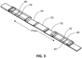
- FIG. 3 is a perspective view of a test device with bidirectional fluid flow paths for discrimination of two analytes in a sample.
- the test device has a base nitrocellulose layer 40 on which the test and control lines are deposited, such as test lines 42, 44 and control lines 46, 48.
- a sample pad 50 is centrally positioned for distribution of sample to each of the test and control lines via a label pad, such as label pads 52, 54.
- the label pads are of a material suitable for receiving the mobilizable species and situated to overlap with the nitrocellulose layer and the sample pad to provide contiguous fluid communication.
- the device described herein is contemplated for use in detection of any pathogenic or infectious agent, and several examples are now described.
- Lyme disease is transmitted by the bite of various species of Ixodes ticks carrying the etiologic agent, a pathogenic Borrelia bacterium (a spirochete).
- Organisms of the Borrelia burgdorferi sensu lato group belong to the family Spirochaetaceae, genus Borrelia. There are at least 11 species in the B. burgdorferi complex and an unknown but large number of substrains. At least three genospecies of the Borrelia burgdorferi sensu lato group have been identified as pathogens: B. burgdorferi sensu stricto, B. afzelli, and B. garinii.
- a test strip with a plurality of peptides immobilized on the first and second test lines in each of the fluid flow paths or mobilizable on the first and second label lines in each of the fluid flow paths is provided.
- the plurality of peptides can be the same or different in each of the test lines and/or label lines.
- the plurality of peptides comprises 3 or more, or 4 or more, or 5 or more, or 6 or more, or 7 or more, or 8 or more, different peptide sequences from a Borellia species, such as B. burgdorferi sensu lato.
- CMKKDDQIAAA MVLRGMAKDGQFALK (SEQ ID NO: 2)
- a shorter peptide from this region such as a peptide with 12-18 contiguous resides from SEQ ID NO: 2 or 12-18 non-contiguous residues from SEQ ID NO: 2, exemplified by the peptide MKKNDQIGAAIALRGVA (SEQ ID NO: 3), or active variants thereof.
- FlaB peptide (p41, SEQ ID NO: 7) is attached by a linker to a C6 peptide (SEQ ID NO: 1, SEQ ID NO: 2, SEQ ID NO: 3, SEQ ID NO: 4, SEQ ID NO: 5 or SEQ ID NO: 6) to form a fusion peptide as exemplified by SEQ ID NO: 16, SEQ ID NO: 17 and SEQ ID NO: 18.
- SEQ ID NO: 16 which consists of a peptide from the p41 (FlaB) region (SEQ ID NO: 2) linked to a peptide from the OspC1 region (SEQ ID NO: 12) linked to a peptide for the C6 region (SEQ ID NO: 1), the three peptides joined by a linker comprised of three amino acid residues (GGG).
- the linker peptide comprises 1, 2, 3, 4, 5, 6, 7, 8, 9, 10 or more amino acids.
- the linker comprises 1 to 2, 1 to 5, 2 to 5, 2 to 4, 1 to 10, 5 to 10, 3 to 6, or 2 to 10 amino acids.
- the linker is G, GG, GGG, or GGGG (SEQ ID NO: 19).
- Suitable linkers for joining any of the peptide sequences disclosed herein can be readily selected and can be of any of a suitable of different lengths, such as from 1 amino acid (e.g., Gly) to 40 amino acids, from 2 amino acids to 15 amino acids, from 3 amino acids to 12 amino acids, including 4 amino acids to 10 amino acids, 5 amino acids to 9 amino acids, 6 amino acids to 8 amino acids, or 7 amino acids to 8 amino acids, and may be 1, 2, 3, 4, 5, 6, or 7 amino acids.
- the linker comprises only glycines. In other embodiments, 1, 2 3 or 4 of the glycines are substituted with serines.
- Non-peptide linker moieties can also be used to join or link a carrier moiety to a mitochondrial fusion modulatory peptide.
- the linker molecules are generally about 6-50 atoms long.
- the linker molecules may also be, for example, aryl acetylene, ethylene glycol oligomers containing 2-10 monomer units, diamines, diacids, amino acids, or combinations thereof.
- Other linker molecules which can bind to polypeptides may be used in light of this disclosure.
- peptides disclosed herein and referenced by incorporation herein are merely exemplary of the peptide sequences that can be used in an assay.
- Embodiments of the assay contemplate use of peptide sequences that have 80%, 85%, 86%, 87%, 89%, 90%, 91%, 92%, 93%, 94%, 95%, 96%, 97%, 98%, or 99% sequence identity to any of the sequences disclosed herein, when the two sequences are compared along their entire length.
- any of the sequences may be biotinylated, including biotinylated at the N-terminus, the C-terminus, or at an amine residue in an amino acid within the first one, two, three, four, five amino acids (counting from the N-terminal amino acid) of the peptide. It will also be appreciated that any of the sequence may be modified at the N-terminus, the C-terminus, or both with a reactive moiety or a chemical linking moiety, such as a hydroxyl, carboxyl, amine or group.
- Examples 1-4 relate to test strips for detection of IgG and IgM antibodies against a pathogenic Borrelia species (Examples 1-3) and for staging as early state or late stage an infection by a pathogenic Borrelia species (Example 4).
- the test strips are designed to detect the presence of immunoglobulins raised by a human subject infected with a pathogenic Borrelia species.
- IgM antibodies develop over a three to six week period, and this is referred to in the art, and herein, as an early stage infection.
- IgG antibodies develop within 4-12 weeks after infection, and are indicative of infection at a later stage, and this is referred to herein as late stage infection.
- an early stage of infection or early stage Lyme disease is indicated by presence of IgM antibodies against a pathogenic Borrelia species in a blood sample taken 3-6 weeks after infection
- a late stage of Lyme disease is indicated by presence of IgG antibodies against a pathogenic Borrelia species in a blood sample taken 4-12 weeks after infection.
- a late state of Lyme disease is additionally indicated by an amount of IgG antibodies in the sample that is greater than the amount of IgM antibodies in the sample.
- the test strip of Example 1 comprises on the first and second label lines a conjugate of a fluorescent label and a non-human (e.g., goat) antihuman immunoglobulin of IgM or IgG specificity against the pathogenic Borrelia species.
- the blood immunoglobulins in the sample travel along the first and second fluid flow paths to the first and second label lines.
- the blood immunoglobulin with specificity for the fluorescent-conjugate goat antihuman immunoglobulin of IgG or IgM binds to the conjugate to form a mobile complex of a specific blood immunoglobulin (IgG or IgM) and a fluorescent conjugate of the antihuman IgG or IgM.
- the complexes travel downstream to their respective first or second test line, where the blood immunoglobulin portion of the complex binds with the peptide antigens deposited on each of the test lines, thus detecting on one test line blood IgG immunoglobulins for the pathogenic Borrelia species and on the other test line blood IgM immunoglobulins for the pathogenic Borrelia species.
- the test strip of Example 1 is intended to interact with an instrument with an optical reader and software to operate the optical reader, gather signal emitted from the test strip and interpret the gathered signal data.
- the test strip includes a reference line used by the instrument as a positional control for determination of the position of the first and second test lines.
- the reference line comprises an immobilized non-human antihuman immunoglobulin specific for IgM (although an immobilized non-human antihuman immunoglobulin specific for IgG could also be used).
- the reference line is positioned downstream of the test line since if a small amount of the analyte of interest to be detected is present, it would be captured on the reference line and the test strip would return a false negative.
- test strip for determining the presence of an immunoglobulin against a pathogenic Borrelia species, and thus indicative of the presence or absence of Lyme disease, is described in Example 2.
- the label line comprises mobilizable, detectable conjugate of one or more peptide antigens with binding specificity to immunoglobulins IgG or IgM in the blood raised against the pathogenic Borrelia species and a detectable label.
- the detectable conjugate binds the blood immunoglobulin, forming a complex that travels downstream to the test line.
- One test line on the test strip comprises a species with binding affinity to IgG immunoglobulin and the other test line comprises a species with binding affinity to IgM immunoglobulin.
- IgG and IgM are discriminated on the test strip at the test lines.
- IgG and IgM are discriminated on the test strip at the label lines.
- a binding pair that is independent of the antigen-antibody pair associated with the pathogenic Borrelia species is used on the reference line. Examples include horse radish peroxidase and anti-horse radish peroxidase and glucose oxidase and anti-glucose oxidase, where one of the binding pair is immobilized on the reference line and the other binding member of the pair is deposited on the test strip upstream of the reference line. Accordingly, test devices with a reference line that comprises an immobilized species with binding affinity for a substance not present in the liquid sample are contemplated.
- test strip for determining the presence of an immunoglobulin against a pathogenic Borrelia species, and thus indicative of the presence or absence of Lyme disease, is described in Example 3.
- One test line on the test strip comprises a species with binding affinity to IgG immunoglobulin and the other test line comprises a species with binding affinity to IgM immunoglobulin. In this way, IgG and IgM are discriminated on the test strip.
- a first label line comprising mobilizable goat anti-human IgM antibodies attached to europium beads.
- a first test line with immobilized peptide antigens for the OspC region e.g., peptide antigens identified as SEQ ID NO: 12 or SEQ ID NO: 13
- the DbpA region e.g., peptide antigen identified as SEQ ID NO: 14
- a reference line with immobilized rabbit anti-goat IgM antibodies is downstream of the first label line.
- a second label line comprising mobilizable mouse anti-human IgG antibodies bound to europium beads and a second test line with immobilized peptide antigens for the C10 (SEQ ID NO: 8 or SEQ ID NO: 10), C6 (SEQ ID NO: 1, SEQ ID NO: 2, SEQ ID NO: 3, SEQ ID NO: 4, SEQ ID NO: 5 or SEQ ID NO: 6) and p41 (FlaB; SEQ ID NO: 7) epitopes of B. burgdorferi.
- the peptide antigens were modified to include a biotin moiety at the N-terminus or in the N-terminal region and anchored to the test strip via binding with an anchored streptavidin molecule.
- test strips that herein provide a means to differentiate separate, distinct antibodies raised against the same infectious agent.
- the test strips comprise an immobilized species on each of the first test line and the second test line that bind each of the separate, distinct antibodies raised against the same infectious agent, and discriminate the two separate, distinct antibodies by the use of a mobilizable, detectable species specific for one of the separate, distinct antibodies on the first label line and on the second label line.
- the tests strip of Example 1-3 can be used to stage as early stage or late stage an infection by a pathogenic Borrelia species.
- the method comprises depositing a fluid sample from a person suspected of or at risk of having been exposed to the pathogenic Borrelia species on a device as described herein, and inspecting the first test line and the second test line for the presence or absence of the mobilizable detectable species.
- depositing and inspecting provides sensitivity to detect an IgM antibody response to B. burgdorferi exposure in greater than 70% of exposed subjects within 2 weeks of exposure.
- depositing and inspecting provides sensitivity to detect an IgG antibody response to B. burgdorferi exposure in greater than 70% of exposed subjects within 2 weeks of exposure.
- a test strip as depicted in FIG. 1A was prepared by positioning a common, single sample pad centrally on a strip of nitrocellulose.
- a plurality of detectable markers e.g., europium beads
- surface bound goat anti-human IgM antibodies to form a first label line.
- a first test line was deposited, the first test line composed of recombinant OspC (SEQ ID NO: 20) and recombinant DbpA (SEQ ID NO: 21), each linked at its N-terminus to biotin and immobilized to the nitrocellulose via streptavidin.
- a rabbit anti-goat IgG antibody Downstream of the first test line was deposited a rabbit anti-goat IgG antibody to form a reference line on the nitrocellulose.
- a plurality of detectable markers e.g., europium beads
- surface bound mouse anti-human IgG antibodies to form a second label line.
- a second test line Downstream of the second label line a second test line was deposited, the second test line composed immobilized peptide antigens for the C10 (SEQ ID NO: 8), C6 (SEQ ID NO: 6) and C6-p41 (SEQ ID NO: 18) epitopes of B. burgdorferi.
- the peptide antigens were modified to include a biotin moiety at the N-terminus or in the N-terminal region and anchored to the nitrocellulose substrate via binding with an anchored streptavidin molecule.
- the C10 peptide antigen was modified at its C-terminus with a hydroxyl group.
- An absorbent pad was positioned on each end of the test strip.
- Blood samples from individuals presenting with acute erythema migrans (an early stage indication of Lyme disease) or with Lyme arthritis (an indicator of late stage Lyme disease) were obtained, as well as blood samples from healthy individuals to serve as a negative control.
- the samples were diluted with buffer and a known volume was placed on the sample pad of the test strip.
- Each test strip was incubated and then inserted into an instrument with an optical system and software to interrogate the test lines and the reference lines for presence or absence of the detectable markers.
- a test strip as described herein provides a sensitivity for detection of IgM that is greater than that provided by enzyme immunoassay, for detection of Lyme disease or for staging Lyme disease as early stage or late stage.
- a test strip as described herein provides a sensitivity for detection of IgG that is essentially the same as that provided by Western blot and/or by enzyme immunoassay, for detection of Lyme disease or for staging Lyme disease as early stage or late stage.
- a test strip as described herein provides a specificity for detection of IgM and/or IgG that is greater or higher than that provided by enzyme immunoassay, for detection of Lyme disease or for staging Lyme disease as early stage or late stage.
- test devices that detect and differentiate or discriminate herpes simplex virus-1 and herpes simplex virus-2 (HSV-1 and HSV-2), influenza A and influenza B (Flu A and Flu B), influenza A+B and respiratory syncytial virus (RSV) are contemplated.
- HSV-1 and HSV-2 herpes simplex virus-1 and herpes simplex virus-2
- influenza A and influenza B influenza A and influenza B
- influenza A+B and respiratory syncytial virus RSV
- a test strip for detection and differentiation of IgA and IgG associate with Zika virus are contemplated.
- the bidirectional test strip with two separate fluid flow paths communicating from a common sample reservoir provide an approach to differentiating the two or more analytes of interest from a sample placed on the common sample reservoir.
- a test strip is contemplated that is comprised of a first label line and a second label line each comprising a mobilizable, detectable anti-human IgG antibody.
- the first test line on the test strip comprises an immobilized antigen with binding affinity for HSV-1 and the second test line on the test strip comprises an immobilized antigen with binding affinity for HSV-2.
- the reference line is downstream of the first test line and comprises a binding member of a binding pair independent from the HSV infectious pathogen or comprises a non-human antibody that binds the mobilizable, detectable anti-human IgG antibody deposited on the label lines.
- a test strip is contemplated that is comprised of a first label line with a mobilizable, detectable anti-flu A nucleoprotein antibody and a second label line with a mobilizable, detectable anti-flu B nucleoprotein antibody.
- the first test line and the second test lines respectively, comprise an immobilized anti-flu A nucleoprotein antibody and an immobilized anti-flu B nucleoprotein antibody.
- the reference line is downstream of the first test line and comprises a binding member of a binding pair independent from the Flu A, Flu B infectious pathogen or comprises a non-human antibody that binds the mobilizable, detectable anti-flu A (or flu B) nucleoprotein antibody deposited on the label line.
- An absorbent pad is positioned on each end of the test strip.
- a lateral flow immunoassay device for detection of the presence or absence of IgG and IgM antibodies in the blood of a human is prepared as follows.
- a test strip is constructed to have a centrally-positioned sample pad that serves as a common sample reservoir for the test strip.
- On one side of the sample reservoir e.g., see Fig. 1A ) in a downstream to upstream direction are deposited a first label line comprising a plurality of peptide antigens from B. burgdorferi, each having specific binding to antibodies against B. burgdorferi and attached to a europium bead; a first test line with immobilized goat-anti-human IgG antibodies; and a reference line with immobilized anti-glucose oxidase antibodies.
- the first label line comprises glucose oxidase enzyme attached to a europium bead that travels downstream with the fluid sample.
- a second label line comprising a plurality of peptide antigens from B. burgdorferi, each having specific binding to antibodies against B. burgdorferi and attached to a europium bead; and a second test line with immobilized goat-anti-human IgM antibodies.
- the peptide antigens deposited on the first and second label lines are:
- An absorbent pad is positioned on each end of the test strip.
- a blood sample from a human suspected of having Lyme disease caused by B. burgdorferi is deposited on the sample reservoir. 15 minutes later, the test strip is inserted into an instrument with an optical system and software to determine presence or absence of europium in the first test line and in the second test line. The instrument uses signal from the reference line to determine position of the first and second test lines.
- a lateral flow immunoassay device for detection of the presence or absence of IgG and IgM antibodies in the blood of a human is prepared as follows.
- a test strip is constructed to have a centrally-positioned sample pad that serves as a common sample reservoir for the test strip.
- On one side of the sample reservoir e.g., see Fig. 1A ) in a downstream to upstream direction are deposited a first label line comprising mobilizable goat anti-human IgM antibodies attached to europium beads, a first test line with immobilized peptide antigens for the OspC region (SEQ ID NO: 12) and the DbpA region (SEQ ID NO: 14) of B.
- a second label line comprising mobilizable mouse anti-human IgG antibodies bound to europium beads and a second test line with immobilized peptide antigens for the C10 (SEQ ID NO: 8 or SEQ ID NO: 10), C6 (SEQ ID NO: 1, SEQ ID NO: 2, SEQ ID NO: 3, SEQ ID NO: 4, SEQ ID NO: 5 or SEQ ID NO: 6) and p41 (FlaB; SEQ ID NO: 7) epitopes of B. burgdorferi.
- the peptide antigens were modified to include a biotin moiety at the N-terminus or in the N-terminal region and anchored to the test strip via binding with an anchored streptavidin molecule.
- An absorbent pad is positioned on each end of the test strip.
- a blood sample from a human suspected of having Lyme disease caused by B. burgdorferi is deposited on the sample reservoir. 15 minutes later, the test strip is inserted into an instrument with an optical system and software to determine presence or absence of europium in the first test line and in the second test line. The instrument uses signal from the reference line to determine position of the first and second test lines.
- a test strip as described in Example 1 is prepared.
- a blood sample is taken from a female subject presenting with symptoms of myalgia, fatigue and erythema migrans.
- the blood sample is deposited on the sample pad of the test strip. 20 minutes later, the test strip is inserted into an instrument with an optical system and software to determine quantity or relative quantities of IgG and IgM antibodies against B. Burgdorferi in the blood sample.
- the test line where IgM antibodies against B. Burgdorferi in the blood sample are captured emits a detectable signal when probed by the instrument.
- the test line where IgG antibodies against B. Burgdorferi in the blood sample are captured emits a detectable signal when probed by the instrument.
- the signal emitted at the test line of IgG antibodies is greater than the signal emitted at the test line of IgM antibodies, indicative of a later stage infection as IgG antibodies are raised 4-12 weeks after infection (E.D. Shapiro and P. Auwaerter, INFECTIOUS DISEASE AND ANTIMICROBIAL AGENTS: Borrelia burgdorferi (Lyme Disease), 2002 Edition).
- a test strip was prepared as follows. A sample pad was centrally positioned on a strip of nitrocellulose. To one side of the single, common sample pad (e.g., see Fig. 1A ) in a downstream to upstream direction was deposited a plurality of europium beads with surface bound goat anti-human IgM antibodies to form a first label line. Downstream of the first label line a first test line was deposited, the first test line composed of recombinant OspC (SEQ ID NO: 20) and recombinant DbpA (SEQ ID NO: 21), each linked at its N-terminus to biotin and immobilized to the nitrocellulose via streptavidin.
- OspC SEQ ID NO: 20
- DbpA SEQ ID NO: 21
- a rabbit anti-goat IgG antibody Downstream of the first test line was deposited a rabbit anti-goat IgG antibody to form a reference line on the nitrocellulose.
- a plurality of europium beads with surface bound mouse anti-human IgG antibodies to form a second label line.
- a second test line Downstream of the second label line a second test line was deposited, the second test line composed immobilized peptide antigens for the C10 (SEQ ID NO: 8), C6 (SEQ ID NO: 6) and C6-p41 (SEQ ID NO: 18) epitopes of B. burgdorferi.
- the peptide antigens were modified to include a biotin moiety at the N-terminus or in the N-terminal region and anchored to the nitrocellulose substrate via binding with an anchored streptavidin molecule.
- the C10 peptide antigen was modified at its C-terminus with a hydroxyl group.
- An absorbent pad was positioned on each end of the test strip.
- Blood samples from individuals presenting with acute erythema migrans (an early stage indication of Lyme disease) or with Lyme arthritis (an indicator of late stage Lyme disease) were obtained, as well as blood samples from healthy individuals to serve as a negative control.
- blood samples were diluted 1: 10 in a buffer (i.e., blood sample volume of 25 ⁇ L in 250 ⁇ L buffer).
- a calibrated micropipette 100 ⁇ L of the diluted blood sample was added to the sample port of a test device for contact with the sample pad.
- test strip was incubated for 10 minutes and then inserted into an instrument with an optical system and software to interrogate the test lines and the reference lines for presence or absence of the detectable europium bead.
- the results from each test device run were interpreted as either positive (above the assay cutoff) or negative (below the assay cutoff).
Landscapes
- Health & Medical Sciences (AREA)
- Life Sciences & Earth Sciences (AREA)
- Immunology (AREA)
- Engineering & Computer Science (AREA)
- Urology & Nephrology (AREA)
- Hematology (AREA)
- Biomedical Technology (AREA)
- Chemical & Material Sciences (AREA)
- Molecular Biology (AREA)
- Medicinal Chemistry (AREA)
- Biochemistry (AREA)
- Cell Biology (AREA)
- Pathology (AREA)
- Biotechnology (AREA)
- Food Science & Technology (AREA)
- General Physics & Mathematics (AREA)
- Physics & Mathematics (AREA)
- Analytical Chemistry (AREA)
- Microbiology (AREA)
- General Health & Medical Sciences (AREA)
- Virology (AREA)
- Tropical Medicine & Parasitology (AREA)
- Peptides Or Proteins (AREA)
- Investigating Or Analysing Biological Materials (AREA)
- Apparatus Associated With Microorganisms And Enzymes (AREA)
- Measuring Or Testing Involving Enzymes Or Micro-Organisms (AREA)
Applications Claiming Priority (5)
| Application Number | Priority Date | Filing Date | Title |
|---|---|---|---|
| US201562210880P | 2015-08-27 | 2015-08-27 | |
| US201562268455P | 2015-12-16 | 2015-12-16 | |
| US201562271101P | 2015-12-22 | 2015-12-22 | |
| EP16759955.4A EP3341730A1 (de) | 2015-08-27 | 2016-08-25 | Immunassaytestvorrichtung mit zwei fluidflusswegen zur detektion und differenzierung von zwei oder mehreren analyten |
| PCT/US2016/048763 WO2017035389A1 (en) | 2015-08-27 | 2016-08-25 | Immunoassay test device with two fluid flow paths for detection and differentiation of two or more analytes |
Related Parent Applications (1)
| Application Number | Title | Priority Date | Filing Date |
|---|---|---|---|
| EP16759955.4A Division EP3341730A1 (de) | 2015-08-27 | 2016-08-25 | Immunassaytestvorrichtung mit zwei fluidflusswegen zur detektion und differenzierung von zwei oder mehreren analyten |
Publications (2)
| Publication Number | Publication Date |
|---|---|
| EP4538705A2 true EP4538705A2 (de) | 2025-04-16 |
| EP4538705A3 EP4538705A3 (de) | 2025-07-02 |
Family
ID=56852437
Family Applications (2)
| Application Number | Title | Priority Date | Filing Date |
|---|---|---|---|
| EP24214974.8A Pending EP4538705A3 (de) | 2015-08-27 | 2016-08-25 | Immunoassay-testvorrichtung mit zwei flüssigkeitsströmungswegen zur detektion und differenzierung von zwei oder mehr analyten |
| EP16759955.4A Ceased EP3341730A1 (de) | 2015-08-27 | 2016-08-25 | Immunassaytestvorrichtung mit zwei fluidflusswegen zur detektion und differenzierung von zwei oder mehreren analyten |
Family Applications After (1)
| Application Number | Title | Priority Date | Filing Date |
|---|---|---|---|
| EP16759955.4A Ceased EP3341730A1 (de) | 2015-08-27 | 2016-08-25 | Immunassaytestvorrichtung mit zwei fluidflusswegen zur detektion und differenzierung von zwei oder mehreren analyten |
Country Status (7)
| Country | Link |
|---|---|
| US (2) | US11131670B2 (de) |
| EP (2) | EP4538705A3 (de) |
| JP (2) | JP2018526645A (de) |
| CN (2) | CN108351355A (de) |
| AU (1) | AU2016311441B2 (de) |
| CA (1) | CA2996984C (de) |
| WO (1) | WO2017035389A1 (de) |
Families Citing this family (48)
| Publication number | Priority date | Publication date | Assignee | Title |
|---|---|---|---|---|
| EP3968004A1 (de) * | 2014-12-26 | 2022-03-16 | Sysmex Corporation | Zellbildgebungsvorrichtung, zellbildgebungsverfahren und probenzelle |
| EP4538705A3 (de) * | 2015-08-27 | 2025-07-02 | Ortho-Clinical Diagnostics, Inc. | Immunoassay-testvorrichtung mit zwei flüssigkeitsströmungswegen zur detektion und differenzierung von zwei oder mehr analyten |
| US11107585B2 (en) | 2016-10-17 | 2021-08-31 | Reliant Immune Diagnostics, Inc | System and method for a digital consumer medical wallet and storehouse |
| US11651866B2 (en) | 2016-10-17 | 2023-05-16 | Reliant Immune Diagnostics, Inc. | System and method for real-time insurance quote in response to a self-diagnostic test |
| US11693002B2 (en) | 2016-10-17 | 2023-07-04 | Reliant Immune Diagnostics, Inc. | System and method for variable function mobile application for providing medical test results using visual indicia to determine medical test function type |
| US9857373B1 (en) * | 2016-10-17 | 2018-01-02 | Reliant Immune Diagnostics, LLC | Pregnancy test to assess disease risk |
| US20190027259A1 (en) * | 2016-10-17 | 2019-01-24 | Reliant Immune Diagnostics, Inc | System and method for remote mapping of gold conjugates |
| US12009078B2 (en) | 2016-10-17 | 2024-06-11 | Reliant Immune Diagnostics, Inc. | System and method for medical escalation and intervention that is a direct result of a remote diagnostic test |
| US9857372B1 (en) | 2016-10-17 | 2018-01-02 | Reliant Immune Diagnostics, LLC | Arbovirus indicative birth defect risk test |
| US11802868B2 (en) | 2016-10-17 | 2023-10-31 | Reliant Immune Diagnostics, Inc. | System and method for variable function mobile application for providing medical test results |
| US11579145B2 (en) | 2016-10-17 | 2023-02-14 | Reliant Immune Diagnostics, Inc. | System and method for image analysis of medical test results |
| US10902951B2 (en) | 2016-10-17 | 2021-01-26 | Reliant Immune Diagnostics, Inc. | System and method for machine learning application for providing medical test results using visual indicia |
| US11125746B2 (en) | 2016-12-14 | 2021-09-21 | Reliant Immune Diagnostics, Inc. | Two-sided flow-through immunoassay |
| US10331924B2 (en) | 2016-12-14 | 2019-06-25 | Reliant Immune Diagnostics, Inc. | System and method for audiovisual response to retail diagnostic product |
| US11164680B2 (en) | 2016-12-14 | 2021-11-02 | Reliant Immune Diagnostics, Inc. | System and method for initiating telemedicine conference using self-diagnostic test |
| US11915810B2 (en) | 2016-12-14 | 2024-02-27 | Reliant Immune Diagnostics, Inc. | System and method for transmitting prescription to pharmacy using self-diagnostic test and telemedicine |
| US11599908B2 (en) | 2016-12-14 | 2023-03-07 | Reliant Immune Diagnostics, Inc. | System and method for advertising in response to diagnostic test |
| US10631031B2 (en) | 2016-12-14 | 2020-04-21 | Reliant Immune Diagnostics, Inc. | System and method for television network in response to input |
| US11594337B2 (en) | 2016-12-14 | 2023-02-28 | Reliant Immune Diagnostics, Inc. | System and method for advertising in response to diagnostic test results |
| US11295859B2 (en) | 2016-12-14 | 2022-04-05 | Reliant Immune Diagnostics, Inc. | System and method for handing diagnostic test results to telemedicine provider |
| US11170877B2 (en) | 2016-12-14 | 2021-11-09 | Reliant Immune Diagnostics, LLC | System and method for correlating retail testing product to medical diagnostic code |
| US10527555B2 (en) | 2016-12-14 | 2020-01-07 | Reliant Immune Disgnostics, Inc. | System and method for visual trigger to perform diagnostic test |
| US11527324B2 (en) | 2017-11-10 | 2022-12-13 | Reliant Immune Diagnostics, Inc. | Artificial intelligence response system based on testing with parallel/serial dual microfluidic chip |
| US11200986B2 (en) | 2017-11-10 | 2021-12-14 | Reliant Immune Diagnostics, Inc. | Database and machine learning in response to parallel serial dual microfluidic chip |
| US10930380B2 (en) | 2017-11-10 | 2021-02-23 | Reliant Immune Diagnostics, Inc. | Communication loop and record loop system for parallel/serial dual microfluidic chip |
| US11041185B2 (en) | 2017-11-10 | 2021-06-22 | Reliant Immune Diagnostics, Inc. | Modular parallel/serial dual microfluidic chip |
| US10930381B2 (en) | 2017-11-10 | 2021-02-23 | Reliant Immune Diagnostics, Inc. | Microfluidic testing system for mobile veterinary applications |
| US11437142B2 (en) | 2017-11-10 | 2022-09-06 | Reliant Immune Diagnostics, Inc. | Biofluidic triggering system and method |
| US11124821B2 (en) | 2017-11-10 | 2021-09-21 | Reliant Immune Diagnostics, Inc. | Microfluidic testing system with cell capture/analysis regions for processing in a parallel and serial manner |
| US11125749B2 (en) | 2018-06-06 | 2021-09-21 | Reliant Immune Diagnostics, Inc. | System and method for remote colorimetry and ratiometric comparison and quantification in analysis of medical test results |
| US11232872B2 (en) | 2018-06-06 | 2022-01-25 | Reliant Immune Diagnostics, Inc. | Code trigger telemedicine session |
| US10636527B2 (en) | 2018-06-06 | 2020-04-28 | Reliant Immune Diagnostics, Inc. | System and method for quantifying, ensuring, and triggering the prescriptive authority for a telemedicine session |
| US11112406B2 (en) | 2018-06-15 | 2021-09-07 | Reliant Immune Diagnostics, Inc. | System and method for digital remote primary, secondary, and tertiary color calibration via smart device in analysis of medical test results |
| US11959917B2 (en) * | 2018-09-18 | 2024-04-16 | Siemens Healthcare Diagnostics Inc. | Methods and reagents for Zika virus immunoassays |
| CN113574384A (zh) | 2019-03-14 | 2021-10-29 | 奎多公司 | 通过测量动力学斜率的免疫测定中的结果确定 |
| CA3164016A1 (en) | 2020-01-10 | 2021-07-15 | Samantha Chang | Substrate with channels for controlled fluid flow in biological assay sampling |
| US20210215715A1 (en) | 2020-01-10 | 2021-07-15 | Quidel Corporation | Test strip with structured substrate for multiplex lateral flow assay for disease diagnostics |
| WO2021216983A1 (en) | 2020-04-24 | 2021-10-28 | Quidel Corporation | Immunoassays for detection of immunoglobulins against sars cov-2 and methods of use |
| CN115836327A (zh) | 2020-05-29 | 2023-03-21 | 奎多公司 | 用于疾病诊断的样本测定的远程评估的系统和方法 |
| US12055542B2 (en) | 2020-05-29 | 2024-08-06 | Quidel Corporation | System and methods for remote assessment of a sample assay for disease diagnostics |
| US11534752B2 (en) | 2020-12-30 | 2022-12-27 | International Business Machines Corporation | Rapid test device having multiple heterogeneous diagnostic methods |
| CA3211910A1 (en) * | 2021-03-19 | 2022-09-22 | Christopher HARDER | Assay membrane test region localization |
| JP2023016530A (ja) * | 2021-07-21 | 2023-02-02 | 東洋紡株式会社 | イムノクロマト試験片 |
| JP7226878B1 (ja) | 2022-06-30 | 2023-02-21 | 積水メディカル株式会社 | 検査方法、イムノクロマトグラフィーテストストリップ、及びイムノクロマトグラフィーキット |
| WO2024091264A1 (en) * | 2022-10-24 | 2024-05-02 | Virginia Commonwealth University | Chimeric recombinant proteins and recombinant protein panels for the diagnosis of lyme disease in animals and humans |
| US11899015B1 (en) | 2022-10-24 | 2024-02-13 | Virginia Commonwealth University | Chimeric recombinant proteins and recombinant protein panels for the diagnosis of Lyme disease in animals and humans |
| WO2024192043A1 (en) | 2023-03-13 | 2024-09-19 | Quidel Corporation | Security protection for medical devices |
| USD1110179S1 (en) | 2024-03-14 | 2026-01-27 | Genlantis Diagnostics Inc. | Testing device |
Citations (8)
| Publication number | Priority date | Publication date | Assignee | Title |
|---|---|---|---|---|
| US5643733A (en) | 1991-10-21 | 1997-07-01 | Abbott Laboratories | Borrelia burgdorferi antigens and uses thereof |
| US6475492B1 (en) | 1999-04-28 | 2002-11-05 | The Administrators Of The Tulane Educational Fund | Peptides and assays for the diagnosis of lyme disease |
| US6660274B2 (en) | 1997-06-30 | 2003-12-09 | The Administrators Of The Tulane Educational Fund | Surface antigens and proteins useful in compositions for the diagnosis and prevention of lyme disease |
| US6716574B2 (en) | 1996-05-02 | 2004-04-06 | Dako A/S | Osp-C derived peptide fragments |
| US6719983B2 (en) | 1996-02-21 | 2004-04-13 | Board Of Regents, The University Of Texas System | VMP-like sequences of pathogenic Borrelia |
| US7887815B2 (en) | 2006-05-10 | 2011-02-15 | Biopeptides Corporation | Peptide diagnostic agent for lyme disease |
| US8338556B1 (en) | 2008-04-11 | 2012-12-25 | The United States Of America As Represented By The Secretary Of The Navy | Patterned ferroelectric layer on a substrate |
| US20150017666A1 (en) | 2012-02-01 | 2015-01-15 | Biopeptides Corp. | Diagnostic peptides for lyme disease |
Family Cites Families (37)
| Publication number | Priority date | Publication date | Assignee | Title |
|---|---|---|---|---|
| AU7672291A (en) | 1990-04-09 | 1991-10-30 | Disease Detection International Inc. | Bi-directional lateral chromatographic test methods |
| US5877028A (en) * | 1991-05-29 | 1999-03-02 | Smithkline Diagnostics, Inc. | Immunochromatographic assay device |
| US5468648A (en) | 1991-05-29 | 1995-11-21 | Smithkline Diagnostics, Inc. | Interrupted-flow assay device |
| ZA951128B (en) * | 1995-02-13 | 1997-04-30 | Smithkline Diagnostics Inc | Interrupted-flow assay device. |
| GB9709821D0 (en) | 1997-05-15 | 1997-07-09 | Clinical Diagnostic Chemicals | Allergy assay |
| EP0973034A1 (de) | 1998-07-16 | 2000-01-19 | Microbe Scope AG | Immunoassays und Vorrichtungen dazu |
| US6528323B1 (en) | 1999-06-14 | 2003-03-04 | Praxsys Biosystems, Inc. | Bidirectional lateral flow test strip and method |
| US6316205B1 (en) * | 2000-01-28 | 2001-11-13 | Genelabs Diagnostics Pte Ltd. | Assay devices and methods of analyte detection |
| US6528321B1 (en) * | 2000-06-26 | 2003-03-04 | Beckman Coulter, Inc. | Opposable-element chromatographic assay device for detection of analytes in whole blood samples |
| JP4726364B2 (ja) | 2001-09-28 | 2011-07-20 | 大塚製薬株式会社 | 単純ヘルプスウイルス抗体の測定方法 |
| US7109023B2 (en) | 2002-11-18 | 2006-09-19 | Princeton Biomeditech Corporation | Immunossay device for diagnosing congestive heart failure and predicting mortality in congestive heart failure patients |
| DE10330983A1 (de) * | 2003-07-09 | 2005-01-27 | Prisma Diagnostika Gmbh | Vorrichtung für Lateral-Fluss-Tests |
| DE202004007021U1 (de) | 2004-04-30 | 2004-07-01 | Genzyme Virotech Gmbh | Dotting- und/oder Blottingmembran-Teststreifen für eine differenzielle Lyme-Borreliose-Diagnostik |
| US20060019406A1 (en) * | 2004-07-23 | 2006-01-26 | Ning Wei | Lateral flow device for the detection of large pathogens |
| US7629127B2 (en) * | 2005-01-21 | 2009-12-08 | Dexall Biomedical Labs, Inc. | Method for the visual detection of specific antibodies by the use of lateral flow assays |
| US7189522B2 (en) | 2005-03-11 | 2007-03-13 | Chembio Diagnostic Systems, Inc. | Dual path immunoassay device |
| US20120003627A1 (en) * | 2007-04-16 | 2012-01-05 | Scholl David R | Portable Fluorescence Reader Device |
| US20100173423A1 (en) * | 2009-01-06 | 2010-07-08 | Inverness Medical Switzerland Gmbh | Multiple testing apparatus and method |
| JP2011069800A (ja) | 2009-09-28 | 2011-04-07 | Bl:Kk | ヒトインフルエンザウイルスh1亜型の免疫学的鑑別検出法 |
| US8568989B2 (en) | 2009-11-17 | 2013-10-29 | Abaxis, Inc. | Peptides and methods for the detection of Lyme disease antibodies |
| US20120164674A1 (en) | 2010-10-28 | 2012-06-28 | Selinfreund Richard H | Devices and washes for biomarker stabilization |
| US20120142023A1 (en) * | 2010-12-02 | 2012-06-07 | Ascoli Carl A | Proteins and method for detection of lyme disease |
| US20140370502A1 (en) | 2011-09-08 | 2014-12-18 | Nexus Dx, Inc. | Multilevel analyte assay |
| US8758772B2 (en) * | 2011-11-04 | 2014-06-24 | Abaxis, Inc. | Peptides and methods for the detection of lyme disease antibodies |
| DK2783216T3 (da) * | 2011-11-21 | 2019-01-02 | Abay Sa | Immunoassays med signalamplifikation |
| US9207181B2 (en) * | 2012-03-01 | 2015-12-08 | Quidel Corporation | System and apparatus for point-of-care diagnostics |
| US20140094383A1 (en) * | 2012-10-02 | 2014-04-03 | Ohio State Innovation Foundation | Tethered Lipoplex nanoparticle Biochips And Methods Of Use |
| SG2013028089A (en) * | 2013-04-12 | 2014-11-27 | Kumar Sil Bijon | Double-chamber bi-directional reverse flow device |
| CN203337669U (zh) | 2013-06-04 | 2013-12-11 | 潍坊三维生物工程集团有限公司 | 一种乙肝病毒前s1与前s2抗原快速联检试纸卡 |
| KR102392227B1 (ko) * | 2014-03-07 | 2022-04-28 | 더 리전트 오브 더 유니버시티 오브 캘리포니아 | 분석물 추출, 농축 및 검출을 통합하기 위한 장치 |
| SG11201608278WA (en) | 2014-04-02 | 2016-10-28 | Chembio Diagnostic Systems Inc | Immunoassay utilizing trapping conjugate |
| ES2842283T3 (es) * | 2014-05-02 | 2021-07-13 | Uab Research Foundation | Métodos y composiciones para el diagnóstico y tratamiento de la meningitis |
| CN104374912B (zh) | 2014-09-19 | 2016-01-20 | 中国科学院寒区旱区环境与工程研究所 | 一种百合隐症病毒、斑驳病毒和黄瓜花叶病毒双向胶体金免疫层析速测卡及制备方法 |
| CN204241484U (zh) | 2014-11-24 | 2015-04-01 | 河南科技学院 | 一种环丙沙星及其抗体残留的免疫学检测试纸卡 |
| CN204241485U (zh) * | 2014-11-24 | 2015-04-01 | 河南科技学院 | 一种雌二醇及其抗体残留的免疫学检测试纸卡 |
| US9476875B2 (en) * | 2015-03-02 | 2016-10-25 | Chembio Diagnostic Systems, Inc. | Integrated buffer dual-path immunoassay device |
| EP4538705A3 (de) * | 2015-08-27 | 2025-07-02 | Ortho-Clinical Diagnostics, Inc. | Immunoassay-testvorrichtung mit zwei flüssigkeitsströmungswegen zur detektion und differenzierung von zwei oder mehr analyten |
-
2016
- 2016-08-25 EP EP24214974.8A patent/EP4538705A3/de active Pending
- 2016-08-25 EP EP16759955.4A patent/EP3341730A1/de not_active Ceased
- 2016-08-25 AU AU2016311441A patent/AU2016311441B2/en active Active
- 2016-08-25 JP JP2018510965A patent/JP2018526645A/ja not_active Ceased
- 2016-08-25 CN CN201680062821.5A patent/CN108351355A/zh active Pending
- 2016-08-25 CN CN202111059449.1A patent/CN113740533A/zh active Pending
- 2016-08-25 CA CA2996984A patent/CA2996984C/en active Active
- 2016-08-25 US US15/247,633 patent/US11131670B2/en active Active
- 2016-08-25 WO PCT/US2016/048763 patent/WO2017035389A1/en not_active Ceased
-
2021
- 2021-08-24 US US17/410,748 patent/US11846637B2/en active Active
- 2021-10-20 JP JP2021171847A patent/JP7384881B2/ja active Active
Patent Citations (10)
| Publication number | Priority date | Publication date | Assignee | Title |
|---|---|---|---|---|
| US5643733A (en) | 1991-10-21 | 1997-07-01 | Abbott Laboratories | Borrelia burgdorferi antigens and uses thereof |
| US6719983B2 (en) | 1996-02-21 | 2004-04-13 | Board Of Regents, The University Of Texas System | VMP-like sequences of pathogenic Borrelia |
| US8071109B2 (en) | 1996-02-21 | 2011-12-06 | Board Of Regents, The University Of Texas System | VMP-like sequences of pathogenic Borrelia |
| US8354240B2 (en) | 1996-02-21 | 2013-01-15 | Board Of Regents, The University Of Texas System | VMP-like sequences of pathogenic Borrelia |
| US6716574B2 (en) | 1996-05-02 | 2004-04-06 | Dako A/S | Osp-C derived peptide fragments |
| US6660274B2 (en) | 1997-06-30 | 2003-12-09 | The Administrators Of The Tulane Educational Fund | Surface antigens and proteins useful in compositions for the diagnosis and prevention of lyme disease |
| US6475492B1 (en) | 1999-04-28 | 2002-11-05 | The Administrators Of The Tulane Educational Fund | Peptides and assays for the diagnosis of lyme disease |
| US7887815B2 (en) | 2006-05-10 | 2011-02-15 | Biopeptides Corporation | Peptide diagnostic agent for lyme disease |
| US8338556B1 (en) | 2008-04-11 | 2012-12-25 | The United States Of America As Represented By The Secretary Of The Navy | Patterned ferroelectric layer on a substrate |
| US20150017666A1 (en) | 2012-02-01 | 2015-01-15 | Biopeptides Corp. | Diagnostic peptides for lyme disease |
Non-Patent Citations (2)
| Title |
|---|
| "GenBank", Database accession no. WP 010890380.1 |
| E.D. SHAPIROP. AUWAERTER: "Lyme Disease", 2002, article "INFECTIOUS DISEASE AND ANTIMICROBIAL AGENTS: Borrelia burgdorferi" |
Also Published As
| Publication number | Publication date |
|---|---|
| EP4538705A3 (de) | 2025-07-02 |
| JP2022023153A (ja) | 2022-02-07 |
| CN113740533A (zh) | 2021-12-03 |
| EP3341730A1 (de) | 2018-07-04 |
| JP7384881B2 (ja) | 2023-11-21 |
| AU2016311441A1 (en) | 2018-03-22 |
| US20240094203A1 (en) | 2024-03-21 |
| US11131670B2 (en) | 2021-09-28 |
| JP2018526645A (ja) | 2018-09-13 |
| CA2996984A1 (en) | 2017-03-02 |
| WO2017035389A1 (en) | 2017-03-02 |
| AU2016311441B2 (en) | 2023-06-01 |
| CA2996984C (en) | 2024-01-02 |
| US20210396752A1 (en) | 2021-12-23 |
| CN108351355A (zh) | 2018-07-31 |
| US11846637B2 (en) | 2023-12-19 |
| US20170059566A1 (en) | 2017-03-02 |
Similar Documents
| Publication | Publication Date | Title |
|---|---|---|
| US11846637B2 (en) | Immunoassay test device with two fluid flow paths for detection and differentiation of two or more analytes | |
| AU2020233741B2 (en) | Method and device for combined detection of viral and bacterial infections | |
| CN106574223B (zh) | 利用俘获缀合物的免疫测定 | |
| US12332242B2 (en) | Method and device for discriminating between viral and bacterial infections | |
| US20210247395A1 (en) | Antibody pairs for use in a rapid influenza b diagnostic test | |
| US12590960B2 (en) | Immunoassay test device with two fluid flow paths for detection and differentiation of two or more analytes | |
| WO2023215783A1 (en) | Method for detecting lyme disease | |
| WO2022027088A1 (en) | Lateral flow device and uses thereof | |
| US20230314434A1 (en) | Semi-quantitative lateral flow devices | |
| HK40028169B (zh) | 用於鉴别病毒性感染与细菌性感染的方法和设备 | |
| HK40053070A (en) | Lateral flow assays for differential isotype detection | |
| OA16407A (en) | An assay device. |
Legal Events
| Date | Code | Title | Description |
|---|---|---|---|
| PUAI | Public reference made under article 153(3) epc to a published international application that has entered the european phase |
Free format text: ORIGINAL CODE: 0009012 |
|
| STAA | Information on the status of an ep patent application or granted ep patent |
Free format text: STATUS: THE APPLICATION HAS BEEN PUBLISHED |
|
| AC | Divisional application: reference to earlier application |
Ref document number: 3341730 Country of ref document: EP Kind code of ref document: P |
|
| AK | Designated contracting states |
Kind code of ref document: A2 Designated state(s): AL AT BE BG CH CY CZ DE DK EE ES FI FR GB GR HR HU IE IS IT LI LT LU LV MC MK MT NL NO PL PT RO RS SE SI SK SM TR |
|
| PUAL | Search report despatched |
Free format text: ORIGINAL CODE: 0009013 |
|
| AK | Designated contracting states |
Kind code of ref document: A3 Designated state(s): AL AT BE BG CH CY CZ DE DK EE ES FI FR GB GR HR HU IE IS IT LI LT LU LV MC MK MT NL NO PL PT RO RS SE SI SK SM TR |
|
| RIC1 | Information provided on ipc code assigned before grant |
Ipc: G01N 33/543 20060101ALI20250528BHEP Ipc: G01N 33/558 20060101AFI20250528BHEP |
|
| STAA | Information on the status of an ep patent application or granted ep patent |
Free format text: STATUS: REQUEST FOR EXAMINATION WAS MADE |
|
| 17P | Request for examination filed |
Effective date: 20251223 |