EP4501216A2 - Verfahren und vorrichtung zur beurteilung des peripheren arteriellen tonus - Google Patents
Verfahren und vorrichtung zur beurteilung des peripheren arteriellen tonus Download PDFInfo
- Publication number
- EP4501216A2 EP4501216A2 EP24199196.7A EP24199196A EP4501216A2 EP 4501216 A2 EP4501216 A2 EP 4501216A2 EP 24199196 A EP24199196 A EP 24199196A EP 4501216 A2 EP4501216 A2 EP 4501216A2
- Authority
- EP
- European Patent Office
- Prior art keywords
- time
- volume
- light
- individual
- point
- Prior art date
- Legal status (The legal status is an assumption and is not a legal conclusion. Google has not performed a legal analysis and makes no representation as to the accuracy of the status listed.)
- Pending
Links
Images


Classifications
-
- A—HUMAN NECESSITIES
- A61—MEDICAL OR VETERINARY SCIENCE; HYGIENE
- A61B—DIAGNOSIS; SURGERY; IDENTIFICATION
- A61B5/00—Measuring for diagnostic purposes; Identification of persons
- A61B5/02—Detecting, measuring or recording for evaluating the cardiovascular system, e.g. pulse, heart rate, blood pressure or blood flow
- A61B5/02007—Evaluating blood vessel condition, e.g. elasticity, compliance
-
- A—HUMAN NECESSITIES
- A61—MEDICAL OR VETERINARY SCIENCE; HYGIENE
- A61B—DIAGNOSIS; SURGERY; IDENTIFICATION
- A61B5/00—Measuring for diagnostic purposes; Identification of persons
- A61B5/02—Detecting, measuring or recording for evaluating the cardiovascular system, e.g. pulse, heart rate, blood pressure or blood flow
- A61B5/026—Measuring blood flow
- A61B5/0261—Measuring blood flow using optical means, e.g. infrared light
-
- A—HUMAN NECESSITIES
- A61—MEDICAL OR VETERINARY SCIENCE; HYGIENE
- A61B—DIAGNOSIS; SURGERY; IDENTIFICATION
- A61B5/00—Measuring for diagnostic purposes; Identification of persons
- A61B5/02—Detecting, measuring or recording for evaluating the cardiovascular system, e.g. pulse, heart rate, blood pressure or blood flow
- A61B5/026—Measuring blood flow
- A61B5/0295—Measuring blood flow using plethysmography, i.e. measuring the variations in the volume of a body part as modified by the circulation of blood therethrough, e.g. impedance plethysmography
-
- A—HUMAN NECESSITIES
- A61—MEDICAL OR VETERINARY SCIENCE; HYGIENE
- A61B—DIAGNOSIS; SURGERY; IDENTIFICATION
- A61B5/00—Measuring for diagnostic purposes; Identification of persons
- A61B5/40—Detecting, measuring or recording for evaluating the nervous system
- A61B5/4029—Detecting, measuring or recording for evaluating the nervous system for evaluating the peripheral nervous systems
- A61B5/4035—Evaluating the autonomic nervous system
-
- A—HUMAN NECESSITIES
- A61—MEDICAL OR VETERINARY SCIENCE; HYGIENE
- A61B—DIAGNOSIS; SURGERY; IDENTIFICATION
- A61B5/00—Measuring for diagnostic purposes; Identification of persons
- A61B5/48—Other medical applications
- A61B5/4806—Sleep evaluation
- A61B5/4815—Sleep quality
-
- A—HUMAN NECESSITIES
- A61—MEDICAL OR VETERINARY SCIENCE; HYGIENE
- A61B—DIAGNOSIS; SURGERY; IDENTIFICATION
- A61B5/00—Measuring for diagnostic purposes; Identification of persons
- A61B5/48—Other medical applications
- A61B5/4806—Sleep evaluation
- A61B5/4818—Sleep apnoea
-
- A—HUMAN NECESSITIES
- A61—MEDICAL OR VETERINARY SCIENCE; HYGIENE
- A61B—DIAGNOSIS; SURGERY; IDENTIFICATION
- A61B5/00—Measuring for diagnostic purposes; Identification of persons
- A61B5/68—Arrangements of detecting, measuring or recording means, e.g. sensors, in relation to patient
- A61B5/6801—Arrangements of detecting, measuring or recording means, e.g. sensors, in relation to patient specially adapted to be attached to or worn on the body surface
- A61B5/6813—Specially adapted to be attached to a specific body part
- A61B5/6825—Hand
- A61B5/6826—Finger
-
- A—HUMAN NECESSITIES
- A61—MEDICAL OR VETERINARY SCIENCE; HYGIENE
- A61B—DIAGNOSIS; SURGERY; IDENTIFICATION
- A61B2505/00—Evaluating, monitoring or diagnosing in the context of a particular type of medical care
- A61B2505/07—Home care
Definitions
- the present invention generally relates, amongst others, to a method and an apparatus for assessing Peripheral Arterial Tone or PAT. More particularly, it relates to robustly monitoring peripheral arterial tone for detecting for example sleep-related events.
- blood is pumped through the vascular system of the individual in a pulsating fashion.
- the volume of blood in for example a finger, a nostril, an ear, a forehead, the inside of a mouth, a toe, a wrist, an ankle, etc., increases and decreases cyclically.
- the blood comprised in arteries is called arterial blood.
- the blood comprised in veins is called venous blood.
- a common method for measuring this fluctuation in blood volume is optical plethysmography, or photoplethysmography, during which an optical plethysmogram, or PhotoPlethysmoGram, or PPG, is used to detect changes in blood volume in the microvascular bed of the tissue.
- a PPG is often obtained by shining light from one or more light sources, such as for example LEDs, onto an investigated volume, and detecting collected light corresponding to the light being reflected or transmitted in the investigated volume on a sensor, wherein the sensor may for example comprise or correspond to a photodetector, such as for example a photodiode.
- the light sources and the sensor can for example be arranged on opposite sides of a finger of an individual, hereby allowing the measurement of a transmission mode PPG, or at the same side of a finger of the individual, hereby allowing the measurement of a reflectance mode PPG.
- the heart pumps arterial blood to the investigated volume.
- the physical events corresponding to a change in arterial blood volume in the investigated volume for example in a cardiac cycle can be captured by optical plethysmography.
- the arterial blood volume in the tissue is also influenced by the diameters of small arteries, or arterioles, in the investigated volume. These arterioles have muscular walls which can contract to reduce the diameter arterioles. In other words, when these arterioles contract and reduce in diameter, the volume of blood comprised in the corresponding arterioles evidently reduces.
- Optical plethysmography is a measurement technique which can be used to monitor changes in the volume of blood comprised in arterioles when these arterioles contract in diameter. Monitoring changes in the arterial blood volume with optical plethysmography therefore empirically provides information on relative changes in muscle tension or 'tone' of the smooth muscle tissue of the arterioles, also referred to as Peripheral Arterial Tone or PAT.
- optical plethysmography can also be used to monitor breathing, hypovolemia, other circulatory conditions, and for example can be used for diagnosing sleep disorders.
- Sleep disorder diagnosis is a medical field wherein a patient's sleep is monitored during a certain time, e.g. one or more nights. Based on the monitoring, different sleep-related events may be identified such as for example apneic events, snoring, or limb movements.
- the reuptake of breathing at the end of a sleep apnea usually coincides with an adrenaline release.
- Adrenaline is released in the bloodstream and binds to adrenergic receptors in the arterioles in the investigated volume, which triggers an increase in muscle tension of the arterioles resulting in a reduction of the arteriole diameter and a reduction of arterial blood volume in the investigated volume.
- Monitoring Peripheral arterial tone through for example optical plethysmography can therefore provide valuable information on the occurrence of sleep-related events such as for example sleep apnea.
- Several scientific publications widely report that a signal acquired by optical plethysmography provides information on changes in arterial tone through changes in the amplitude of the pulses in the signal.
- monitoring changes in the amplitude of pulses in the signal acquired by optical plethysmography does not accurately reflect changes in arterial tone.
- monitoring changes in this amplitude does not allow to accurately determine changes in sympathetic tone.
- the computer-implemented method according to the present disclosure allows determining peripheral arterial tone in an accurate and robust manner.
- the determination of changes in arterial blood volume in the investigated volume between two points in time with the computer-implemented method according to the present disclosure is more accurate than by monitoring changes in amplitude of pulses in the optical plethysmography signal. Therefore, with the computer-implemented method according to the present disclosure, it becomes possible to assess PAT of an individual more accurately and more robustly than with existing prior art solutions.
- the resulting evaluation of changes in arterial blood volume in the investigated volume through the determination of a logarithm or a function approximation thereof of a function of the light intensities therefore provides a more accurate and robust assessment of peripheral arterial tone of the individual.
- an investigated volume of an individual is for example a volume defined in an investigated tissue of the individual which is monitored by optical plethysmography and in which light emitted by optical plethysmography propagates and is collected on a sensor for optical plethysmography.
- an investigated volume of an individual is for example a volume defined in an investigated tissue of the individual for which an optical plethysmography signal is acquired.
- the investigated volume is a peripheral tissue volume of the individual.
- the investigated volume is a volume defined in finger, a tip of a finger, a distal end of a digit of the individual, a nostril, an ear, a forehead, the inside of a mouth, a toe, a tip of a toe, a wrist, an ankle of the individual.
- the investigated volume of an individual comprises the skin of the individual comprised in the investigated volume and further comprises the blood volume present in the investigated volume.
- peripheral arterial tone is understood as arterial tone changes in investigated arterial beds in the investigated volume of an individual.
- determining pulsatile volume changes in the vascular beds of the investigated volume of the individual allows determining or assessing information indicative for muscle tension or 'tone' of the smooth muscle tissue of the arterioles in the investigated volume and therefore allows determining or assessing peripheral arterial tone which is modulated by the sympathetic nervous system.
- Determining peripheral arterial tone is non-invasive and can for example be used to detect heart diseases, erectile dysfunction, sleep apnea, obstructive sleep apnea, cardiovascular conditions, etc..
- an optical plethysmography signal is a signal measured by optical plethysmography.
- the optical plethysmography signal is an optical plethysmogram.
- the optical plethysmography signal is a PPG.
- the optical plethysmography signal is for example measured at the tip of a finger of the individual by an optical plethysmography setup comprising at least one light source and a sensor.
- a light intensity corresponds to the intensity of light collected on the sensor of the optical plethysmography setup, wherein the light collected on the sensor corresponds to light generated by one or more light sources being transmitted through or reflected in the investigated volume of the individual.
- the oxygen saturation estimate or SpO 2 or hemoglobin composition corresponds to a fraction of oxygenated hemoglobin related to a total amount of hemoglobin in the arterial blood volume in the investigated volume.
- the oxygen saturation estimate or SpO 2 or hemoglobin composition corresponds to a ratio of the concentration of oxygenated hemoglobin on the sum of the concentrations of oxygenated and deoxygenated hemoglobin in the arterial blood volume being monitored in the investigated volume.
- the oxygen saturation estimate or SpO 2 or hemoglobin composition corresponds to a ratio of the volume fraction of oxygenated hemoglobin on the sum of the volume fractions of oxygenated and deoxygenated hemoglobin in the arterial blood volume being monitored in the investigated volume.
- deoxygenated hemoglobin is defined as the form of hemoglobin without the bound oxygen, and without any other bound molecule such as for example carbon monoxide, carbon dioxide, or iron.
- oxygenated hemoglobin is defined as the form of hemoglobin with the bound oxygen.
- light emitted by the light sources of an optical plethysmography setup comprises photons which reach the sensor through a probabilistic path of one or multiple scattering events. This optical path is not straight and is often assumed to follow a curved spatial probability distribution. The investigated volume along this curved optical path forms the volume which is sampled or investigated by optical plethysmography.
- the computer-implemented method according to the present disclosure evaluates one or more changes in arterial blood volume in the investigated volume between the two or more points in time and hereby assessing PAT of the individual.
- a change in arterial blood volume in the investigated volume between two points in time corresponds to a relative change between the volume of arterial blood present in the investigated volume at a first point in time and the volume of arterial blood present in the investigated volume at a second point in time different from the first point in time.
- the blood comprised in arteries is called arterial blood.
- the blood comprised in veins is called venous blood.
- a chromophore is a molecular unit which absorbs or scatters light in the investigated volume.
- examples of chromophores are melanin molecules, oxygenated hemoglobin, deoxygenated hemoglobin, etc.
- equation (3') is equal to equation (3) up to a constant 1 ln b .
- Some chromophores remain attached to the epidermis of the individual between the two points in time along the optical plethysmography signal.
- melanin molecules remain fixed to the investigated volume between the two points in time along the optical plethysmography signal.
- the difference in either volume fraction or concentration of such chromophores, such as for example melanin molecules, between the two points in time is therefore null.
- the contribution to the right-hand side of equation (4) of such chromophores is therefore null.
- the main chromophores of which either the volume fraction or the concentration fluctuates between the two points in time along the optical plethysmography signal are the oxygenated and deoxygenated hemoglobin in the arterial blood volume.
- Equation (10) highlights a term on the left-hand side, referred to as the evaluation function, which shows a linear relationship with:
- ⁇ V blood is a linear proxy for arterial blood volume fluctuations within the investigated volume, and hence corresponds to a measurement of peripheral arterial tone.
- the evaluation function will also change with changes in SpO 2 .
- certain compensations methods can be used to compensate for the effect of changes in SpO 2 .
- the function of the light intensities corresponds to a ratio of the light intensities.
- the evaluation function corresponds to the natural logarithm of a function of the light intensities.
- any other evaluation function defined as a function of the light intensities could be used, for example a linear approximation of the logarithm of a function of the light intensities, or for example a Taylor series approximation of a function of the light intensities, or for example a linear approximation of other base logarithms of a function of the light intensities.
- the evaluation function corresponds to a logarithm of a ratio of the light intensities; and the evaluation function depends on one or more of the following:
- At least one of the points in time corresponds to the diastole in a cardiac cycle of the individual and/or wherein at least one of the points in time corresponds to the systole in a cardiac cycle of the individual.
- the volume of arterial blood in the investigated volume of the individual is maximum, resulting in the largest absorption and scattering of light of any point in time within a cardiac cycle, i.e. the period between two heart beats, since hemoglobin is one of the main absorbers and scatters of photons in the investigated volume, hence resulting in the lowest measured light intensity on the sensor of the optical plethysmography setup.
- the volume of arterial blood in the investigated volume of the individual is minimum, resulting in the lowest absorption and scattering of light of any point in time within a cardiac cycle and hence highest measured light intensity on the sensor of the optical plethysmography setup.
- At least one first point in time corresponds for example to the diastole in a first cardiac cycle and/or at least one second point in time corresponds for example to the systole in a second cardiac cycle different from the first cardiac cycle.
- at least one first point in time corresponds for example to the systole in a first cardiac cycle and/or at least one second point in time corresponds for example to the diastole in a second cardiac cycle different from the first cardiac cycle.
- at least one first point in time corresponds for example to the systole or to the diastole in a cardiac cycle and at least one second point in time corresponds to any point in time within the same cardiac cycle or within a different cardiac cycle.
- the method further comprises the steps of:
- Optical plethysmography technology uses a simple and noninvasive setup probe or biosensor.
- the optical plethysmography biosensor non-invasively measures pulsatile arterial volume changes in the investigated volume, and thereby assesses PAT, by collecting the optical plethysmography signal.
- the light source is for example a LED or any other suitable light source which can be miniaturized to fit in the optical plethysmography biosensor.
- the wavelength is for example comprised in the red spectrum. Alternatively, the wavelength is comprised in the infra-red spectrum.
- a physical distance between the light sources and the sensor is for example a few millimeters, such as for example less than 3mm.
- an apparatus comprising at least one processor and at least one memory including computer program code, the at least one memory and computer program code configured to, with the at least one processor, cause the apparatus to perform:
- the apparatus according to the present disclosure allows determining peripheral arterial tone in an accurate and robust manner.
- determining a logarithm or a function approximation therefore of a function of the light intensities the determination of changes in arterial blood volume in the investigated volume between two points in time with the apparatus according to the present disclosure is more accurate than by monitoring changes in amplitude of pulses in the optical plethysmography signal. Therefore, with the apparatus according to the present disclosure, it becomes possible to assess PAT of an individual more accurately and more robustly than with existing prior art solutions.
- the resulting evaluation of changes in arterial blood volume in the investigated volume through the determination of a logarithm or a function approximation thereof of a function of the light intensities therefore provides a more accurate and robust assessment of peripheral arterial tone of the individual.
- a system comprising an apparatus according to a second example aspect of the invention, and further comprises:
- the sensor collects propagated light by optical plethysmography, wherein the propagated light corresponds to the light being transmitted or reflected when propagating in the investigated volume of the individual, such as for example a distal end of a digit of the individual, at the two or more points in time.
- the system further optionally comprises a wireless transmitter comprising a wireless communication interface, wherein the wireless transmitter is configured to transmit the determined peripheral arterial tone wirelessly for further processing by the apparatus.
- the wireless communication interface is preferably a low power communication interface, e.g. a Bluetooth Low Energy, BLE, wireless interface.
- a computer program product comprising computer-executable instructions for causing a system to perform at least the following is provided:
- a computer readable storage medium comprises computer-executable instructions for performing the following steps when the program is run on a computer:
- assessing peripheral arterial tone comprises:
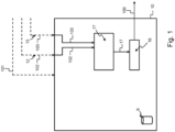
- Fig. 1 illustrates an example embodiment of an apparatus 10 according to the present disclosure.
- the apparatus 10 comprises at least one memory 6, at least one processor, wherein the memory 6 includes computer program code configured to, with the at least one processor, cause the apparatus 10 to perform the following:
- Fig. 2 illustrates an example embodiment of a system 20 according to the present disclosure. Components having identical reference numbers to numbers on Fig. 1 perform the same function.
- the system 20 of Fig. 2 comprises an apparatus 10 according to the present disclosure.
- the system 20 further comprises a light source 2 and a sensor 4.
- a light source 2 is configured to emit light 40.
- the apparatus 10 is configured to:
- Fig. 3 illustrates an example embodiment of a system 20 according to the present disclosure. Components having identical reference numbers to numbers on Fig. 1 or Fig. 2 perform the same function.
- the system 20 of Fig. 3 comprises an apparatus 10 according to the present disclosure.
- the system 20 further comprises at least one light source 2 and a sensor 4, comprised in the apparatus 10.
- a light source 2 is configured to emit light 40.
- the apparatus 10 is configured to:
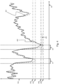
- Fig. 4 illustrates an example comparison between PPG-amplitude-based peripheral arterial tone 401 both in function of time 60 of an individual, wherein the PPG-amplitude-based peripheral arterial tone 401 is assessed by considering changes in amplitude of pulses in an optical plethysmography signal as described in the prior art, and evaluation-function-based peripheral arterial tone 402 in function of time 60 of the same individual, wherein the evaluation-function-based peripheral arterial tone 402 is determined by the computer-implemented method according to the present disclosure, or by the apparatus according to the present disclosure, i.e. wherein the evaluation-function-based peripheral arterial tone 402 is assessed by determining a logarithm of a ratio of light intensities measured from an optical plethysmography signal.
- the evaluation-function-based peripheral arterial tone 402 is plotted by calculating ln I h I l .
- the evaluation-function-based peripheral arterial tone 402 is plotted by calculating ln I l I h .
- the PPG-amplitude-based peripheral arterial tone 401 and the evaluation-function-based peripheral arterial tone 402 evolve in a similar manner as a function of time 60 at a pre-vasoconstriction-event baseline value. But during the time periods 61 and 62, i.e. during occurrence of an event such as for example a sleep-related event, it can be seen on Fig.
- the PPG-amplitude-based peripheral arterial tone 401 and the evaluation-function-based peripheral arterial tone 402 evolve in a similar manner but do not overlap anymore in the corresponding time periods 61 and 62. Indeed, in period 61, the PPG-amplitude-based peripheral arterial tone 401 drops to a level 403 which value is higher than the one of the level 404 to which the evaluation-function-based peripheral arterial tone 402 drops. Similarly, in period 62, the PPG-amplitude-based peripheral arterial tone 401 drops to a level 405 which value is higher than the one of the level 406 to which the evaluation-function-based peripheral arterial tone 402 drops. It can be seen on Fig.
- peripheral arterial tone 100 by determining a logarithm of a function of light intensities acquired by optical plethysmography allows to detect the event occurring more accurately.
- a drop in peripheral arterial tone 100 is indicative for a vasoconstriction of the arteries and the arterioles in the investigated volume under monitoring.
- This vasoconstriction can be related to the occurrence of an event at the individual under monitoring such as for example a sleep-related event, such as for example sleep apnea.
- a sleep-related event such as for example sleep apnea.
- the drop in the PPG-amplitude-based peripheral arterial tone 401 between the pre-vasoconstriction-event baseline value and the lowest point of the PPG-amplitude-based peripheral arterial tone 401 is smaller than the drop in the evaluation-function-based peripheral arterial tone 402 between the pre-vasoconstriction-event baseline value and the lowest point of the evaluation-function-based peripheral arterial tone 402.
- a predetermined threshold value 407 for peripheral arterial tone 100 can be used to detect whether an event is occurring at the individual under monitoring such as for example a sleep-related event, such as for example sleep apnea: when the peripheral arterial tone 100 is above this predetermined threshold value 407, no event is detected, but when the peripheral arterial tone 100 is below this predetermined threshold value 407, an event is detected.
- a sleep-related event such as for example sleep apnea
- the drop in the PPG-amplitude-based peripheral arterial tone 401 between the pre-vasoconstriction-event baseline value and the lowest point of the PPG-amplitude-based peripheral arterial tone 401 is such that the PPG-amplitude-based peripheral arterial tone 401 stays above the predetermined threshold value 407, which results in the absence of detection of an event occurring at the individual under monitoring such as for example a sleep-related event, such as for example sleep apnea.
- the drop in the evaluation-function-based peripheral arterial tone 402 between the pre-vasoconstriction-event baseline value and the lowest point of the evaluation-function-based peripheral arterial tone 402 is such that the evaluation-function-based peripheral arterial tone 402 drops below the predetermined threshold value 407, which results in the detection of an event occurring at the individual under monitoring such as for example a sleep-related event, such as for example sleep apnea.
- peripheral arterial tone 100 by determining a logarithm of a function of light intensities acquired by optical plethysmography allows to more accurately and more robustly detect the occurrence of an event occurring at the individual under monitoring such as for example a sleep-related event, such as for example sleep apnea.
- Fig. 5 illustrates an example embodiment of a computer-implemented method for assessing peripheral arterial tone, PAT, of an individual monitored by optical plethysmography, wherein said method comprises the steps of:
- Fig. 6 shows a suitable computing system 800 enabling to implement embodiments of the system.
- Computing system 800 may in general be formed as a suitable general-purpose computer and comprise a bus 810, a processor 802, a local memory 804, one or more optional input interfaces 814, one or more optional output interfaces 816, a communication interface 812, a storage element interface 806, and one or more storage elements 808.
- Bus 810 may comprise one or more conductors that permit communication among the components of the computing system 800.
- Processor 802 may include any type of conventional processor or microprocessor that interprets and executes programming instructions.
- Local memory 804 may include a random-access memory (RAM) or another type of dynamic storage device that stores information and instructions for execution by processor 802 and/or a read only memory (ROM) or another type of static storage device that stores static information and instructions for use by processor 802.
- Input interface 814 may comprise one or more conventional mechanisms that permit an operator or user to input information to the computing device 800, such as a keyboard 820, a mouse 830, a pen, voice recognition and/or biometric mechanisms, a camera, etc.
- Output interface 816 may comprise one or more conventional mechanisms that output information to the operator or user, such as a display 840, etc.
- Communication interface 812 may comprise any transceiver-like mechanism such as for example one or more Ethernet interfaces that enables computing system 800 to communicate with other devices and/or systems, for example with other computing devices 881, 882, 883.
- the communication interface 812 of computing system 800 may be connected to such another computing system by means of a local area network (LAN) or a wide area network (WAN) such as for example the internet.
- Storage element interface 806 may comprise a storage interface such as for example a Serial Advanced Technology Attachment (SATA) interface or a Small Computer System Interface (SCSI) for connecting bus 810 to one or more storage elements 808, such as one or more local disks, for example SATA disk drives, and control the reading and writing of data to and/or from these storage elements 808.
- SATA Serial Advanced Technology Attachment
- SCSI Small Computer System Interface
- computing system 800 could thus correspond to the apparatus 10 in the embodiment illustrated by Figs. 1 or 2 or 3 .
- circuitry may refer to one or more or all of the following:
- circuitry also covers an implementation of merely a hardware circuit or processor (or multiple processors) or portion of a hardware circuit or processor and its (or their) accompanying software and/or firmware.
- circuitry also covers, for example and if applicable to the particular claim element, a baseband integrated circuit or processor integrated circuit for a mobile device or a similar integrated circuit in a server, a cellular network device, or other computing or network device.
- top, bottom, over, under, and the like are introduced for descriptive purposes and not necessarily to denote relative positions. It is to be understood that the terms so used are interchangeable under appropriate circumstances and embodiments of the invention are capable of operating according to the present invention in other sequences, or in orientations different from the one(s) described or illustrated above.
Landscapes
- Health & Medical Sciences (AREA)
- Life Sciences & Earth Sciences (AREA)
- Heart & Thoracic Surgery (AREA)
- Surgery (AREA)
- Physics & Mathematics (AREA)
- Veterinary Medicine (AREA)
- Biophysics (AREA)
- Pathology (AREA)
- Engineering & Computer Science (AREA)
- Biomedical Technology (AREA)
- Public Health (AREA)
- Medical Informatics (AREA)
- Molecular Biology (AREA)
- General Health & Medical Sciences (AREA)
- Animal Behavior & Ethology (AREA)
- Physiology (AREA)
- Neurology (AREA)
- Cardiology (AREA)
- Hematology (AREA)
- Neurosurgery (AREA)
- Vascular Medicine (AREA)
- Measurement Of The Respiration, Hearing Ability, Form, And Blood Characteristics Of Living Organisms (AREA)
- Measuring Pulse, Heart Rate, Blood Pressure Or Blood Flow (AREA)
Priority Applications (1)
| Application Number | Priority Date | Filing Date | Title |
|---|---|---|---|
| EP24199196.7A EP4501216A3 (de) | 2020-06-26 | 2020-06-26 | Verfahren und vorrichtung zur beurteilung des peripheren arteriellen tonus |
Applications Claiming Priority (2)
| Application Number | Priority Date | Filing Date | Title |
|---|---|---|---|
| EP24199196.7A EP4501216A3 (de) | 2020-06-26 | 2020-06-26 | Verfahren und vorrichtung zur beurteilung des peripheren arteriellen tonus |
| EP20182512.2A EP3928690B1 (de) | 2020-06-26 | 2020-06-26 | Verfahren und vorrichtung zur beurteilung des peripheren arteriellen tonus |
Related Parent Applications (1)
| Application Number | Title | Priority Date | Filing Date |
|---|---|---|---|
| EP20182512.2A Division EP3928690B1 (de) | 2020-06-26 | 2020-06-26 | Verfahren und vorrichtung zur beurteilung des peripheren arteriellen tonus |
Publications (2)
| Publication Number | Publication Date |
|---|---|
| EP4501216A2 true EP4501216A2 (de) | 2025-02-05 |
| EP4501216A3 EP4501216A3 (de) | 2025-04-30 |
Family
ID=71170446
Family Applications (2)
| Application Number | Title | Priority Date | Filing Date |
|---|---|---|---|
| EP20182512.2A Active EP3928690B1 (de) | 2020-06-26 | 2020-06-26 | Verfahren und vorrichtung zur beurteilung des peripheren arteriellen tonus |
| EP24199196.7A Pending EP4501216A3 (de) | 2020-06-26 | 2020-06-26 | Verfahren und vorrichtung zur beurteilung des peripheren arteriellen tonus |
Family Applications Before (1)
| Application Number | Title | Priority Date | Filing Date |
|---|---|---|---|
| EP20182512.2A Active EP3928690B1 (de) | 2020-06-26 | 2020-06-26 | Verfahren und vorrichtung zur beurteilung des peripheren arteriellen tonus |
Country Status (7)
| Country | Link |
|---|---|
| US (1) | US20230263453A1 (de) |
| EP (2) | EP3928690B1 (de) |
| JP (1) | JP2023532318A (de) |
| CN (1) | CN115988984A (de) |
| AU (1) | AU2021295604A1 (de) |
| IL (1) | IL299330A (de) |
| WO (1) | WO2021260192A1 (de) |
Families Citing this family (5)
| Publication number | Priority date | Publication date | Assignee | Title |
|---|---|---|---|---|
| US12059266B2 (en) * | 2020-12-30 | 2024-08-13 | Itamar Medical Ltd. | System and method for arrhythmia detection during an at home sleep test |
| EP4396833A1 (de) | 2021-08-30 | 2024-07-10 | Resmed Digital Health Inc. | Intelligente atemtraining |
| WO2023148190A1 (en) * | 2022-02-03 | 2023-08-10 | Ectosense NV | Systems and methods for screening, diagnosis, detection, monitoring and/or therapy |
| CN120379593A (zh) | 2022-12-20 | 2025-07-25 | 瑞思迈数字健康公司 | 诊断头带 |
| US12579242B2 (en) | 2022-12-21 | 2026-03-17 | Resmed Digital Health Inc. | Systems and methods for digit-based diagnostic chain of custody management |
Citations (1)
| Publication number | Priority date | Publication date | Assignee | Title |
|---|---|---|---|---|
| US20140012110A1 (en) * | 2008-06-30 | 2014-01-09 | Nellcor Puritan Bennett Ireland | Processing and detecting baseline changes in signals |
Family Cites Families (10)
| Publication number | Priority date | Publication date | Assignee | Title |
|---|---|---|---|---|
| US5140990A (en) * | 1990-09-06 | 1992-08-25 | Spacelabs, Inc. | Method of measuring blood pressure with a photoplethysmograph |
| IL120881A (en) * | 1996-07-30 | 2002-09-12 | It M R Medic L Cm 1997 Ltd | Method and device for continuous and non-invasive monitoring of peripheral arterial tone |
| US6856829B2 (en) * | 2000-09-07 | 2005-02-15 | Denso Corporation | Method for detecting physiological condition of sleeping patient based on analysis of pulse waves |
| US7738935B1 (en) * | 2002-07-09 | 2010-06-15 | Pacesetter, Inc. | Methods and devices for reduction of motion-induced noise in pulse oximetry |
| JP5296312B2 (ja) * | 2003-03-12 | 2013-09-25 | イエール ユニバーシティ | 光電容積脈波法を使用した血液量評価方法 |
| AU2004203059A1 (en) * | 2004-06-08 | 2005-12-22 | The Government Of The United States Of America As Represented By The Secretary Of The Department Of Health And Human Services, Centers For Disease Control And Prevention | Apparatus and method for assessing peripheral circulation to evaluate a physiological condition |
| US10973422B2 (en) * | 2016-01-22 | 2021-04-13 | Fitbit, Inc. | Photoplethysmography-based pulse wave analysis using a wearable device |
| EP4403099B1 (de) * | 2016-06-08 | 2026-03-18 | Itamar Medical Ltd. | System zur nichtinvasiven detektion von physiologischen und pathophysiologischen schlafzuständen |
| EP4360546B1 (de) * | 2018-07-11 | 2025-02-19 | Ectosense NV | System zur schlafdiagnose |
| CN110495864B (zh) * | 2019-08-02 | 2022-04-05 | 深圳市德胜医疗科技有限公司 | 人体血管血流收缩力与舒张力测定方法及装置 |
-
2020
- 2020-06-26 EP EP20182512.2A patent/EP3928690B1/de active Active
- 2020-06-26 EP EP24199196.7A patent/EP4501216A3/de active Pending
-
2021
- 2021-06-25 IL IL299330A patent/IL299330A/en unknown
- 2021-06-25 US US18/012,009 patent/US20230263453A1/en active Pending
- 2021-06-25 JP JP2022580956A patent/JP2023532318A/ja active Pending
- 2021-06-25 AU AU2021295604A patent/AU2021295604A1/en active Pending
- 2021-06-25 WO PCT/EP2021/067534 patent/WO2021260192A1/en not_active Ceased
- 2021-06-25 CN CN202180052412.8A patent/CN115988984A/zh active Pending
Patent Citations (1)
| Publication number | Priority date | Publication date | Assignee | Title |
|---|---|---|---|---|
| US20140012110A1 (en) * | 2008-06-30 | 2014-01-09 | Nellcor Puritan Bennett Ireland | Processing and detecting baseline changes in signals |
Non-Patent Citations (2)
| Title |
|---|
| CHATTERJEE ET AL.: "Monte Carlo Analysis of Optical Interactions in Reflectance and Transmittance Finger Photoplethysmography", SENSORS, vol. 19, no. 4, 15 February 2019 (2019-02-15), pages 789 |
| HAMUNEN ET AL.: "Effect of pain on autonomic nervous system indices derived from photoplethysmography in healthy volunteers", BRITISH JOURNAL OF ANESTHESIA, vol. 108, 1 May 2012 (2012-05-01), pages 838 - 844 |
Also Published As
| Publication number | Publication date |
|---|---|
| US20230263453A1 (en) | 2023-08-24 |
| WO2021260192A1 (en) | 2021-12-30 |
| CN115988984A (zh) | 2023-04-18 |
| EP3928690B1 (de) | 2024-09-11 |
| AU2021295604A1 (en) | 2023-02-02 |
| EP4501216A3 (de) | 2025-04-30 |
| IL299330A (en) | 2023-02-01 |
| JP2023532318A (ja) | 2023-07-27 |
| EP3928690A1 (de) | 2021-12-29 |
Similar Documents
| Publication | Publication Date | Title |
|---|---|---|
| EP3928689B1 (de) | Vorrichtung und verfahren zur kompensation der beurteilung des peripheren arteriellen tonus | |
| EP3928690B1 (de) | Verfahren und vorrichtung zur beurteilung des peripheren arteriellen tonus | |
| JP5748160B2 (ja) | 携帯診断装置 | |
| US11160461B2 (en) | Blood pressure measurement using a wearable device | |
| EP3493734B1 (de) | Blutdruckmessung mithilfe einer tragbaren vorrichtung | |
| US9402573B2 (en) | System and method for detecting fluid responsiveness of a patient | |
| US8221326B2 (en) | Detection of oximetry sensor sites based on waveform characteristics | |
| Venkat et al. | Machine learning based spo 2 computation using reflectance pulse oximetry | |
| JP2011521702A (ja) | Co2評価のための方法および装置 | |
| JP5096310B2 (ja) | 身体の部位における血液の灌流を決定するための方法及び装置 | |
| CN107920786A (zh) | 脉搏血氧测定 | |
| EP4208085B1 (de) | Verfahren und vorrichtung zur erkennung von schlafstörungsereignissen aus einem signal, das einen peripheren arteriellen ton einer person anzeigt | |
| US20230040540A1 (en) | Optimizing Sensor Pressure in Blood Pressure Measurements Using a Wearable Device | |
| Campbell | Development of non-invasive, optical methods for central cardiovascular and blood chemistry monitoring | |
| US12364405B1 (en) | Hemodynamic determination of arterial stiffness, arterial age, arterial deposits and HBA1C | |
| Tanveejul et al. | A Study on the Subject and Location Specificity in Reflectance based SpO 2 Estimation using R-value based Calibration Curve | |
| Kanak et al. | Multi-Sensor Suite for Melanin-Adjusted Blood Oxygenation Measurement Progress Report | |
| Bodei | Proof of Concept Design and De-velopment of a Multi-Wavelength Photoplethysmographic System for Physiological Monitoring and Res-piratory Rate Estimation |
Legal Events
| Date | Code | Title | Description |
|---|---|---|---|
| PUAI | Public reference made under article 153(3) epc to a published international application that has entered the european phase |
Free format text: ORIGINAL CODE: 0009012 |
|
| STAA | Information on the status of an ep patent application or granted ep patent |
Free format text: STATUS: THE APPLICATION HAS BEEN PUBLISHED |
|
| AC | Divisional application: reference to earlier application |
Ref document number: 3928690 Country of ref document: EP Kind code of ref document: P |
|
| AK | Designated contracting states |
Kind code of ref document: A2 Designated state(s): AL AT BE BG CH CY CZ DE DK EE ES FI FR GB GR HR HU IE IS IT LI LT LU LV MC MK MT NL NO PL PT RO RS SE SI SK SM TR |
|
| REG | Reference to a national code |
Ref country code: DE Ref legal event code: R079 Free format text: PREVIOUS MAIN CLASS: A61B0005000000 Ipc: A61B0005020000 |
|
| PUAL | Search report despatched |
Free format text: ORIGINAL CODE: 0009013 |
|
| AK | Designated contracting states |
Kind code of ref document: A3 Designated state(s): AL AT BE BG CH CY CZ DE DK EE ES FI FR GB GR HR HU IE IS IT LI LT LU LV MC MK MT NL NO PL PT RO RS SE SI SK SM TR |
|
| RIC1 | Information provided on ipc code assigned before grant |
Ipc: A61B 5/00 20060101ALI20250326BHEP Ipc: A61B 5/0295 20060101ALI20250326BHEP Ipc: A61B 5/026 20060101ALI20250326BHEP Ipc: A61B 5/02 20060101AFI20250326BHEP |
|
| STAA | Information on the status of an ep patent application or granted ep patent |
Free format text: STATUS: REQUEST FOR EXAMINATION WAS MADE |
|
| 17P | Request for examination filed |
Effective date: 20251030 |
|
| STAA | Information on the status of an ep patent application or granted ep patent |
Free format text: STATUS: EXAMINATION IS IN PROGRESS |
|
| 17Q | First examination report despatched |
Effective date: 20260216 |