EP3928689B1 - Vorrichtung und verfahren zur kompensation der beurteilung des peripheren arteriellen tonus - Google Patents
Vorrichtung und verfahren zur kompensation der beurteilung des peripheren arteriellen tonus Download PDFInfo
- Publication number
- EP3928689B1 EP3928689B1 EP20182503.1A EP20182503A EP3928689B1 EP 3928689 B1 EP3928689 B1 EP 3928689B1 EP 20182503 A EP20182503 A EP 20182503A EP 3928689 B1 EP3928689 B1 EP 3928689B1
- Authority
- EP
- European Patent Office
- Prior art keywords
- function
- light
- determining
- individual
- oxygen saturation
- Prior art date
- Legal status (The legal status is an assumption and is not a legal conclusion. Google has not performed a legal analysis and makes no representation as to the accuracy of the status listed.)
- Active
Links
Images


Classifications
-
- A—HUMAN NECESSITIES
- A61—MEDICAL OR VETERINARY SCIENCE; HYGIENE
- A61B—DIAGNOSIS; SURGERY; IDENTIFICATION
- A61B5/00—Measuring for diagnostic purposes; Identification of persons
- A61B5/02—Detecting, measuring or recording for evaluating the cardiovascular system, e.g. pulse, heart rate, blood pressure or blood flow
- A61B5/02007—Evaluating blood vessel condition, e.g. elasticity, compliance
-
- A—HUMAN NECESSITIES
- A61—MEDICAL OR VETERINARY SCIENCE; HYGIENE
- A61B—DIAGNOSIS; SURGERY; IDENTIFICATION
- A61B5/00—Measuring for diagnostic purposes; Identification of persons
- A61B5/02—Detecting, measuring or recording for evaluating the cardiovascular system, e.g. pulse, heart rate, blood pressure or blood flow
- A61B5/026—Measuring blood flow
- A61B5/0261—Measuring blood flow using optical means, e.g. infrared light
-
- A—HUMAN NECESSITIES
- A61—MEDICAL OR VETERINARY SCIENCE; HYGIENE
- A61B—DIAGNOSIS; SURGERY; IDENTIFICATION
- A61B5/00—Measuring for diagnostic purposes; Identification of persons
- A61B5/02—Detecting, measuring or recording for evaluating the cardiovascular system, e.g. pulse, heart rate, blood pressure or blood flow
- A61B5/026—Measuring blood flow
- A61B5/0295—Measuring blood flow using plethysmography, i.e. measuring the variations in the volume of a body part as modified by the circulation of blood therethrough, e.g. impedance plethysmography
-
- A—HUMAN NECESSITIES
- A61—MEDICAL OR VETERINARY SCIENCE; HYGIENE
- A61B—DIAGNOSIS; SURGERY; IDENTIFICATION
- A61B5/00—Measuring for diagnostic purposes; Identification of persons
- A61B5/145—Measuring characteristics of blood in vivo, e.g. gas concentration or pH-value ; Measuring characteristics of body fluids or tissues, e.g. interstitial fluid or cerebral tissue
- A61B5/1455—Measuring characteristics of blood in vivo, e.g. gas concentration or pH-value ; Measuring characteristics of body fluids or tissues, e.g. interstitial fluid or cerebral tissue using optical sensors, e.g. spectral photometrical oximeters
- A61B5/14551—Measuring characteristics of blood in vivo, e.g. gas concentration or pH-value ; Measuring characteristics of body fluids or tissues, e.g. interstitial fluid or cerebral tissue using optical sensors, e.g. spectral photometrical oximeters for measuring blood gases
-
- A—HUMAN NECESSITIES
- A61—MEDICAL OR VETERINARY SCIENCE; HYGIENE
- A61B—DIAGNOSIS; SURGERY; IDENTIFICATION
- A61B5/00—Measuring for diagnostic purposes; Identification of persons
- A61B5/40—Detecting, measuring or recording for evaluating the nervous system
- A61B5/4029—Detecting, measuring or recording for evaluating the nervous system for evaluating the peripheral nervous systems
- A61B5/4035—Evaluating the autonomic nervous system
-
- A—HUMAN NECESSITIES
- A61—MEDICAL OR VETERINARY SCIENCE; HYGIENE
- A61B—DIAGNOSIS; SURGERY; IDENTIFICATION
- A61B5/00—Measuring for diagnostic purposes; Identification of persons
- A61B5/48—Other medical applications
- A61B5/4806—Sleep evaluation
- A61B5/4818—Sleep apnoea
-
- A—HUMAN NECESSITIES
- A61—MEDICAL OR VETERINARY SCIENCE; HYGIENE
- A61B—DIAGNOSIS; SURGERY; IDENTIFICATION
- A61B5/00—Measuring for diagnostic purposes; Identification of persons
- A61B5/68—Arrangements of detecting, measuring or recording means, e.g. sensors, in relation to patient
- A61B5/6801—Arrangements of detecting, measuring or recording means, e.g. sensors, in relation to patient specially adapted to be attached to or worn on the body surface
- A61B5/6813—Specially adapted to be attached to a specific body part
- A61B5/6825—Hand
- A61B5/6826—Finger
-
- A—HUMAN NECESSITIES
- A61—MEDICAL OR VETERINARY SCIENCE; HYGIENE
- A61B—DIAGNOSIS; SURGERY; IDENTIFICATION
- A61B5/00—Measuring for diagnostic purposes; Identification of persons
- A61B5/72—Signal processing specially adapted for physiological signals or for diagnostic purposes
- A61B5/7203—Signal processing specially adapted for physiological signals or for diagnostic purposes for noise prevention, reduction or removal
-
- A—HUMAN NECESSITIES
- A61—MEDICAL OR VETERINARY SCIENCE; HYGIENE
- A61B—DIAGNOSIS; SURGERY; IDENTIFICATION
- A61B2560/00—Constructional details of operational features of apparatus; Accessories for medical measuring apparatus
- A61B2560/02—Operational features
- A61B2560/0223—Operational features of calibration, e.g. protocols for calibrating sensors
Definitions
- the present disclosure generally relates, amongst others, to a method and an apparatus for assessing Peripheral Arterial Tone or PAT. More particularly, it relates to robustly monitoring peripheral arterial tone for detecting for example sleep-related events.
- blood is pumped through the vascular system of the individual in a pulsating fashion.
- the volume of blood in for example a finger, a nostril, an ear, a forehead, the inside of a mouth, a toe, a wrist, an ankle, etc., increases and decreases cyclically.
- the blood comprised in arteries is called arterial blood.
- the blood comprised in veins is called venous blood.
- a common method for measuring this fluctuation in blood volume is optical plethysmography, or photoplethysmography, during which an optical plethysmogram, or PhotoPlethysmoGram, or PPG, is used to detect changes in blood volume in the microvascular bed of the tissue.
- a PPG is often obtained by shining light from one or more light sources, such as for example LEDs, onto an investigated volume, and detecting collected light corresponding to the light being reflected or transmitted in the investigated volume on a sensor, wherein the sensor may for example comprise or correspond to a photodetector, such as for example a photodiode.
- the light sources and the sensor can for example be arranged on opposite sides of a finger of an individual, hereby allowing the measurement of a transmission mode PPG, or at the same side of a finger of the individual, hereby allowing the measurement of a reflectance mode PPG.
- the heart pumps arterial blood to the investigated volume.
- the physical events corresponding to a change in arterial blood volume in the investigated volume for example in a cardiac cycle can be captured by optical plethysmography.
- US20060009700A1 describes an apparatus and method for the non-invasive evaluation, detection, and monitoring of a physiological state or medical condition by assessing peripheral circulation.
- US2019343406A1 describes a method for non-invasively measuring cardiac output, stroke volume, or both by collecting plethysmographic waveform data of a patient.
- the arterial blood volume in the tissue is also influenced by the diameters of small arteries, or arterioles, in the investigated volume. These arterioles have muscular walls which can contract to reduce the diameter arterioles. In other words, when these arterioles contract and reduce in diameter, the volume of arterial blood comprised in the corresponding arterioles evidently reduces.
- Optical plethysmography is a measurement technique which can be used to monitor changes in the volume of arterial blood comprised in arterioles when these arterioles contract in diameter. Monitoring changes in the arterial blood volume with optical plethysmography therefore empirically provides information on relative changes in muscle tension or 'tone' of the smooth muscle tissue of the arterioles, also referred to as Peripheral Arterial Tone or PAT.
- optical plethysmography can also be used to monitor breathing, hypovolemia, other circulatory conditions, and for example can be used for diagnosing sleep disorders.
- Sleep disorder diagnosis is a medical field wherein a patient's sleep is monitored during a certain time, e.g. one or more nights. Based on the monitoring, different sleep-related events may be identified such as for example apneic events, snoring, or limb movements.
- the reuptake of breathing at the end of a sleep apnea usually coincides with an adrenaline release.
- Adrenaline is released in the bloodstream and binds to adrenergic receptors in the arterioles in the investigated volume, which triggers an increase in muscle tension of the arterioles resulting in a reduction of the arteriole diameter and a reduction of arterial blood volume in the investigated volume.
- Monitoring Peripheral arterial tone through for example optical plethysmography can therefore provide valuable information on the occurrence of sleep-related events such as for example sleep apnea.
- Hemoglobin is the main molecule which scatters the light emitted onto the investigated volume during a measurement with optical plethysmography. Hemoglobin comprises oxygenated hemoglobin, also known as HbO 2 , and deoxygenated hemoglobin, also known as Hb. The extent to which hemoglobin in the arterial blood is oxygenated is referred to as the oxygen saturation, or SpO 2 . The degree of absorption and scattering of light due to HbOz is significantly different from that of Hb and additionally depends on the wavelength of the light used for optical plethysmography.
- an optical plethysmogram is not only dependent on the volume of arterial blood in the investigated volume but is also dependent on the hemoglobin composition of that arterial blood volume, which is characterized by the oxygen saturation.
- this measurement can be greatly influenced by changes in the oxygen saturation of the arterial blood volume which is monitored. For example, during apnea, as less oxygen is supplied to the lungs, the proportion of HbOz in a arterial blood volume within the investigated volume of the individual drops.
- WO98/04182 for example describes a method and an apparatus for the detection of medical conditions by monitoring peripheral arterial tone.
- the apparatus of EP1534115A2 performs optical plethysmography relying on a wavelength for which the degree of absorption and scattering for this particular wavelength caused by the HbO2 and the degree of absorption and scattering for this particular wavelength caused by the Hb are identical. Such wavelength is known as isosbestic wavelength.
- the light intensities measured in EP1534115A2 by optical plethysmography when relying on this isosbestic wavelength are therefore less dependent on the hemoglobin composition of the arterial blood volume, in other words, on the value of the oxygen saturation.
- EP1534115A2 requires the use of three light sources to monitor PAT via optical plethysmography: two light sources which do not generate light at an isosbestic wavelength to estimate the oxygen saturation, and a third light source which generates light at an isosbestic wavelength to estimate peripheral arterial tone.
- the use of three light sources increases the device cost and its size compared to a system which comprises only two light sources.
- the computer-implemented method according to the present disclosure allows determining peripheral arterial tone in an accurate and robust manner.
- the hemoglobin composition of the arterial blood volume of the individual at the investigated volume being monitored by optical plethysmography can be determined or estimated.
- the computer-implemented method according to the present disclosure can therefore adjust, or in other words compensate, the optical plethysmography signal such that the light intensities more reliably reflect changes in the arterial blood volume under monitoring by minimizing the influence caused by changes in the hemoglobin composition of the monitored arterial blood volume.
- the computer-implemented method reduces the effect on the optical plethysmography signal of changes in the hemoglobin composition of the arterial blood volume, and thereby minimizes or reduces the effect of changes in the hemoglobin composition of the arterial blood volume on the optical measurement of peripheral arterial tone.
- the computer-implemented method indeed determines a compensation function which is a function of the oxygen saturation estimate and divides a function of the light intensities measured by optical plethysmography by the compensation function for this oxygen saturation estimate.
- the resulting evaluation of a change in arterial blood volume in the investigated volume therefore provides a more accurate and robust assessment of peripheral arterial tone of the individual.
- the computer-implemented method does not rely on the use of a light source emitting light at an isosbestic wavelength.
- the computer-implemented method is compatible with any usual setup for optical plethysmography comprising for example two light sources, wherein the two light sources for example emit light of two different wavelengths, such as for example one red wavelength and another infra-red wavelength.
- the computer-implemented method accurately measures changes in arterial blood volume in an investigated volume with just two light sources, which is the minimum number of light sources needed to determine the oxygen saturation estimate.
- Peripheral arterial tone is then mathematically derived from the optical plethysmography measurement with two light sources. This allows miniaturization compared to a system with three light sources and further allows cost optimization by eliminating the need for a third light source.
- an investigated volume of an individual is for example a volume defined in an investigated tissue of the individual which is monitored by optical plethysmography and in which light emitted by optical plethysmography propagates and is collected on a sensor for optical plethysmography.
- an investigated volume of an individual is for example a volume defined in an investigated tissue of the individual for which an optical plethysmography signal is acquired.
- the investigated volume is a peripheral tissue volume of the individual.
- the investigated volume is a volume defined in finger, a tip of a finger, a distal end of a digit of the individual, a nostril, an ear, a forehead, the inside of a mouth, a toe, a tip of a toe, a wrist, an ankle of the individual.
- the investigated volume of an individual comprises the skin of the individual comprised in the investigated volume and further comprises the arterial blood volume present in the investigated volume.
- peripheral arterial tone is understood as arterial tone changes in investigated arterial beds in the investigated volume of an individual.
- determining pulsatile volume changes in the vascular beds of the investigated volume of the individual allows determining or assessing information indicative for muscle tension or 'tone' of the smooth muscle tissue of the arterioles in the investigated volume and therefore allows determining or assessing peripheral arterial tone which is modulated by the sympathetic nervous system.
- Determining peripheral arterial tone is noninvasive and can for example be used to detect heart diseases, erectile dysfunction, sleep apnea, obstructive sleep apnea, cardiovascular conditions, etc..
- an optical plethysmography signal is a signal measured by optical plethysmography.
- the optical plethysmography signal is an optical plethysmogram.
- the optical plethysmography signal is a PPG.
- the optical plethysmography signal is for example measured at the tip of a finger of the individual by an optical plethysmography setup comprising at least two light sources and a sensor.
- a light intensity corresponds to the intensity of light collected on the sensor of the optical plethysmography setup, wherein the light collected on the sensor corresponds to light generated by one or the two light sources being transmitted through or reflected in the investigated volume of the individual.
- the blood comprised in arteries is called arterial blood.
- the blood comprised in veins is called venous blood.
- the oxygen saturation estimate or SpO 2 or hemoglobin composition corresponds to a fraction of oxygenated hemoglobin related to a total amount of hemoglobin in the arterial blood volume.
- the oxygen saturation estimate or SpO 2 or hemoglobin composition corresponds to a ratio of the concentration of oxygenated hemoglobin on the sum of the concentrations of oxygenated and deoxygenated hemoglobin in the arterial blood volume being monitored in the investigated volume.
- the oxygen saturation estimate or SpO 2 or hemoglobin composition corresponds to a ratio of the volume fraction of oxygenated hemoglobin on the sum of the volume fractions of oxygenated and deoxygenated hemoglobin in the arterial blood volume being monitored in the investigated volume.
- deoxygenated hemoglobin is defined as the form of hemoglobin without the bound oxygen, and without any other bound molecule such as for example carbon monoxide, carbon dioxide, or iron.
- oxygenated hemoglobin is defined as the form of hemoglobin with the bound oxygen.
- light emitted by the light sources of an optical plethysmography setup comprises photons which reach the sensor through a probabilistic path of one or multiple scattering events. This optical path is not straight and is often assumed to follow a curved spatial probability distribution. The investigated volume along this curved optical path forms the volume which is sampled or investigated by optical plethysmography.
- a change in arterial blood volume in the investigated volume between the two points in time corresponds to a relative change between the volume of arterial blood present in the investigated volume at the first point in time and the volume of arterial blood present in the investigated volume at the second point in time.
- the calibration data comprises predetermined calibration coefficients and/or predefined coefficients; and:
- the predefined coefficients are for example known or can be determined from literature, such as for example from scientific publications.
- the predefined coefficients comprise one or more extinction coefficients of chromophores and/or one or more absorption coefficients of chromophores and/or one or more scattering coefficients of chromophores.
- a chromophore is a molecular unit which absorbs or scatters light in the investigated volume.
- examples of chromophores are melanin molecules, oxygenated hemoglobin, deoxygenated hemoglobin, etc.
- equation (3') is equal to equation (3) up to a constant 1 ln b .
- Some chromophores remain attached to the epidermis of the individual between the two points in time along the optical plethysmography signal.
- melanin molecules remain fixed to the investigated volume between the two points in time along the optical plethysmography signal.
- the difference in the volume fraction or concentration of such chromophores, such as for example melanin molecules, between the two points in time is therefore null.
- the contribution to the right-hand side of equation (4) of such chromophores is therefore null.
- the main chromophores of which the volume fraction or concentration fluctuates between the two points in time along the optical plethysmography signal are the oxygenated and deoxygenated hemoglobin in the arterial blood volume.
- Relation (12) is therefore deducted from equation (11): ln I l I h Q 1 SpO 2 + Q 2 ⁇ ⁇ V blood
- Relation (12) highlights a term on the left-hand side which shows a linear relationship with the change ⁇ V blood in total volume fraction or concentration of oxygenated and deoxygenated hemoglobin in the investigated volume between the first and the second points in time.
- ⁇ V blood is a linear proxy for arterial blood volume fluctuations within the investigated volume.
- a change in arterial blood volume in the investigated volume is thus evaluated when determining a ratio of a function of the light intensities collected on the sensor when measuring with optical plethysmography and of a compensation function which is a function of the oxygen saturation estimate.
- At least one of the points in time corresponds to the diastole in a cardiac cycle of the individual and/or wherein at least one of the points in time corresponds to the systole in a cardiac cycle of the individual.
- the volume of arterial blood in the investigated volume of the individual is maximum, resulting in the largest absorption and scattering of light of any point in time within a cardiac cycle, i.e. the period between two heart beats, since hemoglobin is one of the main absorbers and scatters of photons in the investigated volume, hence resulting in the lowest measured light intensity on the sensor of the optical plethysmography setup.
- the volume of arterial blood in the investigated volume of the individual is minimum, resulting in the lowest absorption and scattering of light of any point in time within a cardiac cycle and hence highest measured light intensity on the sensor of the optical plethysmography setup.
- At least one first point in time corresponds for example to the diastole in a first cardiac cycle and/or at least one second point in time corresponds for example to the systole in a second cardiac cycle different from the first cardiac cycle.
- at least one first point in time corresponds for example to the systole in a first cardiac cycle and/or at least one second point in time corresponds for example to the diastole in a second cardiac cycle different from the first cardiac cycle.
- at least one first point in time corresponds for example to the systole or to the diastole in a cardiac cycle and at least one second point in time corresponds to any point in time within the same cardiac cycle or within a different cardiac cycle.
- At least two of the two or more points in time are within one cardiac cycle of the individual.
- the logarithm of the fraction of the light intensities at systole and diastole is linearly related to the changes in the chromophore volume fraction or concentration between systole and diastole.
- it is best to measure at at least two different points in time within one cardiac cycle wherein the at least two points in time demonstrate a large difference in light intensity on the sensor, and as a result, a large difference in arterial blood volume.
- the two points in time which correspond to this largest difference are typically at diastole and at systole within one cardiac cycle.
- the method further comprises the steps of determining an evaluation function which is a function of the light intensities; and wherein the determining a ratio corresponds to determining a ratio of the evaluation function and the compensation function.
- the evaluation function corresponds to the natural logarithm of a function of the light intensities.
- any other evaluation function defined as a function of the light intensities could be used, for example a linear approximation of the natural logarithm of a function of the light intensities, or for example a Taylor series approximation of a function of the light intensities, or for example a linear approximation of other base logarithms of a function of the light intensities.
- the change in arterial blood volume in the investigated volume between the two or more points in time, and hereby peripheral arterial tone, are then assessed by determining a ratio of the evaluation function and of the compensation function.
- the evaluation function corresponds to a logarithm of a function of the light intensities.
- the evaluation function corresponds to the natural logarithm of a function of the light intensities.
- any other evaluation function defined as a function of the light intensities could be used, for example a linear approximation of the logarithm of a function of the light intensities, or for example a Taylor series approximation of a function of the light intensities, or for example a linear approximation of other base logarithms of a function of the light intensities.
- the evaluation function corresponds to a logarithm of a ratio of the light intensities; and wherein the evaluation function depends on one or more of the following:
- Equation (10) therefore corresponds to the evaluation function, i.e. to the logarithm of a ratio of the light intensities measured by the sensor when investigating the investigated volume of the individual with optical plethysmography.
- the method further comprises the steps of:
- the oxygen saturation estimate can be obtained for example from a reference measurement obtained by optical plethysmography.
- the oxygen saturation estimate can be obtained for example by any other suitable apparatus or method which measures oxygen saturation near the investigated volume being assessed.
- the evaluation function can for example be measured by the sensor of the optical plethysmography setup.
- Equation (15) The left-hand side of equation (15), i.e. the calibration ratio, can be computed from the measurements of the sensor of the optical plethysmography setup of both wavelengths ⁇ 1 and ⁇ 2 .
- equation (12) can be rewritten as follows in equation (16): ln I l I h f ⁇ 1 SpO 2 ⁇ ⁇ V blood wherein f ⁇ 1 is corresponds to the compensation function at the first wavelength ⁇ 1 , and which is a non-factorizable function of SpO 2 .
- Optical plethysmography technology uses a simple and noninvasive setup probe or biosensor.
- the optical plethysmography biosensor non-invasively measures pulsatile arterial volume changes in the investigated volume, and thereby assesses PAT, by collecting the optical plethysmography signal.
- the first and/or the second light sources are for example LEDs or any other suitable light sources which can be miniaturized to fit in the optical plethysmography biosensor.
- the first wavelength is for example different from the second wavelength.
- the first wavelength and the second wavelength are comprised in the red spectrum.
- the first wavelength is comprised in the red spectrum
- the second wavelength is comprised in the infra-red spectrum.
- a physical distance between the light sources and the sensor is for example a few millimeters, such as for example less than 3mm.
- the method further comprises the steps of:
- the oxygen saturation estimate depends on a ratio of the concentration of oxygenated hemoglobin on a total concentration of oxygenated and deoxygenated hemoglobin in the arterial blood volume.
- the method further comprises the step of determining the oxygen saturation estimate near the investigated volume of the individual, for example near the distal end of the digit of the individual.
- the compensation function is derived from a regression which maps the oxygen saturation estimate onto the predetermined calibration coefficients.
- Evaluating the compensation function corresponds to determining for example a least-squares regression which maps the oxygen saturation estimate onto the predetermined calibration coefficients.
- a least-squares regression maps the oxygen saturation estimate onto ln I l I h ⁇ 1 ln I l I h ⁇ 2 of equation (15).
- the method further comprises the step of forcing that the regression uses a first-degree rational mapping when evaluating the compensation function.
- the method further comprises the step of forcing that a least-squares regression uses a first-degree rational mapping when estimating the compensation function.
- the least-squares regression maps the oxygen saturation estimate onto ln I l I h ⁇ 1 ln I l I h ⁇ 2 of equation (15), and by forcing that the linear regression uses a first degree rational mapping, i.e. a first degree polynomial in the numerator and a first degree polynomial in the denominator, the least-squares fit yields the best estimate of the predetermined calibration coefficients Q 1, ⁇ 1 , Q 1, ⁇ 2 , Q 2, ⁇ 1 , and Q 2, ⁇ 2 up to a constant scalar.
- the predetermined calibration coefficients up to a constant factor is sufficient.
- compensation function f ⁇ 1 and compensation function f ⁇ 2 can be determined again through a least-squares regression which maps a measured reference oxygen saturation estimate onto a known right-hand side of equation (17).
- the only constraint on the regression is that the mapping makes use of a non-factorizable function of oxygen saturation estimate in the numerator and the denominator.
- an apparatus comprising at least one processor and at least one memory including computer program code, the at least one memory and computer program code configured to, with the at least one processor, cause the apparatus to perform:
- the computer-implemented method according to the present disclosure allows determining peripheral arterial tone in an accurate and robust manner.
- the hemoglobin composition of the arterial blood volume of the individual at the investigated volume being monitored by optical plethysmography can be determined or estimated.
- the computer-implemented method according to the present disclosure can therefore adjust, or in other words compensate, the optical plethysmography signal measured by optical plethysmography such that the light intensities more reliably reflect changes in the arterial blood volume under monitoring by minimizing the influence caused by changes in the hemoglobin composition of the monitored arterial blood volume.
- the computer-implemented method reduces the effect on the optical plethysmography signal of changes in the hemoglobin composition of the arterial blood volume, and thereby minimizes or reduces the effect of changes in the hemoglobin composition of the arterial blood volume on the optical measurement of peripheral arterial tone.
- the computer-implemented method indeed determines a compensation function which is a function of the oxygen saturation estimate and divides a function of the light intensities measured by optical plethysmography by the compensation function for this oxygen saturation estimate.
- the resulting evaluation of a change in arterial blood volume in the investigated volume therefore provides a more accurate and robust assessment of peripheral arterial tone of the individual.
- the apparatus does not rely on the use of a light source emitting light at an isosbestic wavelength.
- the apparatus is compatible with any usual setup for optical plethysmography comprising for example two light sources, wherein the two light sources for example emit light of two different wavelengths, such as for example one red wavelength and another infra-red wavelength.
- the apparatus accurately measures changes in arterial blood volume in an investigated volume through measurement setups with just two light sources, which is the minimum number of light sources needed to determine the oxygen saturation estimate.
- Peripheral arterial tone is then mathematically derived by the apparatus from the optical plethysmography measurement with two light sources. This allows miniaturization compared to an optical plethysmography system comprising three light sources and further allows cost optimization by eliminating the need for a third light source.
- a system comprising an apparatus according to a second example aspect of the invention, and further comprises:
- the sensor collects propagated light by optical plethysmography, wherein the propagated light corresponds to the light being transmitted or reflected when propagating in the investigated volume of the individual, such as for example a distal end of a digit of the individual, at the two or more points in time.
- the system further optionally comprises a wireless transmitter comprising a wireless communication interface, wherein the wireless transmitter is configured to transmit the determined peripheral arterial tone wirelessly for further processing by the apparatus.
- the wireless communication interface is preferably a low power communication interface, e.g. a Bluetooth Low Energy, BLE, wireless interface.
- a computer program product comprising computer-executable instructions for causing a system to perform at least the following is provided:
- a computer readable storage medium comprises computer-executable instructions for performing the following steps when the program is run on a computer:
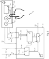
- Fig. 1 illustrates an example embodiment of an apparatus 10 according to the present disclosure.
- the apparatus 10 comprises at least one memory 6, at least one processor, wherein the memory 6 includes computer program code configured to, with the at least one processor, cause the apparatus 10 to perform the following:
- Fig. 2 illustrates an example embodiment of a system 20 according to the present disclosure. Components having identical reference numbers to numbers on Fig. 1 perform the same function.
- the system 20 of Fig. 2 comprises an apparatus 10 according to the present disclosure.
- the system 20 further comprises a light source 2;3 and a sensor 4.
- a light source 2;3 is configured to emit light 40.
- the apparatus 10 is configured to:
- Fig. 3 illustrates an example embodiment of a system 20 according to the present disclosure. Components having identical reference numbers to numbers on Fig. 1 or Fig. 2 perform the same function.
- the system 20 of Fig. 3 comprises an apparatus 10 according to the present disclosure.
- the system 20 further comprises a light source 2;3 and a sensor 4.
- a light source 2;3 is configured to emit light 40.
- the apparatus 10 is configured to:
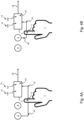
- Figs. 4A and 4B illustrate an example embodiment of the calibration of the apparatus according to the present disclosure.
- Components having identical reference numbers to numbers on Fig. 1 or Fig. 2 or Fig. 3 perform the same function.
- a first light source 2 is provided and emits light 40 at a first wavelength.
- a second light source 3 is provided and emits light 40 at a second wavelength. The second wavelength is preferable different from the first wavelength.
- a sensor 4 is provided. By optical plethysmography, propagated light 41 is collected on the sensor 4 at two points in time 2;3, wherein the propagated light 41 corresponds to the light 40 emitted by the first light source 2 or by the second light source 3 being transmitted or reflected by the investigated volume of an individual when propagating in the investigated volume 11 of the individual 1.
- Fig. 4A for the first wavelength emitted by the light source 2, light intensities 106;107 corresponding to the propagated light 41 collected on the sensor 4 are measured by the sensor 4 at respectively a first point in time 12 and at a second point in time 13.
- Fig. 4B for the second wavelength emitted by the second light source 3, light intensities 108;109 corresponding to the propagated light 41 collected on the sensor 4 are measured by the sensor 4 at respectively a first point in time 12 and at a second point in time 13.
- the sensor 4 determines a first ratio corresponding to a ratio of the first light intensities 106;107 for the first wavelength on Fig. 4A .
- the sensor 4 or, according to an alternative embodiment the apparatus 10 of Fig. 1 or Fig. 2 or Fig. 3 , then determines a second ratio corresponding to a ratio of the second light intensities 108;108 for the second wavelength on Fig. 4B .
- the sensor 4 or, according to an alternative embodiment the apparatus 10 of Fig. 1 or Fig. 2 or Fig. 3 determines a calibration ratio corresponding to the ratio of a function of the first ratio and of a function of the second ratio.
- the determination of the first ratio and/or of the second ratio and/or of the calibration ratio and/or of the predetermined calibration coefficients is performed by any other suitable external device comprising at least one processor and at least one memory including computer program code, the at least one memory and computer program code configured to, with the at least one processor, cause the suitable external device to determine these values.
- Fig. 5 illustrates an example comparison between uncompensated peripheral arterial tone 401 in function of time 60 of an individual, wherein the uncompensated peripheral arterial tone 401 is determined with optical plethysmography without compensation of the influence caused by changes in the hemoglobin composition of the monitored arterial blood volume on the measurement of peripheral arterial tone, and compensated peripheral arterial tone 402 in function of time 60 of the same individual, wherein the compensated peripheral arterial tone 402 is determined by the computer-implemented method according to the present disclosure, or by the apparatus according to the present disclosure, i.e. wherein the compensated peripheral arterial tone 402 is compensated of the influence caused by changes in the hemoglobin composition of the monitored arterial blood volume on the measurement of peripheral arterial tone.
- the peripheral arterial tone estimates are plotted by calculating ln I h I l .
- the peripheral arterial tone estimates are plotted by calculating ln I l I h .
- Fig. 5 also illustrates an example embodiment of the oxygen saturation estimate 104 measured at the same time as the investigated tone 100.
- the level of oxygen saturation estimate 104 is high and the uncompensated peripheral arterial tone 401 and the compensated peripheral arterial tone 402 evolve in a similar manner and overlap as a function of time 60 at a pre-vasoconstriction-event baseline value.
- the value of the oxygen saturation estimate 104 slowly drops, as an indication of an occurrence of an event at the individual under monitoring such as for example a sleep-related event, such as for example sleep apnea.
- a sleep-related event such as for example sleep apnea.
- a drop in peripheral arterial tone 100 is indicative for a vasoconstriction of the arteries and the arterioles in the investigated volume under monitoring.
- This vasoconstriction-event can be related to the occurrence of an event at the individual under monitoring such as for example a sleep-related event, such as for example sleep apnea.
- the drop in the uncompensated peripheral arterial tone 402 between the pre-vasoconstriction-event baseline value and the lowest point of the uncompensated peripheral arterial tone 402 is smaller than the drop in the compensated peripheral arterial tone 401 between the pre-vasoconstriction-event baseline value and the lowest point of the compensated peripheral arterial tone 401.
- a predetermined threshold value 400 for peripheral arterial tone 100 can be used to detect whether an event is occurring at the individual under monitoring such as for example a sleep-related event, such as for example sleep apnea: when the peripheral arterial tone 100 is above this predetermined threshold value 400, no event is detected, but when the peripheral arterial tone 100 is below this predetermined threshold value 400, an event is detected.
- a sleep-related event such as for example sleep apnea
- the drop in the uncompensated peripheral arterial tone 402 between the pre-vasoconstriction-event baseline value and the lowest point of the uncompensated peripheral arterial tone 402 is such that the uncompensated peripheral arterial tone 402 stays above the predetermined threshold value 400, which results in the absence of detection of an event occurring at the individual under monitoring such as for example a sleep-related event, such as for example sleep apnea.
- the drop in the compensated arterial tone 401 between the pre-vasoconstriction-event baseline value and the lowest point of the compensated peripheral arterial tone 401 is such that the compensated peripheral arterial tone 401 drops below the predetermined threshold value 400, which results in the detection of an event occurring at the individual under monitoring such as for example a sleep-related event, such as for example sleep apnea. It can therefore be seen that compensating the influence caused by changes in the hemoglobin composition of the monitored arterial blood volume on the measurement of peripheral arterial tone 100 allows to more accurately and more robustly detect the occurrence of an event occurring at the individual under monitoring such as for example a sleep-related event, such as for example sleep apnea.
- Fig. 6 illustrates an example embodiment of a computer-implemented method for assessing peripheral arterial tone, PAT, of an individual monitored by optical plethysmography, wherein said method comprises the steps of:
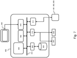
- Fig. 7 shows a suitable computing system 800 enabling to implement embodiments of the system.
- Computing system 800 may in general be formed as a suitable general-purpose computer and comprise a bus 810, a processor 802, a local memory 804, one or more optional input interfaces 814, one or more optional output interfaces 816, a communication interface 812, a storage element interface 806, and one or more storage elements 808.
- Bus 810 may comprise one or more conductors that permit communication among the components of the computing system 800.
- Processor 802 may include any type of conventional processor or microprocessor that interprets and executes programming instructions.
- Local memory 804 may include a random-access memory (RAM) or another type of dynamic storage device that stores information and instructions for execution by processor 802 and/or a read only memory (ROM) or another type of static storage device that stores static information and instructions for use by processor 802.
- Input interface 814 may comprise one or more conventional mechanisms that permit an operator or user to input information to the computing device 800, such as a keyboard 820, a mouse 830, a pen, voice recognition and/or biometric mechanisms, a camera, etc.
- Output interface 816 may comprise one or more conventional mechanisms that output information to the operator or user, such as a display 840, etc.
- Communication interface 812 may comprise any transceiver-like mechanism such as for example one or more Ethernet interfaces that enables computing system 800 to communicate with other devices and/or systems, for example with other computing devices 881, 882, 883.
- the communication interface 812 of computing system 800 may be connected to such another computing system by means of a local area network (LAN) or a wide area network (WAN) such as for example the internet.
- Storage element interface 806 may comprise a storage interface such as for example a Serial Advanced Technology Attachment (SATA) interface or a Small Computer System Interface (SCSI) for connecting bus 810 to one or more storage elements 808, such as one or more local disks, for example SATA disk drives, and control the reading and writing of data to and/or from these storage elements 808.
- SATA Serial Advanced Technology Attachment
- SCSI Small Computer System Interface
- computing system 800 could thus correspond to the apparatus 10 in the embodiment illustrated by Figs. 1 or 2 or 3 .
- circuitry may refer to one or more or all of the following:
- circuitry also covers an implementation of merely a hardware circuit or processor (or multiple processors) or portion of a hardware circuit or processor and its (or their) accompanying software and/or firmware.
- circuitry also covers, for example and if applicable to the particular claim element, a baseband integrated circuit or processor integrated circuit for a mobile device or a similar integrated circuit in a server, a cellular network device, or other computing or network device.
Landscapes
- Health & Medical Sciences (AREA)
- Life Sciences & Earth Sciences (AREA)
- Engineering & Computer Science (AREA)
- Physics & Mathematics (AREA)
- Veterinary Medicine (AREA)
- Animal Behavior & Ethology (AREA)
- Biomedical Technology (AREA)
- Heart & Thoracic Surgery (AREA)
- Medical Informatics (AREA)
- Molecular Biology (AREA)
- Surgery (AREA)
- Pathology (AREA)
- General Health & Medical Sciences (AREA)
- Public Health (AREA)
- Biophysics (AREA)
- Physiology (AREA)
- Neurology (AREA)
- Cardiology (AREA)
- Hematology (AREA)
- Signal Processing (AREA)
- Optics & Photonics (AREA)
- Artificial Intelligence (AREA)
- Computer Vision & Pattern Recognition (AREA)
- Psychiatry (AREA)
- Spectroscopy & Molecular Physics (AREA)
- Neurosurgery (AREA)
- Vascular Medicine (AREA)
- Measurement Of The Respiration, Hearing Ability, Form, And Blood Characteristics Of Living Organisms (AREA)
- Investigating Or Analysing Materials By Optical Means (AREA)
Claims (14)
- Computerimplementiertes Verfahren zur Beurteilung des peripheren arteriellen Tonus, PAT, (100) eines Individuums (1), der durch optische Plethysmographie überwacht wird, wobei das Verfahren umfasst:- Erhalten:- eines optischen Plethysmographiesignals (101), das an einem untersuchten Volumen (11) des Individuums (1) durch einen Sensor einer Anordnung für optische Plethysmographie gemessen wird; und- von Lichtintensitäten (102; 103), die durch optische Plethysmographie zu zwei oder mehr Zeitpunkten (12; 13) entlang des optischen Plethysmographiesignals (101) erfasst werden; und- eine Schätzung der Sauerstoffsättigung (104);- von Kalibrierungsdaten (105);- Bestimmen einer Kompensationsfunktion (14) aus der Schätzung der Sauerstoffsättigung (104) und den Kalibrierungsdaten (105); wobei die Kompensationsfunktion (14) eine Funktion der Schätzung der Sauerstoffsättigung (104) ist;- Bestimmen einer ersten Funktion der Lichtintensitäten (102; 103); und- Bestimmen eines Verhältnisses (15) der ersten Funktion und der Kompensationsfunktion (14), wodurch Veränderungen im arteriellen Blutvolumen (16) in dem untersuchten Volumen (11) zwischen den zwei oder mehr Zeitpunkten (12; 13) evaluiert und dadurch der PAT (100) des Individuums (1) beurteilt werden.
- Verfahren nach Anspruch 1, wobei die Kalibrierungsdaten (105) vorbestimmte Kalibrierungskoeffizienten (25) und/oder vorbestimmte Koeffizienten (26) umfassen; und wobei:- das Bestimmen der Kompensationsfunktion (14) dem Ableiten der Kompensationsfunktion (14) von den vorbestimmten Koeffizienten (26) entspricht; oder- das Bestimmen der Kompensationsfunktion (14) dem Bestimmen der vorbestimmten Kalibrierungskoeffizienten (25) durch Anpassen der Schätzung der Sauerstoffsättigung (104) an ein Kalibrierungsverhältnis entspricht.
- Verfahren nach Anspruch 1 oder 2, wobei mindestens einer (12) der Zeitpunkte (12; 13) der Diastole in einem Herzzyklus des Individuums (1) entspricht und/oder wobei mindestens einer (13) der Zeitpunkte (12; 13) der Systole in einem Herzzyklus des Individuums (1) entspricht.
- Verfahren nach einem der vorhergehenden Ansprüche, wobei das Verfahren ferner die Schritte des Bestimmens einer Evaluierungsfunktion (17) umfasst, die eine Funktion der Lichtintensitäten (102; 103) ist; und wobei das Bestimmen eines Verhältnisses (15) dem Bestimmen eines Verhältnisses der Evaluierungsfunktion (17) und der Kompensationsfunktion (14) entspricht.
- Verfahren nach Anspruch 4, wobei die Evaluierungsfunktion (17) einem Logarithmus einer Funktion der Lichtintensitäten (102; 103) entspricht.
- Verfahren nach Anspruch 4 oder 5, wobei die Evaluierungsfunktion (17) einem Logarithmus eines Verhältnisses der Lichtintensitäten (102, 103) entspricht; und wobei die Evaluierungsfunktion (17) von einem oder mehreren aus Folgendem abhängig ist:- einer Funktion der Schätzung der Sauerstoffsättigung (104);- den Veränderungen im arteriellen Blutvolumen (16) in dem untersuchten Volumen (11).
- Verfahren nach einem der vorhergehenden Ansprüche, wobei das Verfahren ferner die folgenden Schritte umfasst:- Bereitstellen einer ersten Lichtquelle (2), die dazu ausgelegt ist, Licht mit einer ersten Wellenlänge zu emittieren;- Bereitstellen einer zweiten Lichtquelle (3), die dazu ausgelegt ist, Licht mit einer zweiten Wellenlänge zu emittieren;- Bereitstellen eines Sensors (4);- Erfassen, durch optische Plethysmographie und an dem Sensor (4), von sich ausbreitendem Licht, das dem Licht entspricht, das bei der Ausbreitung in dem untersuchten Volumen (11) des Individuums (1) zu den zwei oder mehr Zeitpunkten (12; 13) durchgelassen oder reflektiert wird; und- Bestimmen der Lichtintensitäten (106; 107) des sich ausbreitenden Lichts an dem Sensor (4) zu den zwei oder mehr Zeitpunkten (12; 13) für die erste Wellenlänge;- Bestimmen der Lichtintensitäten (108; 109) des sich ausbreitenden Lichts an dem Sensor (4) zu den zwei oder mehr Zeitpunkten (12; 13) für die zweite Wellenlänge;- Bestimmen eines ersten Verhältnisses entsprechend einem Verhältnis der ersten Lichtintensitäten (106; 107) mit der ersten Wellenlänge;- Bestimmen eines zweiten Verhältnisses entsprechend einem Verhältnis der zweiten Lichtintensitäten (108; 109) mit der zweiten Wellenlänge;- Bestimmen des Kalibrierungsverhältnisses einer Funktion des ersten Verhältnisses und einer Funktion des zweiten Verhältnisses; und- Anpassen der Schätzung der Sauerstoffsättigung (104) an das Kalibrierungsverhältnis und dadurch Bestimmen der vorbestimmten Kalibrierungskoeffizienten (25).
- Verfahren nach Anspruch 7, wobei das Verfahren ferner die folgenden Schritte umfasst:- Erfassen, durch optische Plethysmographie und an dem Sensor (4), von sich ausbreitendem Licht (41), das dem Licht (40) mit der ersten Wellenlänge oder mit der zweiten Wellenlänge entspricht, das bei der Ausbreitung im untersuchten Volumen (11) des Individuums (1) zu den zwei oder mehr Zeitpunkten (12; 13) durchgelassen oder reflektiert wird; und- Bestimmen der Lichtintensitäten (102; 103) des sich ausbreitenden Lichts am Sensor (4) zu den zwei oder mehr Zeitpunkten (12; 13).
- Verfahren nach Anspruch 7 oder 8, wobei das Verfahren ferner den Schritt des Bestimmens der Schätzung der Sauerstoffsättigung (104) in der Umgebung des untersuchten Volumens (11) des Individuums (1) umfasst.
- Verfahren nach einem der vorhergehenden Ansprüche, wobei die Kompensationsfunktion (14) von einer Regression abgeleitet ist, welche die Schätzung der Sauerstoffsättigung (104) auf die vorbestimmten Kalibrierungskoeffizienten (25) abbildet.
- Verfahren nach Anspruch 10, wobei das Verfahren ferner den Schritt des Erzwingens umfasst, dass die Regression eine rationale Abbildung ersten Grades verwendet, wenn die Kompensationsfunktion (14) evaluiert wird.
- Vorrichtung (10), umfassend mindestens einen Prozessor und mindestens einen Speicher (6), der Computerprogrammcode beinhaltet, wobei der mindestens eine Speicher (6) und der Computerprogrammcode dazu ausgelegt sind, mit dem mindestens einen Prozessor die Vorrichtung (10) zu veranlassen,- zu erhalten:- ein optisches Plethysmographiesignal (101), das an einem untersuchten Volumen (11) des Individuums (1) durch einen Sensor einer Anordnung für optische Plethysmographie gemessen wird; und- Lichtintensitäten (102; 103), die durch optische Plethysmographie zu zwei oder mehr Zeitpunkten (12; 13) entlang des optischen Plethysmographiesignals (101) erfasst werden; und- eine Schätzung der Sauerstoffsättigung (104);- Kalibrierungsdaten (105);- eine Kompensationsfunktion (14) aus der Schätzung der Sauerstoffsättigung (104) und den Kalibrierungsdaten (105) zu bestimmen; wobei die Kompensationsfunktion (14) eine Funktion der Schätzung der Sauerstoffsättigung (104) ist;- eine erste Funktion der Lichtintensitäten (102; 103) zu bestimmen; und- ein Verhältnis (15) der ersten Funktion und der Kompensationsfunktion (14) zu bestimmen, wodurch Veränderungen im arteriellen Blutvolumen (16) in dem untersuchten Volumen (11) zwischen den zwei oder mehr Zeitpunkten (12; 13) evaluiert und dadurch der PAT (100) des Individuums (1) beurteilt werden.
- System (20), umfassend eine Vorrichtung nach Anspruch 12, und ferner umfassend:- eine Lichtquelle (2; 3), die dazu ausgelegt ist, Licht zu emittieren, und- einen Sensor (4), der so ausgelegt ist, dass er sich ausbreitendes Licht, das dem Licht entspricht, das bei Ausbreitung in einem distalen Ende eines Fingers des Individuums (1) zu den zwei oder mehr Zeitpunkten (12; 13) durchgelassen oder reflektiert wird, durch optische Plethysmographie erfasst; und ferner so ausgelegt ist, dass er die Lichtintensitäten (102; 103) des sich ausbreitenden Lichts zu den zwei oder mehr Zeitpunkten (12; 13) bestimmt.
- Computerlesbares Speichermedium, umfassend computerausführbare Anweisungen zum Durchführen des Verfahrens der folgenden Schritte, wenn das Programm auf einem Computer ausgeführt wird:- Erhalten:- eines optischen Plethysmographiesignals (101), das an einem untersuchten Volumen (11) eines Individuums (1) gemessen wird; und- von Lichtintensitäten (102; 103), die durch einen Sensor (4) für die optische Plethysmographie zu zwei oder mehr Zeitpunkten (12; 13) entlang des optischen Plethysmographiesignals (101) erfasst werden; und- einer Schätzung der Sauerstoffsättigung (104);- von Kalibrierungsdaten (105);- Bestimmen einer Kompensationsfunktion (14) aus der Schätzung der Sauerstoffsättigung (104) und den Kalibrierungsdaten (105); wobei die Kompensationsfunktion (14) eine Funktion der Schätzung der Sauerstoffsättigung (104) ist;- Bestimmen einer ersten Funktion der Lichtintensitäten (102; 103); und- Bestimmen eines Verhältnisses (15) der ersten Funktion und der Kompensationsfunktion (14), wodurch Veränderungen im arteriellen Blutvolumen (16) in dem untersuchten Volumen (11) zwischen den zwei oder mehr Zeitpunkten (12; 13) evaluiert und dadurch der PAT (100) des Individuums (1) beurteilt werden.
Priority Applications (8)
| Application Number | Priority Date | Filing Date | Title |
|---|---|---|---|
| EP25153804.7A EP4578382A3 (de) | 2020-06-26 | 2020-06-26 | Vorrichtung und verfahren zur kompensation der beurteilung des peripheren arteriellen tonus |
| EP20182503.1A EP3928689B1 (de) | 2020-06-26 | 2020-06-26 | Vorrichtung und verfahren zur kompensation der beurteilung des peripheren arteriellen tonus |
| CN202511682837.3A CN121421527A (zh) | 2020-06-26 | 2021-06-25 | 用于补偿外周动脉张力的评估的装置和方法 |
| AU2021295602A AU2021295602A1 (en) | 2020-06-26 | 2021-06-25 | Apparatus and method for compensating assessment of peripheral arterial tone |
| JP2022580957A JP7811186B2 (ja) | 2020-06-26 | 2021-06-25 | 末梢動脈緊張の評価を補償する装置、方法及びコンピュータ可読記憶媒体 |
| CN202180052414.7A CN115988985B (zh) | 2020-06-26 | 2021-06-25 | 用于补偿外周动脉张力的评估的装置和方法 |
| IL299331A IL299331A (en) | 2020-06-26 | 2021-06-25 | Apparatus and method for compensating assessment of peripheral arterial tone |
| PCT/EP2021/067532 WO2021260190A1 (en) | 2020-06-26 | 2021-06-25 | Apparatus and method for compensating assessment of peripheral arterial tone |
Applications Claiming Priority (1)
| Application Number | Priority Date | Filing Date | Title |
|---|---|---|---|
| EP20182503.1A EP3928689B1 (de) | 2020-06-26 | 2020-06-26 | Vorrichtung und verfahren zur kompensation der beurteilung des peripheren arteriellen tonus |
Related Child Applications (1)
| Application Number | Title | Priority Date | Filing Date |
|---|---|---|---|
| EP25153804.7A Division EP4578382A3 (de) | 2020-06-26 | 2020-06-26 | Vorrichtung und verfahren zur kompensation der beurteilung des peripheren arteriellen tonus |
Publications (2)
| Publication Number | Publication Date |
|---|---|
| EP3928689A1 EP3928689A1 (de) | 2021-12-29 |
| EP3928689B1 true EP3928689B1 (de) | 2025-01-29 |
Family
ID=71170436
Family Applications (2)
| Application Number | Title | Priority Date | Filing Date |
|---|---|---|---|
| EP25153804.7A Pending EP4578382A3 (de) | 2020-06-26 | 2020-06-26 | Vorrichtung und verfahren zur kompensation der beurteilung des peripheren arteriellen tonus |
| EP20182503.1A Active EP3928689B1 (de) | 2020-06-26 | 2020-06-26 | Vorrichtung und verfahren zur kompensation der beurteilung des peripheren arteriellen tonus |
Family Applications Before (1)
| Application Number | Title | Priority Date | Filing Date |
|---|---|---|---|
| EP25153804.7A Pending EP4578382A3 (de) | 2020-06-26 | 2020-06-26 | Vorrichtung und verfahren zur kompensation der beurteilung des peripheren arteriellen tonus |
Country Status (6)
| Country | Link |
|---|---|
| EP (2) | EP4578382A3 (de) |
| JP (1) | JP7811186B2 (de) |
| CN (2) | CN121421527A (de) |
| AU (1) | AU2021295602A1 (de) |
| IL (1) | IL299331A (de) |
| WO (1) | WO2021260190A1 (de) |
Families Citing this family (10)
| Publication number | Priority date | Publication date | Assignee | Title |
|---|---|---|---|---|
| EP4284242A1 (de) | 2021-01-29 | 2023-12-06 | ResMed Sensor Technologies Limited | Systeme und verfahren zur schätzung eines subjektiven komfortniveaus |
| WO2022208368A1 (en) | 2021-03-31 | 2022-10-06 | Resmed Sensor Technologies Limited | Systems and methods for managing blood pressure conditions of a user of a respiratory therapy system |
| EP4322839A1 (de) | 2021-04-16 | 2024-02-21 | ResMed Sensor Technologies Limited | Systeme und verfahren zur charakterisierung einer benutzerschnittstelle oder einer entlüftung mithilfe akustischer daten im zusammenhang mit der entlüftung |
| EP4396833A1 (de) | 2021-08-30 | 2024-07-10 | Resmed Digital Health Inc. | Intelligente atemtraining |
| CN120379593A (zh) | 2022-12-20 | 2025-07-25 | 瑞思迈数字健康公司 | 诊断头带 |
| US12579242B2 (en) | 2022-12-21 | 2026-03-17 | Resmed Digital Health Inc. | Systems and methods for digit-based diagnostic chain of custody management |
| CN120641990A (zh) | 2023-01-18 | 2025-09-12 | 瑞思迈传感器技术有限公司 | 用于使用流量发生器数据来表征用户接口的系统和方法 |
| CN121844363A (zh) | 2023-07-19 | 2026-04-10 | 瑞思迈数字医疗公司 | 使用机器学习和所捕获的图像数据对用户接口进行分类 |
| CN121844390A (zh) | 2023-07-19 | 2026-04-10 | 瑞思迈数字医疗公司 | 使用呼吸治疗数据检测用户接口变更 |
| US12605077B2 (en) | 2023-08-25 | 2026-04-21 | Pranaq Pte. Ltd. | Photoplethysmogram system and method |
Family Cites Families (15)
| Publication number | Priority date | Publication date | Assignee | Title |
|---|---|---|---|---|
| IL120881A (en) | 1996-07-30 | 2002-09-12 | It M R Medic L Cm 1997 Ltd | Method and device for continuous and non-invasive monitoring of peripheral arterial tone |
| JP4040913B2 (ja) * | 2002-06-07 | 2008-01-30 | 株式会社パルメディカル | 非観血動静脈酸素飽和度測定装置 |
| CA2492027C (en) | 2002-07-15 | 2012-10-02 | Itamar Medical Ltd. | Body surface probe, apparatus and method for non-invasively detecting medical conditions |
| AU2004203059A1 (en) * | 2004-06-08 | 2005-12-22 | The Government Of The United States Of America As Represented By The Secretary Of The Department Of Health And Human Services, Centers For Disease Control And Prevention | Apparatus and method for assessing peripheral circulation to evaluate a physiological condition |
| US7512431B2 (en) * | 2005-09-13 | 2009-03-31 | Medtronic, Inc. | Normalization method for a chronically implanted optical sensor |
| US8660799B2 (en) * | 2008-06-30 | 2014-02-25 | Nellcor Puritan Bennett Ireland | Processing and detecting baseline changes in signals |
| US10405762B2 (en) * | 2009-04-22 | 2019-09-10 | Vital Metrix, Inc. | System and method for noninvasively measuring ventricular stroke volume and cardiac output |
| US20110082355A1 (en) * | 2009-07-30 | 2011-04-07 | Oxitone Medical Ltd. | Photoplethysmography device and method |
| US20130310669A1 (en) * | 2012-05-20 | 2013-11-21 | Jerusalem College Of Technology | Pulmonary pulse oximetry method for the measurement of oxygen saturation in the mixed venous blood |
| US20150018654A1 (en) * | 2013-07-09 | 2015-01-15 | Xerox Corporation | Method and apparatus for monitoring a subject for fractional blood oxygen saturation |
| WO2017005016A1 (en) * | 2015-07-03 | 2017-01-12 | Vita-Course Technologies Co., Ltd | System and method for physiological parameter monitoring |
| WO2019026062A1 (en) * | 2017-07-31 | 2019-02-07 | Jerusalem College Of Technology | METHOD FOR MEASURING OXYGEN SATURATION OF ARTERIAL BLOOD AND APPARATUS THEREFOR |
| JP7285843B2 (ja) * | 2017-12-22 | 2023-06-02 | アシスタンス パブリック-ホピトー デ パリ | 平均動脈圧を測定するためのシステム |
| EP4360546B1 (de) * | 2018-07-11 | 2025-02-19 | Ectosense NV | System zur schlafdiagnose |
| CN109924960A (zh) * | 2019-01-31 | 2019-06-25 | 深圳市爱都科技有限公司 | 一种血氧饱和度、心率值和压力等级的计算方法和穿戴设备 |
-
2020
- 2020-06-26 EP EP25153804.7A patent/EP4578382A3/de active Pending
- 2020-06-26 EP EP20182503.1A patent/EP3928689B1/de active Active
-
2021
- 2021-06-25 IL IL299331A patent/IL299331A/en unknown
- 2021-06-25 WO PCT/EP2021/067532 patent/WO2021260190A1/en not_active Ceased
- 2021-06-25 CN CN202511682837.3A patent/CN121421527A/zh active Pending
- 2021-06-25 AU AU2021295602A patent/AU2021295602A1/en active Pending
- 2021-06-25 CN CN202180052414.7A patent/CN115988985B/zh active Active
- 2021-06-25 JP JP2022580957A patent/JP7811186B2/ja active Active
Also Published As
| Publication number | Publication date |
|---|---|
| JP2023532319A (ja) | 2023-07-27 |
| IL299331A (en) | 2023-02-01 |
| CN121421527A (zh) | 2026-01-30 |
| WO2021260190A1 (en) | 2021-12-30 |
| JP7811186B2 (ja) | 2026-02-04 |
| EP4578382A3 (de) | 2025-10-08 |
| EP3928689A1 (de) | 2021-12-29 |
| CN115988985B (zh) | 2025-12-05 |
| EP4578382A2 (de) | 2025-07-02 |
| AU2021295602A1 (en) | 2023-02-02 |
| CN115988985A (zh) | 2023-04-18 |
Similar Documents
| Publication | Publication Date | Title |
|---|---|---|
| EP3928689B1 (de) | Vorrichtung und verfahren zur kompensation der beurteilung des peripheren arteriellen tonus | |
| EP3928690B1 (de) | Verfahren und vorrichtung zur beurteilung des peripheren arteriellen tonus | |
| US8221326B2 (en) | Detection of oximetry sensor sites based on waveform characteristics | |
| JP5748160B2 (ja) | 携帯診断装置 | |
| US9402573B2 (en) | System and method for detecting fluid responsiveness of a patient | |
| JP5096310B2 (ja) | 身体の部位における血液の灌流を決定するための方法及び装置 | |
| JP2011521702A (ja) | Co2評価のための方法および装置 | |
| US12262991B2 (en) | System and method for non-invasive monitoring of hematocrit concentration | |
| EP4208085B1 (de) | Verfahren und vorrichtung zur erkennung von schlafstörungsereignissen aus einem signal, das einen peripheren arteriellen ton einer person anzeigt | |
| EP2895057A1 (de) | Systeme und verfahren zur bestimmung eines flüssigkeitsreaktionsverhaltens | |
| Campbell | Development of non-invasive, optical methods for central cardiovascular and blood chemistry monitoring | |
| CN118662119A (zh) | 光学呼吸速率预测系统 |
Legal Events
| Date | Code | Title | Description |
|---|---|---|---|
| PUAI | Public reference made under article 153(3) epc to a published international application that has entered the european phase |
Free format text: ORIGINAL CODE: 0009012 |
|
| STAA | Information on the status of an ep patent application or granted ep patent |
Free format text: STATUS: THE APPLICATION HAS BEEN PUBLISHED |
|
| AK | Designated contracting states |
Kind code of ref document: A1 Designated state(s): AL AT BE BG CH CY CZ DE DK EE ES FI FR GB GR HR HU IE IS IT LI LT LU LV MC MK MT NL NO PL PT RO RS SE SI SK SM TR |
|
| B565 | Issuance of search results under rule 164(2) epc |
Effective date: 20201127 |
|
| STAA | Information on the status of an ep patent application or granted ep patent |
Free format text: STATUS: REQUEST FOR EXAMINATION WAS MADE |
|
| 17P | Request for examination filed |
Effective date: 20220628 |
|
| RBV | Designated contracting states (corrected) |
Designated state(s): AL AT BE BG CH CY CZ DE DK EE ES FI FR GB GR HR HU IE IS IT LI LT LU LV MC MK MT NL NO PL PT RO RS SE SI SK SM TR |
|
| GRAP | Despatch of communication of intention to grant a patent |
Free format text: ORIGINAL CODE: EPIDOSNIGR1 |
|
| STAA | Information on the status of an ep patent application or granted ep patent |
Free format text: STATUS: GRANT OF PATENT IS INTENDED |
|
| INTG | Intention to grant announced |
Effective date: 20240827 |
|
| GRAS | Grant fee paid |
Free format text: ORIGINAL CODE: EPIDOSNIGR3 |
|
| GRAA | (expected) grant |
Free format text: ORIGINAL CODE: 0009210 |
|
| STAA | Information on the status of an ep patent application or granted ep patent |
Free format text: STATUS: THE PATENT HAS BEEN GRANTED |
|
| RIN1 | Information on inventor provided before grant (corrected) |
Inventor name: VAN PEE, BART Inventor name: VITS, STEVEN Inventor name: MASSIE, FREDERIK Inventor name: GEEURICKX, WOUT |
|
| AK | Designated contracting states |
Kind code of ref document: B1 Designated state(s): AL AT BE BG CH CY CZ DE DK EE ES FI FR GB GR HR HU IE IS IT LI LT LU LV MC MK MT NL NO PL PT RO RS SE SI SK SM TR |
|
| REG | Reference to a national code |
Ref country code: GB Ref legal event code: FG4D |
|
| REG | Reference to a national code |
Ref country code: CH Ref legal event code: EP |
|
| REG | Reference to a national code |
Ref country code: DE Ref legal event code: R096 Ref document number: 602020045421 Country of ref document: DE |
|
| REG | Reference to a national code |
Ref country code: IE Ref legal event code: FG4D |
|
| REG | Reference to a national code |
Ref country code: NL Ref legal event code: MP Effective date: 20250129 |
|
| PG25 | Lapsed in a contracting state [announced via postgrant information from national office to epo] |
Ref country code: NL Free format text: LAPSE BECAUSE OF FAILURE TO SUBMIT A TRANSLATION OF THE DESCRIPTION OR TO PAY THE FEE WITHIN THE PRESCRIBED TIME-LIMIT Effective date: 20250129 |
|
| PG25 | Lapsed in a contracting state [announced via postgrant information from national office to epo] |
Ref country code: RS Free format text: LAPSE BECAUSE OF FAILURE TO SUBMIT A TRANSLATION OF THE DESCRIPTION OR TO PAY THE FEE WITHIN THE PRESCRIBED TIME-LIMIT Effective date: 20250429 |
|
| PG25 | Lapsed in a contracting state [announced via postgrant information from national office to epo] |
Ref country code: FI Free format text: LAPSE BECAUSE OF FAILURE TO SUBMIT A TRANSLATION OF THE DESCRIPTION OR TO PAY THE FEE WITHIN THE PRESCRIBED TIME-LIMIT Effective date: 20250129 |
|
| PG25 | Lapsed in a contracting state [announced via postgrant information from national office to epo] |
Ref country code: PL Free format text: LAPSE BECAUSE OF FAILURE TO SUBMIT A TRANSLATION OF THE DESCRIPTION OR TO PAY THE FEE WITHIN THE PRESCRIBED TIME-LIMIT Effective date: 20250129 |
|
| PGFP | Annual fee paid to national office [announced via postgrant information from national office to epo] |
Ref country code: DE Payment date: 20250620 Year of fee payment: 6 |
|
| PG25 | Lapsed in a contracting state [announced via postgrant information from national office to epo] |
Ref country code: ES Free format text: LAPSE BECAUSE OF FAILURE TO SUBMIT A TRANSLATION OF THE DESCRIPTION OR TO PAY THE FEE WITHIN THE PRESCRIBED TIME-LIMIT Effective date: 20250129 |
|
| PGFP | Annual fee paid to national office [announced via postgrant information from national office to epo] |
Ref country code: GB Payment date: 20250619 Year of fee payment: 6 |
|
| REG | Reference to a national code |
Ref country code: LT Ref legal event code: MG9D |
|
| PG25 | Lapsed in a contracting state [announced via postgrant information from national office to epo] |
Ref country code: NO Free format text: LAPSE BECAUSE OF FAILURE TO SUBMIT A TRANSLATION OF THE DESCRIPTION OR TO PAY THE FEE WITHIN THE PRESCRIBED TIME-LIMIT Effective date: 20250429 Ref country code: IS Free format text: LAPSE BECAUSE OF FAILURE TO SUBMIT A TRANSLATION OF THE DESCRIPTION OR TO PAY THE FEE WITHIN THE PRESCRIBED TIME-LIMIT Effective date: 20250529 |
|
| REG | Reference to a national code |
Ref country code: AT Ref legal event code: MK05 Ref document number: 1762619 Country of ref document: AT Kind code of ref document: T Effective date: 20250129 |
|
| PG25 | Lapsed in a contracting state [announced via postgrant information from national office to epo] |
Ref country code: HR Free format text: LAPSE BECAUSE OF FAILURE TO SUBMIT A TRANSLATION OF THE DESCRIPTION OR TO PAY THE FEE WITHIN THE PRESCRIBED TIME-LIMIT Effective date: 20250129 |
|
| PG25 | Lapsed in a contracting state [announced via postgrant information from national office to epo] |
Ref country code: LV Free format text: LAPSE BECAUSE OF FAILURE TO SUBMIT A TRANSLATION OF THE DESCRIPTION OR TO PAY THE FEE WITHIN THE PRESCRIBED TIME-LIMIT Effective date: 20250129 Ref country code: PT Free format text: LAPSE BECAUSE OF FAILURE TO SUBMIT A TRANSLATION OF THE DESCRIPTION OR TO PAY THE FEE WITHIN THE PRESCRIBED TIME-LIMIT Effective date: 20250529 |
|
| PGFP | Annual fee paid to national office [announced via postgrant information from national office to epo] |
Ref country code: FR Payment date: 20250619 Year of fee payment: 6 |
|
| PG25 | Lapsed in a contracting state [announced via postgrant information from national office to epo] |
Ref country code: GR Free format text: LAPSE BECAUSE OF FAILURE TO SUBMIT A TRANSLATION OF THE DESCRIPTION OR TO PAY THE FEE WITHIN THE PRESCRIBED TIME-LIMIT Effective date: 20250430 Ref country code: BG Free format text: LAPSE BECAUSE OF FAILURE TO SUBMIT A TRANSLATION OF THE DESCRIPTION OR TO PAY THE FEE WITHIN THE PRESCRIBED TIME-LIMIT Effective date: 20250129 |
|
| PG25 | Lapsed in a contracting state [announced via postgrant information from national office to epo] |
Ref country code: AT Free format text: LAPSE BECAUSE OF FAILURE TO SUBMIT A TRANSLATION OF THE DESCRIPTION OR TO PAY THE FEE WITHIN THE PRESCRIBED TIME-LIMIT Effective date: 20250129 |
|
| PG25 | Lapsed in a contracting state [announced via postgrant information from national office to epo] |
Ref country code: SE Free format text: LAPSE BECAUSE OF FAILURE TO SUBMIT A TRANSLATION OF THE DESCRIPTION OR TO PAY THE FEE WITHIN THE PRESCRIBED TIME-LIMIT Effective date: 20250129 |
|
| PG25 | Lapsed in a contracting state [announced via postgrant information from national office to epo] |
Ref country code: SM Free format text: LAPSE BECAUSE OF FAILURE TO SUBMIT A TRANSLATION OF THE DESCRIPTION OR TO PAY THE FEE WITHIN THE PRESCRIBED TIME-LIMIT Effective date: 20250129 |
|
| PG25 | Lapsed in a contracting state [announced via postgrant information from national office to epo] |
Ref country code: DK Free format text: LAPSE BECAUSE OF FAILURE TO SUBMIT A TRANSLATION OF THE DESCRIPTION OR TO PAY THE FEE WITHIN THE PRESCRIBED TIME-LIMIT Effective date: 20250129 |
|
| PG25 | Lapsed in a contracting state [announced via postgrant information from national office to epo] |
Ref country code: IT Free format text: LAPSE BECAUSE OF FAILURE TO SUBMIT A TRANSLATION OF THE DESCRIPTION OR TO PAY THE FEE WITHIN THE PRESCRIBED TIME-LIMIT Effective date: 20250129 |
|
| PG25 | Lapsed in a contracting state [announced via postgrant information from national office to epo] |
Ref country code: CZ Free format text: LAPSE BECAUSE OF FAILURE TO SUBMIT A TRANSLATION OF THE DESCRIPTION OR TO PAY THE FEE WITHIN THE PRESCRIBED TIME-LIMIT Effective date: 20250129 Ref country code: EE Free format text: LAPSE BECAUSE OF FAILURE TO SUBMIT A TRANSLATION OF THE DESCRIPTION OR TO PAY THE FEE WITHIN THE PRESCRIBED TIME-LIMIT Effective date: 20250129 |
|
| PG25 | Lapsed in a contracting state [announced via postgrant information from national office to epo] |
Ref country code: RO Free format text: LAPSE BECAUSE OF FAILURE TO SUBMIT A TRANSLATION OF THE DESCRIPTION OR TO PAY THE FEE WITHIN THE PRESCRIBED TIME-LIMIT Effective date: 20250129 |
|
| PG25 | Lapsed in a contracting state [announced via postgrant information from national office to epo] |
Ref country code: SK Free format text: LAPSE BECAUSE OF FAILURE TO SUBMIT A TRANSLATION OF THE DESCRIPTION OR TO PAY THE FEE WITHIN THE PRESCRIBED TIME-LIMIT Effective date: 20250129 |
|
| REG | Reference to a national code |
Ref country code: DE Ref legal event code: R097 Ref document number: 602020045421 Country of ref document: DE |
|
| PLBE | No opposition filed within time limit |
Free format text: ORIGINAL CODE: 0009261 |
|
| STAA | Information on the status of an ep patent application or granted ep patent |
Free format text: STATUS: NO OPPOSITION FILED WITHIN TIME LIMIT |
|
| REG | Reference to a national code |
Ref country code: CH Ref legal event code: L10 Free format text: ST27 STATUS EVENT CODE: U-0-0-L10-L00 (AS PROVIDED BY THE NATIONAL OFFICE) Effective date: 20251210 |
|
| 26N | No opposition filed |
Effective date: 20251030 |
|
| REG | Reference to a national code |
Ref country code: CH Ref legal event code: H13 Free format text: ST27 STATUS EVENT CODE: U-0-0-H10-H13 (AS PROVIDED BY THE NATIONAL OFFICE) Effective date: 20260127 |
|
| PG25 | Lapsed in a contracting state [announced via postgrant information from national office to epo] |
Ref country code: MC Free format text: LAPSE BECAUSE OF FAILURE TO SUBMIT A TRANSLATION OF THE DESCRIPTION OR TO PAY THE FEE WITHIN THE PRESCRIBED TIME-LIMIT Effective date: 20250129 |
|
| PG25 | Lapsed in a contracting state [announced via postgrant information from national office to epo] |
Ref country code: LU Free format text: LAPSE BECAUSE OF NON-PAYMENT OF DUE FEES Effective date: 20250626 |
|
| REG | Reference to a national code |
Ref country code: BE Ref legal event code: MM Effective date: 20250630 |
|
| PG25 | Lapsed in a contracting state [announced via postgrant information from national office to epo] |
Ref country code: IE Free format text: LAPSE BECAUSE OF NON-PAYMENT OF DUE FEES Effective date: 20250626 |
|
| PG25 | Lapsed in a contracting state [announced via postgrant information from national office to epo] |
Ref country code: BE Free format text: LAPSE BECAUSE OF NON-PAYMENT OF DUE FEES Effective date: 20250630 |