EP4497374A1 - Verfahren zur erzeugung von dreidimensionalen abtastdaten für gesicht - Google Patents
Verfahren zur erzeugung von dreidimensionalen abtastdaten für gesicht Download PDFInfo
- Publication number
- EP4497374A1 EP4497374A1 EP23775251.4A EP23775251A EP4497374A1 EP 4497374 A1 EP4497374 A1 EP 4497374A1 EP 23775251 A EP23775251 A EP 23775251A EP 4497374 A1 EP4497374 A1 EP 4497374A1
- Authority
- EP
- European Patent Office
- Prior art keywords
- data
- face
- tooth
- tooth area
- area
- Prior art date
- Legal status (The legal status is an assumption and is not a legal conclusion. Google has not performed a legal analysis and makes no representation as to the accuracy of the status listed.)
- Pending
Links
Images


Classifications
-
- A—HUMAN NECESSITIES
- A61—MEDICAL OR VETERINARY SCIENCE; HYGIENE
- A61B—DIAGNOSIS; SURGERY; IDENTIFICATION
- A61B5/00—Measuring for diagnostic purposes; Identification of persons
- A61B5/0059—Measuring for diagnostic purposes; Identification of persons using light, e.g. diagnosis by transillumination, diascopy, fluorescence
- A61B5/0062—Arrangements for scanning
- A61B5/0064—Body surface scanning
-
- A—HUMAN NECESSITIES
- A61—MEDICAL OR VETERINARY SCIENCE; HYGIENE
- A61B—DIAGNOSIS; SURGERY; IDENTIFICATION
- A61B5/00—Measuring for diagnostic purposes; Identification of persons
-
- A—HUMAN NECESSITIES
- A61—MEDICAL OR VETERINARY SCIENCE; HYGIENE
- A61B—DIAGNOSIS; SURGERY; IDENTIFICATION
- A61B5/00—Measuring for diagnostic purposes; Identification of persons
- A61B5/0059—Measuring for diagnostic purposes; Identification of persons using light, e.g. diagnosis by transillumination, diascopy, fluorescence
- A61B5/0082—Measuring for diagnostic purposes; Identification of persons using light, e.g. diagnosis by transillumination, diascopy, fluorescence adapted for particular medical purposes
- A61B5/0088—Measuring for diagnostic purposes; Identification of persons using light, e.g. diagnosis by transillumination, diascopy, fluorescence adapted for particular medical purposes for oral or dental tissue
-
- A—HUMAN NECESSITIES
- A61—MEDICAL OR VETERINARY SCIENCE; HYGIENE
- A61C—DENTISTRY; APPARATUS OR METHODS FOR ORAL OR DENTAL HYGIENE
- A61C19/00—Dental auxiliary appliances
- A61C19/04—Measuring instruments specially adapted for dentistry
-
- A—HUMAN NECESSITIES
- A61—MEDICAL OR VETERINARY SCIENCE; HYGIENE
- A61C—DENTISTRY; APPARATUS OR METHODS FOR ORAL OR DENTAL HYGIENE
- A61C9/00—Impression cups, i.e. impression trays; Impression methods
-
- A—HUMAN NECESSITIES
- A61—MEDICAL OR VETERINARY SCIENCE; HYGIENE
- A61C—DENTISTRY; APPARATUS OR METHODS FOR ORAL OR DENTAL HYGIENE
- A61C9/00—Impression cups, i.e. impression trays; Impression methods
- A61C9/004—Means or methods for taking digitized impressions
- A61C9/0046—Data acquisition means or methods
- A61C9/0053—Optical means or methods, e.g. scanning the teeth by a laser or light beam
-
- G—PHYSICS
- G06—COMPUTING OR CALCULATING; COUNTING
- G06T—IMAGE DATA PROCESSING OR GENERATION, IN GENERAL
- G06T17/00—Three-dimensional [3D] modelling for computer graphics
- G06T17/20—Finite element generation, e.g. wire-frame surface description, tesselation
-
- G—PHYSICS
- G06—COMPUTING OR CALCULATING; COUNTING
- G06T—IMAGE DATA PROCESSING OR GENERATION, IN GENERAL
- G06T19/00—Manipulating three-dimensional [3D] models or images for computer graphics
- G06T19/20—Editing of three-dimensional [3D] images, e.g. changing shapes or colours, aligning objects or positioning parts
-
- G—PHYSICS
- G06—COMPUTING OR CALCULATING; COUNTING
- G06T—IMAGE DATA PROCESSING OR GENERATION, IN GENERAL
- G06T7/00—Image analysis
- G06T7/30—Determination of transform parameters for the alignment of images, i.e. image registration
-
- G—PHYSICS
- G06—COMPUTING OR CALCULATING; COUNTING
- G06T—IMAGE DATA PROCESSING OR GENERATION, IN GENERAL
- G06T7/00—Image analysis
- G06T7/50—Depth or shape recovery
- G06T7/55—Depth or shape recovery from multiple images
-
- G—PHYSICS
- G06—COMPUTING OR CALCULATING; COUNTING
- G06V—IMAGE OR VIDEO RECOGNITION OR UNDERSTANDING
- G06V10/00—Arrangements for image or video recognition or understanding
- G06V10/40—Extraction of image or video features
- G06V10/46—Descriptors for shape, contour or point-related descriptors, e.g. scale invariant feature transform [SIFT] or bags of words [BoW]; Salient regional features
-
- G—PHYSICS
- G06—COMPUTING OR CALCULATING; COUNTING
- G06V—IMAGE OR VIDEO RECOGNITION OR UNDERSTANDING
- G06V40/00—Recognition of biometric, human-related or animal-related patterns in image or video data
- G06V40/10—Human or animal bodies, e.g. vehicle occupants or pedestrians; Body parts, e.g. hands
- G06V40/16—Human faces, e.g. facial parts, sketches or expressions
- G06V40/168—Feature extraction; Face representation
- G06V40/171—Local features and components; Facial parts ; Occluding parts, e.g. glasses; Geometrical relationships
-
- G—PHYSICS
- G06—COMPUTING OR CALCULATING; COUNTING
- G06T—IMAGE DATA PROCESSING OR GENERATION, IN GENERAL
- G06T2207/00—Indexing scheme for image analysis or image enhancement
- G06T2207/30—Subject of image; Context of image processing
- G06T2207/30004—Biomedical image processing
- G06T2207/30036—Dental; Teeth
-
- G—PHYSICS
- G06—COMPUTING OR CALCULATING; COUNTING
- G06T—IMAGE DATA PROCESSING OR GENERATION, IN GENERAL
- G06T2207/00—Indexing scheme for image analysis or image enhancement
- G06T2207/30—Subject of image; Context of image processing
- G06T2207/30196—Human being; Person
- G06T2207/30201—Face
-
- G—PHYSICS
- G06—COMPUTING OR CALCULATING; COUNTING
- G06T—IMAGE DATA PROCESSING OR GENERATION, IN GENERAL
- G06T2219/00—Indexing scheme for manipulating 3D models or images for computer graphics
- G06T2219/20—Indexing scheme for editing of 3D models
- G06T2219/2004—Aligning objects, relative positioning of parts
Definitions
- the present disclosure generally relates to a technology for generating three-dimensional (3D) face scan data of a person.
- the present disclosure relates to a technology for generating 3D face scan data, which photographs respective face and tooth areas with a structured light pattern projected thereon to individually acquire 3D data for the face and tooth areas, extract a tooth zone contour using face landmarks, and align and insert tooth zone contour 3D data, which is obtained by separating the extracted tooth zone contour from the tooth area 3D data, into the face 3D data.
- 3D face scan data For cosmetic dental treatment, obtaining 3D face scan data by scanning a face of a patient in three dimensions is necessary. For example, for orthodontic treatment planning, 3D face scan data of the face of the patient is crucial for visualizing the resulting facial appearance throughout the orthodontic treatment process.
- the present disclosure provides a technology for generating 3D face scan data of a person.
- the present disclosure provides a technology for generating 3D face scan data, which photographs respective face and tooth areas with a structured light pattern projected thereon to individually acquire 3D data for the face and tooth areas, extract a tooth zone contour using face landmarks, and align and insert tooth zone contour 3D data, which is obtained by separating the extracted tooth zone contour from the tooth area 3D data, into the face 3D data.
- a method for generating 3D face scan data may include acquiring a face image obtained by photographing a face of a patient, generating face 3D data from the face image acquiring a tooth area image obtained by photographing a tooth area of the patient, generating tooth area 3D data from the tooth area image, extracting a tooth zone contour for the face image or the face 3D data, separating and acquiring tooth zone contour 3D data corresponding to the tooth zone contour from the tooth area 3D data, and aligning and inserting the tooth zone contour 3D data into the face 3D data.
- the acquiring the tooth area image obtained by photographing the tooth area of the patient may include acquiring a plurality of partial tooth area images obtained by photographing the tooth area of the patient from a plurality of angles, and the generating tooth area 3D data from the tooth area image may include generating a plurality of partial tooth area 3D data from the plurality of partial tooth area images, and generating tooth area 3D data for the whole tooth area by aligning the plurality of partial tooth area 3D data.
- the aligning and inserting the tooth zone contour 3D data into the face 3D data may include removing data of the tooth zone contour portion from the face 3D data, and aligning and inserting the tooth zone contour 3D data into the tooth zone contour portion of the face 3D data.
- the method may further include generating auxiliary area 3D data for a preset area of a face and aligning and inserting the auxiliary area 3D data into the face 3D data.
- the face image, the face 3D data, the tooth area image, and the tooth area 3D data may be acquired while a structured light pattern is projected onto the face or the tooth area of the patient.
- the extracting the tooth zone contour for the face image or the face 3D data may include recognizing face landmarks from the face image or the face 3D data, and extracting a tooth zone contour for the face image or the face 3D data on the basis of the face landmarks.
- a computer program is provided, which may be stored in a non-transitory computer recordable storage medium to execute the method for generating 3D face scan data described above on a computer.
- 3D face scan data including high-resolution dental data can be generated through a relatively low-cost device, which has the advantage of effectively assisting in several fields (e.g., cosmetic dental treatment).
- FIG. 1 illustrates a 3D face scan system
- the 3D face scan system includes a structured light projector 101, a face imaging camera 102, tooth imaging cameras 103 and 104, and a data processing unit 200. These components may be implemented as separate devices, or some or all of the components may be integrated into a single unit.
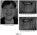
- the structured light projector 101 is a device for generating a structured light pattern and projecting the structured light pattern onto a face A of a patient, as illustrated in FIG. 3 . Meanwhile, other methods such as using a stereo camera instead of structured light, or using time of flight, etc. may be employed.
- the face imaging camera 102 is a device for photographing the face A of the patient to acquire a face image such as that illustrated in FIG. 3A .
- Photographing the face A of the patient may be performed while the structured light pattern is projected onto the face A of the patient.
- photographing the face A of the patient may be performed from at least one of the positions on the front, right and left sides of, and below and above the face of the patient, but is not limited thereto.
- the tooth imaging cameras 103 and 104 are devices for photographing the tooth area of the face A of the patient to acquire tooth area images such as those illustrated in FIGS. 3A, 3B, and 3C . At this time, the tooth area of the face of the patient may be photographed while the structured light pattern is projected onto the face A of the patient.
- the face imaging camera 102 for photographing a face and the tooth imaging cameras 103 and 104 for exclusively photographing tooth area may be configured as separate devices. Only one of the tooth imaging cameras 103 and 104 may be provided, although it is preferable that a plurality of dental cameras are provided to photograph the patient's tooth area from multiple angles and acquire a plurality of tooth area images.
- FIG. 1 illustrates that two tooth imaging cameras 103 and 104 are provided. In this case, some areas may be visible from all of the plurality of cameras 103 and 104, and other areas may be visible only from part of the cameras 103 or 104, in which case the accuracy of the 3D data may be increased by image registration and synthesis.
- the data processing unit 200 is a device that receives the face image acquired by the face imaging camera 102 and the tooth area image acquired by the tooth imaging cameras 103 and 104, and generates 3D face scan data through a data processing process according to FIG. 2 or FIG. 10 .
- FIG. 2 is a flowing chart illustrating a method for generating 3D face scan data.
- Operations S110 and S120 The face image such as that illustrated in FIG. 3A is acquired by photographing, through the face imaging camera 102, the face A of the patient while a preset structured light pattern is projected onto the face A of the patient by the structured light projector 101.
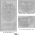
- the face 3D data such as that illustrated in FIG. 4A is generated from the face image with the structured light pattern formed thereon. It is possible to generate the 3D data, because the 3D shape of the face can be identified with the structured light pattern. Since the technology for acquiring 3D data from an image of an object with a structured light pattern projected thereon is already known, detailed description thereof will be omitted.
- tooth area images such as those illustrated in FIGS. 3A, 3B, and 3C are acquired by photographing, through the tooth imaging cameras 103 and 104, the tooth area of a person while the preset structured light pattern is projected onto the face A of the same person by the structured light projector 101.
- the structured light pattern for the face image and the structured light pattern for the tooth area image may be the same or may be different.
- Tooth area 3D data such as those illustrated in FIGS. 4A, 4B, and 4C is generated from the tooth area images with the structured light pattern formed thereon. Comparison between the tooth area images illustrates that the tooth area images of FIGS. 3B and 3C have much higher resolution than the tooth area image included in the face image of FIG. 3A . A natural consequence is that the tooth area 3D data of FIGS. 4B and 4C are much more detailed than the tooth zoon 3D data included in the face 3D data of FIG. 4A .
- a plurality of tooth imaging cameras 103 and 104 are provided so as to acquire a plurality of partial tooth area images by photographing the tooth area from a plurality of angles in S130.
- the operation at S140 may include generating a plurality of partial tooth area 3D data such as those illustrated in FIGS. 4B and 4C from the plurality of partial tooth area images with the structured light pattern formed thereon, such as those illustrated in FIGS. 3B and 3C , and generating the tooth area 3D data for the whole tooth area of the patient by aligning the plurality of partial tooth area 3D data.
- FIG. 5 illustrates the concept of aligning face and tooth area 3D data.
- FIG. 5A illustrates an example of tooth area 3D data generated by aligning two tooth area 3D data of FIGS. 4B and 4C
- FIG. 5B illustrates an example of aligning this tooth area 3D data with face 3D data.
- Operations S150 and S160 Face landmarks of the face are recognized and a tooth zone contour of the face is extracted on the basis of the face landmarks. This process may be performed on the face image such as that illustrated in FIG. 3A or may be performed from the face 3D data such as that illustrated in FIG. 4A .
- FIG. 6 illustrates the concept of extracting face landmarks and tooth zone contour from the face image such as that illustrated in FIG. 3A . Since the concept of the face landmarks and the technology of extracting the face landmarks from the face image are already known, their detailed description will be omitted. For example, a Google search for the keyword "face landmark detection” will yield a variety of techniques.
- the tooth zone contour corresponds to the boundary of the tooth area on the face, and the lip boundary is used as the tooth zone contour in FIG. 6 .
- Operation S170 The tooth zone contour 3D data corresponding to the tooth zone contour extracted in S160 is separated and acquired from the tooth area 3D data (e.g., the tooth area 3D data of FIG. 5A ) generated previously in S140.
- FIG. 7 illustrates the concept of separating and acquiring the tooth zone contour 3D data along the tooth zone contour from the tooth area 3D data.
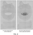
- 3D face scan data is acquired by aligning and inserting the tooth zone contour 3D data separated and acquired in S170 into the face 3D data acquired in S120.
- FIG. 8 illustrates the concept of aligning and inserting the tooth zone contour 3D data into the face 3D data.
- the tooth zone contour 3D data of the tooth zone contour is more detailed high-solution 3D data compared to the face 3D data that covers the whole face area.
- the tooth zone contour 3D data may be directly augmented to the face 3D data
- the data of the tooth zone contour portion may be removed from face 3D data as illustrated in FIG. 8A , before the tooth zone contour 3D data may be aligned and inserted into the tooth zone contour portion of the face 3D data as illustrated in FIG. 8B .
- FIG. 9 illustrates a 3D face scan system
- FIG. 10 is a flowchart illustrating a method for generating 3D face scan data.
- the example shown in FIG. 9 is further provided with an auxiliary 3D camera 105.
- the auxiliary 3D camera 105 is configured to acquire 3D data on areas other than the front of the face, such as the side of the face or the chin area. It is preferable that the subject for photography of the auxiliary 3D camera 105 may be set by the operation of an administrator (e.g., by a dentist).
- auxiliary area 3D data for a preset area or a plurality of preset areas in the face of the patient is generated by the auxiliary 3D camera 105 in S250 and that the process of aligning and inserting the tooth zone contour 3D data and the auxiliary area 3D data into the frontal face 3D data is performed in S280.
- the present disclosure may be implemented in the form of computer-readable code in a computer-readable non-transitory recording medium such as a non-volatile recording medium.
- the non-volatile recording medium includes various forms of storage devices such as hard disks, SSDs, CDROMs, NAS, magnetic tapes, web disks, cloud disks, etc., and may be implemented in the form in which codes are distributed and stored and executed in multiple storage devices connected by a network.
- the aspects of the present disclosure may be implemented in the form of a computer program stored in a medium for executing a specific procedure in conjunction with hardware.
Landscapes
- Health & Medical Sciences (AREA)
- Engineering & Computer Science (AREA)
- Physics & Mathematics (AREA)
- Life Sciences & Earth Sciences (AREA)
- General Health & Medical Sciences (AREA)
- Oral & Maxillofacial Surgery (AREA)
- Theoretical Computer Science (AREA)
- General Physics & Mathematics (AREA)
- Animal Behavior & Ethology (AREA)
- Public Health (AREA)
- Veterinary Medicine (AREA)
- Dentistry (AREA)
- Computer Vision & Pattern Recognition (AREA)
- Biophysics (AREA)
- Biomedical Technology (AREA)
- Epidemiology (AREA)
- Software Systems (AREA)
- Computer Graphics (AREA)
- Multimedia (AREA)
- Pathology (AREA)
- Surgery (AREA)
- Molecular Biology (AREA)
- Medical Informatics (AREA)
- Heart & Thoracic Surgery (AREA)
- Geometry (AREA)
- General Engineering & Computer Science (AREA)
- Architecture (AREA)
- Computer Hardware Design (AREA)
- Human Computer Interaction (AREA)
- Optics & Photonics (AREA)
- Audiology, Speech & Language Pathology (AREA)
- Nuclear Medicine, Radiotherapy & Molecular Imaging (AREA)
- Radiology & Medical Imaging (AREA)
- Dental Tools And Instruments Or Auxiliary Dental Instruments (AREA)
- Measurement Of The Respiration, Hearing Ability, Form, And Blood Characteristics Of Living Organisms (AREA)
- Measuring And Recording Apparatus For Diagnosis (AREA)
- Image Analysis (AREA)
Applications Claiming Priority (2)
| Application Number | Priority Date | Filing Date | Title |
|---|---|---|---|
| KR1020220035664A KR102758841B1 (ko) | 2022-03-22 | 2022-03-22 | 3차원 얼굴 스캔 시스템 및 그에 대한 3차원 얼굴 스캔 데이터 생성 방법 |
| PCT/KR2023/003695 WO2023182755A1 (ko) | 2022-03-22 | 2023-03-21 | 3차원 얼굴 스캔 데이터의 생성 방법 |
Publications (2)
| Publication Number | Publication Date |
|---|---|
| EP4497374A1 true EP4497374A1 (de) | 2025-01-29 |
| EP4497374A4 EP4497374A4 (de) | 2025-10-08 |
Family
ID=88101452
Family Applications (1)
| Application Number | Title | Priority Date | Filing Date |
|---|---|---|---|
| EP23775251.4A Pending EP4497374A4 (de) | 2022-03-22 | 2023-03-21 | Verfahren zur erzeugung von dreidimensionalen abtastdaten für gesicht |
Country Status (6)
| Country | Link |
|---|---|
| US (1) | US20240407892A1 (de) |
| EP (1) | EP4497374A4 (de) |
| JP (1) | JP7736358B2 (de) |
| KR (1) | KR102758841B1 (de) |
| CN (1) | CN118973466A (de) |
| WO (1) | WO2023182755A1 (de) |
Families Citing this family (2)
| Publication number | Priority date | Publication date | Assignee | Title |
|---|---|---|---|---|
| WO2025116509A1 (ko) * | 2023-11-28 | 2025-06-05 | 디웨이브 주식회사 | 개구량을 측정하는 방법 |
| KR20250179261A (ko) * | 2024-06-21 | 2025-12-30 | 주식회사 이노쓰리디 | 구강 스캐닝 시스템 |
Family Cites Families (21)
| Publication number | Priority date | Publication date | Assignee | Title |
|---|---|---|---|---|
| KR100682889B1 (ko) * | 2003-08-29 | 2007-02-15 | 삼성전자주식회사 | 영상에 기반한 사실감 있는 3차원 얼굴 모델링 방법 및 장치 |
| DE102007054907A1 (de) * | 2007-11-15 | 2009-05-28 | Sirona Dental Systems Gmbh | Verfahren zur optischen Vermessung von Objekten unter Verwendung eines Triangulationsverfahrens |
| BRPI1009891B8 (pt) * | 2009-03-20 | 2021-06-22 | 3Shape As | sistema e método para planejamento, visualização e otimização de restaurações dentárias |
| EP3851069B1 (de) * | 2010-06-29 | 2023-01-18 | 3Shape A/S | 2d-bild-anordnung |
| KR101145672B1 (ko) * | 2011-09-20 | 2012-05-24 | 원광대학교산학협력단 | 미소 훈련을 위한 미소 분석 시스템 |
| KR101176770B1 (ko) | 2012-03-22 | 2012-08-23 | 추상완 | 치과용 3차원 스캐너 및 이를 이용한 스캐닝 방법 |
| KR101744080B1 (ko) * | 2014-07-04 | 2017-06-09 | 주식회사 인스바이오 | 치과 시술 시뮬레이션을 위한 치아모델 생성 방법 |
| KR101744079B1 (ko) | 2014-07-04 | 2017-06-09 | 주식회사 인스바이오 | 치과 시술 시뮬레이션을 위한 얼굴모델 생성 방법 |
| KR101638561B1 (ko) * | 2015-04-13 | 2016-07-11 | 주식회사 디오 | 부분스캔을 이용한 치과 임플란트용 크라운 제조방법 |
| US20170103569A1 (en) * | 2015-10-08 | 2017-04-13 | Carestream Health, Inc. | Operator interface for 3d surface display using 2d index image |
| KR101878467B1 (ko) * | 2016-11-01 | 2018-07-13 | 한국과학기술연구원 | 3차원 치아 영상 데이터와 광학식 스캔 치아 모델의 선택적 정합 방법, 장치 및 프로그램 |
| CN108320325A (zh) * | 2018-01-04 | 2018-07-24 | 华夏天宇(北京)科技发展有限公司 | 牙列模型的生成方法及装置 |
| ES2882585T3 (es) | 2018-02-21 | 2021-12-02 | Ivoclar Vivadent Ag | Procedimiento para alinear un modelo tridimensional de una dentadura de un paciente sobre una imagen del rostro del paciente grabada por una cámara |
| JP6793302B2 (ja) * | 2018-02-22 | 2020-12-02 | 株式会社スペースビジョン | 三次元計測装置 |
| US11553988B2 (en) * | 2018-06-29 | 2023-01-17 | Align Technology, Inc. | Photo of a patient with new simulated smile in an orthodontic treatment review software |
| KR20200014645A (ko) | 2018-08-01 | 2020-02-11 | (주)마이핏 | 구강내 스캐닝을 이용한 치과 임플란트용 보철물 제조 방법 |
| KR102221958B1 (ko) | 2019-11-15 | 2021-03-04 | 오스템임플란트 주식회사 | 관심 치아를 자동으로 분할하여 스캔하는 관심 치아 스캔 시스템 및 방법 |
| US11810271B2 (en) * | 2019-12-04 | 2023-11-07 | Align Technology, Inc. | Domain specific image quality assessment |
| KR102728458B1 (ko) * | 2020-01-07 | 2024-11-11 | 주식회사 메디트 | 데이터 정합을 통한 3차원 모델 생성 장치 및 방법 |
| KR102334519B1 (ko) | 2020-12-14 | 2021-12-06 | 이마고웍스 주식회사 | 치과용 3차원 스캔 데이터의 간소화된 랜드마크 자동 검출 방법 및 이를 컴퓨터에서 실행시키기 위한 프로그램이 기록된 컴퓨터로 읽을 수 있는 기록 매체 |
| KR102479466B1 (ko) | 2020-08-21 | 2022-12-21 | 오스템임플란트 주식회사 | 치과용 스캔바디 |
-
2022
- 2022-03-22 KR KR1020220035664A patent/KR102758841B1/ko active Active
-
2023
- 2023-03-21 CN CN202380029126.9A patent/CN118973466A/zh active Pending
- 2023-03-21 EP EP23775251.4A patent/EP4497374A4/de active Pending
- 2023-03-21 WO PCT/KR2023/003695 patent/WO2023182755A1/ko not_active Ceased
- 2023-03-21 JP JP2024543114A patent/JP7736358B2/ja active Active
-
2024
- 2024-08-19 US US18/808,954 patent/US20240407892A1/en active Pending
Also Published As
| Publication number | Publication date |
|---|---|
| JP7736358B2 (ja) | 2025-09-09 |
| JP2025508275A (ja) | 2025-03-24 |
| KR102758841B1 (ko) | 2025-01-23 |
| EP4497374A4 (de) | 2025-10-08 |
| US20240407892A1 (en) | 2024-12-12 |
| WO2023182755A1 (ko) | 2023-09-28 |
| CN118973466A (zh) | 2024-11-15 |
| KR20230137779A (ko) | 2023-10-05 |
Similar Documents
| Publication | Publication Date | Title |
|---|---|---|
| US20240407892A1 (en) | Method of producing 3-dimensional face scan data | |
| EP3673864B1 (de) | Vorrichtung zur erzeugung eines 3d-modellbildes des gebisses eines patienten | |
| KR101878467B1 (ko) | 3차원 치아 영상 데이터와 광학식 스캔 치아 모델의 선택적 정합 방법, 장치 및 프로그램 | |
| KR102135770B1 (ko) | 스테레오 카메라 기반의 3차원 얼굴 복원 방법 및 장치 | |
| CN108052878A (zh) | 人脸识别设备和方法 | |
| JP2015156054A (ja) | 画像処理装置およびその制御方法 | |
| JP4761670B2 (ja) | 動立体モデル生成装置及び方法 | |
| JP2022508372A (ja) | 口腔内スキャナを使用して3dデータを取得する混合方法 | |
| JP2000126134A (ja) | 眼底計測装置及び眼底計測プログラムを記録した記録媒体 | |
| KR102387492B1 (ko) | 시계의 착용자의 안면 인식을 위한 방법 | |
| KR102033426B1 (ko) | 구강 스캐너 | |
| KR20100090457A (ko) | 비강압적 3차원 얼굴 데이터 획득 시스템 및 그 방법 | |
| EP4250248B1 (de) | Identifikationsvorrichtung, identifikationsverfahren und identifikationsprogramm | |
| KR20240009971A (ko) | 구강내 형상 취득 장치 및 구강내 형상 취득 방법 | |
| JP2022094744A (ja) | 被検体動き測定装置、被検体動き測定方法、プログラム、撮像システム | |
| US20150366644A1 (en) | Table for 3d-scanning of dental model | |
| KR101372496B1 (ko) | 스테레오 카메라를 이용한 후두내시경의 3차원 영상 획득 장치 및 그 방법 | |
| KR20060021566A (ko) | 전경체 사영영상과 가상외피에 기반한 가상시점 3차원 장면 재구성 방법 및 시스템 | |
| EP4328862A2 (de) | Gerät, verfahren und programm zur erzeugung von daten für die simulation von zahnbehandlungsergebnissen | |
| KR102891748B1 (ko) | 치아 보철물에 대한 시뮬레이션 수행 방법을 포함하는 컴퓨터 프로그램 | |
| JP2023505396A (ja) | 上顎歯と顔面骨格との間の空間関係を決定する技術 | |
| JP2002260017A (ja) | 3次元形状データ生成方法および装置 | |
| US12343115B2 (en) | Data processing apparatus and data processing method | |
| JP7181557B2 (ja) | 口腔内形状取得装置及び口腔内形状取得方法 | |
| US20210386512A1 (en) | Method Tool And Method Of Operating A Machine Tool |
Legal Events
| Date | Code | Title | Description |
|---|---|---|---|
| STAA | Information on the status of an ep patent application or granted ep patent |
Free format text: STATUS: THE INTERNATIONAL PUBLICATION HAS BEEN MADE |
|
| PUAI | Public reference made under article 153(3) epc to a published international application that has entered the european phase |
Free format text: ORIGINAL CODE: 0009012 |
|
| STAA | Information on the status of an ep patent application or granted ep patent |
Free format text: STATUS: REQUEST FOR EXAMINATION WAS MADE |
|
| 17P | Request for examination filed |
Effective date: 20241016 |
|
| AK | Designated contracting states |
Kind code of ref document: A1 Designated state(s): AL AT BE BG CH CY CZ DE DK EE ES FI FR GB GR HR HU IE IS IT LI LT LU LV MC ME MK MT NL NO PL PT RO RS SE SI SK SM TR |
|
| DAV | Request for validation of the european patent (deleted) | ||
| DAX | Request for extension of the european patent (deleted) | ||
| REG | Reference to a national code |
Ref country code: DE Ref legal event code: R079 Free format text: PREVIOUS MAIN CLASS: A61B0005000000 Ipc: G06T0007550000 |
|
| A4 | Supplementary search report drawn up and despatched |
Effective date: 20250908 |
|
| RIC1 | Information provided on ipc code assigned before grant |
Ipc: G06T 7/55 20170101AFI20250902BHEP Ipc: A61B 5/00 20060101ALI20250902BHEP Ipc: A61C 19/04 20060101ALI20250902BHEP Ipc: G06T 17/20 20060101ALI20250902BHEP Ipc: G06T 7/30 20170101ALI20250902BHEP Ipc: A61C 9/00 20060101ALI20250902BHEP Ipc: G06T 19/20 20110101ALI20250902BHEP Ipc: G06V 10/46 20220101ALI20250902BHEP Ipc: G06V 40/16 20220101ALI20250902BHEP |