EP4495133A2 - Verfahren zur isolierung von t-zellen mit antigenspezifizität für eine krebsspezifische p53-mutation - Google Patents
Verfahren zur isolierung von t-zellen mit antigenspezifizität für eine krebsspezifische p53-mutation Download PDFInfo
- Publication number
- EP4495133A2 EP4495133A2 EP24215129.8A EP24215129A EP4495133A2 EP 4495133 A2 EP4495133 A2 EP 4495133A2 EP 24215129 A EP24215129 A EP 24215129A EP 4495133 A2 EP4495133 A2 EP 4495133A2
- Authority
- EP
- European Patent Office
- Prior art keywords
- cells
- seq
- amino acid
- mutated
- tcr
- Prior art date
- Legal status (The legal status is an assumption and is not a legal conclusion. Google has not performed a legal analysis and makes no representation as to the accuracy of the status listed.)
- Pending
Links
Images



















Classifications
-
- C—CHEMISTRY; METALLURGY
- C07—ORGANIC CHEMISTRY
- C07K—PEPTIDES
- C07K14/00—Peptides having more than 20 amino acids; Gastrins; Somatostatins; Melanotropins; Derivatives thereof
- C07K14/435—Peptides having more than 20 amino acids; Gastrins; Somatostatins; Melanotropins; Derivatives thereof from animals; from humans
- C07K14/46—Peptides having more than 20 amino acids; Gastrins; Somatostatins; Melanotropins; Derivatives thereof from animals; from humans from vertebrates
- C07K14/47—Peptides having more than 20 amino acids; Gastrins; Somatostatins; Melanotropins; Derivatives thereof from animals; from humans from vertebrates from mammals
-
- A—HUMAN NECESSITIES
- A61—MEDICAL OR VETERINARY SCIENCE; HYGIENE
- A61K—PREPARATIONS FOR MEDICAL, DENTAL OR TOILETRY PURPOSES
- A61K40/00—Cellular immunotherapy
- A61K40/10—Cellular immunotherapy characterised by the cell type used
- A61K40/11—T-cells, e.g. tumour infiltrating lymphocytes [TIL] or regulatory T [Treg] cells; Lymphokine-activated killer [LAK] cells
-
- A—HUMAN NECESSITIES
- A61—MEDICAL OR VETERINARY SCIENCE; HYGIENE
- A61K—PREPARATIONS FOR MEDICAL, DENTAL OR TOILETRY PURPOSES
- A61K40/00—Cellular immunotherapy
- A61K40/30—Cellular immunotherapy characterised by the recombinant expression of specific molecules in the cells of the immune system
- A61K40/32—T-cell receptors [TCR]
-
- A—HUMAN NECESSITIES
- A61—MEDICAL OR VETERINARY SCIENCE; HYGIENE
- A61K—PREPARATIONS FOR MEDICAL, DENTAL OR TOILETRY PURPOSES
- A61K40/00—Cellular immunotherapy
- A61K40/40—Cellular immunotherapy characterised by antigens that are targeted or presented by cells of the immune system
- A61K40/41—Vertebrate antigens
- A61K40/42—Cancer antigens
- A61K40/4238—Regulators of development
- A61K40/424—Apoptosis related proteins, e.g. survivin or livin
- A61K40/4241—Apoptosis related proteins, e.g. survivin or livin p53
-
- A—HUMAN NECESSITIES
- A61—MEDICAL OR VETERINARY SCIENCE; HYGIENE
- A61P—SPECIFIC THERAPEUTIC ACTIVITY OF CHEMICAL COMPOUNDS OR MEDICINAL PREPARATIONS
- A61P35/00—Antineoplastic agents
-
- C—CHEMISTRY; METALLURGY
- C07—ORGANIC CHEMISTRY
- C07K—PEPTIDES
- C07K14/00—Peptides having more than 20 amino acids; Gastrins; Somatostatins; Melanotropins; Derivatives thereof
- C07K14/435—Peptides having more than 20 amino acids; Gastrins; Somatostatins; Melanotropins; Derivatives thereof from animals; from humans
- C07K14/46—Peptides having more than 20 amino acids; Gastrins; Somatostatins; Melanotropins; Derivatives thereof from animals; from humans from vertebrates
- C07K14/47—Peptides having more than 20 amino acids; Gastrins; Somatostatins; Melanotropins; Derivatives thereof from animals; from humans from vertebrates from mammals
- C07K14/4701—Peptides having more than 20 amino acids; Gastrins; Somatostatins; Melanotropins; Derivatives thereof from animals; from humans from vertebrates from mammals not used
- C07K14/4746—Peptides having more than 20 amino acids; Gastrins; Somatostatins; Melanotropins; Derivatives thereof from animals; from humans from vertebrates from mammals not used p53
-
- C—CHEMISTRY; METALLURGY
- C12—BIOCHEMISTRY; BEER; SPIRITS; WINE; VINEGAR; MICROBIOLOGY; ENZYMOLOGY; MUTATION OR GENETIC ENGINEERING
- C12N—MICROORGANISMS OR ENZYMES; COMPOSITIONS THEREOF; PROPAGATING, PRESERVING, OR MAINTAINING MICROORGANISMS; MUTATION OR GENETIC ENGINEERING; CULTURE MEDIA
- C12N5/00—Undifferentiated human, animal or plant cells, e.g. cell lines; Tissues; Cultivation or maintenance thereof; Culture media therefor
- C12N5/06—Animal cells or tissues; Human cells or tissues
- C12N5/0602—Vertebrate cells
- C12N5/0634—Cells from the blood or the immune system
- C12N5/0636—T lymphocytes
-
- A—HUMAN NECESSITIES
- A61—MEDICAL OR VETERINARY SCIENCE; HYGIENE
- A61K—PREPARATIONS FOR MEDICAL, DENTAL OR TOILETRY PURPOSES
- A61K2121/00—Preparations for use in therapy
-
- A—HUMAN NECESSITIES
- A61—MEDICAL OR VETERINARY SCIENCE; HYGIENE
- A61K—PREPARATIONS FOR MEDICAL, DENTAL OR TOILETRY PURPOSES
- A61K2300/00—Mixtures or combinations of active ingredients, wherein at least one active ingredient is fully defined in groups A61K31/00 - A61K41/00
-
- C—CHEMISTRY; METALLURGY
- C12—BIOCHEMISTRY; BEER; SPIRITS; WINE; VINEGAR; MICROBIOLOGY; ENZYMOLOGY; MUTATION OR GENETIC ENGINEERING
- C12N—MICROORGANISMS OR ENZYMES; COMPOSITIONS THEREOF; PROPAGATING, PRESERVING, OR MAINTAINING MICROORGANISMS; MUTATION OR GENETIC ENGINEERING; CULTURE MEDIA
- C12N2501/00—Active agents used in cell culture processes, e.g. differentation
- C12N2501/20—Cytokines; Chemokines
- C12N2501/23—Interleukins [IL]
- C12N2501/2302—Interleukin-2 (IL-2)
-
- C—CHEMISTRY; METALLURGY
- C12—BIOCHEMISTRY; BEER; SPIRITS; WINE; VINEGAR; MICROBIOLOGY; ENZYMOLOGY; MUTATION OR GENETIC ENGINEERING
- C12N—MICROORGANISMS OR ENZYMES; COMPOSITIONS THEREOF; PROPAGATING, PRESERVING, OR MAINTAINING MICROORGANISMS; MUTATION OR GENETIC ENGINEERING; CULTURE MEDIA
- C12N2502/00—Coculture with; Conditioned medium produced by
- C12N2502/11—Coculture with; Conditioned medium produced by blood or immune system cells
- C12N2502/1121—Dendritic cells
Definitions
- Adoptive cell therapy can produce positive clinical responses in some cancer patients. Nevertheless, obstacles to the successful use of ACT for the widespread treatment of cancer and other diseases remain. For example, T cells that specifically recognize cancer antigens may be difficult to identify and/or isolate from a patient. Accordingly, there is a need for improved methods of obtaining cancer-reactive T cells.
- An embodiment of the invention provides a method of isolating T cells having antigenic specificity for a mutated p53 amino acid sequence encoded by a cancer-specific p53 mutation, the method comprising: inducing autologous antigen presenting cells (APCs) of a patient to present at least one mutated p53 amino acid sequence; co-culturing autologous T cells of the patient with the autologous APCs that present the mutated p53 amino acid sequence; and selecting the autologous T cells that (a) were co-cultured with the autologous APCs that present the mutated p53 amino acid sequence and (b) have antigenic specificity for the mutated p53 amino acid sequence presented in the context of a major histocompatability complex (MHC) molecule expressed by the patient to provide isolated T cells having antigenic specificity for the mutated p53 amino acid sequence encoded by the cancer-specific p53 mutation.
- APCs autologous antigen presenting cells
- Another embodiment of the invention provides a method of isolating a T cell receptor (TCR), or an antigen-binding portion thereof, having antigenic specificity for a mutated p53 amino acid sequence encoded by a cancer-specific p53 mutation, the method comprising: isolating T cells having antigenic specificity for a mutated p53 amino acid sequence encoded by a cancer-specific p53 mutation according to any of the methods described herein; and isolating a nucleotide sequence that encodes the TCR, or the antigen-binding portion thereof, from the selected autologous T cells, wherein the TCR, or the antigen-binding portion thereof, has antigenic specificity for the mutated p53 amino acid sequence encoded by the cancer-specific p53 mutation.
- TCR T cell receptor
- Still another embodiment of the invention provides a method of preparing a population of cells that express a TCR, or an antigen-binding portion thereof, having antigenic specificity for a mutated p53 amino acid sequence encoded by a cancer-specific p53 mutation, the method comprising: isolating a TCR, or an antigen-binding portion thereof, according to any of the methods described herein, and introducing the nucleotide sequence encoding the isolated TCR, or the antigen-binding portion thereof, into peripheral blood mononuclear cells (PBMC) to obtain cells that express the TCR, or the antigen-binding portion thereof.
- PBMC peripheral blood mononuclear cells
- Another embodiment of the invention provides method of preparing a population of T cells that have antigenic specificity for a mutated p53 amino acid sequence encoded by a cancer-specific p53 mutation, the method comprising: isolating T cells according to any of the methods described herein, and expanding the numbers of selected autologous T cells to obtain a population of T cells that have antigenic specificity for the mutated p53 amino acid sequence encoded by the cancer-specific p53 mutation.
- Additional embodiments of the invention provide related populations of cells, pharmaceutical compositions, and methods of treating or preventing cancer.
- Tumor Protein P53 acts as a tumor suppressor by, for example, regulating cell division.
- the p53 protein is located in the nucleus of the cell, where it binds directly to DNA.
- the p53 protein is involved in determining whether the DNA will be repaired or the damaged cell will undergo apoptosis. If the DNA can be repaired, p53 activates other genes to fix the damage. If the DNA cannot be repaired, the p53 protein prevents the cell from dividing and signals it to undergo apoptosis. By stopping cells with mutated or damaged DNA from dividing, p53 helps prevent the development of tumors.
- Wild-type (WT) full-length p53 comprises the amino acid sequence of SEQ ID NO: 1.
- Mutations in the p53 protein may reduce or eliminate the p53 protein's tumor suppressor function.
- a p53 mutation may be a gain-of-function mutation by interfering with wild type p53 in a dominant negative fashion.
- Mutated p53 protein may be expressed in any of a variety of human cancers such as, for example, cholangiocarcinoma, melanoma, colon cancer, rectal cancer, ovarian cancer, endometrial cancer, non-small cell lung cancer (NSCLC), glioblastoma, uterine cervical cancer, head and neck cancer, breast cancer, pancreatic cancer, or bladder cancer.
- human cancers such as, for example, cholangiocarcinoma, melanoma, colon cancer, rectal cancer, ovarian cancer, endometrial cancer, non-small cell lung cancer (NSCLC), glioblastoma, uterine cervical cancer, head and neck cancer, breast cancer, pancreatic cancer, or bladder cancer.
- An embodiment of the invention provides a method of isolating T cells having antigenic specificity for a p53 mutated amino acid sequence encoded by a cancer-specific p53 mutation.
- Mutations of p53 are defined herein by reference to the amino acid sequence of full-length, WT p53 (SEQ ID NO: 1).
- mutations of p53 are described herein by reference to the amino acid residue present at a particular position, followed by the position number, followed by the amino acid with which that residue has been replaced in the particular mutation under discussion.
- R175" refers to the arginine present at position 175 of SEQ ID NO: 1
- R175H indicates that the arginine present at position 175 of SEQ ID NO: 1 is replaced by histidine
- G245S indicates that the glycine present at position 245 of SEQ ID NO: 1 has been replaced with serine.
- P53 has nine known splice variants. The p53 mutations described herein are conserved over all nine p53 splice variants. An alignment of the nine p53 splice variants is shown in Figure 32 .
- T cells isolated by the inventive methods may have antigenic specificity for any mutated p53 amino acid sequence described herein encoded by any of the nine p53 splice variants.
- positions are as defined by SEQ ID NO: 1
- the actual positions of the amino acid sequence of a particular splice variant of p53 are defined relative to the corresponding positions of SEQ ID NO: 1, and the positions as defined by SEQ ID NO: 1 may be different than the actual positions in a particular splice variant.
- mutations refer to a replacement of an amino acid residue in the amino acid sequence of a particular splice variant of p53 corresponding to the indicated position of the 393-amino acid sequence of SEQ ID NO: 1 with the understanding that the actual positions in the splice variant may be different.
- Embodiments of the invention may provide any one or more of many advantages.
- Mutated p53 is expressed by cancer cells and is not expressed by normal, noncancerous cells.
- T cells isolated by the inventive methods advantageously target the destruction of cancer cells while minimizing or eliminating the destruction of normal, non-cancerous cells, thereby reducing, for example, by minimizing or eliminating, toxicity.
- T cells isolated by the inventive methods may, advantageously, successfully treat or prevent mutated p53-positive cancers that do not respond to other types of treatment such as, for example, chemotherapy, surgery, or radiation.
- T cells isolated by the inventive methods may provide highly avid recognition of mutated p53, which may provide the ability to recognize unmanipulated tumor cells (e.g., tumor cells that have not been treated with interferon (IFN)- ⁇ , transfected with a vector encoding one or both of mutated p53 and the applicable HLA molecule, pulsed with a p53 peptide with the p53 mutation, or a combination thereof).
- unmanipulated tumor cells e.g., tumor cells that have not been treated with interferon (IFN)- ⁇ , transfected with a vector encoding one or both of mutated p53 and the applicable HLA molecule, pulsed with a p53 peptide with the p53 mutation, or a combination thereof.
- IFN interferon
- T cells isolated by the inventive methods may increase the number of patients who may be eligible for treatment with immunotherapy.
- peptides and tandem minigenes (TMG) including p53 mutations can be produced, stocked and validated prior to screening a patient so that once a p53 mutation is identified in a patient's tumor, the patient's T cells can be tested independent of the knowledge of the other mutations in the patient's tumor.
- TMG tandem minigenes
- This may reduce the time required to carry out the method which may be particularly advantageous when evaluating terminally ill patients.
- the duration of the inventive methods may be, e.g., about 3 to about 6 weeks shorter than traditional screening methods.
- the inventive methods may also provide a highly-focused study of p53 mutation responses in the absence of other competing mutations.
- inventive methods may be useful for testing T cell responses in a patient independent of the knowledge of whether the patient had the corresponding mutation or even a tumor.
- inventive methods may be useful for screening patients with a defined HLA haplotype and predicted p53 mutation binding, but are not limited to screening this type of cohort.
- the cancer-specific p53 mutation may be any mutation in the p53 gene which encodes a mutated p53 amino acid sequence (also referred to as a "non-silent mutation") and which is expressed in a cancer cell but not in a normal, noncancerous cell.
- Non-limiting examples of cancer-specific p53 mutations include missense, nonsense, insertion, deletion, duplication, frameshift, and repeat expansion mutations.
- the p53 mutation is a missense mutation.
- the mutated p53 amino acid sequence comprises a human p53 amino acid sequence with a mutation at position 175, 220, 245, 248, 249, 273, or 282 of SEQ ID NO: 1.
- the p53 mutation may be any missense mutation.
- the mutation at position 175, 220, 245, 248, 249, 273, or 282 of SEQ ID NO: 1 may be a substitution of the native (WT) amino acid residue present at position 175, 220, 245, 248, 249, 273, or 282 of SEQ ID NO: 1 with any amino acid residue other than the native (WT) amino acid residue present at the particular position under discussion.
- the mutated p53 amino acid sequence comprises a human p53 amino acid sequence with one of the following human p53 mutations: R175H, Y220C, G245D, G245S, R248L, R248Q, R248W, R249S, R273H, R273C, R273L, or R282W.
- the mutated p53 amino acid sequence may comprise a mutated p53 amino acid sequence selected from the group consisting of SEQ ID NOs: 2-13.
- the method may comprise inducing autologous antigen presenting cells (APCs) of a patient to present at least one mutated p53 amino acid sequence.
- APCs may include any cells which present peptide fragments of proteins in association with major histocompatibility complex (MHC) molecules on their cell surface.
- MHC major histocompatibility complex
- the APCs may include, for example, any one or more of macrophages, DCs, langerhans cells, B-lymphocytes, and T-cells.
- the APCs are DCs.
- the inventive methods may, advantageously, identify T cells that have antigenic specificity for a mutated p53 amino acid sequence encoded by a cancer-specific p53 mutation that is presented in the context of an MHC molecule expressed by the patient.
- the MHC molecule can be any MHC molecule expressed by the patient including, but not limited to, MHC Class I, MHC Class II, HLA-A, HLA-B, HLA-C, HLA-DM, HLA-DO, HLA-DP, HLA-DQ, and HLA-DR molecules.
- inventive methods may, advantageously, identify mutated p53 amino acid sequences presented in the context of any MHC molecule expressed by the patient without using, for example, epitope prediction algorithms to identify MHC molecules or mutated p53 amino acid sequences, which may be useful only for a select few MHC class I alleles and may be constrained by the limited availability of reagents to select p53 mutation-reactive T cells (e.g., an incomplete set of MHC tetramers).
- the inventive methods advantageously induce APCs to present the mutated p53 amino acid sequences in the context of any MHC molecule expressed by the patient and are not limited to any particular MHC molecule.
- the autologous APCs are antigen-negative autologous APCs.
- inducing autologous APCs of the patient to present the at least one mutated p53 amino acid sequence may be carried out using any suitable method known in the art.
- inducing autologous APCs of the patient to present the at least one mutated p53 amino acid sequence comprises pulsing the autologous APCs with a peptide comprising no more than one mutated p53 amino acid sequence or a pool of peptides, each peptide in the pool comprising a different mutated p53 amino acid sequence.
- the autologous APCs may be cultured with a peptide or a pool of peptides comprising the mutated p53 amino acid sequence in a manner such that the APCs internalize the peptide(s) and display the mutated p53 amino acid sequence(s), bound to an MHC molecule, on the cell membrane.
- Methods of pulsing APCs are known in the art and are described in, e.g., Solheim (Ed.), Antigen Processing and Presentation Protocols (Methods in Molecular Biology), Human Press, (2010 ).
- the peptide(s) used to pulse the APCs may include the mutated p53 amino acid(s) encoded by the cancer-specific p53 mutation.
- the peptide(s) may further comprise any suitable number of contiguous amino acids from the endogenous p53 protein encoded by the p53 gene on each of the carboxyl side and the amino side of the mutated amino acid(s).
- the number of contiguous amino acids from the endogenous p53 protein flanking each side of the mutation is not limited and may be, for example, about 4, about 5, about 6, about 7, about 8, about 9, about 10, about 11, about 12, about 13, about 14, about 15, about 16, about 17, about 18, about 19, about 20, or a range defined by any two of the foregoing values.
- the peptide(s) comprise(s) about 12 contiguous amino acids from the endogenous p53 protein on each side of the mutated amino acid(s).
- the peptide or pool of peptides comprise(s) one or more of the mutated p53 peptides of SEQ ID NOs: 2-13 (Table 1).
- inducing autologous APCs of the patient to present the mutated p53 amino acid sequence comprises introducing a nucleotide sequence encoding the mutated p53 amino acid sequence into the APCs.
- the nucleotide sequence is introduced into the APCs so that the APCs express and display the mutated p53 amino acid sequence, bound to an MHC molecule, on the cell membrane.
- the nucleotide sequence encoding the mutated p53 amino acid may be RNA or DNA.
- Introducing a nucleotide sequence into APCs may be carried out in any of a variety of different ways known in the art as described in, e.g., Solheim et al. supra.
- Non-limiting examples of techniques that are useful for introducing a nucleotide sequence into APCs include transformation, transduction, transfection, and electroporation.
- the method may comprise preparing more than one nucleotide sequence, each encoding a different mutated p53 amino acid sequence, and introducing each nucleotide sequence into a different population of autologous APCs.
- multiple populations of autologous APCs, each population expressing and displaying a different mutated p53 amino acid sequence may be obtained.
- the method may comprise introducing a nucleotide sequence encoding the more than one mutated p53 amino acid sequence.
- the nucleotide sequence introduced into the autologous APCs is a tandem minigene (TMG) construct, each minigene comprising a p53 gene, each p53 gene including a cancer-specific p53 mutation that encodes a different mutated p53 amino acid sequence.
- TMG tandem minigene
- Each minigene may encode one p53 mutation flanked on each side of the p53 mutation by any suitable number of contiguous amino acids from the endogenous p53 protein, as described herein with respect to other aspects of the invention.
- the TMG construct encodes one or more mutated p53 peptides of SEQ ID NOs: 2-13.
- the TMG construct may encode the amino acid sequence of SEQ ID NO: 14.
- the APCs express the mutated p53 amino acid sequences encoded by the TMG construct and display the mutated p53 amino acid sequences, bound to an MHC molecule, on the cell membranes.
- the method may comprise preparing more than one TMG construct, each construct encoding a different set of mutated p53 amino acid sequences, and introducing each TMG construct into a different population of autologous APCs.
- multiple populations of autologous APCs, each population expressing and displaying mutated p53 amino acid sequences encoded by different TMG constructs, may be obtained.
- the method may comprise culturing autologous T cells of the patient with the autologous APCs that present the mutated p53 amino acid sequence.
- the T cells can be obtained from numerous sources in the patient, including but not limited to tumor, blood, bone marrow, lymph node, the thymus, or other tissues or fluids.
- the T cells can include any type of T cell and can be of any developmental stage, including but not limited to, CD4+/CD8+ double positive T cells, CD4+ helper T cells, e.g., Th1 and Th2 cells, CD8+ T cells (e.g., cytotoxic T cells), tumor infiltrating cells (e.g., tumor infiltrating lymphocytes (TIL)), peripheral blood T cells, memory T cells, naive T cells, and the like.
- the T cells may be CD8+ T cells, CD4+ T cells, or both CD4+ and CD8+ T cells.
- the method may comprise co-culturing the autologous T cells and autologous APCs so that the T cells encounter the mutated p53 amino acid sequence presented by the APCs in such a manner that the autologous T cells specifically bind to and immunologically recognize a mutated p53 amino acid sequence presented by the APCs.
- the autologous T cells are co-cultured in direct contact with the autologous APCs.
- the method may comprise selecting the autologous T cells that (a) were co-cultured with the autologous APCs that present the mutated p53 amino acid sequence and (b) have antigenic specificity for the mutated p53 amino acid sequence presented in the context of a MHC molecule expressed by the patient.
- antigenic specificity means that the autologous T cells can specifically bind to and immunologically recognize the mutated p53 amino acid sequence encoded by the cancer-specific p53 mutation.
- the selecting may comprise identifying the T cells that have antigenic specificity for the mutated p53 amino acid sequence and separating them from T cells that do not have antigenic specificity for the mutated p53 amino acid sequence.
- the method comprises expanding the numbers of autologous T cells, e.g., by co-culturing with a T cell growth factor, such as interleukin (IL)-2 or IL-15, or as described herein with respect to other aspects of the invention, prior to selecting the autologous T cells.
- a T cell growth factor such as interleukin (IL)-2 or IL-15
- the method does not comprise expanding the numbers of autologous T cells with a T cell growth factor, such as IL-2 or IL-15 prior to selecting the autologous T cells.
- T cells having antigenic specificity for the mutated p53 amino acid sequence may express any one or more of a variety of T cell activation markers which may be used to identify those T cells having antigenic specificity for the mutated p53 amino acid sequence.
- T cell activation markers may include, but are not limited to, programmed cell death 1 (PD-1), lymphocyte-activation gene 3 (LAG-3), T cell immunoglobulin and mucin domain 3 (TIM-3), 4-1BB, OX40, and CD107a.
- selecting the autologous T cells that have antigenic specificity for the mutated p53 amino acid sequence comprises selecting the T cells that express any one or more of PD-1, LAG-3, TIM-3, 4-1BB, OX40, and CD107a.
- Cells expressing one or more T cell activation markers may be sorted on the basis of expression of the marker using any of a variety of techniques known in the art such as, for example, fluorescence-activated cell sorting (FACS) or magnetic-activated cell sorting (MACS) as described in, e.g., Turcotte et al., Clin. Cancer Res., 20(2): 331-43 (2013 ) and Gros et al., J. Clin. Invest., 124(5): 2246-59 (2014 ).
- FACS fluorescence-activated cell sorting
- MCS magnetic-activated cell sorting
- selecting the autologous T cells that have antigenic specificity for the mutated p53 amino acid sequence comprises selecting the T cells (i) that secrete a greater amount of one or more cytokines upon co-culture with APCs that present the mutated p53 amino acid sequence as compared to the amount of the one or more cytokines secreted by a negative control or (ii) in which at least twice as many of the numbers of T cells secrete one or more cytokines upon co-culture with APCs that present the mutated p53 amino acid sequence as compared to the numbers of negative control T cells that secrete the one or more cytokines.
- the one or more cytokines may comprise any cytokine the secretion of which by a T cell is characteristic of T cell activation (e.g., a T cell receptor (TCR) expressed by the T cells specifically binding to and immunologically recognizing the mutated p53 amino acid sequence).
- T cell activation e.g., a T cell receptor (TCR) expressed by the T cells specifically binding to and immunologically recognizing the mutated p53 amino acid sequence.
- TCR T cell receptor
- cytokines, the secretion of which is characteristic of T cell activation include IFN-y, IL-2, and tumor necrosis factor alpha (TNF- ⁇ ), granulocyte/monocyte colony stimulating factor (GM-CSF), IL-4, IL-5, IL-9, IL-10, IL-17, and IL-22.
- the autologous T cells may be considered to have "antigenic specificity" for the mutated p53 amino acid sequence if the T cells secrete at least twice as much IFN- ⁇ upon co-culture with (a) antigen-negative APCs pulsed with a concentration of a peptide comprising the mutated p53 amino acid sequence (e.g., about 0.05 ng/mL to about 10 ⁇ g/mL, e.g., 0.05 ng/mL, 0.1 ng/mL, 0.5 ng/mL, 1 ng/mL, 5 ng/mL, 100 ng/mL, 1 ⁇ g/mL, 5 ⁇ g/mL, or 10 ⁇ g/mL) or (b) APCs into which a nucleotide sequence encoding the mutated p53 amino acid sequence has been introduced as compared to the amount of IFN- ⁇ secreted by a negative control.
- the negative control may be, for example, autologous T cells (e.g., derived from peripheral blood mononuclear cells (PBMC)) co-cultured with (a) antigen-negative APCs pulsed with the same concentration of an irrelevant peptide (e.g., the wild-type amino acid sequence, or some other peptide with a different sequence from the mutated p53 amino acid sequence) or (b) APCs into which a nucleotide sequence encoding an irrelevant peptide sequence has been introduced.
- PBMC peripheral blood mononuclear cells
- the autologous T cells may also have "antigenic specificity" for the mutated p53 amino acid sequence if the T cells secrete a greater amount of IFN- ⁇ upon co-culture with antigen-negative APCs pulsed with higher concentrations of a peptide comprising the mutated p53 amino acid sequence as compared to a negative control, for example, the negative control described above.
- IFN- ⁇ secretion may be measured by methods known in the art such as, for example, enzyme-linked immunosorbent assay (ELISA).
- the autologous T cells may be considered to have "antigenic specificity" for the mutated p53 amino acid sequence if at least twice as many of the numbers of T cells secrete IFN- ⁇ upon co-culture with (a) antigen-negative APCs pulsed with a concentration of a peptide comprising the mutated p53 amino acid sequence or (b) APCs into which a nucleotide sequence encoding the mutated p53 amino acid sequence has been introduced as compared to the numbers of negative control T cells that secrete IFN- ⁇ .
- concentration of peptide and the negative control may be as described herein with respect to other aspects of the invention.
- the numbers of cells secreting IFN- ⁇ may be measured by methods known in the art such as, for example, ELISPOT.
- the autologous T cells may be considered to have "antigenic specificity" for mutated p53 if at least twice as many spots are detected by ELISPOT for the T cells upon co-culture with (a) antigen-negative, applicable HLA molecule positive target cells pulsed with a low concentration of mutated p53 peptide or (b) antigen-negative, applicable HLA molecule positive target cells into which a nucleotide sequence encoding mutated p53 has been introduced such that the target cell expresses mutated p53 as compared to the number of spots detected by ELISPOT for negative control T cells co-cultured with the same target cells.
- concentration of peptide and the negative control may be as described herein with respect to other aspects of the invention.
- the autologous T cells may be considered to have "antigenic specificity" for mutated p53 if greater than about 50 spots are detected by ELISPOT for the T cells expressing the TCR upon co-culture with (a) antigen-negative, applicable HLA molecule positive target cells pulsed with a low concentration of mutated p53 peptide or (b) antigen-negative, applicable HLA molecule positive target cells into which a nucleotide sequence encoding mutated p53 has been introduced such that the target cell expresses mutated p53.
- concentration of peptide may be as described herein with respect to other aspects of the invention.
- T cells having antigenic specificity for the mutated p53 amino acid sequence may both (1) express any one or more T cells activation markers described herein and (2) secrete a greater amount of one or more cytokines as described herein, in an embodiment of the invention, T cells having antigenic specificity for the mutated p53 amino acid sequence may express any one or more T cell activation markers without secreting a greater amount of one or more cytokines or may secrete a greater amount of one or more cytokines without expressing any one or more T cell activation markers.
- selecting the autologous T cells that have antigenic specificity for the mutated p53 amino acid sequence comprises selectively growing the autologous T cells that have antigenic specificity for the mutated p53 amino acid sequence.
- the method may comprise co-culturing the autologous T cells with autologous APCs in such a manner as to favor the growth of the T cells that have antigenic specificity for the mutated p53 amino acid sequence over the T cells that do not have antigenic specificity for the mutated p53 amino acid sequence. Accordingly, a population of T cells is provided that has a higher proportion of T cells that have antigenic specificity for the mutated p53 amino acid sequence as compared to T cells that do not have antigenic specificity for the mutated p53 amino acid sequence.
- the method further comprises obtaining multiple fragments of a tumor from the patient, separately co-culturing autologous T cells from each of the multiple fragments with the autologous APCs that present the mutated p53 amino acid sequence as described herein with respect to other aspects of the invention, and separately assessing the T cells from each of the multiple fragments for antigenic specificity for the mutated p53 amino acid sequence, as described herein with respect to other aspects of the invention.
- selecting the autologous T cells may further comprise separately assessing autologous T cells for antigenic specificity for each of the multiple mutated p53 amino acid sequences.
- the inventive method may further comprise separately inducing autologous APCs of the patient to present each mutated p53 amino acid sequence encoded by the construct (or included in the pool), as described herein with respect to other aspects of the invention (for example, by providing separate APC populations, each presenting a different mutated p53 amino acid sequence encoded by the construct (or included in the pool)).
- the method may further comprise separately co-culturing autologous T cells of the patient with the different populations of autologous APCs that present each mutated p53 amino acid sequence, as described herein with respect to other aspects of the invention.
- the method may further comprise separately selecting the autologous T cells that (a) were co-cultured with the autologous APCs that present the mutated p53 amino acid sequence and (b) have antigenic specificity for the mutated p53 amino acid sequence presented in the context of a MHC molecule expressed by the patient, as described herein with respect to other aspects of the invention.
- the method may comprise determining which mutated p53 amino acid sequence encoded by a TMG construct that encodes multiple mutated p53 amino acid sequences (or included in the pool) are immunologically recognized by the autologous T cells (e.g., by process of elimination).
- expansion of the numbers of T cells is carried out by culturing the T cells with OKT3 antibody, IL-2, and feeder PBMC (e.g., irradiated allogeneic PBMC).
- the inventive methods may, advantageously, generate a large number of T cells having antigenic specificity for the mutated p53 amino acid sequence.
- an embodiment of the invention provides a method of preparing a population of T cells that have antigenic specificity for a mutated p53 amino acid sequence encoded by a cancer-specific p53 mutation, the method comprising isolating T cells as described herein with respect to other aspects of the invention, and expanding the numbers of selected autologous T cells to obtain a population of T cells that have antigenic specificity for the mutated p53 amino acid sequence encoded by the cancer-specific mutation. Expanding the numbers of selected cells may be carried out as described herein with respect to other aspects of the invention.
- Another embodiment of the invention provides a method of isolating a T cell receptor (TCR), or an antigen-binding portion thereof, having antigenic specificity for a mutated p53 amino acid sequence encoded by a cancer-specific p53 mutation.
- the method may comprise isolating T cells having antigenic specificity for a mutated p53 amino acid sequence encoded by a cancer-specific p53 mutation as described herein with respect to other aspects of the invention.
- the method may further comprise isolating a nucleotide sequence that encodes the TCR, or the antigen-binding portion thereof, from the selected T cells, wherein the TCR, or the antigen-binding portion thereof, has antigenic specificity for the mutated p53 amino acid sequence encoded by the cancer-specific p53 mutation.
- the numbers selected T cells that have antigenic specificity for the mutated p53 amino acid sequence may be expanded. Expanding the numbers of selected cells may be carried out as described herein with respect to other aspects of the invention.
- the numbers of selected T cells that have antigenic specificity for the mutated p53 amino acid sequence are not expanded prior to isolating the nucleotide sequence that encodes the TCR, or the antigen-binding portion thereof.
- the TCR, or antigen binding portion thereof may be isolated from a single cell.
- the antigen-binding portion of the TCR refers to any portion comprising contiguous amino acids of the TCR of which it is a part, provided that the antigen-binding portion specifically binds to the mutated p53 amino acid sequence as described herein with respect to other aspects of the invention.
- the term “antigen-binding portion” refers to any part or fragment of the TCR, which part or fragment retains the biological activity of the TCR of which it is a part (the parent TCR).
- Antigen-binding portions encompass, for example, those parts of a TCR that retain the ability to specifically bind to the mutated p53 amino acid sequence, or detect, treat, or prevent cancer, to a similar extent, the same extent, or to a higher extent, as compared to the parent TCR.
- the functional portion can comprise, for instance, about 10%, about 25%, about 30%, about 50%, about 68%, about 80%, about 90%, about 95%, or more, of the parent TCR.
- the antigen-binding portion can comprise an antigen-binding portion of either or both of the ⁇ and ⁇ chains of the TCR, such as a portion comprising one or more of the complementarity determining region (CDR)1, CDR2, and CDR3 of the variable region(s) of the ⁇ chain and/or ⁇ chain of the TCR.
- CDR complementarity determining region
- the antigen-binding portion can comprise the amino acid sequence of the CDR1 of the ⁇ chain (CDR1 ⁇ ), the CDR2 of the ⁇ chain (CDR2 ⁇ ), the CDR3 of the ⁇ chain (CDR3 ⁇ ), the CDR1 of the ⁇ chain (CDR1 ⁇ ), the CDR2 of the ⁇ chain (CDR2 ⁇ ), the CDR3 of the ⁇ chain (CDR3 ⁇ ), or any combination thereof.
- the antigen-binding portion comprises the amino acid sequences of CDR1 ⁇ , CDR2 ⁇ , and CDR3 ⁇ ; the amino acid sequences of CDR1 ⁇ , CDR2 ⁇ , and CDR3 ⁇ ; or the amino acid sequences of all of CDR1 ⁇ , CDR2 ⁇ , CDR3 ⁇ , CDR1 ⁇ , CDR2 ⁇ , and CDR3 ⁇ of the TCR.
- TCR nucleic acid sequence may include, for example, single-cell reverse transcription polymerase chain reaction (RT-PCR), bioinfomatic reconstruction, limiting dilution cloning, the FLUIDIGM system (Fluidigm Corporation, South San Francisco, CA), deep sequencing of TCR ⁇ and TCR ⁇ chains with frequency pairing, PAIRSEQ method of identifying TCR ⁇ and ⁇ chain pairs from Adaptive Biotechnologies (Seattle, WA), and combinations thereof.
- RT-PCR single-cell reverse transcription polymerase chain reaction
- bioinfomatic reconstruction limiting dilution cloning
- FLUIDIGM system Fludigm Corporation, South San Francisco, CA
- PAIRSEQ method of identifying TCR ⁇ and ⁇ chain pairs from Adaptive Biotechnologies (Seattle, WA), and combinations thereof.
- the term "recombinant expression vector” means a genetically-modified oligonucleotide or polynucleotide construct that permits the expression of an mRNA, protein, polypeptide, or peptide by a host cell, when the construct comprises a nucleotide sequence encoding the mRNA, protein, polypeptide, or peptide, and the vector is contacted with the cell under conditions sufficient to have the mRNA, protein, polypeptide, or peptide expressed within the cell.
- the vectors may not be naturally-occurring as a whole. However, parts of the vectors can be naturally-occurring.
- the recombinant expression vectors can comprise any type of nucleotides, including, but not limited to DNA (e.g., complementary DCA (cDNA)) and RNA, which can be single-stranded or double-stranded, synthesized or obtained in part from natural sources, and which can contain natural, non-natural or altered nucleotides.
- the recombinant expression vectors can comprise naturally-occurring, non-naturally-occurring internucleotide linkages, or both types of linkages.
- the non-naturally occurring or altered nucleotides or internucleotide linkages does not hinder the transcription or replication of the vector.
- the recombinant expression vector can be any suitable recombinant expression vector, and can be used to transform or transfect any suitable host cell.
- Suitable vectors include those designed for propagation and expansion or for expression or both, such as plasmids and viruses.
- the vector can be selected from the group consisting of transposon/transposase, the pUC series (Fermentas Life Sciences), the pBluescript series (Stratagene, LaJolla, CA), the pET series (Novagen, Madison, Wl), the pGEX series (Pharmacia Biotech, Uppsala, Sweden), and the pEX series (Clontech, Palo Alto, CA).
- Bacteriophage vectors such as AGT10, ⁇ GT11, AZapll (Stratagene), AEMBL4, and ⁇ NM1149, also can be used.
- plant expression vectors include pBI01, pBI101.2, pBI101.3, pBI121 and pBIN19 (Clontech).
- animal expression vectors include pEUK-CI, pMAM and pMAMneo (Clontech).
- the recombinant expression vector is a viral vector, e.g., a retroviral vector.
- an embodiment of the invention provides a method of preparing a population of cells that express a TCR, or an antigen-binding portion thereof, having antigenic specificity for a mutated p53 amino acid sequence encoded by a cancer-specific p53 mutation, the method comprising isolating a TCR, or an antigen-binding portion thereof, as described herein with respect to other aspects of the invention, and introducing the nucleotide sequence encoding the isolated TCR, or the antigen-binding portion thereof, into host cells to obtain cells that express the TCR, or the antigen-binding portion thereof.
- nucleotide sequence e.g., a recombinant expression vector
- introducing the nucleotide sequence encoding the isolated TCR, or the antigen-binding portion thereof, into host cells may be carried out in any of a variety of different ways known in the art as described in, e.g., Green et al. supra.
- Non-limiting examples of techniques that are useful for introducing a nucleotide sequence into host cells include transformation, transduction, transfection, transposition, and electroporation.
- the host cell into which the nucleotide sequence encoding the TCR, or antigen binding portion thereof, is introduced may be any type of cell that can contain the recombinant expression vector.
- the host cell can be a eukaryotic cell, e.g., plant, animal, fungi, or algae, or can be a prokaryotic cell, e.g., bacteria or protozoa.
- the host cell can be a cultured cell or a primary cell, i.e., isolated directly from an organism, e.g., a human.
- the host cell can be an adherent cell or a suspended cell, i.e., a cell that grows in suspension. Suitable host cells are known in the art and include, for instance, DH5 ⁇ E.
- the host cell is preferably a prokaryotic cell, e.g., a DH5a cell.
- the host cell is preferably a mammalian cell. Most preferably, the host cell is a human cell. While the host cell can be of any cell type, can originate from any type of tissue, and can be of any developmental stage, the host cell preferably is a PBL or a PBMC. More preferably, the host cell is a T cell.
- the PBMC include T cells.
- the T cells may be any type of T cell. Without being bound to a particular theory or mechanism, it is believed that less differentiated, "younger” T cells may be associated with any one or more of greater in vivo persistence, proliferation, and antitumor activity as compared to more differentiated, "older” T cells.
- the inventive methods may, advantageously, identify and isolate a TCR, or an antigen-binding portion thereof, that has antigenic specificity for the mutated p53 amino acid sequence and introduce the TCR, or an antigen-binding portion thereof, into "younger" T cells that may provide any one or more of greater in vivo persistence, proliferation, and antitumor activity as compared to "older" T cells (e.g., effector cells in a patient's tumor).
- the inventive methods identify and isolate TCRs, or the antigen-binding portions thereof, that have antigenic specificity against a mutated p53 amino acid sequence that is encoded by a recurrent (also referred to as "hot-spot") cancer-specific p53 mutation.
- the method may comprise introducing the nucleotide sequence encoding the isolated TCR, or the antigen-binding portion thereof, into host cells that are allogeneic to the patient.
- the method may comprise introducing the nucleotide sequence encoding the isolated TCR, or the antigen-binding portion thereof, into the host cells from another patient whose tumors express the same p53 mutation in the context of the same MHC molecule.
- the method further comprises expanding the numbers of host cells that express the TCR, or the antigen-binding portion thereof.
- the numbers of host cells may be expanded, for example, as described herein with respect to other aspects of the invention.
- the inventive methods may, advantageously, generate a large number of T cells having antigenic specificity for the mutated p53 amino acid sequence.
- the population of cells can be a heterogeneous population comprising the T cells having antigenic specificity for the mutated p53 amino acid sequence encoded by the cancer-specific p53 mutation in addition to at least one other cell, e.g., a PBMC which does not have antigenic specificity for the mutated p53 amino acid sequence encoded by the cancer-specific p53 mutation, or a cell other than a T cell, e.g., a B cell, a macrophage, a neutrophil, an erythrocyte, a hepatocyte, an endothelial cell, an epithelial cells, a muscle cell, a brain cell, etc.
- a B cell e.g., a macrophage, a neutrophil, an erythrocyte, a hepatocyte, an endothelial cell, an epithelial cells, a muscle cell, a brain cell, etc.
- the population of cells can be a substantially homogeneous population, in which the population comprises mainly of (e.g., consisting essentially of) T cells having antigenic specificity for the mutated p53 amino acid sequence encoded by the cancer-specific p53 mutation.
- the population also can be a clonal population of cells, in which all cells of the population are clones of a single T cell, such that all cells of the population have antigenic specificity for the mutated p53 amino acid sequence encoded by the cancer-specific p53 mutation.
- the population of cells is a clonal population comprising T cell having antigenic specificity for the mutated p53 amino acid sequence encoded by the cancer-specific p53 mutation, as described herein.
- about 1% to about 100% for example, about 1%, about 5%, about 10%, about 15%, about 20%, about 25%, about 30%, about 35%, about 40%, about 45%, about 50%, about 55%, about 60%, about 65%, about 70%, about 75%, about 80%, about 85%, about 90%, about 95%, about 96%, about 97%, about 98%, about 99%, or about 100%, or a range defined by any two of the foregoing values, of the population of cells comprises T cells that have antigenic specificity for the mutated p53 amino acid sequence.
- populations of cells that comprise a high proportion of T cells that have antigenic specificity for the mutated p53 amino acid sequence advantageously may have a lower proportion of irrelevant cells that may hinder the function of the T cell, e.g., the ability of the T cell to target the destruction of cancer cells and/or treat or prevent cancer.
- the inventive populations of cells can be formulated into a composition, such as a pharmaceutical composition.
- the invention provides a pharmaceutical composition comprising any of the inventive populations of cells and a pharmaceutically acceptable carrier.
- the inventive pharmaceutical composition can comprise an inventive population of cells in combination with another pharmaceutically active agent(s) or drug(s), such as a chemotherapeutic agents, e.g., asparaginase, busulfan, carboplatin, cisplatin, daunorubicin, doxorubicin, fluorouracil, gemcitabine, hydroxyurea, methotrexate, paclitaxel, rituximab, vinblastine, vincristine, etc.
- chemotherapeutic agents e.g., asparaginase, busulfan, carboplatin, cisplatin, daunorubicin, doxorubicin, fluorouracil, gemcitabine, hydroxyurea, methotrexate, paclitaxel, ritux
- the carrier is a pharmaceutically acceptable carrier.
- the carrier can be any of those conventionally used for the particular inventive population of cells under consideration.
- Such pharmaceutically acceptable carriers are well-known to those skilled in the art and are readily available to the public. It is preferred that the pharmaceutically acceptable carrier be one which has no detrimental side effects or toxicity under the conditions of use.
- Suitable formulations may include any of those for parenteral, subcutaneous, intravenous, intramuscular, intraarterial, intratumoral, intrathecal, or interperitoneal administration. More than one route can be used to administer the inventive population of cells, and in certain instances, a particular route can provide a more immediate and more effective response than another route.
- the inventive population of cells is administered by injection, e.g., intravenously.
- the pharmaceutically acceptable carrier for the cells for injection may include any isotonic carrier such as, for example, normal saline (about 0.90% w/v of NaCl in water, about 300 mOsm/L NaCl in water, or about 9.0 g NaCl per liter of water), NORMOSOL R electrolyte solution (Abbott, Chicago, IL), PLASMA-LYTE A (Baxter, Deerfield, IL), about 5% dextrose in water, or Ringer's lactate.
- the pharmaceutically acceptable carrier is supplemented with human serum albumin.
- inventive populations of cells and pharmaceutical compositions can be used in methods of treating or preventing cancer.
- inventive T cells are believed to bind specifically to a mutated p53 amino acid sequence encoded by a cancer-specific p53 mutation, such that a TCR expressed by the cell is able to mediate an immune response against a target cell expressing the mutated p53 amino acid sequence.
- an embodiment of the invention provides a method of treating or preventing cancer in a mammal, comprising administering to the mammal any of the pharmaceutical compositions or populations of cells described herein, in an amount effective to treat or prevent cancer in the mammal.
- inventive methods can provide any amount of any level of treatment or prevention of cancer in a mammal.
- the treatment or prevention provided by the inventive method can include treatment or prevention of one or more conditions or symptoms of the cancer being treated or prevented.
- treatment or prevention can include promoting the regression of a tumor.
- prevention can encompass delaying the onset of the cancer, or a symptom or condition thereof.
- the amount or dose of the inventive population of cells or pharmaceutical composition administered should be sufficient to effect, e.g., a therapeutic or prophylactic response, in the mammal over a reasonable time frame.
- the dose of the inventive population of cells or pharmaceutical composition should be sufficient to bind to a mutated p53 amino acid sequence encoded by a cancer-specific p53 mutation, or detect, treat or prevent cancer in a period of from about 2 hours or longer, e.g., 12 to 24 or more hours, from the time of administration. In certain embodiments, the time period could be even longer.
- the dose will be determined by the efficacy of the particular inventive population of cells or pharmaceutical composition administered and the condition of the mammal (e.g., human), as well as the body weight of the mammal (e.g., human) to be treated.
- an assay which comprises comparing the extent to which target cells are lysed or IFN- ⁇ is secreted by T cells upon administration of a given dose of such T cells to a mammal among a set of mammals of which is each given a different dose of the T cells, could be used to determine a starting dose to be administered to a mammal.
- the extent to which target cells are lysed or IFN- ⁇ is secreted upon administration of a certain dose can be assayed by methods known in the art.
- the dose of the inventive population of cells or pharmaceutical composition also will be determined by the existence, nature and extent of any adverse side effects that might accompany the administration of a particular inventive population of cells or pharmaceutical composition.
- the attending physician will decide the dosage of the inventive population of cells or pharmaceutical composition with which to treat each individual patient, taking into consideration a variety of factors, such as age, body weight, general health, diet, sex, inventive population of cells or pharmaceutical composition to be administered, route of administration, and the severity of the condition being treated.
- the number of cells administered per infusion may vary, for example, in the range of one million to 100 billion cells; however, amounts below or above this exemplary range are within the scope of the invention.
- the daily dose of inventive host cells can be about 1 million to about 150 billion cells (e.g., about 5 million cells, about 25 million cells, about 500 million cells, about 1 billion cells, about 5 billion cells, about 20 billion cells, about 30 billion cells, about 40 billion cells, about 60 billion cells, about 80 billion cells, about 100 billion cells, about 120 billion cells, about 130 billion cells, about 150 billion cells, or a range defined by any two of the foregoing values), preferably about 10 million to about 130 billion cells (e.g., about 20 million cells, about 30 million cells, about 40 million cells, about 60 million cells, about 70 million cells, about 80 million cells, about 90 million cells, about 10 billion cells, about 25 billion cells, about 50 billion cells, about 75 billion cells, about 90 billion cells, about 100 billion cells, about 110 billion cells,
- the cells can be cells that are allogeneic or autologous to the mammal.
- the cells are autologous to the mammal.
- Another embodiment of the invention provides any of the isolated population of cells or pharmaceutical compositions described herein for use in treating or preventing cancer in a mammal.
- the cancer can be any cancer, including any of acute lymphocytic cancer, acute myeloid leukemia, alveolar rhabdomyosarcoma, bone cancer, brain cancer, breast cancer, cancer of the anus, anal canal, or anorectum, cancer of the eye, cancer of the intrahepatic bile duct, cancer of the joints, cancer of the neck, gallbladder, or pleura, cancer of the nose, nasal cavity, or middle ear, cancer of the oral cavity, cancer of the vagina, cancer of the vulva, chronic lymphocytic leukemia, chronic myeloid cancer, colon cancer, colocrectal cancer, endometrial cancer, esophageal cancer, uterine cervical cancer, gastrointestinal carcinoid tumor, glioma, Hodgkin lymphoma, hypopharynx cancer, kidney cancer, larynx cancer, liver cancer, lung cancer, malignant mesothelioma, melanoma, multiple myelom
- the cancer is a cancer which expresses mutated p53.
- the cancer may express p53 with a mutation at one or more of positions 175, 220, 245, 248, 249, 273, or 282 of SEQ ID NO: 1.
- the cancer may express p53 with one or more of the following human p53 mutations: R175H, Y220C, G245D, G245S, R248L, R248Q, R248W, R249S, R273H, R273C, R273L, or R282W.
- the cancer is an epithelial cancer or cholangiocarcinoma, melanoma, colon cancer, rectal cancer, ovarian cancer, endometrial cancer, non-small cell lung cancer (NSCLC), glioblastoma, uterine cervical cancer, head and neck cancer, breast cancer, pancreatic cancer, or bladder cancer.
- NSCLC non-small cell lung cancer
- glioblastoma uterine cervical cancer, head and neck cancer
- breast cancer pancreatic cancer, or bladder cancer.
- the mammal referred to in the inventive methods can be any mammal.
- the term "mammal” refers to any mammal, including, but not limited to, mammals of the order Rodentia, such as mice and hamsters, and mammals of the order Logomorpha, such as rabbits. It is preferred that the mammals are from the order Carnivora, including Felines (cats) and Canines (dogs). Preferably, the mammals are from the order Artiodactyla, including Bovines (cows) and Swines (pigs) or of the order Perssodactyla, including Equines (horses). Preferably, the mammals are of the order Primates, Ceboids, or Simoids (monkeys) or of the order Anthropoids (humans and apes). A more preferred mammal is the human.
- This example demonstrates the identification of anti-mutated p53 T cells in Patient 4141 by co-culturing autologous APCs induced to express mutated p53 within autologous T cells ("p53 hotspot mutation universal screening"). This example also demonstrates the isolation and specific reactivity of a TCR from patient 4141.
- TIL fragment F12 and infusion bag TIL (Rx1) from patient 4141 and p53-R175H-specific TCR or mock transduced T cells from patient 4196 were used as effectors.
- Co-cultures with T cell effectors and HLA-A*02:01 APCs were either (1) electroporated with TMGs composed of irrelevant, WT p53, or mutated p53 sequences or (2) pulsed with peptide vehicle (DMSO) or purified (>95% by HPLC) 25 amino acid peptides composed of WT p53-R175 sequence or mutated p53-R175H sequence.
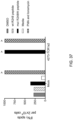
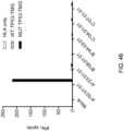
- Cos7 cells (2.5 ⁇ 10 4 per well) were plated on wells of flat-bottom 96 well plates. The following day, cells were co-transfected with individual HLA alleles from patient 4141 and either no extra gene, WT TP53 TMG, or mutated TP53 TMG containing the p53-R175H sequence. TIL with specificity to p53-R175H from Patient 4141 (fragment culture 12) were co-cultured the following day with transfected Cos7 cells and were incubated overnight at 37 °C. Secretion of IFN- ⁇ was evaluated by ELISPOT. The results are shown in Figure 46 .
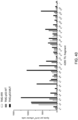
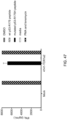
- T cells expressing mock (no TCR) or 4141-TCR1a2 were co-cultured with T2 tumor cells (expressing HLA-A*02:01).
- T2 cells were pulsed for 2 hours at 37 °C with peptide vehicle (DMSO) or purified (>95% by HPLC) peptides composed of WT p53-R175 peptide HMTEWRRC (SEQ ID NO: 532) or mutated p53-R175H peptide HMTEWRHC (SEQ ID NO: 530).
- Media alone and PMA and lonomycin were negative and positive controls, respectively.
- Co-cultures were performed overnight at 37 °C. Secretion of IFN- ⁇ was evaluated by ELISA. The results are shown in Figure 47 .
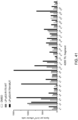
- T cells expressing 4141-TCR1a2 were co-cultured overnight at 37 °C with Saos2 cells (p53-NULL and HLA-A*02:01+), which were either unmanipulated or made to overexpress full length p53-R175H protein.
- Inhibitors of secretion (monensin and brefeldin A) were added to co-cultures to trap cytokines within T cells. After 6 hours of co-culture, cells were fixed and permeabilized then stained for IL-2, CD107a, IFN- ⁇ and tumor necrosis factor-alpha (TNF ⁇ ). Flow cytometry was used to analyze co-cultures based on a lymphocyte gate. The results are shown in Figure 48 .
- TCR 4141-TCR1a2 which was isolated from Patient 4141, is set forth below.
- the first underlined region is the CDR1alpha (SEQ ID NO: 467)
- the second underlined region is the CDR2alpha (SEQ ID NO: 468)
- the third underlined region is the CDR3alpha (SEQ ID NO: 469)
- the fourth underlined region is the CDR1beta (SEQ ID NO: 470)
- the fifth underlined region is the CDR2beta (SEQ ID NO: 471)
- the sixth underlined region is the CDR3beta (SEQ ID NO: 472).
- the bold region is the linker (SEQ ID NO: 26).
- the first italicized region is the alpha chain constant region (SEQ ID NO: 23) and the second italicized region is the beta chain constant region (SEQ ID NO: 25).
- the alpha chain variable region (SEQ ID NO: 473) includes the sequence starting from the amino terminus and ending immediately prior to the start of the alpha chain constant region.
- the beta chain variable region (SEQ ID NO: 474) includes the sequence starting immediately after the linker and ending immediately prior to the start of the beta chain constant region.
- the full-length alpha chain (SEQ ID NO: 475) includes the sequence starting from the amino terminus and ending immediately prior to the start of the linker.
- the full-length beta chain (SEQ ID NO: 476) includes the sequence starting immediately after the linker and ending with the carboxyl terminus.
- TCR Cancer reactive T cells were identified as described below. The TCR was isolated as described below.
- This example demonstrates the identification of anti-mutated p53 T cells in Patient 4130 by co-culturing autologous APCs induced to express mutated p53 within autologous T cells ("p53 hotspot mutation universal screening").
- TIL fragments (F14, F20 and F24) from patient 4130 were co-cultured with autologous APCs (1) electroporated with TMGs composed of irrelevant, WT p53 or mutated p53 sequences or (2) pulsed with peptide vehicle (DMSO) or purified (>95% by HPLC) 25 amino acid peptides composed of WT p53-R273 sequence or mutated p53-R273H sequence.
- Co-cultures were performed overnight at 37 °C.
- Expression of 4-1BB was evaluated by flow cytometry after gating for lymphocytes ⁇ living cells (PI negative) ⁇ CD3+ (T cells). The results are shown in Figure 2 .
- This example demonstrates the identification of anti-mutated p53 T cells in Patient 4259 by co-culturing autologous APCs induced to express mutated p53 within autologous T cells ("p53 hotspot mutation universal screening"). This example also demonstrates the isolation and specific reactivity of a TCR isolated from patient 4259.
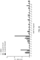
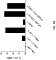
- TIL fragment culture (no. 6) from patient 4259 was co-cultured with autologous APCs either (1) electroporated with TMG composed of irrelevant, WT p53, or mutated p53 sequence or (2) pulsed with peptide vehicle (DMSO) or purified (>95% by HPLC) 25-amino acid peptides composed of WT p53-Y220 sequence or mutated p53-Y220C sequence. Co-cultures were performed overnight at 37 °C. Expression of 4-1BB was evaluated by flow cytometry after gating for lymphocytes ⁇ living cells (PI negative) ⁇ CD3+ (T cells). The results are shown in Figure 49 .
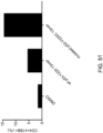
- Autologous antigen presenting cells were pulsed with DMSO, WT p53-Y220 peptide RNTFRHSVVVPYE (SEQ ID NO: 533) or mutated p53-Y220C peptide RNTFRHSVVVPCE (SEQ ID NO: 534) for 2 hours at 37 °C. Excess peptide was washed away. TIL from patient 4259 (fragment culture 6) with specificity to p53-Y220C was co-cultured overnight at 37 °C with peptide-pulsed APCs. Expression of 4-1BB was evaluated by flow cytometry after gating for lymphocytes ⁇ living cells (PI negative) ⁇ CD3+ (T cells). The results are shown in Figure 51 .
- Cos7 cells (2.5 ⁇ 10 4 per well) were plated on wells of flat-bottom 96 well plates. The following day, cells were co-transfected with individual HLA alleles from patient 4259. The next day, DMSO or the p53-Y220C peptide RNTFRHSVWPCE (SEQ ID NO: 534) were pulsed for 2 hours on transfected Cos7 cells. Excess peptide was washed away. TIL fragment culture no.6 from Patient 4259 was added (10 5 cells/well). Co-cultures were incubated overnight at 37 °C. Expression of 4-1BB was assayed by flow cytometry after gating lymphocytes ⁇ live ⁇ CD3+ (T cells) ⁇ CD8-CD4+. The results are shown in Figure 52 .
- APCs autologous to Patient 4259 were pulsed with 25-amino acid peptides corresponding to the WT p53-Y220 or mutated p53-Y220C sequence for 2 hours at 37 °C. Excess peptide was washed away. T cells expressing 4259-F6-TCR were co-cultured overnight at 37 °C with peptide-pulsed APCs. Expression of 4-1BB was evaluated by flow cytometry after gating for lymphocytes ⁇ living cells (PI negative) ⁇ CD3+ (T cells). The introduced TCR was measured by mouse TCRbeta (mTCR). The results are shown in Table 5.
- TC tumor cell
- T#4259 was co-cultured with T cells (10 5 ) expressing mock (no TCR) or p53-Y220C-specific TCR (4259-F6-TCR) overnight at 37 °C.
- the TC#4259 cells were either incubated with nothing, W6/32 pan-HLA Class-I specific blocking antibody, IVA12 pan-HLA Class-II specific blocking antibody or mutated p53-Y220C peptide RNTFRHSVWPCE (SEQ ID NO: 534) for 2 hours at 37 °C.
- the antibodies were kept in the co-culture at 5 ⁇ g/mL final concentration.
- the peptide was incubated at 10 ⁇ g/mL and excess peptide was washed after incubation.
- Media alone (no TC) and PMA and lonomycin were negative and positive controls, respectively.
- Expression of 4-1 BB was evaluated by flow cytometry after gating for lymphocytes ⁇ living cells (PI negative) ⁇ CD3+ (T cells). The results are shown in Figure 53 .
- TCR 4259-F6-TCR The sequence of TCR 4259-F6-TCR, which was isolated from Patient 4259, is set forth below. Starting from the amino terminus, the first underlined region is the CDR1alpha (SEQ ID NO: 477), the second underlined region is the CDR2alpha (SEQ ID NO: 478), the third underlined region is the CDR3alpha (SEQ ID NO: 479), the fourth underlined region is the CDR1beta (SEQ ID NO: 480), the fifth underlined region is the CDR2beta (SEQ ID NO: 481), and the sixth underlined region is the CDR3beta (SEQ ID NO: 482). The bold region is the linker (SEQ ID NO: 26).
- the first italicized region is the alpha chain constant region (SEQ ID NO: 23) and the second italicized region is the beta chain constant region (SEQ ID NO: 25).
- the alpha chain variable region (SEQ ID NO: 483) includes the sequence starting from the amino terminus and ending immediately prior to the start of the alpha chain constant region.
- the beta chain variable region (SEQ ID NO: 484) includes the sequence starting immediately after the linker and ending immediately prior to the start of the beta chain constant region.
- the full-length alpha chain (SEQ ID NO: 485) includes the sequence starting from the amino terminus and ending immediately prior to the start of the linker.
- the full-length beta chain (SEQ ID NO: 486) includes the sequence starting immediately after the linker and ending with the carboxyl terminus.
- TCR Cancer reactive T cells were identified as described below. The TCR was isolated as described below.
- This example demonstrates the identification of anti-mutated p53 T cells in Patient 4127 by co-culturing autologous APCs induced to express mutated p53 within autologous T cells ("p53 hotspot mutation universal screening").
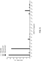
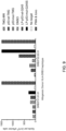
- TIL from patient 4127 were co-cultured with allogeneic (DRB3*01:01:01 or DRB3*02:02:01) APCs which were (1) electroporated with TMGs composed of irrelevant, WT p53 or mutated p53 sequences or (2) pulsed with peptide vehicle (DMSO) or purified (>95% by HPLC) 25 amino acid peptides composed of WT p53-G245 sequence or mutated p53-G245S sequence. Co-cultures were performed overnight at 37 °C. Secretion of IFN- ⁇ was evaluated using ELISPOT assay. The results are shown in Figure 9 .
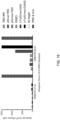
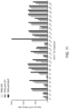
- T cells expressing the 4127-TCR1 specific for p53-G245S were co-cultured with allogeneic (DRB3*01:01:01 or DRB3*02:02:01) APCs which were (1) electroporated with TMGs composed of irrelevant, WT p53 or mutated p53 sequences or (2) pulsed with peptide vehicle (DMSO) or purified (>95% by HPLC) 25 amino acid peptides composed of WT p53-G245 sequence or mutated p53-G245S sequence. Co-cultures were performed overnight at 37 °C. Secretion of IFN- ⁇ was evaluated using ELISPOT assay. The results are shown in Figure 10 .
- T cells from 27 patients were screened by (i) inducing autologous antigen presenting cells (APCs) of a patient to present at least one mutated p53 amino acid sequence; (ii) co-culturing autologous T cells of the patient with the autologous APCs that present the mutated p53 amino acid sequence; and (iii) selecting the autologous T cells that (a) were co-cultured with the autologous APCs that present the mutated p53 amino acid sequence.
- a summary of responses to p53 "hostpot" mutations by T cells using the screening method is provided in Table 7.
- This example demonstrates the identification of anti-mutated p53 T cells in Patient 4273 by co-culturing autologous APCs induced to express mutated p53 within autologous T cells ("p53 hotspot mutation universal screening"). This example also demonstrates the isolation of two anti-mutated p53 TCRs from patient 4273.
- F15 was the most reactive fragment and the responses by F15 to the LP and TMG were comparable and primarily by CD4 T cells.
- So 4273-F15 was co-cultured with APCs pulsed with the R248W LP, and the following day we sorted CD4+41BB+ T cells as single cells into wells a 96 well PCR plate at one cell per well.
- the PCR plate has an RT-PCR solution in each well, which amplifies the TCR alpha and beta CDR3 regions in the same solution.
- the wells of the plate are then split into two 96 well PCR plates and a second PCR round is performed to amplify either the CDR3 alpha or the CDR3 beta as separate reactions.
- the PCR products from each well are sequenced by Sanger sequencing.
- the variable family is determined and fused to the CDR3 and junction (J or DJ) from the translated sequence.
- variable sequence is fused to the murine constant sequence and the reconstructed TCRalpha and TCRbeta are linked by furin-felxible-P2A (RAKR-SGSG-ATNFSLLKQAGDVEENPGP) (SEQ ID NO: 26). Then the sequence is synthesized into DNA de novo and cloned into an expression vector (gamma-retrovirus or SLEEPING BEAUTY (SB) transposon (University of Minnesota, Minneapolis, MN)). T cells are then made to express the TCR using the standard viral transduction or non-viral transposition protocols and T cells expressing murinized TCRs (as detected by mouse TCR beta constant chain) are tested against the putative peptide.
- Autologous APCs were pulsed with 25 amino acid peptides corresponding to the WT or mutated p53-R248W neoepitope for 2 hours at 37 °C.
- TIL from patient 4273 fragment culture 15
- specificity to p53-R248W were co-cultured overnight at 37 °C with peptide-pulsed APCs.
- DMSO was peptide vehicle.
- Expression of 4-1BB was evaluated by flow cytometry after gating for lymphocytes ⁇ living cells (PI negative) ⁇ CD3+ (T cells). The results are shown in Figure 34 .
- Cos7 cells (2.5 ⁇ 10 4 per well) were plated on wells of flat-bottom 96 well plates. The following day, cells were co-transfected with individual HLA alleles from patient 4273 and either WT or mutated TP53 TMG with or without the p53-R248W neoantigen, respectively. The following day, TIL with specificity to p53-R248W from Patient 4273 (fragment culture 15) were co-cultured with transfected Cos7 cells overnight at 37 °C. Secretion of IFN- ⁇ was evaluated by ELISA. The results are shown in Figure 36 .
- T cells expressing mock (no TCR) or 4273-TCR1a2 were co-cultured with autologous APCs which were pulsed with peptide vehicle (DMSO) or purified (>95% by HPLC) 25 amino acid peptides composed of WT p53-R248 sequence or mutated p53-R248W sequence. Media alone and PMA and lonomycin were negative and positive controls, respectively. Co-cultures were performed overnight at 37 °C. Secretion of IFN- ⁇ was evaluated by ELISPOT. The results are shown in Figure 37 .
- the sequence of TCR 4273-TP53-R248W-TCR1a1, which was isolated from Patient 4273, is set forth below. Starting from the amino terminus, the first underlined region is the CDR1alpha (SEQ ID NO: 437), the second underlined region is the CDR2alpha (SEQ ID NO: 438), the third underlined region is the CDR3alpha (SEQ ID NO: 439), the fourth underlined region is the CDR1beta (SEQ ID NO: 440), the fifth underlined region is the CDR2beta (SEQ ID NO: 441), and the sixth underlined region is the CDR3beta (SEQ ID NO: 442). The bold region is the linker (SEQ ID NO: 26).
- the first italicized region is the alpha chain constant region (SEQ ID NO: 23) and the second italicized region is the beta chain constant region (SEQ ID NO: 25).
- the alpha chain variable region (SEQ ID NO: 443) includes the sequence starting from the amino terminus and ending immediately prior to the start of the alpha chain constant region.
- the beta chain variable region (SEQ ID NO: 444) includes the sequence starting immediately after the linker and ending immediately prior to the start of the beta chain constant region.
- the full-length alpha chain (SEQ ID NO: 445) includes the sequence starting from the amino terminus and ending immediately prior to the start of the linker.
- the full-length beta chain (SEQ ID NO: 446) includes the sequence starting immediately after the linker and ending with the carboxyl terminus.
- the sequence of TCR 4273- TP53-R248W- TCR 1 a2, which was isolated from Patient 4273, is set forth below. Starting from the amino terminus, the first underlined region is the CDR1alpha (SEQ ID NO: 447), the second underlined region is the CDR2alpha (SEQ ID NO: 448), the third underlined region is the CDR3alpha (SEQ ID NO: 449), the fourth underlined region is the CDR1beta (SEQ ID NO: 450), the fifth underlined region is the CDR2beta (SEQ ID NO: 451), and the sixth underlined region is the CDR3beta (SEQ ID NO: 452).
- the bold region is the linker (SEQ ID NO: 26). Starting from the amino terminus, the first italicized region is the alpha chain constant region (SEQ ID NO: 23) and the second italicized region is the beta chain constant region (SEQ ID NO: 25).
- the alpha chain variable region (SEQ ID NO: 453) includes the sequence starting from the amino terminus and ending immediately prior to the start of the alpha chain constant region.
- the beta chain variable region (SEQ ID NO: 454) includes the sequence starting immediately after the linker and ending immediately prior to the start of the beta chain constant region.
- the full-length alpha chain (SEQ ID NO: 455) includes the sequence starting from the amino terminus and ending immediately prior to the start of the linker.
- the full-length beta chain (SEQ ID NO: 456) includes the sequence starting immediately after the linker and ending with the carboxyl terminus.
- the statistics for the TCRs of Patient 4273 are set forth in Table 8 below.
- Table 8 96 total wells sorted with 41BB+ T cells after co-culture with mutated p53 protein (TMG or peptide). 77 wells had productive pairs (meaning had (1) a sequence and (2) no stop codons in the sequence) for a pairing frequency of 80.2%. 43 of those pairs were the CDR3A/CDR3B combination to make 4273-TP53-R248W-TCR1a1 (55.8% of the productive pairs). 30 of those pairs were the CDR3A/CDR3B combination to make 4273-TP53-R248W-TCR1a2 (39% of the productive pairs).
- a TCR (4149-TCRa2b1 or 4149-TCRa2b2) was transposed into autologous PBL from patient 4149 and co-cultured with autologous APCs which were (1) electroporated with TMG composed of irrelevant, WT p53, or mutated p53 sequence or (2) pulsed with peptide vehicle (DMSO) or purified (>95% by HPLC) 25-amino acid peptides composed of WT p53-Y220 sequence or mutated p53-Y220C sequence. Combination of PMA and ionomycin was positive control. Co-cultures were performed overnight at 37 °C. Secretion of IFN- ⁇ was evaluated using ELISPOT assay. The results are shown in Figure 15 .
- ELISPOT Result Positive (+) or negative (-) for IFN- ⁇ production Peptide SEQ ID NO: 0 - Vehicle Not applicable 1 + DRNTFRHSVWP C EP 508 2 + RNTFRHSVWP C EPP 509 3 + NTFRHSVWP C EPPE 510 4 + TFRHSVVVP C EPPEV 511 5 + (weak) FRHSVWP C EPPEVG 512 6 - RHSVWP C EPPEVGS 513 7 - HSWVP C EPPEVGSD 514 8 - SVWP C EPPEVGSDC 515 9 - WVP C EPPEVGSDCT 516 10 - WP C EPPEVGSDCTT 517 11 - VP C EPPEVGSDCTTI 518
- Cos7 cells (2.5 ⁇ 10 4 per well) were plated on wells of flat-bottom 96 well plates. After 20 hours, cells were co-transfected with individual HLA alleles with or without TMGs. After 20 hours, autologous DC cells were transfected with TMG in parallel. All HLA Class-II alleles were co-transfected into one set of wells with or without TMG. Cells not transfected with TMG were pulsed with p53-Y220C 15-mer peptide for 2-3 hours at 37 °C at 10 ⁇ g/mL. After washing, 4149-TCRa2b2-transposed T cells (10 5 ) at day+14 of second REP were added to wells and co-cultured overnight at 37 °C.
- DRB3*02 expression was detected in 1367 of 3719 (37%) of DRB_ typed patients in the NCI HLA database and in 5 of 9 (56%) endometrial and ovarian cancer patients at NCI-SB.
- the reported frequency of the DRB3*02 allele is very high.
- TCR 4149TCRa2b2 The sequence of TCR 4149TCRa2b2, which was isolated from Patient 4149, is set forth below. Starting from the amino terminus, the first underlined region is the CDR1alpha (SEQ ID NO: 57), the second underlined region is the CDR2alpha (SEQ ID NO: 58), the third underlined region is the CDR3alpha (SEQ ID NO: 59), the fourth underlined region is the CDR1beta (SEQ ID NO: 60), the fifth underlined region is the CDR2beta (SEQ ID NO: 61), and the sixth underlined region is the CDR3beta (SEQ ID NO: 62). The bold region is the linker (SEQ ID NO: 26).
- the first italicized region is the alpha chain constant region (SEQ ID NO: 23) and the second italicized region is the beta chain constant region (SEQ ID NO: 25).
- the alpha chain variable region (SEQ ID NO: 63) includes the sequence starting from the amino terminus and ending immediately prior to the start of the alpha chain constant region.
- the beta chain variable region (SEQ ID NO: 64) includes the sequence starting immediately after the linker and ending immediately prior to the start of the beta chain constant region.
- the full-length alpha chain (SEQ ID NO: 65) includes the sequence starting from the amino terminus and ending immediately prior to the start of the linker.
- the full-length beta chain (SEQ ID NO: 66) includes the sequence starting immediately after the linker and ending with the carboxyl terminus.
- Cancer reactive T cells were identified using the screening method set forth below.
- the p53 reactive cells for this patient were identified by the method described in U.S. Application No. 2017/0224800 ("Tran method").
- This example demonstrates the identification of anti-mutated p53 T cells in Patient 4213 by co-culturing autologous APCs induced to express mutated p53 within autologous T cells ("p53 hotspot mutation universal screening"). This example also demonstrates the isolation of twelve anti-mutated p53 TCRs from patient 4213.
- TIL fragments (F2 and F24) from patient 4213 were co-cultured with autologous APCs pulsed with peptide vehicle (DMSO) or purified (>95% by HPLC) 25-amino acid peptides composed of the mutated p53-R248Q sequence. Co-cultures were performed overnight at 37 °C. Expression of 4-1BB was evaluated by flow cytometry after gating for lymphocytes -> living cells (PI negative) -> CD3+ (T cells). The results are shown in Figure 19 . CD8+4-1BB+ T cells were sorted into wells of 96 wells plates. TCRs were identified using single-cell RT-PCR.
- CD4+ T cells came from patient 4213's peripheral blood lymphocytes.
- the CD4+ T cell culture was co-cultured with autologous APCs pulsed with peptide vehicle (DMSO) or purified (>95% by HPLC) 25-amino acid peptides composed of the mutated p53-R248Q sequence. Co-cultures were performed overnight at 37 °C. Secretion of IFN- ⁇ was evaluated by ELISPOT. Expression of 4-1BB was evaluated by flow cytometry after gating for lymphocytes ⁇ living cells (PI negative) ⁇ CD3+ (T cells). The results are shown in Figure 20 .
- CD4+41BB+ T cells were sorted into wells of 96 wells plates. TCRs were identified using single-cell RT-PCR.
- the sequence of 4213-F2-TCR1, which was isolated from Patient 4213, is set forth below. Starting from the amino terminus, the first underlined region is the CDR1alpha (SEQ ID NO: 317), the second underlined region is the CDR2alpha (SEQ ID NO: 318), the third underlined region is the CDR3alpha (SEQ ID NO: 319), the fourth underlined region is the CDR1beta (SEQ ID NO: 320), the fifth underlined region is the CDR2beta (SEQ ID NO: 321), and the sixth underlined region is the CDR3beta (SEQ ID NO: 322). The bold region is the linker (SEQ ID NO: 26).
- Cancer reactive T cells were identified using the screening method set forth below. The method used to isolate the TCR is set forth below.
- TCR 4213-F2-TCR1 of Patient 4213 are set forth in Table 13 below.
- Table 13 Parameter # Frequency Total wells 96 100% CDR3alpha 3 3.1% CDR3beta 8 8.3% 4213-F2-TCR1 pairs 2 2.1% Total paired TCRs 24 25.0%
- the sequence of 4213-F2-TCR2, which was isolated from Patient 4213, is set forth below. Starting from the amino terminus, the first underlined region is the CDR1alpha (SEQ ID NO: 327), the second underlined region is the CDR2alpha (SEQ ID NO: 328), the third underlined region is the CDR3alpha (SEQ ID NO: 329), the fourth underlined region is the CDR1beta (SEQ ID NO: 330), the fifth underlined region is the CDR2beta (SEQ ID NO: 331), and the sixth underlined region is the CDR3beta (SEQ ID NO: 332). The bold region is the linker (SEQ ID NO: 26).
- the first italicized region is the alpha chain constant region (SEQ ID NO: 23) and the second italicized region is the beta chain constant region (SEQ ID NO: 25).
- the alpha chain variable region (SEQ ID NO: 333) includes the sequence starting from the amino terminus and ending immediately prior to the start of the alpha chain constant region.
- the beta chain variable region (SEQ ID NO: 334) includes the sequence starting immediately after the linker and ending immediately prior to the start of the beta chain constant region.
- the full-length alpha chain (SEQ ID NO: 335) includes the sequence starting from the amino terminus and ending immediately prior to the start of the linker.
- the full-length beta chain (SEQ ID NO: 336) includes the sequence starting immediately after the linker and ending with the carboxyl terminus.
- Cancer reactive T cells were identified using the screening method set forth below. The method used to isolate the TCR is set forth below.
- the sequence of 4213-F2-TCR3, which was isolated from Patient 4213, is set forth below. Starting from the amino terminus, the first underlined region is the CDR1alpha (SEQ ID NO: 337), the second underlined region is the CDR2alpha (SEQ ID NO: 338), the third underlined region is the CDR3alpha (SEQ ID NO: 339), the fourth underlined region is the CDR1beta (SEQ ID NO: 340), the fifth underlined region is the CDR2beta (SEQ ID NO: 341), and the sixth underlined region is the CDR3beta (SEQ ID NO: 342). The bold region is the linker (SEQ ID NO: 26).
- the first italicized region is the alpha chain constant region (SEQ ID NO: 23) and the second italicized region is the beta chain constant region (SEQ ID NO: 25).
- the alpha chain variable region (SEQ ID NO: 343) includes the sequence starting from the amino terminus and ending immediately prior to the start of the alpha chain constant region.
- the beta chain variable region (SEQ ID NO: 344) includes the sequence starting immediately after the linker and ending immediately prior to the start of the beta chain constant region.
- the full-length alpha chain (SEQ ID NO: 345) includes the sequence starting from the amino terminus and ending immediately prior to the start of the linker.
- the full-length beta chain (SEQ ID NO: 346) includes the sequence starting immediately after the linker and ending with the carboxyl terminus.
- Cancer reactive T cells were identified using the screening method set forth below. The method used to isolate the TCR is set forth below.
- TCR 4213-F2-TCR3 of Patient 4213 are set forth in Table 15 below.
- Table 15 Parameter # Frequency Total wells 96 100% CDR3alpha 19 19.8% CDR3beta 42 43.8% 4213-F2-TCR3 pairs 17 17.7% Total paired TCRs 24 25.0%
- the sequence of 4213-F24-TCRa1, which was isolated from Patient 4213, is set forth below. Starting from the amino terminus, the first underlined region is the CDR1alpha (SEQ ID NO: 347), the second underlined region is the CDR2alpha (SEQ ID NO: 348), the third underlined region is the CDR3alpha (SEQ ID NO: 349), the fourth underlined region is the CDR1beta (SEQ ID NO: 350), the fifth underlined region is the CDR2beta (SEQ ID NO: 351), and the sixth underlined region is the CDR3beta (SEQ ID NO: 352). The bold region is the linker (SEQ ID NO: 26).
- the first italicized region is the alpha chain constant region (SEQ ID NO: 23) and the second italicized region is the beta chain constant region (SEQ ID NO: 25).
- the alpha chain variable region (SEQ ID NO: 353) includes the sequence starting from the amino terminus and ending immediately prior to the start of the alpha chain constant region.
- the beta chain variable region (SEQ ID NO: 354) includes the sequence starting immediately after the linker and ending immediately prior to the start of the beta chain constant region.
- the full-length alpha chain (SEQ ID NO: 355) includes the sequence starting from the amino terminus and ending immediately prior to the start of the linker.
- the full-length beta chain (SEQ ID NO: 356) includes the sequence starting immediately after the linker and ending with the carboxyl terminus.
- Cancer reactive T cells were identified using the screening method set forth below. The method used to isolate the TCR is set forth below.
- the first italicized region is the alpha chain constant region (SEQ ID NO: 23) and the second italicized region is the beta chain constant region (SEQ ID NO: 25).
- the alpha chain variable region (SEQ ID NO: 363) includes the sequence starting from the amino terminus and ending immediately prior to the start of the alpha chain constant region.
- the beta chain variable region (SEQ ID NO: 364) includes the sequence starting immediately after the linker and ending immediately prior to the start of the beta chain constant region.
- the full-length alpha chain (SEQ ID NO: 365) includes the sequence starting from the amino terminus and ending immediately prior to the start of the linker.
- the full-length beta chain (SEQ ID NO: 366) includes the sequence starting immediately after the linker and ending with the carboxyl terminus.
- Cancer reactive T cells were identified using the screening method set forth below. The method used to isolate the TCR is set forth below.
- TCR 4213-F24-TCRa2 of Patient 4213 are set forth in Table 17 below.
- Table 17 Parameter # Frequency Total wells 96 100% CDR3alpha 39 40.6% CDR3beta 80 83.3% 4213-F24-TCRa2 pairs 37 38.5% Total paired TCRs 44 45.8%
- the sequence of 4213-PBL-TCR1, which was isolated from Patient 4213, is set forth below. Starting from the amino terminus, the first underlined region is the CDR1alpha (SEQ ID NO: 367), the second underlined region is the CDR2alpha (SEQ ID NO: 368), the third underlined region is the CDR3alpha (SEQ ID NO: 369), the fourth underlined region is the CDR1beta (SEQ ID NO: 370), the fifth underlined region is the CDR2beta (SEQ ID NO: 371), and the sixth underlined region is the CDR3beta (SEQ ID NO: 372). The bold region is the linker (SEQ ID NO: 26).
- the first italicized region is the alpha chain constant region (SEQ ID NO: 23) and the second italicized region is the beta chain constant region (SEQ ID NO: 25).
- the alpha chain variable region (SEQ ID NO: 373) includes the sequence starting from the amino terminus and ending immediately prior to the start of the alpha chain constant region.
- the beta chain variable region (SEQ ID NO: 374) includes the sequence starting immediately after the linker and ending immediately prior to the start of the beta chain constant region.
- the full-length alpha chain (SEQ ID NO: 375) includes the sequence starting from the amino terminus and ending immediately prior to the start of the linker.
- the full-length beta chain (SEQ ID NO: 376) includes the sequence starting immediately after the linker and ending with the carboxyl terminus.
- Cancer reactive T cells were identified using the screening method set forth below. The method used to isolate the TCR is set forth below.
- TCR 4213-PBL-TCR1 of Patient 4213 are set forth in Table 18 below.
- TABLE 18 Parameter # Frequency Total wells 192 100% CDR3alpha 9 4.7% CDR3beta 6 3.1% 4213-PBL-TCR1 pairs 6 3.1% Total paired TCRs 63 32.8%
- the sequence of 4213-PBL-TCR2, which was isolated from Patient 4213, is set forth below. Starting from the amino terminus, the first underlined region is the CDR1 alpha (SEQ ID NO: 377), the second underlined region is the CDR2alpha (SEQ ID NO: 378), the third underlined region is the CDR3alpha (SEQ ID NO: 379), the fourth underlined region is the CDR1beta (SEQ ID NO: 380), the fifth underlined region is the CDR2beta (SEQ ID NO: 381), and the sixth underlined region is the CDR3beta (SEQ ID NO: 382). The bold region is the linker (SEQ ID NO: 26).
- the first italicized region is the alpha chain constant region (SEQ ID NO: 23) and the second italicized region is the beta chain constant region (SEQ ID NO: 25).
- the alpha chain variable region (SEQ ID NO: 383) includes the sequence starting from the amino terminus and ending immediately prior to the start of the alpha chain constant region.
- the beta chain variable region (SEQ ID NO: 384) includes the sequence starting immediately after the linker and ending immediately prior to the start of the beta chain constant region.
- the full-length alpha chain (SEQ ID NO: 385) includes the sequence starting from the amino terminus and ending immediately prior to the start of the linker.
- the full-length beta chain (SEQ ID NO: 386) includes the sequence starting immediately after the linker and ending with the carboxyl terminus.
- Cancer reactive T cells were identified using the screening method set forth below. The method used to isolate the TCR is set forth below.
- TCR 4213-PBL-TCR2 of Patient 4213 are set forth in Table 19 below.
- TABLE 19 Parameter # Frequency Total wells 192 100% CDR3alpha 8 4.2% CDR3beta 8 4.2% 4213-PBL-TCR2 pairs 5 2.6% Total paired TCRs 63 32.8%
- the sequence of 4213-PBL-TCR3, which was isolated from Patient 4213, is set forth below. Starting from the amino terminus, the first underlined region is the CDR1alpha (SEQ ID NO: 387), the second underlined region is the CDR2alpha (SEQ ID NO: 388), the third underlined region is the CDR3alpha (SEQ ID NO: 389), the fourth underlined region is the CDR1beta (SEQ ID NO: 390), the fifth underlined region is the CDR2beta (SEQ ID NO: 391), and the sixth underlined region is the CDR3beta (SEQ ID NO: 392). The bold region is the linker (SEQ ID NO: 26).
- the first italicized region is the alpha chain constant region (SEQ ID NO: 23) and the second italicized region is the beta chain constant region (SEQ ID NO: 25).
- the alpha chain variable region (SEQ ID NO: 393) includes the sequence starting from the amino terminus and ending immediately prior to the start of the alpha chain constant region.
- the beta chain variable region (SEQ ID NO: 394) includes the sequence starting immediately after the linker and ending immediately prior to the start of the beta chain constant region.
- the full-length alpha chain (SEQ ID NO: 395) includes the sequence starting from the amino terminus and ending immediately prior to the start of the linker.
- the full-length beta chain (SEQ ID NO: 396) includes the sequence starting immediately after the linker and ending with the carboxyl terminus.
- Cancer reactive T cells were identified using the screening method set forth below. The method used to isolate the TCR is set forth below.
- TCR 4213-PBL-TCR3 of Patient 4213 are set forth in Table 20 below.
- Table 20 The statistics for TCR 4213-PBL-TCR3 of Patient 4213 are set forth in Table 20 below. TABLE 20 Parameter # Frequency Total wells 192 100% CDR3alpha 4 2.1% CDR3beta 7 3.6% 4213-PBL-TCR3 pairs 4 2.1% Total paired TCRs 63 32.8%
- the sequence of 4213-PBL-TCR4a1, which was isolated from Patient 4213, is set forth below. Starting from the amino terminus, the first underlined region is the CDR1alpha (SEQ ID NO: 397), the second underlined region is the CDR2alpha (SEQ ID NO: 398), the third underlined region is the CDR3alpha (SEQ ID NO: 399), the fourth underlined region is the CDR1beta (SEQ ID NO: 400), the fifth underlined region is the CDR2beta (SEQ ID NO: 401), and the sixth underlined region is the CDR3beta (SEQ ID NO: 402). The bold region is the linker (SEQ ID NO: 26).
- Cancer reactive T cells were identified using the screening method set forth below. The method used to isolate the TCR is set forth below.
- TCR 4213-PBL-TCR4a1 of Patient 4213 are set forth in Table 21 below.
- TABLE 21 Parameter # Frequency Total wells 192 100% CDR3alpha 2 1.0% CDR3beta 36 18.8% 4213-PBL-TCR4a1 pairs 2 1.0% Total paired TCRs 63 32.8%
- the sequence of 4213-PBL-TCR4a2, which was isolated from Patient 4213, is set forth below. Starting from the amino terminus, the first underlined region is the CDR1alpha (SEQ ID NO: 407), the second underlined region is the CDR2alpha (SEQ ID NO: 408), the third underlined region is the CDR3alpha (SEQ ID NO: 409), the fourth underlined region is the CDR1beta (SEQ ID NO: 410), the fifth underlined region is the CDR2beta (SEQ ID NO: 411), and the sixth underlined region is the CDR3beta (SEQ ID NO: 412). The bold region is the linker (SEQ ID NO: 26).
- the first italicized region is the alpha chain constant region (SEQ ID NO: 23) and the second italicized region is the beta chain constant region (SEQ ID NO: 25).
- the alpha chain variable region (SEQ ID NO: 413) includes the sequence starting from the amino terminus and ending immediately prior to the start of the alpha chain constant region.
- the beta chain variable region (SEQ ID NO: 414) includes the sequence starting immediately after the linker and ending immediately prior to the start of the beta chain constant region.
- the full-length alpha chain (SEQ ID NO: 415) includes the sequence starting from the amino terminus and ending immediately prior to the start of the linker.
- the full-length beta chain (SEQ ID NO: 416) includes the sequence starting immediately after the linker and ending with the carboxyl terminus.
- Cancer reactive T cells were identified using the screening method set forth below. The method used to isolate the TCR is set forth below.
- TCR 4213-PBL-TCR4a2 of Patient 4213 are set forth in Table 22 below.
- TABLE 22 Parameter # Frequency Total wells 192 100% CDR3alpha 2 1.0% CDR3beta 36 18.8% 4213-PBL-TCR4a2 pairs 2 1.0% Total paired TCRs 63 32.8%
- the sequence of 4213-PBL-TCR4a3, which was isolated from Patient 4213, is set forth below. Starting from the amino terminus, the first underlined region is the CDR1alpha (SEQ ID NO: 417), the second underlined region is the CDR2alpha (SEQ ID NO: 418), the third underlined region is the CDR3alpha (SEQ ID NO: 419), the fourth underlined region is the CDR1beta (SEQ ID NO: 420), the fifth underlined region is the CDR2beta (SEQ ID NO: 421), and the sixth underlined region is the CDR3beta (SEQ ID NO: 422). The bold region is the linker (SEQ ID NO: 26).
- the first italicized region is the alpha chain constant region (SEQ ID NO: 23) and the second italicized region is the beta chain constant region (SEQ ID NO: 25).
- the alpha chain variable region (SEQ ID NO: 423) includes the sequence starting from the amino terminus and ending immediately prior to the start of the alpha chain constant region.
- the beta chain variable region (SEQ ID NO: 424) includes the sequence starting immediately after the linker and ending immediately prior to the start of the beta chain constant region.
- the full-length alpha chain (SEQ ID NO: 425) includes the sequence starting from the amino terminus and ending immediately prior to the start of the linker.
- the full-length beta chain (SEQ ID NO: 426) includes the sequence starting immediately after the linker and ending with the carboxyl terminus.
- Cancer reactive T cells were identified using the screening method set forth below. The method used to isolate the TCR is set forth below.
- TCR 4213-PBL-TCR4a3 of Patient 4213 are set forth in Table 23 below.
- TABLE 23 Parameter # Frequency Total wells 192 100% CDR3alpha 4 2.1% CDR3beta 36 18.8% 4213-PBL-TCR4a3 pairs 3 1.6% Total paired TCRs 63 32.8%
- the sequence of 4213-PBL-TCR4a4, which was isolated from Patient 4213, is set forth below. Starting from the amino terminus, the first underlined region is the CDR1alpha (SEQ ID NO: 427), the second underlined region is the CDR2alpha (SEQ ID NO: 428), the third underlined region is the CDR3alpha (SEQ ID NO: 429), the fourth underlined region is the CDR1beta (SEQ ID NO: 430), the fifth underlined region is the CDR2beta (SEQ ID NO: 431), and the sixth underlined region is the CDR3beta (SEQ ID NO: 432). The bold region is the linker (SEQ ID NO: 26).
- the first italicized region is the alpha chain constant region (SEQ ID NO: 23) and the second italicized region is the beta chain constant region (SEQ ID NO: 25).
- the alpha chain variable region (SEQ ID NO: 433) includes the sequence starting from the amino terminus and ending immediately prior to the start of the alpha chain constant region.
- the beta chain variable region (SEQ ID NO: 434) includes the sequence starting immediately after the linker and ending immediately prior to the start of the beta chain constant region.
- the full-length alpha chain (SEQ ID NO: 435) includes the sequence starting from the amino terminus and ending immediately prior to the start of the linker.
- the full-length beta chain (SEQ ID NO: 436) includes the sequence starting immediately after the linker and ending with the carboxyl terminus.
- Cancer reactive T cells were identified using the screening method set forth below. The method used to isolate the TCR is set forth below.
- TCR 4213-PBL-TCR4a4 of Patient 4213 are set forth in Table 24 below.
- TABLE 24 Parameter # Frequency Total wells 192 100% CDR3alpha 2 1.0% CDR3beta 36 18.8% 4213-PBL-TCR4a3 pairs 2 1.0% Total paired TCRs 63 32.8%
- This example demonstrates the identification of anti-mutated p53 T cells in Patient 4268 by co-culturing autologous APCs induced to express mutated p53 within autologous T cells ("p53 hotspot mutation universal screening"). This example also demonstrates the isolation of five anti-mutated p53 TCRs from patient 4268.
- DMSO peptide vehicle
- HPLC purified 25-amino acid peptides composed of wt p53-R248 sequence or mutated p
- the sequence of 4268-TCR1, which was isolated from Patient 4268, is set forth below. Starting from the amino terminus, the first underlined region is the CDR1alpha (SEQ ID NO: 137), the second underlined region is the CDR2alpha (SEQ ID NO: 138), the third underlined region is the CDR3alpha (SEQ ID NO: 139), the fourth underlined region is the CDR1beta (SEQ ID NO: 140), the fifth underlined region is the CDR2beta (SEQ ID NO: 141), and the sixth underlined region is the CDR3beta (SEQ ID NO: 142). The bold region is the linker (SEQ ID NO: 26).
- the first italicized region is the alpha chain constant region (SEQ ID NO: 23) and the second italicized region is the beta chain constant region (SEQ ID NO: 25).
- the alpha chain variable region (SEQ ID NO: 143) includes the sequence starting from the amino terminus and ending immediately prior to the start of the alpha chain constant region.
- the beta chain variable region (SEQ ID NO: 144) includes the sequence starting immediately after the linker and ending immediately prior to the start of the beta chain constant region.
- the full-length alpha chain (SEQ ID NO: 145) includes the sequence starting from the amino terminus and ending immediately prior to the start of the linker.
- the full-length beta chain (SEQ ID NO: 146) includes the sequence starting immediately after the linker and ending with the carboxyl terminus.
- Cancer reactive T cells were identified using the screening method set forth below. The method used to isolate the TCR is set forth below.
- TCR 4268-TCR1 of Patient 4268 are set forth in Table 25 below.
- Table 25 The statistics for TCR 4268-TCR1 of Patient 4268 are set forth in Table 25 below.
- TABLE 25 Parameter # Frequency Total wells 96 100% CDR3alpha 54 56.3% CDR3beta 71 74.0% 4268-TCR1 pairs 48 50.0% Total paired TCRs 61 63.5%
- the sequence of 4268-TCR2, which was isolated from Patient 4268, is set forth below. Starting from the amino terminus, the first underlined region is the CDR1alpha (SEQ ID NO: 147), the second underlined region is the CDR2alpha (SEQ ID NO: 148), the third underlined region is the CDR3alpha (SEQ ID NO: 149), the fourth underlined region is the CDR1beta (SEQ ID NO: 150), the fifth underlined region is the CDR2beta (SEQ ID NO: 151), and the sixth underlined region is the CDR3beta (SEQ ID NO: 152). The bold region is the linker (SEQ ID NO: 26).
- the first italicized region is the alpha chain constant region (SEQ ID NO: 23) and the second italicized region is the beta chain constant region (SEQ ID NO: 25).
- the alpha chain variable region (SEQ ID NO: 153) includes the sequence starting from the amino terminus and ending immediately prior to the start of the alpha chain constant region.
- the beta chain variable region (SEQ ID NO: 154) includes the sequence starting immediately after the linker and ending immediately prior to the start of the beta chain constant region.
- the full-length alpha chain (SEQ ID NO: 155) includes the sequence starting from the amino terminus and ending immediately prior to the start of the linker.
- the full-length beta chain (SEQ ID NO: 156) includes the sequence starting immediately after the linker and ending with the carboxyl terminus.
- Cancer reactive T cells were identified using the screening method set forth below. The method used to isolate the TCR is set forth below.
- TCR 4268-TCR2 of Patient 4268 are set forth in Table 26 below.
- Table 26 Parameter # Frequency Total wells 96 100% CDR3alpha 6 6.3% CDR3beta 4 4.2% 4268-TCR2 pairs 4 4.2% Total paired TCRs 61 63.5%
- the sequence of 4268-TCR3, which was isolated from Patient 4268, is set forth below. Starting from the amino terminus, the first underlined region is the CDR1alpha (SEQ ID NO: 157), the second underlined region is the CDR2alpha (SEQ ID NO: 158), the third underlined region is the CDR3alpha (SEQ ID NO: 159), the fourth underlined region is the CDR1beta (SEQ ID NO: 160), the fifth underlined region is the CDR2beta (SEQ ID NO: 161), and the sixth underlined region is the CDR3beta (SEQ ID NO: 162). The bold region is the linker (SEQ ID NO: 26).
- the first italicized region is the alpha chain constant region (SEQ ID NO: 23) and the second italicized region is the beta chain constant region (SEQ ID NO: 25).
- the alpha chain variable region (SEQ ID NO: 163) includes the sequence starting from the amino terminus and ending immediately prior to the start of the alpha chain constant region.
- the beta chain variable region (SEQ ID NO: 164) includes the sequence starting immediately after the linker and ending immediately prior to the start of the beta chain constant region.
- the full-length alpha chain (SEQ ID NO: 165) includes the sequence starting from the amino terminus and ending immediately prior to the start of the linker.
- the full-length beta chain (SEQ ID NO: 166) includes the sequence starting immediately after the linker and ending with the carboxyl terminus.
- Cancer reactive T cells were identified using the screening method set forth below. The method used to isolate the TCR is set forth below.
- TCR 4268-TCR3 of Patient 4268 are set forth in Table 27 below.
- Table 27 Parameter # Frequency Total wells 96 100% CDR3alpha 42 43.8% CDR3beta 37 38.5% 4268-TCR3 pairs 31 32.3% Total paired TCRs 35 36.5%
- the sequence of 4268-TCR4, which was isolated from Patient 4268, is set forth below. Starting from the amino terminus, the first underlined region is the CDR1alpha (SEQ ID NO: 167), the second underlined region is the CDR2alpha (SEQ ID NO: 168), the third underlined region is the CDR3alpha (SEQ ID NO: 169), the fourth underlined region is the CDR1beta (SEQ ID NO: 170), the fifth underlined region is the CDR2beta (SEQ ID NO: 171), and the sixth underlined region is the CDR3beta (SEQ ID NO: 172). The bold region is the linker (SEQ ID NO: 26).
- the first italicized region is the alpha chain constant region (SEQ ID NO: 23) and the second italicized region is the beta chain constant region (SEQ ID NO: 25).
- the alpha chain variable region (SEQ ID NO: 173) includes the sequence starting from the amino terminus and ending immediately prior to the start of the alpha chain constant region.
- the beta chain variable region (SEQ ID NO: 174) includes the sequence starting immediately after the linker and ending immediately prior to the start of the beta chain constant region.
- the full-length alpha chain (SEQ ID NO: 175) includes the sequence starting from the amino terminus and ending immediately prior to the start of the linker.
- the full-length beta chain (SEQ ID NO: 176) includes the sequence starting immediately after the linker and ending with the carboxyl terminus.
- Cancer reactive T cells were identified using the screening method set forth below. The method used to isolate the TCR is set forth below.
- TCR 4268-TCR4 of Patient 4268 are set forth in Table 28 below. TABLE 28 Parameter # Frequency Total wells 96 100% CDR3alpha 43 44.8% CDR3beta 53 55.2% 4268-TCR4 pairs 40 41.7% Total paired TCRs 42 43.8%
- the sequence of 4268-TCR5, which was isolated from Patient 4268, is set forth below. Starting from the amino terminus, the first underlined region is the CDR1alpha (SEQ ID NO: 177), the second underlined region is the CDR2alpha (SEQ ID NO: 178), the third underlined region is the CDR3alpha (SEQ ID NO: 179), the fourth underlined region is the CDR1beta (SEQ ID NO: 180), the fifth underlined region is the CDR2beta (SEQ ID NO: 181), and the sixth underlined region is the CDR3beta (SEQ ID NO: 182). The bold region is the linker (SEQ ID NO: 26).
- the first italicized region is the alpha chain constant region (SEQ ID NO: 23) and the second italicized region is the beta chain constant region (SEQ ID NO: 25).
- the alpha chain variable region (SEQ ID NO: 183) includes the sequence starting from the amino terminus and ending immediately prior to the start of the alpha chain constant region.
- the beta chain variable region (SEQ ID NO: 184) includes the sequence starting immediately after the linker and ending immediately prior to the start of the beta chain constant region.
- the full-length alpha chain (SEQ ID NO: 185) includes the sequence starting from the amino terminus and ending immediately prior to the start of the linker.
- the full-length beta chain (SEQ ID NO: 186) includes the sequence starting immediately after the linker and ending with the carboxyl terminus.
- Cancer reactive T cells were identified using the screening method set forth below. The method used to isolate the TCR is set forth below.
- TCR 4268-TCR5 of Patient 4268 are set forth in Table 29 below.
- TABLE 29 Parameter # Frequency Total wells 96 100% CDR3alpha 2 2.1% CDR3beta 87 90.6% 4268-TCR5 pairs 2 2.1% Total paired TCRs 17 17.7%
- This example demonstrates the identification of anti-mutated p53 T cells in Patient 4266 by co-culturing autologous APCs induced to express mutated p53 within autologous T cells ("p53 hotspot mutation universal screening"). This example also demonstrates the isolation of four anti-mutated p53 TCRs from patient 4266.
- Cos7 cells (2.5 ⁇ 10 4 per well) were plated on wells of flat-bottom 96 well plates. The following day, cells were co-transfected with individual HLA alleles from patient 4266. The next day cells were pulsed with no peptide, DMSO, WT p53-R248 peptide SSCMGGMNRR (SEQ ID NO: 590) or mutated p53-R248W peptide SSCMGGMNWR (SEQ ID NO: 591) for 2 hours at 37 °C at 1 ⁇ g/mL. TIL cultures from from patient 4266 (10 5 ) were added to wells and co-cultured overnight at 37°C. Expression of 4-1BB was evaluated by flow cytometry after gating for lymphocytes ⁇ living cells (PI negative) ⁇ CD3+ (T cells) ⁇ CD4-CD8+. The results are shown in Fig. 29 .
- T cells expressing mock (no TCR), 4266-TCR1, 4266-TCR2, 4266-TCR3 or 4266-TCR4 with putative specificity to p53-R248W identified from 4266-TIL were co-cultured with autologous APCs which were pulsed with peptide vehicle (DMSO) or purified (>95% by HPLC) 25 amino acid peptides composed of WT p53-R248 sequence or mutated p53-R248W sequence. Media alone and PMA and lonomycin were negative and positive controls, respectively. Co-cultures were performed overnight at 37 °C. Expression of 4-1BB was evaluated by flow cytometry after gating for lymphocytes ⁇ living cells (PI negative) ⁇ CD3+ (T cells) ⁇ CD4-CD8+. The results are shown in Fig. 30 .
- a tumor cell (TC) line was established from a xenografted tumor fragment resected from Patient 4266 then serially passaged through immunocompromised mice (TC#4266).
- the TC#4266 was co-cultured with T cells (10 5 ) expressing mock (no TCR) or p53-R248W-specific TCRs (4266-TCR2, 4266-TCR3 or 4266-TCR4) overnight at 37°C.
- the TC#4266 cells were either incubated with nothing, W6/32 pan-HLA Class-I specific blocking antibody, IVA12 pan-HLA Class-II specific blocking antibody or mutated p53-R248W peptide SSCMGGMNWR (SEQ ID NO: 591) for 2 hours at 37 °C.
- the antibodies were kept in the co-culture at 5 ⁇ g/mL final concentration.
- the peptide was incubated at 1 ⁇ g/mL and excess peptide was washed after incubation. Media alone (no TC) and PMA and lonomycin were negative and positive controls, respectively.
- Expression of 4-1BB was evaluated by flow cytometry after gating for lymphocytes ⁇ living cells (PI negative) ⁇ CD3+ (T cells) ⁇ CD4-CD8+. The results are shown in Fig. 31 .
- the sequence of 4266-TCR1, which was isolated from Patient 4266, is set forth below. Starting from the amino terminus, the first underlined region is the CDR1alpha (SEQ ID NO: 97), the second underlined region is the CDR2alpha (SEQ ID NO: 98), the third underlined region is the CDR3alpha (SEQ ID NO: 99), the fourth underlined region is the CDR1beta (SEQ ID NO: 100), the fifth underlined region is the CDR2beta (SEQ ID NO: 101), and the sixth underlined region is the CDR3beta (SEQ ID NO: 102). The bold region is the linker (SEQ ID NO: 26).
- the first italicized region is the alpha chain constant region (SEQ ID NO: 23) and the second italicized region is the beta chain constant region (SEQ ID NO: 25).
- the alpha chain variable region (SEQ ID NO: 103) includes the sequence starting from the amino terminus and ending immediately prior to the start of the alpha chain constant region.
- the beta chain variable region (SEQ ID NO: 104) includes the sequence starting immediately after the linker and ending immediately prior to the start of the beta chain constant region.
- the full-length alpha chain (SEQ ID NO: 105) includes the sequence starting from the amino terminus and ending immediately prior to the start of the linker.
- the full-length beta chain (SEQ ID NO: 106) includes the sequence starting immediately after the linker and ending with the carboxyl terminus.
- Cancer reactive T cells were identified using the screening method set forth below. The method used to isolate the TCR is set forth below.
- TCR 4266-TCR1 of Patient 4266 The statistics for TCR 4266-TCR1 of Patient 4266 are set forth in Table 30 below. TABLE 30 Parameter # Frequency Total wells 96 100% CDR3alpha 9 9.4% CDR3beta 10 10.4% 4266-TCR1 pairs 9 9.4% Total paired TCRs 53 55.2%
- the sequence of 4266-TCR2, which was isolated from Patient 4266, is set forth below. Starting from the amino terminus, the first underlined region is the CDR1alpha (SEQ ID NO: 107), the second underlined region is the CDR2alpha (SEQ ID NO: 108), the third underlined region is the CDR3alpha (SEQ ID NO: 109), the fourth underlined region is the CDR1beta (SEQ ID NO: 110), the fifth underlined region is the CDR2beta (SEQ ID NO: 111), and the sixth underlined region is the CDR3beta (SEQ ID NO: 112). The bold region is the linker (SEQ ID NO: 26).
- the first italicized region is the alpha chain constant region (SEQ ID NO: 23) and the second italicized region is the beta chain constant region (SEQ ID NO: 25).
- the alpha chain variable region (SEQ ID NO: 113) includes the sequence starting from the amino terminus and ending immediately prior to the start of the alpha chain constant region.
- the beta chain variable region (SEQ ID NO: 114) includes the sequence starting immediately after the linker and ending immediately prior to the start of the beta chain constant region.
- the full-length alpha chain (SEQ ID NO: 115) includes the sequence starting from the amino terminus and ending immediately prior to the start of the linker.
- the full-length beta chain (SEQ ID NO: 116) includes the sequence starting immediately after the linker and ending with the carboxyl terminus.
- Cancer reactive T cells were identified using the screening method set forth below. The method used to isolate the TCR is set forth below.
- TCR 4266-TCR2 of Patient 4266 are set forth in Table 31 below.
- Table 31 Parameter # Frequency Total wells 96 100% CDR3alpha 21 21.9% CDR3beta 18 18.8% 4266-TCR2 pairs 13 13.5% Total paired TCRs 53 55.2%
- the sequence of 4266-TCR3, which was isolated from Patient 4266, is set forth below. Starting from the amino terminus, the first underlined region is the CDR1alpha (SEQ ID NO: 117), the second underlined region is the CDR2alpha (SEQ ID NO: 118), the third underlined region is the CDR3alpha (SEQ ID NO: 119), the fourth underlined region is the CDR1beta (SEQ ID NO: 120), the fifth underlined region is the CDR2beta (SEQ ID NO: 121), and the sixth underlined region is the CDR3beta (SEQ ID NO: 122). The bold region is the linker (SEQ ID NO: 26).
- the first italicized region is the alpha chain constant region (SEQ ID NO: 23) and the second italicized region is the beta chain constant region (SEQ ID NO: 25).
- the alpha chain variable region (SEQ ID NO: 123) includes the sequence starting from the amino terminus and ending immediately prior to the start of the alpha chain constant region.
- the beta chain variable region (SEQ ID NO: 124) includes the sequence starting immediately after the linker and ending immediately prior to the start of the beta chain constant region.
- the full-length alpha chain (SEQ ID NO: 125) includes the sequence starting from the amino terminus and ending immediately prior to the start of the linker.
- the full-length beta chain (SEQ ID NO: 126) includes the sequence starting immediately after the linker and ending with the carboxyl terminus.
- Cancer reactive T cells were identified using the screening method set forth below. The method used to isolate the TCR is set forth below.
- TCR 4266-TCR3 of Patient 4266 are set forth in Table 32 below.
- Table 32 Parameter # Frequency Total wells 96 100% CDR3alpha 19 19.8% CDR3beta 26 27.1 4266-TCR3 pairs 18 18.8% Total paired TCRs 53 55.2%
- the sequence of 4266-TCR4, which was isolated from Patient 4266, is set forth below. Starting from the amino terminus, the first underlined region is the CDR1alpha (SEQ ID NO: 127), the second underlined region is the CDR2alpha (SEQ ID NO: 128), the third underlined region is the CDR3alpha (SEQ ID NO: 129), the fourth underlined region is the CDR1beta (SEQ ID NO: 130), the fifth underlined region is the CDR2beta (SEQ ID NO: 131), and the sixth underlined region is the CDR3beta (SEQ ID NO: 132). The bold region is the linker (SEQ ID NO: 26).
- the first italicized region is the alpha chain constant region (SEQ ID NO: 23) and the second italicized region is the beta chain constant region (SEQ ID NO: 25).
- the alpha chain variable region (SEQ ID NO: 133) includes the sequence starting from the amino terminus and ending immediately prior to the start of the alpha chain constant region.
- the beta chain variable region (SEQ ID NO: 134) includes the sequence starting immediately after the linker and ending immediately prior to the start of the beta chain constant region.
- the full-length alpha chain (SEQ ID NO: 135) includes the sequence starting from the amino terminus and ending immediately prior to the start of the linker.
- the full-length beta chain (SEQ ID NO: 136) includes the sequence starting immediately after the linker and ending with the carboxyl terminus.
- Cancer reactive T cells were identified using the screening method set forth below. The method used to isolate the TCR is set forth below.
- TCR 4266-TCR4 of Patient 4266 are set forth in Table 33 below.
- Table 33 Parameter # Frequency Total wells 96 100% CDR3alpha 5 5.2% CDR3beta 13 13.5 4266-TCR3 pairs 5 5.2% Total paired TCRs 53 55.2%
- This example demonstrates the identification of anti-mutated p53 T cells in Patient 4252 by co-culturing autologous APCs induced to express mutated p53 within autologous T cells ("p53 hotspot mutation universal screening").
- TIL fragments F1, F3, F7 and F19 patient 4252 were used as effectors.
- Co-cultures with T cell effectors and autologous APCs were either (1) electroporated with TMGs composed of irrelevant, WT p53 or mutated p53 sequences or (2) pulsed with peptide vehicle (DMSO) or purified (>95% by HPLC) 25 amino acid peptides composed of WT p53-R175 sequence or mutated p53-R175H sequence.
- T cells only (no target) was negative control and PMA and lono was positive control.
- Co-cultures were performed overnight at 37 °C. Expression of 4-1BB on CD3+CD8-CD4+ T cells was evaluated by flow cytometry. The results are shown in Figure 38 .
- This example demonstrates the identification of anti-mutated p53 T cells in Patient 4270 by co-culturing autologous APCs induced to express mutated p53 within autologous T cells ("p53 hotspot mutation universal screening").
- TIL fragments F13 and F16 patient 4270 were used as effectors.
- Co-cultures with T cell effectors and autologous APCs were either (1) electroporated with TMGs composed of irrelevant, WT p53 or mutated p53 sequences or (2) pulsed with peptide vehicle (DMSO) or purified (>95% by HPLC) 25 amino acid peptides composed of WT p53-R282 sequence or mutated p53-R282W sequence.
- T cells only was negative control and PMA and lono was positive control.
- Co-cultures were performed overnight at 37 °C. Secretion of IFN- ⁇ was evaluated using ELISPOT assay. The results are shown in Figure 39 .
- This example demonstrates the identification of anti-mutated p53 T cells in Patient 4285 by co-culturing autologous APCs induced to express mutated p53 within autologous T cells ("p53 hotspot mutation universal screening").
- Cos7 cells (2.5 ⁇ 10 4 per well) were plated on wells of flat-bottom 96 well plates. The following day, cells were co-transfected with individual HLA alleles from patient 4285. The next day cells were pulsed with DMSO or mutated p53-R175H peptide YKQSQHMTEWRHCPHHERCSDSDG (SEQ ID NO: 2) for 2 hours at 37 °C at 10 ⁇ g/mL. Selected TIL fragment cultures with specificity to p53-R175H from Patient 4285 (4285-F6, 4285-F9 and 4285-F10) were co-cultured with transfected Cos7 cells overnight at 37 °C. Expression of 4-1BB was assayed by flow cytometry after gating lymphocytes ⁇ live ⁇ CD3+ (T cells) ⁇ CD8-CD4+. The results are shown in Figure 54 .
- Autologous APCs were pulsed with decreasing concentrations of 25- or 15-amino acid peptides corresponding to the WT or mutated p53-R175H sequence for 2 hours at 37 °C.
- T cells transposed with 4285-TCR1 from patient 4285 were co-cultured overnight at 37 °C with peptide-pulsed APCs.
- Expression of 4-1BB was assayed by flow cytometry after gating lymphocytes ⁇ live ⁇ CD3+ (T cells) ⁇ CD8-CD4+. The results are shown in Figure 55 .
- the first italicized region is the alpha chain constant region (SEQ ID NO: 23) and the second italicized region is the beta chain constant region (SEQ ID NO: 25).
- the alpha chain variable region (SEQ ID NO: 493) includes the sequence starting from the amino terminus and ending immediately prior to the start of the alpha chain constant region.
- the beta chain variable region (SEQ ID NO: 494) includes the sequence starting immediately after the linker and ending immediately prior to the start of the beta chain constant region.
- the full-length alpha chain (SEQ ID NO: 495) includes the sequence starting from the amino terminus and ending immediately prior to the start of the linker.
- the full-length beta chain (SEQ ID NO: 496) includes the sequence starting immediately after the linker and ending with the carboxyl terminus.
- TCR Cancer reactive T cells were identified as described below. The TCR was isolated as described below.
- This example demonstrates a summary of responses to p53 "hotspot" mutations by T cells.
- Table 36 A summary of responses to p53 "hotspot" mutations by T cells is provided in Table 36. Numbers 1-15 of Table 36 were a retrospective study. Numbers 16-33 of Table 36 were a prospective study. TABLE 36 # Tumor type Patient p53 mut T cell screen HLA 1 Gastric 3446 G245S n/a n/a 2 Gastroesophageal 3788 Y220C N n/a 3 Rectal 3942 R273C n/a n/a 4 Colon 4023 R282W Y Class-II 5 Colon 4095 R282W n/a n/a 6 Ovarian 4127 G245S Y DRB3*02:02 7 Breast 4130 R273H Y Class-II 8 Colon 4141 R175H Y A*02:01 9 Ovarian 4149 Y220C Y DRB3*02:02 10 Colon 4160 R273H N n/a 11 Melanoma 4165 G245D N n
- This example demonstrates the treatment of patients with p53 mutation-reactive TIL.
- TABLE 37 Tumor Type Patient p53 mut Infusion bag screening Total # of Cells ( ⁇ 10 9 ) % of p53 reactive TIL # of p53 reactive TIL ( ⁇ 10 9 ) Response Duration (months) 1 Ovarian 4127 G245S Y 143 2.8 4.0 P.R. 4 2 Colon 4141 R175H Y 69 0.8 0.6 N.R. - 3 Colon 4196 R175H Y 92 3.3 3.0 N.R. - 4 Ovarian 4149 Y220C Y 37 11.1 4.1 N.R. - 5 Colon 4213 R248Q TBS 33 P.R.
- This example demonstrates the overall frequencies of each missense p53 mutation in all of the tumors sequenced.
- TIL tumor cells
- TIL tumor cells
- Tp53 mutations were identified. Out of 150 tumors, 30% expressed WT p53, and 70% expressed mutated p53. Out of 122 tumor samples which expressed mutated p53, 73% expressed a missense mutation, 13% expressed a stop-gain mutation, 11% expressed a frameshift mutation, and 3% expressed an indel mutation.
- TCRs of Table 38 were isolated, expressed in T cells and tested against the relevant antigen. A summary of the results is shown in Table 38.
- TABLE 38 TCR name p53 a.a. substitution TCR tested? p53 mutation reactive? 4127-TP53-G245S-TCR1 G245S yes yes 4149TCRa2 b2 Y220C yes yes 4266-TCR1 R248W yes no 4266-TCR2 R248W yes yes 4266-TCR3 R248W yes yes 4266-TCR4 R248W yes yes 4268-TCR1 R248Q yes no 4268-TCR2 R248Q yes no 4268-TCR3 R248Q yes no 4268-TCR4 R248Q yes no 4268-TCR5 R248Q yes no 4273-TP53-R248W-TCR1a1 R248W yes no 4273-TP53-R248W-TCR1a2 R248W yes Yes 4141-TCR1a
Landscapes
- Health & Medical Sciences (AREA)
- Life Sciences & Earth Sciences (AREA)
- Chemical & Material Sciences (AREA)
- Organic Chemistry (AREA)
- General Health & Medical Sciences (AREA)
- Engineering & Computer Science (AREA)
- Zoology (AREA)
- Genetics & Genomics (AREA)
- Biomedical Technology (AREA)
- Veterinary Medicine (AREA)
- Public Health (AREA)
- Animal Behavior & Ethology (AREA)
- Biochemistry (AREA)
- Immunology (AREA)
- Biotechnology (AREA)
- Bioinformatics & Cheminformatics (AREA)
- Wood Science & Technology (AREA)
- Epidemiology (AREA)
- Medicinal Chemistry (AREA)
- Molecular Biology (AREA)
- Biophysics (AREA)
- Proteomics, Peptides & Aminoacids (AREA)
- Toxicology (AREA)
- Gastroenterology & Hepatology (AREA)
- Cell Biology (AREA)
- Hematology (AREA)
- Microbiology (AREA)
- General Engineering & Computer Science (AREA)
- Pharmacology & Pharmacy (AREA)
- Nuclear Medicine, Radiotherapy & Molecular Imaging (AREA)
- General Chemical & Material Sciences (AREA)
- Chemical Kinetics & Catalysis (AREA)
- Medicines Containing Material From Animals Or Micro-Organisms (AREA)
- Micro-Organisms Or Cultivation Processes Thereof (AREA)
- Developmental Biology & Embryology (AREA)
- Virology (AREA)
- Oncology (AREA)
- Mycology (AREA)
- Measuring Or Testing Involving Enzymes Or Micro-Organisms (AREA)
- Medicinal Preparation (AREA)
Applications Claiming Priority (3)
| Application Number | Priority Date | Filing Date | Title |
|---|---|---|---|
| US201762565464P | 2017-09-29 | 2017-09-29 | |
| EP18782605.2A EP3688142B1 (de) | 2017-09-29 | 2018-09-17 | Verfahren zur isolierung von t-zellen mit antigenspezifizität für eine krebsspezifische p53-mutation |
| PCT/US2018/051280 WO2019067242A1 (en) | 2017-09-29 | 2018-09-17 | METHODS FOR ISOLATING T CELLS HAVING ANTIGENIC SPECIFICITY FOR SPECIFIC MUTATION OF CANCER P53 |
Related Parent Applications (2)
| Application Number | Title | Priority Date | Filing Date |
|---|---|---|---|
| EP18782605.2A Division EP3688142B1 (de) | 2017-09-29 | 2018-09-17 | Verfahren zur isolierung von t-zellen mit antigenspezifizität für eine krebsspezifische p53-mutation |
| EP18782605.2A Division-Into EP3688142B1 (de) | 2017-09-29 | 2018-09-17 | Verfahren zur isolierung von t-zellen mit antigenspezifizität für eine krebsspezifische p53-mutation |
Publications (2)
| Publication Number | Publication Date |
|---|---|
| EP4495133A2 true EP4495133A2 (de) | 2025-01-22 |
| EP4495133A3 EP4495133A3 (de) | 2025-04-09 |
Family
ID=63763012
Family Applications (2)
| Application Number | Title | Priority Date | Filing Date |
|---|---|---|---|
| EP24215129.8A Pending EP4495133A3 (de) | 2017-09-29 | 2018-09-17 | Verfahren zur isolierung von t-zellen mit antigenspezifizität für eine krebsspezifische p53-mutation |
| EP18782605.2A Active EP3688142B1 (de) | 2017-09-29 | 2018-09-17 | Verfahren zur isolierung von t-zellen mit antigenspezifizität für eine krebsspezifische p53-mutation |
Family Applications After (1)
| Application Number | Title | Priority Date | Filing Date |
|---|---|---|---|
| EP18782605.2A Active EP3688142B1 (de) | 2017-09-29 | 2018-09-17 | Verfahren zur isolierung von t-zellen mit antigenspezifizität für eine krebsspezifische p53-mutation |
Country Status (10)
| Country | Link |
|---|---|
| US (1) | US20200316121A1 (de) |
| EP (2) | EP4495133A3 (de) |
| JP (1) | JP7391015B2 (de) |
| KR (1) | KR102809909B1 (de) |
| CN (1) | CN111344394A (de) |
| AU (1) | AU2018342245B2 (de) |
| CA (1) | CA3080274A1 (de) |
| IL (1) | IL273516B2 (de) |
| SG (1) | SG11202002635RA (de) |
| WO (1) | WO2019067242A1 (de) |
Families Citing this family (22)
| Publication number | Priority date | Publication date | Assignee | Title |
|---|---|---|---|---|
| KR20240007775A (ko) | 2016-12-08 | 2024-01-16 | 이매틱스 바이오테크놀로지스 게엠베하 | T 세포 수용체 및 이를 사용하는 면역 요법 |
| DE102016123847B3 (de) * | 2016-12-08 | 2018-04-05 | Immatics Biotechnologies Gmbh | Neue T-Zellrezeptoren und deren Verwendung in Immuntherapie |
| GB201700621D0 (en) | 2017-01-13 | 2017-03-01 | Guest Ryan Dominic | Method,device and kit for the aseptic isolation,enrichment and stabilsation of cells from mammalian solid tissue |
| AU2018269370B2 (en) | 2017-05-16 | 2025-06-05 | The Johns Hopkins University | Manabodies and methods of using |
| TW202525841A (zh) * | 2019-06-27 | 2025-07-01 | 美國衛生與公眾服務部 | 識別p53中之r175h或y220c突變之t細胞受體 |
| CN112390875B (zh) * | 2019-08-16 | 2023-01-24 | 香雪生命科学技术(广东)有限公司 | 一种识别afp的高亲和力t细胞受体 |
| WO2021127184A1 (en) * | 2019-12-17 | 2021-06-24 | The Johns Hopkins University | MANAbodies TARGETING TUMOR ANTIGENS AND METHODS OF USING |
| BR112022011795A2 (pt) | 2019-12-20 | 2022-08-30 | Instil Bio Uk Ltd | Métodos para preparar e isolar uma população terapêutica de linfócitos e para tratar câncer em um indivíduo, população terapêutica de linfócitos, bolsa criopreservada, recipiente flexível, e, sistema para extração de linfócitos |
| CN113621070A (zh) | 2020-05-06 | 2021-11-09 | 华夏英泰(北京)生物技术有限公司 | 一种t细胞抗原受体、其多聚体复合物及其制备方法和应用 |
| TWI900647B (zh) * | 2020-09-04 | 2025-10-11 | 美國衛生與公眾服務部 | 識別p53中r273c或y220c突變的t細胞受體 |
| CN112301088B (zh) * | 2020-10-21 | 2022-12-13 | 杭州纽安津生物科技有限公司 | 一种筛选新生抗原或新生抗原编码序列的方法 |
| WO2022183167A1 (en) | 2021-02-25 | 2022-09-01 | Alaunos Therapeutics, Inc. | Recombinant vectors comprising polycistronic expression cassettes and methods of use thereof |
| JP2024509472A (ja) * | 2021-03-08 | 2024-03-01 | 大鵬薬品工業株式会社 | Fgfr2及び癌ドライバー遺伝子に遺伝子変化が同時発生する患者におけるがんの治療 |
| EP4314027A2 (de) * | 2021-03-31 | 2024-02-07 | The Johns Hopkins University | Verfahren und materialien zum targeting von tumorantigenen |
| JP2024517884A (ja) * | 2021-05-07 | 2024-04-23 | アメリカ合衆国 | p53におけるC135Y、R175H又はM237I変異を認識するT細胞受容体 |
| JP2024525727A (ja) | 2021-07-14 | 2024-07-12 | 2セブンティ バイオ インコーポレイテッド | 抗体由来の結合ドメインに融合した操作されたt細胞受容体 |
| JP2024540197A (ja) * | 2021-10-29 | 2024-10-31 | ホウ,ヤフェイ | P53におけるr175h変異を認識するt細胞受容体およびその用途 |
| CA3255500A1 (en) | 2022-04-08 | 2023-10-12 | Regeneron Pharmaceuticals, Inc. | MULTI-PART RECEIVERS AND SIGNALING COMPLEXES |
| WO2024131372A1 (zh) * | 2022-12-23 | 2024-06-27 | 上海市第一人民医院 | 靶向巨细胞病毒pp65的TCR和表达其的T细胞及应用 |
| KR20260028688A (ko) * | 2023-05-25 | 2026-03-04 | 엘리시오 테라퓨틱스, 인크. | P53 펩티드 양친매체를 함유하는 조성물 및 이의 이용 방법 |
| CN116970058B (zh) * | 2023-09-22 | 2023-12-15 | 成都朗谷生物科技股份有限公司 | 针对tp53基因r249s突变的肿瘤新抗原多肽及其应用 |
| WO2025060149A1 (zh) * | 2023-09-22 | 2025-03-27 | 上海市第一人民医院 | 一种分离的tcr及其用途 |
Citations (5)
| Publication number | Priority date | Publication date | Assignee | Title |
|---|---|---|---|---|
| US8034334B2 (en) | 2002-09-06 | 2011-10-11 | The United States Of America As Represented By The Secretary, Department Of Health And Human Services | Immunotherapy with in vitro-selected antigen-specific lymphocytes after non-myeloablative lymphodepleting chemotherapy |
| US20120244133A1 (en) | 2011-03-22 | 2012-09-27 | The United States of America, as represented by the Secretary, Department of Health and | Methods of growing tumor infiltrating lymphocytes in gas-permeable containers |
| US8383099B2 (en) | 2009-08-28 | 2013-02-26 | The United States Of America, As Represented By The Secretary, Department Of Health And Human Services | Adoptive cell therapy with young T cells |
| US20170218042A1 (en) | 2014-10-02 | 2017-08-03 | The United State of America, as represented by the Secretary, Department of Health and Human Service | Methods of isolating t cell receptors having antigenic specificity for a cancer-specific mutation |
| US20170224800A1 (en) | 2014-10-02 | 2017-08-10 | The United States Of America, As Represented By The Secretary, Department Of Health And Human Services | Methods of isolating t cells having antigenic specificity for a cancer-specific mutation |
Family Cites Families (6)
| Publication number | Priority date | Publication date | Assignee | Title |
|---|---|---|---|---|
| ES2139012T3 (es) * | 1992-05-26 | 2000-02-01 | Univ Leiden | Peptidos de la proteina p53 humana destinados para ser utilizados en composiciones que inducen una reaccion en los linfocitos t humanos, y linfocitos t citotoxicos especificos de la proteina p53 humana. |
| JPH0959175A (ja) * | 1995-06-12 | 1997-03-04 | Meiji Milk Prod Co Ltd | 抗腫瘍免疫活性化剤 |
| GB2440736A (en) * | 2006-08-10 | 2008-02-13 | Medical Res Council | Crystals of mutant p53 polypeptidtes |
| US20170333480A1 (en) * | 2014-11-05 | 2017-11-23 | Board Of Regents, The University Of Texas System | Gene modified immune effector cells and engineered cells for expansion of immune effector cells |
| CN118994363A (zh) * | 2014-11-26 | 2024-11-22 | 美国卫生和人力服务部 | 抗突变的kras的t细胞受体 |
| WO2019067243A1 (en) * | 2017-09-29 | 2019-04-04 | The United States Of America, As Represented By The Secretary, Department Of Health And Human Services | T CELL RECEPTORS RECOGNIZING P53 MUTE |
-
2018
- 2018-09-17 SG SG11202002635RA patent/SG11202002635RA/en unknown
- 2018-09-17 JP JP2020517553A patent/JP7391015B2/ja active Active
- 2018-09-17 EP EP24215129.8A patent/EP4495133A3/de active Pending
- 2018-09-17 IL IL273516A patent/IL273516B2/en unknown
- 2018-09-17 WO PCT/US2018/051280 patent/WO2019067242A1/en not_active Ceased
- 2018-09-17 KR KR1020207012343A patent/KR102809909B1/ko active Active
- 2018-09-17 CA CA3080274A patent/CA3080274A1/en active Pending
- 2018-09-17 AU AU2018342245A patent/AU2018342245B2/en active Active
- 2018-09-17 CN CN201880063656.4A patent/CN111344394A/zh active Pending
- 2018-09-17 US US16/650,696 patent/US20200316121A1/en active Pending
- 2018-09-17 EP EP18782605.2A patent/EP3688142B1/de active Active
Patent Citations (5)
| Publication number | Priority date | Publication date | Assignee | Title |
|---|---|---|---|---|
| US8034334B2 (en) | 2002-09-06 | 2011-10-11 | The United States Of America As Represented By The Secretary, Department Of Health And Human Services | Immunotherapy with in vitro-selected antigen-specific lymphocytes after non-myeloablative lymphodepleting chemotherapy |
| US8383099B2 (en) | 2009-08-28 | 2013-02-26 | The United States Of America, As Represented By The Secretary, Department Of Health And Human Services | Adoptive cell therapy with young T cells |
| US20120244133A1 (en) | 2011-03-22 | 2012-09-27 | The United States of America, as represented by the Secretary, Department of Health and | Methods of growing tumor infiltrating lymphocytes in gas-permeable containers |
| US20170218042A1 (en) | 2014-10-02 | 2017-08-03 | The United State of America, as represented by the Secretary, Department of Health and Human Service | Methods of isolating t cell receptors having antigenic specificity for a cancer-specific mutation |
| US20170224800A1 (en) | 2014-10-02 | 2017-08-10 | The United States Of America, As Represented By The Secretary, Department Of Health And Human Services | Methods of isolating t cells having antigenic specificity for a cancer-specific mutation |
Non-Patent Citations (6)
| Title |
|---|
| "Antigen Processing and Presentation Protocols (Methods in Molecular Biology)", 2010, HUMAN PRESS |
| "Molecular Cloning: A Laboratory Manual", 2012, COLD SPRING HARBOR LABORATORY PRESS |
| DUDLEY ET AL., J. IMMUNOTHER., vol. 26, 2003, pages 332 - 42 |
| GROS ET AL., J. CLIN. INVEST., vol. 124, no. 5, 2014, pages 2246 - 59 |
| RIDDELL ET AL., J. IMMUNOL. METHODS, vol. 128, 1990, pages 189 - 201 |
| TURCOTTE ET AL., CLIN. CANCER RES., vol. 20, no. 2, 2013, pages 331 - 43 |
Also Published As
| Publication number | Publication date |
|---|---|
| CA3080274A1 (en) | 2019-04-04 |
| SG11202002635RA (en) | 2020-04-29 |
| JP7391015B2 (ja) | 2023-12-04 |
| KR20200065026A (ko) | 2020-06-08 |
| IL273516A (en) | 2020-05-31 |
| AU2018342245A1 (en) | 2020-04-16 |
| IL273516B1 (en) | 2025-02-01 |
| WO2019067242A1 (en) | 2019-04-04 |
| AU2018342245B2 (en) | 2024-06-13 |
| US20200316121A1 (en) | 2020-10-08 |
| IL273516B2 (en) | 2025-06-01 |
| JP2020534839A (ja) | 2020-12-03 |
| EP4495133A3 (de) | 2025-04-09 |
| CN111344394A (zh) | 2020-06-26 |
| KR102809909B1 (ko) | 2025-05-22 |
| EP3688142B1 (de) | 2025-04-09 |
| EP3688142A1 (de) | 2020-08-05 |
Similar Documents
| Publication | Publication Date | Title |
|---|---|---|
| EP3688142B1 (de) | Verfahren zur isolierung von t-zellen mit antigenspezifizität für eine krebsspezifische p53-mutation | |
| AU2022203507B2 (en) | Methods of isolating T cells and T cell receptors having antigenic specificity for a cancer-specific mutation from peripheral blood | |
| AU2021202223B2 (en) | Methods of isolating T cells having antigenic specificity for a cancer-specific mutation | |
| US20230203124A1 (en) | Methods of isolating t cell receptors having antigenic specificity for a cancer-specific mutation | |
| US20250136938A1 (en) | Methods of enriching cell populations for cancer-specific t cells using in vitro stimulation of memory t cells | |
| WO2017048614A1 (en) | Methods of isolating tumor-reactive t cell receptors from tumor or peripheral blood | |
| HK40031141A (en) | Methods of isolating t cells having antigenic specificity for a p53 cancer-specific mutation | |
| HK40031141B (en) | Methods of isolating t cells having antigenic specificity for a p53 cancer-specific mutation | |
| JP2024056826A (ja) | がん特異的突然変異に対し抗原特異性を有するt細胞受容体を単離する方法 | |
| WO2019173441A1 (en) | Methods of preparing populations of cells and retroviral reagents for adoptive cell immunotherapy |
Legal Events
| Date | Code | Title | Description |
|---|---|---|---|
| PUAI | Public reference made under article 153(3) epc to a published international application that has entered the european phase |
Free format text: ORIGINAL CODE: 0009012 |
|
| STAA | Information on the status of an ep patent application or granted ep patent |
Free format text: STATUS: THE APPLICATION HAS BEEN PUBLISHED |
|
| AC | Divisional application: reference to earlier application |
Ref document number: 3688142 Country of ref document: EP Kind code of ref document: P |
|
| AK | Designated contracting states |
Kind code of ref document: A2 Designated state(s): AL AT BE BG CH CY CZ DE DK EE ES FI FR GB GR HR HU IE IS IT LI LT LU LV MC MK MT NL NO PL PT RO RS SE SI SK SM TR |
|
| REG | Reference to a national code |
Ref country code: DE Ref legal event code: R079 Free format text: PREVIOUS MAIN CLASS: C07K0014470000 Ipc: C12N0005078300 |
|
| PUAL | Search report despatched |
Free format text: ORIGINAL CODE: 0009013 |
|
| AK | Designated contracting states |
Kind code of ref document: A3 Designated state(s): AL AT BE BG CH CY CZ DE DK EE ES FI FR GB GR HR HU IE IS IT LI LT LU LV MC MK MT NL NO PL PT RO RS SE SI SK SM TR |
|
| RIC1 | Information provided on ipc code assigned before grant |
Ipc: C07K 14/47 20060101ALI20250306BHEP Ipc: A61K 39/00 20060101ALI20250306BHEP Ipc: A61K 35/17 20250101ALI20250306BHEP Ipc: C12N 5/0784 20100101ALI20250306BHEP Ipc: C12N 5/0783 20100101AFI20250306BHEP |
|
| STAA | Information on the status of an ep patent application or granted ep patent |
Free format text: STATUS: REQUEST FOR EXAMINATION WAS MADE |
|
| 17P | Request for examination filed |
Effective date: 20250707 |