EP4494566A2 - Verfahren zur reduktion von metallischen artefakten in der dentalen volumentomographie - Google Patents
Verfahren zur reduktion von metallischen artefakten in der dentalen volumentomographie Download PDFInfo
- Publication number
- EP4494566A2 EP4494566A2 EP24217465.4A EP24217465A EP4494566A2 EP 4494566 A2 EP4494566 A2 EP 4494566A2 EP 24217465 A EP24217465 A EP 24217465A EP 4494566 A2 EP4494566 A2 EP 4494566A2
- Authority
- EP
- European Patent Office
- Prior art keywords
- dimensional
- sinogram
- ray images
- masks
- ray
- Prior art date
- Legal status (The legal status is an assumption and is not a legal conclusion. Google has not performed a legal analysis and makes no representation as to the accuracy of the status listed.)
- Pending
Links
Images




Classifications
-
- A—HUMAN NECESSITIES
- A61—MEDICAL OR VETERINARY SCIENCE; HYGIENE
- A61B—DIAGNOSIS; SURGERY; IDENTIFICATION
- A61B6/00—Apparatus or devices for radiation diagnosis; Apparatus or devices for radiation diagnosis combined with radiation therapy equipment
- A61B6/50—Apparatus or devices for radiation diagnosis; Apparatus or devices for radiation diagnosis combined with radiation therapy equipment specially adapted for specific body parts; specially adapted for specific clinical applications
- A61B6/51—Apparatus or devices for radiation diagnosis; Apparatus or devices for radiation diagnosis combined with radiation therapy equipment specially adapted for specific body parts; specially adapted for specific clinical applications for dentistry
-
- G—PHYSICS
- G06—COMPUTING OR CALCULATING; COUNTING
- G06T—IMAGE DATA PROCESSING OR GENERATION, IN GENERAL
- G06T12/00—Tomographic reconstruction from projections
- G06T12/10—Image preprocessing, e.g. calibration, positioning of sources or scatter correction
-
- A—HUMAN NECESSITIES
- A61—MEDICAL OR VETERINARY SCIENCE; HYGIENE
- A61B—DIAGNOSIS; SURGERY; IDENTIFICATION
- A61B6/00—Apparatus or devices for radiation diagnosis; Apparatus or devices for radiation diagnosis combined with radiation therapy equipment
- A61B6/02—Arrangements for diagnosis sequentially in different planes; Stereoscopic radiation diagnosis
- A61B6/03—Computed tomography [CT]
- A61B6/032—Transmission computed tomography [CT]
-
- A—HUMAN NECESSITIES
- A61—MEDICAL OR VETERINARY SCIENCE; HYGIENE
- A61B—DIAGNOSIS; SURGERY; IDENTIFICATION
- A61B6/00—Apparatus or devices for radiation diagnosis; Apparatus or devices for radiation diagnosis combined with radiation therapy equipment
- A61B6/12—Arrangements for detecting or locating foreign bodies
-
- A—HUMAN NECESSITIES
- A61—MEDICAL OR VETERINARY SCIENCE; HYGIENE
- A61B—DIAGNOSIS; SURGERY; IDENTIFICATION
- A61B6/00—Apparatus or devices for radiation diagnosis; Apparatus or devices for radiation diagnosis combined with radiation therapy equipment
- A61B6/52—Devices using data or image processing specially adapted for radiation diagnosis
- A61B6/5205—Devices using data or image processing specially adapted for radiation diagnosis involving processing of raw data to produce diagnostic data
-
- A—HUMAN NECESSITIES
- A61—MEDICAL OR VETERINARY SCIENCE; HYGIENE
- A61B—DIAGNOSIS; SURGERY; IDENTIFICATION
- A61B6/00—Apparatus or devices for radiation diagnosis; Apparatus or devices for radiation diagnosis combined with radiation therapy equipment
- A61B6/52—Devices using data or image processing specially adapted for radiation diagnosis
- A61B6/5258—Devices using data or image processing specially adapted for radiation diagnosis involving detection or reduction of artifacts or noise
-
- G—PHYSICS
- G06—COMPUTING OR CALCULATING; COUNTING
- G06T—IMAGE DATA PROCESSING OR GENERATION, IN GENERAL
- G06T2210/00—Indexing scheme for image generation or computer graphics
- G06T2210/41—Medical
-
- G—PHYSICS
- G06—COMPUTING OR CALCULATING; COUNTING
- G06T—IMAGE DATA PROCESSING OR GENERATION, IN GENERAL
- G06T2211/00—Image generation
- G06T2211/40—Computed tomography
- G06T2211/441—AI-based methods, deep learning or artificial neural networks
-
- G—PHYSICS
- G06—COMPUTING OR CALCULATING; COUNTING
- G06T—IMAGE DATA PROCESSING OR GENERATION, IN GENERAL
- G06T2211/00—Image generation
- G06T2211/40—Computed tomography
- G06T2211/448—Computed tomography involving metal artefacts, streaking artefacts, beam hardening or photon starvation
Definitions
- the present invention relates to x-ray dental volume tomography (DVT).
- the present invention more particularly relates to a method of metal artefact reduction (MAR) in x-ray dental volume tomography.
- MAR metal artefact reduction
- a three-dimensional (3D) tomographic image of the patient jaw is reconstructed based on two-dimensional (2D) x-ray images.
- Metal objects such as metallic fillings or implants in the patient's mouth can cause metal artefacts in the final three-dimensional tomographic image.
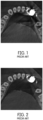
- the metal artefacts appear as dark or light stripes as shown in Fig. 1 . These artefacts can complicate the medical diagnosis.
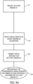
- a conventional method of metal artifact reduction is shown in Fig. 3 . In the conventional method, first the two-dimensional x-ray images are acquired and subsequently used to reconstruct a three-dimensional (3D) tomographic image.
- Fig. 2 shows a tomogram in which the metal artefacts in Fig. 1 have been reduced through the method in Fig. 3 .
- Fig. 8 is a tomogram corresponding to the same section in Fig. 7 and has been reconstructed with the metal artefact reduction method according to the prior art as shown in Fig.3 .
- the metal artefacts in Fig. 7 caused by the metal objects (6a) outside the small volume (V) cannot be corrected through the conventional method of metal artifact reduction shown in Fig. 3 , and thus, the conventional metal artifact reduction method is not reliable.
- the detection of metal objects may be directly performed on the two-dimensional x-ray images.
- the two dimensional-x-ray images or alternatively the sinogram can be analyzed.
- a two-dimensional x-ray image represents the projection of the irradiated three-dimensional object, for instance, a patient jaw as shown in Fig. 4 .
- a sinogram is created when these projections taken at all angles are combined into an array of projections as shown in Fig. 5a .
- the trace of a metal object imaged over time defines curves in the sinogram as shown in Fig.
- the data for reconstructing a 3D artifact-free tomographic image is generally missing.
- artificial intelligence is used to supplement the two-dimensional x-ray images where the data is missing.
- the sites including the missing data are initially determined through thresholding and subsequently supplemented with a trained artificial intelligence to reconstruct a metal artifact reduced three-dimensional tomographic image.
- this approach is prone to the disadvantages of the above-mentioned classical image processing techniques involving the use of thresholding.
- An objective of the present invention is to overcome the above-mentioned disadvantages of the prior art and to provide a method of metal artefact reduction in x-ray dental volume tomography.
- the present invention provides a method of metal artefact reduction in x-ray dental volume tomography.
- the method comprises: a step of obtaining two-dimensional x-ray images or a sinogram of at least part of a patient jaw, acquired through relatively rotating an x-ray source and a detector around the patient jaw; a step of detecting metal objects in the two-dimensional x-ray images or the sinogram by using a trained artificial intelligence algorithm to generate 2D masks which represent the metal objects in the two-dimensional x-ray images or 3D masks which represent the metal objects in the sinogram, respectively; and a step of reconstructing a three dimensional tomographic image respectively based on two-dimensional x-ray images or the sinogram and the 2D masks or the 3D masks as generated.
- a major advantageous effect of the present invention is that the metal objects can be more reliably detected through directly and solely analyzing the 2D x-ray images or the sinogram by using 2D/3D masks that are generated through the trained artificial intelligence algorithm.
- the need for initially reconstructing and analyzing the 3D tomographic image with metal objects can be obviated, and thus the process time can be comparatively shortened, and the process load can be comparatively reduced.
- the speed of reconstructing a metal artefact reduced 3D tomographic images has been improved.
- Another major advantageous effect of the present invention is that metallic objects that are not in the reconstructed volume can be reliably detected, and the metal artifacts caused by these metallic objects can be reduced.
- the metal artefact reduction method of the present invention has an improved tomographic image quality independently from the size of the volume to be reconstructed.
- Another major advantageous effect of the present invention is that the artificial intelligence algorithm can be continually trained with the help of real/virtual data including 2D x-ray images taken from the patient jaw or a calibration body from various angles, and thus the generation of 2D/3D mask for detecting the metal objects can be accordingly improved through such training. Thereby, the medical diagnosis can be increasingly facilitated.
- the three-dimensional tomographic image may be reconstructed in two alternative ways.
- the method further comprises a step of correcting the two-dimensional x-ray images or the sinogram by means of the generated 2D masks or the 3D masks respectively, wherein in the reconstruction step, the three-dimensional tomographic image is reconstructed based on the corrected two-dimensional x-ray images or the corrected sinogram.
- the reconstruction step in the reconstruction step the three-dimensional tomographic image is reconstructed respectively based on the two-dimensional x-ray images or the sinogram as originally obtained and the 2D masks or the 3D masks as generated.
- the 2D/3D masks are preferably 2D/3D binary masks.
- probability masks or confidence masks may be used.
- the two-dimensional x-ray images or the sinogram may be corrected through classical image processing.
- another artificial intelligence algorithm may be used for correcting the two-dimensional x-ray images.
- This artificial intelligence algorithm can be trained with two-dimensional x-ray images data acquired from different patient jaws.
- the reconstruction of the three-dimensional tomographic image may be based solely on the corrected two-dimensional x-ray images or the corrected sinogram such that the metal objects are not present in the final three-dimensional tomographic image.
- the reconstruction of the three-dimensional tomographic image may be additionally based on the uncorrected two-dimensional x-ray images or the uncorrected sinogram such that the metal objects are present in the three-dimensional tomographic image.
- the artificial intelligence algorithm is trained by using data pairs, where each pair consists of a two-dimensional x-ray image and an associated 2D mask which represents the location of any metal objects in the associated two-dimensional x-ray image.
- the artificial intelligence algorithm may be trained with a sinogram and an associated 3D mask.
- the two-dimensional x-ray images are generated through an x-ray source and a detector through a revolution around the patient jaw.
- the 2D masks or the 3D mask used for the training are obtained through the locations of the metal objects in the two-dimensional x-ray images or the sinogram.
- One way of obtaining them is by analyzing a three-dimensional tomographic image in which metal artefacts have not been corrected (e.g.
- the 2D masks and/or the 3D mask used in the training step may be manually generated from two-dimensional x-ray images and/or sinograms in which metal artefacts have not be corrected.
- the two-dimensional x-ray images or the sinogram to be obtained is not limited to the entire patient jaw and may also correspond to a part of the patient jaw such as a small volume.
- Such small volumes may be different in location, size and shape.
- the metal artefact reduction method may be applied to any small volume even when it is free of metal objects and the patient jaw includes one or more metal object outside the small volume.
- the present invention also provides an x-ray dental volume tomography system which has an x-ray unit and a tomographic reconstruction unit.
- the x-ray unit has an acquisition means for acquiring the two-dimensional x-ray images or the sinogram of at least part of a patient jaw i.e., a small volume, through relatively rotating an x-ray source and a detector around the patient jaw.
- the part of the patient jaw to be examined can be selected by the user, and accordingly irradiated to prevent overdosing of the patient.
- the tomographic reconstruction unit also has an image processing means which executes the method according to the present invention for metal artefact reduction in x-ray dental volume tomography.
- Fig. 6 shows an x-ray unit (9) according to an embodiment of the present invention.
- the x-ray unit (9) has an acquisition means for acquiring two-dimensional x-ray images (1) or a sinogram (2) of at least part (V) of a patient jaw (3a) i.e., a small volume, through relatively rotating an x-ray source (4) and a detector (5) around the patient jaw (3a).
- the x-ray unit (9) is connectable to a tomographic reconstruction unit which has an image processing means which is adapted to execute the method of metal artefact reduction in x-ray dental volume tomography according to the present invention.
- the x-ray unit (9) and the tomographic reconstruction unit together configure an x-ray dental volume tomography system according to the present invention.
- Fig. 9a shows the steps (S1,S2,S3,S4) of the method of metal artefact reduction in x-ray dental volume tomography according to a first alternative embodiment of the present invention.
- a first step (S1) two-dimensional x-ray images (1) or a sinogram (2) of at least part (V) of a patient jaw (3a) is obtained from the acquisition means of the x-ray unit (9).
- the metal objects (6) in the two-dimensional x-ray images (1) or the sinogram (2) are detected by using a trained artificial intelligence algorithm which generates either 2D masks (7) that represent the metal objects (6) in the two-dimensional x-ray images (1) or 3D masks which represent the metal objects (6) in the sinogram (2), respectively.
- 2D masks or 3D masks obtained from a classical algorithm can be optionally used in combination to those generated through the trained artificial intelligence algorithm to optimize the detection.
- a third step (S3) the two-dimensional x-ray images (1) or the sinogram (2) are corrected at least by means of the generated 2D masks (7) or the 3D masks respectively.
- This correction may be performed through classical image processing. Alternatively, the correction may be performed through another trained artificial intelligence algorithm.
- step (S4) a three-dimensional tomographic image (8) based on the corrected two-dimensional x-ray images (1) or the corrected sinogram (2) is reconstructed.
- the reconstruction of the three-dimensional tomographic image (8) may be based on the corrected and the uncorrected two-dimensional x-ray images (1) or the corrected and the uncorrected sinogram (2) so that the metal objects (6), if any, are also shown in the three-dimensional tomographic image (8).
- Fig. 9b shows the steps (S1,S2,S5) of the method of metal artefact reduction in x-ray dental volume tomography according to a second alternative embodiment of the present invention.
- the third step (S3) is dispensed with.
- the three-dimensional tomographic image (8) is reconstructed respectively based on the two-dimensional x-ray images (1) or the sinogram (2) as originally obtained and the 2D masks (7) or the 3D masks as generated.
- Fig.7 shows a tomogram according to the prior art with metal artefacts that are caused by metal objects (6a) outside the reconstructed small volume (V).
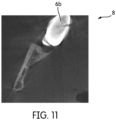
- Fig.11 shows a tomogram corresponding to the same section in Fig. 7 , reconstructed with the metal artefact reduction method according to the present invention.
- each data pair includes a two-dimensional x-ray image (1') and an associated 2D mask (7') which represents the 2D location of any metal object (6') in the associated two-dimensional image (1').
- each data pair includes a sinogram (2') and an associated 3D mask which represents the 3D location of any metal object (6') in the sinogram (2').
- the two-dimensional x-ray images (1') and the sinogram (2') of the data pairs correspond, preferably to the entire patient jaw (3a') of an arbitrary patient (3'), which have been generated through an x-ray source (4') and a detector (5') through a relative revolution preferably completely around the patient jaw (3a').
- the 2D masks (7') and the 3D mask used in the training step may be obtained by analyzing a three-dimensional tomographic image (8') in which the metal artifacts have not be corrected.
- the 2D masks (7') and/or the 3D masks used for the training can be obtained from the prior art method in Fig. 3 as a by-product which result from the analyzing step.
- the 2D masks (7') and/or the 3D masks used in the training step may be manually generated from two-dimensional images (1') and/or sinograms (2') in which metal artefacts have not be corrected.
- the tomographic reconstruction unit has an input means for retrieving the trained artificial intelligence algorithm for generating the 2D masks (7) and the 3D masks.
- the input means may be a wireless connection or wired connections that can be connected to a network or the like for data retrieval.
- the acquisition means is adapted to user-selectively acquire two-dimensional x-ray images (1) or the sinogram (2) that corresponds to one of a plurality of different parts (V) of the patient jaw (3a) which have respectively different volumes.
- the user can select the parts (V) to be irradiated and adjust the size thereof.
- the method is provided in form of a computer-readable program which has codes for causing a computer-based x-ray dental volume tomography system to perform the above-described steps of the metal artefact reduction method in x-ray dental volume tomography.
- the computer-readable program is stored on a computer-readable storage.
Landscapes
- Health & Medical Sciences (AREA)
- Life Sciences & Earth Sciences (AREA)
- Engineering & Computer Science (AREA)
- Medical Informatics (AREA)
- Physics & Mathematics (AREA)
- Radiology & Medical Imaging (AREA)
- Surgery (AREA)
- High Energy & Nuclear Physics (AREA)
- Veterinary Medicine (AREA)
- Nuclear Medicine, Radiotherapy & Molecular Imaging (AREA)
- Optics & Photonics (AREA)
- Pathology (AREA)
- Public Health (AREA)
- Biomedical Technology (AREA)
- Heart & Thoracic Surgery (AREA)
- Molecular Biology (AREA)
- Biophysics (AREA)
- Animal Behavior & Ethology (AREA)
- General Health & Medical Sciences (AREA)
- Theoretical Computer Science (AREA)
- General Physics & Mathematics (AREA)
- Computer Vision & Pattern Recognition (AREA)
- Pulmonology (AREA)
- Dentistry (AREA)
- Oral & Maxillofacial Surgery (AREA)
- Apparatus For Radiation Diagnosis (AREA)
- Image Analysis (AREA)
Priority Applications (1)
| Application Number | Priority Date | Filing Date | Title |
|---|---|---|---|
| EP24217465.4A EP4494566A3 (de) | 2019-04-18 | 2019-04-18 | Verfahren zur reduktion von metallischen artefakten in der dentalen volumentomographie |
Applications Claiming Priority (2)
| Application Number | Priority Date | Filing Date | Title |
|---|---|---|---|
| EP19170279.4A EP3725229B1 (de) | 2019-04-18 | 2019-04-18 | Verfahren zur reduzierung von metallischen artefakten in der dentalen volumentomografie |
| EP24217465.4A EP4494566A3 (de) | 2019-04-18 | 2019-04-18 | Verfahren zur reduktion von metallischen artefakten in der dentalen volumentomographie |
Related Parent Applications (2)
| Application Number | Title | Priority Date | Filing Date |
|---|---|---|---|
| EP19170279.4A Division EP3725229B1 (de) | 2019-04-18 | 2019-04-18 | Verfahren zur reduzierung von metallischen artefakten in der dentalen volumentomografie |
| EP19170279.4A Division-Into EP3725229B1 (de) | 2019-04-18 | 2019-04-18 | Verfahren zur reduzierung von metallischen artefakten in der dentalen volumentomografie |
Publications (2)
| Publication Number | Publication Date |
|---|---|
| EP4494566A2 true EP4494566A2 (de) | 2025-01-22 |
| EP4494566A3 EP4494566A3 (de) | 2025-03-19 |
Family
ID=66239912
Family Applications (2)
| Application Number | Title | Priority Date | Filing Date |
|---|---|---|---|
| EP24217465.4A Pending EP4494566A3 (de) | 2019-04-18 | 2019-04-18 | Verfahren zur reduktion von metallischen artefakten in der dentalen volumentomographie |
| EP19170279.4A Active EP3725229B1 (de) | 2019-04-18 | 2019-04-18 | Verfahren zur reduzierung von metallischen artefakten in der dentalen volumentomografie |
Family Applications After (1)
| Application Number | Title | Priority Date | Filing Date |
|---|---|---|---|
| EP19170279.4A Active EP3725229B1 (de) | 2019-04-18 | 2019-04-18 | Verfahren zur reduzierung von metallischen artefakten in der dentalen volumentomografie |
Country Status (5)
| Country | Link |
|---|---|
| US (2) | US12374003B2 (de) |
| EP (2) | EP4494566A3 (de) |
| JP (2) | JP7577663B2 (de) |
| CN (1) | CN113226184B (de) |
| WO (1) | WO2020212573A1 (de) |
Families Citing this family (9)
| Publication number | Priority date | Publication date | Assignee | Title |
|---|---|---|---|---|
| US11298565B2 (en) | 2017-11-16 | 2022-04-12 | Ebamed Sa | Heart arrhythmia non-invasive treatment device and method |
| EP4494566A3 (de) | 2019-04-18 | 2025-03-19 | DENTSPLY SIRONA Inc. | Verfahren zur reduktion von metallischen artefakten in der dentalen volumentomographie |
| WO2021094824A1 (en) | 2019-11-14 | 2021-05-20 | Ebamed Sa | Cardiac phase gating system for radiation therapy |
| WO2022136925A1 (en) | 2020-12-23 | 2022-06-30 | Ebamed Sa | A multiplanar motion management system |
| US12277627B1 (en) | 2021-08-27 | 2025-04-15 | Queensland University Of Technology | Correction of ultrasound probe-induced metal artifacts in X-ray based imaging systems |
| US12229963B2 (en) * | 2021-09-29 | 2025-02-18 | Siemens Healthineers International Ag | Adaptive auto-segmentation in computed tomography |
| US12229927B2 (en) | 2021-09-29 | 2025-02-18 | Siemens Healthineers International Ag | Metal artifact reduction in computed tomography |
| DE102022201003B3 (de) | 2022-01-31 | 2023-06-22 | Siemens Healthcare Gmbh | Verfahren zum Festlegen einer Aufnahmetrajektorie |
| CN115760935B (zh) * | 2022-11-18 | 2026-02-10 | 杭州柳叶刀机器人有限公司 | 颌部影像的配准方法、装置、计算机设备及可读存储介质 |
Citations (1)
| Publication number | Priority date | Publication date | Assignee | Title |
|---|---|---|---|---|
| WO2018126396A1 (en) | 2017-01-05 | 2018-07-12 | General Electric Company | Deep learning based estimation of data for use in tomographic reconstruction |
Family Cites Families (16)
| Publication number | Priority date | Publication date | Assignee | Title |
|---|---|---|---|---|
| US7548604B2 (en) | 2007-01-04 | 2009-06-16 | General Electric Company | Method and apparatus for reduction of metal artifacts in CT imaging |
| US9943380B2 (en) * | 2007-03-14 | 2018-04-17 | Orthoaccel Technologies, Inc. | Vibrating orthodontic remodelling device |
| DE102009032059A1 (de) * | 2009-07-07 | 2011-01-13 | Siemens Aktiengesellschaft | Sinogrammbearbeitung für die Metallartefaktreduktion in der Computertomographie |
| CN101777177A (zh) * | 2009-12-29 | 2010-07-14 | 上海维宏电子科技有限公司 | 基于衰减滤波的ct图像去金属伪影混合重建法 |
| EP2510898A1 (de) * | 2011-04-14 | 2012-10-17 | Astra Tech AB | Befestigungsvorrichtung |
| WO2013111052A1 (en) | 2012-01-25 | 2013-08-01 | Koninklijke Philips N.V. | Metal artifact reduction from metallic dental fillings and/or crowns in computed tomography (ct) and/or x-ray |
| JP2013233168A (ja) | 2012-05-02 | 2013-11-21 | Morita Mfg Co Ltd | 画像処理装置、x線ct撮影装置および画像処理方法 |
| US20170000430A1 (en) * | 2015-06-30 | 2017-01-05 | Carestream Health, Inc. | Methods and apparatus for jaw motion analysis |
| CN107714069A (zh) | 2016-08-10 | 2018-02-23 | 南京普爱医疗设备股份有限公司 | 一种基于cbct影像的金属伪影的测试方法 |
| WO2018071408A1 (en) * | 2016-10-10 | 2018-04-19 | Dentsply Sirona Inc. | Shade-coordinated, veneering porcelain system for dental prostheses |
| US10395397B2 (en) * | 2016-11-14 | 2019-08-27 | Carestream Health, Inc. | Metal artifacts reduction for cone beam CT |
| US10475214B2 (en) * | 2017-04-05 | 2019-11-12 | General Electric Company | Tomographic reconstruction based on deep learning |
| CN111684492B (zh) * | 2017-06-26 | 2024-03-15 | 医科达有限公司 | 使用深度卷积神经网络来改善锥形束ct图像质量的方法 |
| US20190059827A1 (en) | 2017-08-24 | 2019-02-28 | General Electric Company | System and method for imaging a subject |
| US11126914B2 (en) * | 2017-10-11 | 2021-09-21 | General Electric Company | Image generation using machine learning |
| EP4494566A3 (de) | 2019-04-18 | 2025-03-19 | DENTSPLY SIRONA Inc. | Verfahren zur reduktion von metallischen artefakten in der dentalen volumentomographie |
-
2019
- 2019-04-18 EP EP24217465.4A patent/EP4494566A3/de active Pending
- 2019-04-18 EP EP19170279.4A patent/EP3725229B1/de active Active
-
2020
- 2020-04-17 US US17/604,045 patent/US12374003B2/en active Active
- 2020-04-17 CN CN202080007301.0A patent/CN113226184B/zh active Active
- 2020-04-17 WO PCT/EP2020/060864 patent/WO2020212573A1/en not_active Ceased
- 2020-04-17 JP JP2021540231A patent/JP7577663B2/ja active Active
-
2024
- 2024-08-14 JP JP2024135351A patent/JP2024164073A/ja active Pending
-
2025
- 2025-06-25 US US19/249,830 patent/US20250322566A1/en active Pending
Patent Citations (1)
| Publication number | Priority date | Publication date | Assignee | Title |
|---|---|---|---|---|
| WO2018126396A1 (en) | 2017-01-05 | 2018-07-12 | General Electric Company | Deep learning based estimation of data for use in tomographic reconstruction |
Also Published As
| Publication number | Publication date |
|---|---|
| EP3725229A1 (de) | 2020-10-21 |
| EP4494566A3 (de) | 2025-03-19 |
| US20220207794A1 (en) | 2022-06-30 |
| WO2020212573A1 (en) | 2020-10-22 |
| US20250322566A1 (en) | 2025-10-16 |
| JP2024164073A (ja) | 2024-11-26 |
| CN113226184B (zh) | 2024-06-18 |
| US12374003B2 (en) | 2025-07-29 |
| JP2022530298A (ja) | 2022-06-29 |
| JP7577663B2 (ja) | 2024-11-05 |
| EP3725229B1 (de) | 2025-05-28 |
| CN113226184A (zh) | 2021-08-06 |
Similar Documents
| Publication | Publication Date | Title |
|---|---|---|
| US20250322566A1 (en) | Method of metal artefact reduction in x-ray dental volume tomography | |
| ES3061537T3 (en) | Automated correction of metal affected voxel representations of x-ray data using deep learning techniques | |
| US12182970B2 (en) | X-ray imaging restoration using deep learning algorithms | |
| CN111656405A (zh) | 使用深度学习来减少金属伪影 | |
| US20110044559A1 (en) | Image artifact reduction | |
| CN115209808B (zh) | 学习完毕模型的制作方法、图像生成方法以及图像处理装置 | |
| US12115017B2 (en) | Method of calibrating x-ray projection geometry in x-ray cone beam computed tomography | |
| KR20150095140A (ko) | 컴퓨터 단층 촬영 장치 및 그에 따른 ct 영상 복원 방법 | |
| US20220110596A1 (en) | Improved metal artifacts reduction in cone beam reconstruction | |
| CN118648025A (zh) | 一种使用计算机断层扫描图像重建三维口腔扫描数据的装置和方法 | |
| US7978886B2 (en) | System and method for anatomy based reconstruction | |
| EP3272290A1 (de) | Vorrichtung und verfahren zur rekonstruktion eines medizinischen bildes | |
| US6845143B2 (en) | CT image reconstruction | |
| JP2015231528A (ja) | X線コンピュータ断層撮像装置及び医用画像処理装置 | |
| WO2008065394A1 (en) | Method and apparatus for reducing distortion in a computed tomography image | |
| US8526757B2 (en) | Imaging system and imaging method for imaging a region of interest | |
| KR20230091422A (ko) | 의료영상 처리 장치와 그 의료영상 학습 방법 및 의료영상 처리 방법 | |
| US11317887B2 (en) | Computed tomography reconstruction of moving bodies | |
| CN114868152B (zh) | 用于在高相对螺距下采集的计算机断层摄影x射线数据的装置 | |
| KR101613391B1 (ko) | 치료 계획용 ct 영상을 이용한 산란선 제거 방법 및 시스템 | |
| Choi et al. | Automatic Detection of Inferior Alveolar Nerve Canal from Cone-Beam Computed Tomography Images for Dental Surgery Planning. | |
| JP7258474B2 (ja) | X線ct装置及び放射線治療システム |
Legal Events
| Date | Code | Title | Description |
|---|---|---|---|
| PUAI | Public reference made under article 153(3) epc to a published international application that has entered the european phase |
Free format text: ORIGINAL CODE: 0009012 |
|
| STAA | Information on the status of an ep patent application or granted ep patent |
Free format text: STATUS: THE APPLICATION HAS BEEN PUBLISHED |
|
| AC | Divisional application: reference to earlier application |
Ref document number: 3725229 Country of ref document: EP Kind code of ref document: P |
|
| AK | Designated contracting states |
Kind code of ref document: A2 Designated state(s): AL AT BE BG CH CY CZ DE DK EE ES FI FR GB GR HR HU IE IS IT LI LT LU LV MC MK MT NL NO PL PT RO RS SE SI SK SM TR |
|
| REG | Reference to a national code |
Ref country code: DE Ref legal event code: R079 Free format text: PREVIOUS MAIN CLASS: A61B0006510000 Ipc: A61B0006030000 |
|
| PUAL | Search report despatched |
Free format text: ORIGINAL CODE: 0009013 |
|
| AK | Designated contracting states |
Kind code of ref document: A3 Designated state(s): AL AT BE BG CH CY CZ DE DK EE ES FI FR GB GR HR HU IE IS IT LI LT LU LV MC MK MT NL NO PL PT RO RS SE SI SK SM TR |
|
| RIC1 | Information provided on ipc code assigned before grant |
Ipc: A61B 6/51 20240101ALI20250211BHEP Ipc: A61B 6/12 20060101ALI20250211BHEP Ipc: G06T 11/00 20060101ALI20250211BHEP Ipc: G06T 7/00 20170101ALI20250211BHEP Ipc: A61B 6/00 20240101ALI20250211BHEP Ipc: A61B 6/03 20060101AFI20250211BHEP |
|
| STAA | Information on the status of an ep patent application or granted ep patent |
Free format text: STATUS: REQUEST FOR EXAMINATION WAS MADE |
|
| 17P | Request for examination filed |
Effective date: 20250902 |
|
| GRAP | Despatch of communication of intention to grant a patent |
Free format text: ORIGINAL CODE: EPIDOSNIGR1 |
|
| RIC1 | Information provided on ipc code assigned before grant |
Ipc: A61B 6/03 20060101AFI20251215BHEP Ipc: A61B 6/00 20240101ALI20251215BHEP Ipc: G06T 7/00 20170101ALI20251215BHEP Ipc: G06T 11/00 20060101ALI20251215BHEP Ipc: A61B 6/12 20060101ALI20251215BHEP Ipc: A61B 6/51 20240101ALI20251215BHEP |
|
| STAA | Information on the status of an ep patent application or granted ep patent |
Free format text: STATUS: GRANT OF PATENT IS INTENDED |
|
| INTG | Intention to grant announced |
Effective date: 20260122 |