EP4474474A2 - Verfahren und zusammensetzungen zur behandlung von osteopetrose - Google Patents
Verfahren und zusammensetzungen zur behandlung von osteopetrose Download PDFInfo
- Publication number
- EP4474474A2 EP4474474A2 EP24193746.5A EP24193746A EP4474474A2 EP 4474474 A2 EP4474474 A2 EP 4474474A2 EP 24193746 A EP24193746 A EP 24193746A EP 4474474 A2 EP4474474 A2 EP 4474474A2
- Authority
- EP
- European Patent Office
- Prior art keywords
- csf1r
- mice
- composition
- cre
- cells
- Prior art date
- Legal status (The legal status is an assumption and is not a legal conclusion. Google has not performed a legal analysis and makes no representation as to the accuracy of the status listed.)
- Withdrawn
Links
Images















Classifications
-
- C—CHEMISTRY; METALLURGY
- C12—BIOCHEMISTRY; BEER; SPIRITS; WINE; VINEGAR; MICROBIOLOGY; ENZYMOLOGY; MUTATION OR GENETIC ENGINEERING
- C12N—MICROORGANISMS OR ENZYMES; COMPOSITIONS THEREOF; PROPAGATING, PRESERVING, OR MAINTAINING MICROORGANISMS; MUTATION OR GENETIC ENGINEERING; CULTURE MEDIA
- C12N5/00—Undifferentiated human, animal or plant cells, e.g. cell lines; Tissues; Cultivation or maintenance thereof; Culture media therefor
- C12N5/06—Animal cells or tissues; Human cells or tissues
- C12N5/0602—Vertebrate cells
- C12N5/0634—Cells from the blood or the immune system
- C12N5/0645—Macrophages, e.g. Kuepfer cells in the liver; Monocytes
-
- A—HUMAN NECESSITIES
- A61—MEDICAL OR VETERINARY SCIENCE; HYGIENE
- A61P—SPECIFIC THERAPEUTIC ACTIVITY OF CHEMICAL COMPOUNDS OR MEDICINAL PREPARATIONS
- A61P19/00—Drugs for skeletal disorders
- A61P19/08—Drugs for skeletal disorders for bone diseases, e.g. rachitism, Paget's disease
-
- A—HUMAN NECESSITIES
- A61—MEDICAL OR VETERINARY SCIENCE; HYGIENE
- A61K—PREPARATIONS FOR MEDICAL, DENTAL OR TOILETRY PURPOSES
- A61K38/00—Medicinal preparations containing peptides
- A61K38/16—Peptides having more than 20 amino acids; Gastrins; Somatostatins; Melanotropins; Derivatives thereof
- A61K38/17—Peptides having more than 20 amino acids; Gastrins; Somatostatins; Melanotropins; Derivatives thereof from animals; from humans
- A61K38/1703—Peptides having more than 20 amino acids; Gastrins; Somatostatins; Melanotropins; Derivatives thereof from animals; from humans from vertebrates
- A61K38/1709—Peptides having more than 20 amino acids; Gastrins; Somatostatins; Melanotropins; Derivatives thereof from animals; from humans from vertebrates from mammals
-
- A—HUMAN NECESSITIES
- A61—MEDICAL OR VETERINARY SCIENCE; HYGIENE
- A61K—PREPARATIONS FOR MEDICAL, DENTAL OR TOILETRY PURPOSES
- A61K40/00—Cellular immunotherapy
- A61K40/10—Cellular immunotherapy characterised by the cell type used
- A61K40/17—Monocytes; Macrophages
-
- A—HUMAN NECESSITIES
- A61—MEDICAL OR VETERINARY SCIENCE; HYGIENE
- A61K—PREPARATIONS FOR MEDICAL, DENTAL OR TOILETRY PURPOSES
- A61K40/00—Cellular immunotherapy
- A61K40/20—Cellular immunotherapy characterised by the effect or the function of the cells
- A61K40/24—Antigen-presenting cells [APC]
-
- A—HUMAN NECESSITIES
- A61—MEDICAL OR VETERINARY SCIENCE; HYGIENE
- A61K—PREPARATIONS FOR MEDICAL, DENTAL OR TOILETRY PURPOSES
- A61K40/00—Cellular immunotherapy
- A61K40/40—Cellular immunotherapy characterised by antigens that are targeted or presented by cells of the immune system
-
- A—HUMAN NECESSITIES
- A61—MEDICAL OR VETERINARY SCIENCE; HYGIENE
- A61K—PREPARATIONS FOR MEDICAL, DENTAL OR TOILETRY PURPOSES
- A61K9/00—Medicinal preparations characterised by special physical form
- A61K9/0012—Galenical forms characterised by the site of application
- A61K9/0019—Injectable compositions; Intramuscular, intravenous, arterial, subcutaneous administration; Compositions to be administered through the skin in an invasive manner
-
- A—HUMAN NECESSITIES
- A61—MEDICAL OR VETERINARY SCIENCE; HYGIENE
- A61P—SPECIFIC THERAPEUTIC ACTIVITY OF CHEMICAL COMPOUNDS OR MEDICINAL PREPARATIONS
- A61P19/00—Drugs for skeletal disorders
- A61P19/08—Drugs for skeletal disorders for bone diseases, e.g. rachitism, Paget's disease
- A61P19/10—Drugs for skeletal disorders for bone diseases, e.g. rachitism, Paget's disease for osteoporosis
-
- C—CHEMISTRY; METALLURGY
- C12—BIOCHEMISTRY; BEER; SPIRITS; WINE; VINEGAR; MICROBIOLOGY; ENZYMOLOGY; MUTATION OR GENETIC ENGINEERING
- C12N—MICROORGANISMS OR ENZYMES; COMPOSITIONS THEREOF; PROPAGATING, PRESERVING, OR MAINTAINING MICROORGANISMS; MUTATION OR GENETIC ENGINEERING; CULTURE MEDIA
- C12N5/00—Undifferentiated human, animal or plant cells, e.g. cell lines; Tissues; Cultivation or maintenance thereof; Culture media therefor
- C12N5/06—Animal cells or tissues; Human cells or tissues
- C12N5/0602—Vertebrate cells
- C12N5/0652—Cells of skeletal and connective tissues; Mesenchyme
- C12N5/0654—Osteocytes, Osteoblasts, Odontocytes; Bones, Teeth
-
- C—CHEMISTRY; METALLURGY
- C12—BIOCHEMISTRY; BEER; SPIRITS; WINE; VINEGAR; MICROBIOLOGY; ENZYMOLOGY; MUTATION OR GENETIC ENGINEERING
- C12N—MICROORGANISMS OR ENZYMES; COMPOSITIONS THEREOF; PROPAGATING, PRESERVING, OR MAINTAINING MICROORGANISMS; MUTATION OR GENETIC ENGINEERING; CULTURE MEDIA
- C12N9/00—Enzymes; Proenzymes; Compositions thereof; Processes for preparing, activating, inhibiting, separating or purifying enzymes
- C12N9/14—Hydrolases (3)
- C12N9/48—Hydrolases (3) acting on peptide bonds (3.4)
- C12N9/50—Proteinases, e.g. Endopeptidases (3.4.21-3.4.25)
- C12N9/64—Proteinases, e.g. Endopeptidases (3.4.21-3.4.25) derived from animal tissue
- C12N9/6421—Proteinases, e.g. Endopeptidases (3.4.21-3.4.25) derived from animal tissue from mammals
- C12N9/6472—Cysteine endopeptidases (3.4.22)
-
- A—HUMAN NECESSITIES
- A61—MEDICAL OR VETERINARY SCIENCE; HYGIENE
- A61K—PREPARATIONS FOR MEDICAL, DENTAL OR TOILETRY PURPOSES
- A61K2239/00—Indexing codes associated with cellular immunotherapy of group A61K40/00
- A61K2239/31—Indexing codes associated with cellular immunotherapy of group A61K40/00 characterized by the route of administration
-
- A—HUMAN NECESSITIES
- A61—MEDICAL OR VETERINARY SCIENCE; HYGIENE
- A61K—PREPARATIONS FOR MEDICAL, DENTAL OR TOILETRY PURPOSES
- A61K2239/00—Indexing codes associated with cellular immunotherapy of group A61K40/00
- A61K2239/38—Indexing codes associated with cellular immunotherapy of group A61K40/00 characterised by the dose, timing or administration schedule
-
- A—HUMAN NECESSITIES
- A61—MEDICAL OR VETERINARY SCIENCE; HYGIENE
- A61K—PREPARATIONS FOR MEDICAL, DENTAL OR TOILETRY PURPOSES
- A61K48/00—Medicinal preparations containing genetic material which is inserted into cells of the living body to treat genetic diseases; Gene therapy
Definitions
- the present technology generally relates to methods and compositions for treating, preventing, or ameliorating osteopetrosis. More specifically, the present technology relates to administering a composition comprising a therapeutically effective amount of engineered monocytic cells or wild-type monocytic cells from a healthy donor to a subject suffering from or at risk for osteopetrosis.
- Osteoclasts are multinucleated giant cells that resorb bone, ensuring development and continuous remodeling of the skeleton and the bone marrow hematopoietic niche. Defective osteoclast activity leads to osteopetrosis and bone marrow failure, while excess activity can contribute to bone loss and osteoporosis. Osteopetrosis can be partially treated by bone marrow transplantation in human and mice, in accordance with osteoclasts hematopoietic origin, and studies suggesting that they develop by fusion of hematopoietic stem cell (HSC)-derived monocytic precursors in the presence of CSF 1 and RANK-Ligand.
- HSC hematopoietic stem cell
- the present disclosure provides a method for treating or preventing osteopetrosis in a subject in need thereof, the method comprising administering a composition comprising a therapeutically effective amount of monocytic cells from a healthy donor to the subject.
- the subject is characterized by decreased expression of one or more of CA2, CLCN7, CTSK, CSF1R, IKBKG, ITGB3, OSTM1, PLEKHM1, TCIRG1, TNFRSF11A, and TNFSF11, as compared to the monocytic cells of the donor.
- the osteopetrosis comprises one or more of stunted growth, skeletal deformity, increased likelihood of bone fracture, anemia, recurrent infections, hepatosplenomegaly, facial paralysis, abnormal cortical bone morphology, abnormal form of vertebral bodies, abnormal temperature regulation, abnormality of the ribs, abnormality of vertebral epiphysis morphology, bone pain, cranial nerve paralysis, craniosynostosis, hearing impairment, and hypocalcemia.
- the composition is formulated for intravenous administration by injection, infusion, or transfusion.
- the subject is a mammal.
- the mammalian subject is a human.
- the subject is characterized by a cathepsin K deficiency.
- the present disclosure provides a method for treating or preventing osteopetrosis in a subject in need thereof, the method comprising administering to the subject a composition comprising a therapeutically effective amount of monocytic cells engineered to express one or more genes selected from CA2, CLCN7, CTSK, CSF1R, IKBKG, ITGB3, OSTM1, PLEKHM1, TCIRG1, TNFRSF11A, and TNFSF11.
- the osteopetrosis comprises one or more of stunted growth, skeletal deformity, increased likelihood of bone fracture, anemia, recurrent infections, hepatosplenomegaly, facial paralysis, abnormal cortical bone morphology, abnormal form of vertebral bodies, abnormal temperature regulation, abnormality of the ribs, abnormality of vertebral epiphysis morphology, bone pain, cranial nerve paralysis, craniosynostosis, hearing impairment, and hypocalcemia.
- the composition is formulated for intravenous administration by injection, infusion, or transfusion.
- the subject is a mammal.
- the mammalian subject is a human.
- the subject is characterized by a cathepsin K deficiency.
- the monocytic cells are obtained from the subject.
- the present disclosure provides a donor monocytic cell line engineered to express one or more genes selected from CA2, CLCN7, CTSK, CSF1R, IKBKG, ITGB3, OSTM1, PLEKHM1, TCIRG1, TNFRSF11A, and TNFSF11, wherein the one or more genes is operably linked to a heterologous nucleic acid to form a chimeric nucleic acid construct.
- the heterologous nucleic acid encodes a selectable marker.
- the selectable marker is a bioluminescent protein, a fluorescent protein, a chemiluminescent protein, a xanthine-guanine phosphoribosyl transferase gene (gpt), or any combination thereof.
- the heterologous nucleic acid encodes one or more control sequences suitable for directing expression of the one or more genes in a monocytic cell.
- the one or more control sequences comprises a promoter.
- the donor cells comprise a vector encoding one or more genes selected from CA2, CLCN7, CTSK, CSF1R, IKBKG, ITGB3, OSTM1, PLEKHM1, TCIRG1, TNFRSF11A, and TNFSF11.
- the vector is a mammalian expression vector, a lentiviral vector, or transposon vector.
- a "chimeric nucleic acid” comprises a coding sequence or fragment thereof linked to a nucleotide sequence that is different from the nucleotide sequence with which it is associated in cells in which the coding sequence occurs naturally.
- the terms "effective amount,” or “therapeutically effective amount,” and “pharmaceutically effective amount” refer to a quantity sufficient to achieve a desired therapeutic and/or prophylactic effect, e.g., an amount which results in the prevention of a disease, condition, and/or symptom(s) thereof.
- the amount of a composition administered to the subject will depend on the type and severity of the disease and on the characteristics of the individual, such as general health, age, sex, body weight, and tolerance to the composition drugs. It will also depend on the degree, severity, and type of disease or condition. The skilled artisan will be able to determine appropriate dosages depending on these and other factors. In some embodiments, multiple doses are administered.
- compositions comprising the monocytic cells of the present technology may be administered to a subject having one or more signs, symptoms, or risk factors of osteopetrosis, including, but not limited to stunted growth, skeletal deformity, increased likelihood of bone fracture, anemia, recurrent infections, hepatosplenomegaly, facial paralysis, abnormal cortical bone morphology, abnormal form of vertebral bodies, abnormal temperature regulation, abnormality of the ribs, abnormality of vertebral epiphysis morphology, bone pain, cranial nerve paralysis, craniosynostosis, hearing impairment, and hypocalcemia.
- signs, symptoms, or risk factors of osteopetrosis including, but not limited to stunted growth, skeletal deformity, increased likelihood of bone fracture, anemia, recurrent infections, hepatosplenomegaly, facial paralysis, abnormal cortical bone morphology, abnormal form of vertebral bodies, abnormal temperature regulation, abnormality of the ribs, abnormality of vertebral epiphysis morph
- a "therapeutically effective amount" of the compositions of the present technology includes levels at which the presence, frequency, or severity of one or more signs, symptoms, or risk factors of osteopetrosis are, at a minimum, ameliorated.
- a therapeutically effective amount reduces or ameliorates the physiological effects of osteopetrosis, and/or the risk factors of osteopetrosis, and/or the likelihood of developing osteotpetrosis.
- a therapeutically effective amount is achieved by multiple administrations.
- a therapeutically effective amount is achieved with a single administration.
- engineered is used herein to refer to a cell or organism that has been manipulated to be genetically altered, modified, or changed, e.g., by disruption of the genome.
- engineered monocytic cell refers to a monocytic cell that has been manipulated to be genetically altered, modified, or changed.
- an engineered monocytic cell refers to a monocytic cell that has been transduced with a lentivirus designed to express a nucleotide sequence of interest, e.g., a cDNA coding for the wild type allele of any one or more of CA2, CLCN7, CTSK, CSF1R, IKBKG, ITGB3, OSTM1, PLEKHM1, TCIRG1, TNFRSF11A, or TNFSF11 under a strong promoter.
- a nucleotide sequence of interest e.g., a cDNA coding for the wild type allele of any one or more of CA2, CLCN7, CTSK, CSF1R, IKBKG, ITGB3, OSTM1, PLEKHM1, TCIRG1, TNFRSF11A, or TNFSF11 under a strong promoter.
- Heterologous nucleic acid refers to a nucleic acid, DNA, or RNA, which has been introduced into a cell (or the cell's ancestor), and which is not a copy of a sequence naturally found in the cell into which it is introduced.
- Such heterologous nucleic acid may comprise segments that are a copy of a sequence that is naturally found in the cell into which it has been introduced, or fragments thereof.
- prevention refers to, in a statistical sample, reduction in the occurrence or recurrence of the disorder or condition in treated subjects/samples relative to an untreated controls, or refers delays the onset of one or more symptoms of the disorder or condition relative to the untreated controls.
- subject and “patient” are used interchangeably and refer to a mammalian subject.
- subject means any animal (mammalian, human, or other) patient that can be afflicted with osteopetrosis and when thus afflicted is in need of treatment.
- the subject is a human.
- Treating,” “treat,” “treated,” or “treatment” of a disease or disorder includes: (i) inhibiting the disease or disorder, i.e., arresting its development; (ii) relieving the disease or disorder, i.e., causing its regression; (iii) slowing progression of the disorder; and/or (iv) inhibiting, relieving, or slowing progression of one or more symptoms of the disease or disorder.
- compositions and methods for the treatment of osteopetrosis identify: (i) the developmental origin of osteoclasts, the cells that shape bone architecture; (ii) a mechanism that controls the maintenance of osteoclast function after birth; and (iii) a novel strategy to treat osteopetrosis and to modulate osteoclast activity in vivo.
- the data described herein demonstrate that parabiosis or transfusion of monocytic cells results in long-term gene transfer in osteoclasts in the absence of HSC chimerism and can rescue an adult-onset osteopetrotic phenotype caused by cathepsin-K deficiency. Transfusion of monocytic cells is also sufficient to rescue bone development in early-onset autosomal recessive osteopetrosis in newborn mice.
- Osteopetroses are a heterogeneous group of genetic disorders characterized by increased bone density due to impaired bone resorption by osteoclasts. The increased bone density places the affected individual at an increased risk for bone fracture. Normally, bone growth is a balance between osteoblasts (cells that create bone tissue) and osteoclasts (cells that destroy bone tissue). Individuals with osteopetrosis have a deficiency of osteoclasts, resulting in too little bone resorption and too much bone creation. The types of osteopetrosis are distinguished based on their pattern of inheritance: autosomal dominant, autosomal recessive, or X-linked.
- the signs and symptoms of osteopetrosis may vary depending on the type of the disease, and mild forms of osteopetrosis may be asymptomatic.
- the typical signs and symptoms of osteopetrosis include: stunted growth, skeletal deformity, increased likelihood of bone fracture, anemia, recurrent infections, hepatosplenomegaly, facial paralysis, abnormal cortical bone morphology, abnormal form of vertebral bodies, abnormal temperature regulation, abnormality of the ribs, abnormality of vertebral epiphysis morphology, bone pain, cranial nerve paralysis, craniosynostosis, hearing impairment, and hypocalcemia.
- ADO autosomal dominant osteopetrosis
- Albers-Schönberg disease The signs and symptoms of autosomal dominant osteopetrosis (ADO; also known as Albers-Schönberg disease), include multiple bone fractures, scoliosis, arthritis in the hips, and/or osteomyelitis.
- Autsomal recessive osteopetrosis (ARO) is often characterized by one or more of a high risk of bone fracture resulting from minor bumps or falls, pinched nerves in the head and face, impaired bone marrow function, slow growth, short stature, dental abnormalities, hepatosplenomegaly, intellectual disability, and epilepsy.
- IAO intermediate autosomal osteopetrosis
- a form of osteopetrosis that can have either an autosomal dominant or recessive pattern or inheritance may be characterized by one or more of a high risk of bone fracture and anemia, calcifications in the brain, intellectual disability, and renal tubular acidosis.
- Individuals with the X-linked pattern of inheritance may be characterized by one or more of lymphedema, anhidrotic ectodermal dysplasia, and immunodeficiency.
- a method for treating or preventing osteopetrosis in a subject in need thereof, comprising administering a composition comprising a therapeutically effective amount of monocytic cells from a healthy donor to the subject comprising administering a composition comprising a therapeutically effective amount of monocytic cells from a healthy donor to the subject.
- the subject is characterized by decreased expression of one or more genes implicated in or potentially underlying the development of osteopetrosis, such as CA2 , CLCN7 , CTSK , CSF1R , IKBKG, ITGB3, OSTM1, PLEKHM1, TCIRG1, TNFRSF11A, and TNFSF11.
- the subject is characterized by a cathepsin K deficiency.
- a method for treating or preventing osteopetrosis in a subject in need thereof comprising administering to the subject a composition comprising a therapeutically effective amount of monocytic cells engineered to express one or more genes implicated in or potentially underlying the development of osteopetrosis, such as CA2, CLCN7, CTSK, CSF1R, IKBKG, ITGB3, OSTM1, PLEKHM1, TCIRG1, TNFRSF11A, and TNFSF11 is provided.
- monocytic cells used for treatment are genetically modified to correct a genetic abnormality or to improve or changed cellular functioning according to known genetic engineering protocols.
- a method of treating or preventing osteopetrosis in a subject comprises: (a) obtaining a sample of monocytic cells from the subject; (b) genetically correcting one or more mutations in the monocytic cells, (c) culturing the monocytic cells; and (d) providing the corrected monocytic cells to the subject.
- a method for treating or preventing osteopetrosis comprises: (a) obtaining a sample of monocytic cells from the subject; (b) genetically engineering the cells to express a nucleotide sequence coding for the wild type allele of any one or more of CA2, CLCN7, CTSK, CSF1R, IKBKG, ITGB3, OSTM1, PLEKHM1, TCIRG1, TNFRSF11A, and TNFSF11 with a suitable transducing vector, such as a lentiviral vector; (c) culturing the engineered monocytic cells under conditions sufficient to express the nucleotide sequence; (d) removing the viral particles from the engineered monocytic cells; and (e) providing the engineered monocytic cells to the subject.
- a suitable transducing vector such as a lentiviral vector
- the transducing vector encoding the one or more of CA2 , CLCN7 , CTSK , CSF1R, IKBKG, ITGB3, OSTM1 , PLEKHM1, TCIRG1 , TNFRSF11A , and TNFSF11 is a mammalian expression vector.
- the mammalian expression vector is a lentiviral vector or transposon vector.
- Subjects suffering from osteopetrosis can be identified by any or a combination of diagnostic or prognostic assays known in the art.
- typical symptoms of osteopetrosis include, but are not limited to, stunted growth, skeletal deformity, increased likelihood of bone fracture, anemia, recurrent infections, hepatosplenomegaly, facial paralysis, abnormal cortical bone morphology, abnormal form of vertebral bodies, abnormal temperature regulation, abnormality of the ribs, abnormality of vertebral epiphysis morphology, bone pain, cranial nerve paralysis, craniosynostosis, hearing impairment, and hypocalcemia.
- a composition comprising a therapeutically effective amount of monocytic cells from a health donor and/or monocytic cells engineered to express one or more genes selected from CA2, CLCN7, CTSK, CSF1R, IKBKG, ITGB3, OSTM1 , PLEKHM1 , TCIRG1 , TNFRSF11A , and TNFSF11 is administered to the subject.
- the composition is administered according to any acceptable transfusion regimen.
- the composition is administered one, two, three, four, or five times per day. In some embodiments, the composition is administered more than five times per day.
- the composition is administered every day, every other day, every third day, every fourth day, every fifth day, or every sixth day.
- the composition is administered weekly, bi-weekly, tri-weekly, or monthly.
- the composition is administered for a period of one, two, three, four, or five weeks.
- the composition is administered for six weeks or more.
- the composition is administered for twelve weeks or more.
- the composition is administered for a period of less than one year.
- the composition is administered for a period of more than one year or until a desired therapeutic outcome is observed in the subject.
- treatment of subjects diagnosed with or suspected of having osteopetrosis with one or more compositions of the present technology ameliorates or eliminates one or more of the following symptoms of osteopetrosis: stunted growth, skeletal deformity, increased likelihood of bone fracture, anemia, recurrent infections, hepatosplenomegaly, facial paralysis, abnormal cortical bone morphology, abnormal form of vertebral bodies, abnormal temperature regulation, abnormality of the ribs, abnormality of vertebral epiphysis morphology, bone pain, cranial nerve paralysis, craniosynostosis, hearing impairment, and hypocalcemia.
- the present technology provides a method for preventing or delaying the onset of osteopetrosis or one or more symptoms of osteopetrosis in a subject at risk of having or developing osteopetrosis.
- compositions of the present technology are administered to a subject susceptible to, or otherwise at risk of for osteopetrosis in an amount sufficient to eliminate or reduce the risk, or delay the onset of the disease, including biochemical, histologic and/or behavioral symptoms of the disease, its complications and intermediate pathological phenotypes presenting during development of the disease.
- Administration of a prophylactic compositions can occur prior to the manifestation of symptoms characteristic of the disease or disorder, such that the disease or disorder is prevented or, alternatively, delayed in its progression.
- Csf1r iCre , Csf1r MeriCreMer , Qsf1r flox were kindly provided by Dr Jeffrey Pollard
- Flt3 Cre were kindly provided by Dr Thomas Boehm
- Myb +/- mice were kindly provided by Dr John Frampton
- Vav Cre were kindly provided from Dr Thomas Graf.
- Tnftsf11a flox mice were kindly provided by JM Penninger
- Tnfrsf11a Koba / Cre mice were kindly provided by Dr Yasuhiro Kobayashi
- Ctsk tml(cre)Ska mice were kindly provided by Dr.
- Rosa.26-CreER T2 R26-CreER T2
- Rosa26 LSL-YFP stock number: 006148
- Rosa26 LSL-tdTomato stock number: 007908
- mice were bred and kept under specific pathogen conditions in separated ventilated cages in the animal facility of MSKCC and the Medical Theoretical Center of the TU Dresden. All experiments with osteopetrotic mice that lack teeth were performed with mice maximal 4 weeks of age that were kept with the lactating mother or provided with DietGel 76A (Clear H 2 O, 72-07-5022) to avoid secondary effects from malnutrition. Experiments were performed in adherence to the Institutional Review Board (IACUC 15-04-006) from MSKCC and Austindiretation Dresden and were in compliance with relevant ethical regulations. Mice greater than 7 days old were sacrificed by cervical dislocation (TU Dresden), CO 2 asphyxiation or anesthesia (MSKCC). To harvest embryos, pregnant females were sacrificed and embryos were collected by postmortem cesarean from the uterus and exsanguinated through decapitation in cold PBS (Fisher, 14190).
- Genotyping PCR genotyping was performed according to protocols described previously and indicated in Table 1. The investigators were not blinded to allocation during experiments and outcome assessment. Table 1. Mouse line Primer 1 sequence 5' --> 3' Primer 2 sequence 5' --> 3' Denaturation Annealing Elongation Cycles Final Elongation Expected bands Csf1r iCre Mutant allele 94°C - 45sec 60°C - 45sec 72°C - 45sec 30 72°C - 5 min Mutant: 400 bp CSf1r MeriC reMer WT allele 94°C - 45sec 60°C - 90sec 72°C - 90sec 30 72°C - 5 min WT: 1Kb Mutant allele Mutant: 2 Kb Csf1r flox Mutant allele 94°C - 30sec 60°C - 60sec 72°C - 60sec 35 72°C - 2 min WT: 450 bp Floxed: 500 bp Csf1
- mice were crossed at night, the following day a positive vaginal plug was considered as 0.5 days post-coitum (dpc), as previously described. Embryos were harvested as indicated below. For postnatal time point, pregnant females were monitored for the date of delivery, caesarean sections were carried out at term and neonates were fostered using lactating CD-1 females.
- Csf1r MeriCreMer female mice were crossed with male Rosa26 LSL-YFP / LSL-YFP mice.
- Cre recombination in Csf1r MeriCreMer ;Rosa26 LSL-YFP embryos at embryonic day E8.5 was induced with a single dose of 4-hydroxytamoxifen (4-OHT) injected intraperitoneally in pregnant mothers at a dose of 75 ⁇ g/g of body weight supplemented with 37.5 ⁇ g/g of progesterone as previously described.
- 4-hydroxytamoxifen (4-OHT) 4-hydroxytamoxifen
- Cre-mediated recombination in Rosa.26-CreER T2 tamoxifen was introduced to pregnant mother by a single TAM gavage (5mg) at E10.5, supplemented with progesterone (37.5 ⁇ g/g body weight resolved in Sunflower seed oil, Sigma-Aldrich) was injected i.p. directly after gavage.
- TAM Rosa.26-CreER T2 tamoxifen
- Csf1r iCre mice were crossed to male Rosa26 LSL-YFP / YFP or Rosa26 LSL-tdTomato / tdTomato mice .
- Csf1r iCre ;Rosa26 LSL-YFP and Csf1r iCre ;Rosa26 LSL-tdTomato females were used for parabiosis.
- Parabionts were kept on Sulfamethoxazole/Trimethoprim (Sulfatrim) diet for up to 8 weeks. Ex-parabionts were separated after 4 weeks for cellular complementation and 6 weeks for rescue of CathepsinK activity.
- mice for parabiosis were caged together few days before surgery.
- One day before the surgery the fur from lateral sides of mice was carefully removed with a trimmer followed by depilatory cream (for 3 minutes) at the site of surgery, excess of fur was removed with a moist gauze pad.
- Left side partner is shaved on the right side and vice versa. This procedure was performed under Isofluorane inhalation anesthesia. Mice were fed with food supplemented with Sulfatrim ad libitum one day prior to surgery.
- mice were anesthetized intraperitoneally with 150mg/kg of ketamine and 15mg/kg of xylazine.
- Sterile eye lubricant (Paralube Vet Ointment, 17033-211-38) was applied to both eyes to prevent corneal drying during surgery.
- mice were placed in a supine position on a surgical tray with heat support provided by a heating pad. The surgical site was cleaned 3 times with cotton swabs soaked in povidone-iodine (Betadine) then with 70% ethanol.
- anesthetic agent bupivacaine (Marcaine 0.25-0.5% solution) was applied locally. Surgery was performed by a longitudinal skin incision on the lateral side of mice, approximately 0.5cm above the elbow to 0.5cm below the knee joint. Mice were laid side-by-side in close contact and the ligaments of the two knees and elbows were sutured together using monofilament non-absorbable suture. Then, the skin incisions was closed by apposing and clipping skin to skin of the pair with 9 mm wound clips.
- mice Post-operative procedure: immediately after surgery mice were injected subcutaneously with 2 mg/kg of meloxicam and 0.5mg/kg of buprenorphine and for a maximum of 48 hours postoperatively. Mice are provided with Sulfatrim and Hydrogel (Clear H 2 O, 70-01-5022) in medicups. Wound clips were removed 14 days after surgery under Isofluorane anesthesia.
- mice were anesthetized and prepared for surgery as indicated above. Mice were separated at the site of parabiosis junction. Using scissors, the skin joining both mice is cut longitudinally. The sutures around the elbows and knees are cut and removed. The resulting wound is closed with 9 mm wound clips. Mice were injected subcutaneously with 2 mg/kg of meloxicam and 0.5mg/kg of buprenorphine and for a maximum of 48 hours postoperatively. Wound clips were removed 14 days after surgery.
- Bones were prepared for histology on frozen sections as detailed below, and stained with antibodies to detect fluorescent proteins: anti-GFP biotin, anti-RFP (Abeam), fluorescent TRAP staining and TO-PRO-3 as a nuclear stain. Histological sections of 15 ⁇ m thickness were scanned by confocal microscopy at 1.5 ⁇ m Z stacks. Sections were quantified for the number of TRAP + multinuclear cells (more than 3 nuclei per cell) and their YFP and Tomato expression using Imaris in 3D view and individual Z stacks.
- EdU pulse labeling 12 week old C57BL/6N mice (Charles River) were injected intraperitoneally with 25 ⁇ g/g of a 2.5mg/mL solution of EdU prepared extemporaneously (Fisher C10420, Click-iT EdU Alexa Fluor 488 Flow Cytometry Assay Kit). Blood, bone marrow, and bone samples were collected at 60hrs and 72hrs post injection and bone were prepared for histology of frozen sections as indicated above for adult mice.
- Sections are incubated for 45 minutes at room temperature with 100 ⁇ L of Click-iT EdU reaction buffer then washed with PBS and stain for fluorescent TRAP (ELF 97) and TO-PRO-3 (nuclear stain). Mounting media was 75% glycerol in PBS.
- Sections were analyzed using Imaris in 3D view and individual Z stacks to quantify the percentage of EdU+ TRAP + multinuclear cells and EdU + nuclei per individual osteoclasts.
- Bone marrow monocytes were isolated from 12-16 weeks old donor.
- In fate-mapping experiments 1 ⁇ 10 6 total cells from Csf1r iCre ;Rosa26 LSL-tdTomato mice were transferred at day 0, day 3 and day 6 by retro-orbital injections into recipient Csf1r iCre ;Rosa26 LSL-YFP age and gender matched mice. Mice were sacrificed at day 11 and day 60 after the first transfer.
- Ly6C + cells were calculated by determining the cell number / ml using a Neubauer chamber in combination with a staining for Ly6C analyzed by flow cytometry. Bone samples were dissected and prepared for frozen sections as described above and stained for anti-GFP, anti-RFP, fluorescent TRAP and TO-PRO-3. The percentages of YFP + or Tomato + multinuclear cells was quantified in femurs of recipients.
- Bone samples were fixed in 4% formaldehyde (Fisher, 28908) in PBS for 1 day (embryo) or 3 days (post-natal mice) at 4°C then washed 3 times with PBS and decalcified (for mice older than P7) in a 14% EDTA pH7.1 solution at 4°C for 5 days to 15 days, washed 3 times with PBS and dehydrated in 70% ethanol for 1 day and processed for paraffin sections.
- TRAP Tartrate Resistant Acid Phosphatase staining protocol.
- slides were placed in coplin jars and incubated in a 1% (v/v) Naphtol-Ether substrate solution in basic stock solution for 1hr at 37°C followed by incubation in a solution containing 2% (v/v) sodium nitrate solution and 2% (v/v) basic fuchsin solution in basic stock solution for 20 mins at 37°C.
- Napthol-Ether substrate solution 2% (w/v) Napthol AS-BI Phosphate (Sigma, 70482) in 2-Ethoxyethanol (Sigma, 256374).
- Sodium nitrate solution 4% ⁇ w/v) Sodium nitrate (Sigma, 237213) in water.
- Basic fuchsin solution 5% (w/v) basic fuchsin dye (Sigma, 857343) in 2N HCL (Fisher, A144-500).
- TRAP + multinuclear cells (more than 3 nuclei per cell), associated to bone tissue, were quantified in ImageJ using the Cell Counter plugin. Numerical values were plotted using GraphPad Prism. For Static and dynamic histomorphometry. Young and aged mice were injected (i.p.) twice with 15 mg/kg body weight Calcein (Sigma) dissolved in 1.4% NaHCO3/PBS 2 and 3 days apart, respectively. Mice were sacrificed 2 days after the last calcein injection. Femora and tibiae were fixed in 4% PBS-buffered paraformaldehyde and dehydrated in an ascending ethanol series.
- bones were embedded in methacrylate and cut into 7 ⁇ m sections to assess the fluorescent calcein labels.
- Unstained sections were analyzed using fluorescence microscopy to determine the mineralized surface/bone surface (MS/BS), the mineral apposition rate (MAR), and the bone formation rate/bone surface (BFR/BS).
- MAR mineral apposition rate
- BFR/BS bone formation rate/bone surface
- To determine numbers of osteoclasts bones were decalcified for one week using Osteosoft (Merck), dehydrated, and embedded into paraffin. Tartrate-resistant acid phosphatase staining was used to assess the osteoclast surface per bone surface (Oc.SBS) and number of osteoclasts per bone surface (N.Oc/BS). Bone sections were analyzed using the Osteomeasure software (Osteometrics, USA) following international standards.
- Sections were washed 2 times with PBS for 5 minutes. Sections stained with anti-GFP biotin antibody were first incubated with Biotin/Streptavidin blocking kit (Vector laboratories, SP2002). Streptavidin blocking solution is prepared by adding 4 drops of streptavidin solution to 1mL of PBS/0.25% BSA, samples were incubated for 15 min, then washed once with PBS for 5 min. Biotin blocking solution is prepared by adding 4 drops of biotin solution to 1mL of PBS/0.25% BSA, samples were incubated for 15 min then washed once with PBS for 5 min. Primary and secondary antibodies used are listed in Table 2. Sections were also stained with fluorescent TRAP and nuclear stain. Table 2.
- Fate-mapping models Flt3 Cre ;Rosa26 LSL-YFP , Cfs1r Cre ;Rosa26 LSL-YFP Csf1r MeriCreMer ;Rosa26 LSL- YFP and Tnfrsf11a Cre ; Rosa26 LSL-YFP Conjug -ation Company Catalog number Conce n-tration Dilution Staining Buffer Incubating conditions Primary Antibody Goat polyclonal anti-GFP Biotin Abeam ab6658 1 mg/m L 1::50 PBS/0.25% BSA/0.3% triton 4°C overnight (15hrs) Secondary Antibody Streptavidin AlexaF luor48 8 Thermo Fisher Scientific S-11223 1::10 0 PBS/0.25% BSA 2hrs at room temperature
- Fluorescent TRAP staining Sections were prepared for fluorescent TRAP by incubating with TRAP incubation solution (112 mM sodium acetate, 76 mM sodium tartate, and 11 mM sodium nitrite, pH 4.1-4.3) at room temperature for 10 minutes. Buffer was removed and incubated with ELF97 substrate (Molecular Probes E6589, 2mM) at a concentration of 125 ⁇ M in TRAP incubation solution for 15 min under UV light and washed 2 times with PBS for 5 minutes. Nuclear stain used was TO-PRO-3 Iodide (Fisher T3605) 1:4000 in PBS for 5 minutes. Mounting media was 75% glycerol in PBS.
- NanoSPECT / CT Animal imaging by computed tomography.
- Mice were anesthetized under Isofluorane anesthesia and placed on an imaging table containing an animal bed equipped with a nosecone for gas inhalation and body temperature stabilization. For 3 week old mice a mouse bed was used and for mice 8 weeks and older a rat bed was used.
- Whole-body imaging of mice was acquired using a NanoSPECT/CT scanner (Mediso) for non-invasive and longitudinal monitoring of the 3D skeletal structure.
- Each CT scan averaged 15 minutes and was acquired with an exposure time of 1,000ms and 240 projections set at a pitch of 1 degree.
- the tube energy of the X-ray was 55KVp and 145 ⁇ A.
- the in-plane voxel size was medium generating a voxel size of 147 ⁇ m 3 .
- Reconstructed images were analyzed using In Vivo Scope 2.0 (Bioscan, Inc.) software.
- mice were sacrificed and bones placed in 70% ethanol until scanning. Bone microarchitecture was analyzed using the vivaCT40 (Scanco Medical, Switzerland). Entire femora or humeri were imaged at a resolution of 10.5 ⁇ m (1 slice) with an X-ray energy of 70 kVp, 114 mA, and an integration time of 200ms. The machine was routinely calibrated using hydroxyapatite phantoms for density and geometry. Trabecular bone in femora or humeri from old mice was assessed in the metaphysis 20 slices below the growth plate using 150 slices. The trabecular region within the cortical bones (P21 mice) was determined in the femoral midshaft (100 slices up, 100 slices down). Pre-defined scripts from Scanco were used for the evaluation.
- Yolk sac from E10.5 embryos was digested for 60 min at 37°C in PBS containing 5% FCS, Collagenase Type 4 (Worthington, final 4.2 U/ml) and DNAseI (Sigma-Aldrich, final 100 ⁇ g/ml). The digestion reaction was stopped by incubation with 12.5 mM EDTA. Fetal liver was gently dissociated between the frosted ends of glass slides, and was then digested for 30 minutes using the same digestion enzyme mix as yolk sac.
- Blood was collected from anesthetized mice by retro-orbital venous sinus bleeding or cardiac puncture using a 1mL syringe and a 26G needle rinsed with 100mM EDTA (sigma E4884).
- the collected blood was lysed with 3 mL of red blood cell lysis buffer (155mM NH 4 Cl sigma A9434, 10mM NaHCO 3 sigma S5761 and 0.1mM EDTA sigma E4884) for 5 mins, washed with 10mL of FACS buffer (PBS, 0.5% BSA and 2mM EDTA) and centrifuged at 320g for 7 minutes at 4°C. All mice were perfused with 10ml of PBS after blood withdrawal.
- red blood cell lysis buffer 155mM NH 4 Cl sigma A9434, 10mM NaHCO 3 sigma S5761 and 0.1mM EDTA sigma E4884
- FACS buffer PBS, 0.5% BSA and 2mM
- Bone marrow cells were harvested by crushing or flushing femurs and tibias using a syringe and a 26G needle with 10mL of PBS / 5% FCS or RPMI supplemented with 10% FBS. Bone marrow was dissociated by gently pipetting up and down with a 1mL or 10mL pipette. Spleens were gently dispersed between frosted slides and digested for 30 min at 37 °C in PBS with 5% FCS containing Collagenase Type 4 (Worthington) at a final concentration of 4.2 U/ml and 100 ⁇ g/ml DNAseI (Sigma-Aldrich).
- the cell pellets were resuspended in FACS buffer containing purified anti-mouse CD16/32 antibody (1:100, Biolegend 101301), 5% normal mouse (Fisher, 015-000-120), 5% normal rat (Fisher 012-000-120) and 5% normal rabbit serum (Fisher, 011-000-120) and incubated for 10 minutes on ice or directly stained using blocking antibodies in the staining mixtures. Samples were immunostained with fluorochrome-conjugated antibodies for 30 minutes on ice, and analyzed by flow cytometry using an LSRFortessa or an LSR II (BD-Bioscience). Full list of antibodies for flow cytometry is provided in Table 3.
- osteoclasts arise by fusion of HSC-derived precursors and require expression of Csf1r and Tnfrsf11a (Rank).
- Csf1r Cre Csf1r fl / fl ; and Csf1r Cre ; Tnfrsf11a fl / fl mice were generated.
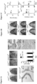
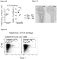
- mice presented with an osteopetrotic phenotype similar to Csf1 ( op / op ), Csf1r, and Tnfrsf11a mutants and characterized in young mice by lack of teeth eruption, skull and skeletal deformities with shortness of long bones, increased bone density, and lack of osteoclasts and hematopoietic cells ( Figure 1A ; Figure 5B ) .
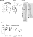
- Vav Cre ;Csf1r fl / fl mice were not osteopetrotic at birth, had normal teeth ( Figure 1B ), and hematopoietic cell numbers in long bones at 4 week of age ( Figure 1C ). However, they developed a late-onset osteopetrosis similar to Flt3 Cre ;Tnfrsf11a fl / fl and Flt3 Cre ;Csf1r fl / fl mice: at ⁇ 60 weeks of age Vav Cre ;Csf1r fl / fl mice had decreased osteoclasts and hematopoietic cell numbers in long bones ( Figure 1C-D ), enlarged trabecular bone ( Figure 1E ) and increased bone mass ( Figure 1F , Figure 7A-E ).
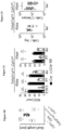
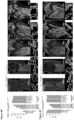
- TRAP + multinucleated cells develop in ⁇ E15 ossification centers from Myb -deficient embryo, which lack HSC but still support the development of EMP-derived macrophages ( Figure 2A , B ).
- TRAP + multinucleated cells are labeled with YFP in Csf1r Mer-iCre-Mer ; Rosa26 LSL-YFP mice pulsed at E8.5 with a single dose of hydroxytamoxifen (4-OHT), which labels EMP but not HSC ( Figure 2C , D; Figure 8A-E ).
- Tnfrsf11a is expressed by osteoclasts, but its expression is also a hallmark of EMP-derived pMacs that colonize the developing embryo.
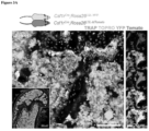
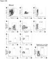
- Cre-mediated expression of a Rosa26 LSL-YFP fluorescent reporter is achieved with high efficiency in fetal macrophages but with low efficiency or not at all in HSC and their progeny in blood and tissues ( Figure 9A-D ). It was therefore hypothesized that conditional deletion of Csf1r in Tnfrsf11a Cre would recapitulate macrophage deficiency observed in Csf1r -deficient mice, while leaving the HSC lineage unaffected.
- Tnfrsf11a Cre ;Csf1r fl / fl mice were generated ( Figure 2E-J ).
- Tnfrsf11a Cre ;Csf1r fl / fl lack tissue macrophages such as brain microglia and epidermal Langerhans cells at birth while development of HSCs and blood cells was preserved ( Figure 6A-N ).
- this analysis supports a model in which EMP-derived embryonic osteoclasts are needed for teeth eruption, normal skull shape and optimal formation of long bones, and the timely colonization of long bones by hematopoietic progenitors, while HSC-derived osteoclasts are important for the maintenance of bone mass after birth and later in life, although they may partially rescue bone development in the absence of EMP-derived osteoclasts in Tnfrs11a Cre ;Csf1r fl / fl mice.
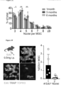
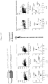
- the proportion of osteoclasts acquiring a new nucleus can be estimated -0.5 to 2 % per day in this model, compatible with individual nuclei being replaced every - 2 months.
- Figure 3G the studies above suggested a model ( Figure 3G ) where: osteoclasts that control skeletal development develop in ossification centers from EPM, while their post-natal maintenance is mediated via the serial acquisition by long-lived syncytia of new nuclei from HSC-derived blood leukocytes, rather than by de novo renewal by lateral fusion or proliferation of osteoclast precursors.
- a prediction from this model is that osteopetrosis due to a recessive mutation affecting osteoclasts function may be rescued or prevented through parabiosis with a wild-type partner.
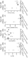
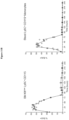
- Parabiosis experiments between 4 week-old cathepsin K deficient mice, which develop an adult-onset form of osteopetrosis known as pycnodysostosis, and cathepsin K +/- or cathepsin K -/- littermates and between wild type mice as control showed a reduction of bone volume in 10 weeks old cathepsin K -/- mice paired with cathepsin K +/- littermates ( Figure 4A ), suggesting that circulating blood cells carrying a wild type cathepsin K allele are sufficient to reduce bone density.
- osteoclasts originating from EMP are essential for normal bone development. Moreover, it is shown that osteoclasts are long-lived in adult and their function is maintained by iterative fusion of individual HSC-derived circulating cells with existing syncytia. In the absence or deficiency of EMP-derived osteoclasts, however, their timely replacement by transfusion with monocytic cells can rescue bone development in early-onset osteopetrotic mice in the absence of bone marrow transplantation. This could contribute to the treatment of early onset osteopetrosis, since the current treatment by bone marrow transplantation.
- Example 3 Ex vivo gene therapy for the treatment of a form of osteopetrosis: pycnodysostosis
- This example demonstrates the prophetic use of monocytic cells obtained from a subject and engineered to express a gene, such as CTSK , for the treatment of osteopetrosis in the subject.
- Pycnodysostosis is an autosomal recessive form of osteopetrosis, due to loss of function mutations in the cathepsin K gene ( CTSK ), compatible with life, and characterized by short stature, deformity of the skull maxilla and phalanges, increased density of the bones, osteosclerosis, and fragility of bone. There is no treatment for this disease as the relatively comparably milder symptoms do not qualify for bone marrow transplantation.
- Subjects suspected of having or diagnosed as having cathepsin K deficiency will be selected for a gene therapy trial and will undergo GMP grade collection of peripheral blood monocytes by apheresis and/or elutriation. Purified monocytes will then be transduced with a lentivirus designed to express a cDNA coding for the wild type allele of CTSK under a strong promoter (e.g., CMV). Transduced monocytes will then be washed to remove contaminating viral particles and rested for 12 hours followed by reinfusion to the patient. Assuming ⁇ 10 9 monocytes can be collected, the number of cells transferred per kilogram of body weight would be >10 7 . Following the auto-transfusion, the subject will be monitored at regular intervals for clinical and radiological signs of pycnodysostosis in order to determine the efficiency of the procedure and the frequency with which it should be repeated.
- a strong promoter e.g., CMV
- Example 4 Ex vivo gene therapy for the treatment of a form of osteopetrosis: severe infantile autosomal recessive osteopetrosis
- This example demonstrates the prophetic use of monocytic cells obtained from a subject and engineered to express a gene, such as TCIRG1, TNFRSF11a, CA2, CLCN7, OSTM1, and/or PLEKHM1, for the treatment of osteopetrosis in the subject.
- a gene such as TCIRG1, TNFRSF11a, CA2, CLCN7, OSTM1, and/or PLEKHM1, for the treatment of osteopetrosis in the subject.
- a range includes each individual member.
- a group having 1-3 cells refers to groups having 1, 2, or 3 cells.
- a group having 1-5 cells refers to groups having 1, 2, 3, 4, or 5 cells, and so forth.
Landscapes
- Health & Medical Sciences (AREA)
- Life Sciences & Earth Sciences (AREA)
- Engineering & Computer Science (AREA)
- General Health & Medical Sciences (AREA)
- Chemical & Material Sciences (AREA)
- Biomedical Technology (AREA)
- Bioinformatics & Cheminformatics (AREA)
- Organic Chemistry (AREA)
- Veterinary Medicine (AREA)
- Public Health (AREA)
- Animal Behavior & Ethology (AREA)
- Zoology (AREA)
- Genetics & Genomics (AREA)
- Wood Science & Technology (AREA)
- Epidemiology (AREA)
- Biotechnology (AREA)
- Medicinal Chemistry (AREA)
- Immunology (AREA)
- Physical Education & Sports Medicine (AREA)
- Pharmacology & Pharmacy (AREA)
- Orthopedic Medicine & Surgery (AREA)
- Microbiology (AREA)
- Rheumatology (AREA)
- Biochemistry (AREA)
- General Engineering & Computer Science (AREA)
- Cell Biology (AREA)
- Gastroenterology & Hepatology (AREA)
- Hematology (AREA)
- Chemical Kinetics & Catalysis (AREA)
- General Chemical & Material Sciences (AREA)
- Nuclear Medicine, Radiotherapy & Molecular Imaging (AREA)
- Dermatology (AREA)
- Molecular Biology (AREA)
- Marine Sciences & Fisheries (AREA)
- Proteomics, Peptides & Aminoacids (AREA)
- Medicines Containing Material From Animals Or Micro-Organisms (AREA)
- Micro-Organisms Or Cultivation Processes Thereof (AREA)
- Medicines That Contain Protein Lipid Enzymes And Other Medicines (AREA)
- Developmental Biology & Embryology (AREA)
- Virology (AREA)
Applications Claiming Priority (3)
| Application Number | Priority Date | Filing Date | Title |
|---|---|---|---|
| US201862679553P | 2018-06-01 | 2018-06-01 | |
| PCT/US2019/034984 WO2019232426A1 (en) | 2018-06-01 | 2019-05-31 | Methods and compositions for the treatment of osteopetrosis |
| EP19809981.4A EP3801589B1 (de) | 2018-06-01 | 2019-05-31 | Verfahren und zusammensetzungen zur behandlung von osteopetrose |
Related Parent Applications (1)
| Application Number | Title | Priority Date | Filing Date |
|---|---|---|---|
| EP19809981.4A Division EP3801589B1 (de) | 2018-06-01 | 2019-05-31 | Verfahren und zusammensetzungen zur behandlung von osteopetrose |
Publications (2)
| Publication Number | Publication Date |
|---|---|
| EP4474474A2 true EP4474474A2 (de) | 2024-12-11 |
| EP4474474A3 EP4474474A3 (de) | 2025-03-05 |
Family
ID=68698991
Family Applications (2)
| Application Number | Title | Priority Date | Filing Date |
|---|---|---|---|
| EP19809981.4A Active EP3801589B1 (de) | 2018-06-01 | 2019-05-31 | Verfahren und zusammensetzungen zur behandlung von osteopetrose |
| EP24193746.5A Withdrawn EP4474474A3 (de) | 2018-06-01 | 2019-05-31 | Verfahren und zusammensetzungen zur behandlung von osteopetrose |
Family Applications Before (1)
| Application Number | Title | Priority Date | Filing Date |
|---|---|---|---|
| EP19809981.4A Active EP3801589B1 (de) | 2018-06-01 | 2019-05-31 | Verfahren und zusammensetzungen zur behandlung von osteopetrose |
Country Status (3)
| Country | Link |
|---|---|
| US (1) | US20210205360A1 (de) |
| EP (2) | EP3801589B1 (de) |
| WO (1) | WO2019232426A1 (de) |
Families Citing this family (1)
| Publication number | Priority date | Publication date | Assignee | Title |
|---|---|---|---|---|
| CN117551619B (zh) * | 2024-01-10 | 2024-04-26 | 广东工业大学 | 一种具备高破骨细胞分化能力的thp-1细胞、破骨细胞及制备与应用 |
Family Cites Families (5)
| Publication number | Priority date | Publication date | Assignee | Title |
|---|---|---|---|---|
| EP1944361A1 (de) * | 2007-01-11 | 2008-07-16 | INSERM (Institut National de la Santé et de la Recherche Médicale) | Methode zur Kultivierung von Monozyten |
| US9050315B2 (en) * | 2010-04-12 | 2015-06-09 | The Trustees Of The University Of Pennsylvania | In vivo and ex vivo expansion of hematopoietic stem cells with a targeted combination of clinically tested, FDA approved drugs |
| WO2013169202A1 (en) * | 2012-05-10 | 2013-11-14 | Biomatcell Ab | Osteogenic differentiation of mesenchymal stem cells |
| EP2943566A1 (de) * | 2013-01-08 | 2015-11-18 | Fred Hutchinson Cancer Research Center | Zusammensetzungen und verfahren zur expansion von embryonalen hämatopoetischen stammzellen |
| ES2960205T3 (es) * | 2015-06-16 | 2024-03-01 | Fond Citta Della Speranza Onlus | Vesículas extracelulares derivadas de células del linaje osteoblástico para uso terapéutico y diagnóstico |
-
2019
- 2019-05-31 WO PCT/US2019/034984 patent/WO2019232426A1/en not_active Ceased
- 2019-05-31 EP EP19809981.4A patent/EP3801589B1/de active Active
- 2019-05-31 US US17/058,830 patent/US20210205360A1/en active Pending
- 2019-05-31 EP EP24193746.5A patent/EP4474474A3/de not_active Withdrawn
Non-Patent Citations (42)
Also Published As
| Publication number | Publication date |
|---|---|
| EP4474474A3 (de) | 2025-03-05 |
| WO2019232426A1 (en) | 2019-12-05 |
| EP3801589A4 (de) | 2022-03-02 |
| EP3801589A1 (de) | 2021-04-14 |
| EP3801589B1 (de) | 2024-08-28 |
| US20210205360A1 (en) | 2021-07-08 |
Similar Documents
| Publication | Publication Date | Title |
|---|---|---|
| Jacome-Galarza et al. | Developmental origin, functional maintenance and genetic rescue of osteoclasts | |
| Pridans et al. | Pleiotropic impacts of macrophage and microglial deficiency on development in rats with targeted mutation of the Csf1r locus | |
| Santos-Zas et al. | Cytotoxic CD8+ T cells promote granzyme B-dependent adverse post-ischemic cardiac remodeling | |
| Ballesteros et al. | Co-option of neutrophil fates by tissue environments | |
| Ke et al. | Embryogenesis and adult life in the absence of intrinsic apoptosis effectors BAX, BAK, and BOK | |
| Bernhardt et al. | Inflammatory cell infiltration and resolution of kidney inflammation is orchestrated by the cold-shock protein Y-box binding protein-1 | |
| Lever et al. | Parabiosis reveals leukocyte dynamics in the kidney | |
| Borchers et al. | Sustained CTL activation by murine pulmonary epithelial cells promotes the development of COPD-like disease | |
| Tucker | Dieseases of the Wistar Rat | |
| BR112017008251B1 (pt) | Usos de um tecido de um porco transgênico que compreende um gene a(1,3) galactosiltransferase, cmah e ß4galnt2 interrompido e de um produto relacionado com a pele, e, métodos para preparação de um material de transplante para xenoenxerto, para produção de um composto de interesse e para produzir um reagente de cultura de células | |
| Zhu et al. | AQP4-A25Q point mutation in mice depolymerizes orthogonal arrays of particles and decreases polarized expression of AQP4 protein in astrocytic Endfeet at the blood–brain barrier | |
| Kousa et al. | IRF6 expression in basal epithelium partially rescues Irf6 knockout mice | |
| Niemi et al. | The conditioning lesion response in dorsal root ganglion neurons is inhibited in oncomodulin knock-out mice | |
| Fujino et al. | c-MAF deletion in adult C57BL/6J mice induces cataract formation and abnormal differentiation of lens fiber cells | |
| EP3801589B1 (de) | Verfahren und zusammensetzungen zur behandlung von osteopetrose | |
| Yu et al. | Nuclear factor of activated T cells 2 is required for osteoclast differentiation and function in vitro but not in vivo | |
| Li et al. | Effects of maternal hypothyroidism on postnatal cardiomyocyte proliferation and cardiac disease responses of the progeny | |
| Liang et al. | The most common human ADAR1p150 Zα domain mutation P193A is well tolerated in mice and does not activate the integrated stress response pathway | |
| Fujino et al. | Generation and mutational analysis of a transgenic murine model of the human MAF mutation | |
| CN113769095B (zh) | 神经调节素1促进皮肤创面修复的方法及其应用 | |
| Gullard et al. | Reduced Dentin Matrix Protein Expression in Camurati‐Engelmann Disease Transgenic Mouse Model | |
| Hadas et al. | Hyaluronan-NK cell Interaction Controls the Primary Vascular Barrier during Early Pregnancy | |
| Singh et al. | Zfp750 prevents oral adhesions and promotes temporary epithelial fusions | |
| Wu et al. | Generation and characterization of fibroblast-specific Basigin knockout mice | |
| Mason | Investigating the Developmental Origins of Pontine Diffuse Midline Glioma |
Legal Events
| Date | Code | Title | Description |
|---|---|---|---|
| PUAI | Public reference made under article 153(3) epc to a published international application that has entered the european phase |
Free format text: ORIGINAL CODE: 0009012 |
|
| STAA | Information on the status of an ep patent application or granted ep patent |
Free format text: STATUS: THE APPLICATION HAS BEEN PUBLISHED |
|
| AC | Divisional application: reference to earlier application |
Ref document number: 3801589 Country of ref document: EP Kind code of ref document: P |
|
| AK | Designated contracting states |
Kind code of ref document: A2 Designated state(s): AL AT BE BG CH CY CZ DE DK EE ES FI FR GB GR HR HU IE IS IT LI LT LU LV MC MK MT NL NO PL PT RO RS SE SI SK SM TR |
|
| REG | Reference to a national code |
Ref country code: DE Ref legal event code: R079 Free format text: PREVIOUS MAIN CLASS: C12N0005077000 Ipc: A61K0038000000 |
|
| PUAL | Search report despatched |
Free format text: ORIGINAL CODE: 0009013 |
|
| AK | Designated contracting states |
Kind code of ref document: A3 Designated state(s): AL AT BE BG CH CY CZ DE DK EE ES FI FR GB GR HR HU IE IS IT LI LT LU LV MC MK MT NL NO PL PT RO RS SE SI SK SM TR |
|
| RIC1 | Information provided on ipc code assigned before grant |
Ipc: C12N 5/10 20060101ALI20250129BHEP Ipc: C12N 5/0786 20100101ALI20250129BHEP Ipc: A61P 19/08 20060101ALI20250129BHEP Ipc: A61K 48/00 20060101ALI20250129BHEP Ipc: A61K 38/00 20060101AFI20250129BHEP |
|
| STAA | Information on the status of an ep patent application or granted ep patent |
Free format text: STATUS: THE APPLICATION IS DEEMED TO BE WITHDRAWN |
|
| 18D | Application deemed to be withdrawn |
Effective date: 20250906 |