EP4459545A1 - Zielregionpositionierungsverfahren, elektronische vorrichtung und medium - Google Patents
Zielregionpositionierungsverfahren, elektronische vorrichtung und medium Download PDFInfo
- Publication number
- EP4459545A1 EP4459545A1 EP23765687.1A EP23765687A EP4459545A1 EP 4459545 A1 EP4459545 A1 EP 4459545A1 EP 23765687 A EP23765687 A EP 23765687A EP 4459545 A1 EP4459545 A1 EP 4459545A1
- Authority
- EP
- European Patent Office
- Prior art keywords
- coordinate information
- information
- magnetic resonance
- nuclear magnetic
- coordinate system
- Prior art date
- Legal status (The legal status is an assumption and is not a legal conclusion. Google has not performed a legal analysis and makes no representation as to the accuracy of the status listed.)
- Pending
Links
Images

Classifications
-
- G—PHYSICS
- G06—COMPUTING OR CALCULATING; COUNTING
- G06N—COMPUTING ARRANGEMENTS BASED ON SPECIFIC COMPUTATIONAL MODELS
- G06N3/00—Computing arrangements based on biological models
- G06N3/02—Neural networks
- G06N3/04—Architecture, e.g. interconnection topology
- G06N3/045—Combinations of networks
-
- G—PHYSICS
- G06—COMPUTING OR CALCULATING; COUNTING
- G06T—IMAGE DATA PROCESSING OR GENERATION, IN GENERAL
- G06T7/00—Image analysis
- G06T7/0002—Inspection of images, e.g. flaw detection
- G06T7/0012—Biomedical image inspection
-
- A—HUMAN NECESSITIES
- A61—MEDICAL OR VETERINARY SCIENCE; HYGIENE
- A61B—DIAGNOSIS; SURGERY; IDENTIFICATION
- A61B5/00—Measuring for diagnostic purposes; Identification of persons
- A61B5/0059—Measuring for diagnostic purposes; Identification of persons using light, e.g. diagnosis by transillumination, diascopy, fluorescence
- A61B5/0077—Devices for viewing the surface of the body, e.g. camera, magnifying lens
-
- A—HUMAN NECESSITIES
- A61—MEDICAL OR VETERINARY SCIENCE; HYGIENE
- A61B—DIAGNOSIS; SURGERY; IDENTIFICATION
- A61B5/00—Measuring for diagnostic purposes; Identification of persons
- A61B5/05—Detecting, measuring or recording for diagnosis by means of electric currents or magnetic fields; Measuring using microwaves or radio waves
- A61B5/055—Detecting, measuring or recording for diagnosis by means of electric currents or magnetic fields; Measuring using microwaves or radio waves involving electronic [EMR] or nuclear [NMR] magnetic resonance, e.g. magnetic resonance imaging
-
- G—PHYSICS
- G06—COMPUTING OR CALCULATING; COUNTING
- G06F—ELECTRIC DIGITAL DATA PROCESSING
- G06F18/00—Pattern recognition
- G06F18/20—Analysing
- G06F18/24—Classification techniques
- G06F18/241—Classification techniques relating to the classification model, e.g. parametric or non-parametric approaches
- G06F18/2413—Classification techniques relating to the classification model, e.g. parametric or non-parametric approaches based on distances to training or reference patterns
- G06F18/24133—Distances to prototypes
- G06F18/24137—Distances to cluster centroïds
- G06F18/2414—Smoothing the distance, e.g. radial basis function networks [RBFN]
-
- G—PHYSICS
- G06—COMPUTING OR CALCULATING; COUNTING
- G06N—COMPUTING ARRANGEMENTS BASED ON SPECIFIC COMPUTATIONAL MODELS
- G06N20/00—Machine learning
-
- G—PHYSICS
- G06—COMPUTING OR CALCULATING; COUNTING
- G06N—COMPUTING ARRANGEMENTS BASED ON SPECIFIC COMPUTATIONAL MODELS
- G06N3/00—Computing arrangements based on biological models
- G06N3/02—Neural networks
- G06N3/04—Architecture, e.g. interconnection topology
-
- G—PHYSICS
- G06—COMPUTING OR CALCULATING; COUNTING
- G06N—COMPUTING ARRANGEMENTS BASED ON SPECIFIC COMPUTATIONAL MODELS
- G06N3/00—Computing arrangements based on biological models
- G06N3/02—Neural networks
- G06N3/08—Learning methods
-
- G—PHYSICS
- G06—COMPUTING OR CALCULATING; COUNTING
- G06T—IMAGE DATA PROCESSING OR GENERATION, IN GENERAL
- G06T5/00—Image enhancement or restoration
- G06T5/70—Denoising; Smoothing
-
- G—PHYSICS
- G06—COMPUTING OR CALCULATING; COUNTING
- G06T—IMAGE DATA PROCESSING OR GENERATION, IN GENERAL
- G06T7/00—Image analysis
- G06T7/70—Determining position or orientation of objects or cameras
- G06T7/73—Determining position or orientation of objects or cameras using feature-based methods
-
- G—PHYSICS
- G06—COMPUTING OR CALCULATING; COUNTING
- G06T—IMAGE DATA PROCESSING OR GENERATION, IN GENERAL
- G06T7/00—Image analysis
- G06T7/90—Determination of colour characteristics
-
- A—HUMAN NECESSITIES
- A61—MEDICAL OR VETERINARY SCIENCE; HYGIENE
- A61B—DIAGNOSIS; SURGERY; IDENTIFICATION
- A61B34/00—Computer-aided surgery; Manipulators or robots specially adapted for use in surgery
- A61B34/20—Surgical navigation systems; Devices for tracking or guiding surgical instruments, e.g. for frameless stereotaxis
- A61B2034/2046—Tracking techniques
-
- A—HUMAN NECESSITIES
- A61—MEDICAL OR VETERINARY SCIENCE; HYGIENE
- A61B—DIAGNOSIS; SURGERY; IDENTIFICATION
- A61B90/00—Instruments, implements or accessories specially adapted for surgery or diagnosis and not covered by any of the groups A61B1/00 - A61B50/00, e.g. for luxation treatment or for protecting wound edges
- A61B90/36—Image-producing devices or illumination devices not otherwise provided for
- A61B90/37—Surgical systems with images on a monitor during operation
- A61B2090/374—NMR or MRI
-
- A—HUMAN NECESSITIES
- A61—MEDICAL OR VETERINARY SCIENCE; HYGIENE
- A61B—DIAGNOSIS; SURGERY; IDENTIFICATION
- A61B90/00—Instruments, implements or accessories specially adapted for surgery or diagnosis and not covered by any of the groups A61B1/00 - A61B50/00, e.g. for luxation treatment or for protecting wound edges
- A61B90/36—Image-producing devices or illumination devices not otherwise provided for
- A61B90/361—Image-producing devices, e.g. surgical cameras
-
- G—PHYSICS
- G06—COMPUTING OR CALCULATING; COUNTING
- G06N—COMPUTING ARRANGEMENTS BASED ON SPECIFIC COMPUTATIONAL MODELS
- G06N3/00—Computing arrangements based on biological models
- G06N3/02—Neural networks
-
- G—PHYSICS
- G06—COMPUTING OR CALCULATING; COUNTING
- G06T—IMAGE DATA PROCESSING OR GENERATION, IN GENERAL
- G06T2207/00—Indexing scheme for image analysis or image enhancement
- G06T2207/10—Image acquisition modality
- G06T2207/10072—Tomographic images
- G06T2207/10088—Magnetic resonance imaging [MRI]
-
- G—PHYSICS
- G06—COMPUTING OR CALCULATING; COUNTING
- G06T—IMAGE DATA PROCESSING OR GENERATION, IN GENERAL
- G06T2207/00—Indexing scheme for image analysis or image enhancement
- G06T2207/20—Special algorithmic details
- G06T2207/20081—Training; Learning
-
- G—PHYSICS
- G06—COMPUTING OR CALCULATING; COUNTING
- G06T—IMAGE DATA PROCESSING OR GENERATION, IN GENERAL
- G06T2207/00—Indexing scheme for image analysis or image enhancement
- G06T2207/20—Special algorithmic details
- G06T2207/20084—Artificial neural networks [ANN]
-
- G—PHYSICS
- G06—COMPUTING OR CALCULATING; COUNTING
- G06T—IMAGE DATA PROCESSING OR GENERATION, IN GENERAL
- G06T2207/00—Indexing scheme for image analysis or image enhancement
- G06T2207/30—Subject of image; Context of image processing
- G06T2207/30004—Biomedical image processing
- G06T2207/30008—Bone
-
- G—PHYSICS
- G06—COMPUTING OR CALCULATING; COUNTING
- G06T—IMAGE DATA PROCESSING OR GENERATION, IN GENERAL
- G06T2207/00—Indexing scheme for image analysis or image enhancement
- G06T2207/30—Subject of image; Context of image processing
- G06T2207/30004—Biomedical image processing
- G06T2207/30096—Tumor; Lesion
-
- Y—GENERAL TAGGING OF NEW TECHNOLOGICAL DEVELOPMENTS; GENERAL TAGGING OF CROSS-SECTIONAL TECHNOLOGIES SPANNING OVER SEVERAL SECTIONS OF THE IPC; TECHNICAL SUBJECTS COVERED BY FORMER USPC CROSS-REFERENCE ART COLLECTIONS [XRACs] AND DIGESTS
- Y02—TECHNOLOGIES OR APPLICATIONS FOR MITIGATION OR ADAPTATION AGAINST CLIMATE CHANGE
- Y02A—TECHNOLOGIES FOR ADAPTATION TO CLIMATE CHANGE
- Y02A90/00—Technologies having an indirect contribution to adaptation to climate change
- Y02A90/30—Assessment of water resources
Definitions
- Embodiments of the present disclosure relate to the technical field of smart medical care, and in particular, to a method for positioning a target area, an electronic device, and a computer-readable storage medium.
- Minimally invasive/non-invasive treatment technology refers to a treatment method of accurately destroying and killing tumors by using focused ultrasound, argonhelium cryoablation, catheter intervention, radiofrequency ablation and other minimally invasive methods through image guidance.
- minimally invasive/non-invasive treatment is referred to as one of the most active and promising technologies in the field of comprehensive tumor treatment currently because it features small trauma, accurate curative effects, strong pertinence, rapid recovery, and the like.
- the complexity of a patient's tissue there are very high requirements on the ability of a doctor to find a lesion of the patient. Especially for an inexperienced doctor, it takes a very long time to find the lesion.
- a doctor determines whether a site observed is a lesion of the patient mainly depending on the subjective judgment of the doctor. As a result, a problem such as improper treatment position caused by a subjective judgment error of the doctor may occur. In addition, the patient's body position changes during the treatment, and the doctor further needs to rely on experience to reposition to continue the treatment, which also takes a very long time.
- Pasting a mark increases discomfort of the patient, and selection of a marking material, training on a pasting position, and the like further increase the workload of a medical worker.
- Embodiments of the present disclosure provide a method for positioning a target area, an electronic device, and a computer-readable storage medium.
- an embodiment of the present disclosure provides a method for positioning a target area, including:
- the identifying the skin surface area from the target image includes:
- the method before the inputting a target image subjected to the image enhancement processing into a trained classification model to obtain first pixel coordinate information of the skin surface area in a pixel coordinate system, the method further includes:
- the determining first device coordinate information of a central position of the skin surface area in a device coordinate system includes:
- the first positional relationship information is a difference between the first device coordinate information and fourth device coordinate information of the central position of the target area in the device coordinate system;
- the method before the collecting, by using a camera, a first image including a skin surface area corresponding to a reactive bone, the method further includes: obtaining the first positional relationship information in advance based on a nuclear magnetic resonance image.
- the obtaining the first positional relationship information in advance based on a nuclear magnetic resonance image includes:
- the determining the first positional relationship information based on the first nuclear magnetic resonance coordinate information and the third nuclear magnetic resonance coordinate information includes:
- an electronic device including:
- an embodiment of the present disclosure provides a computer-readable storage medium, where the computer-readable storage medium stores a computer program thereon, and when the program is executed by a processor, the method for positioning a target area according to any one of the implementations described above is implemented.
- a position of a lesion of a patient is intelligently identified and positioned during the surgical operation, and positioning accuracy of the position of the lesion of the patient is improved. Moreover, there is no need to paste a mark, thereby reducing the workload of a medical worker.
- the term "and/or” includes any and all combinations of at least one related enumerated item.
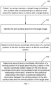
- FIG. 1 is a flowchart of a method for positioning a target area according to an embodiment of the present disclosure.
- the method for positioning a target area includes the following steps.
- Step 100 Collect, by using a camera, a target image including a skin surface area corresponding to a reactive bone, where the reactive bone is a bone with a target feature.
- the camera may be any one of a monocular camera, a binocular camera, a multiocular camera, and a 3D-structured optical camera.
- the reactive bone may be a bone whose spatial positional relationship with a human skin surface does not change in a natural state.
- the reactive bone may be a nasal bone, a sacrococcygeal bone, or the like.
- the skin surface area corresponding to the reactive bone refers to an area that is on the human skin surface, and has the same position as the reactive bone, but has a different depth.
- the skin surface area may be an area including the nose; or when the reactive bone is a sacrococcygeal bone, the skin surface area may be a sacrococcygeal triangular area.
- Step 101 Identify the skin surface area from the target image.
- the identifying the skin surface area from the target image includes: performing image enhancement processing on the target image; and inputting a target image subjected to the image enhancement processing into a trained classification model to obtain first pixel coordinate information of the skin surface area in a pixel coordinate system.
- the identifying the skin surface area from the target image includes: inputting the target image into a trained classification model to obtain first pixel coordinate information of the skin surface area in a pixel coordinate system.
- the pixel coordinate system is a two-dimensional coordinate system established based on the target image.
- An origin of the pixel coordinate system may be any point on the target image or any point on a nontarget image, for example, may be an upper left corner of the target image.
- One axis of the pixel coordinate system is parallel to a row of the target image, and the other axis thereof is parallel to a column of the target image; or one axis of the pixel coordinate system is parallel to the column of the target image, and the other axis thereof is parallel to the row of the target image.
- Pixel coordinate information of a point on the target image in the pixel coordinate system is discrete, and can only be an integer value in pixels.
- image enhancement processing is performed on the target image.
- a method familiar to those skilled in the art may be adopted to perform image enhancement processing on the target image.
- contrast limited adaptive histogram equalization CLAHE
- CLAHE contrast limited adaptive histogram equalization
- the method before the inputting a target image subjected to the image enhancement processing into a trained classification model to obtain first pixel coordinate information of the skin surface area in a pixel coordinate system, or before the inputting the target image into a trained classification model to obtain first pixel coordinate information of the skin surface area in a pixel coordinate system, the method further includes: collecting a sample image including the skin surface area by using the camera; performing image enhancement processing on the sample image; and performing model training based on a sample image subjected to the image enhancement processing to obtain the classification model.
- the method before the inputting a target image subjected to the image enhancement processing into a trained classification model to obtain first pixel coordinate information of the skin surface area in a pixel coordinate system, or before the inputting the target image into a trained classification model to obtain first pixel coordinate information of the skin surface area in a pixel coordinate system, the method further includes: collecting a sample image including the skin surface area by using the camera; and performing model training based on the sample image to obtain the classification model.
- a model familiar to those skilled in the art may be trained to obtain the classification model.
- a mask region-based convolutional neural network (R-CNN) model may be trained to obtain the classification model.
- a process of implementing the mask R-CNN model roughly includes: labeling a skin surface area of a sample image or a sample image subjected to image enhancement processing to generate a mask label data set; filtering and preprocessing the mask label data set, and dividing a filtered and preprocessed data set to obtain data sets with different pose image combinations; inputting the data sets with different pose image combinations into a pre-trained neural network (such as ResNet) to obtain a corresponding body surface feature map; for a region of interest (ROI) of each point in the body surface feature map, obtaining a bounding box based on the ROI; performing binary classification and bounding-box (BB) regression processing on the bounding box to filter points corresponding to part of lower score ROIs; and performing an ROI alignment operation on the remaining points in the
- Step 102 Determine first device coordinate information of a central position of the skin surface area in a device coordinate system.
- the device may be any device that performs a surgical operation, such as a mechanical arm.
- the device coordinate system is a three-dimensional coordinate system established based on the device.
- the determining first device coordinate information of a central position of the skin surface area in a device coordinate system includes: determining second pixel coordinate information of the central position of the skin surface area in a pixel coordinate system; determining camera coordinate information of the central position of the skin surface area in a camera coordinate system based on the second pixel coordinate information and a first transformation relationship, where the first transformation relationship is a transformation relationship between the pixel coordinate system and the camera coordinate system; and determining the first device coordinate information based on the camera coordinate information and a second transformation relationship, where the second transformation relationship is a transformation relationship between the camera coordinate system and the device coordinate system.
- the camera coordinate system is a three-dimensional coordinate system established based on the camera.
- the first transformation relationship may be represented by a first transformation matrix.
- the camera coordinate system is associated with the pixel coordinate system by using an image physical coordinate system
- the first transformation relationship may be obtained based on a transformation relationship between the camera coordinate system and the image physical coordinate system and a transformation relationship between the image physical coordinate system and the pixel coordinate system.
- the transformation relationship between the camera coordinate system and the image physical coordinate system may be expressed by using a third transformation matrix
- the transformation relationship between the image physical coordinate system and the pixel coordinate system may be expressed by using a fourth transformation matrix
- the first transformation matrix may be determined based on the third transformation matrix and the fourth transformation matrix
- the image physical coordinate system is a two-dimensional coordinate system established on an image sensor.
- An origin of the image physical coordinate system is an intersection of an optical axis of the camera and an imaging plane.
- One axis of the image physical coordinate system is parallel to a row of the image sensor, and the other axis thereof is parallel to a column of the image sensor; or one axis of the image physical coordinate system is parallel to the column of the image sensor, and the other axis thereof is parallel to a row of the image sensor.
- Image physical coordinate information of a point on the image sensor in the image physical coordinate system is discrete, with the length as the unit.
- the camera coordinate system is a three-dimensional coordinate system and the image physical coordinate system is a two-dimensional coordinate system. Therefore, the third transformation matrix is a transformation matrix between the three-dimensional coordinate system and the two-dimensional coordinate system. Specifically, it is assumed that there is a point P on the skin surface area, camera coordinate information of the point P in the camera coordinate system is (Xc, Yc, Zc), and a line OcP connecting an intersection Oc of the optical axis of the camera and the imaging plane to the point P has an intersection p with the camera imaging plane, that is, a projection point of the point P on the camera imaging plane. As shown in FIG.
- ⁇ is a number of pixels in unit length in an x-axis direction
- ⁇ is a number of pixels in unit length in a y-axis direction
- (u, v) is the pixel coordinate information of the point P
- (x, y) is the image physical coordinate information of the point p
- (u 0 , v 0 ) is pixel coordinate information of the origin of the image physical coordinate system in the pixel coordinate system.
- the determining camera coordinate information of the central position of the skin surface area in a camera coordinate system based on the second pixel coordinate information and a first transformation relationship includes: determining the camera coordinate information according to formula (4).
- the second transformation relationship may be represented by a second transformation matrix Le.
- the camera coordinate system and the device coordinate system are both three-dimensional coordinate systems. Therefore, the second transformation matrix is a transformation matrix between the two three-dimensional coordinate systems, and at the same time, the skin surface area changes only in spatial position and orientation in the two three-dimensional coordinate systems, but does not change in shape. Therefore, the second transformation matrix Le may be represented by using a rotation matrix R and a translation matrix T. Specifically, it is assumed that there is a point P on the skin surface area, camera coordinate information of the point P in the camera coordinate system is (Xc, Yc, Zc), device coordinate information of the point P in the device coordinate system is (Xe, Ye, Ze), and a transformation relationship between the two pieces of coordinate information is shown in formula (5).
- R is a 3 ⁇ 3 matrix
- R r 11 r 12 r 13 r 21 r 22 r 23 r 31 r 32 r 33
- T is a translation vector
- T t x t y t z
- Le is an extrinsic matrix of the camera in the device coordinate system.
- the determining the first device coordinate information based on the camera coordinate information and a second transformation relationship includes: determining the first device coordinate information based on formula (5).
- Step 103 Determine second device coordinate information of a central position of a target area including a lesion in the device coordinate system based on the first device coordinate information and predetermined first positional relationship information, where the first positional relationship information is positional relationship information between the central position of the skin surface area and the central position of the target area.
- the lesion may be a tumor, such as hysteromyoma.
- the first positional relationship information is a difference between the first device coordinate information and fourth device coordinate information of the central position of the target area in the device coordinate system; and the determining second device coordinate information of a central position of a target area including a lesion in the device coordinate system based on the first device coordinate information and predetermined first positional relationship information includes: determining that the second device coordinate information is a difference between the first device coordinate information and the first positional relationship information.
- the first positional relationship information is a difference between third nuclear magnetic resonance coordinate information of the central position of the skin surface area in a nuclear magnetic resonance coordinate system and first nuclear magnetic resonance coordinate information of the central position of the target area in the nuclear magnetic resonance coordinate system; and the determining second device coordinate information of a central position of a target area including a lesion in the device coordinate system based on the first device coordinate information and predetermined first positional relationship information includes: determining a difference between the first device coordinate information and the fourth device coordinate information based on a third transformation relationship and the difference between the third nuclear magnetic resonance coordinate information and the first nuclear magnetic resonance coordinate information; and determining that the second device coordinate information is a difference between the first device coordinate information and the difference between the first device coordinate information and the fourth device coordinate information.
- the third transformation relationship is a transformation relationship between the nuclear magnetic resonance coordinate system and the device coordinate system that are two two-dimensional coordinate systems, and the third transformation relationship may be expressed by using a fifth transformation matrix.
- the third transformation matrix is similar to the second transformation matrix Le, and details are not described herein.
- the method before the collecting, by using a camera, a first image including a skin surface area corresponding to a reactive bone, the method further includes: obtaining the first positional relationship information in advance based on a nuclear magnetic resonance image.
- the obtaining the first positional relationship information in advance based on a nuclear magnetic resonance image includes: determining first nuclear magnetic resonance coordinate information of the central position of the target area in a nuclear magnetic resonance coordinate system and second nuclear magnetic resonance coordinate information of a target position of the reactive bone in the nuclear magnetic resonance coordinate system based on the nuclear magnetic resonance image; determining third nuclear magnetic resonance coordinate information of the central position of the skin surface area in the nuclear magnetic resonance coordinate system based on the second nuclear magnetic resonance coordinate information and the nuclear magnetic resonance image; and determining the first positional relationship information based on the first nuclear magnetic resonance coordinate information and the third nuclear magnetic resonance coordinate information.
- the target position of the reactive bone may be a sacrococcygeal alternation part.
- the central position of the skin surface area and the sacrococcygeal alternation part are at different depths but at the same position.
- the determining the first positional relationship information based on the first nuclear magnetic resonance coordinate information and the third nuclear magnetic resonance coordinate information includes: determining that the first positional relationship information is a difference between the first nuclear magnetic resonance coordinate information and the third nuclear magnetic resonance coordinate information; or determining a difference between the first nuclear magnetic resonance coordinate information and the third nuclear magnetic resonance coordinate information; and determining that the first positional relationship information is a product of the difference and a third transformation relationship, where the third transformation relationship is a transformation relationship between the nuclear magnetic resonance coordinate system and the device coordinate system.
- a position of a lesion of a patient is intelligently identified and positioned during the surgical operation, and positioning accuracy of the position of the lesion of the patient is improved. Moreover, there is no need to paste a mark, thereby reducing the workload of a medical worker.
- an electronic device including:
- the processor is a device with a data processing capability, and includes, but is not limited to, a central processing unit (CPU).
- the memory is a device with a data storage capability, and includes, but is not limited to, a random access memory (RAM, more specifically, for example, an SDRAM or a DDR), a read-only memory (ROM), an electrically erasable programmable read-only memory (EEPROM), and a flash memory.
- RAM random access memory
- ROM read-only memory
- EEPROM electrically erasable programmable read-only memory
- the processor and the memory are connected to each other by a bus, and then are connected to other components of a computing device.
- the electronic device further includes: a camera, configured to collect a target image including a skin surface area corresponding to a reactive bone, where the reactive bone is a bone with a target feature.
- another embodiment of the present disclosure provides a computer-readable storage medium, where the computer-readable storage medium stores a computer program thereon, and when the program is executed by a processor, the method for positioning a target area according to any one of the implementations described above is implemented.
- FIG. 3 is a block diagram of composition of an apparatus for positioning a target area according to another embodiment of the present disclosure.
- another embodiment of the present disclosure provides an apparatus for positioning a target area, including: an acquisition module 301, configured to collect, by using a camera, a target image including a skin surface area corresponding to a reactive bone, where the reactive bone is a bone with a target feature; an identification module 302, configured to identify the skin surface area from the target image; and a coordinate information determining module 303, configured to determine first device coordinate information of a central position of the skin surface area in a device coordinate system, and determine second device coordinate information of a central position of a target area including a lesion in the device coordinate system based on the first device coordinate information and predetermined first positional relationship information, where the first positional relationship information is positional relationship information between the central position of the skin surface area and the central position of the target area.
- the identification module 302 is specifically configured to perform image enhancement processing on the target image, and input a target image subjected to the image enhancement processing into a trained classification model to obtain first pixel coordinate information of the skin surface area in a pixel coordinate system.
- the acquisition module 301 is further configured to collect a sample image including the skin surface area by using the camera; and the identification module 302 is further configured to perform image enhancement processing on the sample image; and perform model training based on a sample image subjected to the image enhancement processing to obtain the classification model.
- the coordinate information determining module 303 is specifically configured to implement the "determining first device coordinate information of a central position of the skin surface area in a device coordinate system" in the following way: determining second pixel coordinate information of the central position of the skin surface area in a pixel coordinate system; determining camera coordinate information of the central position of the skin surface area in a camera coordinate system based on the second pixel coordinate information and a first transformation relationship, where the first transformation relationship is a transformation relationship between the pixel coordinate system and the camera coordinate system; and determining the first device coordinate information based on the camera coordinate information and a second transformation relationship, where the second transformation relationship is a transformation relationship between the camera coordinate system and the device coordinate system.
- the first positional relationship information is a difference between the first device coordinate information and fourth device coordinate information of the central position of the target area in the device coordinate system; and the coordinate information determining module 303 is specifically configured to implement the "determining second device coordinate information of a central position of a target area including a lesion in the device coordinate system based on the first device coordinate information and predetermined first positional relationship information" in the following way: determining that the second device coordinate information is a difference between the first device coordinate information and the first positional relationship information.
- the acquisition module 301 is further configured to obtain the first positional relationship information in advance based on a nuclear magnetic resonance image.
- the acquisition module 301 is specifically configured to implement the "obtaining the first positional relationship information in advance based on a nuclear magnetic resonance image" in the following way: determining first nuclear magnetic resonance coordinate information of the central position of the target area in a nuclear magnetic resonance coordinate system and second nuclear magnetic resonance coordinate information of a target position of the reactive bone in the nuclear magnetic resonance coordinate system based on the nuclear magnetic resonance image; determining third nuclear magnetic resonance coordinate information of the central position of the skin surface area in the nuclear magnetic resonance coordinate system based on the second nuclear magnetic resonance coordinate information and the nuclear magnetic resonance image; and determining the first positional relationship information based on the first nuclear magnetic resonance coordinate information and the third nuclear magnetic resonance coordinate information.
- the acquisition module 301 is specifically configured to implement the "determining the first positional relationship information based on the first nuclear magnetic resonance coordinate information and the third nuclear magnetic resonance coordinate information" in the following way: determining that the first positional relationship information is a difference between the first nuclear magnetic resonance coordinate information and the third nuclear magnetic resonance coordinate information; or determining a difference between the first nuclear magnetic resonance coordinate information and the third nuclear magnetic resonance coordinate information; and determining that the first positional relationship information is a product of the difference and a third transformation relationship, where the third transformation relationship is a transformation relationship between the nuclear magnetic resonance coordinate system and the device coordinate system.
- a physical component may have multiple functions, or a function or step may be performed by a plurality of physical components in cooperation.
- Some or all of the physical components may be implemented as software executed by a processor, such as a central processing unit, a digital signal processor or a microprocessor, or as hardware, or as an integrated circuit, such as an application-specific integrated circuit.
- a processor such as a central processing unit, a digital signal processor or a microprocessor, or as hardware, or as an integrated circuit, such as an application-specific integrated circuit.
- Such software may be distributed on computer-readable medium.
- the computer-readable medium may include a computer storage medium (or non-transitory medium) and a communication medium (or transitory medium).
- the term computer storage medium includes volatile and nonvolatile, and removable and non-removable media implemented in any method or technology for storing information (such as computer-readable instructions, data structures, program modules or other data).
- the computer storage medium includes, but is not limited to, a RAM, a ROM, an EEPROM, a flash memory or other memories, a CD-ROM, a digital versatile disk (DVD) or other optical disk storages, magnetic boxes, magnetic tapes, magnetic disk storages or other magnetic storages, or any other medium that may be configured to store desired information and can be accessed by a computer.
- the communication medium usually contains computer-readable instructions, data structures, program modules or other data in a modulated data signal such as a carrier wave or other transmission mechanism, and may include any information delivery medium.
Landscapes
- Engineering & Computer Science (AREA)
- Physics & Mathematics (AREA)
- Theoretical Computer Science (AREA)
- Health & Medical Sciences (AREA)
- Life Sciences & Earth Sciences (AREA)
- General Physics & Mathematics (AREA)
- General Health & Medical Sciences (AREA)
- Computer Vision & Pattern Recognition (AREA)
- Data Mining & Analysis (AREA)
- Biophysics (AREA)
- Biomedical Technology (AREA)
- Molecular Biology (AREA)
- General Engineering & Computer Science (AREA)
- Artificial Intelligence (AREA)
- Evolutionary Computation (AREA)
- Medical Informatics (AREA)
- Software Systems (AREA)
- Computing Systems (AREA)
- Mathematical Physics (AREA)
- Computational Linguistics (AREA)
- Public Health (AREA)
- Veterinary Medicine (AREA)
- Nuclear Medicine, Radiotherapy & Molecular Imaging (AREA)
- Pathology (AREA)
- Heart & Thoracic Surgery (AREA)
- Surgery (AREA)
- Animal Behavior & Ethology (AREA)
- Radiology & Medical Imaging (AREA)
- Bioinformatics & Cheminformatics (AREA)
- Bioinformatics & Computational Biology (AREA)
- Evolutionary Biology (AREA)
- Quality & Reliability (AREA)
- High Energy & Nuclear Physics (AREA)
- Image Analysis (AREA)
Applications Claiming Priority (2)
| Application Number | Priority Date | Filing Date | Title |
|---|---|---|---|
| CN202210234627.8A CN114638798B (zh) | 2022-03-10 | 2022-03-10 | 目标区域的定位方法、电子设备、介质 |
| PCT/CN2023/074338 WO2023169108A1 (zh) | 2022-03-10 | 2023-02-03 | 目标区域的定位方法、电子设备、介质 |
Publications (2)
| Publication Number | Publication Date |
|---|---|
| EP4459545A1 true EP4459545A1 (de) | 2024-11-06 |
| EP4459545A4 EP4459545A4 (de) | 2025-08-27 |
Family
ID=81947625
Family Applications (1)
| Application Number | Title | Priority Date | Filing Date |
|---|---|---|---|
| EP23765687.1A Pending EP4459545A4 (de) | 2022-03-10 | 2023-02-03 | Zielregionpositionierungsverfahren, elektronische vorrichtung und medium |
Country Status (4)
| Country | Link |
|---|---|
| EP (1) | EP4459545A4 (de) |
| KR (1) | KR20240128966A (de) |
| CN (1) | CN114638798B (de) |
| WO (1) | WO2023169108A1 (de) |
Cited By (1)
| Publication number | Priority date | Publication date | Assignee | Title |
|---|---|---|---|---|
| US12582848B2 (en) | 2021-06-07 | 2026-03-24 | The Regents Of The University Of Michigan | Minimally invasive histotripsy systems and methods |
Families Citing this family (2)
| Publication number | Priority date | Publication date | Assignee | Title |
|---|---|---|---|---|
| IL308943A (en) | 2021-06-07 | 2024-01-01 | Univ Michigan Regents | All-inclusive ultrasound systems and methods that include histotripsy |
| CN114638798B (zh) * | 2022-03-10 | 2025-10-21 | 重庆海扶医疗科技股份有限公司 | 目标区域的定位方法、电子设备、介质 |
Family Cites Families (18)
| Publication number | Priority date | Publication date | Assignee | Title |
|---|---|---|---|---|
| GB0015683D0 (en) * | 2000-06-28 | 2000-08-16 | Depuy Int Ltd | Apparatus for positioning a surgical instrument |
| KR20140132525A (ko) * | 2013-05-08 | 2014-11-18 | (주)약침학회 | 3차원 영상 촬영 장치를 이용한 경혈 위치 및 자침 깊이 결정 방법 |
| CN103325143B (zh) * | 2013-06-13 | 2016-10-05 | 华南理工大学 | 基于模型匹配的标记点自动注册方法 |
| TWI670681B (zh) * | 2017-06-04 | 2019-09-01 | 鈦隼生物科技股份有限公司 | 判定手術路徑上一個或多個點之方法和系統 |
| CN107464230B (zh) * | 2017-08-23 | 2020-05-08 | 京东方科技集团股份有限公司 | 图像处理方法及装置 |
| CN109754387B (zh) * | 2018-11-23 | 2021-11-23 | 北京永新医疗设备有限公司 | 一种全身骨显像放射性浓聚灶的智能检测定位方法 |
| CN112890865A (zh) * | 2019-11-19 | 2021-06-04 | 深圳迈瑞生物医疗电子股份有限公司 | 自动注释方法、超声成像系统及计算机存储介质 |
| CN113041519A (zh) * | 2019-12-27 | 2021-06-29 | 重庆海扶医疗科技股份有限公司 | 一种智能空间定位方法 |
| CN111408066B (zh) * | 2020-03-19 | 2021-04-16 | 山东大学 | 基于磁共振影像的肿瘤位置标定系统及设备 |
| CN112053400B (zh) * | 2020-09-09 | 2022-04-05 | 北京柏惠维康科技有限公司 | 数据处理方法及机器人导航系统 |
| CN112258494B (zh) * | 2020-10-30 | 2021-10-22 | 北京柏惠维康科技有限公司 | 一种病灶位置确定方法、装置及电子设备 |
| CN112419309B (zh) * | 2020-12-11 | 2023-04-07 | 上海联影医疗科技股份有限公司 | 医学图像相位确定方法、装置、计算机设备和存储介质 |
| CN113034691B (zh) * | 2021-03-22 | 2025-04-29 | 广州虎牙科技有限公司 | 人体模型的骨骼绑定方法、装置及电子设备 |
| CN115089303B (zh) * | 2021-05-10 | 2026-04-21 | 武汉联影智融医疗科技有限公司 | 机器人定位方法及系统 |
| CN113274130A (zh) * | 2021-05-14 | 2021-08-20 | 上海大学 | 用于光学手术导航系统的无标记手术注册方法 |
| CN113822984B (zh) * | 2021-08-12 | 2025-05-13 | 深圳点猫科技有限公司 | 一种三维模型的生成方法、装置、系统及介质 |
| CN114129240B (zh) * | 2021-12-02 | 2022-11-01 | 推想医疗科技股份有限公司 | 一种引导信息生成方法、系统、装置及电子设备 |
| CN114638798B (zh) * | 2022-03-10 | 2025-10-21 | 重庆海扶医疗科技股份有限公司 | 目标区域的定位方法、电子设备、介质 |
-
2022
- 2022-03-10 CN CN202210234627.8A patent/CN114638798B/zh active Active
-
2023
- 2023-02-03 EP EP23765687.1A patent/EP4459545A4/de active Pending
- 2023-02-03 WO PCT/CN2023/074338 patent/WO2023169108A1/zh not_active Ceased
- 2023-02-03 KR KR1020247025063A patent/KR20240128966A/ko active Pending
Cited By (1)
| Publication number | Priority date | Publication date | Assignee | Title |
|---|---|---|---|---|
| US12582848B2 (en) | 2021-06-07 | 2026-03-24 | The Regents Of The University Of Michigan | Minimally invasive histotripsy systems and methods |
Also Published As
| Publication number | Publication date |
|---|---|
| EP4459545A4 (de) | 2025-08-27 |
| CN114638798A (zh) | 2022-06-17 |
| WO2023169108A1 (zh) | 2023-09-14 |
| CN114638798B (zh) | 2025-10-21 |
| KR20240128966A (ko) | 2024-08-27 |
Similar Documents
| Publication | Publication Date | Title |
|---|---|---|
| EP4459545A1 (de) | Zielregionpositionierungsverfahren, elektronische vorrichtung und medium | |
| EP3509013B1 (de) | Identifizieren eines vordefinierten objekts in einem satz von bildern von einem medizinischen bildscanner während eines chirurgischen eingriffs | |
| CN115590623B (zh) | 穿刺路径规划系统 | |
| EP3307169B1 (de) | Echtzeit-kollimation und roi-filterpositionierung in der röntgenbildgebung durch die automatische erkennung der interessenspunkte | |
| Fu et al. | Safe motion planning for steerable needles using cost maps automatically extracted from pulmonary images | |
| CN116570370B (zh) | 一种脊柱针刀穿刺导航系统 | |
| CN111493918B (zh) | 腰椎ct影像的观测面自动定位方法、应用方法及设备 | |
| Yang et al. | Improving catheter segmentation & localization in 3d cardiac ultrasound using direction-fused fcn | |
| CN113274130A (zh) | 用于光学手术导航系统的无标记手术注册方法 | |
| CN109816665B (zh) | 一种光学相干断层扫描图像的快速分割方法及装置 | |
| CN109308735A (zh) | 基于Unity3D体渲染的数据开洞的方法及其存储介质 | |
| CN112184720B (zh) | 一种ct图像的内直肌和视神经分割方法及系统 | |
| CN119359971B (zh) | 一种麻醉穿刺辅助定位方法 | |
| CN113693739B (zh) | 肿瘤导航修正方法、装置及便携式荧光影像导航设备 | |
| RU2839094C2 (ru) | Способ позиционирования целевой области, электронное устройство и носитель | |
| CN112704566A (zh) | 手术耗材核查方法及手术机器人系统 | |
| CN118037650A (zh) | 一种基于弱监督学习的视网膜脱离区定位方法和系统 | |
| CN117934386A (zh) | 颅内血肿穿刺定位方法、装置、电子设备及存储介质 | |
| CN116012286B (zh) | 手术风险区域确定方法、装置、电子设备及可读存储介质 | |
| US20250268661A1 (en) | Apparatus and methods for performing a medical procedure | |
| CN119006496B (zh) | 一种脊柱分割方法、设备、介质和程序产品 | |
| Vivekananda et al. | Deep Learning for Liver Cancer Segmentation: Combining Residual Blocks and Multi-Scale Feature Extraction | |
| CN118053195A (zh) | 超广域眼底图像识别方法、装置、终端设备及存储介质 | |
| CN120918790A (zh) | 一种避障提示方法、装置、介质及设备 | |
| CN119130906A (zh) | 图像处理方法、眼底图像对比方法、密度变化图应用方法 |
Legal Events
| Date | Code | Title | Description |
|---|---|---|---|
| STAA | Information on the status of an ep patent application or granted ep patent |
Free format text: STATUS: THE INTERNATIONAL PUBLICATION HAS BEEN MADE |
|
| PUAI | Public reference made under article 153(3) epc to a published international application that has entered the european phase |
Free format text: ORIGINAL CODE: 0009012 |
|
| STAA | Information on the status of an ep patent application or granted ep patent |
Free format text: STATUS: REQUEST FOR EXAMINATION WAS MADE |
|
| 17P | Request for examination filed |
Effective date: 20240731 |
|
| AK | Designated contracting states |
Kind code of ref document: A1 Designated state(s): AL AT BE BG CH CY CZ DE DK EE ES FI FR GB GR HR HU IE IS IT LI LT LU LV MC ME MK MT NL NO PL PT RO RS SE SI SK SM TR |
|
| DAV | Request for validation of the european patent (deleted) | ||
| DAX | Request for extension of the european patent (deleted) | ||
| A4 | Supplementary search report drawn up and despatched |
Effective date: 20250728 |
|
| RIC1 | Information provided on ipc code assigned before grant |
Ipc: G06T 7/00 20170101AFI20250722BHEP Ipc: A61B 5/00 20060101ALI20250722BHEP Ipc: A61B 5/055 20060101ALI20250722BHEP Ipc: G06N 3/045 20230101ALI20250722BHEP Ipc: G06T 7/73 20170101ALI20250722BHEP Ipc: A61B 34/20 20160101ALI20250722BHEP Ipc: A61B 90/00 20160101ALI20250722BHEP Ipc: G06N 3/02 20060101ALI20250722BHEP |