EP4455635A2 - Probenanalyseverfahren zum nachweis von blastzellen, probenanalysegerät, reagenz und verwendung davon - Google Patents
Probenanalyseverfahren zum nachweis von blastzellen, probenanalysegerät, reagenz und verwendung davon Download PDFInfo
- Publication number
- EP4455635A2 EP4455635A2 EP24172036.6A EP24172036A EP4455635A2 EP 4455635 A2 EP4455635 A2 EP 4455635A2 EP 24172036 A EP24172036 A EP 24172036A EP 4455635 A2 EP4455635 A2 EP 4455635A2
- Authority
- EP
- European Patent Office
- Prior art keywords
- light intensity
- region information
- intensity signal
- sample
- reagent
- Prior art date
- Legal status (The legal status is an assumption and is not a legal conclusion. Google has not performed a legal analysis and makes no representation as to the accuracy of the status listed.)
- Pending
Links
- 210000003969 blast cell Anatomy 0.000 title claims abstract description 297
- 239000003153 chemical reaction reagent Substances 0.000 title claims abstract description 223
- 238000012284 sample analysis method Methods 0.000 title claims abstract description 63
- 230000003287 optical effect Effects 0.000 claims abstract description 162
- 239000003795 chemical substances by application Substances 0.000 claims abstract description 155
- 238000001514 detection method Methods 0.000 claims abstract description 141
- 239000003219 hemolytic agent Substances 0.000 claims abstract description 139
- 238000010186 staining Methods 0.000 claims abstract description 103
- 239000002245 particle Substances 0.000 claims abstract description 88
- 239000007850 fluorescent dye Substances 0.000 claims abstract description 79
- -1 dehydrated sorbitan fatty acid ester Chemical class 0.000 claims abstract description 60
- 239000002736 nonionic surfactant Substances 0.000 claims abstract description 51
- 235000014113 dietary fatty acids Nutrition 0.000 claims abstract description 42
- 239000000194 fatty acid Substances 0.000 claims abstract description 42
- 229930195729 fatty acid Natural products 0.000 claims abstract description 42
- 238000000034 method Methods 0.000 claims abstract description 40
- 210000000265 leukocyte Anatomy 0.000 claims description 74
- 238000011282 treatment Methods 0.000 claims description 52
- 101000738771 Homo sapiens Receptor-type tyrosine-protein phosphatase C Proteins 0.000 claims description 38
- 102100037422 Receptor-type tyrosine-protein phosphatase C Human genes 0.000 claims description 38
- 229920000136 polysorbate Polymers 0.000 claims description 18
- 210000000170 cell membrane Anatomy 0.000 claims description 16
- MTHSVFCYNBDYFN-UHFFFAOYSA-N diethylene glycol Chemical compound OCCOCCO MTHSVFCYNBDYFN-UHFFFAOYSA-N 0.000 claims description 12
- 229920003171 Poly (ethylene oxide) Polymers 0.000 claims description 9
- 210000005259 peripheral blood Anatomy 0.000 claims description 8
- 239000011886 peripheral blood Substances 0.000 claims description 8
- 239000003093 cationic surfactant Substances 0.000 claims description 7
- 229920001213 Polysorbate 20 Polymers 0.000 claims description 6
- NWGKJDSIEKMTRX-AAZCQSIUSA-N Sorbitan monooleate Chemical compound CCCCCCCC\C=C/CCCCCCCC(=O)OC[C@@H](O)[C@H]1OC[C@H](O)[C@H]1O NWGKJDSIEKMTRX-AAZCQSIUSA-N 0.000 claims description 6
- 229930182470 glycoside Natural products 0.000 claims description 6
- 239000000256 polyoxyethylene sorbitan monolaurate Substances 0.000 claims description 6
- 235000010486 polyoxyethylene sorbitan monolaurate Nutrition 0.000 claims description 6
- IYFATESGLOUGBX-YVNJGZBMSA-N Sorbitan monopalmitate Chemical compound CCCCCCCCCCCCCCCC(=O)OC[C@@H](O)[C@H]1OC[C@H](O)[C@H]1O IYFATESGLOUGBX-YVNJGZBMSA-N 0.000 claims description 5
- 150000003242 quaternary ammonium salts Chemical class 0.000 claims description 4
- 229930002600 steroidal saponin Natural products 0.000 claims description 3
- 150000008130 triterpenoid saponins Chemical class 0.000 claims description 3
- 230000001678 irradiating effect Effects 0.000 claims 5
- 210000004369 blood Anatomy 0.000 abstract description 42
- 239000008280 blood Substances 0.000 abstract description 42
- HEMHJVSKTPXQMS-UHFFFAOYSA-M Sodium hydroxide Chemical compound [OH-].[Na+] HEMHJVSKTPXQMS-UHFFFAOYSA-M 0.000 description 66
- 210000004027 cell Anatomy 0.000 description 59
- LYCAIKOWRPUZTN-UHFFFAOYSA-N Ethylene glycol Chemical compound OCCO LYCAIKOWRPUZTN-UHFFFAOYSA-N 0.000 description 42
- IAZDPXIOMUYVGZ-UHFFFAOYSA-N Dimethylsulphoxide Chemical compound CS(C)=O IAZDPXIOMUYVGZ-UHFFFAOYSA-N 0.000 description 24
- 238000002360 preparation method Methods 0.000 description 22
- 239000003146 anticoagulant agent Substances 0.000 description 20
- 229940127219 anticoagulant drug Drugs 0.000 description 20
- 238000004590 computer program Methods 0.000 description 19
- 239000000203 mixture Substances 0.000 description 19
- 150000007523 nucleic acids Chemical class 0.000 description 18
- 102000039446 nucleic acids Human genes 0.000 description 18
- 108020004707 nucleic acids Proteins 0.000 description 18
- 238000010586 diagram Methods 0.000 description 17
- 210000000601 blood cell Anatomy 0.000 description 15
- KCXVZYZYPLLWCC-UHFFFAOYSA-N EDTA Chemical compound OC(=O)CN(CC(O)=O)CCN(CC(O)=O)CC(O)=O KCXVZYZYPLLWCC-UHFFFAOYSA-N 0.000 description 13
- ADWNFGORSPBALY-UHFFFAOYSA-M sodium;2-[dodecyl(methyl)amino]acetate Chemical compound [Na+].CCCCCCCCCCCCN(C)CC([O-])=O ADWNFGORSPBALY-UHFFFAOYSA-M 0.000 description 13
- 238000009472 formulation Methods 0.000 description 12
- JKMHFZQWWAIEOD-UHFFFAOYSA-N 2-[4-(2-hydroxyethyl)piperazin-1-yl]ethanesulfonic acid Chemical compound OCC[NH+]1CCN(CCS([O-])(=O)=O)CC1 JKMHFZQWWAIEOD-UHFFFAOYSA-N 0.000 description 11
- 239000007995 HEPES buffer Substances 0.000 description 11
- 230000005284 excitation Effects 0.000 description 11
- IWZKICVEHNUQTL-UHFFFAOYSA-M potassium hydrogen phthalate Chemical compound [K+].OC(=O)C1=CC=CC=C1C([O-])=O IWZKICVEHNUQTL-UHFFFAOYSA-M 0.000 description 11
- XLYOFNOQVPJJNP-UHFFFAOYSA-N water Substances O XLYOFNOQVPJJNP-UHFFFAOYSA-N 0.000 description 11
- 239000000872 buffer Substances 0.000 description 10
- 239000000427 antigen Substances 0.000 description 9
- 102000036639 antigens Human genes 0.000 description 9
- 108091007433 antigens Proteins 0.000 description 9
- 239000000975 dye Substances 0.000 description 9
- MHMNJMPURVTYEJ-UHFFFAOYSA-N fluorescein-5-isothiocyanate Chemical compound O1C(=O)C2=CC(N=C=S)=CC=C2C21C1=CC=C(O)C=C1OC1=CC(O)=CC=C21 MHMNJMPURVTYEJ-UHFFFAOYSA-N 0.000 description 9
- 208000006664 Precursor Cell Lymphoblastic Leukemia-Lymphoma Diseases 0.000 description 8
- 210000003743 erythrocyte Anatomy 0.000 description 8
- 239000003550 marker Substances 0.000 description 8
- 239000004094 surface-active agent Substances 0.000 description 8
- 230000027455 binding Effects 0.000 description 7
- 239000006184 cosolvent Substances 0.000 description 7
- 239000000126 substance Substances 0.000 description 7
- 235000010482 polyoxyethylene sorbitan monooleate Nutrition 0.000 description 6
- 229920000053 polysorbate 80 Polymers 0.000 description 6
- 208000024893 Acute lymphoblastic leukemia Diseases 0.000 description 5
- 208000014697 Acute lymphocytic leukaemia Diseases 0.000 description 5
- 150000001875 compounds Chemical class 0.000 description 5
- 230000003834 intracellular effect Effects 0.000 description 5
- 239000008363 phosphate buffer Substances 0.000 description 5
- 150000003839 salts Chemical class 0.000 description 5
- LFQSCWFLJHTTHZ-UHFFFAOYSA-N Ethanol Chemical compound CCO LFQSCWFLJHTTHZ-UHFFFAOYSA-N 0.000 description 4
- 239000004721 Polyphenylene oxide Substances 0.000 description 4
- 238000011534 incubation Methods 0.000 description 4
- 210000004698 lymphocyte Anatomy 0.000 description 4
- 210000001616 monocyte Anatomy 0.000 description 4
- 210000000440 neutrophil Anatomy 0.000 description 4
- 229920000570 polyether Polymers 0.000 description 4
- 229920000056 polyoxyethylene ether Polymers 0.000 description 4
- 229940051841 polyoxyethylene ether Drugs 0.000 description 4
- 239000000243 solution Substances 0.000 description 4
- 238000012360 testing method Methods 0.000 description 4
- FFJCNSLCJOQHKM-CLFAGFIQSA-N (z)-1-[(z)-octadec-9-enoxy]octadec-9-ene Chemical compound CCCCCCCC\C=C/CCCCCCCCOCCCCCCCC\C=C/CCCCCCCC FFJCNSLCJOQHKM-CLFAGFIQSA-N 0.000 description 3
- QTBSBXVTEAMEQO-UHFFFAOYSA-N Acetic acid Chemical compound CC(O)=O QTBSBXVTEAMEQO-UHFFFAOYSA-N 0.000 description 3
- WSFSSNUMVMOOMR-UHFFFAOYSA-N Formaldehyde Chemical compound O=C WSFSSNUMVMOOMR-UHFFFAOYSA-N 0.000 description 3
- OKKJLVBELUTLKV-UHFFFAOYSA-N Methanol Chemical compound OC OKKJLVBELUTLKV-UHFFFAOYSA-N 0.000 description 3
- DNIAPMSPPWPWGF-UHFFFAOYSA-N Propylene glycol Chemical compound CC(O)CO DNIAPMSPPWPWGF-UHFFFAOYSA-N 0.000 description 3
- 238000004458 analytical method Methods 0.000 description 3
- KRKNYBCHXYNGOX-UHFFFAOYSA-N citric acid Chemical compound OC(=O)CC(O)(C(O)=O)CC(O)=O KRKNYBCHXYNGOX-UHFFFAOYSA-N 0.000 description 3
- 230000009089 cytolysis Effects 0.000 description 3
- 201000010099 disease Diseases 0.000 description 3
- 208000037265 diseases, disorders, signs and symptoms Diseases 0.000 description 3
- 239000012634 fragment Substances 0.000 description 3
- 208000003747 lymphoid leukemia Diseases 0.000 description 3
- 241000894007 species Species 0.000 description 3
- ALYNCZNDIQEVRV-UHFFFAOYSA-N 4-aminobenzoic acid Chemical compound NC1=CC=C(C(O)=O)C=C1 ALYNCZNDIQEVRV-UHFFFAOYSA-N 0.000 description 2
- RTZKZFJDLAIYFH-UHFFFAOYSA-N Diethyl ether Chemical compound CCOCC RTZKZFJDLAIYFH-UHFFFAOYSA-N 0.000 description 2
- NBIIXXVUZAFLBC-UHFFFAOYSA-N Phosphoric acid Chemical compound OP(O)(O)=O NBIIXXVUZAFLBC-UHFFFAOYSA-N 0.000 description 2
- JUJWROOIHBZHMG-UHFFFAOYSA-N Pyridine Chemical compound C1=CC=NC=C1 JUJWROOIHBZHMG-UHFFFAOYSA-N 0.000 description 2
- PXIPVTKHYLBLMZ-UHFFFAOYSA-N Sodium azide Chemical compound [Na+].[N-]=[N+]=[N-] PXIPVTKHYLBLMZ-UHFFFAOYSA-N 0.000 description 2
- FAPWRFPIFSIZLT-UHFFFAOYSA-M Sodium chloride Chemical compound [Na+].[Cl-] FAPWRFPIFSIZLT-UHFFFAOYSA-M 0.000 description 2
- HVUMOYIDDBPOLL-XWVZOOPGSA-N Sorbitan monostearate Chemical compound CCCCCCCCCCCCCCCCCC(=O)OC[C@@H](O)[C@H]1OC[C@H](O)[C@H]1O HVUMOYIDDBPOLL-XWVZOOPGSA-N 0.000 description 2
- KKEYFWRCBNTPAC-UHFFFAOYSA-N Terephthalic acid Chemical compound OC(=O)C1=CC=C(C(O)=O)C=C1 KKEYFWRCBNTPAC-UHFFFAOYSA-N 0.000 description 2
- 230000002159 abnormal effect Effects 0.000 description 2
- 239000000980 acid dye Substances 0.000 description 2
- 125000000217 alkyl group Chemical group 0.000 description 2
- 210000002390 cell membrane structure Anatomy 0.000 description 2
- 238000003745 diagnosis Methods 0.000 description 2
- 230000000694 effects Effects 0.000 description 2
- 208000014951 hematologic disease Diseases 0.000 description 2
- 230000002949 hemolytic effect Effects 0.000 description 2
- 208000032839 leukemia Diseases 0.000 description 2
- QJGQUHMNIGDVPM-UHFFFAOYSA-N nitrogen group Chemical group [N] QJGQUHMNIGDVPM-UHFFFAOYSA-N 0.000 description 2
- XNGIFLGASWRNHJ-UHFFFAOYSA-N phthalic acid Chemical compound OC(=O)C1=CC=CC=C1C(O)=O XNGIFLGASWRNHJ-UHFFFAOYSA-N 0.000 description 2
- 239000002904 solvent Substances 0.000 description 2
- 230000009870 specific binding Effects 0.000 description 2
- JOXIMZWYDAKGHI-UHFFFAOYSA-N toluene-4-sulfonic acid Chemical compound CC1=CC=C(S(O)(=O)=O)C=C1 JOXIMZWYDAKGHI-UHFFFAOYSA-N 0.000 description 2
- FJKROLUGYXJWQN-UHFFFAOYSA-N 4-hydroxybenzoic acid Chemical compound OC(=O)C1=CC=C(O)C=C1 FJKROLUGYXJWQN-UHFFFAOYSA-N 0.000 description 1
- BSYNRYMUTXBXSQ-FOQJRBATSA-N 59096-14-9 Chemical compound CC(=O)OC1=CC=CC=C1[14C](O)=O BSYNRYMUTXBXSQ-FOQJRBATSA-N 0.000 description 1
- 102000001554 Hemoglobins Human genes 0.000 description 1
- 108010054147 Hemoglobins Proteins 0.000 description 1
- 206010027476 Metastases Diseases 0.000 description 1
- 229910019142 PO4 Inorganic materials 0.000 description 1
- 150000001335 aliphatic alkanes Chemical class 0.000 description 1
- 229910000147 aluminium phosphate Inorganic materials 0.000 description 1
- 150000001408 amides Chemical class 0.000 description 1
- 229960004050 aminobenzoic acid Drugs 0.000 description 1
- SRSXLGNVWSONIS-UHFFFAOYSA-N benzenesulfonic acid Chemical compound OS(=O)(=O)C1=CC=CC=C1 SRSXLGNVWSONIS-UHFFFAOYSA-N 0.000 description 1
- 229940092714 benzenesulfonic acid Drugs 0.000 description 1
- MVIOINXPSFUJEN-UHFFFAOYSA-N benzenesulfonic acid;hydrate Chemical compound O.OS(=O)(=O)C1=CC=CC=C1 MVIOINXPSFUJEN-UHFFFAOYSA-N 0.000 description 1
- 201000006491 bone marrow cancer Diseases 0.000 description 1
- 239000002738 chelating agent Substances 0.000 description 1
- 235000019864 coconut oil Nutrition 0.000 description 1
- 239000003240 coconut oil Substances 0.000 description 1
- 238000004883 computer application Methods 0.000 description 1
- 238000007796 conventional method Methods 0.000 description 1
- 238000011161 development Methods 0.000 description 1
- 230000004069 differentiation Effects 0.000 description 1
- RZXUTLHEWLEYBU-UHFFFAOYSA-N dodecyl 2-hydroxyethanesulfonate;sodium Chemical compound [Na].CCCCCCCCCCCCOS(=O)(=O)CCO RZXUTLHEWLEYBU-UHFFFAOYSA-N 0.000 description 1
- 238000005516 engineering process Methods 0.000 description 1
- 210000003979 eosinophil Anatomy 0.000 description 1
- 235000019387 fatty acid methyl ester Nutrition 0.000 description 1
- 238000000684 flow cytometry Methods 0.000 description 1
- 150000004676 glycans Chemical class 0.000 description 1
- 238000000338 in vitro Methods 0.000 description 1
- 208000015181 infectious disease Diseases 0.000 description 1
- 230000003993 interaction Effects 0.000 description 1
- 239000012528 membrane Substances 0.000 description 1
- 229910052751 metal Inorganic materials 0.000 description 1
- 239000002184 metal Substances 0.000 description 1
- 230000009401 metastasis Effects 0.000 description 1
- 239000011259 mixed solution Substances 0.000 description 1
- 150000002772 monosaccharides Chemical class 0.000 description 1
- 210000001167 myeloblast Anatomy 0.000 description 1
- 208000025113 myeloid leukemia Diseases 0.000 description 1
- 210000003914 myeloid leukocyte Anatomy 0.000 description 1
- 230000003204 osmotic effect Effects 0.000 description 1
- NBIIXXVUZAFLBC-UHFFFAOYSA-K phosphate Chemical compound [O-]P([O-])([O-])=O NBIIXXVUZAFLBC-UHFFFAOYSA-K 0.000 description 1
- 239000010452 phosphate Substances 0.000 description 1
- 229920001282 polysaccharide Polymers 0.000 description 1
- 239000005017 polysaccharide Substances 0.000 description 1
- 239000003755 preservative agent Substances 0.000 description 1
- 230000002335 preservative effect Effects 0.000 description 1
- 150000003138 primary alcohols Chemical class 0.000 description 1
- UMJSCPRVCHMLSP-UHFFFAOYSA-N pyridine Natural products COC1=CC=CN=C1 UMJSCPRVCHMLSP-UHFFFAOYSA-N 0.000 description 1
- 238000012216 screening Methods 0.000 description 1
- 150000003333 secondary alcohols Chemical class 0.000 description 1
- 230000035945 sensitivity Effects 0.000 description 1
- 150000003384 small molecules Chemical class 0.000 description 1
- 229910052708 sodium Inorganic materials 0.000 description 1
- 239000011734 sodium Substances 0.000 description 1
- 239000011780 sodium chloride Substances 0.000 description 1
- 159000000000 sodium salts Chemical class 0.000 description 1
- 238000011895 specific detection Methods 0.000 description 1
- LMBFAGIMSUYTBN-MPZNNTNKSA-N teixobactin Chemical compound C([C@H](C(=O)N[C@@H]([C@@H](C)CC)C(=O)N[C@@H](CO)C(=O)N[C@H](CCC(N)=O)C(=O)N[C@H]([C@@H](C)CC)C(=O)N[C@@H]([C@@H](C)CC)C(=O)N[C@@H](CO)C(=O)N[C@H]1C(N[C@@H](C)C(=O)N[C@@H](C[C@@H]2NC(=N)NC2)C(=O)N[C@H](C(=O)O[C@H]1C)[C@@H](C)CC)=O)NC)C1=CC=CC=C1 LMBFAGIMSUYTBN-MPZNNTNKSA-N 0.000 description 1
- 238000012546 transfer Methods 0.000 description 1
- 230000009466 transformation Effects 0.000 description 1
Images




Classifications
-
- G—PHYSICS
- G01—MEASURING; TESTING
- G01N—INVESTIGATING OR ANALYSING MATERIALS BY DETERMINING THEIR CHEMICAL OR PHYSICAL PROPERTIES
- G01N15/00—Investigating characteristics of particles; Investigating permeability, pore-volume or surface-area of porous materials
- G01N15/10—Investigating individual particles
- G01N15/14—Optical investigation techniques, e.g. flow cytometry
- G01N15/1434—Optical arrangements
- G01N15/1436—Optical arrangements the optical arrangement forming an integrated apparatus with the sample container, e.g. a flow cell
-
- G—PHYSICS
- G01—MEASURING; TESTING
- G01N—INVESTIGATING OR ANALYSING MATERIALS BY DETERMINING THEIR CHEMICAL OR PHYSICAL PROPERTIES
- G01N15/00—Investigating characteristics of particles; Investigating permeability, pore-volume or surface-area of porous materials
- G01N15/10—Investigating individual particles
- G01N15/14—Optical investigation techniques, e.g. flow cytometry
- G01N15/1456—Optical investigation techniques, e.g. flow cytometry without spatial resolution of the texture or inner structure of the particle, e.g. processing of pulse signals
- G01N15/1459—Optical investigation techniques, e.g. flow cytometry without spatial resolution of the texture or inner structure of the particle, e.g. processing of pulse signals the analysis being performed on a sample stream
-
- G—PHYSICS
- G01—MEASURING; TESTING
- G01N—INVESTIGATING OR ANALYSING MATERIALS BY DETERMINING THEIR CHEMICAL OR PHYSICAL PROPERTIES
- G01N15/00—Investigating characteristics of particles; Investigating permeability, pore-volume or surface-area of porous materials
- G01N15/10—Investigating individual particles
- G01N15/14—Optical investigation techniques, e.g. flow cytometry
- G01N15/1429—Signal processing
-
- G—PHYSICS
- G01—MEASURING; TESTING
- G01N—INVESTIGATING OR ANALYSING MATERIALS BY DETERMINING THEIR CHEMICAL OR PHYSICAL PROPERTIES
- G01N21/00—Investigating or analysing materials by the use of optical means, i.e. using sub-millimetre waves, infrared, visible or ultraviolet light
- G01N21/17—Systems in which incident light is modified in accordance with the properties of the material investigated
- G01N21/47—Scattering, i.e. diffuse reflection
- G01N21/49—Scattering, i.e. diffuse reflection within a body or fluid
- G01N21/51—Scattering, i.e. diffuse reflection within a body or fluid inside a container, e.g. in an ampoule
-
- G—PHYSICS
- G01—MEASURING; TESTING
- G01N—INVESTIGATING OR ANALYSING MATERIALS BY DETERMINING THEIR CHEMICAL OR PHYSICAL PROPERTIES
- G01N21/00—Investigating or analysing materials by the use of optical means, i.e. using sub-millimetre waves, infrared, visible or ultraviolet light
- G01N21/62—Systems in which the material investigated is excited whereby it emits light or causes a change in wavelength of the incident light
- G01N21/63—Systems in which the material investigated is excited whereby it emits light or causes a change in wavelength of the incident light optically excited
- G01N21/64—Fluorescence; Phosphorescence
- G01N21/6428—Measuring fluorescence of fluorescent products of reactions or of fluorochrome labelled reactive substances, e.g. measuring quenching effects, using measuring "optrodes"
-
- G—PHYSICS
- G01—MEASURING; TESTING
- G01N—INVESTIGATING OR ANALYSING MATERIALS BY DETERMINING THEIR CHEMICAL OR PHYSICAL PROPERTIES
- G01N15/00—Investigating characteristics of particles; Investigating permeability, pore-volume or surface-area of porous materials
- G01N15/01—Investigating characteristics of particles; Investigating permeability, pore-volume or surface-area of porous materials specially adapted for biological cells, e.g. blood cells
- G01N2015/011—Investigating characteristics of particles; Investigating permeability, pore-volume or surface-area of porous materials specially adapted for biological cells, e.g. blood cells with lysing, e.g. of erythrocytes
-
- G—PHYSICS
- G01—MEASURING; TESTING
- G01N—INVESTIGATING OR ANALYSING MATERIALS BY DETERMINING THEIR CHEMICAL OR PHYSICAL PROPERTIES
- G01N15/00—Investigating characteristics of particles; Investigating permeability, pore-volume or surface-area of porous materials
- G01N15/01—Investigating characteristics of particles; Investigating permeability, pore-volume or surface-area of porous materials specially adapted for biological cells, e.g. blood cells
- G01N2015/016—White blood cells
-
- G—PHYSICS
- G01—MEASURING; TESTING
- G01N—INVESTIGATING OR ANALYSING MATERIALS BY DETERMINING THEIR CHEMICAL OR PHYSICAL PROPERTIES
- G01N15/00—Investigating characteristics of particles; Investigating permeability, pore-volume or surface-area of porous materials
- G01N15/10—Investigating individual particles
- G01N2015/1006—Investigating individual particles for cytology
-
- G—PHYSICS
- G01—MEASURING; TESTING
- G01N—INVESTIGATING OR ANALYSING MATERIALS BY DETERMINING THEIR CHEMICAL OR PHYSICAL PROPERTIES
- G01N15/00—Investigating characteristics of particles; Investigating permeability, pore-volume or surface-area of porous materials
- G01N15/10—Investigating individual particles
- G01N15/14—Optical investigation techniques, e.g. flow cytometry
- G01N2015/1402—Data analysis by thresholding or gating operations performed on the acquired signals or stored data
-
- G—PHYSICS
- G01—MEASURING; TESTING
- G01N—INVESTIGATING OR ANALYSING MATERIALS BY DETERMINING THEIR CHEMICAL OR PHYSICAL PROPERTIES
- G01N15/00—Investigating characteristics of particles; Investigating permeability, pore-volume or surface-area of porous materials
- G01N15/10—Investigating individual particles
- G01N15/14—Optical investigation techniques, e.g. flow cytometry
- G01N2015/1486—Counting the particles
-
- G—PHYSICS
- G01—MEASURING; TESTING
- G01N—INVESTIGATING OR ANALYSING MATERIALS BY DETERMINING THEIR CHEMICAL OR PHYSICAL PROPERTIES
- G01N21/00—Investigating or analysing materials by the use of optical means, i.e. using sub-millimetre waves, infrared, visible or ultraviolet light
- G01N21/17—Systems in which incident light is modified in accordance with the properties of the material investigated
- G01N21/47—Scattering, i.e. diffuse reflection
- G01N2021/4704—Angular selective
- G01N2021/4711—Multiangle measurement
-
- G—PHYSICS
- G01—MEASURING; TESTING
- G01N—INVESTIGATING OR ANALYSING MATERIALS BY DETERMINING THEIR CHEMICAL OR PHYSICAL PROPERTIES
- G01N21/00—Investigating or analysing materials by the use of optical means, i.e. using sub-millimetre waves, infrared, visible or ultraviolet light
- G01N21/62—Systems in which the material investigated is excited whereby it emits light or causes a change in wavelength of the incident light
- G01N21/63—Systems in which the material investigated is excited whereby it emits light or causes a change in wavelength of the incident light optically excited
- G01N21/64—Fluorescence; Phosphorescence
- G01N21/6428—Measuring fluorescence of fluorescent products of reactions or of fluorochrome labelled reactive substances, e.g. measuring quenching effects, using measuring "optrodes"
- G01N2021/6439—Measuring fluorescence of fluorescent products of reactions or of fluorochrome labelled reactive substances, e.g. measuring quenching effects, using measuring "optrodes" with indicators, stains, dyes, tags, labels, marks
Definitions
- the application related to the field of in vitro diagnosis, in particular to a sample analysis method for detecting blast cells, a sample analyzer, a reagent and a use thereof.
- Immature white blood cells do not appear in peripheral blood of healthy people.
- immatological diseases such as leukemia, metastasis of bone marrow cancer, severe infection and other diseases
- immature white blood cells will appear in blood. Therefore, the determination of immature white blood cells in human peripheral blood is of great significance for disease diagnosis.
- the detection methods for immature blood cells such as lymphoblasts and myeloblasts mainly rely on specific antibodies.
- This method has a good accuracy, but the detection cost is high, and this method has the problems of long time-consuming, complicated operation and high cost when applied to the screening of samples containing blast cells in peripheral blood such as leukemia.
- a rapid detection method for analyzing immature white blood cells based on a blood cell analyzer has been developed, in which a fluorescent dye is mainly used to differentially staining intracellular nucleic acids.
- the methods for detecting nucleic acid have the problems of insufficient accuracy and relatively high false positive and false negative.
- the purpose of the present disclosure is to provide a sample analysis method for detecting blast cells, a sample analyzer, a reagent for the detection and a use thereof.
- the analysis of blast cells in blood samples with the reagent by utilizing the cell membrane structure difference between blast cells and mature leukocytes, increase the detection signal difference between the blast cells and mature leukocytes, and thereby significantly improve the detection accuracy.
- the first aspect of the present disclosure provides a sample analysis method.
- the method comprises the following operations.
- a specimen to be tested of a subject is obtained.
- a first sample is prepared, and the first sample contains at least a part of the specimen to be tested and a first reagent, in which the first reagent comprises a hemolytic agent containing at least one nonionic surfactant comprising a dehydrated sorbitan fatty acid esterand a staining agent containing a fluorescent dye.
- the particles in the first sample is caused to pass one-by-one through a detection area of an optical detection device and the particles in the first sample are irradiated with a light source of the optical detection device to obtain optical signals of the particles.
- First characteristic region information and ghost region information are obtained based on the optical signals, in which the first characteristic region information comprises blast cell information and at least part of the ghost region information.
- the blast cell information is obtained based on the first characteristic region information and the ghost region information.
- the optical signals comprise a side scattered light intensity signal, a forward scattered light intensity signal, and a first fluorescence light intensity signal from the staining agent containing the fluorescent dye.
- Obtaining the first characteristic region information and the ghost region information based on the optical signals comprises: obtaining the first characteristic region information based on the side scattered light intensity signal and the forward scattered light intensity signal, and obtaining the ghost region information based on at least the first fluorescence light intensity signal.
- the second aspect of the present disclosure provides another sample analysis method.
- the method comprises the following operations.
- a specimen to be tested of a subject is obtained.
- a second sample is prepared, in which the second sample comprises at least a portion of the specimen to be tested and a second reagent, and the second reagent comprises a hemolytic agent, a staining agent containing a fluorescent dye and an antibody treatment agent containing a fluorescent labeled CD45.
- the particles in the second sample is caused to pass one-by-one through a detection area of an optical detection device and the particles in the second sample are irradiated with a light source of the optical detection device to obtain optical signals of the particles, in which the optical signals comprise a side scattered light intensity signal, a forward scattered light intensity signal, a first fluorescence light intensity signal from the staining agent containing the fluorescent dye, and a second fluorescence light intensity signal from the antibody treatment agent containing the fluorescent labeled CD45.
- First characteristic region information is obtained based on the side scattered light intensity signal and the second fluorescence light intensity signal
- a ghost region information is obtained based on at least the first fluorescence light intensity signal
- the first characteristic region information comprises blast cell information and the at least part of ghost region information.
- the blast cell information is obtained based on the first characteristic region information and the ghost region information.
- the fluorescent labeled CD45 in the second reagent has a concentration of 25 ⁇ g/mL to 1000 ⁇ g/mL, preferably 50 ⁇ g/mL to 250 ⁇ g/mL.
- the hemolytic agent comprises at least one selected from a group consisting of cationic surfactants, nonionic surfactants, alkyl glycosides, triterpenoid saponins, steroidal saponins, and diethylene glycol.
- the cationic surfactants are selected from quaternary ammonium salts; the nonionic surfactants are selected from polyoxyethylenes and dehydrated sorbitan fatty acid esters. More preferably, the hemolytic agent comprises at least one nonionic surfactant comprising a dehydrated sorbitan fatty acid ester.
- another sample analysis method comprises the following operations.
- a specimen to be tested of a subject is obtained.
- a second sample is prepared, in which the second sample comprises at least a portion of the specimen to be tested and a second reagent, and the second reagent comprises a hemolytic agent, a staining agent containing a fluorescent dye and an antibody treatment agent containing a fluorescent labeled antibody.
- the particles in the second sample is caused to pass one-by-one through a detection area of an optical detection device and the particles in the second sample are irradiated with a light source of the optical detection device to obtain optical signals of the particles, in which the optical signals comprise a side scattered light intensity signal, a forward scattered light intensity signal, a first fluorescence light intensity signal from the staining agent containing the fluorescent dye, and a second fluorescence light intensity signal from the antibody treatment agent containing the fluorescent labeled antibody.
- First characteristic region information is obtained based on the side scattered light intensity signal and the second fluorescence light intensity signal
- a ghost region information is obtained based on at least the first fluorescence light intensity signal
- the first characteristic region information comprises blast cell information and at least part of the ghost region information.
- the blast cell information is obtained based on the first characteristic region information and the ghost region information.
- the third aspect of the present disclosure provides yet another sample analysis method.
- the method comprises the following operations.
- a specimen to be tested is obtained from a subject.
- a third sample is prepared, and the third sample comprises at least a portion of the specimen to be tested and a third reagent, wherein the third reagent comprises at least a hemolytic agent capable of treating respective cell membranes of mature white blood cells and those of blast cells differently, and a staining agent containing a fluorescent dye.
- Particles in the third sample is caused to pass one-by-one through a detection area of an optical detection device, the particles in the third sample are irradiated with a light source of the optical detection device to obtain optical signals of the particles, wherein the optical signals comprise a side scattered light intensity signal, a forward scattered light intensity signal, a first fluorescence light intensity signal from the staining agent containing the fluorescent dye.
- a first characteristic region information is obtained based on the side scattered light intensity signal and the forward scattered light intensity signal
- a ghost region information is obtained based on at least the first fluorescence light intensity signal
- the first characteristic region information comprises blast cell information and at least part of ghost region information.
- the blast cell information is obtained based on the first characteristic region information and the at least part of ghost region information.
- obtaining ghost region information based on at least the first fluorescence light intensity signal comprises: obtaining the ghost region information based on the first fluorescence light intensity signal and any one of the side scattered intensity signal and the forward scatted intensity signal.
- obtaining the blast cell information based on the first characteristic region information and the ghost region information comprises: subtracting the at least part of the ghost region information from the first characteristic region information to obtain the blast cell information.
- the hemolytic agent comprises at least one nonionic surfactant comprising a dehydrated sorbitan fatty acid ester.
- the fluorescent dye has a concentration of 1mg/L to 500mg/L, preferably 10mg/L to 50mg/L.
- the blast cell information includes a count and/or an alarm about blast cells.
- the fourth aspect of the present disclosure provides another sample analysis method.
- the method comprises the following operations.
- a specimen to be tested is obtained from a subject.
- a fourth sample is prepared, and the fourth sample comprises at least a portion of the specimen to be tested and a fourth reagent, wherein the fourth reagent comprises a hemolytic agent containing at least one nonionic surfactant comprising a dehydrated sorbitan fatty acid ester.
- Particles in the fourth sample is caused to pass one-by-one through a detection area of an optical detection device, the particles in the fourth sample are irradiated with a light source of the optical detection device to obtain optical signals of the particles, wherein the optical signals comprise a side scattered light intensity signal and a forward scattered light intensity signal.
- a first characteristic region information is obtained based on the side scattered light intensity signal and the forward scattered light intensity signal, wherein the first characteristic region information comprises blast cell information.
- the blast cell information is obtained based on the first characteristic region information.
- the blast cell information includes an alarm about blast cells.
- the dehydrated sorbitan fatty acid ester is selected from a group consisting of Tween and Span, preferably, the dehydrated sorbitan fatty acid ester is selected from a group consisting of Span 40 ⁇ Span 80 and Tween 20 ⁇ Tween 100.
- the at least one nonionic surfactant comprising a dehydrated sorbitan fatty acid ester has a concentration of 0.2g/L to 2g/L, preferably 0.3g/L to 1.5 g/L.
- the specimen to be tested includes peripheral blood.
- the fifth aspect of the present disclosure provides another sample analysis method.
- the method comprises the following operations.
- the operation instruction is received and detection of blast cell is carried out on the specimen, in which the blast cell detection on the specimen comprises the operations of any of the above sample analysis methods.
- the sixth aspect of the present disclosure provides a sample analyzer.
- the sample analyzer comprises a specimen aspiration device, a sample preparation device, a light source, a flow chamber, optical detectors, a processor, and a non-transitory computer-readable storage medium programmed with computer program instructions.
- the specimen aspiration device is configured to aspirate at least a portion of the specimen to be tested and transport it to the sample preparation device.
- the sample preparation device comprises a reagent supply part and a reaction cell, in which the reagent supply part is configured to provide a first reagent to the reaction cell.
- the reaction cell is configured to mix the first reagent with the at least a portion of the specimen to be tested to obtain a first sample and transport the first sample to the flow chamber, in which the first reagent comprises a hemolytic agent containing at least one nonionic surfactant comprising a dehydrated sorbitan fatty acid ester and a staining agent containing fluorescent dye.
- the light source is configured to align a light beam with a detection hole of the flow chamber connected with the reaction cell.
- the flow chamber is configured to allow particles in the sample to pass through one by one.
- the optical detectors are mounted around the flow chamber for detecting optical signals of the particles passing through the detection hole of the flow chamber.
- the processor is operably connected to the optical detectors.
- first characteristic region information and ghost region information is obtained based on the optical signals, which comprises blast cell information and at least part of ghost region information, and the blast cell information is obtained based on the first characteristic region information and the ghost region information.
- the optical signals comprises a side scattered light intensity signal, a forward scattered light intensity signal, and a first fluorescence light intensity signal from the staining agent containing the fluorescent dye; wherein obtaining the first characteristic region information and the ghost region information based on the optical signals comprises: obtaining the first characteristic region information based on the side scattered light intensity signal and the forward scattered light intensity signal, and obtaining the ghost region information based on at least the first fluorescence light intensity signal.
- obtaining the ghost region information base on at least the first fluorescence light intensity signal comprises: obtaining the ghost region information based on the first fluorescence light intensity signal and any one of the side scattered light intensity signal and the forward scattered light intensity signal.
- the reagent supply part comprises a plurality of reagent storage containers, and the hemolytic agent and the staining agent are respectively stored in different storage containers; or the hemolytic agent and the staining agent are stored in a mixed form in one storage container.
- the blast cell information comprises a count and/or an alarm about blast cells.
- the sample analyzer further comprises a user interface configured to display detection results and/or input instructions to the sample analyzer.
- the seventh aspect of the present disclosure provides another sample analyzer.
- the sample analyzer comprises a specimen aspiration device, a sample preparation device, a light source, a flow chamber, optical detectors, a processor, and a non-transitory computer-readable storage medium programmed with computer program instructions.
- the specimen aspiration device is configured to aspirate at least a portion of the specimen to be tested and transport it to the sample preparation device.
- the sample preparation device comprises a reagent supply part and a reaction cell, in which the reagent supply part is configured to provide a second reagent to the reaction cell, the reaction cell is configured to mix the second reagent with the at least a portion of the specimen to be tested to obtain a second sample and transport the second sample to the flow chamber, in which the second reagent comprises a hemolytic agent, a staining agent containing a fluorescent dye and an antibody treatment agent containing a fluorescent labeled CD45.
- the light source is configured to align a light beam with a detection hole of the flow chamber connected with the reaction cell.
- the flow chamber is configured to allow particles in the sample to pass through one by one.
- the optical detectors are mounted around the flow chamber for detecting optical signals of particles passing through the detection hole of the flow chamber, in which the optical signals comprises a side scattered light intensity signal, a forward scattered light intensity signal, a first fluorescence light intensity signal from the staining agent containing the fluorescent dye, and a second fluorescence light intensity signal from the antibody treatment agent containing the fluorescent labeled CD45.
- the processor is operably connected to the optical detectors. When the computer program instructions programed in the non-transitory computer-readable storage medium is executed by the processor, the processor performs the following operations.
- First characteristic region information is obtained based on the side scattered light intensity signal and the second fluorescence light intensity signal
- ghost region information is obtained based on at least the first fluorescence light intensity signal, in which the first characteristic region information comprises blast cell information and at least part of the ghost region information; and the blast cell information is obtained based on the first characteristic region information and the ghost region information.
- obtaining the ghost region information base on at least the first fluorescence light intensity signal comprises: obtaining the ghost region information based on the first fluorescence light intensity signal and any one of the side scattered light intensity signal and the forward scattered light intensity signal.
- obtaining the blast cell information based on the first characteristic region information and the ghost region information comprises: subtracting the at least part of the ghost region information from the first characteristic region information to obtain the blast cell information.
- the reagent supply part comprises a plurality of reagent storage containers, and the hemolytic agent, the staining agent and the antibody treatment agent are respectively stored in different storage containers; or any two or three of the hemolytic agent, the staining agent and the antibody treatment agent are stored in a mixed form in one storage container.
- the blast cell information comprises a count and/or an alarm about blast cells.
- the sample analyzer further comprises a user interface configured to display detection results and/or input instructions to the sample analyzer.
- the eighth aspect of the present disclosure further provides a sample analyzer.
- the sample analyzer comprises a specimen aspiration device, a sample preparation device, a light source, a flow chamber, optical detectors, a processor, and a non-transitory computer-readable storage medium programmed with computer program instructions.
- the specimen aspiration device is configured to aspirate at least a portion of the specimen to be tested and transport it to the sample preparation device.
- the sample preparation device comprises a reagent supply part and a reaction cell, in which the reagent supply part is configured to provide a second reagent to the reaction cell.
- the reaction cell is configured to mix the second reagent with the at least a portion of the specimen to be tested to obtain a second sample and transport the second sample to the flow chamber, in which the second reagent comprises a hemolytic agent, a staining agent containing a fluorescent dye and an antibody treatment agent containing a fluorescent labeled antibody.
- the light source is configured to align a light beam with a detection hole of the flow chamber connected with the reaction cell.
- the flow chamber is configured to allow particles in the sample to pass through one by one.
- the optical detectors are mounted around the flow chamber for detecting optical signals of particles passing through the detection hole of the flow chamber, in which the optical signals include a side scattered light intensity signal, a forward scattered light intensity signal, a first fluorescence light intensity signal from the staining agent containing the fluorescent dye, and a second fluorescence light intensity signal from the antibody treatment agent containing the fluorescent labeled antibody.
- the processor is operably connected to the optical detectors. When the computer program instructions programed in the non-transitory computer-readable storage medium are executed by the processor, the processor performs the following operations.
- First characteristic region information is obtained based on the side scattered light intensity signal and the second fluorescence light intensity signal
- ghost region information is obtained based on at least the first fluorescence light intensity signal, in which the first characteristic region information comprises blast cell information and at least part of the ghost region information; and the blast cell information is obtained based on the first characteristic region information and the ghost region information.
- the ninth aspect of the present disclosure provides another sample analyzer.
- the sample analyzer comprises a specimen aspiration device, a sample preparation device, a light source, a flow chamber, optical detectors, a processor, and a non-transitory computer-readable storage medium programmed with computer program instructions, wherein, the specimen aspiration device is configured to aspirate at least a portion of the specimen to be tested and transport it to the sample preparation device;
- the sample preparation device comprises a reagent supply part and a reaction cell, wherein the reagent supply part is configured to provide a third reagent to the reaction cell, the reaction cell is configured to mix the third reagent with the at least a portion of the specimen to be tested to obtain a third sample and transport the third sample to the flow chamber, wherein the third reagent comprises at least a hemolytic agent capable of treating respective cell membranes of mature white blood cells and those of blast cells in the third sample differently and a staining agent containing fluorescent dye;
- the light source is configured to align a light beam with
- a first characteristic region information is obtained based on the side scattered light intensity signal and the forward scattered light intensity signal, a ghost region information is obtained based on at least the first fluorescence light intensity signal, wherein the first characteristic region information comprises blast cell information and at least part of the ghost region information.
- the blast cell information is obtained based on the first characteristic region information and the at least part of ghost region information.
- the tenth aspect of the present disclosure provides yet another sample analyzer.
- the sample analyzer comprises a specimen aspiration device, a sample preparation device, a light source, a flow chamber, optical detectors, a processor, and a non-transitory computer-readable storage medium programmed with computer program instructions, wherein, the specimen aspiration device is configured to aspirate at least a portion of the specimen to be tested and transport it to the sample preparation device; the sample preparation device comprises a reagent supply part and a reaction cell, wherein the reagent supply part is configured to provide a fourth reagent to the reaction cell, the reaction cell is configured to mix the fourth reagent with the at least a portion of the specimen to be tested to obtain a fourth sample and transport the fourth sample to the flow chamber, wherein the fourth reagent comprises a hemolytic agent containing at least one nonionic surfactant comprising a dehydrated sorbitan fatty acid ester; the light source is configured to align a light beam with a detection hole of the flow chamber connected with
- the eleventh aspect of the present disclosure provides a reagent for detecting blast cells.
- the reagent comprises a hemolytic agent containing at least one nonionic surfactant comprising a dehydrated sorbitan fatty acid ester, and a staining agent containing a fluorescent dye.
- the at least one nonionic surfactant comprising a dehydrated sorbitan fatty acid ester has a concentration of 0.2g/L to 2g/L, preferably 0.3g/L to 1.5 g/L.
- the at least one nonionic surfactant dehydrated sorbitan fatty acid ester is selected from the group consisting of Tween and Span; preferably, Span is selected from Span 40 ⁇ Span 80 and Tween is selected from Tween 20 ⁇ Tween 100.
- the hemolytic agent further comprises: 0.1g/L to 10g/L, preferably 2g/L to 5g/L of a cosolvent; 0.1 g/L to 10g/L of a buffer agent; and 0.05 g/L to 2g/L, preferably 0.2g/L to 1g/L of an anticoagulant.
- the fluorescent dye is selected from a group consisting of compounds represented by Formula I and Formula II:
- the fluorescent dye in the reagent has a concentration of 1mg/L to 500mg/L, preferably 10mg/L to 50mg/L.
- the reagent further comprises an antibody treatment agent comprising a fluorescent labeled CD45.
- the fluorescent labeled CD45 in the reagent has a concentration of 25 ⁇ g/mL to 1000 ⁇ g/mL, preferably 50 ⁇ g/mL to 250 ⁇ g/mL.
- the hemolytic agent, the staining agent and the antibody treatment agent are separately packaged reagents; or any two or three of the hemolytic agent, the staining agent and the antibody treatment agent form one reagent packaged in a mixed form.
- the twelfth aspect of the present disclosure provides the use of the above reagent for detecting blast cells in a sample using a sample analyzer.
- the hemolytic agent capable to treating respective cell membranes of blast cells and those of white blood cells differently (such as including a dehydrated sorbitan fatty acid ester-based nonionic surfactant as a hemolytic agent) and a staining agent containing a fluorescent dye is used to treat the specimen.
- the hemolytic agent can lyse red blood cells on the one hand, and treat respective the cell membranes of blast cells and those of white blood cells differently on the other hand.
- the dye can preferentially enter mature white blood cells to staining nucleic acids, so as to enlarge the optical characteristics, especially the difference of scattered light signals, between blast cells and white blood cells which combine the fluorescent signals from the nucleic acid dye to realize the distinction between blast cells and white blood cells.
- the specimen is treated with a reagent including a dehydrated sorbitan fatty acid ester-based nonionic surfactant as a hemolytic agent.
- the surfactant can lyse red blood cells on the one hand, and treat respective the cell membranes of blast cells and those of white blood cells differently on the other hand, so as to enlarge the optical characteristics, especially the difference of scattered light signals between blast cells and white blood cells, thereby distinguishing the blast cells from white blood cells, and further alarming the specimen containing blast cells.
- the specimen is treated with a reagent comprising a hemolytic agent, an antibody treatment agent containing a fluorescent labeled CD45, and a staining agent containing a fluorescent dye.
- a fluorescent signal from the fluorescent labeled CD45 is obtained by specific binding of the CD45 to a surface antigen of the blast cell.
- the difference in optical characteristics of the blast cells and the white blood cells can be enlarged by combining the fluorescent signal with a scattered light signal, thereby accurately distinguishing the blast cells from the white blood cells.
- sample analysis method provided by the present disclosure is more sensitive and accurate in identifying blast cells originating from acute lymphoblastic leukemia.
- first and second are used for the purposes of describing only and cannot be understood as indicating or implying the relative importance or implying the number of technical features indicated. Thus, the features defined as “first”, “second” may explicitly or implicitly include at least one of the features.
- blast cells refer to the cells at which various blood cells begin to develop. Blast cells do not appear in the blood of healthy people, but they do appear in the blood of patients with some diseases, especially blood diseases.
- the detection of blood samples based on an automatic blood cell analyzer can only give an alarm about the blast cells possibly existed in the samples, and there is a high proportion of false alarms including both false negative and false positive alarms.
- the fluorescence signal produced by the combination of nucleic acid fluorescent dye and intracellular nucleic acid substance, combined with a scattered light signal is usually used to classify and count white blood cells.
- the detection data will be abnormal, which can give an alarm about the existence of blast cells.
- the intracellular nucleic acid content of blast cells is similar to that of white blood cells. Such method cannot distinguish blast cells from white blood cells well, thus leading to insufficient detection accuracy of blast cells.
- the present disclosure provides a sample analysis method. Exemplary embodiments of the sample analysis method are described in detail below.
- the sample analysis method according to the first embodiment is based on the discovery of the inventors.
- the inventors have found that when specimens are treated with different hemolytic agents, due to certain differences in cell membrane structures of blast cells and white blood cells, when samples are treated with dehydrated sorbitan fatty acid ester-based nonionic surfactants such as Tween and/or Span as hemolytic agents, compared with other commonly used hemolytic agents such as quaternary ammonium salts, treatment of cell membranes of blast cells and white blood cells differently can be realized.
- the treatment of cell membranes by this hemolytic agent further leads to the preferential entry of fluorescent dyes into white blood cells and the combination of nucleic acid substances, thus increasing the intracellular structure difference between blast cells and white blood cells, and increasing the difference of light signals, especially scattered light signals, so that blast cells and white blood cells can be better distinguished.
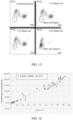
- FIG. 1 is a principle schematic diagram of a sample analysis method according to an embodiment of the present disclosure. It should be understood that this figure only schematically shows the relative positional relationship of the regions of occurrence of various particle swarms in each two-dimensional scatter plot, and for different blood samples, the specific regions of occurrence of various particle clusters may be different from those shown in the figure.
- the left graph of FIG. 1 is a principle schematic diagram of a sample analysis method according to an embodiment of the present disclosure. It should be understood that this figure only schematically shows the relative positional relationship of the regions of occurrence of various particle swarms in each two-dimensional scatter plot, and for different blood samples, the specific regions of occurrence of various particle clusters may be different from those shown in the figure.
- FIG. 1 is a schematic diagram of the regions of various white blood cells, blast cells and fragments of red blood cells after lysis (ghost) in the two-dimensional scatter diagram of side scattered light intensity and forward scattered light intensity obtained after optical detection by jointly treating a specimen with a dehydrated sorbitan fatty acid ester-based nonionic surfactant as hemolytic agents and a staining agent containing a nucleic acid fluorescent dye.
- blast cells may appear in either of the two regions (blast cell region I and blast cell region II) or in both regions at the same time. These two regions can be clearly distinguished from the regions where white blood cells, such as lymphocytes, monocytes and neutrophils appear, and only the lower blast cell region II partially overlaps with the ghost region.
- the first characteristic region information including the blast cell information and at least a portion of the ghost region information can be obtained based on the information of particles in the blast cell region I and the blast cell region II.
- the ghost region is far away from the region where other cells appear in the dimension of fluorescence light intensity (ordinate), so that information of the whole ghost region can be obtained by the fluorescence signal, and information of the blast cells can be obtained based on the first characteristic region information (including the blast cell region I, the blast cell region II and part of the ghost region) obtained by the scattered light signal and the ghost region information obtained by the fluorescence signal.
- FIG. 2 shows a flow diagram of a sample analysis method according to the embodiment of the present disclosure.
- the sample analysis method of this embodiment includes the following operations.
- a first sample is prepared.
- the first sample includes at least a portion of the specimen to be tested and a first reagent, the first reagent includes a hemolytic agent containing at least one nonionic surfactant comprising a dehydrated sorbitan fatty acid ester, and a staining agent containing a fluorescent dye.
- particles in the first sample is caused to pass one-by-one through a detection area of an optical detection device and the particles in the first sample are irradiated with a light source of the optical detection device to obtain optical signals of the particles.
- first characteristic region information and ghost region information are obtained based on the optical signals, the first characteristic region information includes blast cell information and at least part of the ghost region information.
- the blast cell information is obtained based on the first characteristic region information and the ghost region information.
- the specimen to be tested from a subject is obtained.
- the specimen to be tested may be blood from the subject, especially peripheral blood.
- the specimen to be tested or part of the specimen to be tested is treated with the first reagent to prepare the first sample.
- the first reagent includes a hemolytic agent containing at least one nonionic surfactant comprising a dehydrated sorbitan fatty acid, and a staining agent containing a fluorescent dye.
- the concentration of the dehydrated sorbitan fatty acid-based nonionic surfactant as the hemolytic agent is in the range of 0.2g/L to 2g/L. In a preferred embodiment, the concentration of the dehydrated sorbitan fatty acid ester-based nonionic surfactant is in the range of 0.3 g/L to 1.5g/L.
- the present disclosure does not particularly limit the concentration of the dehydrated sorbitan fatty acid-based nonionic surfactant as a hemolytic agent in the first reagent, and a concentration suitable for red blood cells lysis can be selected.
- the dehydrated sorbitan fatty acid ester-based nonionic surfactant is selected from a group consisting of Tween and Span.
- Tween can be selected from types of Tween 20 ⁇ Tween 100
- Span can be selected from types of Span 40 ⁇ Span 80.
- any one of the above types of Tween and Span can be used as the hemolytic agent, or two or more of them can be selected together as the hemolytic agent.
- the differentiated effects on the optical signals of blast cells and mature white blood cells are generally similar.
- the hemolytic agent also includes a cosolvent, a buffer agent, an anticoagulant and the like.
- Cosolvents can help the fluorescent dye enter cells.
- concentration of the cosolvent can be in the range of 0.1g/L to 10g/L, preferably 2 g/L to 5g/L.
- Substances that can be used as cosolvents are amphiphilic small molecules.
- the cosolvent may be selected from sodium lauryl sarcosine, sodium lauryl methyl hydroxyethyl sulfonate, sodium lauryl hydroxyethyl sulfonate and the like, and sodium lauryl sarcosine is preferred.
- the buffer agent is used to maintain the pH value of the hemolytic agent, which is usually 7.4 and can be adjusted according to specific situations.
- the present disclosure does not particularly limit the species and concentration of the buffer agent in the hemolytic agent. Any commonly used buffer pairs and conventional use concentrations may be used.
- a buffer pair may be selected from but are not limited to phosphoric acid and its salt, citric acid and its salt, acetic acid and its salt, and the like.
- the concentration of the buffer agent may be from 0.1 g/L to 10g/L.
- the present disclosure does not particularly limit species and concentration of an anticoagulant in the hemolytic agent. Any commonly used anticoagulants and conventional use concentrations may be used.
- the anticoagulant may be, but is not limited to, EDTA.
- the concentration of the anticoagulant may be from 0.05g/L to 2g/L, preferably 0.2 g/L to 1g/L.
- the staining agent in the first reagent contains a fluorescent dye for binding to intracellular nucleic acid substances of a blood cell in the sample and emitting a fluorescent signal under excitation of a light source of an appropriate wavelength.
- the present disclosure does not particularly limit the species of the fluorescent dye in the staining agent.
- the nucleic acid fluorescent dye used for detecting white blood cells can be used as the staining agent in the first reagent.
- suitable staining agent can be selected according to the excitation wavelength of an excitation light source in different devices.
- the fluorescent dye may be a compound represented by the following Formula I: where R1 and R2 are each independently selected from C2-C8 alkanes.
- specific compounds of Formula I are the following dye A and dye B:
- the dye represented by Formula I can be excited by red light.
- the fluorescent dye may also be a compound represented by the following Formula II: where R is selected from C2-C8 alkyl or H.
- a specific compound of Formula II is the following dye C:
- the dye represented by Formula II can be excited by blue light.
- the fluorescent dye can be dissolved in a suitable solvent (such as methanol, ethanol, ethylene glycol, and the like) at a certain concentration to be used as a staining agent.
- a suitable solvent such as methanol, ethanol, ethylene glycol, and the like
- concentration of the fluorescent dye in the first reagent is from 1mg/L to 500mg/L, preferably from 10mg/L to 50mg/L.
- the above hemolytic agent and staining agent are separate reagents that are mixed to form the first reagent only when or before treating the specimen to be tested.
- the hemolytic agent and the staining agent may also be packaged in a mixed form as the first reagent.
- a specimen is treated using a mixture of the hemolytic agent and the staining agent in a ratio of about 50:1 (e.g., about 1mL of hemolytic agent and about 20 ⁇ L of staining agent) as the first reagent.
- At least part of the specimen to be tested is treated with the first reagent, in particular by mixing the first reagent including the hemolytic agent and the staining agent with the specimen to be tested, and incubating the mixture at a temperature of such as 42°C, for a certain period of time, such as 10 to 30 seconds, for example 15 seconds or 20 seconds.
- particles in the first sample is caused to pass one-by-one through a detection area of an optical detection device and the particles in the first sample are irradiated with a light source of the optical detection device to obtain optical signals of the particles.
- a first characteristic region information and a ghost region information are obtained based on the optical signals, in which the first characteristic region information includes blast cell information and at least part of the ghost region information.
- the obtained optical signals of particles include scattered light signals, in particular a side scattered light intensity signal and a forward scattered light intensity signal, and a first fluorescence light intensity signal from the staining agent containing a fluorescent dye.
- S40 includes obtaining the first characteristic region information from the side scattered light intensity signal and the forward scattered light intensity signal, which includes the blast cell information in the blast cell region I and the blast cell region II, and also includes the ghost region information partially overlapping the blast cell region II.
- S140 further includes obtaining the ghost region information based on at least the first fluorescence light intensity signal.
- the ghost region information may also be obtained from the first fluorescence light intensity signal and the side scattered light intensity signal as shown in the right graph of FIG. 1 .
- the ghost region information may also be obtained from the first fluorescence light intensity signal and the forward scattered light intensity signal.
- the blast cell information is obtained based on the first characteristic region information and the ghost region information.
- the operation includes subtracting the at least part of the ghost region information from the first characteristic region information to obtain the blast cell information.
- the blast cell information includes a count and/or an alarm about blast cells, in particular the alarm.
- a second embodiment of the sample analysis method is also provided.
- the method of this embodiment allows a fluorescent-labeled such antibody binding to the surface antigens of the cells, as such the difference of optical signals between the blast cells and the white blood cells is enlarged under hemolytic situation, so as to distinguish blast cells from leukocytes.
- FIG. 3 shows a principle schematic diagram of the sample analysis method according to the embodiment of the present disclosure.
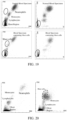
- the left graph of FIG. 3 is a schematic diagram of the regions of various white blood cells, blast cells and fragments of red blood cells after lysis (ghosts) in the two-dimensional scatter diagram of CD45 fluorescence light intensity (WAS) and side scattered light intensity (SSC) obtained from optical detection after treating a specimen with a hemolytic agent, a staining agent containing a nucleic acid fluorescent dye and an antibody treatment agent containing a fluorescent labeled CD45.
- WAS fluorescence light intensity
- SSC side scattered light intensity
- the region of the blast cells is clearly distinguished from the regions where lymphocytes, monocytes and neutrophils appear, and only the lower part of the region slightly overlaps with the ghost region, so that the first characteristic region information including blast cell information and at least part of the ghost region information can be obtained from the blast cell region. According to the first characteristic region information, the blast cell information can be obtained, thereby alarming that blast cells may exist in the specimen.
- the blast cell information obtained only from the first characteristic region information may contain part of the ghost region information, and thus it is not accurate enough. For example, it may lead to false positive results.
- the ghost region information is also further obtained by the fluorescence light intensity (FL) signal, so as to obtain more accurate blast cell information by the first characteristic region information and the ghost region information.
- FL fluorescence light intensity
- FSC forward scattered light intensity
- FIG. 4 is a flow diagram of a sample analysis method according to the embodiment of the present disclosure.
- the sample analysis method of this embodiment includes the following operations.
- a second sample is prepared.
- the second sample contains at least a portion of the specimen to be tested and a second reagent, in which the second reagent includes a hemolytic agent, a staining agent containing a fluorescent dye and an antibody treatment agent containing a fluorescent labeled CD45.
- particles in the second sample is caused to pass e through a detection area of an optical detection device one by on and the particles in the second sample are irradiated with a light source of the optical detection device to obtain optical signals of the particles, in which the optical signals include a side scattered light intensity signal, a forward scattered light intensity signal, a first fluorescence light intensity signal from the staining agent containing the fluorescent dye, and a second fluorescence light intensity signal from the antibody treatment agent containing the fluorescent labeled CD45.
- a first characteristic region information is obtained based on the side scattered light intensity signal and the second fluorescence light intensity signal
- a ghost region information is obtained based on at least the first fluorescence light intensity signal, in which the first characteristic region information includes blast cell information and at least part of the ghost region information.
- blast cell information is obtained based on the first characteristic region information and the ghost region information.
- the specimen to be tested or part of the specimen to be tested is treated with the second reagent to prepare the second sample.
- the second reagent includes a hemolytic agent, a staining agent containing a fluorescent dye, and an antibody treatment agent containing a fluorescent labeled antibody (e.g. CD45).
- the antibody is an antibody capable of binding to the respective surface antigens of blast cells and white blood cells differently.
- the antibody is a monoclonal antibody.
- the term "antibody” mentioned in the present disclosure refers to those that have different binding abilities with surface antigens of various kinds of cells, thereby being able to distinguish blast cells from other white blood cells by antibody fluorescence light intensity and side scattered light intensity.
- the antibody is CD45, and other embodiments may use other antibodies.
- the binding abilities of the antibody between surface antigens of white blood cells and blast cells are different, so that the blast cells can be obviously distinguished from white blood cells by the fluorescence signal emitted by a fluorescent marker linked to the antibody in combination with the side scattered light signals.
- a fluorescent marker linked to the antibody may be any suitable fluorescent marker.
- the present disclosure does not particularly limit the fluorescent marker, so long as the fluorescent marker linked to the antibody does not affect the binding of the antibody to an antigen, nor does it affect the use of other reagents.
- the fluorescent substances that can be excited by the light source can be selected as markers according to the wavelength of the laser light source of a sample analyzer.
- the fluorescence wavelength emitted by a fluorescent marker after being excited should be distinguished from the fluorescence wavelength emitted by the fluorescent dye in the staining agent that is bond with a nucleic acid.
- fluorescent markers include FITC (blue light excitation), APC (red light excitation) and the like, but not limited thereto.
- Fluorescent labeled antibodies that have been commercialized, such as CD45-FITC, CD45-APC and the like, may also be used, but not limited thereto.
- the antibody treatment agent is a buffer (e.g. phosphate buffer) containing the fluorescent labeled antibody. It also comprises other components, such as a preservative (such as sodium azide, ProClin series, etc.), a metal chelating agent (such as sodium salt of EDTA), an osmotic pressure regulator (such as sodium chloride, etc.).
- a preservative such as sodium azide, ProClin series, etc.
- a metal chelating agent such as sodium salt of EDTA
- an osmotic pressure regulator such as sodium chloride, etc.
- the concentration of the fluorescent labeled CD45 in the second reagent is from 25 ⁇ g/mL to 1000 ⁇ g/mL, preferably from 50 ⁇ g/mL to 250 ⁇ g/mL.
- the hemolytic agent may be different from or the same as the hemolytic agent in the first reagent.
- the hemolytic agent can be used as long as it can lyse red blood cells and does not interfere with components of other agents, because the optical signal different from that of white blood cells is mainly generated by the specific binding of the fluorescent labeled CD45 with surface antigens of blast cells.
- the hemolytic agent may include at least one surfactant selected from cationic surfactants, nonionic surfactants, alkyl glycosides, triterpenoid saponins, steroidal saponins, and diethylene glycol.
- the hemolytic agent in the second reagent is selected from alkyl glycosides.
- the alkyl glycoside may be a glycoside compound represented by general formula I: R-(CH 2 )n-CH 3 (I) .
- R is selected from a group consisting of monosaccharide, deoxymonosaccharide and polysaccharide, and n is an integer from 5 to 17.
- All the hemolytic agents disclosed in Chinese patent application No.: CN111602052A can be used as hemolytic agents in the second reagent. The entire contents of the application are incorporated herein by reference.
- the hemolytic agent may include diethylene glycol. Further preferably, formaldehyde which fixes cells may also be included.
- the hemolytic agent includes a cationic surfactant, a nonionic surfactant, an aromatic organic acid, and/or a salt thereof.
- the aromatic organic acid may be one or more selected from the group consisting of terephthalic acid, phthalic acid, hydroxybenzoic acid, acetylsalicylic acid, p-aminobenzoic acid, benzenesulfonic acid, p-toluenesulfonic acid and hydroxybenzenesulfonic acid.
- the cationic surfactant is at least one selected from the group consisting of a quaternary ammonium salt and a pyridine onium salt.
- the nonionic surfactant is at least one selected from the group consisting of polyoxyethylene surfactants, Span and Tween. Specific examples include the hemolytic agents disclosed in Chinese Patent Application No.: CN 202210350953.5 , which is incorporated herein by reference by its entirety.
- the hemolytic agent in the second reagent may be the same as the hemolytic agent in the first reagent, selected from the group consisting of dehydrated sorbitan fatty acid ester-based surfactants, preferably selected from the group consisting of Span and Tween. More preferably, Span is selected from Span 40 ⁇ Span 80, and Tween is selected from Tween 20 ⁇ Tween 100.
- the present disclosure does not particularly limit the concentration of the surfactant as the hemolytic agent in the second reagent, and when different hemolytic agents are used, the conventional concentration of different hemolytic agents can be selected.
- concentration of the dehydrated sorbitan fatty acid ester-based nonionic surfactant is in the range of 0.2 g/L to 2g/L. In a preferred embodiment, the concentration of the surfactant is in the range of 0.3 g/L to 1.5g/L.
- the hemolytic agent also includes a cosolvent, a buffer agent, an anticoagulant and the like. The types and contents of these components may be the same as those of the hemolytic agent in the first reagent described above, and will not be repeated again.
- the hemolytic agent in the second reagent is the same as the hemolytic agent in the first reagent.
- the differences between the blast cells and the white blood cells are further enlarged from a scattered light signal (e.g., the side scattered light intensity), so that the blast cells can be better distinguished from the white blood cells.
- the staining agent in the second reagent is the same as the staining agent in the first reagent and will not be described again.
- the fluorescent dye in the staining agent is preferably a dye that can be excited by blue light.
- the fluorescent dye may be a compound represented by the above Formula II:
- the hemolytic agent, the staining agent and the antibody treatment agent are separately packaged reagents that are mixed to form the second reagent only when or before treating the specimen to be tested.
- any two or three of the hemolytic agent, the staining agent and the antibody treatment agent are packaged in a mixed form as one reagent.
- the fluorescent labeled CD45 may be added to the hemolytic agent at the above concentration to form a reagent packaged in a mixed form.
- At least part of the specimen to be tested is treated with the second reagent, in particular by mixing the antibody treatment agent with the specimen to be tested, and incubating at a temperature such as 37°C to 42°C, for a certain period of time, such as 30 to 180 seconds. Then, a certain amount of the mixed solution is added to and mixed with the hemolytic agent and staining agent, and further incubated at a certain temperature, such as 42°C for a certain period of time, for example, 10 to 30 seconds.
- a temperature such as 37°C to 42°C
- particles in the second sample is caused to pass one-by-one through a detection area of an optical detection device and the particles in the second sample are irradiated with the light source of the optical detection device to obtain optical signals of the particles.
- the optical signals includes a side scattered light intensity signal, a forward scattered light intensity signal, a first fluorescence light intensity signal from the staining agent containing the fluorescent dye, and a second fluorescence light intensity signal from the antibody treatment agent containing the fluorescent labeled antibody (such as CD45).
- a first characteristic region information is obtained based on the side scattered light intensity signal and the second fluorescence light intensity signal
- a ghost region information is obtained based on at least the first fluorescence light intensity signal
- the first characteristic region information includes blast cell information and at least part of ghost region information.
- the blast cell information is calculated based on the first characteristic region information and the ghost region information.
- the ghost region information may also be obtained based on the first fluorescence light intensity signal and any one of the side scattered light intensity signal and the forward scattered light intensity signal.
- S230 to S250 in the second embodiment are similar to S130 to S150 in the first embodiment, except that the first characteristic region information is obtained by the side scattered light intensity signal and the second fluorescence light intensity signal from the fluorescent labeled CD45.
- FIG. 3 a two-dimensional scatter plot of the second fluorescence light intensity of a fluorescent labeled CD45 and the side scattered light intensity of the second sample obtained after the specimen to be tested is treated with the second reagent is schematically shown.
- the region where the blast cells appear is one region (whereas in the first embodiment, the blast cells may appear in either or both of two non-contiguous regions) and is better distinguished from white blood cells (lymphocytes, monocytes, neutrophils and eosinophils).
- the overlapping area of the lower part of the blast cell region and the ghost region is also smaller than that of the first embodiment.
- the ghost region information is also obtained from at least the first fluorescence light intensity signal, and in S250, the blast cell information is obtained based on the first characteristic region information and the ghost region information. Specifically, obtaining the blast cell information also includes subtracting the at least part of the ghost region information from the first characteristic region information to obtain the blast cell information. In a preferred embodiment, the ghost region information overlapped in the blast cell region is subtracted from the first characteristic region information, so that more accurate blast cell information is obtained and false detection results can be avoided.
- the blast cell information includes a count and/or an alarm about blast cells.
- the above-described preferred embodiment of the second embodiment can count the blast cells.
- a third embodiment of the sample analysis method is provided.
- the embodiment is similar to the first embodiment mentioned above.
- a hemolytic agent (not limited to the hemolytic agent in the first reagent mentioned above) is used to treat respective the cell membranes of mature white blood cells and those of blast cells differently in the specimen, The forward scattered light intensity and the side scattered light intensity are used to obtain the first characteristic region including the blast cell information.
- the first fluorescence signal emitted by a nucleic acid dye is used to deduct the ghost information from the first characteristic region, so as to obtain the blast cell information.
- the sample analysis method of this embodiment includes the following operations.
- a specimen to be tested is obtained from a subject.
- a third sample is prepared.
- the third sample comprises at least a portion of the specimen to be tested and a third reagent, in which the third reagent comprises at least a hemolytic agent capable of treating respective cell membranes of mature white blood cells and those of blast cells differently in the third sample, and a staining agent containing a fluorescent dye.
- particles in the third sample is caused to pass one-by-one through a detection area of an optical detection device, the particles in the third sample are irradiated with a light source of the optical detection device to obtain optical signals of the particles.
- the optical signals comprise a side scattered light intensity signal, a forward scattered light intensity signal, a first fluorescence light intensity signal from the staining agent containing the fluorescent dye.
- a first characteristic region information is obtained based on the side scattered light intensity signal and the forward scattered light intensity signal
- a ghost region information is obtained based on at least the first fluorescence light intensity signal
- the first characteristic region information comprises blast cell information and at least part of ghost region information
- the blast cell information is obtained based on the first characteristic region information and the at least part of ghost region information.
- S310 in this embodiment is similar to S110 in the above-mentioned first embodiment.
- S320 is similar to S120 of the above-mentioned first embodiment in that the specimen is treated by the reagent containing a hemolytic agent and a staining agent.
- the hemolytic agent of the third agent is not limited to a dehydrated sorbitan fatty acid ester-based nonionic surfactant, but other hemolytic agents capable of treating respective the cell membranes of mature white blood cells and those of blast cells differently in the specimen can also be used.
- the type of hemolytic agent includes but is not limited to the above-mentioned dehydrated sorbitan fatty acid ester-based nonionic surfactant, polyoxyethylene-based nonionic surfactant, nitrogen-containing nonionic surfactant, and block polyether non-ionic surfactant.
- polyoxyethylene-based nonionic surfactants examples include polyoxyethylene oleyl ether, primary alcohol polyoxyethylene ether, secondary alcohol polyoxyethylene ether, branched-chain isomerized Guerbet alcohol ether, alkyl phenol polyoxyethylene ether, fatty acid methyl ester polyoxyethylene ether, and the like.
- nitrogen-containing non-ionic surfactant examples include Coconut oil alkyl alkyl amide phosphate and the like.
- block polyether non-ionic surfactant examples include propylene glycol block polyether, ethylene glycol block polyether and the like.
- These surfactants serving as a hemolytic agent can treat cells membranes of blast cells and those of mature white blood cells differently, resulting in difference in the amount and type of substance that ends up inside the cells, so as to further enlarge the difference of the light signal between blast cells and mature white blood cells.
- the use concentration can be adjusted according to the properties of different surfactants.
- the polyoxyethylene-based non-ionic surfactants as a hemolytic agent may have a concentration of 0.1g/L to 20g/L, preferably 0.5 g/L to 5 g/L.
- hemolytic agent included in the third agent it also includes other necessary agent, such as co-solvents, buffer agent, anticoagulants, or the like, which will not be repeated here.
- the third sample prepared by mixing and incubating the third reagent with the specimen to be tested is also caused to pass through the detection area of the optical detection device in S330 to obtain the optical signal of the particles therein.
- the optical signal includes a side scattered light intensity signal, a forward scattered light intensity signal, and a first fluorescence light intensity signal from a staining dye containing a fluorescent dye.
- the first characteristic region information is obtained based on the side scattered light intensity signal and the forward scattered light intensity signal.
- FIG. 19 shows a specific example of the treatment and detection of specimens from a healthy subject (upper two graphs) and a patient (lower two graphs) using the third agent containing polyoxyethylene oleyl ether as the hemolytic agent, a two-dimensional scatter plot formed by the side scattered light intensity signal and the forward scattered light intensity signal (left two graphs) and a two-dimensional scatter plot formed by the side scattered light intensity signal and the fluorescent light signal (right two graphs). Similar to the left graph of FIG. 1 , the two regions circled in the left lower graph of FIG. 19 are regions where blast cells may appear, that is the first characteristic region.
- Blast cells may apper in either or both regions. Both the two regions can be clearly distinguished from regions where white blood cells such as lymphocytes, monocytes, and neutrophils occur, with only the lower blast cells region partially overlapping with the ghost region. There is a significant increase in the number of particles in the first characteristic region compared to the scatter plot obtained from the specimen of the healthy subject (upper left graph). In the lower right graph, the ghost region is circled, which is far away from the regions where other cells appear in terms of fluorescence light intensity, so that all the ghost region information can be obtained through the fluorescence signals.
- the blast cells information can be obtained based on the first characteristic region information including a part of ghost region information obtained by the scattered light signal and the ghost region information obtained by the fluorescence signal.
- the ghost region information is obtained at least based on the first fluorescence light intensity signal. Similar to the right graph of FIG. 1 , the fluorescence light intensity in the ghost region of the lower right graph is lower than the fluorescence light intensity of other cells, so it is clear that the ghost region is far away from the regions where other cells appear, so that all the ghost region information can be obtained based on the fluorescence signal.
- the ghost region information can be obtained by only the first fluorescence light intensity signal, or one scattered light signal (side scattered light intensity signal or forward scattered light intensity signal) combined with the first fluorescence light intensity signal.
- the blast cells information is obtained based on the first characteristic region information and at least part of the ghost region information.
- the blast cells information can be obtained by subtracting the ghost region information obtained by the fluorescence signal from the first characteristic region information containing part of the ghost region information obtained by the scattered light signal.
- the blast cells information includes a count and/or an alarm about blast cells, in particular the alarm.
- a fourth embodiment of the sample analysis method is provided.
- a hemolytic agent such as a dehydrated sorbitan fatty acid ester-based nonionic surfactant
- the forward scattered light intensity and the side scattered light intensity are used to obtain the first characteristic region including the blast cell information, so as to obtain the blast cell information.
- the sample analysis method of this embodiment includes the following operations.
- a fourth sample is prepared.
- the fourth sample comprises at least a portion of the specimen to be tested and a fourth reagent, wherein the fourth reagent comprises a hemolytic agent containing at least one nonionic surfactant comprising a dehydrated sorbitan fatty acid ester.
- particles in the fourth sample is caused to pass one-by-one through a detection area of an optical detection device, the particles in the fourth sample are irradiated with a light source of the optical detection device to obtain optical signals of the particles, wherein the optical signals comprise a side scattered light intensity signal and a forward scattered light intensity signal.
- a first characteristic region information is obtained based on the side scattered light intensity signal and the forward scattered light intensity signal, wherein the first characteristic region information comprises blast cell information.
- the blast cell information is obtained based on the first characteristic region information.
- S410 of this embodiment is similar to S110 of the first embodiment above.
- S420 is similar to S120 in the first embodiment above, except that the fourth agent does not include a staining agent, but only a hemolytic agent.
- the hemolytic agent is identical to the hemolytic agent in the first reagent and contains at least one nonionic surfactant comprising a dehydrated sorbitan fatty acid ester, which enables treat respective the cell membranes of mature white blood cells and those of blast cells cells differently in the specimen.
- the dehydrated sorbitan fatty acid ester-based nonionic surfactant is defined as that in the first embodiment, is not repeated here.
- the fourth sample prepared by mixing and incubating the fourth reagent with the specimen to be tested is caused to pass through the detection area of the optical detection device in S430, to obtain the scattered light signal of the particles therein.
- the scattered light signal includes a side scattered light intensity signal and a forward scattered light intensity signal.
- the first characteristic region information including the blast cell information is obtained based on the side scattered light intensity signal and the forward scattered light intensity signal.
- FIG. 20 shows a specific embodiment of the treatment and detection of specimens from a healthy subject (left) and a patient (right) with a fourth agent containing a hemolytic agent but not a fluorescent dye, in which the two-dimensional scatter plot is formed by a side scattered light intensity signal and a forward scattered light intensity signal.
- the regions where blast cell may appear are circled on the right graph, including a large number of particles, while the corresponding regions on the left graph show only a few particles. It can be seen that after the treatment of the fourth reagent, the blast cells can be distinguished from the white blood cells to a certain extent by the scattered light signal of the particles.
- the blast cell information is obtained based on the first characteristic region information.
- the first characteristic region can indicate the presence of blast cells in the sample, so that the blast cell information obtained based on the first characteristic region can alarm the specimen containing blast cells.
- the method of the embodiment does not require the use of fluorescent dyes, thus reducing the cost of detection.
- a fifth embodiment of the sample analysis method is provided.
- the embodiment is different from the two above-mentioned embodiments in that, the method further includes an operation of receiving an operation instruction. After receiving operation instructions, blast cell detection is carried out on a specimen.
- the specific detection operations are the operations of the sample analysis method in any one of the first embodiment or the fourth embodiment and will not be repeated herein.
- the sample analysis method of each embodiment enlargeds the difference of optical signals between the blast cells and the white blood cells by using the first to fourth reagent to treat respective the blast cells and the white blood cells differently, and well distinguish them, so that the blast cells information can be obtained by using the methods, and the detection accuracy is improved.
- the methods can alarm the blast cells in the sample more accurately, and the counting of the blast cells can be realized in a preferred embodiment.
- acute lymphoblastic leukemia has a rapid onset and great harm, but there is a lack of high sensitivity detection methods for blast cells of acute lymphoblastic leukemia at present.
- the sample analysis methods provided by the present disclosure are more sensitive and accurate for blast cell detection of blood specimens from acute lymphoblastic leukemia patients.
- the present disclosure also provides a sample analyzer to perform the above sample analysis method. Exemplary embodiments of the sample analyzer are described in detail below with reference to FIG. 5 to FIG.6 .
- the sample analyzer of the first embodiment refers to FIG. 5 where the sample analyzer is schematically shown.
- the sample analyzer includes a specimen aspiration device 10, a sample preparation device (30), an optical detection device 50 including a light source, a flow chamber, a processor 70, and a non-transitory computer-readable storage medium (not shown) programmed with computer program instructions.
- the specimen aspiration device 10 is configured to aspirate at least a portion of the specimen to be tested and transport it to the sample preparation device. Some of or all of the specimen to be tested can be taken as needed to detect the blast cells in the specimen.
- the specimen aspiration device 10 may aspirate a portion of the specimen to be tested several times for use including the above-mentioned blast cell detection and other detection items such as white blood cell detection, platelet detection, hemoglobin detection and the like, but is not limited thereto.
- the specimen aspiration device can also aspirate a certain amount of the specimen at one time and divide them into several sub-specimens for different treatments and detections, respectively.
- the sample preparation device 30 includes a reagent supply part and a reaction cell.
- the specimen aspiration device 10 and the reagent supply part are respectively connected to the reaction cell through pipelines.
- the reagent supply part may deliver a specific reagent to a specific reaction cell to be mixed with the specimen to be tested provided by the specimen aspiration device, to prepare a sample for a predetermined detection purpose.
- the reagent supply part is configured to supply the first reagent to the reaction cell.
- the first reagent includes a hemolytic agent containing at least one nonionic surfactant comprising a dehydrated sorbitan fatty acid ester (preferably selected from Tween and Span) and a staining agent containing a fluorescent dye, which is not repeated herein.
- the reagent supply part includes a plurality of reagent storage containers, and the hemolytic agent and the staining agent in the first reagent are stored in different storage containers; or the hemolytic agent and the staining agent are stored in a mixed form in one storage container.
- the reaction cell is configured to mix the first reagent with at least a portion of the specimen to be tested therein to obtain the first sample, and to transport the first sample to the flow chamber.
- the reaction cell can be heated to a suitable reaction temperature, so as to obtain the best treatment effect of the sample.
- the specimen to be tested in the reaction cell is treated with the first reagent to prepare the first sample, and the first sample is transported to the optical detection device 50 through the pipeline assembly to obtain the optical signals of each particle in the sample.
- the optical detection device 50 includes a light source, a flow chamber and at least one, typically a plurality of optical detectors.
- the light source is configured to align a light beam with a detection hole of the flow chamber which is connected with the reaction cell.
- the flow chamber is configured to allow particles in the sample to pass through one by one.
- the optical detectors are mounted around the flow chamber for detecting optical signals of particles passing through the detection hole of the flow chamber.
- FIG. 6 a schematic structural block diagram of an optical detection device is shown.
- the optical system 1 and the flow chamber 2 are arranged on an optical axis.
- the optical system 1 includes a light source and a lens group for aligning the light emitted by the light source with a detection hole on the flow chamber 2.
- a plurality of optical detectors are arranged around the flow chamber 2 for detecting light scattered signals and a fluorescence signal of particles passing through the detection hole one by one.
- the optical detectors generally include a first detector 3, a second detector 4 and a fluorescence detector 5.
- the light scattered signal includes a forward light scattered intensity signal emitted from the optical system 1 and transmitted through the flow chamber 2 collected by the first detector 3 arranged on the optical axis, and a side light scattered intensity signal emitted from the flow chamber 2 collected by the second detector 4 arranged on a side of the optical axis.
- the optical detectors further include a fluorescence detector 5 arranged on a side of the optical axis for collecting the fluorescence light intensity signal emitted from the flow chamber 2. According to different treatment of specimens, each detector can collect scattered light intensity signal and fluorescence light intensity signal from each particle in different samples.
- the fluorescence detector 5 is used to collect the fluorescence light intensity signal emitted by the fluorescent dye bound with nucleic acids in the particles.
- the processor 70 is operably connected to the optical detectors, and optical signals of particles detected by the optical detectors are transmitted to the processor 70 for analysis.
- the signals are amplified by an amplifier and transmitted to the processor 70.
- the processor 70 When the computer program instructions programed in the non-transitory computer-readable storage medium are executed by the processor 70, the processor 70 obtains corresponding particle information according to the received optical signals, and executes operations for obtaining the blast cell information according to the sample analysis method in the above embodiments.
- the processor 70 when the computer program instructions programed in the non-transitory computer-readable storage medium are executed by the processor 70, the processor performs the following operations.
- the first characteristic region information and ghost region information is obtained based on the optical signals.
- the first characteristic region information includes blast cell information and at least part of ghost region information.
- the blast cell information is obtained based on the first characteristic region information and the ghost region information.
- the optical signals in this embodiment includes a side scattered light intensity signal, a forward scattered light intensity signal, and a first fluorescence light intensity signal from the staining agent containing a fluorescent dye.
- the operation of obtaining a first characteristic region information and a ghost region information based on the optical signals performed by the processor includes: obtaining the first characteristic region information based on the side scattered light intensity signal and the forward scattered light intensity signal, and obtaining the ghost region information based on at least the first fluorescence light intensity signal.
- Obtaining the ghost region information base on at least the first fluorescence light intensity signal includes obtaining the ghost region information based on the first fluorescence light intensity signal; or obtaining the ghost region information based on the first fluorescence light intensity signal and any one of the side scattered light intensity signal and the forward scattered light intensity signal.
- the operation of obtaining the blast cell information based on the first characteristic region information and the ghost region information performed by the processor includes subtracting at least part of the ghost region information from the first characteristic region information to obtain the blast cell information.
- the present disclosure also provides a sample analyzer of the second embodiment.
- the sample analyzer also includes a specimen aspiration device 10, a sample preparation device 30 including a reagent supply part and a reaction cell, an optical detection device 50 (further referring to FIG. 6 ) including a light source, a flow chamber and optical detectors, a processor 70, and a non-transitory computer-readable storage medium (not shown) programmed with computer program instructions.
- the reagent supply part is configured to supply the second reagent to the reaction cell.
- the second reagent is the same as previously described and includes a hemolytic agent, a staining agent containing a fluorescent dye, and a treatment agent containing a fluorescent labeled antibody (in particular, such as CD45), which will not be repeated here.
- the reagent supply part includes a plurality of reagent storage containers, and the hemolytic agent, the staining agent and the antibody treatment agent are stored in different storage containers separately; or any two or three of the hemolytic agent, the staining agent and the antibody treatment agent are stored in a mixed form in one storage container.
- the reaction cell is configured to mix the second reagent with at least a portion of the specimen to be tested therein to obtain a second sample, and to transport the second sample to the flow chamber.
- the second reagent includes both a staining agent containing a fluorescent dye and an antibody treatment agent containing a fluorescent labeled antibody.
- the light source may be a single light source in which a single wavelength of excitation light emitted by the light source is capable of simultaneously exciting the fluorescent dye and the fluorescent marker bound to the antibody, and the fluorescence emitted by them have different wavelengths that can be detectable separately.
- the light source may also be a dual-light source capable of emitting excitation light of different wavelengths for exciting the fluorescent dye and the fluorescent marker bound to the antibody, respectively.
- the fluorescence detector 5 is used to collect a first fluorescence light intensity signal (if any) from the fluorescent dye to which nucleic acid is bound and the second fluorescence light intensity signal from a fluorescent labeled antibody (e.g. CD45).
- the first fluorescence light intensity signal from the fluorescent dye and the second fluorescence light intensity signal from the fluorescent labeled antibody have different wavelengths and can thus be collected separately.
- the fluorescence detector 5 may include two fluorescence sub-detectors corresponding to different wavelengths of the first and second fluorescence signals respectively, to collect the first and second fluorescence light intensity signals.
- the processor 70 is operably connected to the optical detectors.
- the processor is caused to perform the following operations.
- First characteristic region information is obtained based on the side scattered light intensity signal and the second fluorescence light intensity signal
- ghost region information is obtained based on at least the first fluorescence light intensity signal, in which the first characteristic region information includes blast cell information and at least part of ghost region information.
- the blast cell information is obtained based on the first characteristic region information and the ghost region information.
- Obtaining the ghost region information base on at least the first fluorescence light intensity signal includes obtaining the ghost region information based on the first fluorescence light intensity signal; or obtaining the ghost region information based on the first fluorescence light intensity signal and any one of the side scattered light intensity signal and the forward scattered light intensity signal.
- Obtaining the blast cell information based on the first characteristic region information and the ghost region information includes subtracting at least part of the ghost region information from the first characteristic region information to obtain the blast cell information.
- the disclosure further provides a sample analyzer of a third embodiment.
- the sample analyzer also includes a sample aspiration device 10, a sample preparation device 30 including a reagent supply part and a reaction cell, an optical detection device 50 (further referring to FIG. 6 ) including a light source, a flow chamber and optical detectors, a processor 70, and a non-transitory computer-readable storage medium (not shown) programmed with computer program instructions.
- the reagent supply part is configured to supply the third reagent to the reaction cell.
- the third reagent is the same as previously described and includes a hemolytic agent capable of treating respective cell membranes of mature white blood cells and that of blast cells differently and a staining agent containing a fluorescent dye, which will not be repeated here.
- the reagent supply part includes a plurality of reagent storage containers, and the hemolytic agent and the staining agent are stored in different storage containers separately; or the hemolytic agent and the staining agent are stored in a mixed form in one storage container.
- the reaction cell is configured to mix the third reagent with at least a portion of the specimen to be tested therein to obtain a third sample, and to transport the sample to the flow chamber.
- the third sample after the treatment of the third reagent is caused to pass through the optical detection device 50 (still referring to FIG. 6 , the structure may be identical with the optical detectors in the sample analyzer of the first embodiment) to obtain the light signals of each of the particles.
- the light signal includes the forward scattered light intensity signal, a side scattered light intensity signal and a first fluorescence light intensity signal collected by the first detector 3, the second detector 4 and the fluorescent detector 5, respectively.
- the processor 70 is operably connected to the optical detectors.
- the processor is caused to perform the following operations.
- First characteristic region information is obtained based on the side scattered light intensity signal and the forward scattered light intensity signal
- ghost region information is obtained based on at least the first fluorescence light intensity signal, in which the first characteristic region information includes blast cell information and at least part of ghost region information.
- the blast cell information is obtained based on the first characteristic region information and the at least part of ghost region information.
- Obtaining the ghost region information base on at least the first fluorescence light intensity signal includes obtaining the ghost region information based on the first fluorescence light intensity signal; or obtaining the ghost region information based on the first fluorescence light intensity signal and any one of the side scattered light intensity signal and the forward scattered light intensity signal.
- the disclosure further provides a sample analyzer of a fourth embodiment.
- the sample analyzer also includes a sample aspiration device 10, a sample preparation device 30 including a reagent supply part and a reaction cell, an optical detection device 50 (further referring to FIG. 6 ) including a light source, a flow chamber and optical detectors, a processor 70, and a non-transitory computer-readable storage medium (not shown) programmed with computer program instructions.
- the reagent supply part is configured to supply the fourth reagent to the reaction cell.
- the fourth reagent is the same as previously described and includes a hemolytic agent containing at least one nonionic surfactant comprising a dehydrated sorbitan fatty acid ester, which will not be repeated here.
- the reaction cell is configured to mix the fourth reagent with at least a portion of the specimen to be tested to obtain a fourth sample, and to transport the sample to the flow chamber.
- the fourth sample after the treatment of the fourth reagent is caused to pass through the optical detection device 50 (still referring to FIG. 6 , the structure may be identical with the optical detectors in the sample analyzer of the first embodiment, or include only the first detector 3 and the second detector 4, but not include the fluoreascent detector 5) to obtain the light signals of each of the particles.
- the light signal includes the forward scattered light intensity signal and a side scattered light intensity signal collected by the first detector 3 and the second detector 4, respectively.
- the processor 70 is operably connected to the optical detectors.
- the processor is caused to perform the following operations.
- First characteristic region information is obtained based on the side scattered light intensity signal and the forward scattered light intensity signal.
- the first characteristic region information includes blast cell information. A reminder of the blast cell information is outputted based on the first characteristic region information.
- the processor 70 is further operably connected with the specimen aspiration device 10, the reagent supply part, and the reaction cell to control the specimen aspiration device, the reagent supply part, and the reaction cell to perform the above-mentioned operations of aspiration the specimen to be tested to a specific reaction cell, and mixing and incubating with a specific reagent or reagent combination supplied to the reaction cell by the reagent supply part, in accordance with a computer application programed in the computer-readable storage medium, or upon receipt of a user instruction, to prepare a sample for blast cell detection.
- the processor further controls the reaction cell to transport the prepared sample to the optical detection device 50 to perform the above detection, and the processor 70 executes the above operations according to the received detection data to obtain the blast cell information.
- the sample analyzer also includes an output device 90.
- the output device 90 includes a user interface for human-computer interaction.
- the user interface is configured to display detection results and/or input instructions to the sample analyzer. For example, in one embodiment, an instruction to perform blast cell detection is input by an operator through the user interface.
- the user interface displays detection results, i.e. blast cell information including for example blast cell count and/or alarm about the user.
- the sample analyzer in the above embodiments may also have a first casing 100 and a second casing 200 (referring to FIG. 5 ).
- the sample preparation device 30 is disposed inside the first casing 100, and the output device 90 and the specimen aspiration device 10 are disposed on an outer surface of the first casing 100.
- the optical detection device 50 and the processor 70 are provided inside the second casing 200.
- the present disclosure further provides a reagent for detecting blast cells.
- the reagent is any one of the first reagent and the second reagent, and the composition of the reagent will not be repeated here.
- the blast cells and white blood cells in the specimen to be tested are treated differently by using the reagent containing a specific hemolytic agent, thereby preparing corresponding samples and performing optical detection to obtain optical signals of each particle in the sample.
- the difference of cell membranes of blast cells and white blood cells is utilized to perform differentiation treatment by using the reagent, thus enlarges the difference of optical signals between blast cells and white blood cells, so that blast cells can be distinguished from white blood cells.
- the optical signal is analyzed, and more accurate blast cell information is obtained by utilizing the first characteristic region information including blast cell information and part of the ghost information and the ghost region information including only the ghost information, thus reducing the proportion of false negative and false positive.
- the reagent is highly sensitive to the blast cell detection in the blood of patients with acute lymphoblastic leukemia, and thus more accurate.
- detecting blast cells includes counting and/or alarming about blast cells.
- the advantages of the present invention are further explained below by specific examples.
- the fluorescent dyes used in the examples are the aforementioned dyes A to C.
- the staining agent and hemolytic agent are prepared.
- the left graph of FIG. 7 is a two-dimensional scatter plot of a side scattered light intensity (SSC) versus a forward scattered light intensity (FSC). Because the blood specimen comes from a normal subject, only the blast cell region 1 and the blast cell region 2 are shown in dashed coils, in which there is no signal of the blast cells. In the blast cell region II, part of the ghost region overlaps with this region.
- the right graph of FIG. 7 is a two-dimensional scatter plot of a fluorescence light intensity (FL) versus a side scattered light intensity (SSC). Only the ghost region is shown in dashed coils in the graph. This graph shows that the ghost region is far away from the white blood cells region, and complete ghost information can be obtained. By subtracting the ghost information from the information of the blast cell region 1 and the blast cell region 2, the result that the specimen does not contain blast cells is obtained.
- the method can accurately detect the absence of blast cells in the specimen and avoid false positives.
- staining agent and hemolytic agent are prepared.
- the two graphs above show the detection results of specimens from the healthy subject.
- the two graphs below show the detection results of specimens from the patient.
- the two graphs on the left are two-dimensional scatter graphs of forward scattered light intensity versus side scattered light intensity, in which the blast cell region 1 and the blast cell region 2 are shown by circles.
- the two graphs on the right are two-dimensional scatter plots of forward scattered light intensity versus fluorescence light intensity, and the circle shows the ghost region. This right graphs show that the ghost region is far away from the white blood cells region, and complete ghost information can be obtained.
- the ghost region information obtained from the right graph of FIG. 8 was subtracted from the first characteristic region information including the blast cell region 1 and the blast cell region 2 obtained from the left graph of FIG. 8 .
- the detection did not alarm specimens from the healthy subject, but alarmed the presence of blast cells from specimens from the patient.
- staining agent and hemolytic agent are prepared.
- the left graph of FIG. 9 is a two-dimensional scatter plot of forward scattered light intensity versus side scattered light intensity, in which the blast cell region 1 and the blast cell region 2 are shown in circles.
- the right graph of FIG. 9 is a two-dimensional scatter plot of forward scattered light intensity versus fluorescence light intensity.
- the ghost region is shown in circles in the graph. This graph shows that the ghost region is far away from the white blood cells region, and complete ghost information can be obtained.
- the ghost region information obtained from the right graph of FIG. 9 was subtracted from the first characteristic region information including the blast cell region 1 and the blast cell region 2 obtained from the left graph of FIG. 9 .
- the left graph almost no particles appear in the blast cell region 1, but the detection results show that there are blast cells in the specimen, so the specimen was alarmed.
- the blast cells mainly appear in the blast cell region 2.
- the staining agent, hemolytic agent and the antibody treatment agent are prepared.
- the left graph of FIG. 10 is a two-dimensional scatter plot of forward scattered light intensity versus side scattered light intensity, in which the blast cell region 1 and the blast cell region 2 are shown in circles.
- the right graph of FIG. 10 is a two-dimensional scatter plot of forward scattered light intensity versus fluorescence light intensity, in which the ghost region is shown in the circle. This graph shows that the ghost region is far away from the white blood cells region, and complete ghost information can be obtained.
- the ghost region information obtained from the right graph of FIG. 10 was subtracted from the first characteristic region information including the blast cell region 1 and the blast cell region 2 obtained from the left graph of FIG. 10 , and the result was that the specimen was alarmed.
- the staining agent, hemolytic agent and the antibody treatment agent are prepared.
- the left graph of FIG. 11 is a two-dimensional scatter plot of forward scattered light intensity versus side scattered light intensity, in which the blast cell region 1 and the blast cell region 2 are shown in circles.
- the right graph is a two-dimensional scatter plot of forward scattered light intensity versus fluorescence light intensity.
- the ghost region is shown in circles in the graph. This graph shows that the ghost region is far away from the white blood cells region, and complete ghost information can be obtained.
- the ghost region information obtained from the right graph of FIG. 11 was subtracted from the first characteristic region information including the blast cell region 1 and the blast cell region 2 obtained from the left graph of FIG. 11 , and the result was that the specimen was not alarmed.
- the staining agent, hemolytic agent and the antibody treatment agent are prepared.
- the same reagent A to C was used to measure normal blood specimens of a healthy subject and obtain scatter plots.
- the upper left graph of FIG. 12 shows the scatter diagram of antibody fluorescence light intensity (WAS) versus side scattered light intensity (SSC) from the healthy subject
- the upper right graph shows the scatter diagram of antibody fluorescence light intensity (WAS) versus side scattered light intensity (SSC) from the patient.
- the lower graph of FIG. 12 is a scatter plot of fluorescence light intensity (FL) versus forward scattered light intensity (FSC) from the patient (not shown that from the healthy subject). It can be seen that the ghost is far away from other cell regions, so all the ghost information can be obtained.
- the blast cells count from the patient specimen obtained by subtracting the ghost region information was 22.4%. At the same time, the specimen was examined under microscope, and the blast cells count was 24.4%.
- the staining agent, hemolytic agent and the antibody treatment agent are prepared.
- C Antibody treatment agent (commercial products diluted with phosphate buffer) FITC labeled CD45 antibody 100 ⁇ g/ml.
- Blood specimens from a patient and a healthy subject were tested according to the following methods. 100 ⁇ L of anticoagulant-treated blood specimens was added to 1 ⁇ L of reagent C, incubated at 42°C for 1 to 2 minutes, then 20 ⁇ L of the incubation mixture were added to 1 mL of reagent B, and 20 ⁇ L of reagent A was added and incubated at 42°C for 30 seconds to form a sample.
- the forward low-angle scattered light intensity, the side high-angle scattered light intensity and two types of fluorescence light intensity were obtained by a blood cell analyzer with blue light as the excitation light source, and the scatter plots were obtained respectively.
- the first characteristic region information was obtained from the scatter plot of antibody fluorescence light intensity versus side scattered light intensity
- the ghost region information was obtained from the scatter plot of forward scattered light intensity versus fluorescence light intensity
- the ghost region information was subtracted from the first characteristic region information of the scatter plot of antibody fluorescence light intensity (WAS) versus side scattered light intensity (SSC), respectively.
- WAS antibody fluorescence light intensity
- SSC side scattered light intensity
- the right graph of FIG. 13 shows the detection result of the healthy subject and the left graph shows the detection result of the patient.
- the blast cells count of the patient specimen was 9.3%. At the same time, the specimen was examined under microscope, and the blast cells count was 10.6%.
- the staining agent, hemolytic agent and the antibody treatment agent are prepared.
- C Antibody treatment agent (commercial products diluted with phosphate buffer) FITC labeled CD45 antibody 100 ⁇ g/ml.
- the first characteristic region information was obtained from the scatter plot of antibody fluorescence light intensity (WAS) versus side scattered light intensity (SSC)
- the ghost region information was obtained from the scatter plot of forward scattered light intensity (FSC) versus fluorescence light intensity (FL)
- the ghost region information was subtracted from the first characteristic region information of the scatter plot of antibody fluorescence light intensity (WAS) versus side scattered light intensity (SSC), respectively.
- the results are shown in FIG. 14 .
- the right graph of FIG. 14 shows the detection result of the healthy subject and the left graph shows the detection result of the patient.
- the blast cells count was 9.8%.
- the specimen was examined under microscope, and the blast cells count was 10.6%.
- Specimens with different blast cell contents were quantitatively analyzed using the reagent prepared in Example 8 with the same method as in Example 8.
- the ghost region information of each sample was subtracted from the first characteristic region information of the scatter plot of antibody fluorescence light intensity (WAS) versus side scattered light intensity (SSC) to obtain the blast cell information.
- the detection results (WAS-SSC scatter plot after deducting the ghost information) of specimens with blast cells contents of 0% (normal specimen), 1.2%, 3.3% and 62%, respectively, are shown in FIG. 15 .
- the blast cell count in each specimen was obtained.
- each specimen was examined under microscope, and the blast cells were counted.
- the blast cell count obtained by hematology analyzer using the above method was plotted with respect to the blast cell count obtained by microscopic examination, and linear fitting was performed, and the result is shown in FIG. 16 .
- the staining agent and the antibody treatment agent are prepared.
- the upper left graph of FIG. 17 is a two-dimensional scatter plot of antibody fluorescence light intensity (WAS) versus side scattered light intensity (SSC).
- WAS antibody fluorescence light intensity
- SSC side scattered light intensity
- the blast cell region is shown in circle in the graph. It can be seen that the blast cell region can be well distinguished from other white blood cell regions.
- the upper right graph of FIG. 17 is a two-dimensional scatter plot of forward scattered light intensity (FSC) versus side scattered light intensity (SSC).
- FSC forward scattered light intensity
- SSC side scattered light intensity
- the blast cell region is shown in the circle in the graph. It can be seen that when other hemolytic agents are used, the blast cell region is close to the white blood cell region and is not easy to distinguish.
- the lower graph of FIG. 17 is a two-dimensional scatter plot of fluorescence light intensity (FL) versus side scattered light intensity (SSC).
- FL fluorescence light intensity
- SSC side scattered light intensity
- the ghost region information obtained from the lower graph of FIG. 17 was subtracted from the first characteristic region information including the blast cell region obtained from the upper left graph of FIG. 17 , and the result showed that there were blast cells in the specimen and the specimen was alarmed.
- staining agent and hemolytic agent are prepared.
- the first characteristic region information including the information of blast cell region 1 and the blast cell region 2 was obtained in the two-dimensional scatter plot of the forward scattered light intensity versus the side scattered light intensity, and the ghost region information was obtained in the two-dimensional scatter plot of the forward scattered light intensity and the fluorescence light intensity.
- the ghost region information was subtracted from the first characteristic region information, and when the result showed that the blast cell existed, the measured specimen was alarmed about the blast cells.
- the clinical specimens were detected by Mindray 6000 blood cell analyzer using the matched white blood cell detection reagent with traditional blast cell detection methods at the same time. That is, when the white blood cells were detected, the white blood cells were classified and counted by the fluorescence light intensity signal generated by the fluorescent dye combined with the side scattered light intensity signal, and if the abnormal data of the blast cell region was detected, the measured specimen was alarmed about the blast cells.
- the statistical results of a large number of specimens show that using the above reagents and the blood cell analyzer for blast cell early alarm can reduce the proportion of false negative and false positive of blast cell early alarm about a certain extent compared with the conventional blast cell early alarm method.
- the analysis method of this example can significantly reduce the proportion of false negative, basically realize no missing detection, and has positive clinical significance.
- the staining agent and the hemolytic agent are prepared.
- the lower two graphs show detection results from the specimen from a patient
- the left two graphs are two-dimensional scatter plots of the forward scattered light intensity versus the side scattered light intensity, in which the lower left circle shows the two regions where blast cells may appear
- the right two graphs are two-dimensional scatter plots of forward scattered light intensity versus fluorescence light intensity, and the lower right circle shows the ghost region. From the right graph, it can be seen that the ghost region is far away from the white blood cell region, and the complete ghost information can be obtained.
- the blast cell information was obtained by subtracting the ghost region information obtained from the right graph from the first characteristic region information including the blast cell information obtained from the left graph of FIG. 19 .
- the detection did not alert the specimen from the healthy subject, but the specimen from the patient to the presence of blast cells.
- the results showed that using other nonionic surfactants as hemolytic agents could be used for early warning of the presence of blast cells in peripheral blood.
- the staining agent and the hemolytic agent are prepared.
- the scatter plot of the blood specimen from the patient showed particles in circled areas, indicating the presence of blast cells in the blood. The results show that the detection system can indicate the presence of blast cells.
Landscapes
- Chemical & Material Sciences (AREA)
- Health & Medical Sciences (AREA)
- Physics & Mathematics (AREA)
- Immunology (AREA)
- Biochemistry (AREA)
- Life Sciences & Earth Sciences (AREA)
- Analytical Chemistry (AREA)
- General Health & Medical Sciences (AREA)
- General Physics & Mathematics (AREA)
- Pathology (AREA)
- Dispersion Chemistry (AREA)
- Engineering & Computer Science (AREA)
- Signal Processing (AREA)
- Chemical Kinetics & Catalysis (AREA)
- Optics & Photonics (AREA)
- Nuclear Medicine, Radiotherapy & Molecular Imaging (AREA)
- Investigating Or Analysing Biological Materials (AREA)
Applications Claiming Priority (1)
| Application Number | Priority Date | Filing Date | Title |
|---|---|---|---|
| CN202310469745.1A CN116429668A (zh) | 2023-04-24 | 2023-04-24 | 检测原始细胞的样本分析方法、分析仪、试剂及其用途 |
Publications (2)
| Publication Number | Publication Date |
|---|---|
| EP4455635A2 true EP4455635A2 (de) | 2024-10-30 |
| EP4455635A3 EP4455635A3 (de) | 2024-12-04 |
Family
ID=87079610
Family Applications (1)
| Application Number | Title | Priority Date | Filing Date |
|---|---|---|---|
| EP24172036.6A Pending EP4455635A3 (de) | 2023-04-24 | 2024-04-24 | Probenanalyseverfahren zum nachweis von blastzellen, probenanalysegerät, reagenz und verwendung davon |
Country Status (2)
| Country | Link |
|---|---|
| EP (1) | EP4455635A3 (de) |
| CN (2) | CN116429668A (de) |
Citations (1)
| Publication number | Priority date | Publication date | Assignee | Title |
|---|---|---|---|---|
| CN111602052A (zh) | 2018-04-28 | 2020-08-28 | 深圳迈瑞生物医疗电子股份有限公司 | 一种血液检测方法及血液分析系统 |
Family Cites Families (3)
| Publication number | Priority date | Publication date | Assignee | Title |
|---|---|---|---|---|
| WO2012024546A2 (en) * | 2010-08-18 | 2012-02-23 | Nodality, Inc. | Incorporation of health measurments in analysis and interpretation of functional biological response data |
| WO2016036878A1 (en) * | 2014-09-02 | 2016-03-10 | Nodality, Inc. | Methods for diagnosis, prognosis and methods of treatment |
| CN115219392A (zh) * | 2021-04-14 | 2022-10-21 | 深圳迈瑞生物医疗电子股份有限公司 | 样本分析仪和样本分析方法 |
-
2023
- 2023-04-24 CN CN202310469745.1A patent/CN116429668A/zh active Pending
-
2024
- 2024-04-18 CN CN202410472457.6A patent/CN118329742A/zh active Pending
- 2024-04-24 EP EP24172036.6A patent/EP4455635A3/de active Pending
Patent Citations (1)
| Publication number | Priority date | Publication date | Assignee | Title |
|---|---|---|---|---|
| CN111602052A (zh) | 2018-04-28 | 2020-08-28 | 深圳迈瑞生物医疗电子股份有限公司 | 一种血液检测方法及血液分析系统 |
Also Published As
| Publication number | Publication date |
|---|---|
| CN118329742A (zh) | 2024-07-12 |
| CN116429668A (zh) | 2023-07-14 |
| EP4455635A3 (de) | 2024-12-04 |
Similar Documents
| Publication | Publication Date | Title |
|---|---|---|
| CN101750476B (zh) | 血液分析试剂及其使用方法 | |
| EP2705136B1 (de) | Verfahren und automatisiertes blutanalysegerät zur analyse nukleierter erythrozyten | |
| Dertinger et al. | Simple and reliable enumeration of micronucleated reticulocytes with a single-laser flow cytometer | |
| EP3382035B1 (de) | Analyse von körperflüssigkeiten auf zellulärer ebene | |
| EP3654014B1 (de) | Verfahren zur analyse von knochenmarkflüssigkeit, probenanalysator und nichttransitorisches speichermedium | |
| US5858667A (en) | Method for the enumeration of micronucleated erythrocyte populations with a single laser flow cytometer | |
| JP4042925B2 (ja) | 幼若白血球の分類計数法 | |
| EP0268766B1 (de) | Durchfluss-Zytometrieverfahren zum Klassifizieren von Leukozyten und dazu angewandte Reagenzien | |
| RU2435164C2 (ru) | Реактив и набор реактивов для анализа незрелых лейкоцитов | |
| WO2019206298A1 (zh) | 试剂、分析血小板的方法及血液细胞分析仪 | |
| RU2461004C2 (ru) | Набор реагентов для анализа образца и способ анализа образца | |
| CN113125392B (zh) | 检测隐球菌的样本分析仪、方法以及计算机可读存储介质 | |
| EP4455635A2 (de) | Probenanalyseverfahren zum nachweis von blastzellen, probenanalysegerät, reagenz und verwendung davon | |
| US20250052680A1 (en) | Blood cell analyzer for detecting archaeocyte and detection method | |
| EP0259833B1 (de) | Reagenz und Verfahren zum Klassifizieren von Leukozyten durch Durchfluss-Zytometrie | |
| CN119223849A (zh) | 血液分析仪、血液分析方法及细胞分析试剂和用途 | |
| Ronot et al. | Assessment of cell viability in mammalian cell lines | |
| CN118090597A (zh) | 用于检测原始细胞的试剂、用途、分析方法及分析仪 | |
| Fukumori et al. | New compact‐type latex photometric immunoassay system for hemoglobin and three acute inflammation markers: Neutrophil count, C‐reactive protein, and anti‐streptolysin O | |
| CN120741411A (zh) | 检测未成熟白细胞的样本分析方法、样本分析装置、试剂及其用途 | |
| CN115201161A (zh) | 试剂、血液细胞分析方法及分析仪 |
Legal Events
| Date | Code | Title | Description |
|---|---|---|---|
| PUAI | Public reference made under article 153(3) epc to a published international application that has entered the european phase |
Free format text: ORIGINAL CODE: 0009012 |
|
| STAA | Information on the status of an ep patent application or granted ep patent |
Free format text: STATUS: REQUEST FOR EXAMINATION WAS MADE |
|
| 17P | Request for examination filed |
Effective date: 20240424 |
|
| AK | Designated contracting states |
Kind code of ref document: A2 Designated state(s): AL AT BE BG CH CY CZ DE DK EE ES FI FR GB GR HR HU IE IS IT LI LT LU LV MC ME MK MT NL NO PL PT RO RS SE SI SK SM TR |
|
| PUAL | Search report despatched |
Free format text: ORIGINAL CODE: 0009013 |
|
| AK | Designated contracting states |
Kind code of ref document: A3 Designated state(s): AL AT BE BG CH CY CZ DE DK EE ES FI FR GB GR HR HU IE IS IT LI LT LU LV MC ME MK MT NL NO PL PT RO RS SE SI SK SM TR |
|
| RIC1 | Information provided on ipc code assigned before grant |
Ipc: G01N 15/10 20240101ALN20241031BHEP Ipc: G01N 15/01 20240101ALN20241031BHEP Ipc: G01N 15/14 20240101AFI20241031BHEP |