EP4421189A1 - Verfahren zur diagnose oder prognose von morbus huntington - Google Patents
Verfahren zur diagnose oder prognose von morbus huntington Download PDFInfo
- Publication number
- EP4421189A1 EP4421189A1 EP23382178.4A EP23382178A EP4421189A1 EP 4421189 A1 EP4421189 A1 EP 4421189A1 EP 23382178 A EP23382178 A EP 23382178A EP 4421189 A1 EP4421189 A1 EP 4421189A1
- Authority
- EP
- European Patent Office
- Prior art keywords
- trna
- gcc
- ctc
- gly
- mir
- Prior art date
- Legal status (The legal status is an assumption and is not a legal conclusion. Google has not performed a legal analysis and makes no representation as to the accuracy of the status listed.)
- Withdrawn
Links
Images









Classifications
-
- C—CHEMISTRY; METALLURGY
- C12—BIOCHEMISTRY; BEER; SPIRITS; WINE; VINEGAR; MICROBIOLOGY; ENZYMOLOGY; MUTATION OR GENETIC ENGINEERING
- C12Q—MEASURING OR TESTING PROCESSES INVOLVING ENZYMES, NUCLEIC ACIDS OR MICROORGANISMS; COMPOSITIONS OR TEST PAPERS THEREFOR; PROCESSES OF PREPARING SUCH COMPOSITIONS; CONDITION-RESPONSIVE CONTROL IN MICROBIOLOGICAL OR ENZYMOLOGICAL PROCESSES
- C12Q1/00—Measuring or testing processes involving enzymes, nucleic acids or microorganisms; Compositions therefor; Processes of preparing such compositions
- C12Q1/68—Measuring or testing processes involving enzymes, nucleic acids or microorganisms; Compositions therefor; Processes of preparing such compositions involving nucleic acids
- C12Q1/6876—Nucleic acid products used in the analysis of nucleic acids, e.g. primers or probes
- C12Q1/6883—Nucleic acid products used in the analysis of nucleic acids, e.g. primers or probes for diseases caused by alterations of genetic material
-
- C—CHEMISTRY; METALLURGY
- C12—BIOCHEMISTRY; BEER; SPIRITS; WINE; VINEGAR; MICROBIOLOGY; ENZYMOLOGY; MUTATION OR GENETIC ENGINEERING
- C12Q—MEASURING OR TESTING PROCESSES INVOLVING ENZYMES, NUCLEIC ACIDS OR MICROORGANISMS; COMPOSITIONS OR TEST PAPERS THEREFOR; PROCESSES OF PREPARING SUCH COMPOSITIONS; CONDITION-RESPONSIVE CONTROL IN MICROBIOLOGICAL OR ENZYMOLOGICAL PROCESSES
- C12Q2600/00—Oligonucleotides characterized by their use
- C12Q2600/178—Oligonucleotides characterized by their use miRNA, siRNA or ncRNA
Definitions
- the present invention refers to the medical field. Particularly, the present invention refers to an in vitro method for the diagnosis or prognosis of Huntington's disease (HD), preferably for the early diagnosis or prognosis of HD.
- HD Huntington's disease
- HD is a dominantly inherited neurodegenerative disorder caused by a CAG triplet repeat expansion in the Huntingtin gene (HTT).
- HTT Huntingtin gene
- M-HD the HD manifest stage
- P-HD the premanifest stage
- RNAs small RNAs
- sRNAs small RNAs
- the intrastriatal injection of sRNAs from HD patients' brains triggers motor abnormalities, neuronal toxicity, and transcriptional alteration in naive mice.
- sRNAs can be responsible for transcriptomic deregulation occurring during the disease. Therefore, the understanding of sRNA expression dynamics should provide clues about the progression of the disease and help to elucidate if sRNA perturbations are an early phenomenon in HD.
- extracellular sRNAs can be found as freely circulating, associated to lipid- or protein-complexes, or encapsulated inside different types of vesicles.
- extracellular vesicles EVs are bilayered vesicles produced by the majority of cells with the capacity to package and deliver molecules such as RNAs and proteins between cells, making them attractive in biomarker discovery.
- the present invention is focused on solving this problem and an innovative method aimed at the diagnosis or prognosis of HD is herein in provided.
- the present invention refers to an in vitro method for the diagnosis or prognosis of HD, preferably for the early diagnosis or prognosis of this disease.
- the inventors of the present invention have defined premanifest HD-specific biomarkers sensitive enough to stratify patients, and/or to monitor changes prior to clinical decision.
- the plasma exRNA transcriptome was explored, in healthy volunteers' samples, using an exhaustive pipeline of analysis. Then, the same strategy was conducted focusing on two well-differentiated plasma subfractions, EVs-enriched and Non-EV pools, from HD patients in comparison to control samples. This design may highlight poorly represented exRNA species that can be masked when studying sRNA from total blood/plasma samples. Importantly, the inventors of the present invention observed that most exRNAs in plasma EVs are downregulated in HD and they validated these results in biological and technical replicates by an additional technique.
- Table 2 The statistical results of the individual biomarkers are shown in Table 2 (P-HD vs control).
- Table 2 RNA AUC p-value tRNA Glu/CTC 0.79 0.002 tRNA Lys/TTT 0.625 0.249 miR-451a 0.703 0.031 miR-21-5p 0.745 0.008 tRNA Gly/GCC 0.73 0.013 let-7a-5p 0.728 0.014 miR-26a-5p 0.663 0.092 miR-27a-3p 0.773 0.003
- Table 3 RNA AUC p-value tRNA Glu/CTC 0.793 0.001 tRNA Lys/TTT miR-451a 0.773 0.001 miR-21-5p tRNA Gly/GCC 0.82 0.0001 let-7a miR-26a 0.783 0.002 miR-27a tRNA Glu/CTC 0.82 0.0001 miR-21-5p miR-451a 0.775 0.003 tRNA Lys/TTT tRNA Gly/GCC 0.863 0.0001 miR-27a miR-26a 0.753 0.006 let-7a tRNA Glu/CTC 0.81 0.001 miR-451a miR-21-5p 0.75 0.007 tRNA Lys/TTT tRNA Gly/GCC 0.793 0.002 miR-26a miR-27a 0.83 0.0001 let-7a tRNA Glu/CTC 0.805 0.00
- Table 4 The statistical results of combinations of three biomarkers are shown in Table 4 (P-HD vs control).
- Table 4 RNA AUC p-value tRNA Gly/GCC 0.848 0.0001 let-7a tRNA Glu/CTC tRNA Gly/GCC 0.853 0.0001 let-7a miR-21-5p tRNA Gly/GCC 0.860 0.0001 let-7a miR-27a tRNA Gly/GCC 0.828 0.0001 let-7a miR-451a tRNA Glu/CTC 0.860 0.0001 miR-21-5p tRNA Gly/GCC tRNA Glu/CTC 0.843 0.0001 miR-21-5p let-7a tRNA Glu/CTC 0.835 0.0001 miR-21-5p miR-27a tRNA Glu/CTC 0.833 0.0001 miR-21-5p miR-451a miR-27a 0.86 0.0001 let-7a tRNA Glu/CTC miR-27a 0.8
- Table 6 AUC p-value tRNA Glu/CTC 0.868 0.0001 miR-21-5p tRNA Gly/GCC miR-27a miR-451a
- Table 7 AUC p-value tRNA Glu/CTC 0.88 0.001 NfL tRNA Gly/GCC 0.861 0.001 NfL miR-451a 0.89 0.0001 NfL NfL 0.895 0.0001 miR-27a
- Table 8 The statistical results of the combination of two biomarkers of the invention with NfL are shown in Table 8 (P-HD vs control).
- Table 8 AUC p-value tRNA Glu/CTC 0.919 0.0001 miR-21-5p NfL tRNA Glu/CTC 0.89 0.001 miR-26a NfL tRNA Glu/CTC 0.923 0.0001 let-7a NfL tRNA Glu/CTC 0.909 0.0001 miR-27a NfL tRNA Glu/CTC 0.904 0.0001 miR-451a NfL tRNA Glu/CTC 0.885 0.001 tRNA Gly/GCC NfL tRNA Gly/GCC 0.904 0.0001 miR-21-5p NfL tRNA Gly/GCC 0.866 0.001 miR-26a NfL tRNA Gly/GCC 0.914 0.0001 let-7a NfL tRNA Gly/GCC 0.895 0.0001 mi
- Table 9 The statistical results of the combination of three biomarkers of the invention with NfL are shown in Table 9 (P-HD vs control).
- Table 9 AUC p-value tRNA Gly/GCC 0.904 0.0001 miR-27a tRNA Glu/CTC NfL tRNA Glu/CTC 0.914 0.0001 miR-27a miR-451a NfL tRNA Gly/GCC 0.89 0.0001 miR-26a tRNA Glu/CTC NfL miR-21-5p 0.933 0.0001 miR-27a miR-451a NfL tRNA Gly/GCC 0.885 0.001 tRNA Lys/TTT tRNA Glu/CTC NfL tRNA Gly/GCC 0.928 0.0001 miR-27a miR-451a NfL tRNA Glu/CTC 0.919 0.0001 miR-27a miR-21-5p NfL
- Table 10 The statistical results of the combination of four biomarkers of the invention with NfL are shown in Table 10 (P-HD vs control).
- Table 10 AUC p-value tRNA Gly/GCC 0.914 0.0001 miR-451a tRNA Glu/CTC NfL miR-27a tRNA Gly/GCC 0.923 0.0001 miR-21-5p tRNA Glu/CTC NfL miR-27a let-7 0.928 0.0001 miR-451a tRNA Glu/CTC NfL miR-27a
- tRNA Glu/CTC SEQ ID NO: 1
- tRNA Gly/GCC SEQ ID NO: 5
- any tRNA fragment which comprises the 5'end of tRNA Glu/CTC tRF-5 Glu/CTC
- 5'end of tRNA Gly/GCC tRF-5 Gly/GCC
- Table 13 and Table 14 show other sequences which comprise the 5'end of tRNA Glu/CTC (tRF-5 Glu/CTC ) or 5'end of tRNA Gly/GCC .
- SEQ ID NO: 1 is underlined in Table 13 to show that sequences SEQ ID NO: 9 to 41 comprise the SEQ ID NO: 1; and that SEQ ID NO: 5 is underlined in Table 14 to show that sequences SEQ ID NO: 42 to 75 comprise the SEQ ID NO: 5.
- the first embodiment of the present invention refers to an in vitro method for the diagnosis or prognosis of HD (preferably for the early diagnosis or prognosis of HD) which comprises assessing the level of expression of a tRNA fragment which comprises the 5'end of tRNA Glu/CTC (tRF-5 Glu/CTC ) or 5'end of tRNA Gly/GCC (tRF-5 Gly/GCC ) in a biological sample obtained from the subject, wherein the identification of a deviation in the expression level (i.e., a higher or lower expression level) with respect to a pre-established threshold value determined in healthy subjects is an indication that the subject may be suffering from HD or has a poor prognosis (preferably that the subject may be suffering preclinical alterations associated to Huntington's disease or have a poor prognosis).
- a tRNA fragment which comprises the 5'end of tRNA Glu/CTC (tRF-5 Glu/CTC ) or 5'end of tRNA Gly/GCC (tRF
- the second embodiment of the present invention refers to the in vitro use of a tRF-5 Glu/CTC or tRF-5 Gly/GCC , or of a kit comprising reagents for the detection of tRF-5 Glu/CTC or tRF-5 Gly/GCC , for the diagnosis or prognosis of HD.
- the tRF-5 Glu/CTC sequence is selected from Table 13.
- the present invention comprises assessing the level of expression of a tRF-5 Glu/CTC derived from the following tRNAs: tRNA Glu-CTC-1-1 , tRNA Glu-CTC-2-1 , tRNA Glu-CTC-1-5 , tRNA Glu-CTC-1-4 , tRNA Glu-CTC-1-3 , tRNA GluCTC-1-6 , tRNA Glu-CTC-1-2 or tRNA Glu-CTC-1-7 .
- the tRF-5 Gly/GCC is selected from Table 14.
- the present invention comprises assessing the level of expression of a tRF-5 Gly/GCC derived from the following tRNAs: tRNA5 Gly-GCC-5-1 , tRNA5 Gly-GCC-3-1 , tRNA5 Gly-GCC-2-1 , tRNA5 Gly-GCC-2-5 , tRNA5 Gly-GCC-2-4 , tRNA5 Gly-GCC-2-6 , tRNA5 Gly-GCC-2-2 or tRNA5 Gly-GCC-2-3 .
- the present invention further comprises assessing the level of expression of miR-451a, miR-21-5p, miR-27a-3p, let-7a-5p, miR-26a-5p, a tRNA fragment comprising the 5'end of tRNA Lys/TTT and/or Neurofilament light chain (NfL), or combinations thereof.
- the present invention comprises assessing the level of expression of the combination of biomarkers of Table 12.
- the present invention comprises a) measuring the expression level of the combinations of biomarkers, b) processing the expression level values in order to obtain a risk score, and c) wherein if the risk score obtained has a variation or deviation as compared with a reference value, this is indicative that the subject is suffering from HD or has a poor prognosis.
- the biological sample obtained from the patient is total blood or blood fractions (including plasma, serum, and blood cells), cerebrospinal fluid, urine, saliva, or extracellular vesicles derived from any of these types of samples.
- the biological samples may also be tissue and cell samples (including brain homogenate, cultured fibroblasts, and iPSC derived, all of them from human or mouse models).
- a method for detecting an RNA biomarker in a test sample from a human gene-mutation carrier of HD comprising: a) contacting the test sample with a specific primer for the RNA biomarker, wherein the RNA biomarker comprises a tRNA fragment which comprises the 5'end of tRNA Glu/CTC (tRF-5 Glu/CTC ) or 5'end of tRNA Gly/GCC (tRF-5 Gly/GCC ); b) amplifying biomarker to produce an amplification product in the test sample; and c) measuring the expression level by determining the level of the amplification product in the test sample.
- the present invention is a computer-implemented invention, wherein a processing unit (hardware) and a software are configured to: a) Receive the expression level values of any of the above cited biomarkers or signatures, b) process the expression level values received for finding substantial variations or deviations, and c) provide an output through a terminal display of the variation or deviation of the expression level, wherein the variation or deviation of the expression level indicates that the subject may be suffering from HD.
- a processing unit hardware
- a software are configured to: a) Receive the expression level values of any of the above cited biomarkers or signatures, b) process the expression level values received for finding substantial variations or deviations, and c) provide an output through a terminal display of the variation or deviation of the expression level, wherein the variation or deviation of the expression level indicates that the subject may be suffering from HD.
- a computer program or a computer-readable media containing means for carrying out the computer-implemented invention is a computer program or a computer-readable media containing means for carrying out the computer-implemented invention.
- Example 1.1 Clinical samples
- the disease burden score was used as an index of pathological burden due to lifetime exposure to mutant huntingtin and calculated as age (in years) ⁇ (CAG - 35.5).
- UHDRS Unified Huntington's Disease Rating Scale
- SDMT Symbol Digit Modality Test
- SWRT Stroop-word task
- cUHDRS Composite UHDRS
- Example 1.2 Sample collection and processing
- Blood samples were collected in tubes with 0.109 M 3.2% trisodium citrate (Vacutainer, Becton Dickinson) via antecubital vein puncture in the morning, and processed within 30 min after withdrawal. Tubes were centrifuged at 500 g for 10 min at RT and the supernatant was pipetted off from cell debris. The supernatant was centrifuged again at 2,500 g for 15 min at RT in order to eliminate platelets and one more time at 13,000 g for 15 min at RT to eliminate bigger particles and smaller aggregates. Platelet-free plasma was collected and stored directly at -80°C in one mL aliquots.
- Plasma NfL levels were measured with the Simoa human NF-light Advantage kit using the Single Molecule Array (Simoa) technology (Simoa, Quanterix) in a SR-X Biomarker detection system by following the manufacturer's instructions.
- Simoa human NF-light Advantage kit using the Single Molecule Array (Simoa) technology (Simoa, Quanterix) in a SR-X Biomarker detection system by following the manufacturer's instructions.
- Example 1.4 Plasma fractionation and EVs isolation
- Purified EVs were further concentrated by ultrafiltration using Amicon Ultra 100 KDa Centrifugal Filters (Merck Millipore) according to the manufacturer's instructions, obtaining the concentrated EVs (cEV fraction) and Filtrate fractions. Pooled fractions were kept at - 80°C until later use.
- Non-EVs fraction Eight EV-depleted SEC fractions (identified as Non-EVs fraction) were pooled with the aim to investigate the extravesicular fraction in the patients' and healthy controls' groups.
- NanoSight NS300 NanoSight NS300 instrument
- Data acquisition and processing were performed using the Nanosight NS300 software (version 3.4) recording three 30-second videos with 25 frames per second.
- Camera type was sCMOS with Laser Type Blue488 and Camera Level 12.
- Video analysis settings were set to a detection threshold of 5.
- Protein concentration was measured by Micro BCA Protein Assay Kit (Thermo Fisher Scientific) in the cEVs and Filtrate fractions, samples with low input of protein. Protein concentration in LowP fractions, HighP fractions, and cell lysate samples was determined using Bio-Rad Protein Assay Dye Reagent (BioRad). Equal protein amount of each sample (20 ⁇ g) was mixed with reducing 5X Pierce Lane Marker Reducing Sample Buffer (Thermo Fisher Scientific) and boiled for 5 min at 95°C.
- Membranes were blocked with 5% non-fat dry milk (sc-2325, Santa Cruz Biotech) diluted in 1X Tris-buffered saline with 0.1% Tween20 (TBS-T) for 1 h at RT. Blots were incubated overnight at 4 °C with the following primary antibodies: rabbit anti-CD9 (13174, Cell Signalling Technology), rabbit anti-Alix (12422-1-AP, Proteintech), mouse anti-Flotillin1 (610820, BD Biosciences), rabbit anti-TSG101 (ab30871, Abcam), rabbit anti-Syntenin (ab133267, Abcam), rabbit anti-Calnexin (ab22595, Abcam) or mouse anti-OxPhos Complex IV subunit IV (A-21347, Thermo Fisher Scientific) diluted 1:1000 in TBS-T with 1% Bovine Serum Albumin (BSA, Sigma).
- BSA Bovine Serum Albumin
- Membranes were then washed three times in TBST-T and incubated for 1 h at RT with goat anti-rabbit or goat anti-mouse HRP conjugated secondary antibodies (31460 and 31430, respectively, Thermo Fisher Scientific) diluted 1:10000 in TBS-T. Blots were washed again three times in TBS-T and detected with SuperSignal West Pico Plus or SuperSignal West Femto Maximum Sensitivity chemiluminescent substrates (Thermo Fisher Scientific). Images were acquired in the imaging system Chemidoc (BioRad).
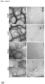
- Plasma subfractions were imaged by cryogenic transmission electron microscopy (cryo-TEM). Vitrified specimens were prepared by placing 3 ⁇ L of a sample on a Quantifoil 1.2/1.3 TEM grid, blotted to a thin film, and plunged into liquid ethane-nitrogen in the Leica EM-CPC cryo-work station. The grids were transferred to a 626 Gatan cryo-holder and maintained at -179 °C. The grids were analyzed with a Jeol JEM 2011 transmission electron microscope operating at an accelerating voltage of 200 kV. Images were recorded on a Gatan Ultrascan 2000 cooled charge-coupled device (CCD) camera with the Digital Micrograph software package (Gatan). Size distribution of EVs was quantified by measuring the diameter of around 200 vesicles in each sample using ImageJ.
- CCD Gatan Ultrascan 2000 cooled charge-coupled device
- Example 1.8 RNA extraction and small RNA sequencing
- RNA samples were eluted in 20 ⁇ L of RNAse-free water. Prior to sequencing, RNA was precipitated using glycogen (20 mg/ mL, Roche), sodium acetate 3M pH 4.8 (Sigma-Aldrich) and 100% ethanol. After 24 h at -20°C, the mixtures were centrifuged at 14,000 rpm for 30 min at 4°C. Carefully, the supernatant was removed, and the pellet was then washed with 75% ethanol followed by centrifugation at 12,000 rpm for 5 min at 4°C. Finally, the supernatant was discarded, and the RNA pellet was dried at RT and dissolved at 65°C for 3 min in 6 ⁇ L of RNAse-free water and stored keeping the cold chain at -80°C.
- RNA samples were analyzed for RNA quality and level of degradation using the Bioanalyzer 2100 (Agilent).
- sRNA libraries were generated using the NEBNext Small RNA Library Preparation Set for Illumina (New England Biolabs) following the manufacturer's instructions. Indexed libraries were equimolarly pooled, and the selected band of interest (145-160 bp) was purified. Single-end sequencing of libraries was then carried out on the Illumina HiSeq 2000 with read lengths of 50 nt, generating at least 8 million reads per sample.
- the quality of the sequenced fastq files was checked using the FastQC software (http://www.bioinformatics.babraham.ac.uk/projects/fastqc/).
- Adapter trimming was performed with Cutadapt and the trimmed reads were aligned to the human genome (version Ensemble hg 19) using STAR aligner (version 2.7.9a).
- the obtained sequences were annotated and quantified using SeqCluster and ExceRpt bioinformatic tools. Prior to further analyses, only sRNAs identified with a minimum of ten counts in at least half of the samples of the tested conditions were considered.
- Partial least squares discriminant analysis (PLS-DA), a multivariate supervised method, based on orthogonal transformations of the initially quantified sRNAs into few uncorrelated components in a way that maximizes the separation of each disease condition, was performed as previously described.
- Three PLS-DA models were applied for each of the three sRNA biotypes considered (miRNAs, tRFs and gene fragments). To elude over-parameterized models with rather poor discriminant properties, we obtained in a first stage of the process, the most important discriminant clusters from the three PLS-DA models. And, in a second stage, we conducted another PLS-DA analysis including the important clusters associated independently to the three different models found in the first stage.
- VIP projection
- sRNAs with VIP > 0.8 were considered for further downstream analysis.
- Principal Component Analysis (PCA) was applied as a non-supervised method to evaluate the separation between disease conditions using the top contributing sRNAs based on PLS-DA analysis. For both PLSA-DA and PCA analyses count matrices normalized and transformed to variance-stabilized expression levels were used.
- Pearson's coefficient (r) was used to screen out the correlation between individual selected top DE sRNAs (DE sRNAs with p-adj ⁇ 0.05 and PLS-DA based VIP > 0.8) and each clinical parameter.
- the selection criteria included sRNAs with r > 0.7 and p-value ⁇ 0.001.
- CCA Canonical-correlation analysis
- PLSR Partial least squares regression
- RNA isolation was used for RNA isolation as described in the section "RNA extraction and small RNA sequencing" with minor modifications.
- synthetic RNA spike-ins Prior to RNA sample lysis using RPL buffer, synthetic RNA spike-ins (UniSp2, UniSp4, UniSpS RNA Spike-in mix; Qiagen) were added and used as controls for RNA isolation, in accordance with the kit protocol.
- Two ⁇ L of isolated RNA were used for reverse transcription (RT) and cDNA synthesis using the miRCURY LNA RT Kit (Qiagen) according to the manufacturer's instructions adding the RNA spike-in UniSp6 as RT control.
- the obtained cDNA was further diluted 1:10 for qPCR and, 3 ⁇ L were used as template for the qPCR in a 10 ⁇ L final volume reaction using the miRCURY LNA SYBR Green PCR Kit (Qiagen) in a StepOnePlus Real-Time PCR System (Applied Biosystems). For each determination, reactions were performed in triplicate.
- miR-100 was selected as the housekeeping gene since it was the most stable sRNA with the least variability among all the sequenced samples.
- RQ relative quantification
- LMM linear mixed-effects models
- ROC receiver operating characteristic
- Example 2.1 Different plasma subfractions show specific sRNA profiles
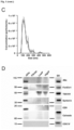
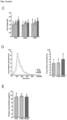
- SEC-eluted fractions enriched in EVs were determined by flow cytometry analyzing the presence of tetraspanins CD9, CD63 and CD81 ( Fig. 1B ).
- the four fractions showing positivity for tetraspanins were pooled (EVs-enriched fractions) and subsequently concentrated by UF (cEVs).
- cEVs were further characterized in compliance with the MISEV guidelines.
- EV concentration yielded 8.33 ⁇ 10 9 ⁇ 2.65 ⁇ 10 8 particles/mL with a mean diameter size of 146.0 ⁇ 2.8 nm in cEVs fractions as determined by NTA ( Fig. 1C ).
- ExceRpt is a reference tool used to characterize all types of circulating sRNAs. During the pre-processing step, it filters out highly abundant sRNAs mapping onto ribosomal RNAs (rRNAs); however, recent data suggest that these fragments of rRNAs are bioactive molecules (doi:10.1038/s41556-021-00652-7).
- ExceRpt only considers the type of isoacceptor when identifying tRNA-derived fragments (tRFs), leading to an oversimplification of the tRFs landscape that has been revealed as highly complex. It has been described that diverse types of tRFs mapping onto different parts of the mature tRNA are differently expressed under physiological and pathological conditions, which makes them appealing biomarkers (doi: 10.1080/15476286.2020.1729584). For these reasons, in order to have a more in-depth and complementary vision of the plasma exRNA transcriptome, we used both tools. Specifically, SeqCluster tool detects units of sRNAs (clusters) using a heuristic iterative algorithm to deal with multi-mapped events.
- Sequences are consistently and non-redundantly grouped if they map onto the same genomic site defining "clusters". Clusters are then classified according to the type of precursor where sRNAs map, including rRNA-clusters and tRF-clusters.
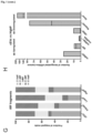
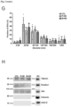
- tRF fragments were classified based on their mapped positions (3'-half, 3'-tRF, i-tRF, 5'-tRF, and 5'-half) and similar distributions were observed in the different subfractions ( Fig. 1 G ). Focusing on the total number of different sRNA clusters identified in each subfraction, we observed that, in accordance with the relative abundance, cEVs fractions contained an increased number of miRNA clusters. This result indicates that although miRNAs were a minor proportion of the total sRNAs, they were concentrated in cEVs compared with other fractions. Despite the higher abundance of gene fragments in HighP fractions, a similar diversity was detected in the different plasma subfractions.
- Example 2.2 No morphological and size differences were found between plasma CTL- and HD-EVs
- Example 2.3 Heterogeneous diversity of extracellular sRNAs in HD and CTL samples
- Fig. 2A To uncover the sRNA diversity in the two plasma fractions selected between HD and control samples, we characterized the sRNA profiles by deep sequencing and applied diverse supervised and unsupervised statistical methods to ascertain sRNA expression differences ( FIG. 2A ). Similar to what we observed in the proof of concept, the majority of reads mapped onto rRNA (51%) followed by gene fragments (34%), and a comparable total number of different sRNA clusters were identified in the three groups. However, we observed heterogeneous proportions of some sRNAs biotypes in the two plasma fractions, including miRNAs and tRFs ( Fig. 2I, 2J ). It is worth mentioning that sequencing analyses revealed low levels of miRNAs which accounted for less than 2% of the EVs-sRNA content ( Fig.
- Non-EVs fractions showed greater intragroup variability ( Fig. 2J ).
- the proportions of sRNAs are more homogeneous between individuals from the same group. All the analyses were also performed using the ExceRpt tool, obtaining similar results.
- Example 2.4 exRNAs-EVs are deregulated in HD and correlated with early symptomatology
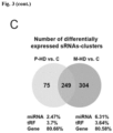
- top DE sRNAs miRNAs, tRFs, and gene fragments
- Example 2.5 Plasma EV-sRNAs as promising early biomarkers in HD
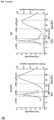
- sRNAs candidates (DE sRNAs showing a p-adj ⁇ 0.05 in P-HD and in M-HD vs CTL patients, with a base mean sequence coverage > 200 and PLS-DA based VIP > 0.8) were selected for validation by qPCR.
- the selected sRNAs were tRNA Glu/CTC , tRNA Gly/GCC , miR-451a, let-7a-5p, miR-26a-5p, miR-27a-3p, miR-21-5p, and tRNA Lys/TTT .
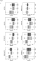
- tRNA Glu/CTC tRNA Gly/GCC , miR-451a, let-7a-5p, miR-26a-5p, miR-27a-3p, miR-21-5p, and tRNA Lys/TTT expression were significantly downregulated in P-HD and M-HD in comparison to CTL samples, consistent with the sequencing findings ( Fig. 5A ).
- NfL neurofilament light chain
Landscapes
- Chemical & Material Sciences (AREA)
- Life Sciences & Earth Sciences (AREA)
- Proteomics, Peptides & Aminoacids (AREA)
- Health & Medical Sciences (AREA)
- Organic Chemistry (AREA)
- Wood Science & Technology (AREA)
- Analytical Chemistry (AREA)
- Zoology (AREA)
- Genetics & Genomics (AREA)
- Engineering & Computer Science (AREA)
- Pathology (AREA)
- Immunology (AREA)
- Microbiology (AREA)
- Molecular Biology (AREA)
- Biotechnology (AREA)
- Biophysics (AREA)
- Physics & Mathematics (AREA)
- Biochemistry (AREA)
- Bioinformatics & Cheminformatics (AREA)
- General Engineering & Computer Science (AREA)
- General Health & Medical Sciences (AREA)
- Measuring Or Testing Involving Enzymes Or Micro-Organisms (AREA)
- Investigating Or Analysing Biological Materials (AREA)
Priority Applications (3)
| Application Number | Priority Date | Filing Date | Title |
|---|---|---|---|
| EP23382178.4A EP4421189A1 (de) | 2023-02-27 | 2023-02-27 | Verfahren zur diagnose oder prognose von morbus huntington |
| PCT/EP2024/053925 WO2024179852A1 (en) | 2023-02-27 | 2024-02-15 | Method for the diagnosis or prognosis of huntington's disease |
| EP24705162.6A EP4673565A1 (de) | 2023-02-27 | 2024-02-15 | Verfahren zur diagnose oder prognose von morbus huntington |
Applications Claiming Priority (1)
| Application Number | Priority Date | Filing Date | Title |
|---|---|---|---|
| EP23382178.4A EP4421189A1 (de) | 2023-02-27 | 2023-02-27 | Verfahren zur diagnose oder prognose von morbus huntington |
Publications (1)
| Publication Number | Publication Date |
|---|---|
| EP4421189A1 true EP4421189A1 (de) | 2024-08-28 |
Family
ID=85384317
Family Applications (2)
| Application Number | Title | Priority Date | Filing Date |
|---|---|---|---|
| EP23382178.4A Withdrawn EP4421189A1 (de) | 2023-02-27 | 2023-02-27 | Verfahren zur diagnose oder prognose von morbus huntington |
| EP24705162.6A Pending EP4673565A1 (de) | 2023-02-27 | 2024-02-15 | Verfahren zur diagnose oder prognose von morbus huntington |
Family Applications After (1)
| Application Number | Title | Priority Date | Filing Date |
|---|---|---|---|
| EP24705162.6A Pending EP4673565A1 (de) | 2023-02-27 | 2024-02-15 | Verfahren zur diagnose oder prognose von morbus huntington |
Country Status (2)
| Country | Link |
|---|---|
| EP (2) | EP4421189A1 (de) |
| WO (1) | WO2024179852A1 (de) |
Citations (1)
| Publication number | Priority date | Publication date | Assignee | Title |
|---|---|---|---|---|
| US20160024575A1 (en) * | 2013-05-02 | 2016-01-28 | The Regents Of The University Of California | Circulating small noncoding rna markers |
-
2023
- 2023-02-27 EP EP23382178.4A patent/EP4421189A1/de not_active Withdrawn
-
2024
- 2024-02-15 WO PCT/EP2024/053925 patent/WO2024179852A1/en not_active Ceased
- 2024-02-15 EP EP24705162.6A patent/EP4673565A1/de active Pending
Patent Citations (1)
| Publication number | Priority date | Publication date | Assignee | Title |
|---|---|---|---|---|
| US20160024575A1 (en) * | 2013-05-02 | 2016-01-28 | The Regents Of The University Of California | Circulating small noncoding rna markers |
Non-Patent Citations (5)
| Title |
|---|
| ANONYMOUS: "Method for the diagnosis or prognosis of Huntington Disease", 2023, Ciber, XP093069789, Retrieved from the Internet <URL:https://www.ciberisciii.es/media/3158100/ficha-tecnolo-gica-hungtington.pptx> * |
| CREUS-MUNCUNILL JORDI ET AL: "Huntington's disease brain-derived small RNAs recapitulate associated neuropathology in mice", ACTA NEUROPATHOLOGICA, vol. 141, no. 4, 6 February 2021 (2021-02-06), pages 565 - 584, XP037408085, ISSN: 0001-6322, DOI: 10.1007/S00401-021-02272-9 * |
| DUBOIS CELINE ET AL: "Small Non-coding RNAs Are Dysregulated in Huntington's Disease Transgenic Mice Independently of the Therapeutic Effects of an Environmental Intervention", MOLECULAR NEUROBIOLOGY, SPRINGER US, NEW YORK, vol. 58, no. 7, 6 March 2021 (2021-03-06), pages 3308 - 3318, XP037499899, ISSN: 0893-7648, [retrieved on 20210306], DOI: 10.1007/S12035-021-02342-9 * |
| GIRSTMAIR HANNAH ET AL: "Depletion of Cognate Charged Transfer RNA Causes Translational Frameshifting within the Expanded CAG Stretch in Huntingtin", CELL REPORTS, vol. 3, no. 1, 1 January 2013 (2013-01-01), US, pages 148 - 159, XP093069805, ISSN: 2211-1247, DOI: 10.1016/j.celrep.2012.12.019 * |
| MATHEW BLESSY AKSA ET AL: "Role of tRNA-Derived Fragments in Neurological Disorders: a Review", MOLECULAR NEUROBIOLOGY, vol. 60, no. 2, 8 November 2022 (2022-11-08), New York, pages 655 - 671, XP093069402, ISSN: 0893-7648, Retrieved from the Internet <URL:https://link.springer.com/content/pdf/10.1007/s12035-022-03078-w.pdf?pdf=button> DOI: 10.1007/s12035-022-03078-w * |
Also Published As
| Publication number | Publication date |
|---|---|
| EP4673565A1 (de) | 2026-01-07 |
| WO2024179852A1 (en) | 2024-09-06 |
Similar Documents
| Publication | Publication Date | Title |
|---|---|---|
| Baraniskin et al. | Identification of microRNAs in the cerebrospinal fluid as marker for primary diffuse large B-cell lymphoma of the central nervous system | |
| CA2840196C (en) | Microrna biomarkers indicative of alzheimer's disease | |
| EP2201134B1 (de) | Genexpressionssignaturen in angereicherten tumorzellproben | |
| CN111630183B (zh) | 透明细胞肾细胞癌生物标志物 | |
| EP3739065A1 (de) | Somatische mutationsbasierte klassifizierung von krebs | |
| Herrero‐Lorenzo et al. | Small RNAs in plasma extracellular vesicles define biomarkers of premanifest changes in Huntington's disease | |
| US20220093251A1 (en) | Novel biomarkers and diagnostic profiles for prostate cancer | |
| CA3152887A1 (en) | Novel biomarkers and diagnostic profiles for prostate cancer integrating clinical variables and gene expression data | |
| CN111235274A (zh) | 喉鳞癌血清外泌体标志物筛选方法和外泌体来源miR-941应用 | |
| James et al. | Mucosal expression of PI3, ANXA1, and VDR discriminates Crohn’s disease from ulcerative colitis | |
| EP3492607A2 (de) | Mirnas in urin für die in vitro diagnose von blasenkrebs | |
| CN104011543B (zh) | 癌症的生物标记 | |
| CN108841949A (zh) | 帕金森病早期检测和诊断试剂盒及装置 | |
| CN113999852B (zh) | circ_0001772作为结直肠癌诊断和治疗标志物的应用 | |
| EP4673565A1 (de) | Verfahren zur diagnose oder prognose von morbus huntington | |
| CN107760688A (zh) | 一种brca2基因突变体及其应用 | |
| CN117568481A (zh) | 一组与肝癌相关的血浆外泌体tsRNAs标志物及其应用 | |
| KR20240054194A (ko) | 혈액 내 rna의 엑손-접합 정보를 이용한 암 진단 방법 | |
| CN112899367B (zh) | 多发性骨髓瘤诊断生物标志物及其应用 | |
| CN112646876A (zh) | 用于银屑病诊断的miRNA及其应用 | |
| EP4249607A1 (de) | Markerpaneel für vorhersage von wiederauftreten der epilepsie in tuberöser sklerose patienten und verwendung davon | |
| EP4189117A1 (de) | Verwendung von zirkulärer rna zur diagnose von multipler sklerose | |
| US20220162699A1 (en) | In vitro method for the diagnosis of synucleinopathies | |
| EP4249606A1 (de) | Markerpaneel für vorhersage von wiederauftreten der epilepsie in tuberöser sklerose patienten und verwendung davon | |
| KR102870611B1 (ko) | 위암 조기진단을 위한 바이오마커 조성물 및 이를 이용한 진단방법 |
Legal Events
| Date | Code | Title | Description |
|---|---|---|---|
| PUAI | Public reference made under article 153(3) epc to a published international application that has entered the european phase |
Free format text: ORIGINAL CODE: 0009012 |
|
| STAA | Information on the status of an ep patent application or granted ep patent |
Free format text: STATUS: THE APPLICATION HAS BEEN PUBLISHED |
|
| AK | Designated contracting states |
Kind code of ref document: A1 Designated state(s): AL AT BE BG CH CY CZ DE DK EE ES FI FR GB GR HR HU IE IS IT LI LT LU LV MC ME MK MT NL NO PL PT RO RS SE SI SK SM TR |
|
| STAA | Information on the status of an ep patent application or granted ep patent |
Free format text: STATUS: THE APPLICATION IS DEEMED TO BE WITHDRAWN |
|
| 18D | Application deemed to be withdrawn |
Effective date: 20250301 |