EP4403192A1 - Verwendung von eisenoxidnanopartikeln bei der herstellung eines parathyroid- und/oder lymphknotenkontrastmittels - Google Patents
Verwendung von eisenoxidnanopartikeln bei der herstellung eines parathyroid- und/oder lymphknotenkontrastmittels Download PDFInfo
- Publication number
- EP4403192A1 EP4403192A1 EP21957301.1A EP21957301A EP4403192A1 EP 4403192 A1 EP4403192 A1 EP 4403192A1 EP 21957301 A EP21957301 A EP 21957301A EP 4403192 A1 EP4403192 A1 EP 4403192A1
- Authority
- EP
- European Patent Office
- Prior art keywords
- ionps
- iron oxide
- parathyroid
- gland
- oxide nanoparticles
- Prior art date
- Legal status (The legal status is an assumption and is not a legal conclusion. Google has not performed a legal analysis and makes no representation as to the accuracy of the status listed.)
- Pending
Links
Images



Classifications
-
- A—HUMAN NECESSITIES
- A61—MEDICAL OR VETERINARY SCIENCE; HYGIENE
- A61K—PREPARATIONS FOR MEDICAL, DENTAL OR TOILETRY PURPOSES
- A61K49/00—Preparations for testing in vivo
- A61K49/0002—General or multifunctional contrast agents, e.g. chelated agents
-
- A—HUMAN NECESSITIES
- A61—MEDICAL OR VETERINARY SCIENCE; HYGIENE
- A61K—PREPARATIONS FOR MEDICAL, DENTAL OR TOILETRY PURPOSES
- A61K49/00—Preparations for testing in vivo
- A61K49/001—Preparation for luminescence or biological staining
- A61K49/0063—Preparation for luminescence or biological staining characterised by a special physical or galenical form, e.g. emulsions, microspheres
- A61K49/0069—Preparation for luminescence or biological staining characterised by a special physical or galenical form, e.g. emulsions, microspheres the agent being in a particular physical galenical form
- A61K49/0089—Particulate, powder, adsorbate, bead, sphere
- A61K49/0091—Microparticle, microcapsule, microbubble, microsphere, microbead, i.e. having a size or diameter higher or equal to 1 micrometer
- A61K49/0093—Nanoparticle, nanocapsule, nanobubble, nanosphere, nanobead, i.e. having a size or diameter smaller than 1 micrometer, e.g. polymeric nanoparticle
-
- A—HUMAN NECESSITIES
- A61—MEDICAL OR VETERINARY SCIENCE; HYGIENE
- A61K—PREPARATIONS FOR MEDICAL, DENTAL OR TOILETRY PURPOSES
- A61K33/00—Medicinal preparations containing inorganic active ingredients
- A61K33/24—Heavy metals; Compounds thereof
- A61K33/26—Iron; Compounds thereof
-
- A—HUMAN NECESSITIES
- A61—MEDICAL OR VETERINARY SCIENCE; HYGIENE
- A61K—PREPARATIONS FOR MEDICAL, DENTAL OR TOILETRY PURPOSES
- A61K49/00—Preparations for testing in vivo
- A61K49/001—Preparation for luminescence or biological staining
- A61K49/006—Biological staining of tissues in vivo, e.g. methylene blue or toluidine blue O administered in the buccal area to detect epithelial cancer cells, dyes used for delineating tissues during surgery
-
- A—HUMAN NECESSITIES
- A61—MEDICAL OR VETERINARY SCIENCE; HYGIENE
- A61K—PREPARATIONS FOR MEDICAL, DENTAL OR TOILETRY PURPOSES
- A61K49/00—Preparations for testing in vivo
- A61K49/04—X-ray contrast preparations
-
- A—HUMAN NECESSITIES
- A61—MEDICAL OR VETERINARY SCIENCE; HYGIENE
- A61K—PREPARATIONS FOR MEDICAL, DENTAL OR TOILETRY PURPOSES
- A61K49/00—Preparations for testing in vivo
- A61K49/06—Nuclear magnetic resonance [NMR] contrast preparations; Magnetic resonance imaging [MRI] contrast preparations
-
- A—HUMAN NECESSITIES
- A61—MEDICAL OR VETERINARY SCIENCE; HYGIENE
- A61K—PREPARATIONS FOR MEDICAL, DENTAL OR TOILETRY PURPOSES
- A61K49/00—Preparations for testing in vivo
- A61K49/06—Nuclear magnetic resonance [NMR] contrast preparations; Magnetic resonance imaging [MRI] contrast preparations
- A61K49/18—Nuclear magnetic resonance [NMR] contrast preparations; Magnetic resonance imaging [MRI] contrast preparations characterised by a special physical form, e.g. emulsions, microcapsules, liposomes
- A61K49/1818—Nuclear magnetic resonance [NMR] contrast preparations; Magnetic resonance imaging [MRI] contrast preparations characterised by a special physical form, e.g. emulsions, microcapsules, liposomes particles, e.g. uncoated or non-functionalised microparticles or nanoparticles
- A61K49/1821—Nuclear magnetic resonance [NMR] contrast preparations; Magnetic resonance imaging [MRI] contrast preparations characterised by a special physical form, e.g. emulsions, microcapsules, liposomes particles, e.g. uncoated or non-functionalised microparticles or nanoparticles coated or functionalised microparticles or nanoparticles
- A61K49/1824—Nuclear magnetic resonance [NMR] contrast preparations; Magnetic resonance imaging [MRI] contrast preparations characterised by a special physical form, e.g. emulsions, microcapsules, liposomes particles, e.g. uncoated or non-functionalised microparticles or nanoparticles coated or functionalised microparticles or nanoparticles coated or functionalised nanoparticles
-
- A—HUMAN NECESSITIES
- A61—MEDICAL OR VETERINARY SCIENCE; HYGIENE
- A61K—PREPARATIONS FOR MEDICAL, DENTAL OR TOILETRY PURPOSES
- A61K49/00—Preparations for testing in vivo
- A61K49/22—Echographic preparations; Ultrasonic imaging preparations
-
- A—HUMAN NECESSITIES
- A61—MEDICAL OR VETERINARY SCIENCE; HYGIENE
- A61K—PREPARATIONS FOR MEDICAL, DENTAL OR TOILETRY PURPOSES
- A61K49/00—Preparations for testing in vivo
- A61K49/22—Echographic preparations; Ultrasonic imaging preparations
- A61K49/222—Echographic preparations; Ultrasonic imaging preparations characterised by a special physical form, e.g. emulsions, liposomes
- A61K49/225—Microparticles, microcapsules
-
- A—HUMAN NECESSITIES
- A61—MEDICAL OR VETERINARY SCIENCE; HYGIENE
- A61P—SPECIFIC THERAPEUTIC ACTIVITY OF CHEMICAL COMPOUNDS OR MEDICINAL PREPARATIONS
- A61P43/00—Drugs for specific purposes, not provided for in groups A61P1/00-A61P41/00
-
- A—HUMAN NECESSITIES
- A61—MEDICAL OR VETERINARY SCIENCE; HYGIENE
- A61P—SPECIFIC THERAPEUTIC ACTIVITY OF CHEMICAL COMPOUNDS OR MEDICINAL PREPARATIONS
- A61P5/00—Drugs for disorders of the endocrine system
- A61P5/14—Drugs for disorders of the endocrine system of the thyroid hormones, e.g. T3, T4
-
- B—PERFORMING OPERATIONS; TRANSPORTING
- B82—NANOTECHNOLOGY
- B82Y—SPECIFIC USES OR APPLICATIONS OF NANOSTRUCTURES; MEASUREMENT OR ANALYSIS OF NANOSTRUCTURES; MANUFACTURE OR TREATMENT OF NANOSTRUCTURES
- B82Y5/00—Nanobiotechnology or nanomedicine, e.g. protein engineering or drug delivery
Definitions
- the present disclosure relates to the technical field of surgical navigation imaging technology, and relates to a negative contrast agent for parathyroid glands, preparation and use thereof, especially to an iron oxide nanoparticle, preparation and use thereof in a negative contrast agent for parathyroid glands.
- the GLOBOCAN reported that the incidence of thyroid gland cancers ranked ninth among all cancers in the world and fifth among female cancers. According to the statistics of imaging data from 25 countries around the world reported by the sub-journal of the Lancet, the incidence of all major thyroid gland cancer subtypes has increased significantly in China. At the same time, low-risk papillary thyroid gland cancers have increased rapidly around the world, including China.
- parathyroid glands are derived from different pharyngeal pouches during embryonic development, there are many anatomical variations, especially in the lower parathyroid gland, which can be ectopic to the mandibular angle, the anterior mediastinum, or even the pericardium, or can also be embedded in the thymus or thyroid gland.
- the in-situ preservation of parathyroid gland remains highly challenging. Therefore, how to protect the parathyroid gland better during surgery is one of the clinical problems to be solved urgently. To date, this remains a clinically important, unmet need.
- the current intraoperative imaging of the parathyroid glands mainly includes both positive imaging and negative imaging.
- the positive contrast agents include methylene blue, aminolaevulinic acid, indocyanine green, etc.
- the positive contrast agents lack the specificity for parathyroid glands, generally resulting in a low staining rate and poor recognition of normal parathyroid glands, and have side effects such as arrhythmia, neurotoxicity, iodine allergy, etc.
- the negative contrast agents are mainly carbon nanomaterials, which are a kind of black carbon nanoparticles (CNPs). Such contrast agents function to negatively image the parathyroid glands by staining the normal thyroid gland to black. It has been used clinically in China. However, its use in non-tumor surgeries is restricted, and its use in secondary surgeries is even more difficult. In addition, carbon nanomaterials have severe potential safety issues that can cause harm to human bodies: carbon nanomaterials cannot be degraded normally in human bodies, which can damage DNA during cell replication, and may induce cancers. Thus, the safety of carbon nanomaterials in human has remained controversial.
- the applied carbon nanomaterials penetrate into lateral cervical lymph nodes through the lymphatic vessels in the thyroid gland area, which are not dissected, therefore, the carbon nanomaterials will remain in the lymph nodes in this area for a long time.
- a long-lasting stains in the skin caused by injection of carbon nanomaterials may occur in endoscopic thyroid surgeries.
- CNPs can also result in a reduction in photosensitivity of the visual cells of zebrafishes, cause toxicity to the glial cells in the nervous system of zebrafishes, and lead to disorders in the movement behavior of zebrafishes.
- Zhao Dandan illustrated the effect of CNPs combined with cold stimulation on the respiratory system and body energy metabolism of mice in her master's thesis .
- Zhou Lixiao showed in her master's thesis that CNPs have a toxic effect on human bronchial epithelial cells.
- an object of the present disclosure is to provide iron oxide nanoparticles (IONPs) and preparation and use thereof in the negative imaging of parathyroid glands.
- the IONPs can help better preserve the parathyroid glands during surgery to maintain their function. It has been proven that the iron oxide nanoparticles can be degraded by the lysosomes of normal human cells and digested into iron ions, which can combine with ferritin and then enter the normal iron metabolism, so that IONPs can be completely absorbed and degraded by human bodies, demonstrating its safety.
- Ferumoxytol IONPs have been approved by the FDA for the clinical treatment of iron deficiency anemia (IDA) in adults.
- the present disclosure provides iron oxide nanoparticles (IONPs) with physicochemical properties of: a nanoparticle structure with a particle size of 1 nm to 1,000 nm, a polydispersity index of less than 0.3, a zeta-potential of -50 mV to 50 mV, and a peak value of ultraviolet-visible absorption spectrum of 100 nm to 500 nm.
- IONPs iron oxide nanoparticles
- the particle size is in a range of 1 nm to 1,000 nm; e.g., 1-5 nm, 1-10 nm, 1-50 nm, 1-100 nm, 1-200 nm, 5-50 nm, 5-100 nm, 5-200 nm, 10-50 nm, 10-100 nm, 10-200 nm, 10-20 nm, 20-30 nm, 30-40 nm, 40-50 nm, 50-60 nm, 60-70 nm, 70-80 nm, 80-90 nm, 90- 100 nm, 100-110 nm, 110-120 nm, 120-130 nm, 130-140 nm, 140-150 nm, 150-200 nm, 200-250 nm, 250-300 nm, 350-400 nm, 450-500 nm, 550-600 nm, 650-700 nm,
- the particle size is in a range of 1-200 nm. More preferably, the particle size is in a range of 1-100 nm. Still more preferably, the particle size is in a range of 1-30 nm. Still more preferably, the particle size is in a range of 10-30 nm. Still more preferably, the particle size is in a range of 10-20 nm. Still more preferably, the particle size is 10 nm.
- the zeta-potential is in a range of -50 mV to 50 mV; e.g., -50 mV to 40 mV, -40 mV to -30 mV, -30 mV to -20 mV, -20 mV to -10 mV, -20 mV to 20 mV, -10 mV to 0 mV, 0 mV to 10 mV, 10 mV to 20 mV, 20 mV to 30 mV, 30 mV to 40 mV, or 40 mV to 50 mV; preferably, -35 mV to 20 mV; more preferably, -35 mV to 0 mV; more preferably, -30.57 mV.
- the peak value of the ultraviolet-visible absorption spectrum is in a range of 100 nm to 500 nm; e.g., 100-120 nm, 120-140 nm, 140-160 nm, 160-180 nm, 180-200 nm, 200-220 nm, 200-300 nm, 220-240 nm, 240-260 nm, 260-280 nm, 280-300 nm, 300-320 nm, 320-340 nm, 340-360 nm, 360-380 nm, 380-400 nm, 400-420 nm, 420-440 nm, 440-460 nm, 460-480 nm, or 480-500 nm.
- the IONPs can also have a specific polydispersity index.
- the polydispersity index is less than 0.3; eg: 0.01, 0.02, 0.03, 0.04, 0.05, 0.06, 0.07, 0.08, 0.09, 0.10, 0.12, 0.14, 0.16, 0.18, 0.20, 0.22, 0.24, 0.26, or 0.28.
- the polydispersity index is in a range of 0.01-0.10. More preferably, the polydispersity index is in a range of 0.01-0.05. More preferably, the polydispersity index is in a range of 0.01-0.03. Still more preferably, the polydispersity index is 0.01, 0.02, or 0.03. Still more preferably, the polydispersity index is 0.03.
- the present disclosure further provides a use of the iron oxide nanoparticles (IONPs) in the preparation of a negative contrast agent for parathyroid glands.
- IONPs iron oxide nanoparticles
- the present disclosure further provides a use of the iron oxide nanoparticles (IONPs) in the preparation of a positive contrast agent for lymph nodes.
- IONPs iron oxide nanoparticles
- the present disclosure further provides a contrast agent, where the negative contrast agent includes the iron oxide nanoparticles (IONPs) described above.
- the contrast agent can serve as a positive contrast agent for thyroid gland, a negative contrast agent for parathyroid glands, or a positive contrast agent for lymph nodes.
- the IONPs of the present disclosure can be preliminarily screened for their diagnostic efficacy by in vivo or in vitro tests, and other methods are also obvious to those skilled in the art.
- the IONPs can be administered alone or in a form of a composition as prepared.
- the composition can be prepared as a contrast agent or a diagnostic tracer for diagnostic purposes.
- the composition can be prepared as a pharmaceutical composition for therapeutic purposes.
- the composition can further include one or more pharmaceutically acceptable carriers or media.
- the carriers and media need to be compatible with the IONPs.
- the acceptable carriers or media can include, e.g., sterile water or normal saline, stabilizers, excipients, antioxidants (ascorbic acid, etc.), buffers (phosphoric acid, citric acid, other organic acids, etc.), preservatives, surface active agents (PEG, Tween, etc.), chelating agents (EDTA, etc.), adhesives, polymers, etc.
- it can also include other low molecular weight peptides; proteins such as serum albumin, gelatin, or immunoglobulin; amino acids such as glycine, glutamine, asparagine, arginine, and lysine; sugars or carbohydrates such as polysaccharides and monosaccharides; and sugar alcohols such as mannitol or sorbitol.
- proteins such as serum albumin, gelatin, or immunoglobulin
- amino acids such as glycine, glutamine, asparagine, arginine, and lysine
- sugars or carbohydrates such as polysaccharides and monosaccharides
- sugar alcohols such as mannitol or sorbitol.
- glucose or other auxiliary drugs such as D-sorbitol, D-mannose, D-mannitol, and sodium chloride
- solubilizer such as alcohol (ethanol, etc.), polyol (propylene glycol, PEG, etc.) and a nonionic surfactant (Tween 80, HCO-50)
- glucose or other auxiliary drugs such as D-sorbitol, D-mannose, D-mannitol, and sodium chloride
- solubilizer such as alcohol (ethanol, etc.), polyol (propylene glycol, PEG, etc.) and a nonionic surfactant (Tween 80, HCO-50)
- the present disclosure further provides a use of the composition in diagnosis of thyroid gland and/or lymph node-associated diseases or disorders, tissue imaging of thyroid gland or lymph nodes, or intraoperative navigation.
- the diagnostic tracer composition can also be prepared into a detection kit.
- the IONPs are prepared along with a drug in a form of pharmaceutical composition for tracking the targeting and therapeutic effect of the drug.
- the amount of IONPs is usually a safe and effective amount, which can be adjustable by those skilled in the art.
- the administration dose of the IONPs usually depends on the patient's weight, application type, as well as status and severity of the disease.
- the administration dose of IONPs can usually be 0.1-1 mg/kg/time.
- compositions provided by the present disclosure can be suitable for any administration mode, which can be oral or parenteral administration, e.g., pulmonary, nasal, rectal, and/or intravenous injection, more specifically, intradermal, subcutaneous, intramuscular, intra-articular, intraperitoneal, pulmonary, oral, sublingual, nasal, transdermal, vaginal, oral or parenteral administration.
- Injection administration includes intravenous injection, intramuscular injection, subcutaneous injection, transdermal administration, etc.
- the dosage form of the composition is selected from the group consisting of injections, sterile powders for injection, tablets, pills, capsules, lozenges, spiritus, powders, granules, syrups, solutions, tinctures, aerosols, powder spray, or suppository. Those skilled in the art can select an appropriate dosage form according to the administration mode.
- dosage forms suitable for oral administration can include, but are not limited to pills, tablets, chewables, capsules, granules, solutions, drops, syrups, aerosols, or powder sprays, etc.; as another example, dosage forms suitable for parenteral administration can include, but are not limited to solutions, suspensions, reconstitutable dry preparations or sprays, etc.; as another example, dosage forms suitable for rectal administration can usually be suppositories; and as another example, dosage forms suitable for injection administration can be injections, sterile powders for injection, etc.
- diagnosis includes, but is not limited to, diagnosing or detecting the presence of thyroid gland diseases, or performing an intraoperative real-time surgical navigation or observing the drug targeting by imaging, and so on.
- the IONPs of the present disclosure can be used in various diagnostic imaging methods for thyroid glands, including ultrasound, magnetic resonance, and X-ray imaging. They can be used in ultrasonic diagnostic imaging and magnetic resonance imaging.
- contrast agent or diagnostic tracer of the present disclosure can be added various additives, such as emulsifiers, coating agents, plasticizers, fillers, cryoprotectants, and/or antioxidants, e.g., to improve the stability, dispersibility, aggregation tendency, and biological properties thereof, or to improve the flexibility and/or polarity of the membrane.
- additives such as emulsifiers, coating agents, plasticizers, fillers, cryoprotectants, and/or antioxidants, e.g., to improve the stability, dispersibility, aggregation tendency, and biological properties thereof, or to improve the flexibility and/or polarity of the membrane.
- the present disclosure further provides a method for negatively imaging parathyroid glands, including: administering the IONPs of the present disclosure to an individual.
- the present disclosure further provides a method for imaging lymph nodes, including: administering the IONPs of the present disclosure to an individual.
- parathyroid glands are small and have large anatomical variations, are located on the back side of the thyroid gland, and are closely related to the thyroid gland.
- the structure of parathyroid glands is high condensed with cells. In comparison, the structure of thyroid glands is relatively loose with lumens and interstitial spaces.
- the IONPs can readily diffuse into the thyroid gland but cannot enter the parathyroid gland due to the tissue structure difference between the parathyroid gland and thyroid gland, which negatively images the parathyroid gland immediately adjacent to the thyroid gland.
- the lymph nodes at the inferior thyroid gland can be imaged by black staining of IONPs, so that the parathyroid gland at the inferior thyroid gland can be negatively imaged.
- the principle for imaging of the lymph node in the present disclosure includes: after the local injection of IONPs (in the lower lip), the IONPs are transported to the submandibular lymph nodes via the anterior cervical lymphatic vessels due to lymphatic drainage.
- the submandibular lymph nodes in rats are relatively superficial, and thus the IONP-stained lymph nodes can be observed by incising the skin.
- the present disclosure further provides a method for diagnosing thyroid gland and/or lymph nodes using the IONPs as described above, including administering the IONPs or composition as described herein as a tissue contrast agent, and obtaining an image of the mammal after the administration of the IONPs or compositions.
- the method further includes evaluating a signal level after administering the diagnostic tracer or contrast agent (including the IONPs or composition as described herein) with a control signal level.
- the present disclosure further provides a method for screening drugs that are effective in treating thyroid gland and/or lymph nodes.
- constructing the IONPs and the drug molecules into the same carrier such as microspheres evaluating the positive imaging effect on the thyroid gland, the negative imaging effect on the parathyroid gland, and/or the positive imaging effect on the lymph node of the IONPs to determine the targeting of the drug molecules on the thyroid gland and/or lymph node diseases to screen the drugs effective in the treatment of thyroid gland and/or lymph nodes.
- the method of the present disclosure has excellent operability, good accuracy, etc.
- iron oxide nanoparticles As used herein, “iron oxide nanoparticles,” “iron oxide nanometric particles” and “nano-iron oxide” have the same meaning.
- carbon nanoparticles As used herein, “carbon nanoparticles (CNPs),” “carbon nanometric particles” and “nano carbon” have the same meaning.
- treatment should be understood in their broadest sense.
- treatment does not necessarily imply that the mammal is treated until fully recovered.
- prevention does not necessarily mean that the subject will not eventually be infected with the disease or disorder.
- the treatment and prevention include alleviating symptoms of a specific disorder or preventing or reducing the risk of developing a specific disorder.
- prevention can be understood as reducing the severity of the attack of a specific disorder.
- the treatment can also be understood as reducing the severity of an existing disorder or the frequency of an acute attack.
- the subject or individual to be therapeutically or prophylactically treated is preferably a mammal, e.g., but not limited to humans, primates, livestock (such as sheep, cattle, horses, donkeys, pigs), pets (such as dogs, cats), laboratory test animals (such as mice, rabbits, rats, guinea pigs, hamsters) or captured wild animals (such as foxes, deer).
- the subject is preferably a primate.
- the subject is most preferably human.
- the iron oxide nanoparticles (IONPs) of the present disclosure and preparation and use thereof have the following beneficial effects:
- the experimental methods, detection methods, and preparation methods disclosed in the present disclosure all adopt conventional technologies in the fields of molecular biology, biochemistry, chromatin structure and assay, analytical chemistry, cell culture, recombinant DNA technology, and related fields.
- Carbon nanoparticles particle size, 150 nm; manufacturer: Chongqing Lummy Pharmaceutical Co., Ltd. (batch number: Z200603; production date: 2020-06-12); black suspension.
- Iron oxide nanoparticles particle size, 10 nm, 50 nm, and 100 nm; magnetic nanoparticle solution.
- Nanoparticle size and Zeta-potential analyzer Zetasizer Nano ZS (Malvern, ZEN3600);
- UV-visible absorption spectrophotometer UV/Vis/NIR Spectrophotometer (PerkinElmer, LAMBDA1050+);
- Morphological structure polydispersity index (dispersity or PDI), particle size, Zeta-potential (surface charge (Dynamic Light Scattering, DLS)), ultraviolet-visible absorption spectrum (Ultraviolet-Visible Spectroscopy, UV-Vis).
- the particle size and zeta-potential were measured using the Zetasizer Nano ZS (Malvern, ZEN3600) nanoparticle size and potential analyzer. Specifically, a 10 nm-IONPs-containing suspension was diluted with normal saline to an IONP concentration of 50ug/mL, and then the IONP samples were directly placed into a sample cell for measurement. The measurement result is -30.57 ⁇ 0.59mV.
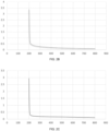
- FIG. 2 The ultraviolet-visible absorption spectrum was measured by an UV-Vis absorption photometer.
- An IONP-containing suspension was diluted with normal saline to an IONP concentration of 10 ⁇ g/mL, and then the IONP samples were directly placed into the sample cell for measurement.
- FIG. 2A is the UV-Vis absorption spectrum of 10 nm IONPs with a peak value of 200-220 nm, which is close to the peak value of the UV-Vis absorption spectrum of carbon nanoparticles
- FIG. 2B is the UV-Vis absorption spectrum of 50 nm IONPs
- FIG. 2C is the UV-Vis absorption spectrum of 100 nm IONPs.
- the zeta-potential of carbon nanoparticles is -4.44 ⁇ 0.25.
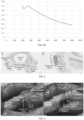
- FIG. 2D shows a UV absorption spectrum curve.
- the particle size is 150 nm; the morphology structure is spherical; the hydrated diameter is 171.50 ⁇ 0.85 nm; and the polydispersity index (PDI) is 0.135.
- PDI polydispersity index
- One 6-week-old SD rat with weight of about 240g-260g was selected and treated in accordance with the steps 1) to 7) of Embodiment 3. Later, the larynx, trachea, thyroid gland, and parathyroid glands within the range from the superior border of the thyroid gland cartilage to the 5 th ring of the trachea were surgically removed, which were then stained with Hematoxylin-Eosin (HE staining for short) and observed under microscope for the pathological results of the rat parathyroid gland.
- HE staining Hematoxylin-Eosin
- FIG. 3A is an image of the laryngotrachea passing across the cross-section of the parathyroid gland, from which it can be seen that the parathyroid gland clings to the thyroid gland, and is located outside and above the thyroid gland.
- FIG. 3B is a locally enlarged view of the thyroid gland and the parathyroid gland, where the characteristic chief cells and oxyphil cells of the parathyroid gland can be observed under the microscope (golden standard).
- 6-week-old female and male SD rats with weights of about 240g-260g were selected.
- the parathyroid glands were negatively imaged according to the blood supply characteristics of the parathyroid gland with selectively traced lymph nodes.
- Experimental groups A, B, and C correspond to IONPs with three different particle sizes of 10 nm, 50 nm, and 100 nm respectively;
- FIG. 5A is the positive control of carbon nanoparticles
- FIGS. 5B to 5D are the IONPs experimental groups of 10 nm, 50 nm, and 100 nm, respectively
- FIG. 5E is the blank control of 0.9% normal saline. As seen from FIG. 5 , the 10 nm IONPs produce the most enhanced imaging effect.
- the side view of the thyroid gland of the SD rat (marked as M) was photographed.
- the thyroid gland was stained to black, and the parathyroid gland remained unstained. Therefore, parathyroid gland can be clearly recognized by the naked eyes effectively.
- a method for measuring the grayscale decrease before and after clinical injection of CNPs was applied, where the method is in particular as follows: An intraoperative photography was carried out after exposure of the surgical cavity, to compare the change in grayscale of the thyroid gland before and after injection.
- the grayscale values before and after the injection in circles with the same size that are on the thyroid gland and adjacent to the border of the parathyroid gland were measured by using the grayscale calculation function in ImageJ software.
- the rate of change in the grayscale of thyroid gland before and after the injection is calculated (by subtracting the grayscale value after the injection from the grayscale value before the injection to obtain a difference, and then dividing the difference by the grayscale value before the injection to obtain the rate of change in the grayscale). The result is shown in FIG. 6 .
- 10 nm IONPs (43.32%) have a similar rate of change in the grayscale to CNPs (44.02%), which produces the best enhancement effect. Therefore, 10 nm IONPs with the best imaging effect were used for subsequent experiments.
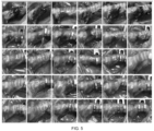
- FIG. 7 The imaging results are shown in FIG. 7 , where each of FIGs. 7A-D includes 7 pictures from left to right (labeled as A1-A7, B1-B7, C1-C7, D1-D7, respectively).
- Images 1, 2, and 3 are blank front views of the rats before the injection (X1, X3, and X5 times); and Images 4, 5, and 6 are the images after the injection of nanomaterials (X1, X3, and X5 times).
- the results in FIG. 7B are similar to those in FIG. 7A .
- FIG. 7B are similar to those in FIG. 7A .
- FIG. 7C whether the IONPs were injected into the left thyroid lobe or the right thyroid lobe, an efficient imaging enhancement can be observed.
- FIG. 7D whether CNPs are injected into the left thyroid lobe or the right thyroid lobe, an efficient imaging enhancement can be observed.
- A7, B7, C7, and D7 show incisions at the middle anterior cervical position, and injections were performed according to the nanoparticle injection method of the respective group to illustrate the comparative degradation of 10 nm IONPs and CNPs in the skin and subcutaneous tissue.
- A7 shows the subcutaneous injection of the 10 nm IONPs and the CNPs into the left and the right sides of the middle anterior cervical incision, respectively; B7 shows the subcutaneous injection of the CNPs and the 10 nm IONPs into the left and the right sides of the middle anterior cervical incision, respectively; C7 shows the subcutaneous injection of the 10 nm IONPs into both the left and the right sides of the anterior cervical incision; and D7 shows the subcutaneous injection of the CNPs into both the left and the right sides of the anterior cervical incision.
- A7 or B7 when different nanoparticles were injected into the left and the right sides of the middle anterior cervical incision of the same rat, respectively, there is no obvious color difference.
- C7 or D7 when the same nanoparticles were injected into both the left and the right sides of rats, there is no obvious color difference.
- One 6-week-old rat with a weight of about 240g-260g was selected and treated according to the steps 1)-7) of Embodiment 3. Then, the anterior cervical area (including the larynx, trachea, esophagus, thyroid gland, and parathyroid gland) of the SD rat was surgically removed. The left thyroid lobe was injected with CNPs, and the right thyroid lobe was injected with 10 nm IONPs. Photographs were taken from the side, as shown in FIG. 8A . The leakage of nanomaterials was observed. After 2.5 hours, the results are shown in FIG. 8B .
- the esophagus On the side to which the CNPs were injected, the esophagus was stained to black (the range shown in the circle), while on the side to which the 10 nm IONPs were injected, the esophagus was not contaminated and showed white (the range shown in the circle), indicating that there is no obvious penetration of the 10 nm IONPs into the parathyroid gland area, and the imaging effect is better.
- the parathyroid glands were negatively imaged according to the blood supply characteristics of the parathyroid gland with selectively traced lymph nodes.
- lymph node imaging after local injection (lower lip) of 10 nm IONPs, the IONPs were transported to the submandibular lymph nodes via the anterior cervical lymphatic vessels.
- the submandibular lymph nodes in rats were relatively superficial, and thus black-stained lymph nodes can be observed after incising the skin.
Landscapes
- Health & Medical Sciences (AREA)
- Life Sciences & Earth Sciences (AREA)
- General Health & Medical Sciences (AREA)
- Engineering & Computer Science (AREA)
- Veterinary Medicine (AREA)
- Public Health (AREA)
- Animal Behavior & Ethology (AREA)
- Epidemiology (AREA)
- Chemical & Material Sciences (AREA)
- Nuclear Medicine, Radiotherapy & Molecular Imaging (AREA)
- Nanotechnology (AREA)
- Biomedical Technology (AREA)
- Medicinal Chemistry (AREA)
- Pharmacology & Pharmacy (AREA)
- Physics & Mathematics (AREA)
- Radiology & Medical Imaging (AREA)
- Bioinformatics & Cheminformatics (AREA)
- Biodiversity & Conservation Biology (AREA)
- Oncology (AREA)
- Optics & Photonics (AREA)
- Acoustics & Sound (AREA)
- Inorganic Chemistry (AREA)
- Chemical Kinetics & Catalysis (AREA)
- General Chemical & Material Sciences (AREA)
- Organic Chemistry (AREA)
- General Engineering & Computer Science (AREA)
- Molecular Biology (AREA)
- Crystallography & Structural Chemistry (AREA)
- Medical Informatics (AREA)
- Diabetes (AREA)
- Endocrinology (AREA)
- Biotechnology (AREA)
- Biophysics (AREA)
- Medicines Containing Antibodies Or Antigens For Use As Internal Diagnostic Agents (AREA)
Applications Claiming Priority (2)
| Application Number | Priority Date | Filing Date | Title |
|---|---|---|---|
| CN202111101501 | 2021-09-18 | ||
| PCT/CN2021/131855 WO2023040037A1 (zh) | 2021-09-18 | 2021-11-19 | 氧化铁纳米粒子在制备甲状旁腺和/或淋巴结显影剂中的应用 |
Publications (2)
| Publication Number | Publication Date |
|---|---|
| EP4403192A1 true EP4403192A1 (de) | 2024-07-24 |
| EP4403192A4 EP4403192A4 (de) | 2025-07-30 |
Family
ID=82517663
Family Applications (1)
| Application Number | Title | Priority Date | Filing Date |
|---|---|---|---|
| EP21957301.1A Pending EP4403192A4 (de) | 2021-09-18 | 2021-11-19 | Verwendung von eisenoxidnanopartikeln bei der herstellung eines parathyroid- und/oder lymphknotenkontrastmittels |
Country Status (4)
| Country | Link |
|---|---|
| US (1) | US20250082788A1 (de) |
| EP (1) | EP4403192A4 (de) |
| CN (1) | CN114796529A (de) |
| WO (1) | WO2023040037A1 (de) |
Families Citing this family (1)
| Publication number | Priority date | Publication date | Assignee | Title |
|---|---|---|---|---|
| CN117351005B (zh) * | 2023-12-01 | 2024-02-06 | 四川纳拓新材料技术有限公司 | 一种涂炭箔涂布缺陷检测方法和系统 |
Family Cites Families (12)
| Publication number | Priority date | Publication date | Assignee | Title |
|---|---|---|---|---|
| AU2005294214A1 (en) * | 2004-10-07 | 2006-04-20 | Emory University | Multifunctional nanoparticles conjugates and their use |
| EP1912564A2 (de) * | 2005-08-09 | 2008-04-23 | Board of Supervisors of Louisiana State University and Agricultural and Mechanical College | In-vivo-abbildung und behandlung mit magnetischen nanopartikel-konjugaten |
| US20090280063A1 (en) * | 2008-05-09 | 2009-11-12 | General Electric Company | Novel pei-peg graft copolymer coating of iron oxide nanoparticles for inflammation imaging |
| EP2600903B1 (de) * | 2010-08-05 | 2017-10-11 | Hanwha Chemical Corporation | Herstellung extrem kleiner paramagnetischer oder pseudoparamagnetischer eisenoxid-nanopartikel von uniformer grösse sowie mrt-t1-kontrastmittel damit |
| KR101642903B1 (ko) * | 2011-02-09 | 2016-07-27 | 한화케미칼 주식회사 | 친수성 물질이 코팅된 산화철 나노입자의 제조방법 및 이를 이용하는 자기공명영상 조영제 |
| WO2012121528A2 (en) * | 2011-03-04 | 2012-09-13 | Samsung Life Public Welfare Foundation | Magnetic nanocomposite specific for thyroid cancer and use thereof |
| CN102145183B (zh) * | 2011-04-14 | 2013-01-02 | 吉林大学 | 一种纳米碳混悬液的制备方法 |
| KR101721570B1 (ko) * | 2011-06-22 | 2017-03-30 | 한화케미칼 주식회사 | 산화철 나노입자 기반 림프절 이미징용 mri 조영제 및 이를 이용하여 림프절을 조영하는 방법 |
| US20150174273A1 (en) * | 2012-07-25 | 2015-06-25 | Emory University | Imaging and therapeutic methods for treating parathyroid tumors |
| WO2016154552A1 (en) * | 2015-03-25 | 2016-09-29 | Lodespin Labs, Llc | The use of iron oxide nanoparticles in magnetically guided sentinel lymph node biopsy |
| CN105664180B (zh) * | 2016-02-04 | 2019-02-12 | 南方医科大学南方医院 | 一种用于淋巴示踪的纳米杂化物分散液及其制备方法和用途 |
| CN108114288A (zh) * | 2018-01-08 | 2018-06-05 | 王亚楠 | 一种用于癌转移淋巴结示踪的荧光特异性纳米氧化铁杂化物分散液及其制备方法和用途 |
-
2021
- 2021-11-19 WO PCT/CN2021/131855 patent/WO2023040037A1/zh not_active Ceased
- 2021-11-19 US US18/692,841 patent/US20250082788A1/en active Pending
- 2021-11-19 EP EP21957301.1A patent/EP4403192A4/de active Pending
-
2022
- 2022-05-18 CN CN202210550782.0A patent/CN114796529A/zh active Pending
Also Published As
| Publication number | Publication date |
|---|---|
| CN114796529A (zh) | 2022-07-29 |
| WO2023040037A1 (zh) | 2023-03-23 |
| US20250082788A1 (en) | 2025-03-13 |
| EP4403192A4 (de) | 2025-07-30 |
Similar Documents
| Publication | Publication Date | Title |
|---|---|---|
| Lu et al. | Enhanced anticancer effect of ROS-boosted photothermal therapy by using fucoidan-coated polypyrrole nanoparticles | |
| Liopo et al. | Melanin nanoparticles as a novel contrast agent for optoacoustic tomography | |
| Vergneau-Grosset et al. | Fish oncology: diseases, diagnostics, and therapeutics | |
| Parivar et al. | Evaluation of iron oxide nanoparticles toxicity on liver cells of BALB/c rats | |
| CN113713124A (zh) | 脂质纳米颗粒在淋巴系统成像中的应用 | |
| Shi et al. | An in vivo study of the biodistribution of gold nanoparticles after intervaginal space injection in the tarsal tunnel | |
| Nakamura et al. | Thiol-organosilica particles internally functionalized with propidium iodide as a multicolor fluorescence and X-ray computed tomography probe and application for non-invasive functional gastrointestinal tract imaging | |
| Bennett et al. | Detection of lymph node metastases by ultra-pH-sensitive polymeric nanoparticles | |
| Kopwitthaya et al. | Biocompatible PEGylated gold nanorods as colored contrast agents for targeted in vivo cancer applications | |
| JP2015522798A (ja) | 赤色および遠赤外蛍光色素を用いた生物組織の細胞レベルにおける特性評価 | |
| EP4403192A1 (de) | Verwendung von eisenoxidnanopartikeln bei der herstellung eines parathyroid- und/oder lymphknotenkontrastmittels | |
| Jiang et al. | Bottle nanomotors amplify tumor oxidative stress for enhanced calcium overload/chemodynamic therapy | |
| Zhou et al. | Theranostic nanoplatform with sequential SDT and ADV effects in response to well-programmed LIFU irradiation for cervical cancer | |
| EP0742724A1 (de) | Mittel zur visuellen markierung von körpergewebe | |
| KR102578726B1 (ko) | 암 세포의 선택적 사멸 유도를 위한 리소좀에서 혼합 전하 나노파티클의 표적 결정화 | |
| Jia et al. | Precise visual distinction of brain glioma from normal tissues via targeted photoacoustic and fluorescence navigation | |
| Saladino et al. | Multimodal imaging approach to track theranostic nanoparticle accumulation in glioblastoma with magnetic resonance imaging and intravital microscopy | |
| KR20150111705A (ko) | 생체영상 및 광역학 치료를 위한 메틸렌 블루 나노 입자 및 이의 용도 | |
| Sun et al. | Role of CT perfusion imaging in patients with variously differentiated gastric adenocarcinoma | |
| Luo et al. | Synergistic approach to combat triple-negative breast cancer: Ti3C2/TiO2-based photothermal therapy combined with sonodynamic effect for tumor ablation | |
| CN117065055A (zh) | 一种核壳型纳米酶探针及其制备方法和应用 | |
| Tan et al. | 2D manganese-doped calcium carbonate nanosheets as activable MRI/US imaging Guided enhanced calcium influx for Boosted regulatory cell death | |
| KR101673260B1 (ko) | 생체영상 및 광역학 치료를 위한 메틸렌 블루 나노 입자 및 이의 용도 | |
| Xu et al. | Personalized design enzyme-like activity theanine coated Pt clusters for catalytic damage of bacterial and infection management | |
| Fang et al. | Application of carbon nanoparticles in tracing sentinel lymph node in endometrial cancer |
Legal Events
| Date | Code | Title | Description |
|---|---|---|---|
| STAA | Information on the status of an ep patent application or granted ep patent |
Free format text: STATUS: THE INTERNATIONAL PUBLICATION HAS BEEN MADE |
|
| PUAI | Public reference made under article 153(3) epc to a published international application that has entered the european phase |
Free format text: ORIGINAL CODE: 0009012 |
|
| STAA | Information on the status of an ep patent application or granted ep patent |
Free format text: STATUS: REQUEST FOR EXAMINATION WAS MADE |
|
| 17P | Request for examination filed |
Effective date: 20240328 |
|
| AK | Designated contracting states |
Kind code of ref document: A1 Designated state(s): AL AT BE BG CH CY CZ DE DK EE ES FI FR GB GR HR HU IE IS IT LI LT LU LV MC MK MT NL NO PL PT RO RS SE SI SK SM TR |
|
| DAV | Request for validation of the european patent (deleted) | ||
| DAX | Request for extension of the european patent (deleted) | ||
| REG | Reference to a national code |
Ref country code: DE Ref legal event code: R079 Free format text: PREVIOUS MAIN CLASS: A61K0049000000 Ipc: A61K0033260000 |
|
| A4 | Supplementary search report drawn up and despatched |
Effective date: 20250630 |
|
| RIC1 | Information provided on ipc code assigned before grant |
Ipc: A61K 33/26 20060101AFI20250624BHEP Ipc: A61K 49/00 20060101ALI20250624BHEP Ipc: B82Y 5/00 20110101ALI20250624BHEP |