EP4397357A2 - Zugangsvorrichtung für ein herz, abnehmbare hämostatische klappeneinheit und system mit einer herzunterstützungseinheit - Google Patents
Zugangsvorrichtung für ein herz, abnehmbare hämostatische klappeneinheit und system mit einer herzunterstützungseinheit Download PDFInfo
- Publication number
- EP4397357A2 EP4397357A2 EP24164015.0A EP24164015A EP4397357A2 EP 4397357 A2 EP4397357 A2 EP 4397357A2 EP 24164015 A EP24164015 A EP 24164015A EP 4397357 A2 EP4397357 A2 EP 4397357A2
- Authority
- EP
- European Patent Office
- Prior art keywords
- heart
- unit
- access device
- port
- valve
- Prior art date
- Legal status (The legal status is an assumption and is not a legal conclusion. Google has not performed a legal analysis and makes no representation as to the accuracy of the status listed.)
- Pending
Links
Images


Classifications
-
- A—HUMAN NECESSITIES
- A61—MEDICAL OR VETERINARY SCIENCE; HYGIENE
- A61B—DIAGNOSIS; SURGERY; IDENTIFICATION
- A61B17/00—Surgical instruments, devices or methods
- A61B17/34—Trocars; Puncturing needles
- A61B17/3417—Details of tips or shafts, e.g. grooves, expandable, bendable; Multiple coaxial sliding cannulas, e.g. for dilating
- A61B17/3421—Cannulas
-
- A—HUMAN NECESSITIES
- A61—MEDICAL OR VETERINARY SCIENCE; HYGIENE
- A61B—DIAGNOSIS; SURGERY; IDENTIFICATION
- A61B17/00—Surgical instruments, devices or methods
- A61B17/00234—Surgical instruments, devices or methods for minimally invasive surgery
-
- A—HUMAN NECESSITIES
- A61—MEDICAL OR VETERINARY SCIENCE; HYGIENE
- A61B—DIAGNOSIS; SURGERY; IDENTIFICATION
- A61B17/00—Surgical instruments, devices or methods
- A61B17/34—Trocars; Puncturing needles
- A61B17/3417—Details of tips or shafts, e.g. grooves, expandable, bendable; Multiple coaxial sliding cannulas, e.g. for dilating
- A61B17/3421—Cannulas
- A61B17/3423—Access ports, e.g. toroid shape introducers for instruments or hands
-
- A—HUMAN NECESSITIES
- A61—MEDICAL OR VETERINARY SCIENCE; HYGIENE
- A61B—DIAGNOSIS; SURGERY; IDENTIFICATION
- A61B17/00—Surgical instruments, devices or methods
- A61B17/34—Trocars; Puncturing needles
- A61B17/3468—Trocars; Puncturing needles for implanting or removing devices, e.g. prostheses, implants, seeds, wires
-
- A—HUMAN NECESSITIES
- A61—MEDICAL OR VETERINARY SCIENCE; HYGIENE
- A61B—DIAGNOSIS; SURGERY; IDENTIFICATION
- A61B17/00—Surgical instruments, devices or methods
- A61B17/34—Trocars; Puncturing needles
- A61B17/3498—Valves therefor, e.g. flapper valves, slide valves
-
- A—HUMAN NECESSITIES
- A61—MEDICAL OR VETERINARY SCIENCE; HYGIENE
- A61M—DEVICES FOR INTRODUCING MEDIA INTO, OR ONTO, THE BODY; DEVICES FOR TRANSDUCING BODY MEDIA OR FOR TAKING MEDIA FROM THE BODY; DEVICES FOR PRODUCING OR ENDING SLEEP OR STUPOR
- A61M39/00—Tubes, tube connectors, tube couplings, valves, access sites or the like, specially adapted for medical use
- A61M39/02—Access sites
- A61M39/0208—Subcutaneous access sites for injecting or removing fluids
-
- A—HUMAN NECESSITIES
- A61—MEDICAL OR VETERINARY SCIENCE; HYGIENE
- A61M—DEVICES FOR INTRODUCING MEDIA INTO, OR ONTO, THE BODY; DEVICES FOR TRANSDUCING BODY MEDIA OR FOR TAKING MEDIA FROM THE BODY; DEVICES FOR PRODUCING OR ENDING SLEEP OR STUPOR
- A61M39/00—Tubes, tube connectors, tube couplings, valves, access sites or the like, specially adapted for medical use
- A61M39/02—Access sites
- A61M39/06—Haemostasis valves, i.e. gaskets sealing around a needle, catheter or the like, closing on removal thereof
-
- A—HUMAN NECESSITIES
- A61—MEDICAL OR VETERINARY SCIENCE; HYGIENE
- A61M—DEVICES FOR INTRODUCING MEDIA INTO, OR ONTO, THE BODY; DEVICES FOR TRANSDUCING BODY MEDIA OR FOR TAKING MEDIA FROM THE BODY; DEVICES FOR PRODUCING OR ENDING SLEEP OR STUPOR
- A61M60/00—Blood pumps; Devices for mechanical circulatory actuation; Balloon pumps for circulatory assistance
- A61M60/10—Location thereof with respect to the patient's body
- A61M60/122—Implantable pumps or pumping devices, i.e. the blood being pumped inside the patient's body
- A61M60/165—Implantable pumps or pumping devices, i.e. the blood being pumped inside the patient's body implantable in, on, or around the heart
- A61M60/191—Implantable pumps or pumping devices, i.e. the blood being pumped inside the patient's body implantable in, on, or around the heart mechanically acting upon the outside of the patient's native heart, e.g. compressive structures placed around the heart
-
- A—HUMAN NECESSITIES
- A61—MEDICAL OR VETERINARY SCIENCE; HYGIENE
- A61M—DEVICES FOR INTRODUCING MEDIA INTO, OR ONTO, THE BODY; DEVICES FOR TRANSDUCING BODY MEDIA OR FOR TAKING MEDIA FROM THE BODY; DEVICES FOR PRODUCING OR ENDING SLEEP OR STUPOR
- A61M60/00—Blood pumps; Devices for mechanical circulatory actuation; Balloon pumps for circulatory assistance
- A61M60/20—Type thereof
- A61M60/289—Devices for mechanical circulatory actuation assisting the residual heart function by means mechanically acting upon the patient's native heart or blood vessel structure, e.g. direct cardiac compression [DCC] devices
-
- A—HUMAN NECESSITIES
- A61—MEDICAL OR VETERINARY SCIENCE; HYGIENE
- A61M—DEVICES FOR INTRODUCING MEDIA INTO, OR ONTO, THE BODY; DEVICES FOR TRANSDUCING BODY MEDIA OR FOR TAKING MEDIA FROM THE BODY; DEVICES FOR PRODUCING OR ENDING SLEEP OR STUPOR
- A61M60/00—Blood pumps; Devices for mechanical circulatory actuation; Balloon pumps for circulatory assistance
- A61M60/80—Constructional details other than related to driving
- A61M60/839—Constructional details other than related to driving of devices for mechanical circulatory actuation
-
- A—HUMAN NECESSITIES
- A61—MEDICAL OR VETERINARY SCIENCE; HYGIENE
- A61M—DEVICES FOR INTRODUCING MEDIA INTO, OR ONTO, THE BODY; DEVICES FOR TRANSDUCING BODY MEDIA OR FOR TAKING MEDIA FROM THE BODY; DEVICES FOR PRODUCING OR ENDING SLEEP OR STUPOR
- A61M60/00—Blood pumps; Devices for mechanical circulatory actuation; Balloon pumps for circulatory assistance
- A61M60/80—Constructional details other than related to driving
- A61M60/855—Constructional details other than related to driving of implantable pumps or pumping devices
- A61M60/861—Connections or anchorings for connecting or anchoring pumps or pumping devices to parts of the patient's body
- A61M60/863—Apex rings
-
- A—HUMAN NECESSITIES
- A61—MEDICAL OR VETERINARY SCIENCE; HYGIENE
- A61M—DEVICES FOR INTRODUCING MEDIA INTO, OR ONTO, THE BODY; DEVICES FOR TRANSDUCING BODY MEDIA OR FOR TAKING MEDIA FROM THE BODY; DEVICES FOR PRODUCING OR ENDING SLEEP OR STUPOR
- A61M60/00—Blood pumps; Devices for mechanical circulatory actuation; Balloon pumps for circulatory assistance
- A61M60/80—Constructional details other than related to driving
- A61M60/855—Constructional details other than related to driving of implantable pumps or pumping devices
- A61M60/865—Devices for guiding or inserting pumps or pumping devices into the patient's body
-
- A—HUMAN NECESSITIES
- A61—MEDICAL OR VETERINARY SCIENCE; HYGIENE
- A61M—DEVICES FOR INTRODUCING MEDIA INTO, OR ONTO, THE BODY; DEVICES FOR TRANSDUCING BODY MEDIA OR FOR TAKING MEDIA FROM THE BODY; DEVICES FOR PRODUCING OR ENDING SLEEP OR STUPOR
- A61M60/00—Blood pumps; Devices for mechanical circulatory actuation; Balloon pumps for circulatory assistance
- A61M60/80—Constructional details other than related to driving
- A61M60/855—Constructional details other than related to driving of implantable pumps or pumping devices
- A61M60/89—Valves
- A61M60/894—Passive valves, i.e. valves actuated by the blood
-
- A—HUMAN NECESSITIES
- A61—MEDICAL OR VETERINARY SCIENCE; HYGIENE
- A61M—DEVICES FOR INTRODUCING MEDIA INTO, OR ONTO, THE BODY; DEVICES FOR TRANSDUCING BODY MEDIA OR FOR TAKING MEDIA FROM THE BODY; DEVICES FOR PRODUCING OR ENDING SLEEP OR STUPOR
- A61M60/00—Blood pumps; Devices for mechanical circulatory actuation; Balloon pumps for circulatory assistance
- A61M60/90—Details not provided for in groups A61M60/40, A61M60/50 or A61M60/80
-
- A—HUMAN NECESSITIES
- A61—MEDICAL OR VETERINARY SCIENCE; HYGIENE
- A61B—DIAGNOSIS; SURGERY; IDENTIFICATION
- A61B17/00—Surgical instruments, devices or methods
- A61B17/00234—Surgical instruments, devices or methods for minimally invasive surgery
- A61B2017/00238—Type of minimally invasive operation
- A61B2017/00243—Type of minimally invasive operation cardiac
-
- A—HUMAN NECESSITIES
- A61—MEDICAL OR VETERINARY SCIENCE; HYGIENE
- A61B—DIAGNOSIS; SURGERY; IDENTIFICATION
- A61B17/00—Surgical instruments, devices or methods
- A61B17/04—Surgical instruments, devices or methods for suturing wounds; Holders or packages for needles or suture materials
- A61B17/06—Needles ; Sutures; Needle-suture combinations; Holders or packages for needles or suture materials
- A61B17/06004—Means for attaching suture to needle
- A61B2017/06019—Means for attaching suture to needle by means of a suture-receiving lateral eyelet machined in the needle
-
- A—HUMAN NECESSITIES
- A61—MEDICAL OR VETERINARY SCIENCE; HYGIENE
- A61B—DIAGNOSIS; SURGERY; IDENTIFICATION
- A61B17/00—Surgical instruments, devices or methods
- A61B17/34—Trocars; Puncturing needles
- A61B17/3417—Details of tips or shafts, e.g. grooves, expandable, bendable; Multiple coaxial sliding cannulas, e.g. for dilating
- A61B2017/3419—Sealing means between cannula and body
-
- A—HUMAN NECESSITIES
- A61—MEDICAL OR VETERINARY SCIENCE; HYGIENE
- A61B—DIAGNOSIS; SURGERY; IDENTIFICATION
- A61B17/00—Surgical instruments, devices or methods
- A61B17/34—Trocars; Puncturing needles
- A61B17/3417—Details of tips or shafts, e.g. grooves, expandable, bendable; Multiple coaxial sliding cannulas, e.g. for dilating
- A61B17/3421—Cannulas
- A61B17/3423—Access ports, e.g. toroid shape introducers for instruments or hands
- A61B2017/3425—Access ports, e.g. toroid shape introducers for instruments or hands for internal organs, e.g. heart ports
-
- A—HUMAN NECESSITIES
- A61—MEDICAL OR VETERINARY SCIENCE; HYGIENE
- A61B—DIAGNOSIS; SURGERY; IDENTIFICATION
- A61B17/00—Surgical instruments, devices or methods
- A61B17/34—Trocars; Puncturing needles
- A61B17/3462—Trocars; Puncturing needles with means for changing the diameter or the orientation of the entrance port of the cannula, e.g. for use with different-sized instruments, reduction ports, adapter seals
- A61B2017/3464—Trocars; Puncturing needles with means for changing the diameter or the orientation of the entrance port of the cannula, e.g. for use with different-sized instruments, reduction ports, adapter seals with means acting on inner surface of valve or seal for expanding or protecting, e.g. inner pivoting fingers
-
- A—HUMAN NECESSITIES
- A61—MEDICAL OR VETERINARY SCIENCE; HYGIENE
- A61B—DIAGNOSIS; SURGERY; IDENTIFICATION
- A61B17/00—Surgical instruments, devices or methods
- A61B17/34—Trocars; Puncturing needles
- A61B2017/348—Means for supporting the trocar against the body or retaining the trocar inside the body
- A61B2017/3482—Means for supporting the trocar against the body or retaining the trocar inside the body inside
- A61B2017/3484—Anchoring means, e.g. spreading-out umbrella-like structure
-
- A—HUMAN NECESSITIES
- A61—MEDICAL OR VETERINARY SCIENCE; HYGIENE
- A61M—DEVICES FOR INTRODUCING MEDIA INTO, OR ONTO, THE BODY; DEVICES FOR TRANSDUCING BODY MEDIA OR FOR TAKING MEDIA FROM THE BODY; DEVICES FOR PRODUCING OR ENDING SLEEP OR STUPOR
- A61M39/00—Tubes, tube connectors, tube couplings, valves, access sites or the like, specially adapted for medical use
- A61M39/02—Access sites
- A61M39/06—Haemostasis valves, i.e. gaskets sealing around a needle, catheter or the like, closing on removal thereof
- A61M2039/062—Haemostasis valves, i.e. gaskets sealing around a needle, catheter or the like, closing on removal thereof used with a catheter
-
- A—HUMAN NECESSITIES
- A61—MEDICAL OR VETERINARY SCIENCE; HYGIENE
- A61M—DEVICES FOR INTRODUCING MEDIA INTO, OR ONTO, THE BODY; DEVICES FOR TRANSDUCING BODY MEDIA OR FOR TAKING MEDIA FROM THE BODY; DEVICES FOR PRODUCING OR ENDING SLEEP OR STUPOR
- A61M39/00—Tubes, tube connectors, tube couplings, valves, access sites or the like, specially adapted for medical use
- A61M39/02—Access sites
- A61M39/06—Haemostasis valves, i.e. gaskets sealing around a needle, catheter or the like, closing on removal thereof
- A61M2039/0626—Haemostasis valves, i.e. gaskets sealing around a needle, catheter or the like, closing on removal thereof used with other surgical instruments, e.g. endoscope, trocar
-
- A—HUMAN NECESSITIES
- A61—MEDICAL OR VETERINARY SCIENCE; HYGIENE
- A61M—DEVICES FOR INTRODUCING MEDIA INTO, OR ONTO, THE BODY; DEVICES FOR TRANSDUCING BODY MEDIA OR FOR TAKING MEDIA FROM THE BODY; DEVICES FOR PRODUCING OR ENDING SLEEP OR STUPOR
- A61M2205/00—General characteristics of the apparatus
- A61M2205/33—Controlling, regulating or measuring
- A61M2205/3331—Pressure; Flow
- A61M2205/3334—Measuring or controlling the flow rate
Definitions
- a similar seal assembly with a bellows is disclosed in United States Patent US 5,492,304 .
- a bellows allows reduction of the overall axial dimension of the seal assembly.
- the device is not suited for permanent implantation, e.g. for use with a cardiac assist device. It is also not suited as a cardiac transapical heart port.
- Another desired property is that access to a heart chamber at a later point is facilitated, e.g. for repair or replacement of components installed inside the heart.
- a transapical heart port provided herein can be inserted at the apex of the heart, for example, using an open surgical incision or percutaneously.
- a transapical heart port itself can provide secure access such that instruments can be exchanged during the intracavitary surgery without concern that one would lose control of the apex of the heart (e.g., to prevent bleeding through or around the transapical heart port and to maintain blood pressure in the patient).
- the transapical heart port provided herein is intended to remain permanently implanted in place after completion of the operation being performed on the heart.
- a sealed access to the interior of the heart is provided in some configurations.
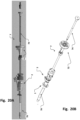
- the access device 1 has for instance in examples a first, open patient (body) configuration.
- An open patient configuration means that the patient body is opened for a surgical procedure, often by a section through the patient skin, and during the surgical procedure - in contrast to a closed patient (body) configuration.
- a removable hemostatic valve unit 2 is removably attached to a proximal side of said apical base plate 100.
- examples include that only the hemostatic valve unit 2 is mated with the apical base 100.
- both the hemostatic valve unit 2 and the sealing unit 3 are mated to the apical base 100.
- This third configuration is also an open patient (body) configuration.
- a transition configuration from the first to the second configuration may include the sealing unit 3 inserted into the hemostatic valve unit 2 for delivery to the apical base plate 100.
- the access device has a first configuration wherein a removable hemostatic valve unit is attached to the base plate.
- the port is thus open for fluid communication and is closable to prevent bleeding, controllable by the valve.
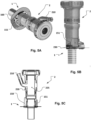
- sealing unit 3 provides for a possible movement of mechanical parts via/across the apex and a separation of a wet zone, e.g. blood in heart chamber, and a dry zone, i.e. gaseous/air environment outside of heart chamber, at the same time without leakage of blood or gas over the separation by e.g. a membrane for instance including a bellows 310.
- a sealing unit 3 may include a detaining unit 330 to provide a detainment of blood and/or gas in order to delay and/or prevent an exchange of blood or gas over the separation e.g. in case of a membrane malfunction.
- the detaining unit may delay and/or prevent a leakage of blood or gas over a membrane if for instance a bellows 310 would break or malfunction.
- the detaining unit is a sponge, or consists of sponge-like material, which allows for slow or inhibited leakage of gas into blood, or vice versa.
- the detaining unit may include a three-dimensional maze that allows for gas to freely pass through but can provide a delay and/or entirely prevent blood from passing through the maze.
- the detaining unit may be positioned inside the sealing unit 3. For instance, the interior channel of the bellows 310 may be provided with the detaining unit.
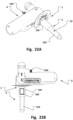
- the detaining unit may be provided in the top of the sealing unit 3 (when assembled positioned in tubular through port 120), see e.g. in the interior space of the tubular through port 120 inside sealing unit 3 such as illustrated in Fig. 22B .
- a detaining unit consisting of a maze would provide a detainment of blood causing a detainment of gas and thus would delay and/or prevent an exchange of blood or gas over the separation.
- the detaining unit may provide a controlled leakage and/or dissolvement of gas in blood, or vice versa, due to the costumed design of the detaining unit.
- a sealing unit with bellows allows for a, e.g. longitudinal and/or axial, movement of medical device parts relative each other, e.g. for piston or rods' movements.
- a sealing unit with bellows allows in addition or alternatively for radial movement of device parts relative each other, in particular from outside the heart to the inside of the heart, while the wet/dry separation is maintained.
- the removable hemostatic valve 2 has a valve through port with proximal and distal openings providing a communication channel with controllable orifice size, e.g. for delivery of various sized medical device, to and from the interior of the heart (chamber).
- sealing member 325 such as at the outside of the sealing unit, e.g. at its proximal end. This prevents blood leakage through the through port when the sealing unit 3 is inserted and affixed to the access device 1.
- the second configuration of the access device 1 is provided as an alternative or in addition to the first configuration thereof.
- the second configuration of the access device 1 may be provided in addition to the first configuration during a transition from the first configuration to the second configuration only, e.g. when the sealing unit 3 is delivered to the apical base plate 100 through the hemostatic valve unit attached to the apical base plate 100. This has the advantage of avoiding blood leakage when changing configuration of the access device 1.
- a transition from the first to the second configuration is illustrated for instance in Figs. 8A and 8B .
- a co-existent first and second configuration is illustrated for instance in Figs. 9A and9B.
- the hemostatic valve unit 2 includes a housing 200 with approximal end and a distal end.
- the housing 200 may be splittable for instance for peel-on and/or peel-off of the hemostatic valve unit 2 as described herein.
- the distal end is affixable to the access device 1.
- the ends include an opening for access to a through channel of the valve 2.
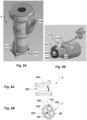
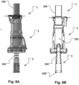
- the though channel includes an inflatable balloon, via an inflation port 210.
- the inflation pressure provides a more or less restricted passage through the through channel of valve 2, i.e. the orifice of the valve through port 225 is variable by the inflation pressure (as illustrated by the double headed arrow in Fig. 4A ).
- the hemostatic valve unit 2 is illustrated removably affixed to an access device 1 for instance in Figs. 5 , 6 , 8 , and 9 .
- a sealing surface 230 is provided for fluid tight sealing against a sealing surface 130 of access device 1, such as at the apical base plate 100.
- the sealing may include a sealing member, such as an O-ring for improved sealing.
- a drive unit 6 may be affixed proximally to the aggregate of access device 1 and sealing unit 3 as shown in Fig. 24A .
- the splittable valve 2 may then advantageously removed from the aggregate upon assembly of the other parts by deflating the balloon 220 and splitting the valve 2.
- the valve 2 is arranged to be peeled off.
- the split parts of valve 2 can then be removed from the patient as valve 2 is no longer needed.
- the splittable valve 2 may be opened only while parts still are connected to each other. For instance, two adjoining parts of a splittable valve with several parts may be separated from each other for opening the splittable valve 2, while still having part being adjoined, e.g.
- Coupling means are described in unpublished PCT application of the same applicant with PCT application number PCT/EP2019/068595 filed 10 July 2019 , which is incorporated herein by reference in its entirety for all purposes, but in particular the description of the coupling unit 200 and extension units 400, including magnetic and linked joint couplings as for instance shown in Figs. 5 and 6 thereof as well as the corresponding description.
- a hemostatic valve unit 2 has a housing 200 with a distal end and a proximal end. It is removably connectable at the distal end thereof to an apical base plate 100 of an access device 1 for a heart 10.
- the valve unit 2 includes a pneumatic valve in a through channel 225 of the valve unit 2 between the distal end and the proximal end thereof.
- the housing 200 has a proximal end with an opening to the through port 225 of the valve unit 2, such as for receiving a medical device to be passed through the inner passage of the valve unit 2.
- the housing 200 is splittable. It has for instance multiple splittable housing parts 202, 204. As shown in Fig. 25 . This allows for the herein described advantageous peel off when the valve is split.
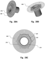
- Fig. 4A a single lobe "donut" balloon is illustrated.
- a balloon with multiple lobes 222 is illustrated.
- An inflation port 210 is connectable to a pressure regulation source for controlling the pressure in the balloon.
- Various pressures provide for various expansions of the balloon, and also for varied pressure on devices introduced into the through port 225 and in apposition to the balloon's exterior wall. Sealing is thus secured over a wide range of cross sections of devices to be entered through the port 225. For instance, a small needle or comparatively large tube can be entered through the valve without bleeding.
- valve 2 is illustrated attached to an access device 1.
- This assembly is illustrated in Fig. 6 being attached to the heart 10 creating a transapical access to a heart chamber.
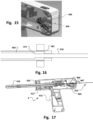
- FIG. 19 A and B the hemostatic valve 2 is illustrated attached to the applicator tool 4 over the tube 410, and in Figs. 20A and B attached to a delivery system 7 for delivering medical devices into the heart, as illustrated in Fig. 21 .
- a delivery tube 70 of the delivery system is configured to be inserted into the heart chamber 20.
- the delivery tube 70 is in the example shown inserted through the access device 1 with attached valve 2 (see Fig. 6 ).
- the distal end of the delivery tube 70 is in the example advanced through the left ventricular chamber towards the left atrial chamber.
- a pusher 74 may be used to advance medical devices or other medical system assembly components through the delivery tube 70 to the inner of the heart.
- the delivery system may include a funnel shaped inserter unit 72 for facilitating insertion of such devices and components into the proximal end of the delivery tube 70.
- an anchor unit 650 may be delivered through the delivery tube 70 to the cardiac valve area as illustrated in Figs. 21 (at distal end of delivery tube 70) and in Fig. 23 (delivered and attached to valve area and also a rod 610).
- the anchor unit is for instance the annuloplasty chain implant mentioned above and described in PCT application number PCT/EP2019/068597 , and in particular the chain annuloplasty ring and its delivery system shown in Figs. 24 to 42 and the corresponding description therein.
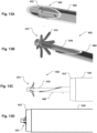
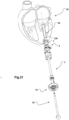
- the applicator tool includes in examples a harpoon 450 insertable through a tube of the applicator tool.
- the harpoon 450 includes a rod member 470 that is housed inside a hollow penetration needle 460 that has a distal tip for penetrating cardiac tissue at an apex of a heart.
- the rod 470 member is preferably a solid rod with an expandable retention member 473 at the distal end 472 of the rod.
- the rod 470 is arranged longitudinally movable in the penetration needle.
- the penetration needle is inserted into the tube 410 and arranged longitudinally movable in the tube 410.
- the rod is preferably kept longitudinally stationary with the penetration needle 460 when it is longitudinally moved.
- the tube 410 has preferably a sharpened edge at a distal end for cutting the cardiac tissue at the apex.
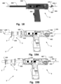
- the tube 410 may be pushed forward, e.g. by operation of a trigger 415 of the grip 400.
- the tube may be suspended freely rotatable around its central axis. Cutting may be facilitated by rotation of the tube when moving through the cardiac tissue. Rotation of the tube may be provided by a control dial 420. In this manner, a quick, precise and efficient transapical hole is made. This is done with improved patient safety as embolization of cut cardiac tissue is prevented.
- the tissue that is cut is safely kept in the tube 410 with the harpoon flanges holding back the tissue in the tube, as illustrated in Fig. 13D .
- the medical device is arranged to be matingly received with an inner channel of the device slidable over the tube's 410 outside. It can for instance be slid onto the tube from the distal end thereof for assembly as shown in Fig. 19A .
- a conical protection unit may temporarily be put on the distal end orifice of the tube 410 for sliding the medical device onto the tube, for instance to cover a sharp edge 412 of the distal end of the tube 410.
- the device incudes an apical access device 1.
- the device may include a hemostatic valve unit 2 removably pre-attached thereto.
- the transapical port is then usable, e.g. for delivery of devices to the heart chamber or performing procedures as desired.
- the sealing unit 3 may then be installed for providing the wet/dry zone separation.
- a medical device like a driving unit of a cardiac assist device can be installed at the dry zone of the access device 1.
- the valve unit may be removed. The procedure can then be concluded, leaving a wet/dry zone separated device implanted in the closed patient body.
- a transapical access system 5 for creating a transapical passage on a beating heart.
- the system includes an access device for a heart according to the afore described aspect of the disclosure.
- the system includes an applicator tool for creating a transapical passage and delivering the access device to an apex of the heart, as described above according to the afore described aspects of the disclosure.
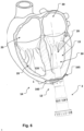
- Fig. 23 illustrates an example of a medical system including a cardiac assist unit transapically implanted.
- a drive unit 6 of the cardiac assist unit is illustrated attached to a sealed apical base plate 1 which is implanted and providing a transapical passage with a wet/dry zone separation as described above.
- the mechanical and electronic parts in the drive unit are in a dry zone (inside its housing and inside the sealing unit 3).
- a driving rod 610 is attached to an anchor unit 650.
- the electronic parts in the drive unit may have a cable through-port for electrical connection 630 of a cable to e.g. an external cell, battery or other power supply. An illustration of such an electrical connection 630 is seen in Figs.
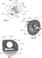
- the apical base plate may comprise various connection interfaces 110 for mating with a medical device.
- a connection interface 110 may be an edge for e.g. matingly engagement with e.g. a locking clip.
- the connection interface 110 may be a screwing hole e.g. for screwing in a screw, for example of M2 size, any suitable size may be used.
- the connection interface 110 may be a connection hole or a connection loop e.g. for matingly engagement with e.g. a suture, a wire, a thread, a fiber or similar.
- Fig. 26 illustrates steps of an example of a method 700 of creating a transapical passage on a beating heart.
- the method or medical procedure includes determining a position 710 on an apex region for creating a transapical passage. This may for instance be done using an imaging modality providing suitable image data for processing and analysis, e.g. CT based, MR based, Ultrasonic based. Alternatively, or in addition, the position may be determined by tactile sensing and/or visual inspection of the heart, e.g. during surgery.
- an imaging modality providing suitable image data for processing and analysis, e.g. CT based, MR based, Ultrasonic based.
- the position may be determined by tactile sensing and/or visual inspection of the heart, e.g. during surgery.
- the method further includes creating a transapical hole 720 at the determined apex region through cardiac tissue, such as by punching and/or cutting through the tissue.
- Creating the transapical hole may advantageously be performed using an applicator tool 4 as described above and illustrated in Figs. 12 to 19 .
- Creating the transapical hole may include penetrating cardiac tissue at the apex with a penetration needle 460 housing a distal tip of a harpoon 450 and through a tube 410.
- the forward penetration movement of the harpoon member and penetration needle 460 may be released by a trigger 465 so that these are "shot” forward as a unit in a one-shot movement, here through the apex wall.
- the one-shot movement is quick and reliable for making the initial puncture of the cardiac tissue.
- a rod member When the harpoon is brought through the cardiac tissue, a rod member may be further advanced out of the penetration needle and one or more retention members may be expanded radially outwards from the rod's 470 distal end 472. The retention member is then withdrawn for apposition against the inner cardiac wall of the heart chamber at the puncture made by the puncture needle 460 (which is also suitably withdrawn into the puncture).
- the sharpened edge 412 of the tube 410 is then pushed through the cardiac tissue towards the retention member. Pushing of the tube relative the rod member distal end 472 may be done by operating the trigger 415 of the pistol grip 400 in a safe and repeatedly standardized manner.
- a control dial 420 may be used by an operator for rotating the tube when cutting for improved cutting.
- a dilator 480 may be used for widening the tissue hole created by the penetration needle 460 and/or the tube 410
- the method further includes attaching a flange unit of the access device to an outside of the heart.
- the flange unit may be attached around the transapical hole by a suture technique called "parachute technique".
- parachute technique both ends of a single suture 165 are sutured through the cardiac tissue around the hole at a suitable distance to the hole. This may be done using a suitable template for a number of sutures around the hole. For instance, 8 to 10 sutures around the periphery of the flange 160 may be sufficient for providing reliable seat of the access device in a sealed manner, i.e. without bleeding from the heart chamber when the channel in tube 120 is suitably closed by e.g. a hemostatic valve, sealing unit or a plug.
- the channel in tube 120 provides a transapical working channel to and from a heart chamber on a beating heart.
- Bleeding is prevented by keeping the tube 410 in the hole.
- the two ends of each same suture are then passed through the flange unit, which is held away from the heart surface on the outside of the tube 410 of tool 4.
- the suturing pattern is repeated using additional sutures, resulting in several suture "pairs" spaced around the hole and tube 410.
- the access device 1 is then lowered or parachuted down against the outer heart wall and advanced until the plate 100 and the flange 160 are in contact with the heart surface and the opening of the tube 120 of the access device 1 is inside the heart chamber. After all the suture pairs are secured, e.g. by suitable knots, the result is a blood tight flange with a tubular port 120 in the transapical hole.
- the tube 410 of the applicator tool may then be retracted. It is withdrawn out of the access device and valve that remain in place at the heart, as shown in Fig. 6 .
- Delivering medical devices to the heart chamber may include deploying an annuloplasty chain ring at a cardiac valve annulus.
- a delivery catheter of a delivery system may be introduced for this purpose through the access device 1 with affixed valve 2.
- the delivery catheter may then be removed out of the patient. Blood leakage from the heart is continued prevented by the hemostatic valve unit 2.
- a sealing device may be slid over the driving rod 610 through the valve 2 and affixed at the access device 1 providing a sealed access device 1 with a wet/dry zone separation.
- the proximal end of the driving rod 610 may then be connected to a drive unit 6 while attaching the drive unit 6 to the access device 1.
- the sealing device 3 may be introduced through the hemostatic valve unit 2 to the access device 1 sealing the transapical passage, e.g. as described above for creating the wet/dry zone separation.
- An element like a rod may pass across the through port of the sealing element 3.
- the distal end port of sealing unit 3 may be provided as a closed element like a membrane or hub.
- a magnetic coupling 340 to an element in the heart chamber may be established with units shown in Fig. 24A, 24B and 24C .
- the valve 2 may then be removed as well as a medical device affixed to the access device prior to concluding the procedure.
- the method 700 may include removing the hemostatic valve unit from the apical base plate by disconnecting the hemostatic valve unit from the apical base plate and withdrawing the hemostatic valve unit out of the patient, or splitting or partitioning the hemostatic valve unit.
- examples of the disclosure may include one or more sensors.
- the access device 1, the hemostatic valve 2, the sealing unit 3, the drive unit for cardiac assist 6, the transapical access system 5, or other medical devices (not shown) attachable to the access device 1 may include such sensor(s) thus implantable into a patient's body.
- Sensors often need to be part of a wet zone and a dry zone, since measurement is often performed in blood (wet zone) and sensors often include electrical parts that need to be separated from blood in a dry zone. Thus, it is important to have a feed-through port 320 in order to facilitate this sensor wet and dry zone separation.
- Such sensor may go through the feed-through port 320 of the bellows 310, instead of the driving rod 610 as previously described.
- there may be more than one (multiple) feed-through ports 320 so that multiple sensors and/or the driving rod 610 could go through different feed-through ports 320 at the same time.
- the feed-through port 320 does not need to be part of the bellows 310, as previous described, but may be a separate partlunit of the sealing unit 3 instead.
- An example of a multiple feed-through, or multi-lumen, ports 320 is for instance including a preferably separate channel for a sensor, such as a pressure sensor, (not shown in the Figures) .
- the senor 620 may include one or more pressure sensors e.g. connectable to a port distally ending in the chamber of the heart. This will provide intracardiac pressure of e.g. the left and/or right ventricle of the heart.
- the pressure data not only provides important clinical data but may also be used in control algorithms of an implanted medical device. Other relevant clinical parameters, e.g. the heart rate of the patient and/or various heart arrhythmias, may also be extracted from the pressure data.
- the sensor 620 may include additionally, or alternatively, one or more optical and/or electrical sensors used for obtaining blood flow and/or blood volume measurements in the heart.
- Such sensors may e.g. be placed in an optical port and/or window (not shown) facing towards the wet zone of the heart. This may e.g. provide measurement data for intracardiac blood volume of the left ventricle of the heart.
- the blood flow/volume data not only provides important clinical data but may also be used in control algorithms of an implanted medical device.
- Other relevant clinical parameters e.g. the heart rate of the patient and/or stroke volume and/or cardiac output and/or SpO2, may also be extracted from the blood flow/volume data.
- the sensor 620 may include in addition, or alternatively, one or more movement sensors e.g. connectable to a port distally ending in the heart and/or connected to the driving rod 610. This will provide measurement data for intracardiac movement and/or activity data of the heart.
- the movement data may e.g. represent the up and down movement of the mitral valve and/or the atria/ventricle plane.
- the movement data not only provides important diagnostic clinical data but may also be used in control algorithms of an implanted medical device. Other relevant clinical parameters, e.g. the heart rate of the patient and/or various heart arrhythmias, may also be extracted from the movement data.
- Examples of movement sensors include magnetic based, such as a Hall effect sensor, and/or optical based. Examples of movement sensors may also include one or more accelerometers.
- the measurement data obtained from the sensor(s) 620 may for instance be used to control a medical device, e.g. be part of a control algorithm implemented in the hardware and/or software of the medical device such as a cardiac assist system.
- the data from the sensors may for instance also be used to monitor important physiological properties and/or use the sensor data to extract and/or calculate critical clinical parameters that need to be monitored. Besides from monitoring the physiological properties and/or clinical parameters, they may be part of a surveillance system.
- Obtaining and managing patient data is not only important for e.g. the safety of the patient and the functionality of a medical device, but also due to regulatory requirements for medical devices since it will be mandatory to collect, retain, and analyze post-market clinical data.
- Fig. 27 illustrates steps of an example of a method 800 of transapically implanting a cardiac assist system.
- the method 800 or medical procedure includes attaching 810 a cardiac assist unit 6 to an access device 1 including a sealed apical base plate 100.
- the apical base plate 100 has a sealed tubular through port arranged across cardiac tissue to a heart chamber.
- a flange unit is attached to the base plate 100 and to the heart 10.
- the access device 1 has a sealing unit 3 attached thereto.
- the method may include inserting a delivery tube through a hemostatic valve attached to the access device prior to sealing the access device for providing a wet/dry zone as described above.
- the applicator tool of any of examples 31 to 33 comprising an access device (1) for a heart having a tubular through port (120) adapted to be arranged across said cardiac tissue when advanced over said tube (410).
- Example 39 A transapical access system for creating a transapical passage on a beating heart, said system including an access device (1) for a heart according to any of originally filed claims 1 to 21, and/or an applicator tool (4) for creating a transapical passage and delivering said access device (1) to an apex of said heart according to any of examples 31 to 38.
- said access device having a sealed tubular through port adapted to be arranged across cardiac tissue to a heart chamber, and a flange unit sealingly affixing said access device to said heart.
Landscapes
- Health & Medical Sciences (AREA)
- Heart & Thoracic Surgery (AREA)
- Engineering & Computer Science (AREA)
- Life Sciences & Earth Sciences (AREA)
- Public Health (AREA)
- Veterinary Medicine (AREA)
- Biomedical Technology (AREA)
- Animal Behavior & Ethology (AREA)
- General Health & Medical Sciences (AREA)
- Cardiology (AREA)
- Hematology (AREA)
- Anesthesiology (AREA)
- Surgery (AREA)
- Mechanical Engineering (AREA)
- Medical Informatics (AREA)
- Molecular Biology (AREA)
- Nuclear Medicine, Radiotherapy & Molecular Imaging (AREA)
- Pathology (AREA)
- Pulmonology (AREA)
- Vascular Medicine (AREA)
- Surgical Instruments (AREA)
- External Artificial Organs (AREA)
- Prostheses (AREA)
- Infusion, Injection, And Reservoir Apparatuses (AREA)
Applications Claiming Priority (4)
| Application Number | Priority Date | Filing Date | Title |
|---|---|---|---|
| PCT/EP2019/087182 WO2021136585A1 (en) | 2019-12-30 | 2019-12-30 | An access device for a heart, a removable hemostatic valve unit, and a system and a method of creating a transapical passage on a beating heart |
| US16/990,903 US12478403B2 (en) | 2019-12-30 | 2020-08-11 | Access device for a heart, a removable hemostatic valve unit, and a system and a method of creating a transapical passage on a beating heart |
| PCT/EP2020/088064 WO2021136822A2 (en) | 2019-12-30 | 2020-12-30 | An access device for a heart, a removable hemostatic valve unit, and a system including a cardiac assist unit |
| EP20842247.7A EP3975890B1 (de) | 2019-12-30 | 2020-12-30 | Zugangsvorrichtung für ein herz, abnehmbare hämostaseventileinheit und system mit einer herzunterstützungseinheit |
Related Parent Applications (2)
| Application Number | Title | Priority Date | Filing Date |
|---|---|---|---|
| EP20842247.7A Division-Into EP3975890B1 (de) | 2019-12-30 | 2020-12-30 | Zugangsvorrichtung für ein herz, abnehmbare hämostaseventileinheit und system mit einer herzunterstützungseinheit |
| EP20842247.7A Division EP3975890B1 (de) | 2019-12-30 | 2020-12-30 | Zugangsvorrichtung für ein herz, abnehmbare hämostaseventileinheit und system mit einer herzunterstützungseinheit |
Publications (2)
| Publication Number | Publication Date |
|---|---|
| EP4397357A2 true EP4397357A2 (de) | 2024-07-10 |
| EP4397357A3 EP4397357A3 (de) | 2024-08-14 |
Family
ID=69374262
Family Applications (5)
| Application Number | Title | Priority Date | Filing Date |
|---|---|---|---|
| EP20842247.7A Active EP3975890B1 (de) | 2019-12-30 | 2020-12-30 | Zugangsvorrichtung für ein herz, abnehmbare hämostaseventileinheit und system mit einer herzunterstützungseinheit |
| EP24164015.0A Pending EP4397357A3 (de) | 2019-12-30 | 2020-12-30 | Zugangsvorrichtung für ein herz, abnehmbare hämostatische klappeneinheit und system mit einer herzunterstützungseinheit |
| EP21217733.1A Active EP4008278B1 (de) | 2019-12-30 | 2020-12-30 | Zugangsvorrichtung für ein herz |
| EP21217732.3A Active EP4008277B1 (de) | 2019-12-30 | 2020-12-30 | System mit einer herzunterstützungseinheit |
| EP21217731.5A Active EP4008276B1 (de) | 2019-12-30 | 2020-12-30 | Abnehmbare hämostaseventileinheit und system mit einer herzunterstützungseinheit |
Family Applications Before (1)
| Application Number | Title | Priority Date | Filing Date |
|---|---|---|---|
| EP20842247.7A Active EP3975890B1 (de) | 2019-12-30 | 2020-12-30 | Zugangsvorrichtung für ein herz, abnehmbare hämostaseventileinheit und system mit einer herzunterstützungseinheit |
Family Applications After (3)
| Application Number | Title | Priority Date | Filing Date |
|---|---|---|---|
| EP21217733.1A Active EP4008278B1 (de) | 2019-12-30 | 2020-12-30 | Zugangsvorrichtung für ein herz |
| EP21217732.3A Active EP4008277B1 (de) | 2019-12-30 | 2020-12-30 | System mit einer herzunterstützungseinheit |
| EP21217731.5A Active EP4008276B1 (de) | 2019-12-30 | 2020-12-30 | Abnehmbare hämostaseventileinheit und system mit einer herzunterstützungseinheit |
Country Status (11)
| Country | Link |
|---|---|
| US (8) | US12478403B2 (de) |
| EP (5) | EP3975890B1 (de) |
| JP (1) | JP2023514939A (de) |
| KR (1) | KR20220143650A (de) |
| CN (1) | CN115243629A (de) |
| AU (1) | AU2020416622A1 (de) |
| BR (1) | BR112022012954A2 (de) |
| CA (1) | CA3166309A1 (de) |
| IL (1) | IL294259A (de) |
| MX (1) | MX2022008049A (de) |
| WO (2) | WO2021136585A1 (de) |
Families Citing this family (8)
| Publication number | Priority date | Publication date | Assignee | Title |
|---|---|---|---|---|
| EP4413955A3 (de) | 2017-07-24 | 2024-11-13 | Emory University | Herzklappensegelverstärkungsvorrichtungen |
| CN116889680B (zh) * | 2023-06-08 | 2024-06-28 | 上海心恒睿医疗科技有限公司 | 心室辅助装置 |
| CN116943016B (zh) * | 2023-07-26 | 2025-10-31 | 航天泰心科技有限公司 | 一种心脏植入设备用固定器及其安装工具 |
| US12433749B2 (en) | 2023-08-25 | 2025-10-07 | Emory University | Systems, devices, and methods for reducing heart valve regurgitation |
| CN117717705B (zh) * | 2024-02-08 | 2024-04-16 | 生命盾医疗技术(苏州)有限公司 | 可实现非灌注密封的介入式导管泵 |
| US12396855B1 (en) | 2024-07-25 | 2025-08-26 | Nyra Medical, Inc. | Systems, devices, and methods for reducing heart valve regurgitation |
| DE102024128036A1 (de) * | 2024-09-27 | 2026-04-02 | Albert-Ludwigs-Universität Freiburg, Körperschaft des öffentlichen Rechts | Medizinische Schleusenanordnung sowie medizinisches Set |
| CN120094091A (zh) * | 2025-05-08 | 2025-06-06 | 槃实科技(深圳)有限公司 | 一种基于血液泵的脉动模拟方法及系统 |
Citations (7)
| Publication number | Priority date | Publication date | Assignee | Title |
|---|---|---|---|---|
| GB1514019A (en) | 1976-05-06 | 1978-06-14 | Gist Brocades Nv | Catheter device |
| US5492304A (en) | 1993-06-16 | 1996-02-20 | United States Surgical Corporation | Seal assembly for accommodating introduction of surgical instruments |
| WO2009100198A2 (en) | 2008-02-08 | 2009-08-13 | Mayo Foundation For Medical Education And Research | Transapical heart port |
| WO2011017440A2 (en) | 2009-08-06 | 2011-02-10 | Mayo Foundation For Medical Education And Research | Implanting organ ports |
| WO2011119101A1 (en) | 2010-03-25 | 2011-09-29 | Jan Otto Solem | A device and a method to controllably assist movement of a mitral valve |
| US20160081715A1 (en) | 2013-02-21 | 2016-03-24 | Covidien Lp | Surgical access device including lateral moving seal cooperating with bellows attached to proximal wall of cannula housing |
| WO2018167059A1 (en) | 2017-03-13 | 2018-09-20 | Academisch Ziekenhuis Maastricht | A transapical heart port for insertion into a heart of a human or an animal subject |
Family Cites Families (20)
| Publication number | Priority date | Publication date | Assignee | Title |
|---|---|---|---|---|
| US5085636A (en) * | 1989-01-13 | 1992-02-04 | Scimed Life Systems, Inc. | Balloon catheter with inflation-deflation valve |
| US6732501B2 (en) * | 2002-06-26 | 2004-05-11 | Heartware, Inc. | Ventricular connector |
| US7323004B2 (en) | 2002-09-30 | 2008-01-29 | Ethicon, Inc. | Device for providing automatic stitching of an incision |
| US7582071B2 (en) | 2005-03-28 | 2009-09-01 | Tyco Healthcare Group Lp | Introducer seal assembly |
| AU2006335227B2 (en) * | 2005-10-14 | 2013-06-13 | Correx, Inc. | Apparatus and method for forming a hole in a hollow organ |
| US9682180B2 (en) | 2009-11-15 | 2017-06-20 | Thoratec Corporation | Attachment system, device and method |
| US20110118833A1 (en) * | 2009-11-15 | 2011-05-19 | Thoratec Corporation | Attachment device and method |
| US8152845B2 (en) * | 2009-12-30 | 2012-04-10 | Thoratec Corporation | Blood pump system with mounting cuff |
| US20110251450A1 (en) | 2010-04-12 | 2011-10-13 | Pagani Francis D | Method and Device for Attachment of an Inflow Conduit to the Heart and to a Pump |
| US8870739B2 (en) | 2010-08-06 | 2014-10-28 | Heartware, Inc. | Conduit device for use with a ventricular assist device |
| US8696557B2 (en) * | 2010-12-21 | 2014-04-15 | Covidien Lp | Access assembly including inflatable seal member |
| WO2012158968A2 (en) | 2011-05-18 | 2012-11-22 | Thoratec Corporation | Needle guard, assembly and method of implanting a heart assist system |
| US9731057B2 (en) * | 2011-07-28 | 2017-08-15 | Fineheart | Removable heart pump, and method implemented in such a pump |
| US10022520B2 (en) | 2012-12-17 | 2018-07-17 | Nico Corporation | Surgical access system |
| WO2018007244A1 (en) * | 2016-07-04 | 2018-01-11 | Atropos Limited | An access device |
| US10894116B2 (en) * | 2016-08-22 | 2021-01-19 | Tc1 Llc | Heart pump cuff |
| WO2018156897A1 (en) * | 2017-02-24 | 2018-08-30 | Tc1 Llc | Minimally invasive methods and devices for ventricular assist device implantation |
| CN110650756B (zh) * | 2017-05-16 | 2022-12-27 | 心脏器械股份有限公司 | 心室内流动可植入pv环路系统 |
| US11167122B2 (en) | 2018-03-05 | 2021-11-09 | Harmony Development Group, Inc. | Force transducting implant system for the mitigation of atrioventricular pressure gradient loss and the restoration of healthy ventricular geometry |
| RU2696685C1 (ru) | 2018-09-18 | 2019-08-05 | Федеральное государственное автономное образовательное учреждение высшего образования "Национальный исследовательский университет "Московский институт электронной техники" | Устройство для подключения насоса вспомогательного кровообращения к желудочку сердца человека |
-
2019
- 2019-12-30 WO PCT/EP2019/087182 patent/WO2021136585A1/en not_active Ceased
-
2020
- 2020-08-11 US US16/990,903 patent/US12478403B2/en active Active
- 2020-12-30 CA CA3166309A patent/CA3166309A1/en active Pending
- 2020-12-30 EP EP20842247.7A patent/EP3975890B1/de active Active
- 2020-12-30 CN CN202080097755.1A patent/CN115243629A/zh active Pending
- 2020-12-30 IL IL294259A patent/IL294259A/en unknown
- 2020-12-30 EP EP24164015.0A patent/EP4397357A3/de active Pending
- 2020-12-30 EP EP21217733.1A patent/EP4008278B1/de active Active
- 2020-12-30 JP JP2022539278A patent/JP2023514939A/ja active Pending
- 2020-12-30 BR BR112022012954A patent/BR112022012954A2/pt not_active Application Discontinuation
- 2020-12-30 EP EP21217732.3A patent/EP4008277B1/de active Active
- 2020-12-30 MX MX2022008049A patent/MX2022008049A/es unknown
- 2020-12-30 AU AU2020416622A patent/AU2020416622A1/en not_active Abandoned
- 2020-12-30 EP EP21217731.5A patent/EP4008276B1/de active Active
- 2020-12-30 WO PCT/EP2020/088064 patent/WO2021136822A2/en not_active Ceased
- 2020-12-30 KR KR1020227026495A patent/KR20220143650A/ko not_active Withdrawn
-
2022
- 2022-06-06 US US17/805,649 patent/US20220323105A1/en active Pending
- 2022-06-06 US US17/805,639 patent/US20220409235A1/en active Pending
- 2022-06-06 US US17/805,647 patent/US12502202B2/en active Active
- 2022-06-06 US US17/805,659 patent/US12383304B2/en active Active
- 2022-06-06 US US17/805,640 patent/US20220296272A1/en active Pending
-
2025
- 2025-07-02 US US19/258,391 patent/US20250331889A1/en active Pending
- 2025-09-23 US US19/337,678 patent/US20260013902A1/en active Pending
Patent Citations (7)
| Publication number | Priority date | Publication date | Assignee | Title |
|---|---|---|---|---|
| GB1514019A (en) | 1976-05-06 | 1978-06-14 | Gist Brocades Nv | Catheter device |
| US5492304A (en) | 1993-06-16 | 1996-02-20 | United States Surgical Corporation | Seal assembly for accommodating introduction of surgical instruments |
| WO2009100198A2 (en) | 2008-02-08 | 2009-08-13 | Mayo Foundation For Medical Education And Research | Transapical heart port |
| WO2011017440A2 (en) | 2009-08-06 | 2011-02-10 | Mayo Foundation For Medical Education And Research | Implanting organ ports |
| WO2011119101A1 (en) | 2010-03-25 | 2011-09-29 | Jan Otto Solem | A device and a method to controllably assist movement of a mitral valve |
| US20160081715A1 (en) | 2013-02-21 | 2016-03-24 | Covidien Lp | Surgical access device including lateral moving seal cooperating with bellows attached to proximal wall of cannula housing |
| WO2018167059A1 (en) | 2017-03-13 | 2018-09-20 | Academisch Ziekenhuis Maastricht | A transapical heart port for insertion into a heart of a human or an animal subject |
Also Published As
Similar Documents
| Publication | Publication Date | Title |
|---|---|---|
| US12383304B2 (en) | Method of accessing a heart with a hemostatic device and an interface | |
| EP2747679B1 (de) | Vorrichtung zur herstellung eines vorübergehenden zugriffs und nachfolgende schliessung | |
| US5249574A (en) | Implantation of leads | |
| AU2012212215B2 (en) | Systems for implanting and using a conduit within a tissue wall | |
| CA2657008A1 (en) | Surgical tools for left ventricular assist device (lvad)implantation | |
| WO2009042816A2 (en) | Applicator, assembly, and method for connecting an inlet conduit to a hollow organ | |
| CA2863939A1 (en) | A device and method for temporary or permanent suspension of an implantable scaffolding containing an orifice for placement of a prosthetic or bio-prosthetic valve | |
| EP3389739B1 (de) | Hohldübel | |
| HK40070831A (en) | Removable hemostatic valve unit and system including a cardiac assist unit | |
| HK40070832A (en) | System including a cardiac assist unit | |
| HK40070833A (en) | Access device for a heart |
Legal Events
| Date | Code | Title | Description |
|---|---|---|---|
| PUAI | Public reference made under article 153(3) epc to a published international application that has entered the european phase |
Free format text: ORIGINAL CODE: 0009012 |
|
| STAA | Information on the status of an ep patent application or granted ep patent |
Free format text: STATUS: REQUEST FOR EXAMINATION WAS MADE |
|
| REG | Reference to a national code |
Ref country code: DE Ref legal event code: R079 Free format text: PREVIOUS MAIN CLASS: A61M0060900000 Ipc: A61B0017340000 |
|
| 17P | Request for examination filed |
Effective date: 20240328 |
|
| AC | Divisional application: reference to earlier application |
Ref document number: 3975890 Country of ref document: EP Kind code of ref document: P |
|
| AK | Designated contracting states |
Kind code of ref document: A2 Designated state(s): AL AT BE BG CH CY CZ DE DK EE ES FI FR GB GR HR HU IE IS IT LI LT LU LV MC MK MT NL NO PL PT RO RS SE SI SK SM TR |
|
| PUAL | Search report despatched |
Free format text: ORIGINAL CODE: 0009013 |
|
| AK | Designated contracting states |
Kind code of ref document: A3 Designated state(s): AL AT BE BG CH CY CZ DE DK EE ES FI FR GB GR HR HU IE IS IT LI LT LU LV MC MK MT NL NO PL PT RO RS SE SI SK SM TR |
|
| RIC1 | Information provided on ipc code assigned before grant |
Ipc: A61M 60/90 20210101ALI20240709BHEP Ipc: A61M 60/894 20210101ALI20240709BHEP Ipc: A61M 60/863 20210101ALI20240709BHEP Ipc: A61M 60/839 20210101ALI20240709BHEP Ipc: A61M 60/289 20210101ALI20240709BHEP Ipc: A61M 60/191 20210101ALI20240709BHEP Ipc: A61M 39/06 20060101ALI20240709BHEP Ipc: A61B 17/34 20060101AFI20240709BHEP |
|
| STAA | Information on the status of an ep patent application or granted ep patent |
Free format text: STATUS: EXAMINATION IS IN PROGRESS |
|
| 17Q | First examination report despatched |
Effective date: 20251014 |