EP4368718A1 - Mikrogliavorläuferzellen, verfahren zur herstellung von mikroglia und hergestellte mikrogliavorläuferzellen und mikroglia - Google Patents
Mikrogliavorläuferzellen, verfahren zur herstellung von mikroglia und hergestellte mikrogliavorläuferzellen und mikroglia Download PDFInfo
- Publication number
- EP4368718A1 EP4368718A1 EP22837704.0A EP22837704A EP4368718A1 EP 4368718 A1 EP4368718 A1 EP 4368718A1 EP 22837704 A EP22837704 A EP 22837704A EP 4368718 A1 EP4368718 A1 EP 4368718A1
- Authority
- EP
- European Patent Office
- Prior art keywords
- microglia
- cells
- progenitor cells
- microglial progenitor
- hemangioblasts
- Prior art date
- Legal status (The legal status is an assumption and is not a legal conclusion. Google has not performed a legal analysis and makes no representation as to the accuracy of the status listed.)
- Pending
Links
Images
Classifications
-
- C—CHEMISTRY; METALLURGY
- C12—BIOCHEMISTRY; BEER; SPIRITS; WINE; VINEGAR; MICROBIOLOGY; ENZYMOLOGY; MUTATION OR GENETIC ENGINEERING
- C12N—MICROORGANISMS OR ENZYMES; COMPOSITIONS THEREOF; PROPAGATING, PRESERVING, OR MAINTAINING MICROORGANISMS; MUTATION OR GENETIC ENGINEERING; CULTURE MEDIA
- C12N5/00—Undifferentiated human, animal or plant cells, e.g. cell lines; Tissues; Cultivation or maintenance thereof; Culture media therefor
- C12N5/06—Animal cells or tissues; Human cells or tissues
- C12N5/0602—Vertebrate cells
- C12N5/0618—Cells of the nervous system
- C12N5/0622—Glial cells, e.g. astrocytes, oligodendrocytes; Schwann cells
-
- C—CHEMISTRY; METALLURGY
- C12—BIOCHEMISTRY; BEER; SPIRITS; WINE; VINEGAR; MICROBIOLOGY; ENZYMOLOGY; MUTATION OR GENETIC ENGINEERING
- C12N—MICROORGANISMS OR ENZYMES; COMPOSITIONS THEREOF; PROPAGATING, PRESERVING, OR MAINTAINING MICROORGANISMS; MUTATION OR GENETIC ENGINEERING; CULTURE MEDIA
- C12N15/00—Mutation or genetic engineering; DNA or RNA concerning genetic engineering, vectors, e.g. plasmids, or their isolation, preparation or purification; Use of hosts therefor
- C12N15/09—Recombinant DNA-technology
- C12N15/63—Introduction of foreign genetic material using vectors; Vectors; Use of hosts therefor; Regulation of expression
- C12N15/79—Vectors or expression systems specially adapted for eukaryotic hosts
- C12N15/85—Vectors or expression systems specially adapted for eukaryotic hosts for animal cells
-
- C—CHEMISTRY; METALLURGY
- C12—BIOCHEMISTRY; BEER; SPIRITS; WINE; VINEGAR; MICROBIOLOGY; ENZYMOLOGY; MUTATION OR GENETIC ENGINEERING
- C12N—MICROORGANISMS OR ENZYMES; COMPOSITIONS THEREOF; PROPAGATING, PRESERVING, OR MAINTAINING MICROORGANISMS; MUTATION OR GENETIC ENGINEERING; CULTURE MEDIA
- C12N2500/00—Specific components of cell culture medium
- C12N2500/02—Atmosphere, e.g. low oxygen conditions
-
- C—CHEMISTRY; METALLURGY
- C12—BIOCHEMISTRY; BEER; SPIRITS; WINE; VINEGAR; MICROBIOLOGY; ENZYMOLOGY; MUTATION OR GENETIC ENGINEERING
- C12N—MICROORGANISMS OR ENZYMES; COMPOSITIONS THEREOF; PROPAGATING, PRESERVING, OR MAINTAINING MICROORGANISMS; MUTATION OR GENETIC ENGINEERING; CULTURE MEDIA
- C12N2501/00—Active agents used in cell culture processes, e.g. differentation
- C12N2501/10—Growth factors
- C12N2501/115—Basic fibroblast growth factor (bFGF, FGF-2)
-
- C—CHEMISTRY; METALLURGY
- C12—BIOCHEMISTRY; BEER; SPIRITS; WINE; VINEGAR; MICROBIOLOGY; ENZYMOLOGY; MUTATION OR GENETIC ENGINEERING
- C12N—MICROORGANISMS OR ENZYMES; COMPOSITIONS THEREOF; PROPAGATING, PRESERVING, OR MAINTAINING MICROORGANISMS; MUTATION OR GENETIC ENGINEERING; CULTURE MEDIA
- C12N2501/00—Active agents used in cell culture processes, e.g. differentation
- C12N2501/10—Growth factors
- C12N2501/125—Stem cell factor [SCF], c-kit ligand [KL]
-
- C—CHEMISTRY; METALLURGY
- C12—BIOCHEMISTRY; BEER; SPIRITS; WINE; VINEGAR; MICROBIOLOGY; ENZYMOLOGY; MUTATION OR GENETIC ENGINEERING
- C12N—MICROORGANISMS OR ENZYMES; COMPOSITIONS THEREOF; PROPAGATING, PRESERVING, OR MAINTAINING MICROORGANISMS; MUTATION OR GENETIC ENGINEERING; CULTURE MEDIA
- C12N2501/00—Active agents used in cell culture processes, e.g. differentation
- C12N2501/10—Growth factors
- C12N2501/15—Transforming growth factor beta (TGF-β)
-
- C—CHEMISTRY; METALLURGY
- C12—BIOCHEMISTRY; BEER; SPIRITS; WINE; VINEGAR; MICROBIOLOGY; ENZYMOLOGY; MUTATION OR GENETIC ENGINEERING
- C12N—MICROORGANISMS OR ENZYMES; COMPOSITIONS THEREOF; PROPAGATING, PRESERVING, OR MAINTAINING MICROORGANISMS; MUTATION OR GENETIC ENGINEERING; CULTURE MEDIA
- C12N2501/00—Active agents used in cell culture processes, e.g. differentation
- C12N2501/10—Growth factors
- C12N2501/155—Bone morphogenic proteins [BMP]; Osteogenins; Osteogenic factor; Bone inducing factor
-
- C—CHEMISTRY; METALLURGY
- C12—BIOCHEMISTRY; BEER; SPIRITS; WINE; VINEGAR; MICROBIOLOGY; ENZYMOLOGY; MUTATION OR GENETIC ENGINEERING
- C12N—MICROORGANISMS OR ENZYMES; COMPOSITIONS THEREOF; PROPAGATING, PRESERVING, OR MAINTAINING MICROORGANISMS; MUTATION OR GENETIC ENGINEERING; CULTURE MEDIA
- C12N2501/00—Active agents used in cell culture processes, e.g. differentation
- C12N2501/10—Growth factors
- C12N2501/165—Vascular endothelial growth factor [VEGF]
-
- C—CHEMISTRY; METALLURGY
- C12—BIOCHEMISTRY; BEER; SPIRITS; WINE; VINEGAR; MICROBIOLOGY; ENZYMOLOGY; MUTATION OR GENETIC ENGINEERING
- C12N—MICROORGANISMS OR ENZYMES; COMPOSITIONS THEREOF; PROPAGATING, PRESERVING, OR MAINTAINING MICROORGANISMS; MUTATION OR GENETIC ENGINEERING; CULTURE MEDIA
- C12N2501/00—Active agents used in cell culture processes, e.g. differentation
- C12N2501/20—Cytokines; Chemokines
- C12N2501/22—Colony stimulating factors (G-CSF, GM-CSF)
-
- C—CHEMISTRY; METALLURGY
- C12—BIOCHEMISTRY; BEER; SPIRITS; WINE; VINEGAR; MICROBIOLOGY; ENZYMOLOGY; MUTATION OR GENETIC ENGINEERING
- C12N—MICROORGANISMS OR ENZYMES; COMPOSITIONS THEREOF; PROPAGATING, PRESERVING, OR MAINTAINING MICROORGANISMS; MUTATION OR GENETIC ENGINEERING; CULTURE MEDIA
- C12N2501/00—Active agents used in cell culture processes, e.g. differentation
- C12N2501/20—Cytokines; Chemokines
- C12N2501/23—Interleukins [IL]
- C12N2501/2303—Interleukin-3 (IL-3)
-
- C—CHEMISTRY; METALLURGY
- C12—BIOCHEMISTRY; BEER; SPIRITS; WINE; VINEGAR; MICROBIOLOGY; ENZYMOLOGY; MUTATION OR GENETIC ENGINEERING
- C12N—MICROORGANISMS OR ENZYMES; COMPOSITIONS THEREOF; PROPAGATING, PRESERVING, OR MAINTAINING MICROORGANISMS; MUTATION OR GENETIC ENGINEERING; CULTURE MEDIA
- C12N2501/00—Active agents used in cell culture processes, e.g. differentation
- C12N2501/20—Cytokines; Chemokines
- C12N2501/23—Interleukins [IL]
- C12N2501/2306—Interleukin-6 (IL-6)
-
- C—CHEMISTRY; METALLURGY
- C12—BIOCHEMISTRY; BEER; SPIRITS; WINE; VINEGAR; MICROBIOLOGY; ENZYMOLOGY; MUTATION OR GENETIC ENGINEERING
- C12N—MICROORGANISMS OR ENZYMES; COMPOSITIONS THEREOF; PROPAGATING, PRESERVING, OR MAINTAINING MICROORGANISMS; MUTATION OR GENETIC ENGINEERING; CULTURE MEDIA
- C12N2501/00—Active agents used in cell culture processes, e.g. differentation
- C12N2501/20—Cytokines; Chemokines
- C12N2501/23—Interleukins [IL]
- C12N2501/2334—Interleukin-34 (IL-34)
-
- C—CHEMISTRY; METALLURGY
- C12—BIOCHEMISTRY; BEER; SPIRITS; WINE; VINEGAR; MICROBIOLOGY; ENZYMOLOGY; MUTATION OR GENETIC ENGINEERING
- C12N—MICROORGANISMS OR ENZYMES; COMPOSITIONS THEREOF; PROPAGATING, PRESERVING, OR MAINTAINING MICROORGANISMS; MUTATION OR GENETIC ENGINEERING; CULTURE MEDIA
- C12N2501/00—Active agents used in cell culture processes, e.g. differentation
- C12N2501/40—Regulators of development
- C12N2501/415—Wnt; Frizzeled
-
- C—CHEMISTRY; METALLURGY
- C12—BIOCHEMISTRY; BEER; SPIRITS; WINE; VINEGAR; MICROBIOLOGY; ENZYMOLOGY; MUTATION OR GENETIC ENGINEERING
- C12N—MICROORGANISMS OR ENZYMES; COMPOSITIONS THEREOF; PROPAGATING, PRESERVING, OR MAINTAINING MICROORGANISMS; MUTATION OR GENETIC ENGINEERING; CULTURE MEDIA
- C12N2501/00—Active agents used in cell culture processes, e.g. differentation
- C12N2501/70—Enzymes
- C12N2501/72—Transferases [EC 2.]
- C12N2501/727—Kinases (EC 2.7.)
-
- C—CHEMISTRY; METALLURGY
- C12—BIOCHEMISTRY; BEER; SPIRITS; WINE; VINEGAR; MICROBIOLOGY; ENZYMOLOGY; MUTATION OR GENETIC ENGINEERING
- C12N—MICROORGANISMS OR ENZYMES; COMPOSITIONS THEREOF; PROPAGATING, PRESERVING, OR MAINTAINING MICROORGANISMS; MUTATION OR GENETIC ENGINEERING; CULTURE MEDIA
- C12N2506/00—Differentiation of animal cells from one lineage to another; Differentiation of pluripotent cells
- C12N2506/45—Differentiation of animal cells from one lineage to another; Differentiation of pluripotent cells from artificially induced pluripotent stem cells
-
- C—CHEMISTRY; METALLURGY
- C12—BIOCHEMISTRY; BEER; SPIRITS; WINE; VINEGAR; MICROBIOLOGY; ENZYMOLOGY; MUTATION OR GENETIC ENGINEERING
- C12N—MICROORGANISMS OR ENZYMES; COMPOSITIONS THEREOF; PROPAGATING, PRESERVING, OR MAINTAINING MICROORGANISMS; MUTATION OR GENETIC ENGINEERING; CULTURE MEDIA
- C12N2800/00—Nucleic acids vectors
- C12N2800/10—Plasmid DNA
- C12N2800/106—Plasmid DNA for vertebrates
- C12N2800/107—Plasmid DNA for vertebrates for mammalian
-
- C—CHEMISTRY; METALLURGY
- C12—BIOCHEMISTRY; BEER; SPIRITS; WINE; VINEGAR; MICROBIOLOGY; ENZYMOLOGY; MUTATION OR GENETIC ENGINEERING
- C12N—MICROORGANISMS OR ENZYMES; COMPOSITIONS THEREOF; PROPAGATING, PRESERVING, OR MAINTAINING MICROORGANISMS; MUTATION OR GENETIC ENGINEERING; CULTURE MEDIA
- C12N2840/00—Vectors comprising a special translation-regulating system
- C12N2840/002—Vectors comprising a special translation-regulating system controllable or inducible
Definitions
- the present invention relates to microglial progenitor cells, a method for producing microglia, and produced microglial progenitor cells and microglia.
- the brain is composed of nerve cells and glial cells.
- Microglia are a type of glial cells and account for approximately 5% to 15% of cells in the central nervous system. Microglia are known to function as immunocompetent cells in the central nervous system. In addition to the immune function, microglia also play a variety of physiological functions. In addition, it has become clear that recently identified risk factor genes for neurodegenerative diseases are highly expressed in microglia. For the above reasons, microglia are becoming increasingly important as a research subject.
- Microglia are derived from mesoderm, and pluripotent stem cells differentiate into microglia through hemangioblasts and microglial progenitor cells.
- Non-Patent Document 1 discloses that human-induced pluripotent stem cells (hiPSCs) are cultured with a plurality of cytokines over a long period of time, and target cells are separated and collected through cell sorting or the like to produce human microglia-like cells.
- hiPSCs human-induced pluripotent stem cells
- Non-Patent Document 1 Abud E. M., et al., iPSC-Derived Human Microglia-like Cells to Study Neurological Diseases, Neuron 2017; 94:278-765, 293.e9
- Non-Patent Document 1 the method described in Non-Patent Document 1 and similar methods have room for improvement from the viewpoints of efficiency (qualitatively, quantitatively, and periodically) and costs.
- an object of the present invention is to provide a highly efficient and inexpensive method for producing microglia, a method for producing microglial progenitor cells for use in the production method, and microglia that enable subculture and function as microglia.
- the present invention has the following aspects.
- microglia-like cells having properties closer to intracerebral microglia with high efficiency, at low cost, and in a short period of time.
- human genes and human proteins will be denoted by uppercase letters.
- mouse genes will be denoted by an uppercase letter for the first letter and lowercase letters for the rest.
- mouse proteins are denoted by uppercase letters.
- human genes, mouse genes, and genes of other species may be expressed without strictly distinguishing them.
- human proteins, mouse proteins, and proteins of other species may be expressed without strictly distinguishing them.
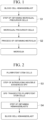
- FIG. 1 is a flowchart showing a method for producing microglia according to one embodiment.
- the present invention provides a method for producing microglia, including: Step S1 of inducing differentiation of hemangioblasts to obtain microglial progenitor cells; and Step S2 of inducing differentiation of the microglial progenitor cells to obtain microglia, in which, in the step of obtaining microglial progenitor cells, expression of PU.1 transcription factor encoded by an exogenous gene is induced.
- microglia artificial microglia-like cells
- microglial progenitor cells can be produced with high efficiency, at low cost, and in a short period of time by obtaining microglial progenitor cells and inducing differentiation of the microglial progenitor cells through the method for producing microglia of the present embodiment.
- Hemangioblasts used as starting materials in the present embodiment can be produced through, for example, a method for producing hemangioblasts to be described below.
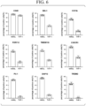
- Modules for example, plasmids containing HyPBase, rtTA, and SP11 shown in FIG. 5 ) necessary for inducible expression of exogenous PU.1 transcription factor have been preliminarily genetically introduced into the hemangioblasts used in the present embodiment.
- Exogenous genes refer to genes which are not endogenous genes but are additionally provided to cells through gene introduction.
- a base sequence of a protein-encoding region may be the same as that of a corresponding endogenous gene, or may have mutations such as amino acid substitutions, insertions, and deletions.
- exogenous genes may be derived from heterologous organisms.
- a non-coding region of an exogenous gene may be linked to 5'UTR (untranslated region) 3'UTR or 5'UTR and 3'UTR required for artificial induction of expression.
- the induction of expression of an exogenous gene refers to artificial induction of expression of an exogenous gene. Specifically, by turning on an inducible promoter of an exogenous gene (in the present embodiment, the SP11 gene encoding the PU.1 transcription factor) that is under control of the inducible promoter, the amount of exogenous gene transcribed is increased and expression of gene products is induced.
- an inducible promoter of an exogenous gene in the present embodiment, the SP11 gene encoding the PU.1 transcription factor
- Step S1 of obtaining microglial progenitor cells expression of PU.1 transcription factor may be induced by inducing transcription of the exogenous SPI1 gene under control of the inducible promoter. Accordingly, microglial progenitor cells that are more suitable for producing microglia are easily produced.
- inducible promoters drug-inducible promoters can be used.
- drug-inducible promoters promoters whose expression induction is turned on and off by addition of doxycycline can be used.
- temperature-sensitive inducible promoters can be used.
- promoters which have a TRE sequence and whose expression induction is turned on by addition of doxycycline can be used.
- the expression level of the exogenous gene at a transcription level increases more than twice as much as when the expression is not induced.
- the degree of increase may be 5 times or more, 10 times or more, 25 times or more, 50 times or more, 100 times or more, or 200 times or more.
- the upper limit of the degree of increase is not particularly limited but may be 1,000 times or less.
- the expression level at the transcription level can be evaluated through, for example, quantitative reverse transcription PCR (qRT-PCR) using one obtained by reverse-transcribing mRNA prepared from target cells as a template.
- qRT-PCR quantitative reverse transcription PCR
- Step S1 of obtaining microglial progenitor cells includes Step S1a of inducing expression of PU.1 transcription factor in the presence of FGF2, SCF, IL-3, IL-6, VEGF, and IWR-le.
- Step S1a of inducing expression of PU.1 transcription factor in the presence of FGF2, SCF, IL-3, IL-6, VEGF, and IWR-le By inducing differentiation from microglial progenitor cells obtained through Step S1a into microglia, it is possible to appropriately induce differentiation from hemangioblasts into microglial progenitor cells and to produce microglial progenitor cells suitable for producing microglia.
- Step S1 of obtaining microglial progenitor cells preferably includes Step S1b of subjecting the cells after Step S1a of inducing expression of PU.1 transcription factor in the presence of FGF2, SCF, IL-3, IL-6, VEGF, and IWR-le to induction of expression of PU.1 transcription factor in the presence of FGF2, SCF, IL-3, and IL-6.
- Step S1 of obtaining microglial progenitor cells it is possible to use StemPro TM -34 SFM (1 ⁇ ) (Gibco TM , 1063011) supplemented with GlutaMax (Gibco TM , 1 ⁇ ), holo-transferrin (Naacalai Tesque, 20 ⁇ g/mL), L-ascorbic acid (Sigma-Aldrich Co. LLC., 500 ⁇ M), monothioglycerol (Sigma-Aldrich Co. LLC., 450 ⁇ M), and penicillin/streptomycin and passed through a 0.45 um filter (hereinafter referred to as "hiHPC diff. medium").
- the concentration of FGF2 may be in a range of 5 ng/mL plus or minus 30% or plus or minus 15%.
- the concentration of SCF may be in a range of 50 ng/mL plus or minus 30% or plus or minus 15%.
- the concentration of IL-3 may be in a range of 30 ng/mL plus or minus 30% or plus or minus 15%.
- the concentration of IL-6 may be in a range of 10 ng/mL plus or minus 30% or plus or minus 15%.
- the concentration of VEGF may be in a range of 10 ng/mL plus or minus 30% or plus or minus 15%.
- the concentration of IWR-le may be in a range of 5 ⁇ M plus or minus 30% or plus or minus 15%.
- the concentration of doxycycline may be in a range of 1 ⁇ g/mL plus or minus 30% or plus or minus 15%.
- Step S2 of inducing differentiation of microglial progenitor cells to obtain microglia preferably includes Step S2a of culturing the microglial progenitor cells obtained in Step S1 of obtaining microglial progenitor cells in a medium containing IL-34, TGF ⁇ 1, M-CSF, CD200, and CX3CL1. Accordingly, microglia (microglia-like cells) having properties similar to intracerebral microglia are likely to be efficiently produced from microglial progenitor cells.
- DMEM/F12 (1:1, Gibco TM ) supplemented with GlutaMax (Gibco TM , 1 ⁇ ), NEAA (Thermo Fisher Scientific Inc., 1 ⁇ ), B27 (Thermo Fisher Scientific Inc., 1 ⁇ ), N2 (Thermo Fisher Scientific Inc., 0.5 ⁇ ), ITS-G (Thermo Fisher Scientific Inc., 2%), insulin (Sigma-Aldrich Co. LLC., 5 ⁇ g/mL), monothioglycerol (Sigma-Aldrich Co. LLC., 200 ⁇ M), and penicillin/streptomycin and passed through a 0.45 um filter.
- DMEM can also be replaced with the same amount of IMDM (Gibco TM ) (hereinafter referred to as "hiMGL diff. medium").
- the concentration of IL-34 may be in a range of 100 ng/mL plus or minus 30% or plus or minus 15%.
- the concentration of TGF ⁇ 1 may be in a range of 50 ng/mL plus or minus 30% or plus or minus 15%.
- the concentration of M-CSF may be in a range of 25 ng/mL plus or minus 30% or plus or minus 15%.
- the concentration of CD200 may be in a range of 100 ng/mL plus or minus 30% or plus or minus 15%.
- the concentration of CX3CL1 may be in a range of 100 ng/mL plus or minus 30% or plus or minus 15%.
- FIG. 2 is a flowchart showing a method for producing hemangioblasts from pluripotent stem cells.
- Hemangioblasts used as starting materials in the present embodiment include Step S3 of introducing exogenous gene SPI1 under control of an inducible promoter into pluripotent stem cells and S4 of inducing differentiation of the pluripotent stem cells into which SP11 is introduced to obtain hemangioblasts.
- the SPI1 gene is a gene encoding PU.1 transcription factor.
- the above-described pluripotent stem cells may be, for example, ES cells or induced pluripotent stem cells (iPSCs).
- the above-described pluripotent stem cells may be human-derived cells or may be cells derived from non-human animals such as mice, rats, pigs, goats, sheep, and monkeys.
- pluripotent stem cells are human-derived cells
- microglia-like cells suitable as experimental materials in human microglia research can be prepared with high efficiency, at low cost, and in a short period of time.
- the above-described pluripotent stem cells may be induced pluripotent stem cells derived from a healthy subject or may be induced pluripotent stem cells derived from a patient with a neurological disease.
- the obtained microglia can be used as a neurological disease model. Such microglia are useful for elucidating mechanisms of neurological diseases.
- Step S3 of introducing exogenous gene SPI1 into pluripotent stem cells a module (for example, a gene construct to be described below) necessary for inducible expression of exogenous PU.1 transcription factor is genetically introduced (transfected) into pluripotent stem cells.
- a module for example, a gene construct to be described below
- Gene introduction is performed, for example, using Genejuice (Sigma-Aldrich Co. LLC., 70967) as a transfection reagent according to the instruction manual.
- Genejuice Sigma-Aldrich Co. LLC., 70967
- Pluripotent stem cells may be cultured and maintained feeder-free.
- Step S4 of obtaining hemangioblasts preferably includes Step S4a of culturing pluripotent stem cells in a medium containing BMP4 and CHIR99021, Step S4b of culturing the cells after Step S4a in a medium containing BMP4, VEGF, and FGF2, and Step S4c of culturing the cells after Step S4b in a medium containing VEGF and FGF2.
- hemangioblasts suitable for starting materials of the present embodiment can be produced.
- medium can be used as a medium in Step S4 of obtaining hemangioblasts.
- the concentration of BMP4 may be in a range of 20 ng/mL plus or minus 30% or plus or minus 15%.
- the concentration of CHIR99021 may be in a range of 2 ⁇ M plus or minus 30% or plus or minus 15%.
- the concentration of VEGF may be in a range of 50 ng/mL plus or minus 30% or plus or minus 15%.
- the concentration of FGF2 may be in a range of 20 ng/mL plus or minus 30% or plus or minus 15%.
- the concentration of VEGF in Step S4c may be in a range of 15 ng/mL plus or minus 30% or plus or minus 15%.
- the concentration of FGF2 in Step S4c may be in a range of 5 ng/mL plus or minus 30% or plus or minus 15%.
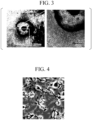
- FIG. 3 is a micrograph showing microglial progenitor cells.
- the large mass in the center is a hemangioblast, and the many small granular cells around it are microglial progenitor cells.
- FIG. 4 is a micrograph showing microglia according to one embodiment.
- the present invention provides microglial progenitor cells which are produced by inducing expression of PU.1 transcription factor encoded by an exogenous gene in hemangioblasts and are to be subjected to differentiation induction to produce microglia exhibiting characteristics of intracerebral microglia.
- the present invention provides microglia which are produced by inducing differentiation of the microglial progenitor cells according to one embodiment described above and exhibit characteristics of intracerebral microglia.
- microglia-like cells it is not known that it is possible to produce microglia-like cells more efficiently by inducing differentiation of hemangioblasts into microglial progenitor cells while inducing expression of PU.1 transcription factor during production of the microglia-like cells.
- microglial progenitor cells can be produced through the above-described production method and microglia can be produced through the above-described method in which the microglial progenitor cells are used as starting materials to more efficiently produce microglia-like cells.
- microglial progenitor cells of the present embodiment are floating cells, double positive for CD11b/CD45, and positive for CD235a.
- microglia of the present embodiment are adherent cells, positive for 1BA1 and CX3CR, and positive for TMEM119 (or P2RY12). In microglia, the expression levels of DAP12 and TREM2 are high.
- microglial progenitor cells and microglia of the present embodiment are induced to differentiate from pluripotent stem cells into which inducible exogenous SPI1 is introduced. For this reason, by evaluating the presence or absence of this inducible exogenous SPI1 as genetic information, it is possible to confirm whether those are derived from hemangioblasts obtained through Step S3 of introducing exogenous gene SP11 and Step S4 of obtaining hemangioblasts shown in FIG. 2 .
- the above-described evaluation can be performed, for example, through PCR using a forward primer complementary to the exogenous DNA fragment and a reverse primer complementary to the SPI1 coding sequence using the total DNA of a target cell as a template.
- primers a forward primer complementary to the SPI1 coding sequence and a reverse primer complementary to the exogenous DNA fragment can also be used.
- microglial progenitor cells and microglia of the present embodiment may be housed in a container at a cell density of 1 ⁇ 10 5 cells/mL or more, for example, 1 ⁇ 10 4 cells/mL or more or 1 ⁇ 10 6 cells/mL or more.
- the microglial progenitor cells and microglia of the present embodiment are in a frozen state, and may be housed in a cryotube or the like that is normally used for cryopreservation of cells.
- a commercially available cryopreservation liquid can be used.
- serum-free cryopreservation liquids include CELLBANKER (registered trademark) 2 (Nippon Zenyaku Kogyo Co., Ltd.) or Banbanker (registered trademark) (Lymphotec Inc.).
- FIG. 5 shows parts of three types of plasmids which are gene constructs used in the present embodiment.
- FIG. 5 shows, in order from the top, a part containing a hyperactive piggyBac transposase (HyPBase) coding region and a puromycin resistance gene, a part containing a reverse tetracycline transactivator (rtTA) coding region and a hygromycin resistance gene, and a part containing a lacZ-neomycin fusion gene ( ⁇ -geo) and an exogenous PU.1 coding region under control of an inducible promoter.
- HyPBase hyperactive piggyBac transposase
- rtTA reverse tetracycline transactivator
- ⁇ -geo lacZ-neomycin fusion gene
- the gene construct shown at the top is used in combination for the purpose of improving gene introduction efficiency, and is not essential.
- the gene construct shown in the center contains a gene encoding rtTA, a factor that binds to a TRE sequence and promotes transcription of downstream genes in the presence of doxycycline. This gene construct is necessary in a case of inducing expression of PU. 1 transcription factor with a doxycycline-inducible promoter.
- the gene construct is unnecessary in a case where inducible promoter other than the doxycycline-inducible promoter is used.
- the SP11 gene is linked downstream of a promoter which has a TRE sequence and whose expression induction is turned on by addition of doxycycline.
- doxycycline In a case where doxycycline is added to a culture medium, it is taken into cells and binds to rtTA. Doxycycline-bound rtTA binds to the TRE sequence and promotes transcription of SPI1.
- IRES sequence contributing to efficient translation of transcripts and a ⁇ -geo sequence functioning as a selection marker are linked downstream of SP11.
- hiPSC human-induced pluripotent stem cell
- hiPSC line 201B7 ( Cell 2007; 131:861-872 ), WD39 ( Mol. Brain 2012; 5: 35 ), and RPC802 (ReproCell Inc., cat# RCRP002N) were cultured feeder-free. These iPSCs were maintained in a 12-well plate (Corning Inc.) coated with iMatrix-511. StemFit/AK02N (Ajinomoto) supplemented with penicillin and streptomycin was used as a medium. A humidified incubator (37°C, 5% CO 2 ) was used for culture. iPSCs were subcultured weekly, and the medium was replaced every 2 days.
- Genejuice (Sigma-Aldrich Co. LLC., 70967) was used as a transfection reagent. 9 ⁇ L of Genejuice was mixed with 200 ⁇ L of Opti-MEM medium (Gibco TM , 31985070), and the mixture was allowed to stand at room temperature for 5 minutes.
- Opti-MEM medium Gibco TM , 31985070
- the plasmids including the expression modules shown in FIG. 5 (pCMV-HyPBase_PGK-Puro (0.4 ⁇ g), pG-PB-CAG-rtTA3G-IH (0.4 ⁇ g), and PB-tet-PHS-SPI1 (0.8 ⁇ g)) were added thereto, and the mixture was left to stand at room temperature for another 5 minutes.
- the human pluripotent stem cells cultured feeder-free were treated with Y-27632 (Wako, 253-00511 (10 ⁇ M/AK02N)) for 1 hour.
- the pluripotent stem cells were detached from the surface of the culture container using TrypLE Select (Life Technogogies, A12859-01, 0.5 ⁇ , incubated at 37°C for 10 minutes). After centrifugation at 1,000 rpm for 5 minutes, the collected cells were stained with trypan blue, and the number of live cells was counted.
- TrypLE Select Life Technogogies, A12859-01, 0.5 ⁇ , incubated at 37°C for 10 minutes. After centrifugation at 1,000 rpm for 5 minutes, the collected cells were stained with trypan blue, and the number of live cells was counted.
- the cells were evenly seeded in 6 wells of a 12-well plate.
- 0.8 mL of AK02N supplemented with penicillin/streptomycin, Y-27632 (10 ⁇ m), and iMatrix0511 was added to the plate.
- the medium was replaced and Y-27632 was removed from the culture liquid.
- Hygromaycin (20 ⁇ g/mL) was added to the replaced medium to select human iPSCs.
- puromycin (2 ⁇ g/mL) was added to the medium.
- the final concentrations of hygromycin and puromycin were 200 ⁇ g/mL and 10 ⁇ g/mL, respectively.
- the colonies were mechanically isolated using a P10 tip under a microscope.
- the isolated colonies were individually seeded in a 24-well plate with a medium supplemented with Y-27632 and iMatrix-511.
- Human microglia were induced to differentiate from the human induced pluripotent stem cell lines established using the above-described three types of cell lines.
- CK protocol A technique obtained by modifying a technique disclosed in well-known literature was used as a basic hiMGL production technique (hereinafter referred to as "CK protocol").
- PU protocol a hiMGL production method further involving inducible expression of exogenous SPI1 heredity
- microglia from iPSCs can be broadly divided into two steps.
- the first step corresponds to day 0 to day 18 from the start of differentiation induction.
- differentiation is induced from human pluripotent stem cells to hemangioblasts and from the hemangioblasts to microglial progenitor cells.
- the second step corresponds to day 19 or beyond.
- differentiation is induced from the microglial progenitor cells to microglia.
- Days 0 to 4 correspond to the period of induction of differentiation from hiPSCs to mesodermal cells.
- Days 4 to 6 correspond to the period of induction of differentiation from the mesodermal cells to hemangioblasts.
- hiPSCs on a 6-well plate into which exogenous SPI1 was introduced were washed once with PBS. Thereafter, 2 mL of hiHPC diff. medium supplemented with BMP4 (PeproTech, 20 ng/mL) and CRIR9901 (Focus Biomolecules, 2 ⁇ M) was added to each well. The cells were allowed to stand in a hypoxic incubator (5% O 2 , 5% CO 2 , 37°C) until day 6. The cells subcultured while maintaining the colony size smaller than normal.
- the cells were collected into a 15-mL tube and centrifuged at 300 g at room temperature for 5 minutes. Thereafter, half of the medium was removed from the supernatant, and approximately 1 mL of the remaining medium containing the collected cells was returned to the wells.
- hiHPC diff. medium supplemented with 1 mL of VEGF (30 ng/mL), FGF2 (10 ng/mL), SCF (PeproTech, 100 ng/mL), IL-3 (PeproTech, 60 ng/mL), IL-6 (PeproTech, 20 ng/mL), and Wnt Inhibitor (IWR-1e, Thermo Fisher Scientific Inc., 5 ⁇ M) was added to each well.
- the cells were allowed to stand in a normoxic incubator (20% O 2 , 5% CO 2 , 37°C).
- the outer edges of the hiPSC cell aggregations became unclear, and the presence of small cells dispersed around the hiPCS cell aggregations was observed.
- the above-described cell aggregations whose outer edges became unclear are hemangioblasts.
- doxycycline (1 ⁇ g/mL) was added to the culture liquid from day 6 to day 18 from the start of culture.
- the expression level of the SP11 gene evaluated through qRT-PCR increased by at least about 50 times compared to a case without doxycycline.
- the expression level of the SP11 gene was significantly low regardless of the presence or absence of doxycycline (approximately 1-2 times).
- Days 7 to 14 correspond to the period of induction of differentiation from the hemangioblasts to primary hematopoietic stem cells.
- Days 14 to 18 correspond to the period of induction of differentiation from primary hematopoietic stem cells to microglial progenitor cells.
- the cells were collected in a falcon tube and centrifuged at 300 ⁇ g for 5 minutes. Thereafter, the supernatant was carefully and completely removed.
- the cells on days 4, 8, 12, and 16 from the start of differentiation induction were evaluated for the presence or absence of CD34, CD43, CD11b, and CD45 markers.
- the evaluation results are shown in Table 1.
- evaluation results of cultured cells without addition of doxycycline and evaluation results of cultured cells without addition of doxycycline and Wnt Inhibitor (IWR-1e) are shown.
- CD34/CD43 markers no significant differences were observed in contrast to the control cells, but regarding the CD11b/CD45 markers, significant differences were observed in contrast to the control cells.
- Human microglia were induced to differentiate from microglial progenitor cells.
- Day 19 or beyond corresponds to a maturation period of microglia.
- the cells were collected into a 15-mL tube and centrifuged at 300 g at room temperature for 5 minutes. Thereafter, the supernatant was removed, and the collected cells (microglial progenitor cells) were evenly seeded in a plurality of wells through the method described below.
- the microglial progenitor cells were seeded in a 12-well plate at 40,000 to 50,000 cells in 0.8 mL of hiMGL diff. medium.
- the medium used was hiMGL diff. medium supplemented with three kinds of cytokine cocktails (IL-34 100 ng/mL; TCF ⁇ 1 50 ng/mL; and M-CSF 25 ng/mL (all are PeproTech)).
- the plate used was one coated with Poly-D-Lysine (PDL, R&D Systems) at least 1 hour before seeding and washed three times with PBS.
- microglial progenitor cells were induced to differentiate into microglia from day 19.
- the medium used after day 19 was one supplemented with five kinds of cytokine cocktails (IL-34 100 ng/mL; TGF ⁇ 1 50 ng/mL; and M-CSF 25 ng/mL, CD200 100 ng/mL; and CX3CL1 100 ng/mL (all are PeproTech)).
- IL-34 100 ng/mL
- CD200 100 ng/mL
- CX3CL1 100 ng/mL all are PeproTech
- microglia-like cells obtained through the above-described method will be referred to as human-induced microglia-like cells (hiMGLs).
- microglia microglia-like cells
- THP-1 a human monocyte cell line
- Marker expression of CD45, IBA1, CD11b, P2RY12, TMEM119, CR3CR1, PU.1, DAP12, and TREM2 was analyzed through flow cytometry. The results are shown in Fig. 6 .
- CD45 is a hematopoietic stem cell marker
- IBA1, PU.1, and CX3CR1 are myeloid lineage markers
- TMEM119 and P2RY12 are microglia-specific markers.
- the CD45 positive cell rate was at the same level between hiMGL and THP-1.
- the positive cell rates for IBA1, CD11b, P2RY12, TMEM119, CR3CR1, PU.1, DAP12, and TREM2 were significantly higher in hiMGL than in THP-1.
- microglia-like cells expressing microglia-specific markers could be produced in a highly pure state and in a short period of time using the PU protocol.
- Poly(A)+ RNA was prepared from target cells and converted into cDNA library fragments (average length: 350 bp). Adapters were linked to both ends of the cross section for base sequence determination.
- a KAPA mRNA capture Kit (KK8440, Kapa Biosystems), a KAPA RNA HyperPrep Kit (KK8542, Kapa Biosystems), a KAPA Pure Beads (KK8443, Kapa Biosystems), and a SeqCap Adapter Kit A (Roche) were used according to the attached instruction manual.
- the cDNA library was quantified using a KAPA Library Quantification Kit (KK4828, Kapa Biosystems). Illumina HiSqX was used for sequence determination.
- VST variance-stabilizing transformation
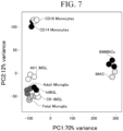
- Expression profiles of the cells produced using the CK protocol and PU protocol on day 25 after induction of differentiation were obtained through the above-described method and compared with expression profiles of the deposited monocytes, adult primary human microglia, fetus primary human microglia, and other hiPSC-derived microglia-like cells.
- PCA principal component analysis
- hierarchical cluster analysis as a comprehensive transcription profile evaluation.
- FIG. 7 shows a part of the above-described analysis and shows analysis results of PCA.
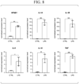
- phagocytic ability As an evaluation of physiological functions of the microglia produced using the PU protocol, phagocytic ability, inflammatory cytokine secretion ability, and inflammasome generation ability were evaluated.
- Latex beads and fibrillar A ⁇ -42 were used for evaluating phagocytic ability.
- NKkB1, IL-1A, IL-1B, IL-6, IL-10, and TNF as inflammatory cytokines were evaluated based on the presence or absence of stimulation with lipopolysaccharides (LPS) ( FIG. 8 ).
- microglia produced using the PU protocol were shown to have phagocytic ability, inflammatory cytokine secretion ability, and inflammasome generation ability.
- microglia produced using the PU protocol were co-cultured with mouse neuron primary cells (derived from the hippocampus), and change in microglia morphology, increase in cytoplasmic area of the microglia, and expression of microglia-specific markers were evaluated.
- microglia produced using the PU protocol changed from an amoeboid shape to a branched shape when co-cultured with mouse neuron primary cells.
- the cytoplasmic area of the microglia was significantly increased.
- expression of IBA1, a microglia-specific marker, was also recognized.
- the hiMGLs produced using the PU protocol can interact with mouse neuron primary cells and can appropriately reproduce intracerebral microglia with respect to specific functions.
- the hiMGLs produced using the PU protocol are thought to be useful experimental materials for studying functions of microglia.
- Intracerebral microglia of a living mouse were replaced with microglia derived from hiPSCs produced using the PU protocol through nasal transplantation, and colonization of the microglia and change in cell morphology were observed.
- PLX5622 was administered to the mouse (8 weeks old after birth, wild type C57BL/6J mouse) for 1 week starting 8 days before transplantation. Thereafter, normal diet was administered for 24 hours.
- hyaluronidase manufactured by the manufacturer, catalog number
- cytokine hCSF1 250 ng/mL
- hTGF- ⁇ 1 100 ng/mL
- brain tissue of the mouse was excised, and the prepared slice sample was subjected to immunohistological staining.
- the hiMGLs produced using the PU protocol can interact with mouse neuron primary cells and can appropriately reproduce intracerebral microglia with respect to specific functions.
- the hiMGLs produced using the PU protocol are thought to be useful experimental materials for studying functions of microglia.
- microglia-like cells having properties closer to intracerebral microglia with high efficiency, at low cost, and in a short period of time.
Landscapes
- Health & Medical Sciences (AREA)
- Engineering & Computer Science (AREA)
- Biomedical Technology (AREA)
- Life Sciences & Earth Sciences (AREA)
- Genetics & Genomics (AREA)
- Chemical & Material Sciences (AREA)
- Organic Chemistry (AREA)
- Biotechnology (AREA)
- Zoology (AREA)
- Wood Science & Technology (AREA)
- Bioinformatics & Cheminformatics (AREA)
- General Engineering & Computer Science (AREA)
- Microbiology (AREA)
- Biochemistry (AREA)
- General Health & Medical Sciences (AREA)
- Neurology (AREA)
- Neurosurgery (AREA)
- Cell Biology (AREA)
- Plant Pathology (AREA)
- Molecular Biology (AREA)
- Physics & Mathematics (AREA)
- Biophysics (AREA)
- Micro-Organisms Or Cultivation Processes Thereof (AREA)
Applications Claiming Priority (2)
| Application Number | Priority Date | Filing Date | Title |
|---|---|---|---|
| US202163218502P | 2021-07-06 | 2021-07-06 | |
| PCT/JP2022/026843 WO2023282290A1 (ja) | 2021-07-06 | 2022-07-06 | ミクログリア前駆細胞並びにミクログリア製造方法、製造されたミクログリア前駆細胞及びミクログリア |
Publications (2)
| Publication Number | Publication Date |
|---|---|
| EP4368718A1 true EP4368718A1 (de) | 2024-05-15 |
| EP4368718A4 EP4368718A4 (de) | 2025-10-08 |
Family
ID=84801780
Family Applications (1)
| Application Number | Title | Priority Date | Filing Date |
|---|---|---|---|
| EP22837704.0A Pending EP4368718A4 (de) | 2021-07-06 | 2022-07-06 | Mikrogliavorläuferzellen, verfahren zur herstellung von mikroglia und hergestellte mikrogliavorläuferzellen und mikroglia |
Country Status (5)
| Country | Link |
|---|---|
| US (1) | US20240309322A1 (de) |
| EP (1) | EP4368718A4 (de) |
| JP (1) | JPWO2023282290A1 (de) |
| CN (1) | CN117813385A (de) |
| WO (1) | WO2023282290A1 (de) |
Families Citing this family (1)
| Publication number | Priority date | Publication date | Assignee | Title |
|---|---|---|---|---|
| US20220220441A1 (en) * | 2019-05-27 | 2022-07-14 | Westfälische Wilhelms-Universität Münster | Rapid and deterministic generation of microglia from human pluripotent stem cells |
Family Cites Families (2)
| Publication number | Priority date | Publication date | Assignee | Title |
|---|---|---|---|---|
| JP2022511385A (ja) * | 2018-09-28 | 2022-01-31 | メモリアル スローン ケタリング キャンサー センター | 幹細胞由来ヒトミクログリア細胞、作製する方法および使用方法 |
| US20220220441A1 (en) * | 2019-05-27 | 2022-07-14 | Westfälische Wilhelms-Universität Münster | Rapid and deterministic generation of microglia from human pluripotent stem cells |
-
2022
- 2022-07-06 US US18/575,385 patent/US20240309322A1/en active Pending
- 2022-07-06 CN CN202280047457.0A patent/CN117813385A/zh active Pending
- 2022-07-06 JP JP2023533166A patent/JPWO2023282290A1/ja active Pending
- 2022-07-06 WO PCT/JP2022/026843 patent/WO2023282290A1/ja not_active Ceased
- 2022-07-06 EP EP22837704.0A patent/EP4368718A4/de active Pending
Also Published As
| Publication number | Publication date |
|---|---|
| US20240309322A1 (en) | 2024-09-19 |
| EP4368718A4 (de) | 2025-10-08 |
| JPWO2023282290A1 (de) | 2023-01-12 |
| CN117813385A (zh) | 2024-04-02 |
| WO2023282290A1 (ja) | 2023-01-12 |
Similar Documents
| Publication | Publication Date | Title |
|---|---|---|
| Yzaguirre et al. | The role of Runx1 in embryonic blood cell formation | |
| Petrova et al. | 3D in vitro model of a functional epidermal permeability barrier from human embryonic stem cells and induced pluripotent stem cells | |
| EP3245289B1 (de) | Differenzierung von makrophagen und pluripotenten stammzellen | |
| EP2909313B1 (de) | Herstellung von roten blutzellen und -plättchen aus stammzellen | |
| CN1791668A (zh) | 内胚层干细胞的制备 | |
| US20250136935A1 (en) | Systems and methods for producing retinal progenitors | |
| Wang et al. | Single-cell transcriptomics reveals the molecular anatomy of sheep hair follicle heterogeneity and wool curvature | |
| Lager et al. | Rapid functional genetics of the oligodendrocyte lineage using pluripotent stem cells | |
| WO2021172542A1 (ja) | 成熟心筋細胞の製造法 | |
| EP4368718A1 (de) | Mikrogliavorläuferzellen, verfahren zur herstellung von mikroglia und hergestellte mikrogliavorläuferzellen und mikroglia | |
| Li et al. | Characterization of gene regulatory networks underlying key properties in human hematopoietic stem cell ontogeny | |
| Ye et al. | The miR-290 and miR-302 clusters are essential for reprogramming of fibroblasts to induced pluripotent stem cells | |
| EA019719B1 (ru) | Способ получения плюрипотентных клеток | |
| JP6436491B2 (ja) | 骨髄異形成症候群等の治療/予防薬のスクリーニング方法 | |
| JP7148134B2 (ja) | 肝芽細胞から胆管上皮前駆細胞への段階的誘導方法 | |
| EP4310178A1 (de) | Verfahren zur herstellung eines gehirnorganoids mit aggregiertem tau-protein | |
| JP7548494B2 (ja) | 細胞の製造方法 | |
| AU2019376495B2 (en) | Method for producing erythroid cells | |
| Xu et al. | Establishment and transcriptome analysis of single blastomere-derived cell lines from zebrafish | |
| CN120025979B (zh) | 内侧神经节隆起神经前体细胞分化方法 | |
| WO2024182558A1 (en) | Fluorescent cone reporter ips cells, retinal organoids and uses thereof | |
| Roshan | Study of Human Retinal Ganglion Cell Development and Direct Conversion | |
| Fernandez | The Role of Retinoic Acid Signaling in Development of the Human Embryonic Hematopoietic System | |
| JP2023086705A (ja) | 筋疲労/筋損傷の細胞モデル、その製造方法及びその用途 | |
| Hansen et al. | Efficient production of erythroid, megakaryoid and myeloid cells, using single cell-derived iPSC colony differentiation |
Legal Events
| Date | Code | Title | Description |
|---|---|---|---|
| STAA | Information on the status of an ep patent application or granted ep patent |
Free format text: STATUS: THE INTERNATIONAL PUBLICATION HAS BEEN MADE |
|
| PUAI | Public reference made under article 153(3) epc to a published international application that has entered the european phase |
Free format text: ORIGINAL CODE: 0009012 |
|
| STAA | Information on the status of an ep patent application or granted ep patent |
Free format text: STATUS: REQUEST FOR EXAMINATION WAS MADE |
|
| 17P | Request for examination filed |
Effective date: 20240102 |
|
| AK | Designated contracting states |
Kind code of ref document: A1 Designated state(s): AL AT BE BG CH CY CZ DE DK EE ES FI FR GB GR HR HU IE IS IT LI LT LU LV MC MK MT NL NO PL PT RO RS SE SI SK SM TR |
|
| DAV | Request for validation of the european patent (deleted) | ||
| DAX | Request for extension of the european patent (deleted) | ||
| A4 | Supplementary search report drawn up and despatched |
Effective date: 20250905 |
|
| RIC1 | Information provided on ipc code assigned before grant |
Ipc: C12N 15/63 20060101AFI20250901BHEP Ipc: C12N 5/079 20100101ALI20250901BHEP Ipc: C12N 5/10 20060101ALI20250901BHEP |