EP4353834B1 - Nachweis von nukleinsäuren aus zellen eines krebstyps - Google Patents
Nachweis von nukleinsäuren aus zellen eines krebstyps Download PDFInfo
- Publication number
- EP4353834B1 EP4353834B1 EP23217634.7A EP23217634A EP4353834B1 EP 4353834 B1 EP4353834 B1 EP 4353834B1 EP 23217634 A EP23217634 A EP 23217634A EP 4353834 B1 EP4353834 B1 EP 4353834B1
- Authority
- EP
- European Patent Office
- Prior art keywords
- cancer
- genomic regions
- scenarios
- cfdna
- target genomic
- Prior art date
- Legal status (The legal status is an assumption and is not a legal conclusion. Google has not performed a legal analysis and makes no representation as to the accuracy of the status listed.)
- Active
Links
Images


Classifications
-
- C—CHEMISTRY; METALLURGY
- C12—BIOCHEMISTRY; BEER; SPIRITS; WINE; VINEGAR; MICROBIOLOGY; ENZYMOLOGY; MUTATION OR GENETIC ENGINEERING
- C12Q—MEASURING OR TESTING PROCESSES INVOLVING ENZYMES, NUCLEIC ACIDS OR MICROORGANISMS; COMPOSITIONS OR TEST PAPERS THEREFOR; PROCESSES OF PREPARING SUCH COMPOSITIONS; CONDITION-RESPONSIVE CONTROL IN MICROBIOLOGICAL OR ENZYMOLOGICAL PROCESSES
- C12Q1/00—Measuring or testing processes involving enzymes, nucleic acids or microorganisms; Compositions therefor; Processes of preparing such compositions
- C12Q1/68—Measuring or testing processes involving enzymes, nucleic acids or microorganisms; Compositions therefor; Processes of preparing such compositions involving nucleic acids
- C12Q1/6876—Nucleic acid products used in the analysis of nucleic acids, e.g. primers or probes
- C12Q1/6883—Nucleic acid products used in the analysis of nucleic acids, e.g. primers or probes for diseases caused by alterations of genetic material
- C12Q1/6886—Nucleic acid products used in the analysis of nucleic acids, e.g. primers or probes for diseases caused by alterations of genetic material for cancer
-
- A—HUMAN NECESSITIES
- A61—MEDICAL OR VETERINARY SCIENCE; HYGIENE
- A61K—PREPARATIONS FOR MEDICAL, DENTAL OR TOILETRY PURPOSES
- A61K45/00—Medicinal preparations containing active ingredients not provided for in groups A61K31/00 - A61K41/00
-
- A—HUMAN NECESSITIES
- A61—MEDICAL OR VETERINARY SCIENCE; HYGIENE
- A61P—SPECIFIC THERAPEUTIC ACTIVITY OF CHEMICAL COMPOUNDS OR MEDICINAL PREPARATIONS
- A61P35/00—Antineoplastic agents
-
- C—CHEMISTRY; METALLURGY
- C12—BIOCHEMISTRY; BEER; SPIRITS; WINE; VINEGAR; MICROBIOLOGY; ENZYMOLOGY; MUTATION OR GENETIC ENGINEERING
- C12Q—MEASURING OR TESTING PROCESSES INVOLVING ENZYMES, NUCLEIC ACIDS OR MICROORGANISMS; COMPOSITIONS OR TEST PAPERS THEREFOR; PROCESSES OF PREPARING SUCH COMPOSITIONS; CONDITION-RESPONSIVE CONTROL IN MICROBIOLOGICAL OR ENZYMOLOGICAL PROCESSES
- C12Q1/00—Measuring or testing processes involving enzymes, nucleic acids or microorganisms; Compositions therefor; Processes of preparing such compositions
- C12Q1/68—Measuring or testing processes involving enzymes, nucleic acids or microorganisms; Compositions therefor; Processes of preparing such compositions involving nucleic acids
- C12Q1/6806—Preparing nucleic acids for analysis, e.g. for polymerase chain reaction [PCR] assay
-
- C—CHEMISTRY; METALLURGY
- C12—BIOCHEMISTRY; BEER; SPIRITS; WINE; VINEGAR; MICROBIOLOGY; ENZYMOLOGY; MUTATION OR GENETIC ENGINEERING
- C12Q—MEASURING OR TESTING PROCESSES INVOLVING ENZYMES, NUCLEIC ACIDS OR MICROORGANISMS; COMPOSITIONS OR TEST PAPERS THEREFOR; PROCESSES OF PREPARING SUCH COMPOSITIONS; CONDITION-RESPONSIVE CONTROL IN MICROBIOLOGICAL OR ENZYMOLOGICAL PROCESSES
- C12Q1/00—Measuring or testing processes involving enzymes, nucleic acids or microorganisms; Compositions therefor; Processes of preparing such compositions
- C12Q1/68—Measuring or testing processes involving enzymes, nucleic acids or microorganisms; Compositions therefor; Processes of preparing such compositions involving nucleic acids
- C12Q1/6809—Methods for determination or identification of nucleic acids involving differential detection
-
- C—CHEMISTRY; METALLURGY
- C12—BIOCHEMISTRY; BEER; SPIRITS; WINE; VINEGAR; MICROBIOLOGY; ENZYMOLOGY; MUTATION OR GENETIC ENGINEERING
- C12Q—MEASURING OR TESTING PROCESSES INVOLVING ENZYMES, NUCLEIC ACIDS OR MICROORGANISMS; COMPOSITIONS OR TEST PAPERS THEREFOR; PROCESSES OF PREPARING SUCH COMPOSITIONS; CONDITION-RESPONSIVE CONTROL IN MICROBIOLOGICAL OR ENZYMOLOGICAL PROCESSES
- C12Q1/00—Measuring or testing processes involving enzymes, nucleic acids or microorganisms; Compositions therefor; Processes of preparing such compositions
- C12Q1/68—Measuring or testing processes involving enzymes, nucleic acids or microorganisms; Compositions therefor; Processes of preparing such compositions involving nucleic acids
- C12Q1/6813—Hybridisation assays
- C12Q1/6827—Hybridisation assays for detection of mutation or polymorphism
-
- C—CHEMISTRY; METALLURGY
- C12—BIOCHEMISTRY; BEER; SPIRITS; WINE; VINEGAR; MICROBIOLOGY; ENZYMOLOGY; MUTATION OR GENETIC ENGINEERING
- C12Q—MEASURING OR TESTING PROCESSES INVOLVING ENZYMES, NUCLEIC ACIDS OR MICROORGANISMS; COMPOSITIONS OR TEST PAPERS THEREFOR; PROCESSES OF PREPARING SUCH COMPOSITIONS; CONDITION-RESPONSIVE CONTROL IN MICROBIOLOGICAL OR ENZYMOLOGICAL PROCESSES
- C12Q1/00—Measuring or testing processes involving enzymes, nucleic acids or microorganisms; Compositions therefor; Processes of preparing such compositions
- C12Q1/68—Measuring or testing processes involving enzymes, nucleic acids or microorganisms; Compositions therefor; Processes of preparing such compositions involving nucleic acids
- C12Q1/6813—Hybridisation assays
- C12Q1/6832—Enhancement of hybridisation reaction
-
- C—CHEMISTRY; METALLURGY
- C12—BIOCHEMISTRY; BEER; SPIRITS; WINE; VINEGAR; MICROBIOLOGY; ENZYMOLOGY; MUTATION OR GENETIC ENGINEERING
- C12Q—MEASURING OR TESTING PROCESSES INVOLVING ENZYMES, NUCLEIC ACIDS OR MICROORGANISMS; COMPOSITIONS OR TEST PAPERS THEREFOR; PROCESSES OF PREPARING SUCH COMPOSITIONS; CONDITION-RESPONSIVE CONTROL IN MICROBIOLOGICAL OR ENZYMOLOGICAL PROCESSES
- C12Q1/00—Measuring or testing processes involving enzymes, nucleic acids or microorganisms; Compositions therefor; Processes of preparing such compositions
- C12Q1/68—Measuring or testing processes involving enzymes, nucleic acids or microorganisms; Compositions therefor; Processes of preparing such compositions involving nucleic acids
- C12Q1/6869—Methods for sequencing
- C12Q1/6874—Methods for sequencing involving nucleic acid arrays, e.g. sequencing by hybridisation
-
- G—PHYSICS
- G16—INFORMATION AND COMMUNICATION TECHNOLOGY [ICT] SPECIALLY ADAPTED FOR SPECIFIC APPLICATION FIELDS
- G16B—BIOINFORMATICS, i.e. INFORMATION AND COMMUNICATION TECHNOLOGY [ICT] SPECIALLY ADAPTED FOR GENETIC OR PROTEIN-RELATED DATA PROCESSING IN COMPUTATIONAL MOLECULAR BIOLOGY
- G16B20/00—ICT specially adapted for functional genomics or proteomics, e.g. genotype-phenotype associations
- G16B20/20—Allele or variant detection, e.g. single nucleotide polymorphism [SNP] detection
-
- G—PHYSICS
- G16—INFORMATION AND COMMUNICATION TECHNOLOGY [ICT] SPECIALLY ADAPTED FOR SPECIFIC APPLICATION FIELDS
- G16B—BIOINFORMATICS, i.e. INFORMATION AND COMMUNICATION TECHNOLOGY [ICT] SPECIALLY ADAPTED FOR GENETIC OR PROTEIN-RELATED DATA PROCESSING IN COMPUTATIONAL MOLECULAR BIOLOGY
- G16B40/00—ICT specially adapted for biostatistics; ICT specially adapted for bioinformatics-related machine learning or data mining, e.g. knowledge discovery or pattern finding
- G16B40/20—Supervised data analysis
-
- C—CHEMISTRY; METALLURGY
- C12—BIOCHEMISTRY; BEER; SPIRITS; WINE; VINEGAR; MICROBIOLOGY; ENZYMOLOGY; MUTATION OR GENETIC ENGINEERING
- C12Q—MEASURING OR TESTING PROCESSES INVOLVING ENZYMES, NUCLEIC ACIDS OR MICROORGANISMS; COMPOSITIONS OR TEST PAPERS THEREFOR; PROCESSES OF PREPARING SUCH COMPOSITIONS; CONDITION-RESPONSIVE CONTROL IN MICROBIOLOGICAL OR ENZYMOLOGICAL PROCESSES
- C12Q2521/00—Reaction characterised by the enzymatic activity
- C12Q2521/50—Other enzymatic activities
- C12Q2521/539—Deaminase
-
- C—CHEMISTRY; METALLURGY
- C12—BIOCHEMISTRY; BEER; SPIRITS; WINE; VINEGAR; MICROBIOLOGY; ENZYMOLOGY; MUTATION OR GENETIC ENGINEERING
- C12Q—MEASURING OR TESTING PROCESSES INVOLVING ENZYMES, NUCLEIC ACIDS OR MICROORGANISMS; COMPOSITIONS OR TEST PAPERS THEREFOR; PROCESSES OF PREPARING SUCH COMPOSITIONS; CONDITION-RESPONSIVE CONTROL IN MICROBIOLOGICAL OR ENZYMOLOGICAL PROCESSES
- C12Q2523/00—Reactions characterised by treatment of reaction samples
- C12Q2523/10—Characterised by chemical treatment
- C12Q2523/125—Bisulfite(s)
-
- C—CHEMISTRY; METALLURGY
- C12—BIOCHEMISTRY; BEER; SPIRITS; WINE; VINEGAR; MICROBIOLOGY; ENZYMOLOGY; MUTATION OR GENETIC ENGINEERING
- C12Q—MEASURING OR TESTING PROCESSES INVOLVING ENZYMES, NUCLEIC ACIDS OR MICROORGANISMS; COMPOSITIONS OR TEST PAPERS THEREFOR; PROCESSES OF PREPARING SUCH COMPOSITIONS; CONDITION-RESPONSIVE CONTROL IN MICROBIOLOGICAL OR ENZYMOLOGICAL PROCESSES
- C12Q2525/00—Reactions involving modified oligonucleotides, nucleic acids, or nucleotides
- C12Q2525/10—Modifications characterised by
- C12Q2525/204—Modifications characterised by specific length of the oligonucleotides
-
- C—CHEMISTRY; METALLURGY
- C12—BIOCHEMISTRY; BEER; SPIRITS; WINE; VINEGAR; MICROBIOLOGY; ENZYMOLOGY; MUTATION OR GENETIC ENGINEERING
- C12Q—MEASURING OR TESTING PROCESSES INVOLVING ENZYMES, NUCLEIC ACIDS OR MICROORGANISMS; COMPOSITIONS OR TEST PAPERS THEREFOR; PROCESSES OF PREPARING SUCH COMPOSITIONS; CONDITION-RESPONSIVE CONTROL IN MICROBIOLOGICAL OR ENZYMOLOGICAL PROCESSES
- C12Q2535/00—Reactions characterised by the assay type for determining the identity of a nucleotide base or a sequence of oligonucleotides
- C12Q2535/122—Massive parallel sequencing
-
- C—CHEMISTRY; METALLURGY
- C12—BIOCHEMISTRY; BEER; SPIRITS; WINE; VINEGAR; MICROBIOLOGY; ENZYMOLOGY; MUTATION OR GENETIC ENGINEERING
- C12Q—MEASURING OR TESTING PROCESSES INVOLVING ENZYMES, NUCLEIC ACIDS OR MICROORGANISMS; COMPOSITIONS OR TEST PAPERS THEREFOR; PROCESSES OF PREPARING SUCH COMPOSITIONS; CONDITION-RESPONSIVE CONTROL IN MICROBIOLOGICAL OR ENZYMOLOGICAL PROCESSES
- C12Q2537/00—Reactions characterised by the reaction format or use of a specific feature
- C12Q2537/10—Reactions characterised by the reaction format or use of a specific feature the purpose or use of
- C12Q2537/143—Multiplexing, i.e. use of multiple primers or probes in a single reaction, usually for simultaneously analyse of multiple analysis
-
- C—CHEMISTRY; METALLURGY
- C12—BIOCHEMISTRY; BEER; SPIRITS; WINE; VINEGAR; MICROBIOLOGY; ENZYMOLOGY; MUTATION OR GENETIC ENGINEERING
- C12Q—MEASURING OR TESTING PROCESSES INVOLVING ENZYMES, NUCLEIC ACIDS OR MICROORGANISMS; COMPOSITIONS OR TEST PAPERS THEREFOR; PROCESSES OF PREPARING SUCH COMPOSITIONS; CONDITION-RESPONSIVE CONTROL IN MICROBIOLOGICAL OR ENZYMOLOGICAL PROCESSES
- C12Q2537/00—Reactions characterised by the reaction format or use of a specific feature
- C12Q2537/10—Reactions characterised by the reaction format or use of a specific feature the purpose or use of
- C12Q2537/159—Reduction of complexity, e.g. amplification of subsets, removing duplicated genomic regions
-
- C—CHEMISTRY; METALLURGY
- C12—BIOCHEMISTRY; BEER; SPIRITS; WINE; VINEGAR; MICROBIOLOGY; ENZYMOLOGY; MUTATION OR GENETIC ENGINEERING
- C12Q—MEASURING OR TESTING PROCESSES INVOLVING ENZYMES, NUCLEIC ACIDS OR MICROORGANISMS; COMPOSITIONS OR TEST PAPERS THEREFOR; PROCESSES OF PREPARING SUCH COMPOSITIONS; CONDITION-RESPONSIVE CONTROL IN MICROBIOLOGICAL OR ENZYMOLOGICAL PROCESSES
- C12Q2537/00—Reactions characterised by the reaction format or use of a specific feature
- C12Q2537/10—Reactions characterised by the reaction format or use of a specific feature the purpose or use of
- C12Q2537/164—Methylation detection other then bisulfite or methylation sensitive restriction endonucleases
-
- C—CHEMISTRY; METALLURGY
- C12—BIOCHEMISTRY; BEER; SPIRITS; WINE; VINEGAR; MICROBIOLOGY; ENZYMOLOGY; MUTATION OR GENETIC ENGINEERING
- C12Q—MEASURING OR TESTING PROCESSES INVOLVING ENZYMES, NUCLEIC ACIDS OR MICROORGANISMS; COMPOSITIONS OR TEST PAPERS THEREFOR; PROCESSES OF PREPARING SUCH COMPOSITIONS; CONDITION-RESPONSIVE CONTROL IN MICROBIOLOGICAL OR ENZYMOLOGICAL PROCESSES
- C12Q2600/00—Oligonucleotides characterized by their use
- C12Q2600/112—Disease subtyping, staging or classification
-
- C—CHEMISTRY; METALLURGY
- C12—BIOCHEMISTRY; BEER; SPIRITS; WINE; VINEGAR; MICROBIOLOGY; ENZYMOLOGY; MUTATION OR GENETIC ENGINEERING
- C12Q—MEASURING OR TESTING PROCESSES INVOLVING ENZYMES, NUCLEIC ACIDS OR MICROORGANISMS; COMPOSITIONS OR TEST PAPERS THEREFOR; PROCESSES OF PREPARING SUCH COMPOSITIONS; CONDITION-RESPONSIVE CONTROL IN MICROBIOLOGICAL OR ENZYMOLOGICAL PROCESSES
- C12Q2600/00—Oligonucleotides characterized by their use
- C12Q2600/154—Methylation markers
Definitions
- DNA methylation plays an important role in regulating gene expression. Aberrant DNA methylation has been implicated in many disease processes, including cancer. DNA methylation profiling using methylation sequencing (e.g., whole genome bisulfite sequencing (WGBS)) is increasingly recognized as a valuable diagnostic tool for detection, diagnosis, and/or monitoring of cancer. For example, specific patterns of differentially methylated regions may be useful as molecular markers for various diseases.
- WGBS whole genome bisulfite sequencing
- determining differentially methylated regions in a disease group only holds weight in comparison with a group of control subjects, such that if the control group is small in number, the determination loses confidence with the small control group.
- methylation status can vary which can be difficult to account for when determining the regions are differentially methylated in a disease group.
- methylation of a cytosine at a CpG site is strongly correlated with methylation at a subsequent CpG site. To encapsulate this dependency is a challenge in itself.
- compositions comprising a plurality of different bait oligonucleotides, wherein the plurality of different bait oligonucleotides is configured to collectively hybridize to DNA molecules derived from at least 200 target genomic regions, wherein each genomic region of the at least 200 target genomic regions is differentially methylated in at least one cancer type relative to another cancer type or relative to a non-cancer type, and wherein the at least 200 target genomic regions comprise, for at least 80% of all possible pairs of cancer types selected from a set comprising at least 10 cancer types, at least one target genomic region that is differentially methylated between the pair of cancer types.
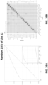
- the cancer types are selected from thyroid cancer, melanoma, sarcoma, myeloid neoplasm, renal cancer, prostate cancer, breast cancer, uterine cancer, ovarian cancer, bladder cancer, urothelial cancer, cervical cancer, anorectal cancer, head & neck cancer, colorectal cancer, liver cancer, bile duct cancer, pancreatic cancer, gallbladder cancer, upper GI cancer, multiple myeloma, lymphoid neoplasm, and lung cancer.
- the at least 200 target genomic regions are selected from any one of lists 1-16.
- the at least 200 target genomic regions comprise at least 500, 1,000, 5,000, 10,000, 15,000, 20,000, 30,000, 40,000, or 50,000 target genomic regions in any one of lists 8-11. In some scenarios, the at least 200 target genomic regions comprise at least 40%, 50%, 60%, or 70% of the target genomic regions listed in List 4. In some scenarios, wherein the at least 200 target genomic regions comprise, for at least 90% or for 100% of all possible pairs of cancer types selected from a set comprising at least 10 cancer types, at least one target genomic region that is differentially methylated between the pair of cancer types.
- the at least 100 target genomic regions comprises at least 200 target genomic regions. In some scenarios, the at least 100 target genomic regions are selected from any one of lists 1-16. In some scenarios, the at least 100 target genomic regions comprise at least 20%, 30%, 40%, 50%, 60%, 70%, 80%, 90% or 95% of the target genomic regions in any one of lists 1-16. In some scenarios, the at least 100 target genomic regions comprise at least 500, 1,000, 5,000, 10,000, 15,000, 20,000, 30,000, 40,000, or 50,000 target genomic regions in any one of lists 1-16. In some scenarios, the at least 100 target genomic regions are selected from any one of lists 1-3.
- the cancer type is selected from thyroid cancer, melanoma, sarcoma, myeloid neoplasm, renal cancer, prostate cancer, breast cancer, uterine cancer, ovarian cancer, bladder cancer, urothelial cancer, cervical cancer, anorectal cancer, head & neck cancer, colorectal cancer, liver cancer, bile duct cancer, pancreatic cancer, gallbladder cancer, upper GI cancer, multiple myeloma, lymphoid neoplasm, and lung cancer.
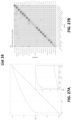
- the cancer type is sarcoma and the likelihood of a detecting sarcoma is at least 35% or at least 40%.
- the likelihood of detecting stage III or stage IV renal cancer is at least 50% or at least 70%. In some scenarios, the likelihood of detecting stage III or stage IV breast cancer is at least 70% or at least 85%. In some scenarios, the likelihood of detecting stage III or stage IV uterine cancer is at least 50%. In some scenarios, the likelihood of detecting ovarian cancer is at least 60% or at least 80%. In some scenarios, the likelihood of detecting bladder cancer is at least 35% or at least 40%. In some scenarios, the likelihood of detecting anorectal cancer is at least 60% or 70%. In some scenarios, the likelihood of detecting head and neck cancer is at least 75% or at least 80%. In some scenarios, the likelihood of detecting stage 1 head and neck cancer is at least 80%.
- the likelihood of detecting colorectal cancer is at least 50% or at least 59%. In some scenarios, the likelihood of detecting liver cancer is at least 75% or 80%. In some scenarios, the likelihood of detecting pancreas and gallbladder cancer is at least 64% or at least 70%. In some scenarios, the likelihood of detecting upper GI cancer is at least at least 60% or at least 68%. In some scenarios, the likelihood of detecting multiple myeloma is at least 65% or at least 75%. In some scenarios, the likelihood of detecting type I multiple myeloma is at least 60%. In some scenarios, the likelihood of detecting lymphoid neoplasm is at least 65% or at least 69%.
- the likelihood of detecting lung cancer is at least 50% or at least 58%.
- the composition comprising oligonucleotide baits is a composition provided above.
- the plurality of genomic regions comprises no more than 95,000 genomic regions, no more than 60,000 genomic regions, no more than 40,000 genomic regions, no more than 35,000 genomic regions, no more than 20,000 genomic regions, no more than 15,000 genomic regions, no more than 8,000 genomic regions, no more than 4,000 genomic regions, no more than 2,000 genomic regions, or no more than 1,400 genomic regions.
- the total size of the plurality of genomic regions is less than 4 MB, less than 2 MB, less than 1 MB, less than 0.7 MB, or less than 0.4 MB.
- the subject has an elevated risk of one or more cancer types. In some scenarios, the subject manifests symptoms associated with one or more cancer types. In some scenarios, the subject has not been diagnosed with a cancer. In some scenarios, the classifier was trained on converted DNA sequences derived from a least 100 subjects with a first cancer type, at least 100 subjects with a second cancer type, and at least 100 subjects with no cancer. In some scenarios, the first cancer type is ovarian cancer. In some scenarios, the first cancer type is liver cancer.
- the first cancer type is selected from thyroid cancer, melanoma, sarcoma, myeloid neoplasm, renal cancer, prostate cancer, breast cancer, uterine cancer, ovarian cancer, bladder cancer, urothecal cancer, cervical cancer, anorectal cancer head & neck cancer, colorectal cancer, liver cancer, pancreatic cancer, gallbladder cancer, esophageal cancer, stomach cancer, multiple myeloma, lymphoid neoplasm, lung cancer, or leukemia.
- the classifier was trained on converted DNA sequences derived from at least 1000, at least 2000, or at least 4000 target genomic regions selected from any one of Lists 1-16.
- the classifier is trained on converted DNA sequences derived from at least 1000, at least 2000, or at least 4000 target genomic regions selected from any one of Lists 1-16.
- the trained classifier determines the presence or absence of cancer or a cancer type by (a) generating a set of features for the sample, wherein each feature in the set of features comprises a numerical value; (b) inputting the set of features into the classifier, wherein the classifier comprises a multinomial classifier; (c) based on the set of features, determining, at the classifier, a set of probability scores, wherein the set of probability scores comprises one probability score per cancer type class and per non-cancer type class; and (d) thresholding the set of probability scores based on one or more values determined during training of the classifier to determine a final cancer classification of the sample.
- the set of features comprises a set of binarized features.
- the numerical value comprises a single binary value.
- the multinomial classifier comprises a multinomial logistic regression ensemble trained to predict a source tissue for the cancer.
- the classifier determines a final cancer classification based on a top-two probability score differential relative to a minimum value, wherein the minimum value corresponds to a predefined percentage of training cancer samples that had been assigned the correct cancer type as their highest score during training of the classifier.
- the classifier assigns a cancer label corresponding to the highest probability score determined by the classifier as the final cancer classification when it is determined that the top-two probability score differential exceeds the minimum value; and assigns an indeterminate cancer label as the final cancer classification when it is determined that the top-two probability score differential does not exceed the minimum value.
- the anti-cancer agent is a chemotherapeutic agent selected from the group consisting of alkylating agents, antimetabolites, anthracyclines, anti-tumor antibiotics, cytoskeletal disruptors (taxans), topoisomerase inhibitors, mitotic inhibitors, corticosteroids, kinase inhibitors, nucleotide analogs, and platinum-based agents.
- cancer assay panels comprising: at least 500 pairs of probes, wherein each pair of the at least 500 pairs comprise two probes configured to overlap each other by an overlapping sequence, wherein the overlapping sequence comprises a 30-nucleotide sequence, and wherein the 30-nucleotide sequence is configured to hybridize to a converted cfDNA molecule corresponding to, or derived from one or more of genomic regions, wherein each of the genomic regions comprises at least five methylation sites, and wherein the at least five methylation sites have an abnormal methylation pattern in cancerous samples.
- each of the at least 500 pairs of probes is conjugated to a non-nucleotide affinity moiety.
- the non-nucleotide affinity moiety is a biotin moiety.
- the cancerous samples are from subjects having cancer selected from the group consisting of breast cancer, uterine cancer, cervical cancer, ovarian cancer, bladder cancer, urothelial cancer of renal pelvis, renal cancer other than urothelial, prostate cancer, anorectal cancer, colorectal cancer, hepatobiliary cancer arising from hepatocytes, hepatobiliary cancer arising from cells other than hepatocytes, pancreatic cancer, squamous cell cancer of the upper gastrointestinal tract, upper gastrointestinal cancer other than squamous, head and neck cancer, lung adenocarcinoma, small cell lung cancer, squamous cell lung cancer and cancer other than adenocarcinoma or small cell lung cancer, neuroendocrine cancer, mela
- the abnormal methylation pattern has at least a threshold p-value rarity in the cancerous samples.
- each of the probes is designed to have less than 20 off-target genomic regions.
- the less than 20 off-target genomic regions are identified using a k-mer seeding strategy.
- the less than 20 off-target genomic regions are identified using k-mer seeding strategy combined to local alignment at seed locations.
- the cancer assay panel comprises at least 10,000, 50,000, 100,000, 200,000, 300,000, 400,000, 500,000, 600,000, 700,000 or 800,000 probes.
- the at least 500 pairs of probes together comprise at least 2 million, 3 million, 4 million, 5 million, 6 million, 8 million, 10 million, 12 million, 14 million, or 15 million nucleotides.
- each of the probes comprises at least 50, 75, 100, or 120 nucleotides.
- each of the probes comprises less than 300, 250, 200, or 150 nucleotides.
- each of the probes comprises 100-150 nucleotides.
- each of the probes comprises less than 20, 15, 10, 8, or 6 methylation sites.
- at least 80, 85, 90, 92, 95, or 98% of the at least five methylation sites are either methylated or unmethylated in the cancerous samples.
- each of the probes comprise multiple binding sites to the methylation sites of the converted cfDNA molecule, wherein at least 80, 85, 90, 92, 95, or 98% of the multiple binding sites comprise exclusively either CpG or CpA.
- each of the probes is configured to have less than 15, 10 or 8 off-target genomic regions.
- at least 30% of the genomic regions are in exons or introns.
- at least 15% of the genomic regions are in exons.
- at least 20% of the genomic regions are in exons.
- less than 10% of the genomic regions are in intergenic regions.
- the genomic regions are selected from any one of Lists 1-3 or Lists 4-16. In some scenarios, the genomic regions comprise at least 20%, 30%, 40%, 50%, 60%, 70%, 80%, 90% or 95% of the genomic regions in any one of Lists 1-3 or Lists 4-16. In some scenarios, the genomic regions comprise at least 500, 1,000, 5000, 10,000, or 15,000, 20,000, 30,000, 40,000, 50,000, 60,000, or 70,000 genomic regions in any one of Lists 1-3 or Lists 4-16.
- cancer assay panels comprising a plurality of probes, wherein each of the plurality of probes is configured to hybridize to a converted cfDNA molecule corresponding to one or more of the genomic regions in any one of Lists 1-3 or Lists 4-16.
- the plurality of probes together is configured to hybridize to a plurality of converted cfDNA molecules corresponding to at least 20%, 30%, 40%, 50%, 60%, 70%, 80%, or 90%, 95% or 100% of the genomic regions of any one of Lists 1-3 or Lists 4-16.
- the plurality of probes together is configured to hybridize to a plurality of converted cfDNA molecules corresponding to at least 500, 1,000, 5000, 10,000, 15,000, 20,000, 30,000, 40,000, or 50,000 genomic regions of any one of Lists 1-3 or Lists 4-16. In some scenarios, at least 3%, 5%, 10%, 15%, or 20% of the probes comprise no G (Guanine). In some scenarios, each of the probes comprise multiple binding sites to methylation sites of the converted cfDNA molecule, wherein at least 80, 85, 90, 92, 95, or 98% of the multiple binding sites comprise exclusively either CpG or CpA. In some scenarios, each of the probes is conjugated to a non-nucleotide affinity moiety. In some scenarios, the non-nucleotide affinity moiety is a biotin moiety.
- TOO tissue of origin
- Some scenarios further comprise the step of: determining a health condition by evaluating the set of sequence reads, wherein the health condition is a presence or absence of cancer; a presence or absence of cancer of a tissue of origin (TOO); a presence or absence of a cancer cell type; or a presence or absence of at least 5, 10, 15, or 20 different types of cancer.
- the sample comprising a plurality of cfDNA molecules was obtained from a human subject.
- Also provided herein are methods for detecting a cancer, comprising the steps of: obtaining a set of sequence reads by sequencing a set of nucleic acid fragments from a subject, wherein the nucleic acid fragments are corresponding to, or derived from a plurality of genomic regions selected from any one of Lists 1-3 or Lists 4-16; for each of the nucleic acid fragments, determining methylation status at a plurality of CpG sites; and detecting a health condition of the subject by evaluating the methylation status for the sequence reads, wherein the health condition is (i) a presence or absence of cancer; (ii) a presence or absence of cancer of a tissue of origin (TOO); (iii) a presence or absence of a cancer cell type; or (iv) a presence or absence of at least 5, 10, 15, or 20 different types of cancer.
- the health condition is (i) a presence or absence of cancer; (ii) a presence or absence of cancer of a tissue of origin (TOO); (
- the plurality of genomic regions comprises at least 20%, 30%, 40%, 50%, 60%, 70%, 80%, 90%, 95%, or 100% of the genomic regions of any one of Lists 1-3 or lists 4-16. In some scenarios, the plurality of genomic regions comprises 500, 1,000, 5000, 10,000, 15,000, 20,000, 30,000, 40,000, 50,000, 60,000, 70,000, or 80,000 of the genomic regions of any one of Lists 1-3 or Lists 4-16.
- Also provided herein are methods of designing a cancer assay panel for diagnosing cancer of a tissue of origin (TOO) comprising the steps of: identifying a plurality of genomic regions, wherein each of the plurality of genomic regions (i) comprises at least 30 nucleotides, and (ii) comprises at least five methylation sites, selecting a subset of the genomic regions, wherein the selection is made when cfDNA molecules corresponding to, or derived from each of the genomic regions in cancerous samples have an abnormal methylation pattern, wherein the abnormal methylation pattern comprises at least five methylation sites either hypomethylated or hypermethylated, and designing a cancer assay panel comprising a plurality of probes, wherein each of the probes is configured to hybridize to a converted cfDNA molecule corresponding to or derived from one or more of the subset of the genomic regions.
- TOO tissue of origin
- the first cancer type and the second cancer type are selected from uterine cancer, upper GI squamous cancer, all other upper GI cancers, thyroid cancer, sarcoma, urothelial renal cancer, all other renal cancers, prostate cancer, pancreatic cancer, ovarian cancer, neuroendocrine cancer, multiple myeloma, melanoma, lymphoma, small cell lung cancer, lung adenocarcinoma, all other lung cancers, leukemia, hepatobiliary hepatocellular carcinoma, hepatobiliary biliary, head and neck cancer, colorectal cancer, cervical cancer, breast cancer, bladder cancer, and anorectal cancer.
- enriching the cfDNA comprises amplifying, via PCR, portions of the cell-free DNA fragments using primers configured to hybridize to a plurality of genomic regions selected from any one of Lists 1-16. In some scenarios, enriching the cfDNA sample comprises contacting the cell-free DNA with a plurality of probes configured to hybridize to converted fragments obtained from the cfDNA molecules corresponding to or derived from the genomic regions in any one of Lists 1-16.
- the cfDNA sample comprises contacting the cell-free DNA with a plurality of probes configured to hybridize to converted fragments obtained from the cfDNA molecules corresponding to or derived from at least 30%, 40%, 50%, 60%, 70%, 80%, 90%, 95% of the genomic regions in any one of Lists 1-16.
- the genomic regions are selected from any one of Lists 1-3.
- the genomic regions are selected from any one of Lists 4-12.
- the genomic regions are selected from any one of Lists 4, 6, or 8-12.
- the genomic regions are selected from List 8.
- the cfDNA sample is enriched by a method provided above.
- the method further comprises determining a cancer classification by evaluating the set of sequence reads, wherein the cancer classification is a presence or absence of cancer; or a presence or absence of a type of cancer.
- the step of determining a cancer classification comprises: generating a test feature vector based on the set of sequence reads; and applying the test feature vector to a classifier.
- the classifier comprises a model that is trained by a training process with a first cancer set of fragments from one or more training subjects with a first cancer type and a second cancer set of fragments from one or more training subjects with a second cancer type, wherein both the first cancer set of fragments and the second cancer set of fragments comprise a plurality of training fragments.
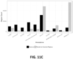
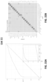
- the cancer classification is a presence or absence of cancer. In some scenarios, has an area under a receiver operating characteristic curve of at least 0.8. In some scenarios, the cancer classification is a type of cancer. In some scenarios, the type of cancer is selected from among at least 12, 14, 16, 18, or 20 cancer types.
- the cancer types are selected from anal cancer, bladder cancer, colorectal cancer, esophageal cancer, head and neck cancer, liver/bile-duct cancer, lung cancer, lymphoma, ovarian cancer, pancreatic cancer, plasma cell neoplasm, and stomach cancer.
- the sensitivity of the method for head and neck cancer is at least 79% or at least 84%; at 99% specificity the sensitivity of the method for liver cancer is at least 82% or at least 85%; at 99% specificity the sensitivity of the method for upper GI tract cancer is at least 62% or at least 68%; wherein at 99% specificity the sensitivity of the method for pancreatic or gallbladder cancer is at least 62% or at least 68%; at 99% specificity the sensitivity of the method for colorectal cancer is at least 60% or at least 65%; at 99% specificity the sensitivity of the method for ovarian cancer is at least 75% or at least 80%; at 99% specificity the sensitivity of the method for lung cancer is at least 60% or at least 65%; at 99% specificity the sensitivity of the method for multiple myeloma is at least 68% or at least 75%; at 99% specificity the sensitivity of the method for lymphoid neoplasm is at least 65% or at least
- the cancer classification is a presence or absence of a type of cancer.
- the step of determining a cancer classification comprises: generating a test feature vector based on the set of sequence reads; and applying the test feature vector to a classifier.
- the classifier comprises a model that is trained by a training process with a first cancer type set of converted DNA sequences from one or more training subjects with a first cancer type and a second cancer type set of converted DNA sequences from one or more training subjects with a second cancer type, wherein both the first cancer type set of converted DNA sequences and the second cancer type set of converted DNA sequences comprise a plurality of training converted DNA sequences.
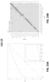
- the type of cancer is selected from the group consisting of head and neck cancer, liver/bile duct cancer, upper GI cancer, pancreatic/gallbladder cancer; colorectal cancer, ovarian cancer, lung cancer, multiple myeloma, lymphoid neoplasms, melanoma, sarcoma, breast cancer, and uterine cancer.
- the type of cancer is head and neck cancer, and the method, at 99.0% specificity, has a sensitivity of at least 79% or at least 84%.
- the type of cancer is liver cancer, and the method, at 99.0% specificity, has a sensitivity of at least 82% or at least 85%.
- the type of cancer is an upper GI tract cancer, and the method, at 99.0% specificity, has a sensitivity of at least 62% or at least 68%. In some scenarios, the type of cancer is a pancreatic or gallbladder cancer, and the method, at 99.0% specificity, has a sensitivity of at least 62% or at least 68%. In some scenarios, the type of cancer is colorectal cancer, and the method, at 99.0% specificity, has a sensitivity of at least 60% or at least 65%. In some scenarios, the type of cancer is ovarian cancer, and the method, at 99.0% specificity, has a sensitivity of at least 75% or at least 80%.
- the type of cancer is lung cancer, and the method, at 99.0% specificity, has a sensitivity of at least60% or at least 65%. In some scenarios, the type of cancer is multiple myeloma, and the method, at 99.0% specificity, has a sensitivity of at least 68% or at least 75%. In some scenarios, the type of cancer is a lymphoid neoplasm, and the method, at 99.0% specificity, has a sensitivity of at least 65% or at least 70%. In some scenarios, the type of cancer is anorectal cancer, and the method, at 99.0% specificity, has a sensitivity of at least 60% or at least 65%.
- the type of cancer is bladder cancer
- the method at 99.0% specificity, has a sensitivity of at least 40% or at least 44%.
- the total size of the target genomic regions is less than 4 Mb, less than 2 Mb, less than 1 Mb, less than 0.7 Mb or less than 0.4 Mb.
- the step of determining a cancer classification comprises:
- test feature vector based on the set of sequence reads; and applying the test feature vector to a model obtained by a training process with a cancer set of fragments from one or more training subjects with a cancer and a non-cancer set of fragments from one or more training subjects without cancer, wherein both the cancer set of fragments and the non-cancer set of fragments comprise a plurality of training fragments.
- the training process comprises: obtaining sequence information of training fragments from a plurality of training subjects; for each training fragment, determining whether that training fragment is hypomethylated or hypermethylated, wherein each of the hypomethylated and hypermethylated training fragments comprises at least a threshold number of CpG sites with at least a threshold percentage of the CpG sites being unmethylated or methylated, respectively, for each training subject, generating a training feature vector based on the hypomethylated training fragments and hypermethylated training fragments, and training the model with the training feature vectors from the one or more training subjects without cancer and the training feature vectors from the one or more training subjects with cancer.
- the training process comprises: obtaining sequence information of training fragments from a plurality of training subjects; for each training fragment, determining whether that training fragment is hypomethylated or hypermethylated, wherein each of the hypomethylated and hypermethylated training fragments comprises at least a threshold number of CpG sites with at least a threshold percentage of the CpG sites being unmethylated or methylated, respectively, for each of a plurality of CpG sites in a reference genome: quantifying a count of hypomethylated training fragments which overlap the CpG site and a count of hypermethylated training fragments which overlap the CpG site; and generating a hypomethylation score and a hypermethylation score based on the count of hypomethylated training fragments and hypermethylated training fragments; for each training fragment, generating an aggregate hypomethylation score based on the hypomethylation score of the CpG sites in the training fragment and an aggregate hypermethylation score based on the hypermethylation score of the CpG sites in the training fragment; for each training subject: ranking the plurality
- the model comprises one of a kernel logistic regression classifier, a random forest classifier, a mixture model, a convolutional neural network, and an autoencoder model.
- the method further comprises the steps of: obtaining a cancer probability for the test sample based on the model; and comparing the cancer probability to a threshold probability to determine whether the test sample is from a subject with cancer or without cancer.
- the method further comprises the steps of: obtaining a cancer type probability for the test sample based on the model; and comparing the cancer type probability to a threshold probability to determine whether the test sample is from a subject with the cancer type or another cancer type or without cancer.
- the method further comprises administering an anti-cancer agent to the subject.
- the anti-cancer agent is a chemotherapeutic agent selected from the group consisting of alkylating agents, antimetabolites, anthracyclines, anti-tumor antibiotics, cytoskeletal disruptors (taxans), topoisomerase inhibitors, mitotic inhibitors, corticosteroids, kinase inhibitors, nucleotide analogs, and platinum-based agents.
- chemotherapeutic agent selected from the group consisting of alkylating agents, antimetabolites, anthracyclines, anti-tumor antibiotics, cytoskeletal disruptors (taxans), topoisomerase inhibitors, mitotic inhibitors, corticosteroids, kinase inhibitors, nucleotide analogs, and platinum-based agents.
- Also provided herein are methods comprising the steps of: obtaining a set of sequence reads of modified test fragments, wherein the modified test fragments are or have been obtained by processing a set of nucleic acid fragments from a test subject, wherein each of the nucleic acid fragments corresponds to or is derived from a plurality of genomic regions selected from any one of Lists 1-16; and applying the set of sequence reads or a test feature vector obtained based on the set of sequence reads to a model obtained by a training process with a first set of fragments from a plurality of training subjects with a first cancer type and a second set of fragments from a plurality of training subjects with a second cancer type, wherein both the first set of fragments and the second set of fragments comprise a plurality of training fragments.
- the terms “comprises,” “comprising,” “includes,” “including,” “has,” “having” or any other variation thereof, are intended to cover a non-exclusive inclusion.
- a process, method, article, or apparatus that comprises a list of elements is not necessarily limited to only those elements but may include other elements not expressly listed or inherent to such process, method, article, or apparatus.
- “or” refers to an inclusive or and not to an exclusive or. For example, a condition A or B is satisfied by any one of the following: A is true (or present) and B is false (or not present), A is false (or not present) and B is true (or present), and both A and B are true (or present).
- methylation refers to a process by which a methyl group is added to a DNA molecule.
- a hydrogen atom on the pyrimidine ring of a cytosine base can be converted to a methyl group, forming 5-methylcytosine.
- the term also refers to a process by which a hydroxymethyl group is added to a DNA molecule, for example by oxidation of a methyl group on the pyrimidine ring of a cytosine base. Methylation and hydroxymethylation tend to occur at dinucleotides of cytosine and guanine referred to herein as "CpG sites.”
- methylation can also refer to the methylation status of a CpG site.
- a CpG site with a 5-methylcytosine moiety is methylated.
- a CpG site with a hydrogen atom on the pyrimidine ring of the cytosine base is unmethylated.
- methylation status at a site i.e., presence or absence of a methyl group.
- a methylated site i.e., a methylated site / absence of a methyl group is an unmethylated site or non-methylated site.
- wet laboratory assay used to detect methylation may vary from those described herein as is well known in the art.
- CpG site refers to a region of a DNA molecule where a cytosine nucleotide is followed by a guanine nucleotide in the linear sequence of bases along its 5' to 3' direction.
- CpG is a shorthand for 5'-C-phosphate-G-3' that is cytosine and guanine separated by only one phosphate group. Cytosines in CpG dinucleotides can be methylated to form 5-methylcytosine.
- hypomethylated refers to a methylation status of a DNA molecule containing multiple CpG sites (e.g., more than 3, 4, 5, 6, 7, 8, 9, 10, etc.) where a high percentage of the CpG sites (e.g., more than 80%, 85%, 90%, or 95%, or any other percentage within the range of 50%-100%) are unmethylated or methylated, respectively.
- fragment can refer to a fragment of a nucleic acid molecule.
- a fragment can refer to a cfDNA molecule in a blood or plasma sample, or a cfDNA molecule that has been extracted from a blood or plasma sample.
- An amplification product of a cfDNA molecule may also be referred to as a "fragment.”
- fragment refers to a sequence read, or set of sequence reads, that have been processed for subsequent analysis (e.g., for in machine-learning based classification), as described herein.
- raw sequence reads can be aligned to a reference genome and matching paired end sequence reads assembled into a longer fragment for subsequent analysis.
- the term "individual” refers to a human individual.
- the term “healthy individual” refers to an individual presumed not to have a cancer or disease.
- subject refers to an individual whose DNA is being analyzed.
- a subject may be a test subject whose DNA is be evaluated using a targeted panel as described herein to evaluate whether the person has cancer or another disease.
- a subject may also be part of a control group known not to have cancer or another disease.
- a subject may also be part of a cancer or other disease group known to have cancer or another disease. Control and cancer/disease groups may be used to assist in designing or validating the targeted panel.
- sequence reads refers to nucleotide sequences reads from a sample. Sequence reads can be obtained through various methods provided herein or as known in the art.
- sequencing depth refers to the count of the number of times a given target nucleic acid within a sample has been sequenced (e.g., the count of sequence reads at a given target region). Increasing sequencing depth can reduce required amounts of nucleic acids required to assess a disease state (e.g., cancer or cancer tissue of origin).
- tissue of origin refers to the organ, organ group, body region or cell type that a cancer arises or originates from.
- the identification of a tissue of origin or cancer cell type typically allows for identification of the most appropriate next steps in the care continuum of cancer to further diagnose, stage and decide on treatment.
- transition generally refers to changes in base composition from one purine to another purine, or from one pyrimidine to another pyrimidine. For instance, the following changes are transitions: C ⁇ U, U ⁇ C, G ⁇ A, A ⁇ G, C ⁇ T, and T ⁇ C.
- a panel or bait set generally refers to all of the probes delivered with a specified panel or bait set.
- a panel or bait set may include both (1) probes having features specified herein (e.g., probes for binding to cell-free DNA fragments corresponding to or derived from genomic regions set forth herein in one or more Lists) and (2) additional probes that do not contain such feature(s).
- the entirety of probes of a panel generally refers to all probes delivered with the panel or bait set, including such probes that do not contain the specified feature(s).
- the present description provides a cancer assay panel comprising a plurality of probes or a plurality of probe pairs.
- the assay panels described herein can alternatively be referred to as bait sets or as compositions comprising bait oligonucleotides.
- the probes can be polynucleotide-containing probes that are specifically designed to target one or more genomic regions differentially methylated between cancer and non-cancer samples, between different cancer tissue of origin (TOO) types, between different cancer cell types, between samples of different stages of cancer, as identified by methods provided herein.
- the target genomic regions are selected to maximize classification accuracy, subject to a size budget (which is determined by sequencing budget and desired depth of sequencing).
- the analytics system may generate variable sizes of the cancer assay panel, e.g., where a small sized cancer assay panel includes probes targeting the most informative genomic regions, a medium sized cancer assay panel includes probes from the small sized cancer assay panel and additional probes targeting a second tier of informative genomic regions, and a large sized cancer assay panel includes probes from the small-sized and the medium-sized cancer assay panels along with even more probes targeting a third tier of informative genomic regions.
- the analytics system may train classifiers with various classification techniques to predict a sample's likelihood of having a particular outcome or state, e.g., cancer, specific cancer type, other disorder, other disease, etc.
- the cancer assay panel comprises at least 500 pairs of probes, wherein each pair of the at least 500 pairs comprises two probes configured to overlap each other by an overlapping sequence, wherein the overlapping sequence comprises at least 30-nucleotides, and wherein each probe is configured to hybridize to the same strand of an (optionally converted) DNA molecule (e.g., a cfDNA molecule) corresponding to one or more genomic regions.
- each of the genomic regions comprises at least five methylation sites, and wherein the at least five methylation sites have an abnormal methylation pattern in cancerous samples or a different methylation pattern between samples of a different TOO.
- the target genomic regions can be selected from List 1.
- the target genomic regions can be selected from List 2.
- the target genomic regions can be selected from List 3.
- the target genomic regions can be selected from List 4.
- the target genomic regions can be selected from List 5.
- the target genomic regions can be selected from List 6.
- the target genomic regions can be selected from List 7.
- the target genomic regions can be selected from List 8.
- the target genomic regions can be selected from List 9.
- the target genomic regions can be selected from List 10.
- the target genomic regions can be selected from List 11.
- the target genomic regions can be selected from List 12.
- the target genomic regions can be selected from List 13.
- the target genomic regions can be selected from List 14.
- the target genomic regions can be selected from List 15.
- the target genomic regions can be selected from List 16.
- the probes are configured to hybridize to a converted DNA or cfDNA molecule corresponding to, or derived from, one or more genomic regions, the probes can have a sequence different from the targeted genomic region.
- a DNA containing an unmethylated CpG site will be converted to include UpG instead of CpG because unmethylated cytosines are converted to uracils by a conversion reaction (e.g., bisulfite treatment).
- a probe is configured to hybridize to a sequence including UpG instead of a naturally-existing unmethylated CpG.
- a complementary site in the probe to the unmethylated site can comprise CpA instead of CpG, and some probes targeting a hypomethylated site where all methylation sites are unmethylated can have no guanine (G) bases.
- G guanine
- at least 3%, 5%, 10%, 15%, or 20% of the probes comprise no CpG sequences.
- the cancer assay panel can be used to detect the presence or absence of cancer generally and/or provide a cancer classification such as cancer type, stage of cancer such as I, II, III, or IV, or provide the TOO where the cancer is believed to originate.
- the panel may include probes targeting genomic regions differentially methylated between general cancerous (pan-cancer) samples and non-cancerous samples, or only in cancerous samples with a specific cancer type (e.g., lung cancer-specific targets).
- a cancer assay panel is designed to include differentially methylated genomic regions based on converted (e.g., bisulfite) sequencing data generated from the cfDNA from cancer and non-cancer individuals.
- Each of the probes may be designed to target one or more target genomic regions.
- the target genomic regions can be selected based on several criteria designed to increase selective enriching of informative cfDNA fragments while decreasing noise and non-specific bindings.
- a panel can include probes that can selectively bind to and enrich cfDNA fragments that are differentially methylated in cancerous samples. In this case, sequencing of the enriched fragments can provide information relevant to detection of cancer.
- the probes (or a portion thereof) are designed to target genomic regions that are determined to have an abnormal methylation pattern in cancer samples, or in samples from certain cancer types, tissue types or cell types.
- probes are designed to target genomic regions determined to be hypermethylated or hypomethylated in certain cancers or cancer types to provide additional selectivity and specificity of the detection.
- a panel comprises probes targeting hypomethylated fragments.
- a panel comprises probes targeting hypermethylated fragments.
- a panel comprises both a first set of probes targeting hypermethylated fragments and a second set of probes targeting hypomethylated fragments.
- a cancer assay panel includes not only probes that are designed to target a region that has a first methylation status (e.g., hypomethylation), but also includes probes that are designed to hybridize to the same target region with the opposite methylation status (e.g., hypermethylation).
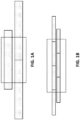
- the targeting of probes to both hypo- and hyper-methylated fragments from the same regions can be referred to as "binary" targeting (see information in the Sequence Listing) ( FIG. 1C ).
- genomic regions i.e., genomic regions giving rise to differentially methylated DNA molecules (or anomalously methylated DNA molecules) between cancer and non-cancer samples, between different cancer tissue of origin (TOO) types, between different cancer cell type, or between samples from different stages of cancer are provided in detail herein and methods of identifying anomalously methylated DNA molecules or fragments that are identified as indicative of cancer are also provided in detail herein.
- TOO cancer tissue of origin
- genomic regions can be selected when the genomic regions give rise to anomalously methylated DNA molecules in cancer samples or samples with known cancer tissue of origin (TOO) types.
- TOO cancer tissue of origin
- a Markov model trained on a set of non-cancerous samples can be used to identify genomic regions that give rise to anomalously methylated DNA molecules (i.e., DNA molecules having a methylation pattern below a p-value threshold).
- the smallest target genomic region is 30 or 31 bp.
- the new target region of 30bp can be centered on a specific CpG site of interest. Then, it is checked whether each edge of this new target is close enough to other targets such that they can be merged. This is based on a "merge distance" parameter which can be 200bp by default but can be tuned. This allows close but distinct target regions to be enriched with overlapping probes.
- the new target can be merged with nothing (increasing the number of panel targets by one), merged with just one target either to the left or the right (not changing the number of panel targets), or merged with existing targets both to the left and right (reducing the number of panel targets by one).
- methods of selecting target genomic regions for detecting cancer and/or a TOO can be used to design and manufacture probes for a cancer assay panel. Methylation status of DNA or cfDNA molecules corresponding to, or derived from, the target genomic regions can be screened using the cancer assay panel.
- Alternative methods for example by WGBS or other methods known in the art, can be also implemented to detect methylation status of DNA molecules or fragments corresponding to, or derived from, the target genomic regions.
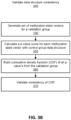
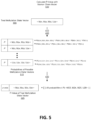
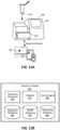
- FIG. 7A is a flowchart of a process 100 for processing a nucleic acid sample and generating methylation state vectors for DNA fragments, according to one scenario.
- the method includes, but is not limited to, the following steps.
- any step of the method may comprise a quantitation sub-step for quality control or other laboratory assay procedures known to one skilled in the art.
- the extracted sample may comprise cfDNA and/or ctDNA.
- the human body may naturally clear out cfDNA and other cellular debris. If a subject has a cancer or disease, cfDNA and/or ctDNA in an extracted sample may be present at a detectable level for detecting the cancer or disease.
- the cfDNA fragments are treated to convert unmethylated cytosines to uracils.
- the method uses a bisulfite treatment of the DNA which converts the unmethylated cytosines to uracils without converting the methylated cytosines.
- a commercial kit such as the EZ DNA Methylation TM - Gold, EZ DNA Methylation TM - Direct or an EZ DNA Methylation TM - Lightning kit (available from Zymo Research Corp (Irvine, CA)) is used for the bisulfite conversion.
- the conversion of unmethylated cytosines to uracils is accomplished using an enzymatic reaction.
- the conversion can use a commercially available kit for conversion of unmethylated cytosines to uracils, such as APOBEC-Seq (NEBiolabs, Ipswich, MA).
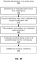
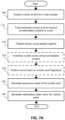
- FIG. 3A is a flowchart describing a process 300 of generating a data structure for a healthy control group, according to a scenario.
- the analytics system obtains information related to methylation status of a plurality of CpG sites on sequence reads derived from a plurality of DNA molecules or fragments from a plurality of healthy subjects.
- the method provided herein for creating a healthy control group data structure can be performed similarly for subjects with cancer, subjects with cancer of a TOO, subjects with a known cancer type, or subjects with another known disease state.
- a methylation state vector is generated for each DNA molecule or fragment, for example via the process 100.
- the analytics system calculates 420 probabilities 515 for the enumerated possibilities of methylation state vectors. As methylation is conditionally dependent on methylation status of nearby CpG sites, one way to calculate the probability of observing a given methylation state vector possibility is to use Markov chain model.
- the analytics system accesses the control group's data structure, specifically the counts of various strings of CpG sites and states.
- S n-k-2 , ..., S n-1 ) the analytics system takes a ratio of the stored count of the number of strings from the data structure matching ⁇ S n-k-2 , ..., S n-1 , M n > divided by the sum of the stored count of the number of strings from the data structure matching ⁇ S n-k-2 , ..., S n-1 , M n > and ⁇ S n-k-2 , ..., S n-1 , U n >.
- S n-k-2 , ..., S n-1 ) is calculated ratio having the form: # of S n ⁇ k ⁇ 2 , ... , S n ⁇ 1 , M n # of S n ⁇ k ⁇ 2 , ... , S n ⁇ 1 , M n + # of S n ⁇ k ⁇ 2 , ... , S n ⁇ 1 , U n
- the calculation may additionally implement a smoothing of the counts by applying a prior distribution.
- the prior distribution is a uniform prior as in Laplace smoothing.
- a constant is added to the numerator and another constant (e.g., twice the constant in the numerator) is added to the denominator of the above equation.
- an algorithmic technique such as Knesser-Ney smoothing is used.
- the above denoted formulas are applied to the test methylation state vector 505 covering sites 23 - 26.
- the analytics system calculates 430 a p-value score 525 that sums the probabilities that are less than or equal to the probability of possibility of methylation state vector matching the test methylation state vector 505.
- the computational burden of calculating probabilities and/or p-value scores may be further reduced by caching at least some calculations.
- the analytic system may cache in transitory or persistent memory calculations of probabilities for possibilities of methylation state vectors (or windows thereof). If other fragments have the same CpG sites, caching the possibility probabilities allows for efficient calculation of p-value scores without needing to re-calculate the underlying possibility probabilities.
- the analytics system may calculate p-value scores for each of the possibilities of methylation state vectors associated with a set of CpG sites from vector (or window thereof). The analytics system may cache the p-value scores for use in determining the p-value scores of other fragments including the same CpG sites.
- the p-value scores of possibilities of methylation state vectors having the same CpG sites may be used to determine the p-value score of a different one of the possibilities from the same set of CpG sites.
- the window In calculating p-values for a methylation state vector larger than the window, the window identifies the sequential set of CpG sites from the vector within the window starting from the first CpG site in the vector.
- the analytic system calculates a p-value score for the window including the first CpG site.
- the analytics system then "slides" the window to the second CpG site in the vector, and calculates another p-value score for the second window.
- each methylation state vector will generate m-l+1 p-value scores.
- the analytics system aggregates the p-value scores for the methylation state vectors to generate an overall p-value score.
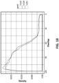
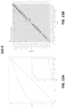
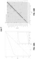
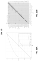
- Example probability calculations are shown in FIG. 5 , but generally the number of possibilities of methylation state vectors increases exponentially by a factor of 2 with the size of the methylation state vector. To give a realistic example, it is possible for fragments to have upwards of 54 CpG sites. Instead of computing probabilities for 2 ⁇ 54 ( ⁇ 1.8 ⁇ 10 ⁇ 16) possibilities to generate a single p-value, the analytics system can instead use a window of size 5 (for example) which results in 50 p-value calculations for each of the 50 windows of the methylation state vector for that fragment.
- Each of the 50 calculations enumerates 2 ⁇ 5 (32) possibilities of methylation state vectors, which total results in 50 ⁇ 2 ⁇ 5 (1.6 ⁇ 10 ⁇ 3) probability calculations. This results in a vast reduction of calculations to be performed, with no meaningful hit to the accurate identification of anomalous fragments. This additional step can also be applied when validating 340 the control group with the validation group's methylation state vectors.
- the analytics system can identify 460 genomic regions indicative of cancer. To identify these informative regions, the analytics system calculates an information gain for each genomic region or more specifically each CpG site that describes an ability to distinguish between various outcomes.
- the analytics system considers portions of the methylation state vector and determines whether the portion is hypomethylated or hypermethylated, and may distinguish that portion to be hypomethylated or hypermethylated. This alternative resolves missing methylation state vectors which are large in size but contain at least one region of dense hypomethylation or hypermethylation. This process of defining hypomethylation and hypermethylation can be applied in step 450 of FIG. 4 .
- the fragments indicative of cancer may be defined according to likelihoods outputted from trained probabilistic models.
- the analytics system generates 620 a hypomethylation score (P hypo ) and a hypermethylation score (P hyper ) per CpG site in the genome.
- the classifier takes four counts at that CpG site - (1) count of (methylations state) vectors of the cancer set labeled hypomethylated that overlap the CpG site; (2) count of vectors of the cancer set labeled hypermethylated that overlap the CpG site; (3) count of vectors of the non-cancer set labeled hypomethylated that overlap the CpG site; and (4) count of vectors of the non-cancer set labeled hypermethylated that overlap the CpG site.
- the process may normalize these counts for each group to account for variance in group size between the non-cancer group and the cancer group.
- the scores may be more broadly defined as counts of fragments indicative of cancer at each genomic region and/or CpG site.
- the process takes a ratio of (1) over (1) summed with (3).
- the hypermethylation score is calculated by taking a ratio of (2) over (2) and (4). Additionally, these ratios may be calculated with an additional smoothing technique as discussed above.
- the hypomethylation score and the hypermethylation score relate to an estimate of cancer probability given the presence of hypomethylation or hypermethylation of fragments from the cancer set.
- the analytics system generates 630 an aggregate hypomethylation score and an aggregate hypermethylation score for each anomalous methylation state vector.
- the aggregate hyper and hypo methylation scores are determined based on the hyper and hypo methylation scores of the CpG sites in the methylation state vector. In one scenario, the aggregate hyper and hypo methylation scores are assigned as the largest hyper and hypo methylation scores of the sites in each state vector, respectively. However, in alternate scenarios, the aggregate scores could be based on means, medians, or other calculations that use the hyper/hypo methylation scores of the sites in each vector.
- the analytics system ranks 640 all of that subject's methylation state vectors by their aggregate hypomethylation score and by their aggregate hypermethylation score, resulting in two rankings per subject.
- the process selects aggregate hypomethylation scores from the hypomethylation ranking and aggregate hypermethylation scores from the hypermethylation ranking.
- the classifier With the selected scores, the classifier generates 650 a single feature vector for each subject.
- the scores selected from either ranking are selected with a fixed order that is the same for each generated feature vector for each subject in each of the training groups.
- the classifier selects the first, the second, the fourth, and the eighth aggregate hyper methylation score, and similarly for each aggregate hypo methylation score, from each ranking and writes those scores in the feature vector for that subject.
- the number of non-cancer samples or different cancer type(s) (n other ) and the number of cancer samples or cancer type(s) (n cancer ) having an anomalously methylated fragment overlapping a CpG site are counted. Then the probability that a sample is cancer is estimated by a score ("S") that positively correlates to n cancer and inversely correlated to n other .
- the score can be calculated using the equation: (n cancer + 1) / (n cancer + n other + 2) or (n cancer ) / (n cancer + n other ).
- the analytics system computes 670 an information gain for each cancer type and for each genomic region or CpG site to determine whether the genomic region or CpG site is indicative of cancer.
- the information gain is computed for training samples with a given cancer type compared to all other samples.
- two random variables 'anomalous fragment' ('AF') and 'cancer type' ('CT') are used.
- AF is a binary variable indicating whether there is an anomalous fragment overlapping a given CpG site in a given samples as determined for the anomaly score / feature vector above.
- CT is a random variable indicating whether the cancer is of a particular type.
- the analytics system computes the mutual information with respect to CT given AF. That is, how many bits of information about the cancer type are gained if it is known whether there is an anomalous fragment overlapping a particular CpG site.
- the analytics system uses this information to rank CpG sites based on how cancer specific they are. This procedure is repeated for all cancer types under consideration. If a particular region is commonly anomalously methylated in training samples of a given cancer but not in training samples of other cancer types or in healthy training samples, then CpG sites overlapped by those anomalous fragments will tend to have high information gains for the given cancer type.
- the ranked CpG sites for each cancer type are greedily added (selected) to a selected set of CpG sites based on their rank for use in the cancer classifier.
- the analytics system defines a feature vector for each sample, by a count of DNA fragments for each genomic region for each cancer type that provides a calculated log-likelihood ratio for the fragment above a plurality of thresholds, wherein each count is a value in the feature vector.
- the analytics system uses the defined feature vectors to calculate an informative score for each genomic region describing that genomic region's ability to distinguish between each pair of cancer types. For each pair of cancer types, the analytics system ranks regions based on the informative scores. The analytics system may select regions based on the ranking according to informative scores.
- the analytics system calculates 695 an informative score for each region describing that region's ability to distinguish between each pair of cancer types.
- the analytics system may specify one type as a positive type and the other as a negative type.
- a region's ability to distinguish between the positive type and the negative type is based on mutual information, calculated using the estimated fraction of cfDNA samples of the positive type and of the negative type for which the feature would be expected to be non-zero in the final assay, i.e., at least one fragment of that tier that would be sequenced in a targeted methylation assay.
- Those fractions are estimated using the observed rates at which the feature occurs in healthy cfDNA, and in high-signal cfDNA and/or tumor samples of each cancer type. For example, if a feature occurs frequently in healthy cfDNA, then it will also be estimated to occur frequently in cfDNA of any cancer type and would likely result in a low informative score.
- the analytics system may choose a certain number of regions for each pair of cancer types from the ranking, e.g., 1024.
- the analytics system further identifies predominantly hypermethylated or hypomethylated regions from the ranking of regions.
- the analytics system may load the set of fragments in the positive type(s) for a region that was identified as informative.
- the analytics system evaluates whether the loaded fragments are predominantly hypermethylated or hypomethylated. If the loaded fragments are predominately hypermethylated or hypomethylated, the analytics system may select probes corresponding to the predominant methylation pattern. If the loaded fragments are not predominantly hypermethylated or hypomethylated, the analytics system may use a mixture of probes for targeting both hypermethylation and hypomethylation.
- the analytics system may further identify a minimal set of CpG sites that overlap more than some percentage of the fragments.
- the analytics system after ranking the regions based on informative scores, labels each region with the lowest informative ranking across all pairs of cancer types. For example, if a region was the 10th-most-informative region for distinguishing breast from lung, and the 5th-most-informative for distinguishing breast from colorectal, then it would be given an overall label of "5".
- the analytics system may design probes starting with the lowest-labeled regions while adding regions to the panel, e.g., until the panel's size budget has been exhausted.
- probes targeting selected genomic regions are further filtered 475 based on the number of their off-target regions. This is for screening probes that pull down too many cfDNA fragments corresponding to, or derived from, off-target genomic regions. Exclusion of probes having many off-target regions can be valuable by decreasing off-target rates and increasing target coverage for a given amount of sequencing.
- An off-target genomic region is a genomic region that has sufficient homology to a target genomic region, such that DNA molecules or fragments derived from off-target genomic regions are hybridized to and pulled down by a probe designed to hybridize to a target genomic region.
- An off-target genomic region can be a genomic region (or a converted sequence of that same region) that aligns to a probe along at least 35 bp, 40 bp, 45 bp, 50 bp, 60 bp, 70 bp, or 80 bp with at least an 80%, 85%, 90%, 95%, or 97% match rate.
- an off-target genomic region is a genomic region (or a converted sequence of that same region) that aligns to a probe along at least 45bp with at least a 90% match rate.
- Various methods known in the art can be adopted to screen off-target genomic regions.
- a k-mer seeding strategy (which can allow one or more mismatches) is combined to local alignment at the seed locations.
- exhaustive searching of good alignments can be guaranteed based on k-mer length, number of mismatches allowed, and number of k-mer seed hits at a particular location.
- This requires doing dynamic programing local alignment at a large number of locations, so this approach is highly optimized to use vector CPU instructions (e.g., AVX2, AVX512) and also can be parallelized across many cores within a machine and also across many machines connected by a network.
- vector CPU instructions e.g., AVX2, AVX512
- probes having sequence homology with off-target genomic regions, or DNA molecules corresponding to, or derived from off-target genomic regions comprising more than a threshold number are excluded (or filtered) from the panel.
- probes having sequence homology with off-target genomic regions, or DNA molecules corresponding to, or derived from off-target genomic regions from more than 30, more than 25, more than 20, more than 18, more than 15, more than 12, more than 10, or more than 5 off-target regions are excluded.
- probes are divided into 2, 3, 4, 5, 6, or more separate groups depending on the numbers of off-target regions. For example, probes having sequence homology with no off-target regions or DNA molecules corresponding to, or derived from off-target regions are assigned to high-quality group, probes having sequence homology with 1-18 off-target regions or DNA molecules corresponding to, or derived from 1-18 off-target regions, are assigned to low-quality group, and probes having sequence homology with more than 19 off-target regions or DNA molecules corresponding to, or derived from 19 off-target regions, are assigned to poor-quality group. Other cut-off values can be used for the grouping.
- probes in the lowest quality group are excluded. In some scenarios, probes in groups other than the highest-quality group are excluded. In some scenarios, separate panels are made for the probes in each group. In some scenarios, all the probes are put on the same panel, but separate analysis is performed based on the assigned groups.
- a panel comprises a larger number of high-quality probes than the number of probes in lower groups. In some scenarios, a panel comprises a smaller number of poor-quality probes than the number of probes in other group. In some scenarios, more than 95%, 90%, 85%, 80%, 75%, or 70% of probes in a panel are high-quality probes. In some scenarios, less than 35%, 30%, 20%, 10%, 5%, 4%, 3%, 2% or 1% of the probes in a panel are low-quality probes. In some scenarios, less than 5%, 4%, 3%, 2% or 1% of the probes in a panel are poor-quality probes. In some scenarios, no poor-quality probes are included in a panel.
- probes having below 50%, below 40%, below 30%, below 20%, below 10% or below 5% are excluded. In some scenarios, probes having above 30%, above 40%, above 50%, above 60%, above 70%, above 80%, or above 90% are selectively included in a panel.
- methods of using a cancer assay panel are provided.
- the methods can comprise steps of treating DNA molecules or fragments to convert unmethylated cytosines to uracils (e.g., using bisulfite treatment), applying a cancer panel (as described herein) to the converted DNA molecules or fragments, enriching a subset of converted DNA molecules or fragments that bind to the probes in the panel, and sequencing the enriched cfDNA fragments.
- Sequence reads obtained by the methods provided herein are further processed by automated algorithms.
- the analytics system is used to receive sequencing data from a sequencer and perform various aspects of processing as described herein.
- the analytics system can be one of a personal computer (PC), a desktop computer, a laptop computer, a notebook, a tablet PC, a mobile device.
- a computing device can be communicatively coupled to the sequencer through a wireless, wired, or a combination of wireless and wired communication technologies.
- the computing device is configured with a processor and memory storing computer instructions that, when executed by the processor, cause the processor to perform steps as described in the remainder of this document.
- the amount of genetic data and data derived therefrom is sufficiently large, and the amount of computational power required so great, so as to be impossible to be performed on paper or by the human mind alone.
- the clinical interpretation of methylation status of targeted genomic regions is a process that includes classifying the clinical effect of each or a combination of the methylation status and reporting the results in ways that are meaningful to a medical professional.
- the clinical interpretation can be based on comparison of the sequence reads with database specific to cancer or non-cancer subjects, and/or based on numbers and types of the cfDNA fragments having cancer-specific methylation patterns identified from a sample.
- targeted genomic regions are ranked or classified based on their likeness to be differentially methylated in cancer samples, and the ranks or classifications are used in the interpretation process.
- the ranks and classifications can include (1) the type of clinical effect, (2) the strength of evidence of the effect, and (3) the size of the effect.
- Various methods for clinical analysis and interpretation of genome data can be adopted for analysis of the sequence reads.
- the clinical interpretation of the methylation states of such differentially methylated regions can be based on machine learning approaches that interpret a current sample based on a classification or regression method that was trained using the methylation states of such differentially methylated regions from samples from cancer and non-cancer patients with known cancer status, cancer type, cancer stage, TOO, etc.
- the clinically meaning information can include the presence or absence of cancer generally, presence or absence of certain types of cancers, cancer stage, or presence or absence of other types of diseases.
- the information relates to a presence or absence of one or more cancer types, selected from the group consisting of breast cancer, endometrial cancer, cervical cancer, ovarian cancer, bladder cancer, urothelial cancer of renal pelvis, renal cell carcinoma, prostate cancer, anorectal cancer, anal cancer, colorectal cancer, hepatocellular cancer, liver/bile-duct cancer, cholangiocarcinoma and hepatobiliary cancer, pancreatic cancer, upper GI adenocarcinoma, esophageal squamous cell cancer, head and neck cancer, lung cancer, squamous cell lung cancer, lung adenocarcinoma, small cell lung cancer, neuroendocrine cancer, melanoma, thyroid cancer, sarcoma, plasma cell neoplasm, multiple myeloma, mye
- the information relates to a presence or absence of one or more cancer types, selected from the group consisting of uterine cancer, upper GI squamous cancer, all other upper GI cancers, thyroid cancer, sarcoma, urothelial renal cancer, all other renal cancers, prostate cancer, pancreatic cancer, ovarian cancer, neuroendocrine cancer, multiple myeloma, melanoma, lymphoma, small cell lung cancer, lung adenocarcinoma, all other lung cancers, leukemia, hepatobiliary carcinoma (hcc), hepatobiliary biliary, head and neck cancer, colorectal cancer, cervical cancer, breast cancer, bladder cancer, and anorectal cancer.
- cancer types selected from the group consisting of uterine cancer, upper GI squamous cancer, all other upper GI cancers, thyroid cancer, sarcoma, urothelial renal cancer, all other renal cancers, prostate cancer, pancreatic cancer, ovarian cancer
- the information relates to a presence or absence of one or more cancer types, selected from the group consisting of anal cancer, bladder cancer, colorectal cancer, esophageal cancer, head and neck cancer, liver/bile-duct cancer, lung cancer, lymphoma, ovarian cancer, pancreatic cancer, plasma cell neoplasm, and stomach cancer.
- cancer types selected from the group consisting of anal cancer, bladder cancer, colorectal cancer, esophageal cancer, head and neck cancer, liver/bile-duct cancer, lung cancer, lymphoma, ovarian cancer, pancreatic cancer, plasma cell neoplasm, and stomach cancer.
- the information relates to a presence or absence of one or more cancer types, selected from the group consisting of thyroid cancer, melanoma, sarcoma, myeloid neoplasm, renal cancer, prostate cancer, breast cancer, uterine cancer, ovarian cancer, bladder cancer, urothelial cancer, cervical cancer, anorectal cancer, head & neck cancer, colorectal cancer, liver cancer, bile duct cancer, pancreatic cancer, gallbladder cancer, upper GI cancer, multiple myeloma, lymphoid neoplasm, and lung cancer.
- the samples are not cancerous and are from subjects having white blood cell clonal expansion or no cancer.
- the assay panel described herein can be used with a cancer type classifier that predicts a disease state for a sample, such as a cancer or non-cancer prediction, a tissue of origin prediction, and/or an indeterminate prediction.
- the cancer type classifier can generate features based on sequence reads by taking into account methylated or unmethylated fragments of DNA at certain genomic areas of interest. For instance, if the cancer type classifier determines that a methylation pattern at a fragment resembles that of a certain cancer type, then the cancer type classifier can set a feature for that fragment as 1, and otherwise if no such fragment is present, then the feature can be set as 0.
- the cancer type classifier can produce a set of binary features (merely by way of example, 30,000 features) for each sample. Further, in some examples, all or a portion of the set of binary features for a sample can be input into the cancer type classifier to provide a set of probability scores, such as one probability score per cancer type class and for a non-cancer type class. Furthermore, in some examples, the cancer type classifier can incorporate or otherwise be used in conjunction with thresholding to determine whether a sample is to be called as cancer or non-cancer, and/or indeterminate thresholding to reflect confidence in a specific TOO call. Such methods are described further below.
- the analytics system can obtain a set of training samples.
- each training sample includes fragment file(s) (e.g., file containing sequence read data), a label corresponding to a type of cancer (TOO) or non-cancer status of the sample, and/or sex of the individual of the sample.
- the analytics system can utilize the training set to train the cancer type classifier to predict the disease state of the sample.
- the analytics system divides the genome (e.g., whole genome) or a subset of the genome (e.g., targeted methylation regions) into regions.
- portions of the genome can be separated into "blocks" of CpGs, whereby a new block begins whenever there is a separation between nearest-neighbor CpGs is at least a minimum separation distance (e.g., at least 500 bp).
- each block can be divided into 1000 bp regions and positioned such that neighboring regions have a certain amount (e.g., 50% or 500 bp) of overlap.
- the analytics system can split the training set into K subsets or folds to be used in a K-fold cross-validation.
- the folds can be balanced for cancer/non-cancer status, tissue of origin, cancer stage, age (e.g., grouped in 10yr buckets), and/or smoking status.
- the training set is split into 5 folds, whereby 5 separate classifiers are trained, in each case training on 4/5 of the training samples and using the remaining 1/5 for validation.
- the analytics system can, for each cancer type (and for healthy cfDNA), fit a probabilistic model to the fragments deriving from the samples of that type.
- a "probabilistic model” is any mathematical model capable of assigning a probability to a sequence read based on methylation status at one or more sites on the read.
- the analytics system fits sequence reads derived from one or more samples from subjects having a known disease and can be used to determine sequence reads probabilities indicative of a disease state utilizing methylation information or methylation state vectors. In particular, in some cases, the analytics system determines observed rates of methylation for each CpG site within a sequence read.
- the rate of methylation represents a fraction or percentage of base pairs that are methylated within a CpG site.
- the trained probabilistic model can be parameterized by products of the rates of methylation.
- any known probabilistic model for assigning probabilities to sequence reads from a sample can be used.
- the probabilistic model can be a binomial model, in which every site (e.g., CpG site) on a nucleic acid fragment is assigned a probability of methylation, or an independent sites model, in which each CpG's methylation is specified by a distinct methylation probability with methylation at one site assumed to be independent of methylation at one or more other sites on the nucleic acid fragment.
- the probabilistic model is a Markov model, in which the probability of methylation at each CpG site is dependent on the methylation state at some number of preceding CpG sites in the sequence read, or nucleic acid molecule from which the sequence read is derived. See, e.g., U.S. Pat. Appl. No. 16/352,602 (published as US 2019/0287652 A1), entitled “Anomalous Fragment Detection and Classification,” and filed March 13, 2019 .
- the probabilistic model is a "mixture model" fitted using a mixture of components from underlying models.
- the mixture components can be determined using multiple independent sites models, where methylation (e.g., rates of methylation) at each CpG site is assumed to be independent of methylation at other CpG sites.
- methylation e.g., rates of methylation
- the probability assigned to a sequence read, or the nucleic acid molecule from which it derives is the product of the methylation probability at each CpG site where the sequence read is methylated and one minus the methylation probability at each CpG site where the sequence read is unmethylated.
- the analytics system determines rates of methylation of each of the mixture components.
- the mixture model is parameterized by a sum of the mixture components each associated with a product of the rates of methylation.
- m i ⁇ ⁇ 0, 1 ⁇ represents the fragment's observed methylation status at position i of a reference genome, with 0 indicating unmethylation and 1 indicating methylation.
- the probability of methylation at position i in a CpG site of mixture component k is ⁇ ki .
- the probability of unmethylation is 1 - ⁇ ki .
- the number of mixture components n can be 1, 2, 3, 4, 5, 6, 7, 8, 9, 10, etc.
- the analytics system fits the probabilistic model using maximum-likelihood estimation to identify a set of parameters ⁇ ⁇ ki , f k ⁇ that maximizes the log-likelihood of all fragments deriving from a disease state, subject to a regularization penalty applied to each methylation probability with regularization strength r .
- the maximized quantity for N total fragments can be represented as: ⁇ j N ln Pr fragment j ⁇ ki f k + r ⁇ ln ⁇ ki 1 ⁇ ⁇ ki
- the analytics system performs fits separately for each cancer type and for healthy cfDNA.
- other means can be used to fit the probabilistic models or to identify parameters that maximize the log-likelihood of all sequence reads derived from the reference samples.
- Bayesian fitting using e.g., Markov chain Monte Carlo
- each parameter is not assigned a single value but instead is associated to a distribution
- gradient-based optimization in which the gradient of the likelihood (or log-likelihood) with respect to the parameter values is used to step through parameter space towards an optimum, is used.
- expectation-maximization in which a set of latent parameters (such as identities of the mixture component from which each fragment is derived) are set to their expected values under the previous model parameters, and then the model's parameters are assigned to maximize the likelihood conditional on the assumed values of those latent variables. The two-step process is then repeated until convergence.
- latent parameters such as identities of the mixture component from which each fragment is derived
- the analytics system can generate features for each sample in the training set. For example, for each sample (regardless of label), in each region, for each cancer type, for each fragment, the analytics system can evaluate the log-likelihood ratio R with the fitted probabilistic models according to: R cancer type A fragment ⁇ ln Pr fragment cancer type A Pr fragment healthy cf DNA Next, for each sample, for each region, for each cancer type, for each of a set of "tier" values, the analytics system can count the number of fragments with Rcancer type > tier and assign those counts as non-negative integer-valued features. For example, the tiers include threshold values of 1, 2, 3, 4, 5, 6, 7, 8, and 9, resulting in each region hosting 9 features per cancer type.
- the analytics system can select certain features for inclusion in a feature vector for each sample. For example, for each pair of distinct cancer types, the analytics system can specify one type as the "positive type” and the other as the "negative type” and rank the features by their ability to distinguish those types. In some cases, the ranking is based on mutual information calculated by the analytics system. For example, the mutual information can be calculated using the estimated fraction of samples of the positive type and negative type (e.g., cancer types A and B) for which the feature is expected to be nonzero in a resulting assay. For instance, if a feature occurs frequently in healthy cfDNA, the analytics system determines the feature is unlikely to occur frequently in cfDNA associated with various types of cancer.
- the mutual information can be calculated using the estimated fraction of samples of the positive type and negative type (e.g., cancer types A and B) for which the feature is expected to be nonzero in a resulting assay. For instance, if a feature occurs frequently in healthy cfDNA, the analytics system determines the feature is unlikely to
- variable X is a certain feature (e.g., binary) and variable Y represents a disease state, e.g., cancer type A or B:
- the joint probability mass function of X and Y is p ( x, y ) and the marginal probability mass functions are p ( x ) and p ( y ) .
- the probability of observing (e.g., in cfDNA) a given binary feature of cancer type A is represented by p (1
- the analytics system uses log-loss as a performance metric, whereby the log-loss is calculated by taking the negative logarithm of the prediction for the correct label for each sample, and then summing over samples (i.e. a perfect prediction of 1.0 for the correct label would give a log-loss of 0).
- feature values are calculated using the same method described above, but restricted to features (region/positive class combinations) selected under the chosen topK value. Generated features are then used to create a prediction using the logistic regression model trained above.
- the analytics trains a two-stage classifier.
- the analytics system trains a binary cancer classifier to distinguish between the labels, cancer and non-cancer, based on the feature vectors of the training samples.
- the binary classifier outputs a prediction score indicating the likelihood of the presence or absence of cancer.
- the analytics system trains a multiclass cancer classifier to distinguish between many cancer types.
- the cancer classifier is trained to determine a cancer prediction that comprises a prediction value for each of the cancer types being classified for.
- the prediction values can correspond to a likelihood that a given sample has each of the cancer types.
- the cancer classifier returns a cancer prediction including a prediction value for breast cancer, lung cancer, and non-cancer.
- the cancer classifier may return a cancer prediction for a test sample including a prediction score for breast cancer, lung cancer, and/or no cancer.
- the analytics system can train the cancer classifier according to any one of a number of methods.
- the binary cancer classifier may be a L2-regularized logistic regression classifier that is trained using a log-loss function.
- the multi-cancer (TOO) classifier may be a multinomial logistic regression. In practice either type of cancer classifier may be trained using other techniques. These techniques are numerous including potential use of kernel methods, machine learning algorithms such as multilayer neural networks, etc. In particular, methods as described in PCT/US2019/022122 and U.S. Patent. App. No. 16/352,602 (published as US 2019/0287652 A1 ) can be used for various scenarios.
- the TOO classifier is trained only on cancer samples that were successfully called as cancer by the binary classifier, thereby ensuring sufficient cancer signal in the cancer sample.
- the binary classifier is trained on the training samples regardless of TOO.
- the sequencer 820 receives an enriched nucleic acid sample 810.
- the sequencer 820 can include a graphical user interface 825 that enables user interactions with particular tasks (e.g., initiate sequencing or terminate sequencing) as well as one more loading stations 830 for loading a sequencing cartridge including the enriched fragment samples and/or for loading necessary buffers for performing the sequencing assays. Therefore, once a user of the sequencer 820 has provided the necessary reagents and sequencing cartridge to the loading station 830 of the sequencer 820, the user can initiate sequencing by interacting with the graphical user interface 825 of the sequencer 820. Once initiated, the sequencer 820 performs the sequencing and outputs the sequence reads of the enriched fragments from the nucleic acid sample 810.
- the sequencer 820 is communicatively coupled with the analytics system 800.
- the analytics system 800 includes some number of computing devices used for processing the sequence reads for various applications such as assessing methylation status at one or more CpG sites, variant calling or quality control.
- the sequencer 820 may provide the sequence reads in a BAM file format to the analytics system 800.
- the analytics system 800 can be communicatively coupled to the sequencer 820 through a wireless, wired, or a combination of wireless and wired communication technologies.
- the analytics system 800 is configured with a processor and non-transitory computer-readable storage medium storing computer instructions that, when executed by the processor, cause the processor to process the sequence reads or to perform one or more steps of any of the methods or processes disclosed herein.
- a cancer log-odds ratio can indicate the effectiveness of a cancer treatment. For example, an increase in the cancer log-odds ratio over time (e.g., at a second, after treatment) can indicate that the treatment was not effective. Similarly, a decrease in the cancer log-odds ratio over time (e.g., at a second, after treatment) can indicate successful treatment. In another scenario, if the cancer log-odds ratio is greater than 1, greater than 1.5, greater than 2, greater than 2.5, greater than 3, greater than 3.5, or greater than 4, one or more appropriate treatments are prescribed.
- the treatment is one or more cancer therapeutic agents selected from the group consisting of a chemotherapy agent, a targeted cancer therapy agent, a differentiating therapy agent, a hormone therapy agent, and an immunotherapy agent.
- the treatment can be one or more chemotherapy agents selected from the group consisting of alkylating agents, antimetabolites, anthracyclines, anti-tumor antibiotics, cytoskeletal disruptors (taxans), topoisomerase inhibitors, mitotic inhibitors, corticosteroids, kinase inhibitors, nucleotide analogs, platinum-based agents and any combination thereof.
- the treatment is one or more targeted cancer therapy agents selected from the group consisting of signal transduction inhibitors (e.g.
- the treatment is one or more immunotherapy agents selected from the group comprising monoclonal antibody therapies such as rituximab (RITUXAN) and alemtuzumab (CAMPATH), non-specific immunotherapies and adjuvants, such as BCG, interleukin-2 (IL-2), and interferon-alfa, immunomodulating drugs, for instance, thalidomide and lenalidomide (REVLIMID).
- monoclonal antibody therapies such as rituximab (RITUXAN) and alemtuzumab (CAMPATH)
- non-specific immunotherapies and adjuvants such as BCG, interleukin-2 (IL-2), and interferon-alfa
- immunomodulating drugs for instance, thalidomide and lenalidomide (REVLIMID).
- REVLIMID thalidomide and lenalidomide
- sequences that can align to the probe along at least 45bp with at least a 90% match rate are candidates for off-target pulldown.
- each probe was scored based on the number of off-target regions. The majority of probes have a score of 1, meaning they match in only one place. Probes with scores between 2-19 were accepted but probes with scores of more than 20 were discarded. Other cutoff values can be used for specific samples. Probes targeting hypermethylated regions tend to have significantly less off-target regions than probes targeting other regions.
- Target genomic regions identified by the process outlined in FIG. 4 were analyzed to understand features of the target regions. Specifically, selected target genomic regions were aligned to a reference genome to determine alignment positions. The alignment position information was collected for each selected target genomic region, including the chromosome number, beginning nucleotide base, end nucleotide base, and the genomic annotations of the given genomic region. Target genomic regions were positioned in introns, exons, intergenic regions, 5'UTRs, 3'UTRs, or controlling regions such as promoters or enhancers. The number of target genomic regions that fall within each genomic annotation were counted and plotted in the graph provided in FIG. 11. FIG. 11 also compares numbers of the selected target genomic regions (black bars) or numbers of randomly selected genomic regions (gray bars) that fall within each genomic annotation.
- the discriminatory value of the target genomic regions of Lists 4-16 was evaluated by testing the ability of a cancer classifier to detect cancer and any of 20 different cancer types according to the methylation status of these target genomic regions. Performance was evaluated over a set of 1,532 cancer samples and 1,521 non-cancer samples that were not used to train the classifier, as shown in TABLE 5. For each sample, differentially methylated cfDNA was enriched using a bait set comprising all of the target genomic regions of Lists 1-16. The classifier was then constrained to provide cancer determinations based only on the methylation status of the target genomic regions of the List being evaluated.
- Cancer type classification accuracy with various genomic target regions Cancer Type List 11 List 12 List 13 List 14 List 15 List 16 % Fxn % Fxn % Fxn % Fxn % Fxn % Fxn % Fxn All Types 92 627/684 91 620/681 91 416/456 91 450/497 92 488/533 91 513/565 Thyroid n/a 0/0 n/a 0/0 n/a 0/0 n/a 0/0 n/a 0/0 n/a 0/0 Melanoma n/a 0/0 n/a 0/0 n/a 0/0 n/a 0/0 n/a 0/0 n/a 0/0 n/a 0/0 Sarcoma 100 3/3 100 3/3 100 2/2 100 1/1 100 2/2 100 3/3 Myeloid Neoplasm 100 3/3 100 3/3 100 3/3 100 1/1 100 1/1 100 2/2 100 2/2
- Cancer Type List 4 Random 50% of List 4 List 12 Random 10% of List 12 Random 25% of List 12 % Fxn % Fxn % Fxn % Fxn % Fxn % Fxn All Types 92 613/666 91 578/635 91 620/681 91 531/586 91 567/622 Thyroid n/a 0/0 n/a 0/0 n/a 0/0 n/a 0/0 n/a 0/0 Melanoma n/a 0/0 n/a 0/0 n/a 0/0 n/a 0/0 n/a 0/0 n/a 0/0 Sarcoma 100 3/3 100 3/3 100 3/3 100 3/3 Myeloid Neoplasm 100 3/3 100 3/3 100 3/3 100 3/3 100 3/3 100 3/3 100 3/3 100 3/3 100 3/3 Renal n/a 0/0 n/a 0/0 n/a 0/0 n/
- Blood samples are collected from a group of individuals previously diagnosed with cancer of a TOO ("test group"), and other groups of individuals without cancer or diagnosed with a different type of cancer ("other group”).
- cfDNA fragments are extracted from the blood samples and treated with bisulfite to convert unmethylated cytosines to uracils.
- the cancer assay panel described herein was applied to the bisulfite treated samples. Unbound cfDNA fragments are washed and cfDNA fragments bound to the probes are collected.
- the collected cfDNA fragments are amplified and sequenced. The sequence reads confirm that the probes specifically enrich cfDNA fragments having methylation patterns indicative of cancer of a TOO and samples from the test group include significantly more of the differentially methylated cfDNA fragments compared to other groups.
Landscapes
- Chemical & Material Sciences (AREA)
- Life Sciences & Earth Sciences (AREA)
- Health & Medical Sciences (AREA)
- Organic Chemistry (AREA)
- Engineering & Computer Science (AREA)
- Proteomics, Peptides & Aminoacids (AREA)
- Zoology (AREA)
- Wood Science & Technology (AREA)
- Analytical Chemistry (AREA)
- Physics & Mathematics (AREA)
- Bioinformatics & Cheminformatics (AREA)
- General Health & Medical Sciences (AREA)
- Genetics & Genomics (AREA)
- Biophysics (AREA)
- Biotechnology (AREA)
- Immunology (AREA)
- Molecular Biology (AREA)
- General Engineering & Computer Science (AREA)
- Microbiology (AREA)
- Biochemistry (AREA)
- Pathology (AREA)
- Medical Informatics (AREA)
- Chemical Kinetics & Catalysis (AREA)
- Bioinformatics & Computational Biology (AREA)
- Data Mining & Analysis (AREA)
- Evolutionary Biology (AREA)
- Theoretical Computer Science (AREA)
- Spectroscopy & Molecular Physics (AREA)
- Public Health (AREA)
- Oncology (AREA)
- Hospice & Palliative Care (AREA)
- Epidemiology (AREA)
- Databases & Information Systems (AREA)
- Computer Vision & Pattern Recognition (AREA)
- Evolutionary Computation (AREA)
- Artificial Intelligence (AREA)
- Software Systems (AREA)
- Bioethics (AREA)
- Medicinal Chemistry (AREA)
- Animal Behavior & Ethology (AREA)
Claims (14)
- Verfahren zum Testen einer Probe auf das Vorhandensein von Nukleinsäuren aus Zellen eines Krebstyps in einem Subjekt, wobei das Verfahren das Einfangen von Fragmenten konvertierter zellfreier DNA (cfDNA) aus der Probe oder Amplifikationsprodukten davon mit einer Zusammensetzung, umfassend eine Vielzahl verschiedener Köderoligonukleotide, und die Sequenzierung der eingefangenen konvertierte-cfDNA-Fragmente oder Amplifikationsprodukte davon zum Erstellen von Sequenzierungsablesungen umfasst, wobei:(a) jedes Köderoligonukleotid in der Vielzahl verschiedener Köderoligonukleotide eine Länge von mindestens 45 Nukleotiden aufweist;(b) die Vielzahl verschiedener Köderoligonukleotide entworfen ist, um kollektiv an konvertierte cfDNA aus mindestens 200 genomischen Zielregionen zu hybridisieren;(c) die mindestens 200 genomischen Zielregionen in cfDNA von Referenzsubjekten mit wenigstens einem Krebstyp relativ zu einem unterschiedlichen Krebstyp oder relativ zu Nicht-Krebs differentiell methyliert sind;(d) die mindestens 200 genomischen Zielregionen, für alle möglichen Paare von Krebstypen, ausgewählt aus einem Satz umfassend wenigstens 10 Krebstypen, mindestens eine genomische Zielregion umfassen, die als zwischen dem Paar von Krebstypen in der cfDNA der Referenzsubjekte differentiell methyliert identifiziert wird;(e) der Nachweis, durch einen Klassifikator, einer Menge von Sequenzen von cfDNA-Fragmenten aus einer Vielzahl der mindestens 200 genomischen Zielregionen in der Probe, die hypermethyliert oder hypomethyliert sind, oberhalb einer Schwellenwert-Menge den Krebstyp in dem Subjekt identifiziert; und(f) der Klassifikator ein trainierter Klassifikator ist, der unter Verwendung der konvertierten cfDNA aus den Referenzsubjekten trainiert wird; und(g) konvertierte cfDNA cfDNA umfasst, die zum Desaminieren unmethylierter Cytosine behandelt wurde.
- Verfahren nach Anspruch 1, ferner umfassend das Herstellen der konvertierten cfDNA der Probe vor dem Einfangen durch Desaminieren, Denaturieren und Dephosphorylieren der cfDNA.
- Verfahren nach Anspruch 1 oder 2, ferner umfassend das Anfügen einzelsträngiger Adapter an die konvertierte cfDNA zum Erzeugen adaptierter cfDNA vor dem Einfangen.
- Verfahren nach Anspruch 3, ferner umfassend das Amplifizieren der adaptierten cfDNA zum Erzeugen amplifizierter cfDNA vor dem Einfangen.
- Verfahren nach Anspruch 4, ferner umfassend das Anfügen zweiter Adapter an die amplifizierte cfDNA vor dem Einfangen.
- Verfahren nach Anspruch 1, ferner umfassend das Anwenden des trainierten Klassifikators auf die Sequenzierungsablesungen, um das Vorhandensein oder die Abwesenheit von einem oder mehreren der wenigstens 10 Krebstypen zu bestimmen, wobei:(i) der trainierte Klassifikator eine Anzahl von Sequenzierungsablesungen für eine Vielzahl der mindestens 200 genomischen Zielregionen, die in den cfDNA-Fragmenten als hypermethyliert oder hypomethyliert identifiziert werden, mit einem Schwellenwert für jeden der wenigstens 10 Krebstypen vergleicht; und(ii) der Nachweis oberhalb des Schwellenwerts für den jeweiligen Krebstyp das Vorhandensein der Zellen des Krebstyps in dem Subjekt identifiziert.
- Verfahren nach Anspruch 1, wobei die mindestens 200 genomischen Zielregionen:(a) mindestens 20 %, 30 %, 40 %, 50 %, 60 %, 70 %, 80 %, 90 % oder 95 % der genomischen Zielregionen in einer der Listen 1-16, oder Komplemente davon, umfassen;(b) mindestens 500 genomische Zielregionen in einer der Listen 1-16, oder Komplemente davon, umfassen;(c) mindestens 20 % der genomischen Zielregionen in einer der Listen 1-3, oder Komplemente davon, umfassen;(d) mindestens 10 % der genomischen Zielregionen in einer der Listen 13-16, oder Komplemente davon, umfassen;(e) mindestens 10 % der genomischen Zielregionen in der Liste 12, oder Komplemente davon, umfassen;(f) mindestens 20 % der genomischen Zielregionen in einer der Listen 8-11, oder Komplemente davon, umfassen;(g) mindestens 40 % der genomischen Zielregionen in der Liste 4, oder Komplemente davon, umfassen; oder(h) ausgewählt sind aus einer der Listen 1-16, oder Komplementen davon.
- Verfahren nach Anspruch 1, wobei jedes Köderoligonukleotid der Vielzahl verschiedener Köderoligonukleotide:(a) an mindestens 45 Nukleotide einer genomischen Zielregion, ausgewählt aus den mindestens 200 genomischen Zielregionen, hybridisiert;(b) an eine Affinitätseinheit konjugiert ist; optional wobei es sich bei der Affinitätseinheit um Biotin handelt; oder(c) eine Länge zwischen 50 und 300 Basen aufweist.
- Verfahren nach Anspruch 1, wobei die mindestens 200 genomischen Zielregionen eine Gesamtgröße von 0,2 MB bis 30 MB umfassen.
- Verfahren nach Anspruch 1, wobei der Klassifikator ein Klassifikator ist, trainiert anhand konvertierter cfDNA-Sequenzen, abgeleitet aus mindestens 100 Subjekten mit einem ersten Krebstyp, mindestens 100 Subjekten mit einem zweiten Krebstyp und mindestens 100 Subjekten ohne Krebs.
- Verfahren nach Anspruch 1, wobei jede der mindestens 200 genomischen Zielregionen mindestens 5 CpG-Dinukleotide umfasst.
- Verfahren nach Anspruch 1, wobei die Vielzahl verschiedener Köderoligonukleotide eine Vielzahl von Sätzen von zwei oder mehr Köderoligonukleotiden umfasst, wobei jedes Köderoligonukleotid innerhalb eines Satzes von Köderoligonukleotiden zum Binden an konvertierte DNA-Moleküle aus der gleichen genomischen Zielregion mit unterschiedlichen Methylierungszuständen konfiguriert ist.
- Verfahren nach Anspruch 1, wobei:(i) die Vielzahl verschiedener Köderoligonukleotide ein oder mehr Paare aus einem ersten Köderoligonukleotid und einem zweiten Köderoligonukleotid umfasst;(ii) jedes Köderoligonukleotid ein 5'-Ende und ein 3'-Ende umfasst;(iii) eine Sequenz von mindestens X Nukleotidbasen am 3'-Ende des ersten Köderoligonukleotids identisch ist mit einer Sequenz von X Nukleotidbasen am 5'-Ende des zweiten Köderoligonukleotids; und(iv) X mindestens 20, mindestens 25 oder mindestens 30 beträgt.
- Verfahren nach Anspruch 1, wobei:(i) mindestens 3 % von der Vielzahl verschiedener Köderoligonukleotide kein G (Guanin) umfassen; oder(ii) jedes Köderoligonukleotid der Vielzahl verschiedener Köderoligonukleotide mehrere Bindungsstellen für Methylierungsstellen von konvertierten cfDNA-Molekülen, abgeleitet aus den mindestens 200 genomischen Zielregionen, umfasst, wobei mindestens 80 % der mehreren Bindungsstellen ausschließlich entweder CpG oder CpA umfassen.
Priority Applications (1)
| Application Number | Priority Date | Filing Date | Title |
|---|---|---|---|
| EP25187669.4A EP4640843A3 (de) | 2019-01-25 | 2020-01-24 | Nachweis von krebs, krebsgewebe des ursprungs und/oder eines krebszellentyps |
Applications Claiming Priority (5)
| Application Number | Priority Date | Filing Date | Title |
|---|---|---|---|
| US201962797176P | 2019-01-25 | 2019-01-25 | |
| US201962797170P | 2019-01-25 | 2019-01-25 | |
| US201962797174P | 2019-01-25 | 2019-01-25 | |
| PCT/US2020/015082 WO2020154682A2 (en) | 2019-01-25 | 2020-01-24 | Detecting cancer, cancer tissue of origin, and/or a cancer cell type |
| EP20710332.6A EP3914736B1 (de) | 2019-01-25 | 2020-01-24 | Nachweis von krebs, ursprungskrebsgewebe, und/oder eines krebszellentyps |
Related Parent Applications (1)
| Application Number | Title | Priority Date | Filing Date |
|---|---|---|---|
| EP20710332.6A Division EP3914736B1 (de) | 2019-01-25 | 2020-01-24 | Nachweis von krebs, ursprungskrebsgewebe, und/oder eines krebszellentyps |
Related Child Applications (1)
| Application Number | Title | Priority Date | Filing Date |
|---|---|---|---|
| EP25187669.4A Division EP4640843A3 (de) | 2019-01-25 | 2020-01-24 | Nachweis von krebs, krebsgewebe des ursprungs und/oder eines krebszellentyps |
Publications (4)
| Publication Number | Publication Date |
|---|---|
| EP4353834A2 EP4353834A2 (de) | 2024-04-17 |
| EP4353834A3 EP4353834A3 (de) | 2024-05-01 |
| EP4353834B1 true EP4353834B1 (de) | 2025-07-09 |
| EP4353834C0 EP4353834C0 (de) | 2025-07-09 |
Family
ID=69780286
Family Applications (3)
| Application Number | Title | Priority Date | Filing Date |
|---|---|---|---|
| EP20710332.6A Active EP3914736B1 (de) | 2019-01-25 | 2020-01-24 | Nachweis von krebs, ursprungskrebsgewebe, und/oder eines krebszellentyps |
| EP25187669.4A Pending EP4640843A3 (de) | 2019-01-25 | 2020-01-24 | Nachweis von krebs, krebsgewebe des ursprungs und/oder eines krebszellentyps |
| EP23217634.7A Active EP4353834B1 (de) | 2019-01-25 | 2020-01-24 | Nachweis von nukleinsäuren aus zellen eines krebstyps |
Family Applications Before (2)
| Application Number | Title | Priority Date | Filing Date |
|---|---|---|---|
| EP20710332.6A Active EP3914736B1 (de) | 2019-01-25 | 2020-01-24 | Nachweis von krebs, ursprungskrebsgewebe, und/oder eines krebszellentyps |
| EP25187669.4A Pending EP4640843A3 (de) | 2019-01-25 | 2020-01-24 | Nachweis von krebs, krebsgewebe des ursprungs und/oder eines krebszellentyps |
Country Status (13)
| Country | Link |
|---|---|
| US (2) | US20220119890A1 (de) |
| EP (3) | EP3914736B1 (de) |
| CN (1) | CN113728115A (de) |
| CA (1) | CA3127762A1 (de) |
| DK (1) | DK3914736T3 (de) |
| ES (1) | ES2974178T3 (de) |
| FI (1) | FI3914736T3 (de) |
| IL (2) | IL325180A (de) |
| LT (1) | LT3914736T (de) |
| PL (1) | PL3914736T3 (de) |
| PT (1) | PT3914736T (de) |
| SI (1) | SI3914736T1 (de) |
| WO (1) | WO2020154682A2 (de) |
Families Citing this family (29)
| Publication number | Priority date | Publication date | Assignee | Title |
|---|---|---|---|---|
| CN119753091A (zh) | 2018-04-02 | 2025-04-04 | 格里尔公司 | 用于扩增与癌症相关联的cfDNA分子的化验板 |
| CN118892493A (zh) | 2018-09-25 | 2024-11-05 | 爱默蕾大学 | 用于细胞识别和整合的核酸 |
| IL281741B2 (en) | 2018-09-27 | 2026-04-01 | Grail Inc | Methylation markers and a targeted methylation detection panel |
| EP3921445B1 (de) * | 2019-02-05 | 2024-09-04 | Grail, Inc. | Nachweis von krebs, ursprungskrebsgewebe, und/oder eines krebszellentyps |
| EP4425506A3 (de) | 2019-05-22 | 2024-12-04 | Grail, Inc. | Systeme und verfahren zur feststellung, ob ein individuum an krebs erkrankt ist, unter verwendung von übertragungslernen |
| KR102103885B1 (ko) * | 2019-10-08 | 2020-04-24 | 주식회사 레피다인 | 생물학적 시료의 간 조직 유래 여부를 판별하는 방법 |
| WO2021092476A1 (en) * | 2019-11-06 | 2021-05-14 | The Board Of Trustees Of The Leland Stanford Junior University | Methods and systems for analyzing nucleic acid molecules |
| AU2020408215A1 (en) | 2019-12-18 | 2022-06-09 | Grail, Inc. | Systems and methods for estimating cell source fractions using methylation information |
| AU2021227920A1 (en) | 2020-02-28 | 2022-09-08 | Grail, Llc | Systems and methods for calling variants using methylation sequencing data |
| AU2021228737A1 (en) | 2020-02-28 | 2022-09-22 | Grail, LLC. | Identifying methylation patterns that discriminate or indicate a cancer condition |
| EP4115427A1 (de) | 2020-03-04 | 2023-01-11 | Grail, LLC | Systeme und verfahren zur bestimmung des krebszustands mit autoencodern |
| KR20230167070A (ko) * | 2021-04-06 | 2023-12-07 | 그레일, 엘엘씨 | 국재화 정확도를 위한 조건부 기원 조직 리턴 |
| CN117858954A (zh) * | 2021-04-21 | 2024-04-09 | 赫利奥健康公司 | 细胞异质性调整的克隆甲基化(chalm):甲基化定量方法 |
| US11783912B2 (en) | 2021-05-05 | 2023-10-10 | The Board Of Trustees Of The Leland Stanford Junior University | Methods and systems for analyzing nucleic acid molecules |
| US20220411878A1 (en) * | 2021-05-14 | 2022-12-29 | Universal Diagnostics, S.L. | Methods for disease detection |
| US20230057154A1 (en) | 2021-08-05 | 2023-02-23 | Grail, Llc | Somatic variant cooccurrence with abnormally methylated fragments |
| US20230279498A1 (en) * | 2021-11-24 | 2023-09-07 | Centre For Novostics Limited | Molecular analyses using long cell-free dna molecules for disease classification |
| AU2023231558A1 (en) * | 2022-03-08 | 2024-09-26 | Aeena Dx, Inc. | Methods for disease detection |
| CN115896027A (zh) * | 2022-04-07 | 2023-04-04 | 广州燃石医学检验所有限公司 | 一种生物组合物、其制备方法及应用 |
| EP4299764A1 (de) * | 2022-06-28 | 2024-01-03 | Universal Diagnostics, S.A. | Verfahren zum nachweis von bauchspeicheldrüsenkrebs unter verwendung von dna-methylierungsmarkern |
| KR20250096737A (ko) * | 2022-10-19 | 2025-06-27 | 그레일, 인코포레이티드 | Dna 샘플에서 조직 식별을 위한 성분 혼합 모델 |
| IL321198A (en) * | 2023-01-20 | 2025-07-01 | Grail Inc | Methods for analysis of cell-free nucleic acids in urine |
| CN121693563A (zh) * | 2023-04-13 | 2026-03-17 | 5端生物科技股份有限公司 | 上调逆转录和降低rna测序中序列偏好性的组合物和方法 |
| KR20260047315A (ko) * | 2023-07-27 | 2026-04-07 | 그레일, 인코포레이티드 | 혈액학적 예후 분류기의 개발 및 활용을 위한 시스템과 방법 |
| WO2025032045A1 (en) * | 2023-08-04 | 2025-02-13 | Universiteit Antwerpen | Multi-cancer detection assay |
| IL326690A (en) * | 2023-08-23 | 2026-04-01 | Grail Inc | Methods and compositions for cell-free biomarker analysis |
| TW202603168A (zh) * | 2024-03-22 | 2026-01-16 | 美商艾拉倫製藥公司 | 用於靜默肝臟中的生長因子受體結合蛋白14(GRB14)之 iRNA 組合物及方法 |
| WO2026000126A1 (zh) * | 2024-06-24 | 2026-01-02 | 京东方科技集团股份有限公司 | 组合物及用于富集经转换的cfDNA片段的方法 |
| WO2026020325A1 (zh) * | 2024-07-23 | 2026-01-29 | 京东方科技集团股份有限公司 | 组合物、试剂盒及医疗样本的分类方法、设备、存储介质 |
Family Cites Families (14)
| Publication number | Priority date | Publication date | Assignee | Title |
|---|---|---|---|---|
| WO2010037001A2 (en) | 2008-09-26 | 2010-04-01 | Immune Disease Institute, Inc. | Selective oxidation of 5-methylcytosine by tet-family proteins |
| WO2010085343A1 (en) * | 2009-01-23 | 2010-07-29 | Cold Spring Harbor Laboratory | Methods and arrays for profiling dna methylation |
| WO2011127136A1 (en) | 2010-04-06 | 2011-10-13 | University Of Chicago | Composition and methods related to modification of 5-hydroxymethylcytosine (5-hmc) |
| JP2017501730A (ja) * | 2013-12-31 | 2017-01-19 | エフ.ホフマン−ラ ロシュ アーゲーF. Hoffmann−La Roche Aktiengesellschaft | Dnaメチル化の状態を通してゲノム機能のエピジェネティックな調節を評価する方法ならびにそのためのシステムおよびキット |
| CN106232833A (zh) | 2014-01-30 | 2016-12-14 | 加利福尼亚大学董事会 | 用于非侵入性诊断的甲基化单体型分析(monod) |
| EP4234722A3 (de) * | 2014-03-31 | 2023-09-20 | Mayo Foundation for Medical Education and Research | Detektion von kolorektalem neoplasma |
| US9984201B2 (en) * | 2015-01-18 | 2018-05-29 | Youhealth Biotech, Limited | Method and system for determining cancer status |
| GB201511152D0 (en) * | 2015-06-24 | 2015-08-05 | Ucl Business Plc | Method of diagnosing bladder cancer |
| EP3390657B1 (de) * | 2015-12-17 | 2020-09-16 | Illumina, Inc. | Unterscheidung von methylierungsspiegeln in komplexen biologischen proben |
| EP4488686A3 (de) * | 2016-04-14 | 2025-04-30 | Guardant Health, Inc. | Verfahren zur früherkennung von krebs |
| US11200963B2 (en) * | 2016-07-27 | 2021-12-14 | Sequenom, Inc. | Genetic copy number alteration classifications |
| CA3046007A1 (en) * | 2016-12-22 | 2018-06-28 | Guardant Health, Inc. | Methods and systems for analyzing nucleic acid molecules |
| BR112019018272A2 (pt) * | 2017-03-02 | 2020-07-28 | Youhealth Oncotech, Limited | marcadores metilação para diagnosticar hepatocelular carcinoma e câncer |
| CN111989407B (zh) | 2018-03-13 | 2024-10-29 | 格里尔公司 | 异常的片段检测及分类 |
-
2020
- 2020-01-24 EP EP20710332.6A patent/EP3914736B1/de active Active
- 2020-01-24 ES ES20710332T patent/ES2974178T3/es active Active
- 2020-01-24 LT LTEPPCT/US2020/015082T patent/LT3914736T/lt unknown
- 2020-01-24 PT PT207103326T patent/PT3914736T/pt unknown
- 2020-01-24 DK DK20710332.6T patent/DK3914736T3/da active
- 2020-01-24 PL PL20710332.6T patent/PL3914736T3/pl unknown
- 2020-01-24 EP EP25187669.4A patent/EP4640843A3/de active Pending
- 2020-01-24 FI FIEP20710332.6T patent/FI3914736T3/fi active
- 2020-01-24 SI SI202030392T patent/SI3914736T1/sl unknown
- 2020-01-24 IL IL325180A patent/IL325180A/en unknown
- 2020-01-24 CN CN202080023806.6A patent/CN113728115A/zh active Pending
- 2020-01-24 EP EP23217634.7A patent/EP4353834B1/de active Active
- 2020-01-24 WO PCT/US2020/015082 patent/WO2020154682A2/en not_active Ceased
- 2020-01-24 CA CA3127762A patent/CA3127762A1/en active Pending
-
2021
- 2021-07-23 US US17/384,241 patent/US20220119890A1/en active Pending
- 2021-07-23 US US17/384,251 patent/US20220090207A1/en active Pending
- 2021-07-25 IL IL285126A patent/IL285126B1/en unknown
Also Published As
| Publication number | Publication date |
|---|---|
| IL285126B1 (en) | 2026-01-01 |
| IL285126A (en) | 2021-09-30 |
| EP4353834A3 (de) | 2024-05-01 |
| WO2020154682A2 (en) | 2020-07-30 |
| EP3914736B1 (de) | 2023-12-20 |
| EP3914736A2 (de) | 2021-12-01 |
| LT3914736T (lt) | 2024-06-10 |
| PT3914736T (pt) | 2024-03-27 |
| AU2020212057A1 (en) | 2021-08-26 |
| PL3914736T3 (pl) | 2024-06-17 |
| US20220090207A1 (en) | 2022-03-24 |
| IL325180A (en) | 2026-02-01 |
| DK3914736T3 (da) | 2024-03-25 |
| FI3914736T3 (fi) | 2024-03-19 |
| EP4640843A2 (de) | 2025-10-29 |
| CA3127762A1 (en) | 2020-07-30 |
| ES2974178T3 (es) | 2024-06-26 |
| SI3914736T1 (sl) | 2024-05-31 |
| EP4353834A2 (de) | 2024-04-17 |
| EP4353834C0 (de) | 2025-07-09 |
| WO2020154682A3 (en) | 2020-10-01 |
| EP4640843A3 (de) | 2025-12-24 |
| CN113728115A (zh) | 2021-11-30 |
| US20220119890A1 (en) | 2022-04-21 |
Similar Documents
| Publication | Publication Date | Title |
|---|---|---|
| EP4353834B1 (de) | Nachweis von nukleinsäuren aus zellen eines krebstyps | |
| EP3921444B1 (de) | Nachweis von krebs, ursprungskrebsgewebe, und/oder eines krebszellentyps | |
| US12435375B2 (en) | Methylation markers and targeted methylation probe panel | |
| US11410750B2 (en) | Methylation markers and targeted methylation probe panel | |
| CN113826167B (zh) | 基于模型的特征化和分类 | |
| WO2020163410A1 (en) | Detecting cancer, cancer tissue of origin, and/or a cancer cell type | |
| AU2020212057B2 (en) | Detecting cancer, cancer tissue of origin, and/or a cancer cell type | |
| HK40065713B (en) | Detecting cancer, cancer tissue of origin, and/or a cancer cell type | |
| HK40065713A (en) | Detecting cancer, cancer tissue of origin, and/or a cancer cell type | |
| HK40065120A (en) | Detecting cancer, cancer tissue of origin, and/or a cancer cell type | |
| HK40063166A (en) | Detecting cancer, cancer tissue of origin, and/or a cancer cell type | |
| HK40065348A (en) | Detecting cancer, cancer tissue of origin, and/or a cancer cell type |
Legal Events
| Date | Code | Title | Description |
|---|---|---|---|
| PUAI | Public reference made under article 153(3) epc to a published international application that has entered the european phase |
Free format text: ORIGINAL CODE: 0009012 |
|
| STAA | Information on the status of an ep patent application or granted ep patent |
Free format text: STATUS: THE APPLICATION HAS BEEN PUBLISHED |
|
| REG | Reference to a national code |
Ref country code: DE Ref legal event code: R079 Free format text: PREVIOUS MAIN CLASS: C12Q0001683200 Ipc: C12Q0001688600 Ref country code: DE Ref legal event code: R079 Ref document number: 602020054457 Country of ref document: DE Free format text: PREVIOUS MAIN CLASS: C12Q0001683200 Ipc: C12Q0001688600 |
|
| PUAL | Search report despatched |
Free format text: ORIGINAL CODE: 0009013 |
|
| AC | Divisional application: reference to earlier application |
Ref document number: 3914736 Country of ref document: EP Kind code of ref document: P |
|
| AK | Designated contracting states |
Kind code of ref document: A2 Designated state(s): AL AT BE BG CH CY CZ DE DK EE ES FI FR GB GR HR HU IE IS IT LI LT LU LV MC MK MT NL NO PL PT RO RS SE SI SK SM TR |
|
| AK | Designated contracting states |
Kind code of ref document: A3 Designated state(s): AL AT BE BG CH CY CZ DE DK EE ES FI FR GB GR HR HU IE IS IT LI LT LU LV MC MK MT NL NO PL PT RO RS SE SI SK SM TR |
|
| RIC1 | Information provided on ipc code assigned before grant |
Ipc: C12Q 1/6832 20180101ALI20240326BHEP Ipc: C12Q 1/6886 20180101AFI20240326BHEP |
|
| RAP3 | Party data changed (applicant data changed or rights of an application transferred) |
Owner name: GRAIL, INC. |
|
| STAA | Information on the status of an ep patent application or granted ep patent |
Free format text: STATUS: REQUEST FOR EXAMINATION WAS MADE |
|
| 17P | Request for examination filed |
Effective date: 20241025 |
|
| RBV | Designated contracting states (corrected) |
Designated state(s): AL AT BE BG CH CY CZ DE DK EE ES FI FR GB GR HR HU IE IS IT LI LT LU LV MC MK MT NL NO PL PT RO RS SE SI SK SM TR |
|
| GRAP | Despatch of communication of intention to grant a patent |
Free format text: ORIGINAL CODE: EPIDOSNIGR1 |
|
| GRAJ | Information related to disapproval of communication of intention to grant by the applicant or resumption of examination proceedings by the epo deleted |
Free format text: ORIGINAL CODE: EPIDOSDIGR1 |
|
| GRAP | Despatch of communication of intention to grant a patent |
Free format text: ORIGINAL CODE: EPIDOSNIGR1 |
|
| STAA | Information on the status of an ep patent application or granted ep patent |
Free format text: STATUS: GRANT OF PATENT IS INTENDED |
|
| GRAJ | Information related to disapproval of communication of intention to grant by the applicant or resumption of examination proceedings by the epo deleted |
Free format text: ORIGINAL CODE: EPIDOSDIGR1 |
|
| STAA | Information on the status of an ep patent application or granted ep patent |
Free format text: STATUS: REQUEST FOR EXAMINATION WAS MADE |
|
| GRAP | Despatch of communication of intention to grant a patent |
Free format text: ORIGINAL CODE: EPIDOSNIGR1 |
|
| STAA | Information on the status of an ep patent application or granted ep patent |
Free format text: STATUS: GRANT OF PATENT IS INTENDED |
|
| INTG | Intention to grant announced |
Effective date: 20250121 |
|
| INTC | Intention to grant announced (deleted) | ||
| INTG | Intention to grant announced |
Effective date: 20250205 |
|
| GRAS | Grant fee paid |
Free format text: ORIGINAL CODE: EPIDOSNIGR3 |
|
| GRAA | (expected) grant |
Free format text: ORIGINAL CODE: 0009210 |
|
| STAA | Information on the status of an ep patent application or granted ep patent |
Free format text: STATUS: THE PATENT HAS BEEN GRANTED |
|
| AC | Divisional application: reference to earlier application |
Ref document number: 3914736 Country of ref document: EP Kind code of ref document: P |
|
| AK | Designated contracting states |
Kind code of ref document: B1 Designated state(s): AL AT BE BG CH CY CZ DE DK EE ES FI FR GB GR HR HU IE IS IT LI LT LU LV MC MK MT NL NO PL PT RO RS SE SI SK SM TR |
|
| REG | Reference to a national code |
Ref country code: GB Ref legal event code: FG4D |
|
| REG | Reference to a national code |
Ref country code: CH Ref legal event code: EP |
|
| REG | Reference to a national code |
Ref country code: IE Ref legal event code: FG4D |
|
| REG | Reference to a national code |
Ref country code: DE Ref legal event code: R096 Ref document number: 602020054457 Country of ref document: DE |
|
| U01 | Request for unitary effect filed |
Effective date: 20250804 |
|
| U07 | Unitary effect registered |
Designated state(s): AT BE BG DE DK EE FI FR IT LT LU LV MT NL PT RO SE SI Effective date: 20250813 |
|
| PG25 | Lapsed in a contracting state [announced via postgrant information from national office to epo] |
Ref country code: IS Free format text: LAPSE BECAUSE OF FAILURE TO SUBMIT A TRANSLATION OF THE DESCRIPTION OR TO PAY THE FEE WITHIN THE PRESCRIBED TIME-LIMIT Effective date: 20251109 |
|
| PG25 | Lapsed in a contracting state [announced via postgrant information from national office to epo] |
Ref country code: NO Free format text: LAPSE BECAUSE OF FAILURE TO SUBMIT A TRANSLATION OF THE DESCRIPTION OR TO PAY THE FEE WITHIN THE PRESCRIBED TIME-LIMIT Effective date: 20251009 |
|
| PG25 | Lapsed in a contracting state [announced via postgrant information from national office to epo] |
Ref country code: HR Free format text: LAPSE BECAUSE OF FAILURE TO SUBMIT A TRANSLATION OF THE DESCRIPTION OR TO PAY THE FEE WITHIN THE PRESCRIBED TIME-LIMIT Effective date: 20250709 |
|
| PG25 | Lapsed in a contracting state [announced via postgrant information from national office to epo] |
Ref country code: GR Free format text: LAPSE BECAUSE OF FAILURE TO SUBMIT A TRANSLATION OF THE DESCRIPTION OR TO PAY THE FEE WITHIN THE PRESCRIBED TIME-LIMIT Effective date: 20251010 |
|
| PG25 | Lapsed in a contracting state [announced via postgrant information from national office to epo] |
Ref country code: PL Free format text: LAPSE BECAUSE OF FAILURE TO SUBMIT A TRANSLATION OF THE DESCRIPTION OR TO PAY THE FEE WITHIN THE PRESCRIBED TIME-LIMIT Effective date: 20250709 |
|
| PG25 | Lapsed in a contracting state [announced via postgrant information from national office to epo] |
Ref country code: RS Free format text: LAPSE BECAUSE OF FAILURE TO SUBMIT A TRANSLATION OF THE DESCRIPTION OR TO PAY THE FEE WITHIN THE PRESCRIBED TIME-LIMIT Effective date: 20251009 |
|
| PG25 | Lapsed in a contracting state [announced via postgrant information from national office to epo] |
Ref country code: ES Free format text: LAPSE BECAUSE OF FAILURE TO SUBMIT A TRANSLATION OF THE DESCRIPTION OR TO PAY THE FEE WITHIN THE PRESCRIBED TIME-LIMIT Effective date: 20250709 |
|
| U20 | Renewal fee for the european patent with unitary effect paid |
Year of fee payment: 7 Effective date: 20260108 |
|
| PG25 | Lapsed in a contracting state [announced via postgrant information from national office to epo] |
Ref country code: SM Free format text: LAPSE BECAUSE OF FAILURE TO SUBMIT A TRANSLATION OF THE DESCRIPTION OR TO PAY THE FEE WITHIN THE PRESCRIBED TIME-LIMIT Effective date: 20250709 |
|
| PGFP | Annual fee paid to national office [announced via postgrant information from national office to epo] |
Ref country code: GB Payment date: 20260127 Year of fee payment: 7 |