EP4344729B1 - Method and device for determining the operating state of a light implant - Google Patents
Method and device for determining the operating state of a light implant Download PDFInfo
- Publication number
- EP4344729B1 EP4344729B1 EP23198137.4A EP23198137A EP4344729B1 EP 4344729 B1 EP4344729 B1 EP 4344729B1 EP 23198137 A EP23198137 A EP 23198137A EP 4344729 B1 EP4344729 B1 EP 4344729B1
- Authority
- EP
- European Patent Office
- Prior art keywords
- light
- receiver
- implant
- living
- determining
- Prior art date
- Legal status (The legal status is an assumption and is not a legal conclusion. Google has not performed a legal analysis and makes no representation as to the accuracy of the status listed.)
- Active
Links
Images
Classifications
-
- A—HUMAN NECESSITIES
- A61—MEDICAL OR VETERINARY SCIENCE; HYGIENE
- A61N—ELECTROTHERAPY; MAGNETOTHERAPY; RADIATION THERAPY; ULTRASOUND THERAPY
- A61N5/00—Radiation therapy
- A61N5/06—Radiation therapy using light
- A61N5/0613—Apparatus adapted for a specific treatment
- A61N5/0622—Optical stimulation for exciting neural tissue
-
- G—PHYSICS
- G01—MEASURING; TESTING
- G01J—MEASUREMENT OF INTENSITY, VELOCITY, SPECTRAL CONTENT, POLARISATION, PHASE OR PULSE CHARACTERISTICS OF INFRARED, VISIBLE OR ULTRAVIOLET LIGHT; COLORIMETRY; RADIATION PYROMETRY
- G01J1/00—Photometry, e.g. photographic exposure meter
- G01J1/42—Photometry, e.g. photographic exposure meter using electric radiation detectors
- G01J1/44—Electric circuits
-
- A—HUMAN NECESSITIES
- A61—MEDICAL OR VETERINARY SCIENCE; HYGIENE
- A61B—DIAGNOSIS; SURGERY; IDENTIFICATION
- A61B5/00—Measuring for diagnostic purposes; Identification of persons
- A61B5/0059—Measuring for diagnostic purposes; Identification of persons using light, e.g. diagnosis by transillumination, diascopy, fluorescence
-
- A—HUMAN NECESSITIES
- A61—MEDICAL OR VETERINARY SCIENCE; HYGIENE
- A61N—ELECTROTHERAPY; MAGNETOTHERAPY; RADIATION THERAPY; ULTRASOUND THERAPY
- A61N5/00—Radiation therapy
- A61N5/06—Radiation therapy using light
- A61N5/0601—Apparatus for use inside the body
-
- A—HUMAN NECESSITIES
- A61—MEDICAL OR VETERINARY SCIENCE; HYGIENE
- A61N—ELECTROTHERAPY; MAGNETOTHERAPY; RADIATION THERAPY; ULTRASOUND THERAPY
- A61N5/00—Radiation therapy
- A61N5/06—Radiation therapy using light
- A61N2005/063—Radiation therapy using light comprising light transmitting means, e.g. optical fibres
-
- G—PHYSICS
- G01—MEASURING; TESTING
- G01J—MEASUREMENT OF INTENSITY, VELOCITY, SPECTRAL CONTENT, POLARISATION, PHASE OR PULSE CHARACTERISTICS OF INFRARED, VISIBLE OR ULTRAVIOLET LIGHT; COLORIMETRY; RADIATION PYROMETRY
- G01J1/00—Photometry, e.g. photographic exposure meter
- G01J1/42—Photometry, e.g. photographic exposure meter using electric radiation detectors
- G01J2001/4247—Photometry, e.g. photographic exposure meter using electric radiation detectors for testing lamps or other light sources
-
- G—PHYSICS
- G01—MEASURING; TESTING
- G01J—MEASUREMENT OF INTENSITY, VELOCITY, SPECTRAL CONTENT, POLARISATION, PHASE OR PULSE CHARACTERISTICS OF INFRARED, VISIBLE OR ULTRAVIOLET LIGHT; COLORIMETRY; RADIATION PYROMETRY
- G01J1/00—Photometry, e.g. photographic exposure meter
- G01J1/42—Photometry, e.g. photographic exposure meter using electric radiation detectors
- G01J1/44—Electric circuits
- G01J2001/444—Compensating; Calibrating, e.g. dark current, temperature drift, noise reduction or baseline correction; Adjusting
-
- G—PHYSICS
- G01—MEASURING; TESTING
- G01J—MEASUREMENT OF INTENSITY, VELOCITY, SPECTRAL CONTENT, POLARISATION, PHASE OR PULSE CHARACTERISTICS OF INFRARED, VISIBLE OR ULTRAVIOLET LIGHT; COLORIMETRY; RADIATION PYROMETRY
- G01J1/00—Photometry, e.g. photographic exposure meter
- G01J1/42—Photometry, e.g. photographic exposure meter using electric radiation detectors
- G01J1/44—Electric circuits
- G01J2001/4446—Type of detector
- G01J2001/446—Photodiode
Definitions
- the present invention relates to a method for determining the operating state of a light implant.
- the invention also relates to a device for diagnosing the operation of the light implant.
- This principle is notably used in the form of an intracranial cerebral implant, comprising a light source responsible for illuminating an area of the brain in order to treat the pathology.
- the implant can notably be in the form of an optical fiber at the end of which the light is diffused.
- Patent applications US 2014/288386 A1 , US 2021/178175 A1 , EP3302687A1 , EP3723851A1 And EP3834884A1 describe such intracranial probes.
- the aim of the invention is to propose a technical solution enabling this objective to be achieved.
- This aim is achieved by a method for determining the operating state of a light implant implanted in the brain of a living being, said light implant comprising a light source responsible for emitting light into the brain of the living being, said method using a diagnostic device which comprises a receiver of a light signal transmitted through a first eye of the living being and means for determining the operating state of the light implant from the received transmitted light signal,
- said reference data is pre-stored or acquired during a calibration step of the diagnostic device.
- the calibration step consists of a step of measuring a reference light signal, carried out when the light implant is deactivated.
- the step of determining the operating state of the light implant consists of determining at least one difference between said reference data and one of said data representative of the light signal received and comparing said difference with a predetermined threshold value.
- the method comprises a step of positioning a point light source opposite a second eye of the living being and a step of activating said point light source.
- the invention also relates to a diagnostic device used to implement the method as defined above, the device comprising a mechanical support on which said receiver is fixed and means for determining the operating state of said light implant, connected to said receiver and configured to process data from said receiver.
- the receiver is a camera.
- the receiver comprises one or more photodiodes.
- the device comprises a point light source fixed to said support and intended to be arranged opposite a second eye of the living being, in parallel with the receiver.
- the mechanical support comprises an optical plate provided with a chin rest and on which said receiver is fixed.
- the mechanical support includes a mask to be placed around the head of the living being.
- the mask comprises two glasses, a first glass receiving said receiver and a second glass receiving a point light source.
- the invention relates to a device 3 for diagnosing the functioning of a light implant 1 intended to be implanted in the brain 2 of a living being.
- this light implant 1 is intended to emit light to at least one area of the brain 2 of the living being.
- light or light signal we mean any electromagnetic radiation which goes from ultraviolet to far infrared via the visible.
- the implant 1 uses at least one light source 10 and comprises a probe 11 through which the light is diffused.
- the probe 11 may be in the form of an optical fiber responsible for conveying the light from the light source 10 to its distal end, located close to the tissues to be treated.
- the principle of production of the implant may be varied.
- Patent applications EP3302687A1 , EP3723851A1 And EP3834884A1 describe such intracranial probes.
- implant we mean that the device has at least one part implanted in the brain of the living being, even if it may possibly have parts located outside the body of the living being.
- the objective of the diagnostic device 3 is to determine the operating state of the light implant 1 placed inside the skull of the living being by detecting the presence or absence of a light signal S transmitted through the eye 20 of the living being.
- the diagnostic device 3 has the particularity of being non-invasive, that is to say that it is placed outside the body of the living being.
- the receiver 31 is configured to collect the photons emitted by the light implant 1 and which are scattered by the tissues and exit via the eyeball.
- the receiver 31 must be able to measure very low optical powers (between femtoWatt and nanoWatt) and with sufficiently short integration times (of the order of a minute or less) so that the examination is compatible with a measurement on a living being.
- the receiver 31 may in particular be a commercial multi-pixel photon counting detector, based on avalanche photodiodes in Geiger mode, or a large diameter silicon photodiode allowing a strong solid angle of detection.
- This type of detector does not provide an image, but a point value; however, their sensitivity can be much higher than that of a camera, and they are more compact and faster. It is possible to add a lens with a large numerical aperture in front of the detector, to collect the most photons. This lens then plays the role of a condenser.
- the observation must be carried out with a minimum of stray light.
- the observation is advantageously carried out in conditions of total darkness to avoid any light pollution due to the environment.
- the ambient light level must be at least one order of magnitude lower than that of the light signal transmitted through the eye.
- data representative of a light signal we mean a maximum light intensity, for example taken at the level of one or more pixels of the captured image (by the camera), an average of several measured light intensities... Any other data could be considered.
- the means 32 for determining the operating state of a light implant 1 can conclude that the light implant 1 is defective. If one or more of the data from the second signal received differ from those representative of the first signal, the means 32 for determining can conclude that the light implant 1 is functional. It is possible to set a threshold above which the means 32 for determining the operating state of the implant determine that one or more of the data from the second signal differ sufficiently from those representative of the first signal.
- the mechanical support is in the form of an optical plate 30 on which the receiver 31, for example the camera, is fixed opposite one of the two eyes (for example the eye 20) of the living being.
- the optical plate 30 can be movable on several axes in order to be able to adapt the position of the camera opposite the targeted eye. It can be equipped with a chin rest (not shown) to stabilize the head of the living being opposite the system.
- a point light source 33 will be positioned in front of the other eye 21, to ensure that the living being maintains its gaze immobile: the living being fixes this point of light with one eye 21, and the camera observes the other eye 20.
- an intracerebral light implant 1 is used, made in the form of an optical fiber implanted in the brain 2, so that the end of the optical fiber is located between the two black substances, at the level of the midbrain and therefore close to the optic chiasm.
- the light implant 1 has, for example, a peak power of 15 mW, the light being pulsed with a duty cycle of 8%. Dilation of the pupil is not necessary. The room is completely dark, and the patient is positioned on the chin rest of the support.
- the light source 33 allowing the gaze to be fixed emits at a certain wavelength (green for example, 550 nm) while the receiver 31 (camera or detector) observing the other eye 20 is only sensitive to the emission wavelength of the light implant 1 (for example by means of a high-pass filter which would only allow wavelengths greater than 600 nm to pass).
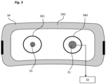
- the mechanical support comes in the form of a mask 34 to be applied around the head of the living being.
- the mask 34 carries two glasses 340, 341 intended to be placed respectively opposite the two eyes 20, 21 of the living being, one glass carrying the receiver 31 (detector or camera) and one glass carrying the light source 33 used to capture the gaze of the living being.
- the mask 34 comprises occultation means 342, used to create a channel isolated from any light pollution between the first eye 20 of the living being and the capture zone of the receiver 31 and between the light source 33 and the second eye 21 of the living being.
- This embodiment may have an adapter to allow the light source 33 and the receiver 31 to be interchanged, and thus to be able to observe the right eye or the left eye of the living being.
- both the transmitter and the sensor may be on the same eye, or even on both eyes.
- the distance between the light source and the point detector could be adjusted to adapt to the interocular distance of the patient.
- the pulsed signal from the optical stimulation device (for example detected electrically by induction on the laser power cable, or optically with a photodetector at the level of the skull, or digitally in RF) is then used as an external source to exacerbate the optical signal measured via the eye.
- the external source can also come from a second optical detector placed on the patient's skull, at the level of the pulsed light source, on the scalp. A detector measures the signal diffused via the scalp, this signal acting as a carrier to increase the signal-to-noise ratio of the detector observing the eye, as for synchronous detection.
Landscapes
- Health & Medical Sciences (AREA)
- Engineering & Computer Science (AREA)
- Biomedical Technology (AREA)
- Life Sciences & Earth Sciences (AREA)
- Pathology (AREA)
- Animal Behavior & Ethology (AREA)
- General Health & Medical Sciences (AREA)
- Public Health (AREA)
- Veterinary Medicine (AREA)
- Nuclear Medicine, Radiotherapy & Molecular Imaging (AREA)
- Radiology & Medical Imaging (AREA)
- Physics & Mathematics (AREA)
- Biophysics (AREA)
- General Physics & Mathematics (AREA)
- Spectroscopy & Molecular Physics (AREA)
- Heart & Thoracic Surgery (AREA)
- Medical Informatics (AREA)
- Molecular Biology (AREA)
- Surgery (AREA)
- Neurosurgery (AREA)
- Measurement Of The Respiration, Hearing Ability, Form, And Blood Characteristics Of Living Organisms (AREA)
- Investigating Or Analysing Materials By Optical Means (AREA)
- Measurement And Recording Of Electrical Phenomena And Electrical Characteristics Of The Living Body (AREA)
- Measuring And Recording Apparatus For Diagnosis (AREA)
Description
La présente invention se rapporte à un procédé de détermination de l'état de fonctionnement d'un implant lumineux. L'invention se rapporte également à un dispositif de diagnostic du fonctionnement de l'implant lumineux.The present invention relates to a method for determining the operating state of a light implant. The invention also relates to a device for diagnosing the operation of the light implant.
Récemment, il a été proposé de ralentir la progression d'une pathologie en utilisant la lumière (tout rayonnement électromagnétique qui va de l'ultraviolet à l'infrarouge lointain en passant par le visible). Ce principe est notamment employé sous la forme d'un implant cérébral, intracrânien, comprenant une source lumineuse chargée de venir éclairer une zone du cerveau en vue de traiter la pathologie. L'implant peut notamment se présenter sous la forme d'une fibre optique à l'extrémité de laquelle la lumière est diffusée.Recently, it has been proposed to slow the progression of a pathology by using light (any electromagnetic radiation ranging from ultraviolet to far infrared through visible light). This principle is notably used in the form of an intracranial cerebral implant, comprising a light source responsible for illuminating an area of the brain in order to treat the pathology. The implant can notably be in the form of an optical fiber at the end of which the light is diffused.
Les demandes de brevets
Cependant, une fois que ces sondes sont implantées, il est difficile de savoir si la lumière est bien diffusée en bout de sonde. En effet, la source lumineuse peut être défectueuse, la fibre optique peut être cassée ou mal implantée. Il en ressort que l'état de fonctionnement de l'implant est parfois difficile à connaître, sans avoir à le retirer pour démontage et vérification.However, once these probes are implanted, it is difficult to know whether the light is properly diffused at the tip of the probe. Indeed, the light source may be defective, the optical fiber may be broken or poorly implanted. As a result, the operating status of the implant is sometimes difficult to determine without having to remove it for disassembly and inspection.
Il existe donc un besoin de disposer d'une solution simple pour déterminer l'état de fonctionnement d'un implant lumineux intracrânien tel que décrit dans les documents de l'état de la technique.There is therefore a need for a simple solution for determining the operating status of an intracranial light implant as described in prior art documents.
Le but de l'invention est de proposer une solution technique permettant de remplir cet objectif.The aim of the invention is to propose a technical solution enabling this objective to be achieved.
Ce but est atteint par un procédé de détermination de l'état de fonctionnement d'un implant lumineux implanté dans le cerveau d'un être vivant, ledit implant lumineux comportant une source lumineuse chargée d'émettre de la lumière dans le cerveau de l'être vivant, ledit procédé utilisant un dispositif de diagnostic qui comporte un récepteur d'un signal lumineux transmis à travers un premier œil de l'être vivant et des moyens de détermination de l'état de fonctionnement de l'implant lumineux à partir du signal lumineux transmis reçu,This aim is achieved by a method for determining the operating state of a light implant implanted in the brain of a living being, said light implant comprising a light source responsible for emitting light into the brain of the living being, said method using a diagnostic device which comprises a receiver of a light signal transmitted through a first eye of the living being and means for determining the operating state of the light implant from the received transmitted light signal,
Ledit procédé comportant :
- Une étape de positionnement du récepteur du dispositif de diagnostic en vis-à-vis du premier œil de l'être vivant,
- Une étape de mesure d'un signal lumineux reçu par le récepteur lorsque l'implant lumineux est activé,
- Une étape de comparaison entre les données représentatives du signal lumineux reçu par le récepteur et au moins une donnée de référence,
- Une étape de détermination de l'état de fonctionnement de l'implant lumineux entre un état fonctionnel et un état non-fonctionnel en tenant compte du résultat de l'étape de comparaison.
- A step of positioning the receiver of the diagnostic device opposite the first eye of the living being,
- A step of measuring a light signal received by the receiver when the light implant is activated,
- A comparison step between the data representative of the light signal received by the receiver and at least one reference data,
- A step of determining the operating state of the light implant between a functional state and a non-functional state taking into account the result of the comparison step.
Selon une particularité, ladite donnée de référence est pré-mémorisée ou acquise lors d'une étape de calibration du dispositif de diagnostic.According to a particular feature, said reference data is pre-stored or acquired during a calibration step of the diagnostic device.
Selon une autre particularité, l'étape de calibration consiste en une étape de mesure d'un signal lumineux de référence, réalisée lorsque l'implant lumineux est désactivé.According to another feature, the calibration step consists of a step of measuring a reference light signal, carried out when the light implant is deactivated.
Selon une autre particularité, l'étape de détermination de l'état de fonctionnement de l'implant lumineux consiste à déterminer au moins un écart entre ladite donnée de référence et une donnée desdites données représentatives du signal lumineux reçu et à comparer ledit écart avec une valeur seuil prédéterminée.According to another particularity, the step of determining the operating state of the light implant consists of determining at least one difference between said reference data and one of said data representative of the light signal received and comparing said difference with a predetermined threshold value.
Selon une autre particularité, le procédé comporte une étape de positionnement d'une source de lumière ponctuelle en vis-à-vis d'un deuxième œil de l'être vivant et une étape d'activation de ladite source de lumière ponctuelle.According to another feature, the method comprises a step of positioning a point light source opposite a second eye of the living being and a step of activating said point light source.
L'invention concerne également un dispositif de diagnostic utilisé pour mettre en œuvre le procédé tel que défini ci-dessus, le dispositif comportant un support mécanique sur lequel est fixé ledit récepteur et des moyens de détermination de l'état de fonctionnement dudit implant lumineux, connectés audit récepteur et configurés pour traiter des données en provenance dudit récepteur.The invention also relates to a diagnostic device used to implement the method as defined above, the device comprising a mechanical support on which said receiver is fixed and means for determining the operating state of said light implant, connected to said receiver and configured to process data from said receiver.
Selon une réalisation particulière, le récepteur est une caméra.According to a particular embodiment, the receiver is a camera.
Selon une autre réalisation particulière, le récepteur comporte une ou plusieurs photodiodes.According to another particular embodiment, the receiver comprises one or more photodiodes.
Selon une particularité, le dispositif comporte une source de lumière ponctuelle fixée audit support et destinée à être arrangée en vis-à-vis d'un deuxième œil de l'être vivant, en parallèle du récepteur.According to a particular feature, the device comprises a point light source fixed to said support and intended to be arranged opposite a second eye of the living being, in parallel with the receiver.
Selon une réalisation particulière, le support mécanique comporte un plateau optique muni d'une mentonnière et sur lequel est fixé ledit récepteur.According to a particular embodiment, the mechanical support comprises an optical plate provided with a chin rest and on which said receiver is fixed.
Selon une autre réalisation particulière, le support mécanique comporte un masque à placer autour de la tête de l'être-vivant.According to another particular embodiment, the mechanical support includes a mask to be placed around the head of the living being.
Selon une particularité, le masque comporte deux lunettes, une première lunette accueillant ledit récepteur et une deuxième lunette accueillant une source de lumière ponctuelle.According to a particular feature, the mask comprises two glasses, a first glass receiving said receiver and a second glass receiving a point light source.
D'autres caractéristiques et avantages vont apparaître dans la description détaillée qui suit faite en regard des dessins annexés dans lesquels :
- La
figure 1 illustre le principe de fonctionnement d'un implant lumineux utilisé dans le cadre de l'invention et celui du dispositif de diagnostic de l'invention ; - La
figure 2 représente un premier exemple de réalisation du dispositif de diagnostic conforme à l'invention ; - La
figure 3 représente un deuxième exemple de réalisation du dispositif de diagnostic conforme à l'invention ;
- There
Figure 1 illustrates the operating principle of a light implant used in the context of the invention and that of the diagnostic device of the invention; - There
Figure 2 represents a first example of embodiment of the diagnostic device according to the invention; - There
Figure 3 represents a second example of embodiment of the diagnostic device according to the invention;
En référence à la
L'implant 1 utilise au moins une source lumineuse 10 et comporte une sonde 11 par laquelle la lumière est diffusée. La sonde 11 peut se présenter sous la forme d'une fibre optique chargée d'acheminer la lumière de la source lumineuse 10 jusqu'à son extrémité distale, située à proximité des tissus à traiter. Le principe de réalisation de l'implant peut être varié. Les demandes de brevets
Selon un aspect particulier de l'invention, le dispositif 3 de diagnostic comporte :
- Eventuellement un support mécanique ;
- Un
récepteur 31 d'un signal lumineux transmis à travers l'œil de l'être vivant, fixé audit support mécanique ; - Des
moyens 32 de détermination de l'état de fonctionnement de l'implant lumineux ;
- Possibly mechanical support;
- A
receiver 31 of a light signal transmitted through the eye of the living being, fixed to said mechanical support; - Means 32 for determining the operating state of the light implant;
L'objectif du dispositif 3 de diagnostic est de déterminer l'état de fonctionnement de l'implant lumineux 1 placé à l'intérieur du crâne de l'être vivant en détectant la présence ou l'absence d'un signal lumineux S transmis à travers l'œil 20 de l'être vivant. Le dispositif 3 de diagnostic présente la particularité d'être non invasif, c'est-à-dire qu'il est placé à l'extérieur du corps de l'être vivant.The objective of the
Le récepteur 31 est configuré pour collecter les photons émis par l'implant lumineux 1 et qui sont diffusés par les tissus et ressortent via le globe oculaire.The
Différentes variantes de réalisation de récepteur peuvent être envisagées :
- Un détecteur utilisant une ou plusieurs photodiodes,
- Une caméra très sensible.
- A detector using one or more photodiodes,
- A very sensitive camera.
Dans tous les cas, le récepteur 31 doit permettre de mesurer des puissances optiques très faibles (entre le femtoWatt et le nanoWatt) et avec des temps d'intégration suffisamment courts (de l'ordre de la minute ou moins) pour que l'examen soit compatible avec une mesure sur un être vivant.In all cases, the
De manière non limitative, le récepteur 31 peut notamment être un détecteur à comptage de photons multi-pixels commercial, basé sur des photodiodes à avalanche en mode Geiger, ou d'une photodiode silicium de large diamètre permettant un fort angle solide de détection. Ce type de détecteur ne fournit pas une image, mais une valeur ponctuelle ; cependant leur sensibilité peut être largement supérieure à celle d'une caméra, et ils sont plus compacts et plus rapides. Il est possible d'ajouter une lentille à large ouverture numérique devant le détecteur, pour collecter le plus de photons. Cette lentille joue alors le rôle de condenseur.In a non-limiting manner, the
Selon un aspect particulier de l'invention, l'observation doit être réalisée avec un minimum de lumière parasite. Autrement dit, l'observation se fait avantageusement dans des conditions d'obscurité totale pour éviter toute pollution lumineuse due à l'environnement. A titre d'exemple et de manière non limitative, le niveau de lumière ambiant doit être d'au moins un ordre de grandeur inférieur à celui du signal lumineux transmis à travers l'œil.According to a particular aspect of the invention, the observation must be carried out with a minimum of stray light. In other words, the observation is advantageously carried out in conditions of total darkness to avoid any light pollution due to the environment. By way of example and without limitation, the ambient light level must be at least one order of magnitude lower than that of the light signal transmitted through the eye.
Selon un aspect particulier de l'invention, les moyens 32 de détermination de l'état de fonctionnement de l'implant lumineux peuvent comporter un microprocesseur sur lequel vient se connecter le récepteur 31. Ils sont chargés d'interpréter les données représentatives des signaux reçus par le récepteur. Différents modes de traitement peuvent être envisagés pour déterminer l'état de fonctionnement de l'implant lumineux :
- Premier mode de traitement :
- Il peut s'agir de récupérer un premier signal lumineux de référence, implant lumineux désactivé, et de mémoriser une ou plusieurs données de référence représentatives de ce premier signal lumineux ; Cette mesure est avantageusement réalisée dans l'obscurité totale ;
- Ensuite, après activation de l'implant, on mesure un deuxième signal lumineux au niveau du récepteur 31 ;
- On compare une ou plusieurs des données représentatives du deuxième signal lumineux avec une ou plusieurs des données de référence correspondantes ;
- Deuxième mode de traitement :
- Une ou plusieurs données de référence sont pré-mémorisées en usine et correspondent à un état de référence dans lequel le système est placé dans l'obscurité totale ;
- Le reste du traitement est identique à celui décrit ci-dessus ;
- First treatment mode:
- This may involve recovering a first reference light signal, with the light implant deactivated, and storing one or more reference data representative of this first light signal; This measurement is advantageously carried out in total darkness;
- Then, after activation of the implant, a second light signal is measured at the
receiver 31; - One or more of the data representative of the second light signal is compared with one or more of the corresponding reference data;
- Second treatment mode:
- One or more reference data are pre-stored in the factory and correspond to a reference state in which the system is placed in total darkness;
- The rest of the treatment is the same as described above;
Par données représentatives d'un signal lumineux, on entend une intensité lumineuse maximale, par exemple prise au niveau d'un ou plusieurs pixels de l'image capturée (par la caméra), une moyenne de plusieurs intensités lumineuses mesurées... Toute autre donnée pourrait être envisagée.By data representative of a light signal, we mean a maximum light intensity, for example taken at the level of one or more pixels of the captured image (by the camera), an average of several measured light intensities... Any other data could be considered.
Si les données comparées sont identiques, les moyens 32 de détermination de l'état de fonctionnement d'un implant lumineux 1 peuvent en conclure que l'implant lumineux 1 est défectueux. Si une ou plusieurs des données issues du deuxième signal reçu diffèrent de celles représentatives du premier signal, les moyens 32 de détermination peuvent en conclure que l'implant lumineux 1 est fonctionnel. Il est possible de régler un seuil au-dessus duquel les moyens 32 de détermination de l'état de fonctionnement de l'implant déterminent qu'une ou plusieurs des données issues du deuxième signal diffèrent suffisamment de celles représentatives du premier signal.If the compared data are identical, the
De manière non limitative, un mode de traitement simple est le suivant :
- Une première image,
implant lumineux 1 intracérébral éteint, permet de vérifier les conditions d'obscurité : le signal ou l'image mesurée ne corresponde qu'au bruit du détecteur ou de la caméra ; Ensuite, - Une deuxième image,
implant lumineux 1 activé, permet, si l'implant lumineux 1 est parfaitement fonctionnel, de mettre en évidence la contribution au signal ou à l'image mesurée des photons issus de l'implant intracérébral et diffusés dans les tissus.
- A first image, with the intracerebral
light implant 1 switched off, allows the dark conditions to be checked: the signal or the measured image only corresponds to the noise of the detector or the camera; Then, - A second image,
light implant 1 activated, allows, iflight implant 1 is perfectly functional, to highlight the contribution to the signal or to the measured image of the photons coming from the intracerebral implant and diffused in the tissues.
D'autres modes de fonctionnement peuvent bien entendu être envisagés, selon les moyens disponibles et mis en œuvre.Other operating modes can of course be considered, depending on the means available and implemented.
Dans un premier mode de réalisation illustré par la
Avantageusement, sur ce plateau 30 optique, on viendra positionner une source de lumière 33 ponctuelle devant l'autre œil 21, pour faire en sorte que l'être vivant maintienne son regard immobile : l'être vivant fixe ce point lumineux avec un œil 21, et la caméra observe l'autre œil 20.Advantageously, on this
Dans le cadre de ce premier mode de réalisation, à titre d'exemple, on utilise un implant lumineux 1 intracérébral réalisé sous la forme d'une fibre optique implantée dans le cerveau 2, de sorte que l'extrémité de la fibre optique se situe entre les deux substances noires, au niveau du mésencéphale et donc à proximité du chiasma optique. L'implant lumineux 1 a par exemple une puissance crête de 15mW, la lumière étant pulsée avec un rapport cyclique de 8%. Une dilatation de la pupille n'est pas nécessaire. Le noir absolu est fait dans la pièce, et le patient se positionne sur la mentonnière du support.In this first embodiment, for example, an intracerebral
L'entraxe entre la source de lumière 33 fixe (pour maintenir le regard fixe) et le récepteur 31 (détecteur ou la caméra) est réglé pour correspondre à la distance interoculaire du patient. Dans un exemple particulier, la caméra est une caméra 16 bits, 2750x2200 pixels, binning 2, pixels de 4.54µm, refroidie à -12°C et positionné à environ 5 cm de l'œil. Le temps d'exposition est fixé à 20 secondes. Toutes les sources lumineuses environnantes sont occultées, et l'implant intracérébral est éteint. Comme déjà décrit ci-dessus, le mode de fonctionnement est par exemple le suivant :
Implant lumineux 1 désactivé, on acquière une première image qui doit être "noire", c'est-à-dire avec uniquement la contribution du bruit interne à la caméra.- On allume alors l'implant lumineux 1 et on répète l'opération qui met en évidence un spot diffus correspondant aux photons venant de l'implant lumineux 1 intracérébral.
- With
light implant 1 deactivated, a first image is acquired which must be "black", that is to say with only the contribution of internal noise to the camera. - We then turn on the
light implant 1 and repeat the operation which highlights a diffuse spot corresponding to the photons coming from the intracerebrallight implant 1.
Avantageusement, la source de lumière 33 permettant de fixer le regard émet à une certaine longueur d'onde (vert par exemple, 550nm) tandis que le récepteur 31 (caméra ou détecteur) observant l'autre œil 20 ne sont sensibles qu'à la longueur d'onde d'émission de l'implant lumineux 1 (par exemple au moyen d'un filtre passe haut qui ne laisserait passer que les longueurs d'onde supérieures à 600 nm).Advantageously, the
Selon une variante de réalisation représentée sur la
Le masque 34 porte deux lunettes 340, 341 destinées à être placées respectivement en vis-à-vis des deux yeux 20, 21 de l'être vivant, une lunette portant le récepteur 31 (détecteur ou caméra) et une lunette portant la source de lumière 33 utilisée pour capter le regard de l'être vivant. Le masque 34 comporte des moyens d'occultation 342, utilisés pour créer un canal isolé de toute pollution lumineuse entre le premier œil 20 de l'être vivant et la zone de capture du récepteur 31 et entre la source de lumière 33 et le deuxième œil 21 de l'être vivant.The
Cette réalisation peut disposer d'un adaptateur afin de pouvoir intervertir la source de lumière 33 et le récepteur 31, et ainsi pouvoir observer l'œil droit ou l'œil gauche de l'être vivant. Alternativement, on peut avoir sur un même œil, à la fois l'émetteur et le capteur, voire même sur les deux yeux. De plus, l'entraxe entre la source de lumière et le détecteur ponctuel pourrait être réglé afin de pouvoir s'adapter à la distance interoculaire du patient.This embodiment may have an adapter to allow the
Le principe de fonctionnement de ce deuxième mode de réalisation est identique à celui décrit ci-dessus pour le premier mode de réalisation.The operating principle of this second embodiment is identical to that described above for the first embodiment.
Avantageusement, il est possible de réaliser une détection synchrone, afin d'augmenter le rapport signal-bruit : le signal pulsé issu du dispositif de stimulation optique (par exemple détecté électriquement par induction sur le câble d'alimentation du laser, ou optiquement avec un photodétecteur au niveau du crâne, ou numériquement en RF) est alors utilisé comme source externe pour exacerber le signal optique mesuré via l'œil. La source externe peut également venir d'un second détecteur optique posé sur le crâne du patient, au niveau de la source de lumière pulsée, sur le cuir chevelu. Un détecteur mesure le signal diffusé via le cuir chevelu, ce signal jouant le rôle de porteuse pour augmenter le rapport signal à bruit du détecteur observant l'œil, comme pour une détection synchrone.Advantageously, it is possible to perform synchronous detection, in order to increase the signal-to-noise ratio: the pulsed signal from the optical stimulation device (for example detected electrically by induction on the laser power cable, or optically with a photodetector at the level of the skull, or digitally in RF) is then used as an external source to exacerbate the optical signal measured via the eye. The external source can also come from a second optical detector placed on the patient's skull, at the level of the pulsed light source, on the scalp. A detector measures the signal diffused via the scalp, this signal acting as a carrier to increase the signal-to-noise ratio of the detector observing the eye, as for synchronous detection.
L'invention présente ainsi de nombreux avantages, parmi lesquels :
- Une facilité de mise en œuvre ;
- Une solution non-invasive ;
- Une solution qui utilise des composant disponibles couramment ;
- Ease of implementation;
- A non-invasive solution;
- A solution that uses commonly available components;
Claims (12)
- Method for determining the operating state of a light-emitting implant (1) implanted in the brain (2) of a living being, said light-emitting implant (1) comprising a light source (10) responsible for emitting light into the brain of the living being, said method using a diagnosing device (3) that comprises a receiver (31) of a light signal transmitted through a first eye (20) of the living being and means for determining the operating state of the light-emitting implant (1) based on the received transmitted light signal,
characterized in that it comprises:- a step of positioning the receiver (31) of the diagnosing device facing the first eye (20) of the living being,- a step of measuring a light signal received by the receiver (31) when the light-emitting implant (1) is activated,- a step of comparing data representative of the light signal received by the receiver (31) and at least one reference datum,- a step of determining the operating state of the light-emitting implant (1) between a functional state and a non-functional state given the result of the comparing step. - Method according to Claim 1, characterized in that said reference datum is stored beforehand or acquired in a step of calibrating the diagnosing device (3).
- Method according to Claim 2, characterized in that the calibrating step consists in a step of measuring a reference light signal, carried out when the light-emitting implant (1) is deactivated.
- Method according to one of Claims 1 to 3, characterized in that the step of determining the operating state of the light-emitting implant (1) consists in determining at least one difference between said reference datum and a datum of said data representative of the received light signal and in comparing said difference with a predetermined threshold value.
- Method according to one of Claims 1 to 4, characterized in that it comprises a step of positioning a point light source (33) facing a second eye (21) of the living being and a step of activating said point light source (33).
- Diagnosing device (3) used to implement the method such as defined in one of Claims 1 to 5, characterized in that it comprises a mechanical mount (30, 34) to which said receiver (31) is fastened and means (32) for determining the operating state of said light-emitting implant (1), said means being connected to said receiver (31) and configured to process data delivered by said receiver.
- Device according to Claim 6, characterized in that the receiver (31) is a camera.
- Device according to Claim 6, characterized in that the receiver (31) comprises one or more photodiodes.
- Device according to one of Claims 6 to 8, characterized in that it comprises a point light source (33) fastened to said mount and intended to be arranged facing a second eye (21) of the living being, in parallel with the receiver (31).
- Diagnosing device according to Claim 6, characterized in that the mechanical mount comprises an optical platform (30) provided with a chin rest, and to which said receiver (31) is fastened.
- Device according to Claim 6, characterized in that the mechanical mount comprises a headset (34) to be placed around the head of the living being.
- Device according to Claim 11, characterized in that the headset (34) comprises two eyeglasses, a first eyeglass (340) accommodating said receiver (31) and a second eyeglass (341) accommodating a point light source (33).
Applications Claiming Priority (1)
| Application Number | Priority Date | Filing Date | Title |
|---|---|---|---|
| FR2209903A FR3140285B1 (en) | 2022-09-29 | 2022-09-29 | Method and determination of the operating state of a light implant |
Publications (2)
| Publication Number | Publication Date |
|---|---|
| EP4344729A1 EP4344729A1 (en) | 2024-04-03 |
| EP4344729B1 true EP4344729B1 (en) | 2025-06-25 |
Family
ID=84331143
Family Applications (1)
| Application Number | Title | Priority Date | Filing Date |
|---|---|---|---|
| EP23198137.4A Active EP4344729B1 (en) | 2022-09-29 | 2023-09-19 | Method and device for determining the operating state of a light implant |
Country Status (3)
| Country | Link |
|---|---|
| US (1) | US12498266B2 (en) |
| EP (1) | EP4344729B1 (en) |
| FR (1) | FR3140285B1 (en) |
Family Cites Families (16)
| Publication number | Priority date | Publication date | Assignee | Title |
|---|---|---|---|---|
| US8706243B2 (en) * | 2009-02-09 | 2014-04-22 | Rainbow Medical Ltd. | Retinal prosthesis techniques |
| JP2012095803A (en) * | 2010-11-01 | 2012-05-24 | Nara Institute Of Science & Technology | Living body light bidirectional information exchange system and control method of the system |
| US9004687B2 (en) * | 2012-05-18 | 2015-04-14 | Sync-Think, Inc. | Eye tracking headset and system for neuropsychological testing including the detection of brain damage |
| US10219696B2 (en) * | 2013-03-07 | 2019-03-05 | The Board Of Trustees Of The Leland Stanford Junior University | Implantable pressure sensors for telemetric measurements through bodily tissues |
| WO2014153428A1 (en) | 2013-03-19 | 2014-09-25 | Surgisense Corporation | Apparatus, systems and methods for determining tissue oxygenation |
| FR3036623B1 (en) | 2015-05-28 | 2017-05-19 | Commissariat Energie Atomique | DEVICE FOR DEEP ELECTRICAL AND OPTICAL STIMULATION OF THE BRAIN |
| FR3045821B1 (en) * | 2015-12-17 | 2018-11-23 | Commissariat A L'energie Atomique Et Aux Energies Alternatives | DEVICE FOR DETECTING A LEAK IN A HERMETIC ENCLOSURE |
| WO2018160711A1 (en) | 2017-02-28 | 2018-09-07 | Amo Wavefront Sciences, Llc | Method and system for pupil retro illumination using sample arm of oct interferometer |
| WO2019115909A1 (en) | 2017-12-11 | 2019-06-20 | Commissariat A L'Énergie Atomique Et Aux Energies Alternatives | Implantable localised illuminating device with improved architecture |
| JP7491218B2 (en) * | 2018-10-10 | 2024-05-28 | 株式会社ニデック | Ophthalmic device and ophthalmic device control program |
| ES2916713T3 (en) * | 2018-10-30 | 2022-07-05 | Inst De Fisica Daltes Energies | artificial vision system |
| DE102019114537A1 (en) * | 2019-05-29 | 2020-12-03 | OSRAM Opto Semiconductors Gesellschaft mit beschränkter Haftung | OPTOELECTRONIC SENSOR COMPONENT FOR LIGHT MEASUREMENT WITH BUILT-IN REDUNDANCY |
| EP4041374A4 (en) * | 2019-10-04 | 2024-01-24 | Nalu Medical, Inc. | STIMULATION DEVICE |
| FR3104449B1 (en) | 2019-12-12 | 2021-12-24 | Commissariat Energie Atomique | Illumination device implantable in a living being |
| FR3104448B1 (en) * | 2019-12-12 | 2022-08-26 | Commissariat Energie Atomique | Illumination device implantable in a living being |
| US11395620B1 (en) | 2021-06-03 | 2022-07-26 | Ofer Moshe | Methods and systems for transformation between eye images and digital images |
-
2022
- 2022-09-29 FR FR2209903A patent/FR3140285B1/en active Active
-
2023
- 2023-09-19 EP EP23198137.4A patent/EP4344729B1/en active Active
- 2023-09-28 US US18/476,487 patent/US12498266B2/en active Active
Also Published As
| Publication number | Publication date |
|---|---|
| FR3140285A1 (en) | 2024-04-05 |
| FR3140285B1 (en) | 2024-08-30 |
| US20240110828A1 (en) | 2024-04-04 |
| EP4344729A1 (en) | 2024-04-03 |
| US12498266B2 (en) | 2025-12-16 |
Similar Documents
| Publication | Publication Date | Title |
|---|---|---|
| EP2670294B1 (en) | Method and device for high-resolution retinal imaging | |
| EP2351518B1 (en) | Perioperative bi-spectral optical probe | |
| EP1875209B1 (en) | Fluorescence imaging device with two wavelength reflection | |
| FR3036187B1 (en) | METHOD OF CORRECTING A FLUORESCENCE IMAGE | |
| EP2309249B1 (en) | Device and method for diffused excitation in images | |
| FR2699677A1 (en) | Method and device for determining the color of a transparent, diffusing and absorbing object, such as in particular a tooth. | |
| US20150245768A1 (en) | Optical probe, optical measurement method, and optical measurement device | |
| EP3614904B1 (en) | System and method for multi-scale retinal imaging | |
| EP2555667B1 (en) | Optical system for following ocular movements and associated support device | |
| EP4344729B1 (en) | Method and device for determining the operating state of a light implant | |
| FR3087539A1 (en) | MEASURING INSTRUMENT WITH MEASUREMENT SPOT VISUALIZATION SYSTEM AND VISUALIZATION ACCESSORY FOR SUCH MEASURING INSTRUMENT | |
| FR3032527A1 (en) | DEVICE FOR MEASURING AN OPTICAL SIGNAL RETRODUCED BY A SAMPLE | |
| WO2007118954A1 (en) | Methylene blue based fibred fluorescence microscopy | |
| EP2677920B1 (en) | Method and device for high-resolution retinal imaging | |
| FR2612391A1 (en) | EYE OBSERVATION DEVICE USING INFRARED REFLECTION ON THE EYE GLOBE | |
| FR3126318A1 (en) | Implantable brain illumination device with optical probe monitoring solution | |
| CA3184950A1 (en) | Device for determining the level of haemoglobin or haematocrit of a circulating liquid | |
| FR3106740A1 (en) | Methods and systems for monitoring eye alignment in an ophthalmic imaging machine | |
| FR2708735A1 (en) | Device making it possible to evaluate the effects of light on the skin and its application to detection of pathologies of the skin | |
| FR3153981A1 (en) | Eye data acquisition device and assembly for eye data acquisition | |
| WO2019207253A1 (en) | System for laser photocoagulation of the retina | |
| EP4322830A1 (en) | Device and method for imaging mobile targets | |
| FR3095519A1 (en) | Non-invasive method of determining the gender of an egg | |
| FR3000660A1 (en) | Display device for detection and intraoperative visualization of component within drainage ganglia, has detection unit detecting part of beam output from radiation, and nuclear radiation detection unit detecting another type of radiation |
Legal Events
| Date | Code | Title | Description |
|---|---|---|---|
| PUAI | Public reference made under article 153(3) epc to a published international application that has entered the european phase |
Free format text: ORIGINAL CODE: 0009012 |
|
| STAA | Information on the status of an ep patent application or granted ep patent |
Free format text: STATUS: REQUEST FOR EXAMINATION WAS MADE |
|
| 17P | Request for examination filed |
Effective date: 20230919 |
|
| AK | Designated contracting states |
Kind code of ref document: A1 Designated state(s): AL AT BE BG CH CY CZ DE DK EE ES FI FR GB GR HR HU IE IS IT LI LT LU LV MC ME MK MT NL NO PL PT RO RS SE SI SK SM TR |
|
| RAP3 | Party data changed (applicant data changed or rights of an application transferred) |
Owner name: UNIVERSITE GRENOBLE ALPES Owner name: CENTRE HOSPITALIER UNIVERSITAIRE GRENOBLE ALPES Owner name: COMMISSARIAT A L'ENERGIE ATOMIQUE ET AUX ENERGIESALTERNATIVES |
|
| GRAP | Despatch of communication of intention to grant a patent |
Free format text: ORIGINAL CODE: EPIDOSNIGR1 |
|
| STAA | Information on the status of an ep patent application or granted ep patent |
Free format text: STATUS: GRANT OF PATENT IS INTENDED |
|
| RIC1 | Information provided on ipc code assigned before grant |
Ipc: A61B 5/00 20060101ALI20250203BHEP Ipc: A61N 5/06 20060101AFI20250203BHEP |
|
| INTG | Intention to grant announced |
Effective date: 20250213 |
|
| GRAS | Grant fee paid |
Free format text: ORIGINAL CODE: EPIDOSNIGR3 |
|
| GRAA | (expected) grant |
Free format text: ORIGINAL CODE: 0009210 |
|
| STAA | Information on the status of an ep patent application or granted ep patent |
Free format text: STATUS: THE PATENT HAS BEEN GRANTED |
|
| AK | Designated contracting states |
Kind code of ref document: B1 Designated state(s): AL AT BE BG CH CY CZ DE DK EE ES FI FR GB GR HR HU IE IS IT LI LT LU LV MC ME MK MT NL NO PL PT RO RS SE SI SK SM TR |
|
| REG | Reference to a national code |
Ref country code: GB Ref legal event code: FG4D Free format text: NOT ENGLISH |
|
| REG | Reference to a national code |
Ref country code: CH Ref legal event code: EP |
|
| REG | Reference to a national code |
Ref country code: DE Ref legal event code: R096 Ref document number: 602023004272 Country of ref document: DE |
|
| REG | Reference to a national code |
Ref country code: CH Ref legal event code: EP |
|
| REG | Reference to a national code |
Ref country code: IE Ref legal event code: FG4D Free format text: LANGUAGE OF EP DOCUMENT: FRENCH |
|
| PG25 | Lapsed in a contracting state [announced via postgrant information from national office to epo] |
Ref country code: FI Free format text: LAPSE BECAUSE OF FAILURE TO SUBMIT A TRANSLATION OF THE DESCRIPTION OR TO PAY THE FEE WITHIN THE PRESCRIBED TIME-LIMIT Effective date: 20250625 |
|
| PGFP | Annual fee paid to national office [announced via postgrant information from national office to epo] |
Ref country code: DE Payment date: 20250919 Year of fee payment: 3 |
|
| REG | Reference to a national code |
Ref country code: LT Ref legal event code: MG9D |
|
| PG25 | Lapsed in a contracting state [announced via postgrant information from national office to epo] |
Ref country code: NO Free format text: LAPSE BECAUSE OF FAILURE TO SUBMIT A TRANSLATION OF THE DESCRIPTION OR TO PAY THE FEE WITHIN THE PRESCRIBED TIME-LIMIT Effective date: 20250925 Ref country code: GR Free format text: LAPSE BECAUSE OF FAILURE TO SUBMIT A TRANSLATION OF THE DESCRIPTION OR TO PAY THE FEE WITHIN THE PRESCRIBED TIME-LIMIT Effective date: 20250926 |
|
| PG25 | Lapsed in a contracting state [announced via postgrant information from national office to epo] |
Ref country code: BG Free format text: LAPSE BECAUSE OF FAILURE TO SUBMIT A TRANSLATION OF THE DESCRIPTION OR TO PAY THE FEE WITHIN THE PRESCRIBED TIME-LIMIT Effective date: 20250625 |
|
| PG25 | Lapsed in a contracting state [announced via postgrant information from national office to epo] |
Ref country code: HR Free format text: LAPSE BECAUSE OF FAILURE TO SUBMIT A TRANSLATION OF THE DESCRIPTION OR TO PAY THE FEE WITHIN THE PRESCRIBED TIME-LIMIT Effective date: 20250625 |
|
| PGFP | Annual fee paid to national office [announced via postgrant information from national office to epo] |
Ref country code: AT Payment date: 20251020 Year of fee payment: 3 Ref country code: FR Payment date: 20250917 Year of fee payment: 3 |
|
| PG25 | Lapsed in a contracting state [announced via postgrant information from national office to epo] |
Ref country code: RS Free format text: LAPSE BECAUSE OF FAILURE TO SUBMIT A TRANSLATION OF THE DESCRIPTION OR TO PAY THE FEE WITHIN THE PRESCRIBED TIME-LIMIT Effective date: 20250925 |
|
| PG25 | Lapsed in a contracting state [announced via postgrant information from national office to epo] |
Ref country code: LV Free format text: LAPSE BECAUSE OF FAILURE TO SUBMIT A TRANSLATION OF THE DESCRIPTION OR TO PAY THE FEE WITHIN THE PRESCRIBED TIME-LIMIT Effective date: 20250625 |
|
| REG | Reference to a national code |
Ref country code: NL Ref legal event code: MP Effective date: 20250625 |
|
| PG25 | Lapsed in a contracting state [announced via postgrant information from national office to epo] |
Ref country code: NL Free format text: LAPSE BECAUSE OF FAILURE TO SUBMIT A TRANSLATION OF THE DESCRIPTION OR TO PAY THE FEE WITHIN THE PRESCRIBED TIME-LIMIT Effective date: 20250625 |
|
| PG25 | Lapsed in a contracting state [announced via postgrant information from national office to epo] |
Ref country code: PT Free format text: LAPSE BECAUSE OF FAILURE TO SUBMIT A TRANSLATION OF THE DESCRIPTION OR TO PAY THE FEE WITHIN THE PRESCRIBED TIME-LIMIT Effective date: 20251027 |
|
| REG | Reference to a national code |
Ref country code: AT Ref legal event code: MK05 Ref document number: 1805871 Country of ref document: AT Kind code of ref document: T Effective date: 20250625 |
|
| PG25 | Lapsed in a contracting state [announced via postgrant information from national office to epo] |
Ref country code: IS Free format text: LAPSE BECAUSE OF FAILURE TO SUBMIT A TRANSLATION OF THE DESCRIPTION OR TO PAY THE FEE WITHIN THE PRESCRIBED TIME-LIMIT Effective date: 20251025 |
|
| PG25 | Lapsed in a contracting state [announced via postgrant information from national office to epo] |
Ref country code: AT Free format text: LAPSE BECAUSE OF FAILURE TO SUBMIT A TRANSLATION OF THE DESCRIPTION OR TO PAY THE FEE WITHIN THE PRESCRIBED TIME-LIMIT Effective date: 20250625 Ref country code: SM Free format text: LAPSE BECAUSE OF FAILURE TO SUBMIT A TRANSLATION OF THE DESCRIPTION OR TO PAY THE FEE WITHIN THE PRESCRIBED TIME-LIMIT Effective date: 20250625 |
|
| PG25 | Lapsed in a contracting state [announced via postgrant information from national office to epo] |
Ref country code: CZ Free format text: LAPSE BECAUSE OF FAILURE TO SUBMIT A TRANSLATION OF THE DESCRIPTION OR TO PAY THE FEE WITHIN THE PRESCRIBED TIME-LIMIT Effective date: 20250625 |
|
| PG25 | Lapsed in a contracting state [announced via postgrant information from national office to epo] |
Ref country code: PL Free format text: LAPSE BECAUSE OF FAILURE TO SUBMIT A TRANSLATION OF THE DESCRIPTION OR TO PAY THE FEE WITHIN THE PRESCRIBED TIME-LIMIT Effective date: 20250625 |
|
| PG25 | Lapsed in a contracting state [announced via postgrant information from national office to epo] |
Ref country code: EE Free format text: LAPSE BECAUSE OF FAILURE TO SUBMIT A TRANSLATION OF THE DESCRIPTION OR TO PAY THE FEE WITHIN THE PRESCRIBED TIME-LIMIT Effective date: 20250625 |
|
| PG25 | Lapsed in a contracting state [announced via postgrant information from national office to epo] |
Ref country code: SK Free format text: LAPSE BECAUSE OF FAILURE TO SUBMIT A TRANSLATION OF THE DESCRIPTION OR TO PAY THE FEE WITHIN THE PRESCRIBED TIME-LIMIT Effective date: 20250625 |
|
| PG25 | Lapsed in a contracting state [announced via postgrant information from national office to epo] |
Ref country code: ES Free format text: LAPSE BECAUSE OF FAILURE TO SUBMIT A TRANSLATION OF THE DESCRIPTION OR TO PAY THE FEE WITHIN THE PRESCRIBED TIME-LIMIT Effective date: 20250625 |
|
| PG25 | Lapsed in a contracting state [announced via postgrant information from national office to epo] |
Ref country code: RO Free format text: LAPSE BECAUSE OF FAILURE TO SUBMIT A TRANSLATION OF THE DESCRIPTION OR TO PAY THE FEE WITHIN THE PRESCRIBED TIME-LIMIT Effective date: 20250625 |