EP4335488A1 - Multimodale vorrichtung und verfahren zur erhöhung der wirksamkeit von transthorakaler kardioversion oder herzstimulation bei patienten mit perfusionsrhythmen - Google Patents
Multimodale vorrichtung und verfahren zur erhöhung der wirksamkeit von transthorakaler kardioversion oder herzstimulation bei patienten mit perfusionsrhythmen Download PDFInfo
- Publication number
- EP4335488A1 EP4335488A1 EP23195427.2A EP23195427A EP4335488A1 EP 4335488 A1 EP4335488 A1 EP 4335488A1 EP 23195427 A EP23195427 A EP 23195427A EP 4335488 A1 EP4335488 A1 EP 4335488A1
- Authority
- EP
- European Patent Office
- Prior art keywords
- subsystem
- tcp
- cetees
- control module
- countershock
- Prior art date
- Legal status (The legal status is an assumption and is not a legal conclusion. Google has not performed a legal analysis and makes no representation as to the accuracy of the status listed.)
- Pending
Links
- 230000000747 cardiac effect Effects 0.000 title claims abstract description 35
- 238000000034 method Methods 0.000 title abstract description 68
- 238000013194 cardioversion Methods 0.000 title abstract description 58
- 230000033764 rhythmic process Effects 0.000 title description 10
- 239000000090 biomarker Substances 0.000 claims abstract description 72
- 239000003623 enhancer Substances 0.000 claims abstract description 47
- 206010003119 arrhythmia Diseases 0.000 claims abstract description 30
- 238000002560 therapeutic procedure Methods 0.000 claims abstract description 21
- 230000000638 stimulation Effects 0.000 claims description 44
- 210000003205 muscle Anatomy 0.000 claims description 34
- 210000001186 vagus nerve Anatomy 0.000 claims description 33
- 239000012636 effector Substances 0.000 claims description 21
- 230000002708 enhancing effect Effects 0.000 claims description 17
- 230000007383 nerve stimulation Effects 0.000 claims description 12
- 230000000004 hemodynamic effect Effects 0.000 claims description 8
- 230000001154 acute effect Effects 0.000 claims description 7
- 230000006835 compression Effects 0.000 claims description 6
- 238000007906 compression Methods 0.000 claims description 6
- 210000003489 abdominal muscle Anatomy 0.000 claims description 4
- 230000001419 dependent effect Effects 0.000 claims 2
- 230000000717 retained effect Effects 0.000 claims 2
- 230000002685 pulmonary effect Effects 0.000 claims 1
- 230000004936 stimulating effect Effects 0.000 claims 1
- 230000001965 increasing effect Effects 0.000 abstract description 20
- 230000004044 response Effects 0.000 abstract description 17
- 208000010496 Heart Arrest Diseases 0.000 abstract description 4
- 238000005259 measurement Methods 0.000 description 51
- 238000005457 optimization Methods 0.000 description 49
- 210000004165 myocardium Anatomy 0.000 description 47
- 238000011282 treatment Methods 0.000 description 45
- 210000000038 chest Anatomy 0.000 description 43
- 238000002604 ultrasonography Methods 0.000 description 42
- 210000004072 lung Anatomy 0.000 description 35
- 230000035939 shock Effects 0.000 description 27
- 238000009423 ventilation Methods 0.000 description 27
- 230000003519 ventilatory effect Effects 0.000 description 26
- 230000007384 vagal nerve stimulation Effects 0.000 description 25
- 230000007246 mechanism Effects 0.000 description 24
- 238000004422 calculation algorithm Methods 0.000 description 23
- 206010003658 Atrial Fibrillation Diseases 0.000 description 22
- 210000000115 thoracic cavity Anatomy 0.000 description 22
- 239000000853 adhesive Substances 0.000 description 20
- 230000006870 function Effects 0.000 description 18
- 238000010801 machine learning Methods 0.000 description 18
- 230000001515 vagal effect Effects 0.000 description 18
- 230000000670 limiting effect Effects 0.000 description 17
- 230000000694 effects Effects 0.000 description 16
- 230000037361 pathway Effects 0.000 description 15
- 230000008569 process Effects 0.000 description 14
- 238000004611 spectroscopical analysis Methods 0.000 description 14
- 230000003247 decreasing effect Effects 0.000 description 13
- 102100026827 Protein associated with UVRAG as autophagy enhancer Human genes 0.000 description 12
- 101710102978 Protein associated with UVRAG as autophagy enhancer Proteins 0.000 description 12
- 230000002107 myocardial effect Effects 0.000 description 10
- 210000005036 nerve Anatomy 0.000 description 10
- 238000013195 electrical cardioversion Methods 0.000 description 9
- 238000010586 diagram Methods 0.000 description 8
- 230000001070 adhesive effect Effects 0.000 description 7
- 230000001276 controlling effect Effects 0.000 description 7
- 230000006872 improvement Effects 0.000 description 6
- 239000012212 insulator Substances 0.000 description 6
- 230000029058 respiratory gaseous exchange Effects 0.000 description 6
- 230000001360 synchronised effect Effects 0.000 description 6
- 230000002123 temporal effect Effects 0.000 description 6
- 210000001519 tissue Anatomy 0.000 description 6
- 206010039897 Sedation Diseases 0.000 description 5
- 210000000056 organ Anatomy 0.000 description 5
- 238000013186 photoplethysmography Methods 0.000 description 5
- 230000036280 sedation Effects 0.000 description 5
- 238000001228 spectrum Methods 0.000 description 5
- 230000003044 adaptive effect Effects 0.000 description 4
- 208000037265 diseases, disorders, signs and symptoms Diseases 0.000 description 4
- 239000003814 drug Substances 0.000 description 4
- 230000009977 dual effect Effects 0.000 description 4
- 238000010348 incorporation Methods 0.000 description 4
- 230000010354 integration Effects 0.000 description 4
- 239000000758 substrate Substances 0.000 description 4
- 241000282414 Homo sapiens Species 0.000 description 3
- 230000002159 abnormal effect Effects 0.000 description 3
- 230000004913 activation Effects 0.000 description 3
- 230000004075 alteration Effects 0.000 description 3
- 230000008901 benefit Effects 0.000 description 3
- 230000004087 circulation Effects 0.000 description 3
- 238000011161 development Methods 0.000 description 3
- 208000035475 disorder Diseases 0.000 description 3
- 238000009826 distribution Methods 0.000 description 3
- 229940079593 drug Drugs 0.000 description 3
- 210000003238 esophagus Anatomy 0.000 description 3
- 238000003384 imaging method Methods 0.000 description 3
- 238000000126 in silico method Methods 0.000 description 3
- 230000004048 modification Effects 0.000 description 3
- 238000012986 modification Methods 0.000 description 3
- 238000012544 monitoring process Methods 0.000 description 3
- 238000001320 near-infrared absorption spectroscopy Methods 0.000 description 3
- 230000036961 partial effect Effects 0.000 description 3
- 230000000144 pharmacologic effect Effects 0.000 description 3
- 210000001139 rectus abdominis Anatomy 0.000 description 3
- 210000003019 respiratory muscle Anatomy 0.000 description 3
- 230000003595 spectral effect Effects 0.000 description 3
- 230000000287 tissue oxygenation Effects 0.000 description 3
- 230000007704 transition Effects 0.000 description 3
- 206010003662 Atrial flutter Diseases 0.000 description 2
- 206010029458 Nodal arrhythmia Diseases 0.000 description 2
- 241000746998 Tragus Species 0.000 description 2
- 230000003187 abdominal effect Effects 0.000 description 2
- 238000007792 addition Methods 0.000 description 2
- 238000004458 analytical method Methods 0.000 description 2
- 210000003484 anatomy Anatomy 0.000 description 2
- 230000001746 atrial effect Effects 0.000 description 2
- 206010003668 atrial tachycardia Diseases 0.000 description 2
- 239000003990 capacitor Substances 0.000 description 2
- 238000001514 detection method Methods 0.000 description 2
- 230000006866 deterioration Effects 0.000 description 2
- 230000002526 effect on cardiovascular system Effects 0.000 description 2
- 239000004744 fabric Substances 0.000 description 2
- 230000008713 feedback mechanism Effects 0.000 description 2
- 238000002847 impedance measurement Methods 0.000 description 2
- 238000000338 in vitro Methods 0.000 description 2
- 230000000977 initiatory effect Effects 0.000 description 2
- 230000003993 interaction Effects 0.000 description 2
- 238000012804 iterative process Methods 0.000 description 2
- 238000012417 linear regression Methods 0.000 description 2
- 238000007477 logistic regression Methods 0.000 description 2
- 239000000463 material Substances 0.000 description 2
- 238000005399 mechanical ventilation Methods 0.000 description 2
- 230000003287 optical effect Effects 0.000 description 2
- 230000008520 organization Effects 0.000 description 2
- 230000036407 pain Effects 0.000 description 2
- 238000012545 processing Methods 0.000 description 2
- 230000000241 respiratory effect Effects 0.000 description 2
- 239000000932 sedative agent Substances 0.000 description 2
- 230000001624 sedative effect Effects 0.000 description 2
- 230000001953 sensory effect Effects 0.000 description 2
- 238000004513 sizing Methods 0.000 description 2
- 238000003860 storage Methods 0.000 description 2
- 230000001988 toxicity Effects 0.000 description 2
- 231100000419 toxicity Toxicity 0.000 description 2
- 208000021816 ventricular bradycardia Diseases 0.000 description 2
- 206010047302 ventricular tachycardia Diseases 0.000 description 2
- 210000001835 viscera Anatomy 0.000 description 2
- 230000003245 working effect Effects 0.000 description 2
- 208000033986 Device capturing issue Diseases 0.000 description 1
- 208000033988 Device pacing issue Diseases 0.000 description 1
- 208000001953 Hypotension Diseases 0.000 description 1
- 244000141359 Malus pumila Species 0.000 description 1
- 229910003798 SPO2 Inorganic materials 0.000 description 1
- 101100478210 Schizosaccharomyces pombe (strain 972 / ATCC 24843) spo2 gene Proteins 0.000 description 1
- 208000018452 Torsade de pointes Diseases 0.000 description 1
- 208000002363 Torsades de Pointes Diseases 0.000 description 1
- 208000027418 Wounds and injury Diseases 0.000 description 1
- 238000002679 ablation Methods 0.000 description 1
- 230000003288 anthiarrhythmic effect Effects 0.000 description 1
- 239000003416 antiarrhythmic agent Substances 0.000 description 1
- 235000021016 apples Nutrition 0.000 description 1
- 238000003491 array Methods 0.000 description 1
- 230000036772 blood pressure Effects 0.000 description 1
- 210000000988 bone and bone Anatomy 0.000 description 1
- 210000000748 cardiovascular system Anatomy 0.000 description 1
- 230000008859 change Effects 0.000 description 1
- 238000004590 computer program Methods 0.000 description 1
- 238000005094 computer simulation Methods 0.000 description 1
- 230000002596 correlated effect Effects 0.000 description 1
- 239000013078 crystal Substances 0.000 description 1
- 230000006378 damage Effects 0.000 description 1
- 238000007418 data mining Methods 0.000 description 1
- 238000013135 deep learning Methods 0.000 description 1
- 238000009795 derivation Methods 0.000 description 1
- 238000012774 diagnostic algorithm Methods 0.000 description 1
- 238000002405 diagnostic procedure Methods 0.000 description 1
- 201000010099 disease Diseases 0.000 description 1
- 238000001647 drug administration Methods 0.000 description 1
- 238000001827 electrotherapy Methods 0.000 description 1
- 238000005516 engineering process Methods 0.000 description 1
- 230000002349 favourable effect Effects 0.000 description 1
- 238000002695 general anesthesia Methods 0.000 description 1
- 230000010247 heart contraction Effects 0.000 description 1
- 230000036571 hydration Effects 0.000 description 1
- 238000006703 hydration reaction Methods 0.000 description 1
- 208000021822 hypotensive Diseases 0.000 description 1
- 230000001077 hypotensive effect Effects 0.000 description 1
- 230000002401 inhibitory effect Effects 0.000 description 1
- 208000014674 injury Diseases 0.000 description 1
- 238000007726 management method Methods 0.000 description 1
- 238000013178 mathematical model Methods 0.000 description 1
- 238000010297 mechanical methods and process Methods 0.000 description 1
- 230000005226 mechanical processes and functions Effects 0.000 description 1
- 238000005192 partition Methods 0.000 description 1
- 230000001575 pathological effect Effects 0.000 description 1
- 230000008375 physiological alteration Effects 0.000 description 1
- 230000035479 physiological effects, processes and functions Effects 0.000 description 1
- 238000003825 pressing Methods 0.000 description 1
- 230000001536 pro-arrhythmogenic effect Effects 0.000 description 1
- 230000007420 reactivation Effects 0.000 description 1
- 230000002787 reinforcement Effects 0.000 description 1
- 230000004202 respiratory function Effects 0.000 description 1
- 239000004065 semiconductor Substances 0.000 description 1
- 238000012163 sequencing technique Methods 0.000 description 1
- 238000009097 single-agent therapy Methods 0.000 description 1
- 230000000087 stabilizing effect Effects 0.000 description 1
- 238000007619 statistical method Methods 0.000 description 1
- 210000001562 sternum Anatomy 0.000 description 1
- 238000012360 testing method Methods 0.000 description 1
- 238000003325 tomography Methods 0.000 description 1
- 238000012549 training Methods 0.000 description 1
- 238000011277 treatment modality Methods 0.000 description 1
- 238000010200 validation analysis Methods 0.000 description 1
Images


Classifications
-
- A—HUMAN NECESSITIES
- A61—MEDICAL OR VETERINARY SCIENCE; HYGIENE
- A61N—ELECTROTHERAPY; MAGNETOTHERAPY; RADIATION THERAPY; ULTRASOUND THERAPY
- A61N1/00—Electrotherapy; Circuits therefor
- A61N1/18—Applying electric currents by contact electrodes
- A61N1/32—Applying electric currents by contact electrodes alternating or intermittent currents
- A61N1/36—Applying electric currents by contact electrodes alternating or intermittent currents for stimulation
- A61N1/362—Heart stimulators
- A61N1/3625—External stimulators
-
- A—HUMAN NECESSITIES
- A61—MEDICAL OR VETERINARY SCIENCE; HYGIENE
- A61N—ELECTROTHERAPY; MAGNETOTHERAPY; RADIATION THERAPY; ULTRASOUND THERAPY
- A61N1/00—Electrotherapy; Circuits therefor
- A61N1/18—Applying electric currents by contact electrodes
- A61N1/32—Applying electric currents by contact electrodes alternating or intermittent currents
- A61N1/36—Applying electric currents by contact electrodes alternating or intermittent currents for stimulation
- A61N1/362—Heart stimulators
- A61N1/365—Heart stimulators controlled by a physiological parameter, e.g. heart potential
- A61N1/36585—Heart stimulators controlled by a physiological parameter, e.g. heart potential controlled by two or more physical parameters
-
- A—HUMAN NECESSITIES
- A61—MEDICAL OR VETERINARY SCIENCE; HYGIENE
- A61N—ELECTROTHERAPY; MAGNETOTHERAPY; RADIATION THERAPY; ULTRASOUND THERAPY
- A61N1/00—Electrotherapy; Circuits therefor
- A61N1/18—Applying electric currents by contact electrodes
- A61N1/32—Applying electric currents by contact electrodes alternating or intermittent currents
- A61N1/38—Applying electric currents by contact electrodes alternating or intermittent currents for producing shock effects
- A61N1/39—Heart defibrillators
- A61N1/3904—External heart defibrillators [EHD]
- A61N1/39044—External heart defibrillators [EHD] in combination with cardiopulmonary resuscitation [CPR] therapy
-
- A—HUMAN NECESSITIES
- A61—MEDICAL OR VETERINARY SCIENCE; HYGIENE
- A61N—ELECTROTHERAPY; MAGNETOTHERAPY; RADIATION THERAPY; ULTRASOUND THERAPY
- A61N1/00—Electrotherapy; Circuits therefor
- A61N1/18—Applying electric currents by contact electrodes
- A61N1/32—Applying electric currents by contact electrodes alternating or intermittent currents
- A61N1/38—Applying electric currents by contact electrodes alternating or intermittent currents for producing shock effects
- A61N1/39—Heart defibrillators
- A61N1/395—Heart defibrillators for treating atrial fibrillation
-
- A—HUMAN NECESSITIES
- A61—MEDICAL OR VETERINARY SCIENCE; HYGIENE
- A61N—ELECTROTHERAPY; MAGNETOTHERAPY; RADIATION THERAPY; ULTRASOUND THERAPY
- A61N1/00—Electrotherapy; Circuits therefor
- A61N1/18—Applying electric currents by contact electrodes
- A61N1/32—Applying electric currents by contact electrodes alternating or intermittent currents
- A61N1/38—Applying electric currents by contact electrodes alternating or intermittent currents for producing shock effects
- A61N1/39—Heart defibrillators
- A61N1/3987—Heart defibrillators characterised by the timing or triggering of the shock
-
- A—HUMAN NECESSITIES
- A61—MEDICAL OR VETERINARY SCIENCE; HYGIENE
- A61N—ELECTROTHERAPY; MAGNETOTHERAPY; RADIATION THERAPY; ULTRASOUND THERAPY
- A61N1/00—Electrotherapy; Circuits therefor
- A61N1/18—Applying electric currents by contact electrodes
- A61N1/32—Applying electric currents by contact electrodes alternating or intermittent currents
- A61N1/38—Applying electric currents by contact electrodes alternating or intermittent currents for producing shock effects
- A61N1/39—Heart defibrillators
- A61N1/3968—Constructional arrangements, e.g. casings
Definitions
- the invention disclosed here relates in general to the field of medical devices.
- devices and methods for improving the clinical outcome of patients suffering from cardiac dysrhythmias without cardiac arrest are known.
- Atrial fibrillation Among the most common is atrial fibrillation. Others include: atrial tachycardia, atrial flutter, ventricular tachycardia, and atrial, junctional or ventricular bradycardia, among others. In atrial fibrillation, atrial flutter, atrial tachycardia, and ventricular tachycardia, the heart rate may be too fast. In atrial, junctional, or ventricular bradycardia, it may be too slow. Ultimately, patients suffering these dysrhythmias may need an electrophysiologic procedure such as ablation or placement of a permanent pacemaker.
- transthoracic and transcutaneous may be used interchangeably, and have a similar meaning, specifically the application of electromagnetic energy to myocardium by means of transcutaneous transthoracic countershock or pacing from one or more cutaneous electrode locations.
- countershock it is intended to mean the transthoracic application of electrical energy with the intention of terminating a dysrhythmia such that a more normal cardiac rhythm is restored. Termination of atrial fibrillation has been called defibrillation.
- cardioversion refers to an electrical shock being applied to the heart.
- a device that is intended to enhance the probability that electrical cardioversion has occurred successfully and the heart has been changed from a certain state of dysrhythmia to a more organized rhythm is best described as a cardioverter.
- pacer pacemaker
- pacemaker are used interchangeably.
- Both cardioversion and pacing are forms of Acute Electrical Therapy (AET), and as used herein, the terms electrical therapy, acute electrical therapy, and countershock can refer to cardioversion and/or pacing. Both cardioversion and pacing can work by delivering an electric shock through the thorax of a patient, also referred to as transthoracic cardioversion or pacing. Either form of acute electrical therapy can be delivered by a device that can be referred to as a Transthoracic Cardioverter Pacer (TCP).
- TCP Transthoracic Cardioverter Pacer
- a TCP can be a cardioverter that delivers electrical therapy in the form of cardioversion.
- a TCP can be a pacer that electrical therapy in the form of pacing.
- a TCP can provide cardioversion and/or pacing in different situations.
- the term TCP can refer to a pacer, a cardioverter, or a combination pacer and/or cardioverter that can function as a pacer or a cardioverter in different situations.
- ECG electrocardiogram
- transthoracic resistance when using alternating current is transthoracic impedance, and measurement of transthoracic impedance with a small current may be used as a surrogate indicator of transthoracic resistance.
- the performance of countershock can be improved by using an anterior-posterior vector.
- an anterior-posterior vector Kirchhof et al. 2002.
- one paddle is placed in the front of the patient, and another is placed posteriorly between the shoulder blades.
- the efficacy of electrical countershock or pacing may be improved by application of multiple transthoracic pathways that are electrified near simultaneously or sequentially, as described in U.S. Patent No. 9,174,061 to Freeman , titled MULTI-PATH TRANSTHORACIC DEFIBRILLATION AND CARDIOVERSION, the entire teachings of which are incorporated herein by reference.
- Ventilatory Cycle - Air is an insulator, so the ventilatory cycle of the lungs may affect electrical countershock by means of affecting transthoracic resistance. It has been found that an association exists between the ventilatory cycle and transthoracic impedance. (Ewy et al. 1980). An association has been found between the ventilatory cycle and transthoracic impedance, with higher transthoracic impedances with inspiration, and a significant decrease in countershock success when shocks were delivered in inspiration compared to expiration. Physiologically, electrical countershock may be more effective if it is timed to synchronize with the lungs at maximal expiration.
- FES transcutaneous functional electrical stimulation
- Vibrational or Acoustic Energy An additional non-electrical transthoracic mechanism that may affect the status of the myocardium is application vibrational or acoustic energy.
- Acoustic, ultrasound and vibrational energy are the same in nature and differ only in their frequencies.
- vibrational energy can include both acoustic and ultrasound energy. This technique has been proposed as: 1) a sole-therapy alternative to electrical countershock, as discussed in US Patent No.
- acoustical energy for transthoracic countershock or pacing are shown in 7,006,864, Table 3 to have efficacy optima. More specifically, there were optimal patterns with respect to: Frequency (MHz) Burst length (cycles), Burst rate (Hz), Duty cycle (%), Duration (msec) and Myocardial Coverage (%).
- acoustical or vibratory energy for countershock or pacing, it is generally believed that the acoustic energy should be directed such that it engages the largest possible fraction of the cardiac anatomy involved in the dysrhythmia. Avoidance of bony structures, such as the sternum or ribs, or air containing structures such as the lungs, may be associated with greater acoustical energy reaching the myocardium. However, vibrational or acoustic mechanisms to achieve countershock or pacing may be less effective than electrical countershock and has not been developed for use clinically.
- Vagal nerve stimulation for the treatment of numerous medical conditions is widely known, and it has already been recognized that vagal nerve stimulation may be used to prevent or treat atrial fibrillation and other cardiac rhythm disorders, as taught in US Patent Publication No. 2013/0131746 to Simon , titled NON-INVASIVE VAGUS NERVE STIMULATION DEVICES AND METHODS TO TREAT OR AVERT ATRIAL FIBRILLATION, the entire disclosure of which is incorporated herein by reference.
- vagal nerve stimulation can be accomplished transcutaneously by means of electrical stimulation.
- stimulation of the vagus nerve is inhibitory of the cardiovascular system in general and the transcutaneous stimulation of the vagus nerve may be utilized for treatment of atrial fibrillation in particular. This may be done transcutaneously at the ear or neck.
- transcutaneous electrical stimulation of the auricular branch of the right vagus nerve at the tragus of the ear has been demonstrated to suppress atrial fibrillation.
- Anatomical studies of the ear indicate that the tragus, concha, and cymba concha may be used for vagal nerve stimulation.
- the nerve may be electrically stimulated transcutaneously at the neck, with electrodes placed near the sternocleidomastoid muscle.
- ECG Predictors of Countershock Success Treatments for atrial fibrillation have risks associated with them.
- Pharmacological cardioversion terminal of the dysrhythmia with drugs exposes the patient to the toxicities of the drugs. Principal among these is QT prolongation, which can degenerate to Torsade de pointes, which is hemodynamically unstable. There is an old adage that every antiarrhythmic may also be proarrhythmic. In some respects, electrical cardioversion may appear safer because it is not associated with these pharmacologic risks.
- the pain of cardioversion, and in some cases transthoracic pacing requires the use of sedation or even general anesthesia, which have significant inherent risks. Because of these inherent toxicities, there has been some effort to identify methods to predict the likelihood of efficacy.
- Zeemering et al also listed the best-performing single metric (Spectral Variability), the best-performing metric (single or combination) from a single lead (Dominant Frequency at lead 2, Organization Index at lead 3, and Spectral Entropy at lead 1), and the best-performing single metric from a single lead (Dominant Frequency at lead 2). None of these outperform the "Combined" parameter model.
- transthoracic electrical countershock or pacing are often effective in terminating dysrhythmias in patients with perfusing hemodynamics, these therapies are not always successful. In almost every series cases published, approximately 10% of patients in whom transthoracic cardioversion is attempted electrically remain in the dysrhythmia. The rate at which transthoracic pacing fails is not as well known, but failure to capture the heart electrically and or mechanically is quite common. These significant failure rates are despite significant effort at improving the transthoracic countershock or pacing electrical waveform.
- Failed transthoracic cardioversion is problematical in part because patients need to be sedated because of the pain caused by the electrical discharge.
- the dysrhythmia is not terminated, the patient remains in a pathological circulatory state and has been subjected to the risks of sedation without benefit.
- transthoracic pacing fails to capture the myocardium, the patient may be left hypotensive and require emergency placement of a transvenous pacer.
- This present disclosure overcomes disadvantages of the prior art by providing a method, apparatus, and multimodal system to improve the efficacy of transthoracic cardioversion or pacing in patients with a perfusing circulation.
- This may be achieved by a method and/or device that integrates automatic mechanical, pneumatic, acoustic and electrophysiologic capabilities with electrical countershock or pacing capabilities such that the probability of successful cardioversion is increased.
- the sequence, forces, and electrical properties of the subsystems can be computer controlled.
- the control system may use pre-defined a priori sequences or may optimize all components based on biomarker or subsystem status feedback.
- a priori refers to parameters, conditions, timing, sequences, etc.
- optimization refers to the process of improving parameters in an iterative process that works towards ideal parameters. Optimization can mean iteratively adjusting and updating various parameters in response to biomarkers or other data collected from sensors that can be part of afferent limbs and/or effector limbs, explained more fully below.
- the device is intended to enhance the acute efficacy of both the cardiac electrical therapies of cardioversion (including defibrillation) and pacing by means of synchronizing adjunctive interventions.
- a device and/or method may be called an Cardiac Electrical Therapy Efficacy Enhancement System (CETEES).
- CETEES Cardiac Electrical Therapy Efficacy Enhancement System
- Devices and subsystems intended to achieve cardioversion or defibrillation will, for the purposes of this description, be referred to as cardioverters.
- Devices and subsystems intended to capture and achieve cardiac peacemaking will, for the purposes of this description, be referred to as pacers or pacemakers interchangeably.
- the electrical subsystem that delivers an electric shock intended to achieve cardioversion or pacing of the heart may be combined into a single cardioversion-pacemaker subsystem that can be referred to as a Transthoracic Cardioverter Pacer (TCP).
- TCP Transthoracic Cardioverter Pacer
- the term TCP can describe a cardioverter, a pacemaker, and/or a combined cardioverter and pacemaker that have been combined into a single system capable of either function.
- the patterns of electrical discharge from such a subsystem may be determined by a connected controller.
- Multimodal medical devices may have an overall structure that includes biomarker based afferent limbs, control modules, and effector limbs.
- biomarkers chosen from a list that includes: ECG, ECG derived biomarkers such as fast-Fourier transform based power spectra, ET-CO2, indicators of tissue oxygenation or energetics (i.e. NIRS), indicators of hemodynamics such as photoplethysmography, among others.
- Afferent limbs may be subsystems that provide measurements to the control module. Afferent limbs can also be referred to as afferent subsystems. Afferent limbs inputting to the control module may include measurements of clinically relevant biomarkers such as ECG, cardiovascular photoplethysmography, measure of ventilatory status, and others. Effector limbs may also be referred to as effector subsystems. Effector limbs may be those subsystems that affect the status of the patient in some manner. The overall system may be closed loop in which the effect of the effector subsystems is measured by the afferent limb and the control module subsequently adjusts the effector subsystems.
- the present disclosure overcomes disadvantages of the prior art by enhancing the efficacy of transthoracic countershock or pacing using self-adhesive gel electrode pads by means of increased contact pressure, synchronization with ventilation, utilization of multiple current paths, vibrational or acoustic energy, vagal stimulation or other mechanical or physiological means.
- the present disclosure includes descriptions of countershock or pacing electrodes as being self-adhesive gel electrode pads, it should be clear that the present disclosure also apples to non-adhesive and/or non-gel electrodes, and the disclosure is not intended to be limited to only self-adhesive gel electrode pads.
- the present disclosure can provide an easily utilized device and method to adjunctively improve the efficacy of transthoracic countershock or pacing in patients suffering cardiac dysrhythmia by means of adding one or more of the following:
- This Cardiac Electrical Therapy Efficacy Enhancement System described herein includes a method and devices to adjunctively enhance the efficacy of transthoracic electrical cardioversion or pacing in patients with perfusing circulations. This can be achieved by the multimodal system that integrates automatic mechanical, pneumatic, acoustic and/or electrophysiologic capabilities with electrical countershock or pacing capabilities such that the probability of successful cardioversion or is increased.
- the CETEES described herein can enhance efficacy of self-adhesive gel electrode pads for transthoracic countershock or pacing by means of integrating increased contact pressure, synchronization with ventilation, utilization of multiple current paths, vibrational or acoustic energy, vagal stimulation and/or other mechanical or physiological means.
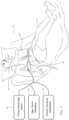
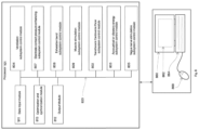
- Fig. 1 is a schematic diagram of a CETEES 100, showing subsystem interactions with a schematic patient and the flow of subsystem and biomarker inputs and outputs, according to an illustrative embodiment.
- the sequence, forces, and electrical properties of the subsystems can be controlled by computer controller 101.
- Computer controller 101 can also be referred to as a processor 101 or a control system 101.
- a CETEES can be controlled by a processor 101 that can include a plurality of modules, data inputs, and control outputs, and a user interface.
- the processor can be contained within a general purpose, or a dedicated, computing device, such as a PC, laptop, tablet or smartphone. In various embodiments, the computing device can be built into, or incorporated within, the housing of the CETEES.
- the computing device can include a user interface that can be a keyboard, mouse, touch screen or similar device, and a display that can include a graphic user interface screen.
- a user can input instructions for a procedure through the user interface into the processor.
- the modules and/or sub-modules can then process the instructions, collect measurements, and provide information to the various subsystems of the CETEES.
- the control system 101 may use pre-defined a priori parameters and/or sequences, and/or the control system may optimize all components based on biomarker or subsystem status feedback.
- the CETEES can start with a priori parameters and/or sequences, and can optimize those parameters and/or sequences during treatment in response to biomarker or subsystem status feedback.
- the parameters and/or sequences can be optimized, or adjusted, as treatment is ongoing to provide customized individual treatment that is adapted in real time to each patient in order to provide superior treatment to that patient.
- the controller 101 can provide outputs 112 from the controller 101 to the various subsystems to control the subsystems.
- an embodiment of a CETEES 100 can integrate gel electrode pads with one or more additional subsystems.
- the CETEES 100 can include one or more cardioverter(s) and/or pacemaker(s) that can also be referred to as Transthoracic Cardioverter Pacers (TCPs) 102.
- TCPs Transthoracic Cardioverter Pacers
- the TCP 102 can apply shocks to the patient through electrode pads.
- the electrode pads can be gel electrode pads or self-adhesive gel electrode pads.
- the controller 101 can provide outputs 112 from the controller 101 to the TCP 102 which can control the TCP 102 to provide electric shocks through the electrodes to the patient 110.
- the CETEES 100 can include electrode contact force enhancers 107.
- the electrode contact force enhancers can apply force that presses electrodes against the patient. This results in increased contact pressure, i.e. applying force to the pads or paddles. The increased contact pressure can reduce impedance, thereby improving the likelihood of successful cardioversion.
- the controller can control the contact force enhancers to provide increased contact pressure during pacing.
- the controller 101 can control the contact force enhancers 107 to provide increased contact pressure before the shock is delivered, and reduce contact pressure after the shock is delivered.
- the controller 101 can control the contact force enhancers 107 to reduce contact pressure between shocks, and increase contact pressure during shocks.
- the CETEES 100 can include a vagus nerve stimulator 105.
- the vagus nerve stimulator 105 can provide stimulation to the vagus nerve of the patient.
- the controller 101 can control the vagus nerve stimulator 105.
- the stimulation of the vagus nerve can be transcutaneous, for example, at the ear or neck.
- the stimulation of the vagus nerve can be electromagnetic and/or acoustical.
- the CETEES 100 can include a myocardium energy emitter 103 that can direct energy towards the myocardium of the patient.
- the myocardium energy emitter 103 can emit acoustic energy towards the myocardium of the patient 110.
- the acoustic energy emitted towards the myocardium can be ultrasound energy.
- the CETEES can include one or more subsystems that can help to reduce the volume of air in the lungs for pacing shock delivery.
- the CETEES can include a ventilator 104, an exhalation band 108, such as a constrictor band, and/or a muscle electrical stimulator 109.
- the exhalation band 108 may be either mechanical or pneumatic in nature, and mechanical exhalation bands and/or a pneumatic exhalation bands can both be referred to as an exhalation band 108.
- the controller 101 can provide output instructions 112 to the ventilator 104 so that the controller can coordinate the ventilator with the delivery of pacing shocks, thereby helping to coordinate so that pacing shocks can be delivered at a minimal lung volume.
- the controller 101 can provide output instructions to the exhalation band 108 which can constrict the torso of the patient 110 to reduce the volume of air in the lungs.
- the controller can coordinate the exhalation band with the delivery of pacing shocks, thereby helping to coordinate so that pacing shocks can be delivered at a minimal long volume.
- the controller 101 can provide output instructions to the muscle electrical stimulator 109 which can stimulate the rectus abdominus muscle, thereby causing the patient to exhale.
- the controller can coordinate the muscle electrical stimulator 109 with the delivery of pacing shocks, so that the patient is stimulated to exhale and reduce the volume of air in the lungs for the delivery of a pacing shock.
- the CETEES may be a single unit with multiple subsystems.
- the controller may be connected to, and provide controlling output instructions to various subsystems that may be separate components or components that may otherwise be capable of operating separately. Regardless of the extent or degree of connection between the controller and the various subsystems, the CETEES can coordinate each of the subsystems to work together to provide coordinated treatment and improved likelihood of successful cardioversion.
- various cables, hoses, wireless connections, and/or other connections can exist between the controller and the various subsystems so that the controller can control and coordinate the various subsystems.
- the various subsystems of the CETEES can provide feedback in the form of inputs 111 to the controller 101.
- the inputs can include the status of various subsystems, such as readiness to deliver a shock, exhalation status, and others.
- the CETEES can include various sensors, including biomarkers, ECG, photopleth, ventilation, and/or other sensors.
- the various sensors can provide patient biomarker data 106 that can be used as inputs 111 to the controller 101 about the status of the patient.
- the controller 101 can adapt the treatment of the patient in response to the various inputs.
- the controller can provide outputs 112 that control and coordinate the various subsystems.
- controller 101 can adjunctively provide additional benefit to the patient, or increase the likelihood of successful cardioversion by various means, including, but not limited to:
- the controller can adjunctively improve the efficacy of transthoracic countershock or pacing by means of:
- Fig. 2 is a perspective view of the CETEES applied to a patient, according to an illustrative embodiment
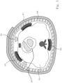
- Fig. 3 is a partially cut-away view of the CETEES around a patient's thorax, showing inner workings including cardioversion pads and contact force enhancers, according to an illustrative embodiment.
- cardioversion pads can also be referred to as pacing pads, TCP pads, or countershock pads, and the terms can be used interchangeably throughout this document.
- the CETEES can include an inelastic outer shell 201 that can contain and maintain the location and orientation of various patient facing subsystems.
- This shell also provides a structure against which patient-facing components may push so as to allow counter-force to be directed inward toward the patient. This can include providing counterforce for the contact force enhancers 303 so that they can press the electrode pads 302 against the patient.
- This shell may be vest-like in shape and adjustable for patients of varying size and circumference. There can be a closure mechanism such as hook and loop tape, buckles, or other closures
- the electrode pads 302 can be gel electrode pads, and can be self-adhesive gel electrode pads.
- the contact force enhancers 107 can be inflatable bladders 303 that can selectively apply force to electrode pads 302, and can be hydraulic or pneumatic.
- the contact force enhancers 107 can be solenoids or other mechanical pushing mechanisms 307 that can selectively apply force to electrode pads 302.
- the contact force enhancers 107 can include a circumferential constrictor 306 that can be a circumferential bladder, multiple circumferential bladders, a constrictable inelastic belt, or various other circumferential constrictors.
- the contact force enhancers 107 can include various combinations of different types of contact force enhancers.
- the contact force enhancers 107 can all be the same type of force enhancer or multiple types of force enhancers.
- the CETEES can include an exhalation constrictor band 108 that can squeeze the torso of the patient, thereby decreasing air volume in the lungs and changing the conformation of the torso to bring the anterior and posterior of the torso closer together.
- the exhalation constrictor band 108 can include one or more bladders that can inflate to squeeze the patient, similar to a blood pressure cuff.
- the exhalation constrictor band 108 can include multiple bladders arranged in parallel around the patient.
- the exhalation constrictor band 108 can be inside of the inelastic shell 301, and the shell 301 can provide a counterforce so that the bladders can press against the patient as they inflate.
- the inflatable bladder(s) of the exhalation constrictor band 108 can be hydraulic or pneumatic.
- the exhalation constrictor band can be a mechanical device that tightens an inelastic belt around the patient to squeeze the torso of the patient.
- the exhalation constrictor band 108 can also be the circumferential constrictor 306 of the contact force enhancer 107, or the CETEES can have distinct and separate exhalation constrictor band 108 and contact force enhancer circumferential constrictor 306.
- one or more components of the one or more contact force enhancers 107 can act as and/or can work to assist the exhalation constrictor band 108.
- the CETEES 100 can include one or more acoustic energy emitters 103 that can transmit acoustical, ultrasound, or vibratory energy to the myocardium.
- the acoustic energy emitter 103 can be an ultrasound emitter.
- the CETEES 100 can include one or more vagus nerve stimulators 105 which can stimulate the vagus nerve transcutaneously using electromagnetic and/or acoustic stimulation.
- the CETEES can also include transcutaneous muscle stimulation electrodes 109. Transcutaneous muscle stimulation electrodes 109 can stimulate the rectus abdominus muscle to cause the patient to exhale.
- the CETEES 100 can also include a number of sensors 206 that can collect patient biomarker data. These sensors can include ECG, photopleth, ventilation sensors, and/or various other sensors. Sensors 206 can provide patient biomarker data 106 as inputs 111 to the controller 101.
- the TCP 102 can have a significantly expanded spectrum of capabilities. It can do more than simply charge the electrodes 302 and release current from the storage capacitors. It can further interface with the control processor, provide input 111 status data to the controller 101 and receive output 112 instructions from the controller 101. It can countershock or provide pacing via multiple electrode pairs, as described in US Patent No. 4,708,145 .
- the CETEES 100 may incorporate transthoracic pacing subsystems 102, or may be a separate unit from the transthoracic pacing subsystems and connected to them via outputs 112 from the controller 101.
- the efficacy of the function of the TCP 102 can be adjunctively enhanced by inclusion of, and coordination with, one or more other subsystems that can include the contact force enhancers 107, the acoustic, ultrasound or vibratory energy emitter 103, the ventilator 104, the muscle electrical stimulator 109, and/or the vagal nerve simulator 105.
- a CETEES system may allow current to be released from one or more combinations of electrodes 302.
- the use of multiple combinations of electrodes 302 can include using multiple paths across the chest, as is described in US Patent No. 9,174,061 .
- the use of multiple paths across the chest can include applying two or more counter shocks simultaneously along paths that are at an angle to each other.
- two counter shocks can be delivered simultaneously along paths that can be at a 90 ° angle to one another.
- multiple electrode pads 302 can be used to create multiple transthoracic pathways through the patient. Electric current can be directed through selected countershock electrodes 302 to create various transthoracic pathways through the patient.
- countershock electrodes can also be referred to as defibrillation electrodes, pacing electrodes, cardioversion electrodes, or TCP electrodes, and the terms can be used interchangeably throughout this document.
- Incorporation of the cardioversion electrodes 302 into the patient facing surfaces of the CETEES allows for multiple countershock subsystems 102 to be discharged simultaneously or sequentially, and allows for the use of one or more transthoracic pathways between various cardioversion electrodes 302.
- Various embodiments described herein can include incorporating a dual/triple/(N) sequential or simultaneous countershock capability into a multimodal automatic system.
- a controller 101 can also activate selective additional subsystems that can adjunctively improve upon the pacing.
- additional adjunctive subsystems can include any of the subsystems described herein, including pneumatic, mechanical, acoustical, or other subsystems, to enhance countershock or pacing efficacy.
- the controller 101 can provide instructions to the pacer(s) 102 to provide electrical countershock that may be single, dual simultaneous/sequential, or N simultaneous/sequential, which can be delivered via two or more simultaneous/sequential electrode pairs.
- One of the electrode pairs may include a positive or negative electrode within the esophagus of the patient.
- the CETEES can have one pair of countershock electrodes 302, or can have more than two countershock electrodes, and in various embodiments the CETEES can have four or more countershock electrodes. In various embodiments, the CETEES can have a pair of electrodes that can be positioned to apply posterior-anterior countershocks.
- the electrodes 302 used for myocardial capture during transthoracic cardiac pacing are also pushed on at the moment of each electrical discharge.
- the countershock or pacing electrodes 302 may have contact force enhancers 107, such as pneumatic bladders 303, mechanical actuators 307, and/or circumferential constrictors 306 behind them to provide force on the electrodes.
- the contact pressure enhancing subsystem 107 to enhance contact pressure may have one or more mechanisms for applying force.
- These mechanisms for applying force can include one or more of: 1) a selective pneumatic bladders 303, and/or mechanical actuators 307 that push on gel electrode pads 302, 2) a circumferential constriction pneumatic bladder 306 that pushes on all electrode pads 302 at the moment of electrical discharge or 3) a combination of those mechanisms.
- selective or full circumferential enhancement of contact force pressure may be achieved by mechanical means such as screws or piston mechanisms. Optimization of contact pressure in electrical countershock or pacing as monotherapy for dysrhythmias has found that at least 50 Newtons of force may be needed to achieve enhanced current flow.
- the controller can begin with at least 90 Newtons of force being applied a priori at the beginning of treatment, and in various embodiments, the controller can adapt the force in response to inputs such as sensed biomarkers and subsystem feedback to optimize the treatment for each individual patient. Generally, the force applied would be below the thresholds associated with skeletal or visceral organ injury.
- Fig. 4 is a schematic of the contact pressure enhancer subsystem including the feedback mechanisms 401 and effector mechanisms 107 along with the controller 101.
- air acts as an insulator that impairs current flow.
- the lungs act as air based insulators.
- the circumferential constrictors and/or electrode contact force enhancers of the CETEES may alter the ventilatory state of the lungs at, just before, or throughout the moment of electrical discharge so as to lower transthoracic resistance.
- one or more of the various subsystems for altering ventilator status can force exhalation and minimize the diameter of the thorax by placing the lungs at maximal expiration at the time of countershock or pacing electrical discharge.
- Fig. 5 is a schematic of the ventilation at exhalation subsystems including the feedback mechanisms 501 and effector mechanisms 502 along with the controller 101.
- residual volume such a value being defined as within 20 percent of a patient's residual volume.
- acoustic, ultrasound or vibratory energy can be combined adjunctively with electrical energy to more efficiently and effectively achieve cardioversion or pacing.
- a CETEES 100 can place the myocardium 305 into a state more amenable to cardioversion or pacing.
- acoustic, ultrasound or vibratory energy are referred to the other energy frequencies are included.
- acoustic, ultrasound or vibratory energy can have a salutary effect on electrophysiologic characteristics of the myocardial substrate that is initiating or maintaining the dysrhythmia. While inefficient as a sole treatment of dysrhythmias, acoustic, ultrasound or vibratory energy can be useful as an adjunct to enhance the efficacy of electrical cardioversion or pacing when combined with other adjunctive techniques.
- the acoustic, ultrasound or vibratory energy may be applied to the myocardium 305: 1) for a period of time before electrical countershock, 2) coincident with electrical countershock, 3) after electrical countershock, 4) a combination of these phases.
- Application of the acoustic, ultrasound or vibratory energy before countershock can place the myocardium in condition for the countershock to be more effective.
- the controller can begin with acoustic, ultrasound or vibratory energy being applied a priori at the beginning of treatment, and in various embodiments, the controller can adapt the acoustic, ultrasound or vibratory energy in response to inputs such as sensed biomarkers and subsystem feedback to optimize the treatment for each individual patient.
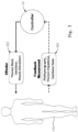
- Fig. 6 is a schematic of the cardiac acoustic subsystems including the feedback measurement and effector mechanisms 103 along with the controller 101.
- a CETEES may additionally place the myocardium 305 into a state more amenable to cardioversion or pacing by means of vagal nerve stimulation.
- Application of this stimulation may be used adjunctively to enhance to probability of acute electrical cardioversion of atrial fibrillation or similarly as an assist in transthoracic pacing.
- vagal nerve stimulation energy may begin to be applied: 1) for a period of time before electrical countershock, 2) coincident with electrical countershock, 3) after electrical countershock, 4) a combination of these phases. Because the effect of vagal nerve stimulation likely takes time intervals on the order of minutes to affect the electrophysiologic state of the myocardium 305, the controller can synchronize the vagal stimulation to begin 5 to 10 minutes before countershock or pacing.
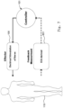
- Fig. 7 is a schematic view of the vagal stimulation subsystem including the feedback measurements and effector mechanisms 105 along with the controller 101.
- the CETEES can have a controller 101 that can input biomarker inputs and inputs from various subsystems, and can coordinate the electrical discharge in response to these inputs.
- Biomarkers inputs may include but not limited to: ECG, ventilatory status, impedance measurements, and ventilatory gas measurements, among others.
- the electrical system for the countershock circuitry may measure one or more of: thoracic resistance, capacitance, impedance, or current flow.
- the controller can receive biomarker inputs from various afferent sensors provided at locations around the patient for collecting biomarker data. The controller can also receive status update from one or more of the effector subsystems.
- Such a control system could allow electrical countershock optimized by the measurement of one or more of thoracic resistance, capacitance, impedance, or current flow; electrical countershock at the optimal portion of the ventilatory cycle; optimization of the electrical countershock by applying current through a selected subset of the patient-facing electrodes; adjustment of the force, location or timing parameters of chest constriction so as to optimize one or more of thoracic resistance, capacitance, impedance, or current flow; and/or adjustment of the parameters of synchronized ventilation so as to optimize one or more of thoracic resistance, capacitance, impedance, or current flow.
- the controller can optimize the relationships in response to various inputs.
- Each subsystem's energy characteristics can be pre-set at a priori optima, and/or can be optimized in real-time based on acquired inputs including tissue and organ status.
- the subsystem energies or frequencies may be varied while ECG parameters known to be associated with, and likely predictive of, successful countershock or pacing (i.e. Zeemering et al discussed above) are measured. These methods can include optimization through closed-loop control. Once the optima are achieved, countershock current can be applied. In the case of cardiac pacing, capture of the myocardium 305 may be attempted at the same time that the optimization of the subsystem(s) is occurring.
- the subsystem energies or frequencies may be varied while the value of one or more previously determined synthetic machine learning derived algorithms or equations are measured. Once the optimal or near-optimal values are achieved, countershock current can be applied. In the case of cardiac pacing, capture of the myocardium 305 may be attempted at the same time that the optimization of the subsystem, based on the machine learning derived equation, is occurring.
- the measurements of the ECG can be useful and readily available in patients suffering cardiac rhythm disorders.
- the ECG QRS rate, QRS rate spectroscopy, and atrial fibrillation waveform spectroscopy can be indicative of the state of the myocardium 305 and likely predictive of dysrhythmia termination or myocardial pacing capture.
- the processor controlling the cardioversion or pacing can apply the current for electrical countershock to differing combinations of electrodes 302 such that multiple paths across the chest can be utilized simultaneously or in sequence.
- the processor controlling the timing can apply the current for electrical cardioversion or pacing to combinations of electrodes so that two countershocks at an angle to one another can be applied simultaneously.
- the processor controlling the cardioversion or pacing can apply the current for electrical countershock to a series of electrodes such that the pathway of current flow through the chest can start in one or more directions and can transition into a different set of directions.
- the mechanical, pneumatic, or hydraulic components can vary the force or pattern of chest compression or constriction so as to enhance the efficacy of cardioversion or pacing.
- the processor can receive measurement data from one or more of thoracic resistance, capacitance, impedance, and/or current flow, and can adapt treatment in real time in response to measured data to provide customized treatment to individual patients.
- the device described herein can adjunctively enhance the efficacy of electrical countershock or pacing by means of enhancing current flow and or altering the myocardial substrate.
- the temporal relationships, also referred to as the synchronizations, between current flow and the activation of various adjunctive subsystems may be different.
- these synchronizations may be in the range of:
- the patient-facing components for electrical countershock such as adhesive gel electrodes 302
- the associated electronics such as the processor
- the processor may be fully incorporated into the housing of an automated mechanical/pneumatic CETEES system.
- one or more of the subsystems may be housed separately from the main device.
- the countershock subsystem it may be housed separately and connected to the main device by electrical cables.
- a system integrating multiple hemodynamic enhancements with cardioversion or pacing may, at any given moment, only be applying a subset of its multiple modalities.
- an inelastic outer shell 301 capable of being placed circumferentially around the thorax at the level of the myocardium 305.
- This shell may be made of various fabric materials and incorporate a closure mechanism allowing appropriate sizing.
- This shell allows subsystems applying mechanical, acoustical, and electrical forces to have a physical structure to maintain optimal configuration and apply Newtonian counterforce against.
- an embodiment can include a fully integrated electromechanical or electropneumatic CETEES system that can include one or more of the following components:
- the countershock/pacemaker subsystem 102 and its associated electronics may be physically integrated into the device, or it can be separate, with or without monitor, connected to the controller wirelessly or by a cable.
- the electrical countershock electrodes 302 can be physically integrated into patient-facing portions of other subsystems, including, for example, the thoracic circumferential constrictor subsystem 306.
- TCP electrodes 302 can be used to create multiple transthoracic pathways through the patient 110. Electric current can be directed through selected countershock electrodes 302 to create various transthoracic pathways through the patient 110. Countershock or pacing may be improved by application of multiple transthoracic pathways that are electrified near simultaneously or sequentially. Incorporation of the TCP electrodes 302 into patient facing surfaces of the device allows for multiple electrodes to be discharged simultaneously or sequentially, and allows for the use of one or more transthoracic pathways between various electrodes. In some cases, an anterior-posterior transthoracic pathway may be optimal. The potentially optimal anterior-posterior electrode placement and current path may be utilized singly, or as part of a multi-shock simultaneous or sequential pattern. Incorporation of the TCP electrodes into the body of the CETEES device allows for fast and easy placement of electrodes in various locations around the patient, including the anterior and posterior of the thorax.
- a CETEES can be controlled by a controller, or processor, 101 that can include, or be connected to, a plurality of modules, data inputs, and control outputs, and a user interface.
- the processor can be contained within a general purpose, or a dedicated, computing device, such as a PC, laptop, tablet, or smartphone.
- the controller 101 can be built into, or incorporated within, the housing of the CETEES.
- the computing device can include a user interface that can be a keyboard, mouse, touch screen or similar device, and a display that can include a graphic user interface screen.
- a user can input instructions for a procedure through the user interface into the processor.
- the modules and/or submodules of the processor can then process the instructions, collect measurements, and provide information and/or instructions to the various subsystems of the CETEES.
- the controller 101 can activate selective pneumatic or mechanical adjuncts, described above, to increase electrode contact pressure.
- the controller 101 can then provide electrical countershock that may be single, dual simultaneous/sequential, or N simultaneous/sequential, which can be two or more simultaneous/sequential countershocks.
- One of the electrode pairs may include a positive or negative electrode within the esophagus.
- the countershock electrodes can be adhesive gel electrodes that can be a disposable component that is pre-manufactured so as to be easily inserted into or removed from the patient-facing mechanical or pneumatic components.
- an illustrative embodiment of a CETEES can integrate standard self-adhesive gel electrode pads with one or more additional subsystems.
- the additional subsystems can be adjunctive to the countershock treatment. These additional subsystems may achieve:
- a CETEES may also achieve improved cardioversion or pacing by varying the combination, location, or current pathways of the electrode pads to use multiple paths across the chest. This can include applying two or more countershocks simultaneously along paths that are at an angle to each other. In various embodiments, two counter shocks can be delivered simultaneously along paths that can be at a 90 ° angle to one another and/or applying multiple countershocks at different angles relative to each other in succession.
- a CETEES can be: 1) adjunctive in application of multiple subsystems, 2) automated, 3) integrated, 4) computer controlled, 5) electromechanical, 6) electrophysiologic 7) optimized, and/or 8) controlled through closed-loop feedback.
- a CETEES can include devices or subsystems that provide countershock or transthoracic pacing, and in various embodiments, a CETEES may be distinct from separate devices that provide transthoracic cardioversion or pacing and be connected to them via a controller 101. Such separate devices are common clinically.
- a CETEES can include an integrated countershock cardioverter and/or transthoracic pacemaker.
- the countershock cardioverter and/or transthoracic pacemaker can be a single device that can perform both functions, or the countershock and/or transthoracic pacemaker can be distinct devices.
- a CETEES may have one or more subsystems that can include: a cardioverter, a transthoracic pacemaker, contact force enhancement subsystem, an acoustic, ultrasound or vibratory energy generator subsystem, a ventilatory subsystem, a vagal nerve simulator subsystem, a controller, and/or an inelastic outer band or outer shell.
- the efficacy of the countershock cardioverter and/or transthoracic pacemaker functions can be adjunctively enhanced by one or more of the other subsystems including: the contact force enhancer subsystem, the acoustic, ultrasound or vibratory energy emitter subsystem, the ventilator subsystem, and/or the vagal nerve simulator subsystem.
- the contact force enhancer subsystem the acoustic, ultrasound or vibratory energy emitter subsystem
- the ventilator subsystem and/or the vagal nerve simulator subsystem.
- subsystems including the transthoracic countershock cardioverter subsystem and the subsystems that can be used to adjunctively enhance the transthoracic cardioverter functions of the CETEES are described below.
- the term countershock subsystem can have an expanded spectrum of capabilities.
- the countershock system can do more than simply charge the electrodes and release current from the storage capacitors. It can further interface with the control processor, provide status data to the controller, and receiving instructions from the controller. In various embodiments, it can countershock via multiple positive-negative electrode pairs.
- a CETEES can control the combinations of electrode pads that are electrically discharged by the cardioverter such that that current is released from one or more combinations of electrode pads, so that the CETEES can utilize multiple paths across the chest.
- Multiple electrode pads can be used to create multiple transthoracic pathways through the patient.
- the use of multiple paths can include applying two or more counter shocks simultaneously along paths that are at an angle to each other.
- two counter shocks can be delivered simultaneously along paths that can be at a 90 ° angle to one another. Electric current can be directed through selected countershock electrodes to create various transthoracic pathways through the patient. The electrodes used for each shock can be selected by the controller.
- a controller can activate selected pneumatic, mechanical, acoustical, or other subsystems, as described herein, to enhance countershock or pacing efficacy.
- the controller can provide electrical countershock that may be single, dual simultaneous/sequential, or N simultaneous/sequential, which can include two or more simultaneous/sequential electrode pairs.
- one of the electrode pairs may include a positive or negative electrode within the esophagus of the patient.
- the CETEES can have one pair of countershock electrodes, or can have more than two countershock electrodes, and in various embodiments the CETEES can have four or more countershock electrodes.
- the CETEES can have a pair of electrodes positioned to apply posterior-anterior countershocks.
- adjunctive enhancements to efficacy can occur if the electrodes used for myocardial capture during transthoracic cardiac pacing are pushed on at the moment of each electrical discharge.
- countershock electrodes can have electrode contact enhancers behind them to provide force on the electrodes.
- CETEES electrode contact force enhancement system may have one or more mechanisms that can apply force to the pads or paddles.
- These mechanisms to apply force can include, but are not limited to, electrode contact enhancing inflatable bladders 303, mechanical actuators 307, and/or circumferential constrictors 306. These systems can apply force to an electrode 302 to push the electrode against the patient 110 and/or apply force to the patient to push the patient against the electrode.
- electrode contact force can be enhanced by a circumferential constrictor subsystem 306 that can include a pneumatic or hydraulic bladder, a series of bladders, and/or a constricting band to push on electrode pads at the moment of electrical discharge.
- a circumferential constrictor subsystem 306 can include a pneumatic or hydraulic bladder, a series of bladders, and/or a constricting band to push on electrode pads at the moment of electrical discharge.
- a circumferential constrictor subsystem 306 can include a pneumatic or hydraulic bladder, a series of bladders, and/or a constricting band
- selective or full circumferential enhancement of contact force pressure may be achieved by pneumatic or hydraulic means, and/or by mechanical means such as screws or piston mechanisms. At least 50 Newtons of force may be needed to achieve enhanced current flow, however, the contact pressure for electrical countershock pacing for dysrhythmias can be optimized for each patient, as explained more fully below.
- Acoustic, ultrasound or vibratory energy can be applied adjunctively in combination with electrical energy to more efficiently and effectively achieve countershock or pacing.
- This energy can be applied transcutaneously from skin surface based acoustical or vibratory emitters 304.
- the acoustic, ultrasound or vibratory emitters 304 can be crystal or semiconductor arrays. Applying acoustic and/or vibrational energy to the patient 110 can place the myocardium 305 into a state more amenable to cardioversion or pacing.
- Acoustic or vibratory energy can have at least a salutary effect on electrophysiologic characteristics of the myocardial substrate that is initiating or maintaining the dysrhythmia, and acoustic, ultrasound or vibratory energy alone can sometimes pace the myocardium 305. While often inefficient alone as a sole treatment of dysrhythmias, acoustic, ultrasound or vibratory energy can be useful as an adjunct to enhance the efficacy of electrical cardioversion or pacing. With respect to countershock, the acoustic, ultrasound or vibratory energy may be applied to the myocardium: 1) for a period of time before electrical countershock, 2) coincident with electrical countershock, 3) after electrical countershock, and/or 4) a combination of these phases. The use of acoustic, ultrasound or vibratory energy as an adjunctive intervention can be optimized for each patient. Application of acoustic, ultrasound or vibratory energy may be continuous or pulsed during pacing or continuous for a period of time before countershock.
- air acts as an insulator that impairs current flow.
- the lungs can act as air-based insulators during countershock.
- the circumferential constriction mechanisms or electrode contact force enhancers of the CETEES may alter the ventilatory state of the lungs at, just before, or throughout the moment of electrical discharge so as to lower transthoracic resistance. More specifically, this subsystem can force exhalation, minimize lung volume, and minimize the diameter of the thorax by placing the lungs in a state of maximal expiration at the time of countershock or pacing electrical discharge.
- Incorporation of electrodes into the patient facing surface of a circumferential constrictor can allow both enhanced electrode contact pressure and ventilatory end-exhalation for improved transthoracic resistance and countershock success.
- a single circumferential constrictor can provide the enhanced contact pressure and the exhalation constriction.
- Lung volume minimizers can also include transcutaneous Functional Electrical Stimulation (FES) which can cause one or more respiratory muscles to contract, as described in U.S. Patent Application Publication No. 2002/0128686 titled ABDOMINAL BELT WITH ADJUSTABLE ELECTRODES, the entire disclosure of which is incorporated herein by reference.
- FES of the rectus abdominis can act to assist exhalation. If a patient is having their breathing assisted by a ventilator, the ventilator can also act as a lung volume minimizer. The ventilation can be synchronized with the countershocks to deliver countershocks during a period of minimum lung volume.
- Synchronization of the ventilator and the countershocks can include delivering countershocks during the optimal expiratory phase of ventilation, at the moment in a ventilation cycle when the lung volume is at a minimum.
- Synchronization of the ventilator and the countershocks can include delivering countershocks at the moment before forced inspiration.
- Synchronization of the ventilator and countershocks can also include delaying forced inspiration until after the countershock. Maximal exhalation may be achieved by one or multiple means.
- a CETEES device may place the myocardium into a state more amenable to cardioversion or pacing by means of vagal stimulation.
- Application of electrical vagal nerve stimulation may help to suppress episodes of paroxysmal atrial fibrillation.
- Adjunctive application of vagal nerve stimulation may be used to increase the likelihood of successful electrical cardioversion of atrial fibrillation and/or to increase the efficacy of transthoracic pacing.
- vagal nerve stimulation energy may begin to be applied: 1) for a period of time before electrical countershock, 2) coincident with electrical countershock, 3) after electrical countershock, 4) a combination of these phases. Because the effect of vagal nerve stimulation likely takes time intervals on the order of minutes to affect the electrophysiologic state of the myocardium, the synchronization relationship that is likely most effective would be for vagal stimulation to initiate 5 to 10 minutes for countershock or pacing.
- Fig. 9 is a flow diagram of an exemplary clinical process 900 for treatment using a CETEES, according to an illustrative embodiment.
- the method or device may utilize the following sequence: At box 905, the device can be applied to the patient's chest.
- various biomarkers can be collected from the patient and provided as an input to the controller of the CETEES.
- An operator provide input to the CETEES confirming the intention to perform cardioversion or pacing, and a desired time to intervention.
- the vagal nerve stimulation subsystem can apply vagal nerve stimulation, and the vagal nerve stimulation can be maintained through completion of countershock or pacing. At least 10 minutes of vagal stimulation may be considered optimal.
- the acoustical or vibratory subsystem initiates application of acoustical or vibratory energy to the myocardium five to 10 minutes before cardioversion or pacing. Acoustical or vibratory energy application can be maintained through completion of countershock or pacing.
- a sedative can be administered to the patient.
- the drug is administered at the appropriate interval before countershock or pacing.
- an operator input can confirm that patient sedation has been achieved.
- the thoracic constriction subsystem, the ventilator control mechanism, and/or the muscle stimulation for exhalation subsystem are activated and synchronized 2 to 3 seconds before countershock. Thoracic constriction, ventilator exhalation, and muscle stimulation for exhalation, can be maintained through completion of countershock.
- the electrode contact force enhancement subsystem applies force 100-500ms before countershock or pacing and maintains this force through each countershock or pace.
- control system discontinues subsystem activation. In the case of cardiac pacing there is a cyclical reactivation sequence.
- the pacer For external transthoracic cardiac pacing to be effective, the pacer must capture the myocardium electrophysiologically with each desired heartbeat.
- a CETEES is potentially a useful adjunct in enhancing the likelihood of initial and continued pacemaker capture of the myocardium.
- the overall configuration and the specific parameters of the subsystem will likely be different when a CETEES is used for cardiac pacing in comparison to one-time cardioversion.
- the adjunctive enhancement of pacemaker electrical capture should be maintained, and some components cycled with respect to each induced cardiac contraction.
- the vagus nerve stimulation and acoustic enhancement may be maintained throughout the time that transthoracic cardiac pacing is being utilized.
- Electrode contact force enhancement may have multiple modes. In one mode, the contact force enhancement may be maintained continuously. In another mode, it may be cycled with each cardiac beat. Because this is going to be applied for periods of time longer than required for one-time cardioversion, it is possible that the force applied will be less. Transthoracic cardiac pacing is often uncomfortable for patients, requiring sedation. In various embodiments, electrode contact force enhancement may, either continuous or cycles, be increased if sedation is used.
- Various patient biomarkers can be measured and used to improve the efficacy of treatment in real time.
- Patient biomarkers can be used to improve the efficacy of countershock pacing, and patient biomarkers can be used to synchronize, coordinate, and/or optimize each of the different adjunctive treatment modalities.
- the CETEES can have a controller unit with a processor that can coordinate biomarker inputs, the subsystem activities, and the electrical discharge.
- Biomarkers may include but are not limited to: ECG, ventilatory status, impedance measurements, and/or ventilatory gas measurements, among others.
- the electrical control system 101 for the countershock circuitry may measure one or more of: thoracic resistance, capacitance, impedance, or current flow. In various embodiments, these measurements can be collected through various afferent sensors provided at locations around the patent for collecting biomarker data, including but not limited to: ECG, force transducers, accelerometers, and/or electrodes for impedance.
- the electrical control system 101 can receive status updates from one or more of the effector subsystems. In various embodiments, these measurements can be collected through the countershock electrodes.
- Such a control system can optimize electrical countershock by adjusting the parameters of the electrical countershock based on the measurement of one or more of thoracic resistance, capacitance, impedance, or current flow.
- the control system can use measurements of patient breathing and the breathing cycle to coordinate electrical countershock to be delivered at the optimal portion of the ventilatory cycle.
- the control system 101 can use biomarker measurements 106 such as thoracic resistance, capacitance, impedance, and/or current flow to identify the electrodes most likely to be most efficacious and optimize application of the electrical countershock by applying current through a selected subset of the patient-facing electrodes.
- the control system can use biomarker measurements of thoracic resistance, capacitance, impedance, and/or current flow to adjust the force, location and/or timing parameters of chest constriction so as to optimize one or more of thoracic resistance, capacitance, impedance, or current flow. Optimizing thoracic resistance, capacitance, impedance, and/or current flow can result in improved efficacy of countershock or pacing.
- Each subsystem's energy or force characteristics may either be pre-set at a priori optima, and/or can be optimized in real-time based on measured tissue and/or organ status.
- the probability for successful or failed countershock or pacing can be known at differing subsystem energies, frequencies, or forces, and the pre-set a priori optima can be the energies, frequencies, or forces known before treatment starts to be the most likely to lead to successful countershock or pacing.
- the controller can begin treatment with pre-set a priori optima, and can then optimize in real time to improve treatment as the treatment is occurring by collecting biomarker measurements and adjusting various subsystems' energy or force characteristics in response to the collected biomarker measurements.
- the subsystem energies or frequencies may be varied in response to ECG parameters known to be associated with, and likely predictive of, successful countershock or pacing. Once the subsystem parameters predicted to be most effective are determined and applied, countershock or pacing current can be applied. In the case of cardiac pacing, capture of the myocardium may be attempted at the same time that the optimization of the subsystem is occurring.
- the subsystem parameters including but not limited to energies, frequencies and/or forces may be varied while the value of one or more previously determined synthetic machine learning derived algorithms or equations are measured. Once the optimal or near-optimal values are achieved, countershock current can be applied. In the case of cardiac pacing, capture of the myocardium may be attempted at the same time that the optimization of the subsystem, based on the machine learning derived equation, is occurring.
- the measurements of the ECG are useful and readily available in patients suffering cardiac rhythm disorders.
- the ECG QRS rate, QRS rate spectroscopy, and atrial fibrillation waveform spectroscopy can be indicative of the state of the myocardium and likely predictive of dysrhythmia termination or myocardial pacing capture and can be used to improve the efficacy of both cardioversion and pacing.
- plateaus in measured biomarkers may be used in determining subsystem optima.
- biomarker can include, as non-limiting examples, various measurements of the heart and heart activity, respiration, etc., and can include transthoracic impedance or other electrical measurements taken from the patient.
- a parameter of a subsystem such as force, frequency, or energy
- the force, energy or frequency can be decreased to the range around the onset of plateau and the subsystem can be considered optimized. This optimization can be performed to determine optimal settings for one or more subsystems in real time to provide the best treatment for an individual patient.
- ECG QRS rate QRS rate spectroscopy
- QRS rate spectroscopy or atrial fibrillation waveform spectroscopy
- a synthetic multivariable equation or algorithm incorporating more than one of these measurements may be determined by techniques called machine learning and may have performance superior to any individual measurement alone.
- Machine learning can be performed in real time as the treatment is occurring so that one or more personalized algorithms can be created for each specific patient, thereby improving treatment for that patient.
- the pneumatic or mechanical force applied to the gel electrode pads 302 could be increased while the transthoracic impedance-resistance is measured. Once the measured biomarker plateaus, that is to say, once there is no further improvement in transthoracic impedance-resistance with application of additional force, then the force is decreased to the range around the onset of plateau and the subsystem is considered optimized.
- one or more of the ECG QRS rate, QRS rate spectroscopy, or atrial fibrillation waveform spectroscopy can be measured and the force associated with onset of plateau of the biomarker identified.
- this can be in combination with measurements of impedance, capacitance, or other measurements of physical characteristics.
- a synthetic multivariable equation or algorithm incorporating more than one of these measurements and/or other measurements described herein may be determined by techniques called machine learning and may have performance superior to any individual measurement alone.
- the time course for application of increasing force to the electrode pads by the contact force enhancer subsystem might occur during the 1 to 2 seconds before projected countershock.
- the subsystem that applies acoustic, ultrasound or vibratory energy to the myocardium may be optimized. More specifically, one or more of the ECG QRS rate, QRS rate spectroscopy, or atrial fibrillation waveform spectroscopy, can be measured while the acoustic, ultrasound or vibratory energy is increased. Once the measured biomarker plateaus, that is to say, once there is no further improvement with application of additional energy, then the energy is decreased to the range around the onset of plateau and the subsystem can be considered optimized.
- the time course for application of increasing acoustic, ultrasound or vibratory energy to the myocardium can occur during the 1 to 2 minutes before projected countershock.
- the one or more subsystems that place the lungs as close as possible to full exhalation may be optimized. More specifically, one or more of the transthoracic resistance, measured lung volume, ECG QRS rate, QRS rate spectroscopy, or atrial fibrillation waveform spectroscopy, can be measured while one or more of 1) the chest is constricted or 2) the ventilator applied relative vacuum, or 3) transcutaneous muscle stimulation is applied. Once the measured biomarker plateaus, that is to say, once there is no further improvement with application of additional exhalation, then the lung volume can be adjusted to the range around the onset of plateau and the subsystem is considered optimized. By way of non-limiting example, the time course for placing the lungs at or near full exhalation can occur during the 1 to 2 seconds before projected countershock.
- Vagal nerve stimulation generally causes the heart rate (QRS rate) to decrease, and the degree of decrease may be used as a biomarker indicator of the degree of stimulation. More specifically, one or more of the ECG QRS rate, QRS rate spectroscopy, or atrial fibrillation waveform spectroscopy, can be measured while the transcutaneous energy is increased. Once the measured biomarker plateaus, that is to say, once there is no further improvement with application of additional energy, then the energy can be decreased to the range around the onset of plateau and the subsystem can be considered optimized.
- the time course for application of vagal nerve stimulation might occur during the 1 to 5 minutes before projected countershock.
- a synthetic multivariable equation or algorithm incorporating more than one of these measurements may be determined by techniques called machine learning and may have performance superior to any individual measurement alone in optimization of each subsystem.
- the optimization of multiple subsystems may occur simultaneously with alternating windows during which each subsystem is allowed to affect ECG derived biomarkers or transthoracic impedance.
- Each subsystem's parameters including but not limited to timing, energy, or force characteristics can be pre-set at predetermined values most likely to lead to the best treatment efficacy, and/or can be optimized in real-time based on measured biomarker data such as tissue and organ status.
- the control system can begin treatment with predetermined parameter values, and can then optimize in real time to improve treatment as the treatment is occurring by collecting biomarker measurements and adjusting various parameter values in response to the collected biomarker measurements.
- the subsystem parameter values such as force, energy, and/or frequency may be varied while ECG biomarker measurements known to be associated with, and likely predictive of, successful countershock or pacing are measured.
- Synthetic predictors of countershock success may be developed by machine learning.
- U.S. Patent No. 9,719,842 titled METHOD FOR THE DISCOVERY, VALIDATION, AND CLINICAL APPLICATION OF MULTIPLEX BIOMARKER ALGORITHMS BASED ON OPTICAL, PHYSICAL, AND/OR ELECTROMAGNETIC PATTERNS to Paradis, the entire disclosure of which is incorporated herein by reference, describes methods for development of synthetic multivariable diagnostic and prognostic algorithms.
- Machine learning techniques can also be applied to data obtained during atrial fibrillation or other cardiac dysrhythmias.
- Useful mathematical diagnostic algorithms may be developed using the in silico techniques variously called machine learning, data mining, and big data, among other terms.
- machine learning will be used to represent all possible mathematical in silico techniques for creation of useful algorithms from large data sets.
- algorithm will be utilized in reference to the clinically useful mathematical equations or computer programs produced by the process disclosed. It should be clear that the present disclosure is not describing the application of a particular known algorithm in a clinical setting, but rather the present disclosure provides a method of developing new patient-specific algorithms, individualized for each patient, in real time for each patient as that patient is receiving treatment. This allows for the treatment efficacy to be improved as the treatment is taking place.
- the performance of machine learning derived algorithms can be independent of the specific in silico software routine used for its derivation.
- the numerous specific in silico techniques may be categorized as supervised, unsupervised, reinforcement, and association rule learning, statistical classification, partition and hierarchical clustering, and deep learning techniques, among others.
- supervised, unsupervised, reinforcement, and association rule learning statistical classification, partition and hierarchical clustering, and deep learning techniques, among others.
- Among the most commonly used are the various forms of regression, including multiple linear and logistic regressions. If the same training data set is used, techniques as different as supervised learning, unsupervised learning, association rule learning, hierarchical clustering, multiple linear and logistic regressions are likely to produce algorithms whose clinical performance is indistinguishable or nearly indistinguishable.
- CETEES a CETEES
- ECG may be measured from standardized locations and transthoracic impedance may be measured across the chest.
- An input parameter is used to modify the pre-test probability distribution of other inputs of the mathematical model, or even the final multiplex algorithm.
- Individual inputs to the algorithms, or pattern of multiple inputs to the algorithms, may be used in combination with any other input to create a synthetic biomarker whose diagnostic performance is superior to that of the individual inputs.
- Such development processes may be iterative, and the first-pass algorithm discovery and optimization may be further optimized by inclusion of patient, clinical, hospital or epidemiology data as inputs to the machine learning process.
- Biomarker patterns or algorithms may be adaptive, improving over time or as a function of feedback within a specific epidemiologically useful unit.
- the biomarker algorithm may be different in hospitals whose incidence of the disease in question are different.
- Some of these inputs may be adaptable at the bedside, as for instance, the patient's age or sex.
- Some or all of the input parameters may also be obtained internally from the patient via, for example, tomography or imaging.
- Temporal patterns are the change in one or more components over time. Different temporal patterns may be used in relation to differing inputs. Temporal patterns that may be used clinically in algorithms can include one or more of: stable and unchanged, baseline to present, increasing or decreasing instability, accelerating deterioration, stabilizing deterioration, and/or improvement.
- Patterns of measured biomarkers may be used in combination with non-optical data such as patient demographics, vital signs, in-vitro diagnostics, and/or any other input whose effect on the probability distribution of the likelihood of countershock success and/or probability distribution indicating subsystem parameters that will increase the likelihood of countershock success is favorable to diagnostic performance.
- Maximizing the efficacy of computational models may include incorporating data such as patient demographics, other vital signs, or in vitro data, among others.
- data collected from various sensors can be input into the processor for the purpose of optimization and/or synchronization of electrical cardioversion or pacing and various adjunctive subsystems, and may originate from one or more of: an electrocardiogram, an accelerometer, a force transducer, ET-CO2, SPO2, an acoustical microphone, and/or the mechanical or electrical subsystems.
- the controller 101 can input the sensory data from the various biomarker sensors and the status data from the various subsystems, and the controller 101 can control the various subsystems in response to the input data 111.
- the controller can provide instructions to the various subsystems.
- the various subsystems of the CETEES act on the patient 110 and provide data regarding subsystem status and/or patient status, and the various meters, also referred to as sensors, of the CETEES can collect data from the patient 110.
- the ventilation subsystem 104, the electrode contact force enhancing subsystem 107, the forced exhalation subsystem, including the muscle stimulation subsystem 109, and the circumferential constriction subsystem 108 can act on the thorax of the patient.
- the countershock subsystem 102 and the acoustical or vibratory energy subsystem 103 can act on the myocardium 305 of the patient.
- the vagus nerve stimulation subsystem 105 can act on the vagus nerve of the patient.
- the ventilation subsystem 104 can include a ventilator that provides air to and/or controls the breathing of a sedated patient 110.
- the controller 101 can coordinate and synchronize the ventilation subsystem 104 and the countershock subsystem 102.
- the controller can provide instructions to the ventilation subsystem 104 and to the countershock subsystem 102, so that the countershock subsystem 102 can deliver a shock to the patient at the end of expiration when the patient's lungs are at or near maximum exhalation.
- the forced exhalation subsystem(s) can include multiple separate subsystems.
- the force exhalation subsystem(s) can include one or more pneumatic bladders, one or more mechanical actuators, muscle electrical stimulators, and/or one or more constriction belts, or other means for squeezing the air out of a patient and minimizing lung volume.
- This can include the circumferential constriction subsystem, various pneumatic bladders, non-distensible belts, or various other means for constricting or squeezing the thorax of the patient 110 and/or muscle stimulators that can cause the patient to exhale.
- the controller can coordinate and synchronize the forced exhalation subsystem(s) and the countershock subsystem 102.
- the controller can provide instructions to the forced exhalation subsystem(s) and to the countershock subsystem 102, so that the countershock subsystem 102 can deliver a shock to the patient at the end of expiration when the patient's lungs are at or near a minimum volume.
- a controller can coordinate and synchronize the timing of the forced exhalation subsystem and the countershock subsystem so that the forced exhalation subsystem can start to force exhalation at least from approximately 20% of the respiratory cycle length before electrical discharge through completion of electrical discharge. This allows adequate time for movement of the anatomic chest structures. Alteration of thoracic and lung anatomy so as to achieve minimized lung and transthoracic volumes may requires 2 to 3 seconds to be achieved.
- the muscle stimulation subsystem 109 can include one or more cutaneous electrodes for muscle stimulation to force the sedated patient to exhale.
- the muscle stimulation subsystem 109 can include transcutaneous functional electrical stimulation (FES) that can cause one or more respiratory muscles to contract, as described in U.S. Patent Application Publication No. 2002/0128686 titled ABDOMINAL BELT WITH ADJUSTABLE ELECTRODES, the entire disclosure of which is incorporated herein by reference.
- FES of the rectus abdominus can act to assist exhalation by triggering the muscles of the patient to force air out of the patient's lungs.
- the controller can coordinate and synchronize the muscle stimulation subsystem 109 and the countershock subsystem 102.
- the controller can provide instructions to the muscle stimulation subsystem 109 and to the countershock subsystem 102, so that the countershock subsystem 102 can deliver a shock to the patient when the patient has exhaled.
- the acoustical or vibratory energy subsystem 103 can apply acoustical and/or vibratory energy to the heart.
- the controller can coordinate and synchronize the acoustical or vibratory energy subsystem 103 and the countershock subsystem 102.
- the controller can provide instructions to the acoustical or vibratory energy subsystem 103 and to the countershock subsystem 102, so that controller can coordinate the timing when the countershock subsystem 102 delivers a shock to the patient and when the acoustical or vibratory energy subsystem 103 delivers energy to the heart.
- the longest possible period of application of acoustical or vibratory energy may enhance the adjunctive effect.
- the acoustical or vibratory energy may affect the myocardium on a timeframe of seconds to minutes.
- the acoustic, ultrasound or vibratory energy might initiate a number of seconds before electrical discharge and end coincident with termination of electrical discharge.
- the acoustical or vibratory energy might be transmitted continuously during pacing or coincident with each electrical discharge.
- the vagal nerve stimulator 105 can stimulate the vagus nerve of the patient.
- the controller can coordinate and synchronize the vagal nerve stimulator subsystem 105 and the countershock subsystem 102.
- the controller can provide instructions to the vagal nerve stimulation subsystem 105 and to the countershock subsystem 102, so that controller can coordinate the timing when the countershock subsystem 102 delivers a shock to the patient and when the vagal nerve stimulation subsystem 105 provides stimulation to the vagal nerve.
- Vagal nerve stimulation may affect the myocardium on a timeframe of tens of minutes. In some cases, the longest possible period of application may enhance the effect.
- the vagal nerve stimulator might initiate a number of seconds or minutes before electrical discharge and end coincident with termination of electrical discharge.
- the vagus nerve stimulator subsystem might transmit continuously during pacing or coincident with each electrical discharge.
- the various subsystems of the CETEES that interact with the patient including the ventilation subsystem 104, the electrode contact force enhancing subsystem 107, the forced exhalation subsystem(s), the muscle stimulation subsystem 109, the acoustical or vibratory energy subsystem 103, and/or the vagus nerve stimulation subsystem 105 can be optimized and synchronized with the countershock subsystem 102 so as the improve the efficacy of countershock.
- the various subsystems can be optimized and synchronized by the processor 101.
- the control system may use pre-defined a priori sequences and parameter values, and/or may optimize all components in response to biomarker or subsystem status feedback, as explained above.
- the CETEES can adjunctively enhance the efficacy of electrical countershock or pacing by means of enhancing current flow and/or altering the myocardial substrate.
- the temporal relationships, more specifically the synchronizations, between current flow and the activation of the other subsystems may be different.
- the synchronization of each different subsystem can be optimized individually for each patent.
- a CETEES can have many or all of the various components and subsystems preinstalled within an inelastic outer shell capable of being placed circumferentially around the thorax at the level of the myocardium 305.
- This shell may be made of various fabric materials and incorporate a closure mechanism allowing appropriate sizing.
- This shell allows subsystems applying mechanical, acoustical, and electrical forces to have mechanical structure to maintain optimal configuration and apply Newtonian counterforce against.
- the shell also allows doctors and/or medical providers to operate with improved efficiency as all of the incorporated components can be applied to the patient quickly at one time instead of applying each component to the patient separately.
- the patient-facing components for electrical countershock such as adhesive gel electrodes, and the associated electronics such as the processor may be fully incorporated into the housing of an automated mechanical/pneumatic CETEES system.
- one or more components of the electrical countershock subsystem can be housed separately from the main device. In the case of the countershock subsystem, it can be housed separately and connected to the main device by electrical cables.
- a system integrating multiple adjunctive enhancements with cardioversion or pacing can, at any given moment, only be applying a subset of its multiple modalities.
- FIG. 8 is an overview of a processing and controlling system for a CETEES, according to an illustrative embodiment.
- a CETEES can be controlled by a controller, also called a processor, 101 that can include a plurality of modules, data inputs, and control outputs, and a user interface.
- the processor can be contained within a general purpose, or a dedicated, computing device 860, such as a PC, laptop, tablet, or smartphone.
- the computing device 860 can be built into, or incorporated within, the housing of the CETEES.
- the computing device can include a user interface that can be a keyboard 864, mouse 866, touch screen or similar device, and a display 862 that can include a graphic user interface screen.
- a user can input instructions for a procedure through the user interface 864 into the processor 101.
- the modules and/or sub-modules can then process the instructions, collect measurements, and provide information to the various subsystems of the CETEES.
- the processor unit 101 can include a data input module 811 and an optimization and synchronization module 813 that can be used by the processor to optimize the performance of the CETEES.
- the data input module 811 can input data 111 collected from various meters, also referred to as afferent limbs, and the data input module 811 can input data 111 collected from various subsystems, also referred to as effector limbs.
- the optimization and synchronization module can use the collected data to optimize and synchronize the various subsystems.
- the optimization and synchronization module 813 can optimize various subsystems by, for example, monitoring subsystem operating parameters as the parameters are adjusted and monitoring biomarker data as the parameters are adjusted to identify the parameter values at the onset of biomarker plateaus, as explained above, so that those parameter values can be used in the optimization of treatment.
- the optimization and synchronization module 813 can also apply machine learning to the various data to create patient specific algorithms for identifying which biomarkers and/or what biomarker values can be used to most effectively increase treatment efficacy.
- the processor unit 101 can start with predetermined parameter values most likely to lead to the best treatment efficacy, and the optimization and synchronization module 813 can optimize various parameter values in real-time in response to data collected by the data input module 811.
- the control system can begin treatment with predetermined parameter values, and then the optimization and synchronization module 813 can optimize in real time to improve treatment as the treatment is occurring by collecting biomarker measurements and adjusting various subsystems in response to the collected data.
- the control subsystem can also execute precision adaptive sequences targeted toward pre-determined optima.
- the output module 812 can provide outputs 112 to the various subsystems including instructions and parameters determined by the optimization and synchronization module 813.
- the processor unit 101 can include a ventilation subsystem control module 804 that can control the functions of the ventilation subsystem.
- the processor unit 101 can include an electrode contact pressure enhancing subsystem control module 807 that can control the functions of the electrode contact pressure enhancing subsystem.
- the processor unit 101 can include an exhalation band subsystem control module 808 that can control the functions of the forced exhalation subsystem.
- the processor unit 101 can include a muscle stimulation subsystem control module 809 that can control the functions of the muscle stimulation subsystem.
- the processor unit 101 can include a countershock subsystem control module 802 that can control the functions of the countershock subsystem.
- the processor unit 101 can include an acoustical or vibratory energy subsystem control module 803 that can control the functions of the acoustical or vibratory energy subsystem.
- the processor unit 101 can include a vagus nerve stimulation subsystem control module 805 that can control the functions of the vagus nerve stimulation subsystem.
- the processor unit 101 can include a bus 820.
- the CETEES may incorporate any type of countershock electrode upon or within the patient.
- the CETEES may incorporate any pattern of electrodes upon or within the patient.
- the CETEES may incorporate any type of control system for the subsystems, including but not limited to: electronic circuits, electronic controllers, or computers.
- the CETEES may incorporate any one of a multiplicity of sensors for adaptive modification of the subsystems. Accordingly, this description is meant to be taken only by way of example, and not to otherwise limit the scope of this invention. Additionally, where the term “substantially” or “approximately” is employed with respect to a given measurement, value or characteristic, it refers to a quantity that is within a normal operating range to achieve desired results, but that includes some variability due to inherent inaccuracy and error within the allowed tolerances (e.g. 1-2 %) of the system.
- process and/or “processor” should be taken broadly to include a variety of electronic hardware and/or software based functions and components. Moreover, a depicted process or processor can be combined with other processes and/or processors or divided into various sub-processes or processors. Such sub-processes and/or sub-processors can be variously combined according to embodiments herein. Likewise, it is expressly contemplated that any function, process and/or processor herein can be implemented using electronic hardware, software consisting of a non-transitory computer-readable medium of program instructions, or a combination of hardware and software. Accordingly, this description is meant to be taken only by way of example, and not to otherwise limit the scope of this invention.
- Afferent limbs may be subsystems that provide measurements to the control module. Afferent limbs can also be referred to as afferent subsystems. Afferent limbs inputting to the control module may include measurements of clinically relevant biomarkers such as ECG, cardiovascular photoplethysmography, measure of ventilatory status, and others. Effector limbs may be those subsystems that affect the status of the patient in some manner. Effector limbs may also be referred to as effector subsystems.
- the overall system may be a closed loop in which the effect of the effector subsystems is measured by the afferent limb and the control module subsequently adjusts the effector subsystems.
- the sequence, forces, and electrical properties of the subsystems can be computer controlled.
- the control system may use pre-defined a priori sequences and/or may optimize all components based on biomarker or subsystem status feedback.
- a priori refers to parameters, conditions, timing, sequences, force levels, energy levels, etc. that have been previously determined to be effective, and can be pre-programed into the device so that they can be applied by the device immediately and automatically at the beginning of treatment.
- optimization refers to the process of improving parameters in an iterative process that works towards ideal parameters. Optimization can mean iteratively adjusting and updating various parameters in response to biomarkers or other data collected from sensors that can be part of afferent limbs and/or effector limbs, explained more fully below.
Applications Claiming Priority (1)
| Application Number | Priority Date | Filing Date | Title |
|---|---|---|---|
| US17/942,041 US20230001220A1 (en) | 2018-07-17 | 2022-09-09 | Multimodal device and method to increase the efficacy of transthoracic cardioversion or cardiac pacing in patients with perfusing rhythms |
Publications (1)
| Publication Number | Publication Date |
|---|---|
| EP4335488A1 true EP4335488A1 (de) | 2024-03-13 |
Family
ID=87933736
Family Applications (1)
| Application Number | Title | Priority Date | Filing Date |
|---|---|---|---|
| EP23195427.2A Pending EP4335488A1 (de) | 2022-09-09 | 2023-09-05 | Multimodale vorrichtung und verfahren zur erhöhung der wirksamkeit von transthorakaler kardioversion oder herzstimulation bei patienten mit perfusionsrhythmen |
Country Status (1)
| Country | Link |
|---|---|
| EP (1) | EP4335488A1 (de) |
Citations (14)
| Publication number | Priority date | Publication date | Assignee | Title |
|---|---|---|---|---|
| US4184493A (en) | 1975-09-30 | 1980-01-22 | Mieczyslaw Mirowski | Circuit for monitoring a heart and for effecting cardioversion of a needy heart |
| US4559946A (en) | 1982-06-18 | 1985-12-24 | Mieczyslaw Mirowski | Method and apparatus for correcting abnormal cardiac activity by low energy shocks |
| US4708145A (en) | 1982-06-01 | 1987-11-24 | Medtronic, Inc. | Sequential-pulse, multiple pathway defibrillation method |
| US20010018562A1 (en) * | 1998-06-19 | 2001-08-30 | Sherman Darren R. | Chest compression device with electro-stimulation |
| US20020128686A1 (en) | 1999-01-11 | 2002-09-12 | Minogue Michael Conor | Electrotherapy device and method |
| US20040230140A1 (en) * | 2003-05-12 | 2004-11-18 | Stig Steen | Systems and procedures for treating cardiac arrest |
| US7006864B2 (en) | 2003-06-17 | 2006-02-28 | Ebr Systems, Inc. | Methods and systems for vibrational treatment of cardiac arrhythmias |
| US20100094149A1 (en) | 2008-09-12 | 2010-04-15 | Philadelphia Health & Education Corporation, D/B/A Drexel University College Of Medicine | Noninvasive Ultrasound Cardiac Pacemaker and Defibrillator |
| US7809438B2 (en) | 2003-08-18 | 2010-10-05 | Ebr Systems, Inc. | Methods and systems for treating arrhythmias using a combination of vibrational and electrical energy |
| US20130131746A1 (en) | 2009-03-20 | 2013-05-23 | ElectroCore, LLC. | Non-invasive vagus nerve stimulation devices and methods to treat or avert atrial fibrillation |
| US9174061B2 (en) | 2003-11-13 | 2015-11-03 | Zoll Medical Corporation | Multi-path transthoracic defibrillation and cardioversion |
| US9719842B2 (en) | 2010-07-06 | 2017-08-01 | Norman A. Paradis | Method for the discovery, validation and clinical application of multiplex biomarker algorithms based on optical, physical and/or electromagnetic patterns |
| US20170266078A1 (en) * | 2003-02-14 | 2017-09-21 | Physio-Control, Inc. | External chest compression and defibrillation systems, devices and methods of operation |
| US20200155862A1 (en) * | 2018-07-17 | 2020-05-21 | Norman Alan Paradis | Incorporation of the electrodes for defibrillation into the patient-facing components of automated cardiopulmonary resuscitation systems |
-
2023
- 2023-09-05 EP EP23195427.2A patent/EP4335488A1/de active Pending
Patent Citations (14)
| Publication number | Priority date | Publication date | Assignee | Title |
|---|---|---|---|---|
| US4184493A (en) | 1975-09-30 | 1980-01-22 | Mieczyslaw Mirowski | Circuit for monitoring a heart and for effecting cardioversion of a needy heart |
| US4708145A (en) | 1982-06-01 | 1987-11-24 | Medtronic, Inc. | Sequential-pulse, multiple pathway defibrillation method |
| US4559946A (en) | 1982-06-18 | 1985-12-24 | Mieczyslaw Mirowski | Method and apparatus for correcting abnormal cardiac activity by low energy shocks |
| US20010018562A1 (en) * | 1998-06-19 | 2001-08-30 | Sherman Darren R. | Chest compression device with electro-stimulation |
| US20020128686A1 (en) | 1999-01-11 | 2002-09-12 | Minogue Michael Conor | Electrotherapy device and method |
| US20170266078A1 (en) * | 2003-02-14 | 2017-09-21 | Physio-Control, Inc. | External chest compression and defibrillation systems, devices and methods of operation |
| US20040230140A1 (en) * | 2003-05-12 | 2004-11-18 | Stig Steen | Systems and procedures for treating cardiac arrest |
| US7006864B2 (en) | 2003-06-17 | 2006-02-28 | Ebr Systems, Inc. | Methods and systems for vibrational treatment of cardiac arrhythmias |
| US7809438B2 (en) | 2003-08-18 | 2010-10-05 | Ebr Systems, Inc. | Methods and systems for treating arrhythmias using a combination of vibrational and electrical energy |
| US9174061B2 (en) | 2003-11-13 | 2015-11-03 | Zoll Medical Corporation | Multi-path transthoracic defibrillation and cardioversion |
| US20100094149A1 (en) | 2008-09-12 | 2010-04-15 | Philadelphia Health & Education Corporation, D/B/A Drexel University College Of Medicine | Noninvasive Ultrasound Cardiac Pacemaker and Defibrillator |
| US20130131746A1 (en) | 2009-03-20 | 2013-05-23 | ElectroCore, LLC. | Non-invasive vagus nerve stimulation devices and methods to treat or avert atrial fibrillation |
| US9719842B2 (en) | 2010-07-06 | 2017-08-01 | Norman A. Paradis | Method for the discovery, validation and clinical application of multiplex biomarker algorithms based on optical, physical and/or electromagnetic patterns |
| US20200155862A1 (en) * | 2018-07-17 | 2020-05-21 | Norman Alan Paradis | Incorporation of the electrodes for defibrillation into the patient-facing components of automated cardiopulmonary resuscitation systems |
Non-Patent Citations (8)
| Title |
|---|
| DEAKIN CDMCLAREN RMPETLEY GWCLEWLOW FDALRYMPLE-HAY MJ: "Effects of positive end-expiratory pressure on transthoracic impedance--implications for defibrillation", RESUSCITATION, vol. 37, no. 1, 1998, pages 9 - 12 |
| EWY GAHELLMAN DAMCCLUNG STAREN D: "Influence of ventilation phase on transthoracic impedance and defibrillation effectiveness", CRIT CARE MED, vol. 8, 1980, pages 164 - 166 |
| KERBER REGRAYZEL JHOYT RMARCUS MKENNEDY J: "Transthoracic resistance in human defibrillation. Influence of body weight, chest size, serial shocks, paddle size and paddle contact pressure", CIRCULATION, vol. 63, no. 3, 1981, pages 676 - 82 |
| KIRCHHOF PECKARDT LLOH P ET AL.: "Anterior-posterior versus anterior-lateral electrode positions for external cardioversion of atrial fibrillation: a randomised trial", LANCET, vol. 360, no. 9342, 2002, pages 1275 - 9, XP004794530, DOI: 10.1016/S0140-6736(02)11315-8 |
| RAMIREZ FDFISET SLCLELAND MJ ET AL.: "Effect of Applying Force to Self-Adhesive Electrodes on Transthoracic Impedance: Implications for Electrical Cardioversion", PACING AND CLINICAL ELECTROPHYSIOLOGY, vol. 39, no. 10, 22 September 2016 (2016-09-22), pages 1141 - 1147 |
| TOWE BCRHO R: "Ultrasonic cardiac pacing in the porcine model", IEEE TRANS BIOMED ENG, vol. 53, no. 7, 2006, pages 1446 - 8 |
| YAP JYYKEATCH CLAMBERT EWOODS WSTODDART PRKAMENEVA T: "Critical Review of Transcutaneous Vagus Nerve Stimulation: Challenges for Translation to Clinical Practice", FRONT NEUROSCI, vol. 14, 2020, pages 284 |
| ZEEMERING SLANKVELD TARBONIZZI P ET AL.: "The electrocardiogram as a predictor of successful pharmacological cardioversion and progression of atrial fibrillation", EUROPACE, vol. 20, no. 7, 2018, pages e96 - e104 |
Similar Documents
| Publication | Publication Date | Title |
|---|---|---|
| US10213613B2 (en) | System and method for using diagnostic pulses in connection with defibrillation therapy | |
| US11331035B2 (en) | Recovery of cardiac event sensing and rhythm detection following electrical stimulation pulse delivery | |
| US9364680B2 (en) | Coordinated resuscitation perfusion support | |
| CN107072576B (zh) | 不受身体姿势影响的节律鉴别器 | |
| US20210267843A1 (en) | Monitoring cpr by a wearable medical device | |
| AU2008264145B2 (en) | Synchronization of defibrillation and chest compressions | |
| US20060195149A1 (en) | Implantable cardiac device with dyspnea measurement | |
| US20220233875A1 (en) | Automated resuscitation system integrating hemodynamic and defibrillatory capabilities | |
| US9421391B2 (en) | Coordinated medium voltage therapy for improving effectiveness of defibrillation therapy | |
| EP2167190A1 (de) | Implantierbarer defibrillator | |
| US9144684B2 (en) | Medium voltage therapy applied as a test of a physiologic state | |
| US9750948B2 (en) | Remote capable cardiac treatment method and apparatus | |
| EP4335488A1 (de) | Multimodale vorrichtung und verfahren zur erhöhung der wirksamkeit von transthorakaler kardioversion oder herzstimulation bei patienten mit perfusionsrhythmen | |
| US20230001220A1 (en) | Multimodal device and method to increase the efficacy of transthoracic cardioversion or cardiac pacing in patients with perfusing rhythms | |
| CN106938121A (zh) | 体外除颤起搏系统 | |
| CN212118770U (zh) | 一种体外人造房颤心脏起搏仪 | |
| JP2019506954A (ja) | 心臓除細動ペーシング方法とシステム | |
| CN115206151A (zh) | 一种人体生命体征多参数同步模拟装置 | |
| WO2013045957A2 (ru) | Способ и устройство для контрпульсационной терапии |
Legal Events
| Date | Code | Title | Description |
|---|---|---|---|
| PUAI | Public reference made under article 153(3) epc to a published international application that has entered the european phase |
Free format text: ORIGINAL CODE: 0009012 |
|
| STAA | Information on the status of an ep patent application or granted ep patent |
Free format text: STATUS: THE APPLICATION HAS BEEN PUBLISHED |
|
| AK | Designated contracting states |
Kind code of ref document: A1 Designated state(s): AL AT BE BG CH CY CZ DE DK EE ES FI FR GB GR HR HU IE IS IT LI LT LU LV MC ME MK MT NL NO PL PT RO RS SE SI SK SM TR |