EP4332552A1 - Dispositif pour détecter des agents pathogènes industriels - Google Patents
Dispositif pour détecter des agents pathogènes industriels Download PDFInfo
- Publication number
- EP4332552A1 EP4332552A1 EP23724896.8A EP23724896A EP4332552A1 EP 4332552 A1 EP4332552 A1 EP 4332552A1 EP 23724896 A EP23724896 A EP 23724896A EP 4332552 A1 EP4332552 A1 EP 4332552A1
- Authority
- EP
- European Patent Office
- Prior art keywords
- sample
- pathogen
- reaction tube
- reaction
- compartments
- Prior art date
- Legal status (The legal status is an assumption and is not a legal conclusion. Google has not performed a legal analysis and makes no representation as to the accuracy of the status listed.)
- Pending
Links
- 244000052769 pathogen Species 0.000 title claims abstract description 41
- 238000006243 chemical reaction Methods 0.000 claims abstract description 40
- 230000001717 pathogenic effect Effects 0.000 claims abstract description 28
- 238000005286 illumination Methods 0.000 claims abstract description 6
- 238000012545 processing Methods 0.000 claims abstract description 5
- 238000001514 detection method Methods 0.000 claims description 21
- 239000000126 substance Substances 0.000 claims description 7
- 239000013642 negative control Substances 0.000 claims description 6
- 241000589994 Campylobacter sp. Species 0.000 claims description 2
- 241000193464 Clostridium sp. Species 0.000 claims description 2
- 241000542499 Cronobacter sp. Species 0.000 claims description 2
- 241000588724 Escherichia coli Species 0.000 claims description 2
- 241000589268 Legionella sp. Species 0.000 claims description 2
- 241001084338 Listeria sp. Species 0.000 claims description 2
- 241001263478 Norovirus Species 0.000 claims description 2
- 241000607149 Salmonella sp. Species 0.000 claims description 2
- 241001147693 Staphylococcus sp. Species 0.000 claims description 2
- 241000223996 Toxoplasma Species 0.000 claims description 2
- 241000607284 Vibrio sp. Species 0.000 claims description 2
- 241000131891 Yersinia sp. Species 0.000 claims description 2
- 230000000007 visual effect Effects 0.000 claims 1
- 238000010438 heat treatment Methods 0.000 abstract description 3
- 239000000523 sample Substances 0.000 description 30
- 238000000034 method Methods 0.000 description 17
- 238000004458 analytical method Methods 0.000 description 10
- 208000007764 Legionnaires' Disease Diseases 0.000 description 7
- 238000005259 measurement Methods 0.000 description 7
- 230000003287 optical effect Effects 0.000 description 7
- 238000007781 pre-processing Methods 0.000 description 7
- 241000589248 Legionella Species 0.000 description 6
- 230000009089 cytolysis Effects 0.000 description 6
- NIXOWILDQLNWCW-UHFFFAOYSA-N acrylic acid group Chemical group C(C=C)(=O)O NIXOWILDQLNWCW-UHFFFAOYSA-N 0.000 description 5
- 238000001914 filtration Methods 0.000 description 5
- XLYOFNOQVPJJNP-UHFFFAOYSA-N water Substances O XLYOFNOQVPJJNP-UHFFFAOYSA-N 0.000 description 5
- 238000012800 visualization Methods 0.000 description 4
- 241000894006 Bacteria Species 0.000 description 3
- HEMHJVSKTPXQMS-UHFFFAOYSA-M Sodium hydroxide Chemical compound [OH-].[Na+] HEMHJVSKTPXQMS-UHFFFAOYSA-M 0.000 description 3
- 230000008901 benefit Effects 0.000 description 3
- 239000003153 chemical reaction reagent Substances 0.000 description 3
- 238000002474 experimental method Methods 0.000 description 3
- 208000015181 infectious disease Diseases 0.000 description 3
- 238000002156 mixing Methods 0.000 description 3
- 230000008569 process Effects 0.000 description 3
- 230000001681 protective effect Effects 0.000 description 3
- 238000002965 ELISA Methods 0.000 description 2
- 208000004023 Legionellosis Diseases 0.000 description 2
- 241000186781 Listeria Species 0.000 description 2
- 241000186779 Listeria monocytogenes Species 0.000 description 2
- 206010024641 Listeriosis Diseases 0.000 description 2
- 239000002033 PVDF binder Substances 0.000 description 2
- 230000003321 amplification Effects 0.000 description 2
- 238000003556 assay Methods 0.000 description 2
- 238000013461 design Methods 0.000 description 2
- 235000013305 food Nutrition 0.000 description 2
- 230000003993 interaction Effects 0.000 description 2
- 230000007246 mechanism Effects 0.000 description 2
- 244000005700 microbiome Species 0.000 description 2
- 238000010606 normalization Methods 0.000 description 2
- 238000003199 nucleic acid amplification method Methods 0.000 description 2
- 238000003752 polymerase chain reaction Methods 0.000 description 2
- 229920002981 polyvinylidene fluoride Polymers 0.000 description 2
- 238000002360 preparation method Methods 0.000 description 2
- 102000004169 proteins and genes Human genes 0.000 description 2
- 108090000623 proteins and genes Proteins 0.000 description 2
- 230000005180 public health Effects 0.000 description 2
- 230000001105 regulatory effect Effects 0.000 description 2
- 230000035945 sensitivity Effects 0.000 description 2
- 238000003860 storage Methods 0.000 description 2
- 208000024891 symptom Diseases 0.000 description 2
- 238000012360 testing method Methods 0.000 description 2
- 238000007794 visualization technique Methods 0.000 description 2
- 206010003757 Atypical pneumonia Diseases 0.000 description 1
- 206010010904 Convulsion Diseases 0.000 description 1
- 241000196324 Embryophyta Species 0.000 description 1
- 206010019233 Headaches Diseases 0.000 description 1
- 238000007397 LAMP assay Methods 0.000 description 1
- 241000589242 Legionella pneumophila Species 0.000 description 1
- 208000035353 Legionnaires disease Diseases 0.000 description 1
- 241001465754 Metazoa Species 0.000 description 1
- 206010052904 Musculoskeletal stiffness Diseases 0.000 description 1
- 208000000112 Myalgia Diseases 0.000 description 1
- 206010028813 Nausea Diseases 0.000 description 1
- 108091034117 Oligonucleotide Proteins 0.000 description 1
- 229920005372 Plexiglas® Polymers 0.000 description 1
- 206010035718 Pneumonia legionella Diseases 0.000 description 1
- 206010054161 Pontiac fever Diseases 0.000 description 1
- 206010037660 Pyrexia Diseases 0.000 description 1
- 238000011529 RT qPCR Methods 0.000 description 1
- 235000011034 Rubus glaucus Nutrition 0.000 description 1
- 244000235659 Rubus idaeus Species 0.000 description 1
- 235000009122 Rubus idaeus Nutrition 0.000 description 1
- 241000700605 Viruses Species 0.000 description 1
- JLCPHMBAVCMARE-UHFFFAOYSA-N [3-[[3-[[3-[[3-[[3-[[3-[[3-[[3-[[3-[[3-[[3-[[5-(2-amino-6-oxo-1H-purin-9-yl)-3-[[3-[[3-[[3-[[3-[[3-[[5-(2-amino-6-oxo-1H-purin-9-yl)-3-[[5-(2-amino-6-oxo-1H-purin-9-yl)-3-hydroxyoxolan-2-yl]methoxy-hydroxyphosphoryl]oxyoxolan-2-yl]methoxy-hydroxyphosphoryl]oxy-5-(5-methyl-2,4-dioxopyrimidin-1-yl)oxolan-2-yl]methoxy-hydroxyphosphoryl]oxy-5-(6-aminopurin-9-yl)oxolan-2-yl]methoxy-hydroxyphosphoryl]oxy-5-(6-aminopurin-9-yl)oxolan-2-yl]methoxy-hydroxyphosphoryl]oxy-5-(6-aminopurin-9-yl)oxolan-2-yl]methoxy-hydroxyphosphoryl]oxy-5-(6-aminopurin-9-yl)oxolan-2-yl]methoxy-hydroxyphosphoryl]oxyoxolan-2-yl]methoxy-hydroxyphosphoryl]oxy-5-(5-methyl-2,4-dioxopyrimidin-1-yl)oxolan-2-yl]methoxy-hydroxyphosphoryl]oxy-5-(4-amino-2-oxopyrimidin-1-yl)oxolan-2-yl]methoxy-hydroxyphosphoryl]oxy-5-(5-methyl-2,4-dioxopyrimidin-1-yl)oxolan-2-yl]methoxy-hydroxyphosphoryl]oxy-5-(5-methyl-2,4-dioxopyrimidin-1-yl)oxolan-2-yl]methoxy-hydroxyphosphoryl]oxy-5-(6-aminopurin-9-yl)oxolan-2-yl]methoxy-hydroxyphosphoryl]oxy-5-(6-aminopurin-9-yl)oxolan-2-yl]methoxy-hydroxyphosphoryl]oxy-5-(4-amino-2-oxopyrimidin-1-yl)oxolan-2-yl]methoxy-hydroxyphosphoryl]oxy-5-(4-amino-2-oxopyrimidin-1-yl)oxolan-2-yl]methoxy-hydroxyphosphoryl]oxy-5-(4-amino-2-oxopyrimidin-1-yl)oxolan-2-yl]methoxy-hydroxyphosphoryl]oxy-5-(6-aminopurin-9-yl)oxolan-2-yl]methoxy-hydroxyphosphoryl]oxy-5-(4-amino-2-oxopyrimidin-1-yl)oxolan-2-yl]methyl [5-(6-aminopurin-9-yl)-2-(hydroxymethyl)oxolan-3-yl] hydrogen phosphate Polymers Cc1cn(C2CC(OP(O)(=O)OCC3OC(CC3OP(O)(=O)OCC3OC(CC3O)n3cnc4c3nc(N)[nH]c4=O)n3cnc4c3nc(N)[nH]c4=O)C(COP(O)(=O)OC3CC(OC3COP(O)(=O)OC3CC(OC3COP(O)(=O)OC3CC(OC3COP(O)(=O)OC3CC(OC3COP(O)(=O)OC3CC(OC3COP(O)(=O)OC3CC(OC3COP(O)(=O)OC3CC(OC3COP(O)(=O)OC3CC(OC3COP(O)(=O)OC3CC(OC3COP(O)(=O)OC3CC(OC3COP(O)(=O)OC3CC(OC3COP(O)(=O)OC3CC(OC3COP(O)(=O)OC3CC(OC3COP(O)(=O)OC3CC(OC3COP(O)(=O)OC3CC(OC3COP(O)(=O)OC3CC(OC3COP(O)(=O)OC3CC(OC3CO)n3cnc4c(N)ncnc34)n3ccc(N)nc3=O)n3cnc4c(N)ncnc34)n3ccc(N)nc3=O)n3ccc(N)nc3=O)n3ccc(N)nc3=O)n3cnc4c(N)ncnc34)n3cnc4c(N)ncnc34)n3cc(C)c(=O)[nH]c3=O)n3cc(C)c(=O)[nH]c3=O)n3ccc(N)nc3=O)n3cc(C)c(=O)[nH]c3=O)n3cnc4c3nc(N)[nH]c4=O)n3cnc4c(N)ncnc34)n3cnc4c(N)ncnc34)n3cnc4c(N)ncnc34)n3cnc4c(N)ncnc34)O2)c(=O)[nH]c1=O JLCPHMBAVCMARE-UHFFFAOYSA-N 0.000 description 1
- 230000009830 antibody antigen interaction Effects 0.000 description 1
- 238000013473 artificial intelligence Methods 0.000 description 1
- 210000002421 cell wall Anatomy 0.000 description 1
- 230000008859 change Effects 0.000 description 1
- 230000000295 complement effect Effects 0.000 description 1
- 230000001010 compromised effect Effects 0.000 description 1
- 239000012468 concentrated sample Substances 0.000 description 1
- 238000011109 contamination Methods 0.000 description 1
- 230000001276 controlling effect Effects 0.000 description 1
- 230000034994 death Effects 0.000 description 1
- 230000001419 dependent effect Effects 0.000 description 1
- 208000037265 diseases, disorders, signs and symptoms Diseases 0.000 description 1
- 238000009826 distribution Methods 0.000 description 1
- 230000009977 dual effect Effects 0.000 description 1
- 230000000694 effects Effects 0.000 description 1
- 230000007613 environmental effect Effects 0.000 description 1
- 230000005183 environmental health Effects 0.000 description 1
- 230000002255 enzymatic effect Effects 0.000 description 1
- 238000000605 extraction Methods 0.000 description 1
- 230000002068 genetic effect Effects 0.000 description 1
- 231100000869 headache Toxicity 0.000 description 1
- 230000001900 immune effect Effects 0.000 description 1
- 210000000987 immune system Anatomy 0.000 description 1
- 238000011534 incubation Methods 0.000 description 1
- 238000003780 insertion Methods 0.000 description 1
- 230000037431 insertion Effects 0.000 description 1
- 229940115932 legionella pneumophila Drugs 0.000 description 1
- 238000011068 loading method Methods 0.000 description 1
- 208000018883 loss of balance Diseases 0.000 description 1
- 239000006166 lysate Substances 0.000 description 1
- 238000010801 machine learning Methods 0.000 description 1
- 238000004519 manufacturing process Methods 0.000 description 1
- 239000003550 marker Substances 0.000 description 1
- 239000000463 material Substances 0.000 description 1
- 239000012528 membrane Substances 0.000 description 1
- 238000009629 microbiological culture Methods 0.000 description 1
- 208000013465 muscle pain Diseases 0.000 description 1
- 230000008693 nausea Effects 0.000 description 1
- 108020004707 nucleic acids Proteins 0.000 description 1
- 102000039446 nucleic acids Human genes 0.000 description 1
- 150000007523 nucleic acids Chemical class 0.000 description 1
- 229920003023 plastic Polymers 0.000 description 1
- 239000004033 plastic Substances 0.000 description 1
- 239000004926 polymethyl methacrylate Substances 0.000 description 1
- 229940071643 prefilled syringe Drugs 0.000 description 1
- 238000004321 preservation Methods 0.000 description 1
- 238000004445 quantitative analysis Methods 0.000 description 1
- 239000011541 reaction mixture Substances 0.000 description 1
- 239000002689 soil Substances 0.000 description 1
- 241000894007 species Species 0.000 description 1
- 238000011895 specific detection Methods 0.000 description 1
- 239000007921 spray Substances 0.000 description 1
- 230000002195 synergetic effect Effects 0.000 description 1
- 230000009466 transformation Effects 0.000 description 1
- 238000000844 transformation Methods 0.000 description 1
Images


Classifications
-
- G—PHYSICS
- G01—MEASURING; TESTING
- G01N—INVESTIGATING OR ANALYSING MATERIALS BY DETERMINING THEIR CHEMICAL OR PHYSICAL PROPERTIES
- G01N21/00—Investigating or analysing materials by the use of optical means, i.e. using sub-millimetre waves, infrared, visible or ultraviolet light
- G01N21/62—Systems in which the material investigated is excited whereby it emits light or causes a change in wavelength of the incident light
- G01N21/63—Systems in which the material investigated is excited whereby it emits light or causes a change in wavelength of the incident light optically excited
- G01N21/64—Fluorescence; Phosphorescence
- G01N21/6486—Measuring fluorescence of biological material, e.g. DNA, RNA, cells
-
- G—PHYSICS
- G01—MEASURING; TESTING
- G01N—INVESTIGATING OR ANALYSING MATERIALS BY DETERMINING THEIR CHEMICAL OR PHYSICAL PROPERTIES
- G01N21/00—Investigating or analysing materials by the use of optical means, i.e. using sub-millimetre waves, infrared, visible or ultraviolet light
- G01N21/62—Systems in which the material investigated is excited whereby it emits light or causes a change in wavelength of the incident light
- G01N21/63—Systems in which the material investigated is excited whereby it emits light or causes a change in wavelength of the incident light optically excited
- G01N21/64—Fluorescence; Phosphorescence
- G01N21/645—Specially adapted constructive features of fluorimeters
-
- G—PHYSICS
- G01—MEASURING; TESTING
- G01N—INVESTIGATING OR ANALYSING MATERIALS BY DETERMINING THEIR CHEMICAL OR PHYSICAL PROPERTIES
- G01N21/00—Investigating or analysing materials by the use of optical means, i.e. using sub-millimetre waves, infrared, visible or ultraviolet light
- G01N21/17—Systems in which incident light is modified in accordance with the properties of the material investigated
- G01N21/25—Colour; Spectral properties, i.e. comparison of effect of material on the light at two or more different wavelengths or wavelength bands
- G01N21/255—Details, e.g. use of specially adapted sources, lighting or optical systems
-
- B—PERFORMING OPERATIONS; TRANSPORTING
- B01—PHYSICAL OR CHEMICAL PROCESSES OR APPARATUS IN GENERAL
- B01L—CHEMICAL OR PHYSICAL LABORATORY APPARATUS FOR GENERAL USE
- B01L7/00—Heating or cooling apparatus; Heat insulating devices
- B01L7/52—Heating or cooling apparatus; Heat insulating devices with provision for submitting samples to a predetermined sequence of different temperatures, e.g. for treating nucleic acid samples
-
- C—CHEMISTRY; METALLURGY
- C12—BIOCHEMISTRY; BEER; SPIRITS; WINE; VINEGAR; MICROBIOLOGY; ENZYMOLOGY; MUTATION OR GENETIC ENGINEERING
- C12Q—MEASURING OR TESTING PROCESSES INVOLVING ENZYMES, NUCLEIC ACIDS OR MICROORGANISMS; COMPOSITIONS OR TEST PAPERS THEREFOR; PROCESSES OF PREPARING SUCH COMPOSITIONS; CONDITION-RESPONSIVE CONTROL IN MICROBIOLOGICAL OR ENZYMOLOGICAL PROCESSES
- C12Q1/00—Measuring or testing processes involving enzymes, nucleic acids or microorganisms; Compositions therefor; Processes of preparing such compositions
- C12Q1/68—Measuring or testing processes involving enzymes, nucleic acids or microorganisms; Compositions therefor; Processes of preparing such compositions involving nucleic acids
- C12Q1/6876—Nucleic acid products used in the analysis of nucleic acids, e.g. primers or probes
- C12Q1/6888—Nucleic acid products used in the analysis of nucleic acids, e.g. primers or probes for detection or identification of organisms
- C12Q1/689—Nucleic acid products used in the analysis of nucleic acids, e.g. primers or probes for detection or identification of organisms for bacteria
-
- G—PHYSICS
- G01—MEASURING; TESTING
- G01N—INVESTIGATING OR ANALYSING MATERIALS BY DETERMINING THEIR CHEMICAL OR PHYSICAL PROPERTIES
- G01N21/00—Investigating or analysing materials by the use of optical means, i.e. using sub-millimetre waves, infrared, visible or ultraviolet light
- G01N21/01—Arrangements or apparatus for facilitating the optical investigation
- G01N21/03—Cuvette constructions
- G01N2021/0325—Cells for testing reactions, e.g. containing reagents
-
- G—PHYSICS
- G01—MEASURING; TESTING
- G01N—INVESTIGATING OR ANALYSING MATERIALS BY DETERMINING THEIR CHEMICAL OR PHYSICAL PROPERTIES
- G01N21/00—Investigating or analysing materials by the use of optical means, i.e. using sub-millimetre waves, infrared, visible or ultraviolet light
- G01N21/62—Systems in which the material investigated is excited whereby it emits light or causes a change in wavelength of the incident light
- G01N21/63—Systems in which the material investigated is excited whereby it emits light or causes a change in wavelength of the incident light optically excited
- G01N21/64—Fluorescence; Phosphorescence
- G01N21/6428—Measuring fluorescence of fluorescent products of reactions or of fluorochrome labelled reactive substances, e.g. measuring quenching effects, using measuring "optrodes"
- G01N2021/6439—Measuring fluorescence of fluorescent products of reactions or of fluorochrome labelled reactive substances, e.g. measuring quenching effects, using measuring "optrodes" with indicators, stains, dyes, tags, labels, marks
-
- G—PHYSICS
- G01—MEASURING; TESTING
- G01N—INVESTIGATING OR ANALYSING MATERIALS BY DETERMINING THEIR CHEMICAL OR PHYSICAL PROPERTIES
- G01N2201/00—Features of devices classified in G01N21/00
- G01N2201/02—Mechanical
- G01N2201/022—Casings
- G01N2201/0221—Portable; cableless; compact; hand-held
-
- G—PHYSICS
- G01—MEASURING; TESTING
- G01N—INVESTIGATING OR ANALYSING MATERIALS BY DETERMINING THEIR CHEMICAL OR PHYSICAL PROPERTIES
- G01N2201/00—Features of devices classified in G01N21/00
- G01N2201/06—Illumination; Optics
- G01N2201/062—LED's
Definitions
- the invention pertains to the field of detecting industrial pathogens, more specifically, bacteria, viruses, protozoa, or other organisms capable of affecting public and/or environmental health.
- the invention relates to a portable and easy-to-use pathogen detection device, e.g. Legionella or Listeria.
- Industrial pathogens are defined as those microorganisms, whose presence in an industrial human operating environment is associated with the generation of public health and/or environmental problems.
- Legionellosis The infection that causes Legionella is called Legionellosis. It can present as a febrile illness, either mild in character (Pontiac fever), or severe as an atypical pneumonia (Legionnaires disease). It is contracted through the airways by inhalation of sprays. For this reason, air conditioners, humidifiers, dew machines, etc. are possible sources of contagion. Recently, a significant increase in the number of cases has been observed and the authorities have reacted with legislative changes that require more frequent control of the presence of Legionella.
- the bacterium Listeria monocytogenes resides in different types of environments, soil, water, vegetation or even in animals.
- the infection in humans usually comes from food contamination, since this bacterium has the ability to survive, and even grow in refrigerated foods or treated with other preservation measures.
- Listeriosis disease Infection with Listeria monocytogenes causes listeriosis disease, which depending on the severity can cause moderate symptoms such as fever, muscle pain or nausea, or severe symptoms such as headache, muscle stiffness, confusion, loss of balance or seizures. Listeriosis can cause death in very young people, the elderly, or people with compromised immune systems.
- Microbiological cultures This is the reference technique and there are several standard tests. This technique requires extended incubation periods (up to 7 days), very specific culture means, lengthy sample preparation processes, etc.
- Immunological tests They are based on the antigen-antibody interaction, although this interaction has to be associated with visualization techniques. Depending on the visualization technique used, the specific characteristics of the detection change. In some cases, specialized laboratories and longer assay times are required, such as Enzyme-Linked Immunosorbent Assays (ELISA). In other cases, however, lateral flow strips may be used to reveal the result in a short time, albeit with less sensitivity and no ability to quantify pathogen levels.
- ELISA Enzyme-Linked Immunosorbent Assays
- An object of the invention is a device according to the independent claim which is conceived in view of the problems identified. Particular embodiments of the invention are defined in the dependent claims.
- a general advantage of the device is that data can be acquired in a dual manner. Two independent signals of different nature are recorded, one digital signal coming from the photographic camera, and another of analog nature coming from the photodetector, later also converted into a digital signal.
- Another advantage of certain embodiments of the device is that it allows for two modes of operation.
- digital and analog images are acquired with identical optical filters for two compartments one with the sample, the other one with sterile substance.
- detection accuracy and robustness are maximized.
- Both signals are processed independently, so that only when both are signals are indicative of positive detection, the end result of the analysis shown by the device will be positive.
- two independent signals are detected in each compartment.
- Each signal can be operated independently, by using a certain optical filter for the detection reaction of each pathogen, and allowing the simultaneous detection of two different pathogens.
- Fig. 1A shows the external aspect of the device 1.
- the size of the device 1 is reduced, e.g. by dimensions 125x253x50mm.
- Figs. 1B-1C show the internal structure of the device, which is portable, allowing rapid detection of industrial pathogens, and which can be used easily by nonspecialized personnel.
- Fig. 1B shows an exploded view of the device 1.
- a top lid 2 and a touch-screen 3 for displaying and controlling, as a user interface, the operation and the result (for example, the model E349_Keyboard of the manufacturer Jun-Saxifragelec) can be seen.
- a first housing 10 for treating the samples and a second housing 11 mainly for the electronic part, an on/off switch 5 (e.g. model PRASA1-16F-BB000 from the manufacturer TE Connectivity / P&B) are also observed.
- a lower lid 6 and a frame 7 are illustrated that grant structural integrity to the device, and also a connection for electrical power 8 that serves for powering the device.
- Fig. 1C shows the inside of the device. It is appreciated how the first housing 10 is intended for reactions and is divided into two individual compartments 4. In each compartment 4 there is a receptacle 9 for placing and fixing (for example, via insertion) a tube with a sample (not shown). Also in the first housing 10 there is a pair of LEDs 15 pointing towards the individual compartments 4.
- the second housing 11 is mainly intended for electronic components, for example, a processor 11a.
- FIGs. 2A-2C are planes with more details of the first housing 10 for reactions.
- Fig. 2A is a perspective view.
- Fig. 2B is a plan view.
- Fig. 2C is a section on line B-B that has been enlarged. They are all described together.
- a reaction tube 12 is housed in a receptacle 9 where it is fitted and fixed.
- Each receptacle 9 is located at a certain distance from a photographic camera 13, (for example, this camera may be the Raspberry Pi Camera 3099 model from the manufacturer Adafruit).
- the photographic camera 13 is preceded by an acrylic filter 14a (e.g., PLEXIGLAS ® ORANGE 2C04 GT model).
- the acrylic filter 14a allows to select wavelengths compatible with the detection mechanism.
- the sample in each compartment 4 may be illuminated with a blue LED diode 15, (e.g. Cree LED model C503B-BCN-CV0Z0462).
- a photodetector, in particular, a photodiode 16 generates an electrical (analog) signal proportional to the intensity of the detected optical signal (for example, a broad spectrum LED such as Hamamatsu Photonics model S1223-01).
- a preferably resistive, flexible, siliconetype heater 17 (e.g., RS Components model 245-528) is installed in the base of the housing 10.
- an amplifier 18 for amplifying the electrical signals (e.g. a MOSFET transistor) a physical separating wall 19 between the sample compartments 4, as well as lenses with protective covers 20.
- each compartment 4 is arranged the reaction tube 12 directly illuminated by its corresponding LED 15. Perpendicular to the line connecting the LED 15 to the reaction tube 12 is the analog optical set formed by the protective lens cover 20, an acrylic filter 14b and the photodiode 16.
- a wall 19 separates both compartments 4 from each other. As seen in the Figs., both sides of the housing 10 are symmetrical with respect to said wall 19.
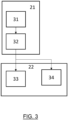
- Fig. 3 depicts in flowchart form several stages of the procedure 20 for detecting a pathogen relating to sample processing and assembly of the analysis reaction.
- a first sample pre-processing stage 21 in which the sample is prepared.
- the sample pre-processing stage 21 in turn includes several stages, a first mixing step 31 with a set of reagents and chemical solutions, specifically formulated to achieve the necessary transformations that allow the biological detection assay to be carried out.
- a second step of the processing stage 21 is a lysis step 32 for rupturing the membrane and/or cell wall of the microorganisms so that DNA or other genetic material is accessible in the medium.
- a simple flow of operations for the lysis step 32 includes: transferring the sample by use of a disposable plastic pipette onto a sample extraction canister, containing a lysate solution, e.g., sodium hydroxide at a concentration of 100 mM.
- a lysate solution e.g., sodium hydroxide at a concentration of 100 mM.
- lysis step 32 includes loading the sample into a syringe and filtering it with a filter of between 0.20 and 0.50 ⁇ m, for example, of the material polyvinylidene fluoride (PVDF), connecting a new pre-filled syringe with the lysis solution on that same filter, collecting the eluate in a canister with drop dispenser for the following operations.
- PVDF material polyvinylidene fluoride
- a second reaction stage 22 including a step of placing the sample 33 already mixed with chemical and biological reagents necessary to carry out the biological detection.
- step of placing a sterile substance 34 also mixed to establish a negative control.
- the basis of detection is an enzymatic nucleic acid amplification reaction, which may occur at constant temperature, e.g., recombinant polymerase amplification (RPA), or loop-mediated isothermal amplification; or with temperature cycles, such as polymerase chain reaction (PCR).
- RPA recombinant polymerase amplification
- PCR polymerase chain reaction
- a fluorescent marker is included whose fluorescence emission intensity under a certain condition and wavelength will be proportional to the amount of pathogen in the sample.
- oligonucleotides and/or probes In order to carry out the specific detection of each pathogen it is necessary for design and manufacture of short sequences of DNA, between 20 and 40 base pairs, called oligonucleotides and/or probes. The correct design of these sequences is essential to carry out the detection of the pathogen correctly. These components will be added to the rest of the chemical reagents, resulting in a reaction mixture, to which the sample or negative control will be added.
- a third data acquisition stage 23 includes a step of collecting analog information 34 generated by the photodetector and a step of collecting digital information 35 generated by the camera, which can be done in parallel.
- the presence of the pathogen activates the reaction in a specific manner, the emission of an optical signal occurring in an intensity proportional to the amount of pathogen present in the sample.
- the device features a wavelength-determined illumination source, and two complementary detection mechanisms: photodetectors and a photographic camera.
- a fourth data pre-processing stage 24 which includes an analog noise filtering step 37 (in the photodiode voltage signal) and a digital noise filtering step 38 (in the image of the photographic camera).
- an analog signal is digitalized.
- a normalization step 40 is carried out and a moving average is applied to these numerical values of the pixels (digital information) and also to the voltage values (analog information) measured by the photodetectors.
- a storage step 41 the results of such operations are stored in a temporary database, e.g. InfluxDB.
- the method continues with a fifth prediction stage 25 where stored information of the data pre-processing stage 24 is collected from the database.
- the prediction stage 25 includes a step of applying a decision algorithm 42, which may incorporate artificial intelligence models, for example, of the machine learning type, to improve prediction accuracy or sensitivity. If a negative control has been performed, there is a step of measuring the increase in fluorescence 43 produced in the sample during the experiment with respect to the negative control. Based on the growth value of said fluorescence level, it is determined whether the result of the sample is positive or negative. Additionally, the abundance of the pathogen detected can be determined from the signal intensity and, by comparison, from database of known values of the pathogen in question.
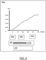
- Fig. 5 is an example of a graphical representation of the results of the visualization stage. They are displayed on a screen that may be touchable to act as a user interface and facilitate interaction with the device, allowing with a start/stop button 26a to begin or end measurements of the device.
- the state 26b in which the device is located is shown, which automatically regulates the temperature, and which indicates the following states: heating and ready. Heating, when the device is, automatically, regulating the temperature to reach the optimum reaction temperature. Ready, when the optimal temperature has been reached and the device may begin to take measurements.
- the process progress bar 26c is updated in real time and a display 26d of the measurements obtained in the prediction stage is presented.
- the time in minutes is shown on the axis of abscissas, while the fluorescence intensity measured in relative fluorescence units (RFU) is shown on the coordinate axis.
- the generated data can be exported through the export button 26e to generate the analysis of the historical series of results.
- the measurement result 26f is shown indicating whether or not the pathogen has been detected and, if applicable, a quantitative value proportional to the amount of pathogen detected.
- Portability The device is easily transportable, does not require any specific condition in the place where it is to be used.
- the device allows to complete the complete flow of analysis of industrial pathogens, including the concentration of the samples, if necessary.
- the use of genetic recognition techniques allows to unequivocally determine one or several specific pathogens in the sample, such as Legionella sp., Listeria sp., Salmonella sp., E. coli, Clostridium sp., Campylobacter sp., Norovirus, Toxoplasma, Staphylococcus sp., Cronobacter sp., Vibrio sp. or Yersinia sp., among others.
- pathogens in the sample such as Legionella sp., Listeria sp., Salmonella sp., E. coli, Clostridium sp., Campylobacter sp., Norovirus, Toxoplasma, Staphylococcus sp., Cronobacter sp., Vibrio sp. or Yersinia sp., among others.
- Versatility allows analyzing the presence of pathogens in a wide variety of samples, for example, water, surfaces or air
- Quantitative analysis capacity The equipment allows to quantify the abundance of a certain pathogen by comparison of the measured signal intensity with that of known values.
- Technological blending The combination of software, hardware and biotechnological elements has a synergistic effect that improves the analytical properties of the device.
- Digitization of data The results of the analysis are obtained in digital format, so they can be easily shared and integrated into industrial facilities management platforms.
- Modularity slight changes in configuration allow the parallel analysis of several samples or different pathogens in the same sample, by selecting different optical lengths for each pathogen (e.g. two different bacteria).
Landscapes
- Health & Medical Sciences (AREA)
- Life Sciences & Earth Sciences (AREA)
- Physics & Mathematics (AREA)
- General Physics & Mathematics (AREA)
- Pathology (AREA)
- Analytical Chemistry (AREA)
- Biochemistry (AREA)
- General Health & Medical Sciences (AREA)
- Chemical & Material Sciences (AREA)
- Immunology (AREA)
- Nuclear Medicine, Radiotherapy & Molecular Imaging (AREA)
- Engineering & Computer Science (AREA)
- Biomedical Technology (AREA)
- Molecular Biology (AREA)
- Spectroscopy & Molecular Physics (AREA)
- Apparatus Associated With Microorganisms And Enzymes (AREA)
- Measuring Or Testing Involving Enzymes Or Micro-Organisms (AREA)
Applications Claiming Priority (2)
| Application Number | Priority Date | Filing Date | Title |
|---|---|---|---|
| ES202230778A ES2939909A1 (es) | 2022-08-30 | 2022-08-30 | Dispositivo para detectar patogenos industriales |
| PCT/ES2023/070266 WO2023222933A1 (fr) | 2022-08-30 | 2023-04-26 | Dispositif pour détecter des agents pathogènes industriels |
Publications (1)
| Publication Number | Publication Date |
|---|---|
| EP4332552A1 true EP4332552A1 (fr) | 2024-03-06 |
Family
ID=86096118
Family Applications (1)
| Application Number | Title | Priority Date | Filing Date |
|---|---|---|---|
| EP23724896.8A Pending EP4332552A1 (fr) | 2022-08-30 | 2023-04-26 | Dispositif pour détecter des agents pathogènes industriels |
Country Status (4)
| Country | Link |
|---|---|
| EP (1) | EP4332552A1 (fr) |
| ES (1) | ES2939909A1 (fr) |
| TW (1) | TW202409264A (fr) |
| WO (1) | WO2023222933A1 (fr) |
Family Cites Families (7)
| Publication number | Priority date | Publication date | Assignee | Title |
|---|---|---|---|---|
| ITMO20030170A1 (it) * | 2003-06-12 | 2004-12-13 | Map S R L | Metodo e apparecchio per analizzare la concentrazione |
| US20100075312A1 (en) * | 2006-09-28 | 2010-03-25 | Stokes Bio Limited | Qpcr analysis apparatus |
| DE102007007040A1 (de) * | 2007-02-07 | 2008-08-14 | Carl Zeiss Microlmaging Gmbh | Messeinrichtung zur optischen und spektroskopischen Untersuchung einer Probe |
| WO2015054695A2 (fr) * | 2013-10-11 | 2015-04-16 | Immunetics, Inc. | Lecteur d'analyse à diodes électroluminescentes ayant une commande par écran tactile et une identification d'échantillon par code à barres |
| WO2017025984A1 (fr) * | 2015-08-07 | 2017-02-16 | Council Of Scientific And Industrial Research | Dispositif de diagnostic moléculaire en temps réel intégré dans un téléphone intelligent |
| EP3432785B1 (fr) * | 2016-03-25 | 2021-03-10 | The General Hospital Corporation | Détection d'acide nucléique fluorescent basée sur la polarisation |
| CN109632666A (zh) * | 2018-12-24 | 2019-04-16 | 枣庄学院 | 基于智能手机的便携式多通道分光光度计及测定吸光度的方法 |
-
2022
- 2022-08-30 ES ES202230778A patent/ES2939909A1/es active Pending
-
2023
- 2023-04-26 WO PCT/ES2023/070266 patent/WO2023222933A1/fr active Application Filing
- 2023-04-26 EP EP23724896.8A patent/EP4332552A1/fr active Pending
- 2023-05-19 TW TW112118742A patent/TW202409264A/zh unknown
Also Published As
| Publication number | Publication date |
|---|---|
| ES2939909A1 (es) | 2023-04-27 |
| WO2023222933A1 (fr) | 2023-11-23 |
| TW202409264A (zh) | 2024-03-01 |
Similar Documents
| Publication | Publication Date | Title |
|---|---|---|
| Lee et al. | Optofluidic Raman-activated cell sorting for targeted genome retrieval or cultivation of microbial cells with specific functions | |
| CN102803959B (zh) | 用于样品中的微生物剂的快速的识别和/或表征的系统和方法 | |
| JP2016073288A (ja) | 固体又は半固体培地上の微生物のキャラクタリゼーション方法 | |
| US10233481B2 (en) | Multi-sample laser-scatter measurement instrument with incubation feature and systems for using the same | |
| CN101218507A (zh) | 用于快速分析液体样本中的微生物物质的系统 | |
| JPH06510666A (ja) | 液体監視装置 | |
| KR20090003220A (ko) | 생체인식 분자에 접합된 마이크로비드를 사용하여 병원체를검출하는 방법 | |
| US20150284763A1 (en) | Method of Using Laser-Induced Breakdown Spectroscopy for the Identification and Classification of Bacteria | |
| US20160161404A1 (en) | System Using Laser-Scatter Measurement Instrument For Organism Identification And Related Network | |
| CN105548114B (zh) | 一种基于酵母菌实时在线分析大气颗粒物毒性的方法 | |
| CN100462714C (zh) | 一种多样本微生物污染快速筛拣的方法 | |
| EP4332552A1 (fr) | Dispositif pour détecter des agents pathogènes industriels | |
| US20200156057A1 (en) | Systems and methods using bacteriophage-mediated lysis for detection and identification of microorganisms in a fluid sample | |
| JP6706267B2 (ja) | 迅速内部蛍光法を使用した耐性遺伝子を含む細菌の無試薬同定 | |
| EP3227664B1 (fr) | Instrument de mesure à diffusion laser à multiples échantillons avec caractéristique d'incubation, et systèmes pour l'utiliser | |
| US20230027503A1 (en) | System and method of biochemical molecule synthesis and detection in a point of collection setting | |
| RU93990U1 (ru) | Устройство для мультисубстратной флуоресцентной идентификации биологических микрообъектов и их биологических свойств | |
| JP5799086B2 (ja) | 分類学的階層分類を用いる微生物因子の同定及び/又はキャラクタリゼーション | |
| US9464329B2 (en) | Portable systems and methods for amplifying nucleotides and detecting nucleotide sequences | |
| CN106872341A (zh) | 一种基于智能手机的移动即时微生物诊断仪 | |
| JP5030015B2 (ja) | 微生物計測システム | |
| Iyengar et al. | Spectral analysis and sorting of microbial organisms using a spectral sorter | |
| US20230109581A1 (en) | Color reading for diagnostic tests | |
| Fluch et al. | What's on the menu? A novel molecular gut content analysis to investigate the feeding behavior of phytophagous insects | |
| Narayana Iyengar et al. | Identifying antibiotic-resistant strains via cell sorting and elastic-light-scatter phenotyping |
Legal Events
| Date | Code | Title | Description |
|---|---|---|---|
| STAA | Information on the status of an ep patent application or granted ep patent |
Free format text: STATUS: UNKNOWN |
|
| STAA | Information on the status of an ep patent application or granted ep patent |
Free format text: STATUS: THE INTERNATIONAL PUBLICATION HAS BEEN MADE |
|
| PUAI | Public reference made under article 153(3) epc to a published international application that has entered the european phase |
Free format text: ORIGINAL CODE: 0009012 |
|
| STAA | Information on the status of an ep patent application or granted ep patent |
Free format text: STATUS: REQUEST FOR EXAMINATION WAS MADE |
|
| 17P | Request for examination filed |
Effective date: 20231127 |
|
| AK | Designated contracting states |
Kind code of ref document: A1 Designated state(s): AL AT BE BG CH CY CZ DE DK EE ES FI FR GB GR HR HU IE IS IT LI LT LU LV MC ME MK MT NL NO PL PT RO RS SE SI SK SM TR |