EP4286528A1 - Verfahren und vorrichtung zum nachweis von transglutaminasen - Google Patents

Verfahren und vorrichtung zum nachweis von transglutaminasen Download PDF

Info

Publication number
EP4286528A1
EP4286528A1 EP22176713.0A EP22176713A EP4286528A1 EP 4286528 A1 EP4286528 A1 EP 4286528A1 EP 22176713 A EP22176713 A EP 22176713A EP 4286528 A1 EP4286528 A1 EP 4286528A1
Authority
EP
European Patent Office
Prior art keywords
donor
glutamine
transglutaminase
sample
fxiii
Prior art date
Legal status (The legal status is an assumption and is not a legal conclusion. Google has not performed a legal analysis and makes no representation as to the accuracy of the status listed.)
Pending
Application number
EP22176713.0A
Other languages
English (en)
French (fr)
Inventor
Joachim Hoenes
Christian Weber
Katja Schmitz
Natascha Krestan
Tobias MECKEL
Current Assignee (The listed assignees may be inaccurate. Google has not performed a legal analysis and makes no representation or warranty as to the accuracy of the list.)
Technische Universitaet Darmstadt
Original Assignee
Technische Universitaet Darmstadt
Priority date (The priority date is an assumption and is not a legal conclusion. Google has not performed a legal analysis and makes no representation as to the accuracy of the date listed.)
Filing date
Publication date
Application filed by Technische Universitaet Darmstadt filed Critical Technische Universitaet Darmstadt
Priority to EP22176713.0A priority Critical patent/EP4286528A1/de
Priority to US18/326,099 priority patent/US20230392186A1/en
Publication of EP4286528A1 publication Critical patent/EP4286528A1/de
Pending legal-status Critical Current

Links

Images

Classifications

    • CCHEMISTRY; METALLURGY
    • C12BIOCHEMISTRY; BEER; SPIRITS; WINE; VINEGAR; MICROBIOLOGY; ENZYMOLOGY; MUTATION OR GENETIC ENGINEERING
    • C12QMEASURING OR TESTING PROCESSES INVOLVING ENZYMES, NUCLEIC ACIDS OR MICROORGANISMS; COMPOSITIONS OR TEST PAPERS THEREFOR; PROCESSES OF PREPARING SUCH COMPOSITIONS; CONDITION-RESPONSIVE CONTROL IN MICROBIOLOGICAL OR ENZYMOLOGICAL PROCESSES
    • C12Q1/00Measuring or testing processes involving enzymes, nucleic acids or microorganisms; Compositions therefor; Processes of preparing such compositions
    • C12Q1/56Measuring or testing processes involving enzymes, nucleic acids or microorganisms; Compositions therefor; Processes of preparing such compositions involving blood clotting factors, e.g. involving thrombin, thromboplastin, fibrinogen
    • CCHEMISTRY; METALLURGY
    • C12BIOCHEMISTRY; BEER; SPIRITS; WINE; VINEGAR; MICROBIOLOGY; ENZYMOLOGY; MUTATION OR GENETIC ENGINEERING
    • C12QMEASURING OR TESTING PROCESSES INVOLVING ENZYMES, NUCLEIC ACIDS OR MICROORGANISMS; COMPOSITIONS OR TEST PAPERS THEREFOR; PROCESSES OF PREPARING SUCH COMPOSITIONS; CONDITION-RESPONSIVE CONTROL IN MICROBIOLOGICAL OR ENZYMOLOGICAL PROCESSES
    • C12Q1/00Measuring or testing processes involving enzymes, nucleic acids or microorganisms; Compositions therefor; Processes of preparing such compositions
    • C12Q1/48Measuring or testing processes involving enzymes, nucleic acids or microorganisms; Compositions therefor; Processes of preparing such compositions involving transferase
    • BPERFORMING OPERATIONS; TRANSPORTING
    • B01PHYSICAL OR CHEMICAL PROCESSES OR APPARATUS IN GENERAL
    • B01LCHEMICAL OR PHYSICAL LABORATORY APPARATUS FOR GENERAL USE
    • B01L3/00Containers or dishes for laboratory use, e.g. laboratory glassware; Droppers
    • B01L3/50Containers for the purpose of retaining a material to be analysed, e.g. test tubes
    • B01L3/502Containers for the purpose of retaining a material to be analysed, e.g. test tubes with fluid transport, e.g. in multi-compartment structures
    • B01L3/5023Containers for the purpose of retaining a material to be analysed, e.g. test tubes with fluid transport, e.g. in multi-compartment structures with a sample being transported to, and subsequently stored in an absorbent for analysis
    • GPHYSICS
    • G01MEASURING; TESTING
    • G01NINVESTIGATING OR ANALYSING MATERIALS BY DETERMINING THEIR CHEMICAL OR PHYSICAL PROPERTIES
    • G01N33/00Investigating or analysing materials by specific methods not covered by groups G01N1/00 - G01N31/00
    • G01N33/48Biological material, e.g. blood, urine; Haemocytometers
    • G01N33/50Chemical analysis of biological material, e.g. blood, urine; Testing involving biospecific ligand binding methods; Immunological testing
    • G01N33/53Immunoassay; Biospecific binding assay; Materials therefor
    • G01N33/573Immunoassay; Biospecific binding assay; Materials therefor for enzymes or isoenzymes
    • BPERFORMING OPERATIONS; TRANSPORTING
    • B01PHYSICAL OR CHEMICAL PROCESSES OR APPARATUS IN GENERAL
    • B01LCHEMICAL OR PHYSICAL LABORATORY APPARATUS FOR GENERAL USE
    • B01L2300/00Additional constructional details
    • B01L2300/06Auxiliary integrated devices, integrated components
    • B01L2300/069Absorbents; Gels to retain a fluid
    • BPERFORMING OPERATIONS; TRANSPORTING
    • B01PHYSICAL OR CHEMICAL PROCESSES OR APPARATUS IN GENERAL
    • B01LCHEMICAL OR PHYSICAL LABORATORY APPARATUS FOR GENERAL USE
    • B01L2300/00Additional constructional details
    • B01L2300/08Geometry, shape and general structure
    • B01L2300/0809Geometry, shape and general structure rectangular shaped
    • B01L2300/0825Test strips
    • GPHYSICS
    • G01MEASURING; TESTING
    • G01NINVESTIGATING OR ANALYSING MATERIALS BY DETERMINING THEIR CHEMICAL OR PHYSICAL PROPERTIES
    • G01N2333/00Assays involving biological materials from specific organisms or of a specific nature
    • G01N2333/90Enzymes; Proenzymes
    • G01N2333/91Transferases (2.)
    • G01N2333/91045Acyltransferases (2.3)
    • G01N2333/91074Aminoacyltransferases (general) (2.3.2)
    • G01N2333/9108Aminoacyltransferases (general) (2.3.2) with definite EC number (2.3.2.-)
    • G01N2333/91085Transglutaminases; Factor XIIIq (2.3.2.13)

Definitions

  • the present invention relates to a method and a device for detecting transglutaminases, in particular blood coagulation factor XIII (FXIII).
  • FXIII blood coagulation factor XIII
  • the invention is based on a rapid, point-of-care test principle for transglutaminases and enables precise determination in a short time and with easy handling.
  • Blood in the body serves as a transport and communication system and is essential for maintaining body functions. If a vascular injury occurs, the wound must be closed quickly to keep blood loss as low as possible. It occurs through the optimal interaction between platelets and coagulation factors.
  • the main substance of the thrombus is fibrin monomers assembled into polymers. These fibrin monomers are created by the cleavage of low molecular weight peptides from fibrinogen by the serine protease thrombin.
  • blood clotting factor XIII also called fibrin stabilizing factor, catalyzes a covalent cross-linking of the fibrin molecules with each other. This further stabilization protects the thrombus from premature lysis.
  • the processes mentioned represent the final steps of the complex blood coagulation cascade.
  • Blood clotting factor XIII also referred to as "factor It is present in blood plasma as an inactive heterotetramer with two A and B subunits each.
  • the heterotetramer represents the proenzyme; under physiological conditions, FXIII is activated by thrombin.
  • the N-terminal activation peptides are first cleaved by thrombin (FXIII) and the dissociation equilibrium is shifted towards the monomers. Low calcium concentrations are then sufficient for activation (FXIII a ).
  • Non-physiological, thrombin-independent activation by high calcium concentrations can also dissociate the complex into active monomers (FXIII 0 ).
  • Activated FXIII as a transglutaminase, links proteins by forming an isopeptide bond between a ⁇ -glutamine residue and the ⁇ -amino residue in an exposed lysine side chain. This reaction releases ammonia.
  • the common three-letter code is also used to designate amino acids or amino acid residues, for example "Gln” for glutamine or "Lys” for lysine.
  • FXIII FXIII
  • hereditary FXIII deficiency is usually diagnosed in infancy due to umbilical stump bleeding. This hereditary form of FXIII deficiency is very rare. An acquired deficiency occurs more frequently and is triggered, for example, by a limited synthetic capacity of the liver, chronic inflammatory bowel disease, sepsis or consumption coagulopathies. Conventional coagulation tests such as thromboplastin time or activated partial prothrombin time (aPTT) are not suitable for detecting FXIII deficiency. Platelet count and fibrinogen concentration also do not provide any information about the FXIII.
  • FXIII can currently be determined photometrically in the clinical chemistry laboratory, for example.
  • FXIII of the sample is activated by thrombin and links a glutamine donor substrate with an amine donor, for example glycine ethyl ester, with the release of ammonia.
  • the resulting ammonia can be determined in a coupled enzymatic reaction.
  • NADH, ⁇ -ketoglutarate and free ammonia are converted to NAD + and glutamate by glutamate dehydrogenase.
  • the decrease in NADH is measured via absorbance at 340 nm and is directly proportional to FXIII activity.
  • factor XIII can be detected by antibodies in an enzyme-linked immunosorbent assay (ELISA).
  • ELISA enzyme-linked immunosorbent assay
  • Factor XIII binds to a specific antibody that is immobilized in the test vessel. After other substances have been washed out, a second antibody binds to the immobilized factor XIII.
  • An enzyme conjugated to the antibody converts the signaling substrate that is subsequently added. The substance formed can be quantified photometrically or fluorimetrically and the FXIII level can be determined from this.
  • tests require the sample to be transported to the clinical chemistry laboratory, centrifuged to obtain plasma, the actual test to be carried out as well as evaluation and feedback to the client.
  • Such processes involving the clinical laboratory are (even with other test procedures) too slow in acute cases to assess the need for an administration and to determine the correct dosage of the preparations.
  • Alternative test methods are thromboelastometry (more qualitative and quite prone to interference) and enzyme immunoassays (very complex and expensive, not activity but protein determination).
  • Point of Care (POC) rapid tests could be used on site (e.g. in the operating room) and enable a correct decision within a few minutes, but are currently not available for a variety of reasons.
  • POC Point of Care
  • Blood is a highly complex mixture of cells (main component erythrocytes) and components dissolved in the serum, such as proteins and lipids.
  • the strong light absorption in hemoglobin makes photometric or fluorimetric determinations in whole blood difficult or impossible. Electrochemical determinations are also severely disturbed by the cells and the viscosity of the blood matrix. A (partial) correction is possible using methods such as impedance spectroscopy, but this also involves a lot of effort.
  • the erythrocytes A quick separation of the erythrocytes is possible using glass fiber fleeces.
  • soluble blood components remain in the blood plasma obtained in this way.
  • the high concentrations of serum albumin and fibrinogen have the potential to interfere with assays for FXIII.
  • the dansylcadaverine test is known, in which a fluorescently labeled amine is incorporated into a glutamine-containing protein, e.g. dimethyl casein.
  • the blue shift and enhancement of fluorescence due to the changed chemical environment of the fluorophore can be used as a measure of the activity of FXIII.
  • serum albumin in physiological concentrations binds dansylcadaverine, withdraws it from the enzymatic reaction and advances the fluorescence spectrum so that any residual reaction that may still be taking place is no longer visible.
  • Fibrinogen is another interfering component: physiological activation of FXIII requires cleavage by thrombin; Alternatively, you can work with high Ca 2+ concentrations. In both cases the coagulation reaction is initiated; the clot hinders the detection reactions due to light scattering and high viscosity.
  • the prior art does not offer a method with which an accurate determination of transglutaminases, in particular of FXIII, would be possible in a reasonably short time and with easy handling.
  • the object of the present invention is to provide a rapid, POC-suitable test principle from human blood.
  • the method of the present invention is suitable for all transglutaminases. Determining the enzymatic activity of blood clotting factor XIII (FXIII) in a sample is particularly preferred.
  • Binding occurs to an immobilized glutamine donor. This can also be the substrate used for detection, but does not have to be. To demonstrate activity, additional substrate can be added, either the same glutamine donor or a different one.
  • the detection can either be carried out by washing out unbound amine donor and detecting that remaining in the reactive field or, preferably, by a spectral shift of the chromophore on the amine donor due to the binding.
  • a number of fluorophore residues can be used that react directly to the changed polarity of the environment, e.g. dansyl, Dapoxyl, 4-chloro-7-nitrobenzofurazan (NBD), 6-dimethylamino-2,3-naphthalimide.
  • NBD 4-chloro-7-nitrobenzofurazan
  • Such fluorophores can be summarized under the term “spectroscopic polarity sensors”.
  • the "amount" of incorporated amine can be determined in particular indirectly spectroscopically;
  • the activity of the transglutaminase can be determined in particular via the temporal change in the spectroscopic signal.
  • Diamines preferably putrescine, cadaverine and/or carbohydrazide, are particularly suitable as linkers between chromophore and glutamine.
  • the signal is preferably proportional to the rate of binding of the amine donor to the glutamine donor substrate catalyzed by the transglutaminase.
  • the immobilized glutamine donor is preferably the glutamine donor substrate to which the amine donor is bound by the glutaminase.
  • the transglutaminase is preferably selected from the group of human transglutaminases.
  • the transglutaminase blood coagulation factor XIII (FXIII) is particularly preferred.
  • the sample preferably contains exactly one transglutaminase.
  • the sample is preferably a blood sample, in particular a human blood sample.
  • the sample can also be a tissue sample, for example an extract from a tissue.
  • the sample contains transglutaminase, in particular FXIII, preferably in an activity of 5% to 200%, for example from 25% to 175%, from 50% to 150%, from 75% to 125%, or from 90% to 110% based on the activity in standard plasma.
  • the activity is preferably at least 5%, at least 25%, at least 50%, at least 75%, or at least 90% based on the activity in standard plasma.
  • the activity is preferably at most 200%, at most 175%, at most 150%, at most 125%, or at most 110% based on the activity in standard plasma.
  • an immobilized glutamine donor in particular an immobilized glutamine donor substrate, preferably an immobilized FXIII substrate
  • the (FXIII) substrate may be a physiological (FXIII) substrate.
  • the (FXIII) substrate is not a physiological (FXIII) substrate.
  • (FXIII) substrate it is meant in particular that the transglutaminase (in particular FXIII) can catalyze the reaction of a glutamine residue of the substrate with the amino group of an amine donor. If the glutamine donor includes at least one glutamine residue, but the transglutaminase (especially FXIII) cannot catalyze a bond between the glutamine residue and the amine donor, the glutamine donor is not a (FXIII) substrate.
  • the glutamine donor can in particular be a peptide, preferably a polypeptide or protein.
  • the glutamine donor contains at least one glutamine residue.
  • the glutamine donor preferably contains several glutamine residues, for example at least two, at least three, or at least five glutamine residues.
  • Proteins with a molecular weight of 10 to 300 kDa, for example 10 to 100 kDa or 12 to 30 kDa, are preferred.
  • the molecular weight of the glutamine donor can be, for example, at least 10 kDa or at least 12 kDa.
  • the molecular weight of the glutamine donor can, for example, be at most 300 kDa, at most 100 kDa or at most 30 kDa.
  • the glutamine content of the glutamine donor is preferably in a range from 3% to 15% based on the total number of residues, for example in a range from 5% to 12% or from 7% to 10%.
  • the glutamine content of the glutamine donor can be, for example, at least 3%, at least 5% or at least 7%.
  • the glutamine content of the glutamine donor can, for example, be at most 15%, at most 12% or at most 10%.
  • the glutamine donor is particularly preferably a substrate of transglutaminase, also called a glutamine donor substrate.
  • a glutamine donor substrate This has the advantage that the immobilized glutamine donor can act as a glutamine donor substrate to which the transglutaminase binds the amine donor. This means that there is no need to add additional transglutaminase substrate.
  • the immobilized glutamine donor is not identical to the glutamine donor substrate of step e) of the method of the present invention.
  • glutamine donor substrate is added, to which the amine donor can be bound by the transglutaminase.
  • step e it is possible, for example, to provide a mixture or solution which comprises amine donor and glutamine donor substrate before carrying out step e), and to bring this into contact with the transglutaminase bound to the glutamine donor in step e), so that the binding between amine donor and glutamine donor substrate occurs the transglutaminase can be formed.
  • the addition of glutamine donor substrate can also take place if the immobilized glutamine donor itself is already a glutamine donor substrate.
  • peptide or “peptides” collectively describe molecules containing amino acids linked together via peptide bonds. This includes oligopeptides (peptides with up to ten amino acid residues) and polypeptides (peptides with more than ten amino acid residues). Polypeptides with more than 50 amino acid residues are also called proteins.
  • peptide or “peptides” as used in the present disclosure also expressly include proteins.
  • the glutamine donor in particular the FXIII substrate
  • the immobilization includes in particular the binding of the glutamine donor to a carrier, preferably Sepharose, cellulose or paper, each preferably functionalized with DEAE (diethylaminoethyl group) or other cationic groups.
  • the immobilization can additionally or alternatively also include precipitation of the glutamine donor, in particular the FXIII substrate, in the presence of the carrier, for example precipitation with Ca 2+ .
  • the immobilized glutamine donor can exist in various forms.
  • the immobilized glutamine donor can be in the form of a suspension, in particular in a reaction vessel. It is also possible to immobilize the glutamine donor on a solid, for example on a paper support in a test strip.
  • the immobilized glutamine donor can undergo modifications, for example chemical have modifications. Modifications of lysine residues are particularly preferred.
  • the glutamine donor can be methylated, acetylated or succinylated, for example dimethylated, in particular N,N-dimethylated or also acetamidinated.
  • the glutamine donor can be methylated, for example dimethylated, in particular N,N-dimethylated. Dimethylation occurs in particular by reacting the glutamine donor with formaldehyde and sodium borohydride. Alternatively or additionally, one or more other reducing agents can be used. The reaction depends on the accessibility of the lysine - easily accessible reacts fastest. The degree of dimethylation is controlled by the amount of reagents added and the reaction time. The highest possible degree of dimethylation of the freely accessible lysines is preferred because this prevents cross-linking within a protein or between proteins. The degree of dimethylation is preferably more than 90%.
  • the immobilized glutamine donor can preferably be succinylated or acetamidinated.
  • the glutamine donor is particularly preferably selected from the group consisting of casein, in particular dimethylcasein, and lysozyme, in particular succinylated lysozyme, preferably succinylated chicken protein lysozyme ( Gallus gallus ) .
  • casein from cow's milk ( Bos taurus ) is preferred.
  • Dimethylcasein is well known to those skilled in the art because it is usually used to determine proteases by detecting newly formed terminal amino groups.
  • the transglutaminase present in the sample is activated.
  • activation may occur by thrombin.
  • clotting should be avoided by adding inhibitors of fibrin aggregation, for example the tetrapeptide GPRP.
  • High Ca 2+ concentrations are preferably used for activation. This is particularly true when the transglutaminase is FXIII, but also with other transglutaminases.
  • the Ca 2+ concentration is preferably in a range from 100 to 500 mM, for example from 200 to 400 mM.
  • the Ca 2+ concentration can be, for example, at least 100 mM or at least 200 mM.
  • the Ca 2+ concentration can, for example, be at most 500 mM or at most 400 mM.
  • Lower Ca 2+ concentrations, for example concentrations of 1 to ⁇ 100 mM, in particular 1 to 10 mM are also possible in conjunction with high ionic strength.
  • a high ionic strength can for example, by NaCl concentration in a range from 200 to 1000 mM, in particular from 400 to 700 mM.
  • FXIII is preferably present as FXIII a (cleaved by thrombin) or as FXIII 0 (activated by Ca 2+ ions).
  • the sample is brought into contact with the immobilized glutamine donor.
  • the details of the contact essentially depend on the type of immobilization of the glutamine donor. If the immobilized glutamine donor is present as a suspension, in particular in a reaction vessel, the contacting can be carried out, for example, by adding the sample to the suspension, for example by adding the sample to the reaction vessel. If, on the other hand, the glutamine donor is immobilized on a solid, such as a test strip, the contact can be made by bringing the sample and the solid, in particular the test strip, into contact. The sample can, for example, be applied to the solid at the point where the glutamine donor is also immobilized.
  • the sample it is also possible to apply the sample to a location where the glutamine donor is not immobilized.
  • This can be a preferred option in particular if the solid body, in particular the test strip, is designed in such a way that the sample can migrate through the solid body, for example by capillary forces, and can thus be brought into contact with the immobilized glutamine donor.
  • the method preferably does not contain any step of pre-treating the sample. It is a particular advantage of the present invention that the sample does not require any pretreatment. In particular, it is possible to bring the sample, for example a (human) blood sample, into contact with the immobilized glutamine donor after its collection without pretreatment. Calcium can be added. If the glutamine donor is precipitated with Ca 2+ , the calcium in the precipitated glutamine donor can also contribute to or cause activation of the transglutaminase.
  • the contacting according to step c) of the method can include an incubation step.
  • the incubation step is particularly advantageous in order to further increase the extent of binding of the transglutaminase to the glutamine donor.
  • the incubation time is preferably in a range from 10 seconds to 5 minutes, for example from 0.5 to 5 minutes or from 1 to 3 minutes.
  • the incubation period can, for example, be at least 10 seconds, at least 30 seconds or at least 60 seconds.
  • the incubation time can, for example, be a maximum of 300 seconds or a maximum of 180 seconds.
  • the incubation time refers in particular to the period from the start of the contact in step c) to the start of the removal of components of the sample that are not bound to the glutamine donor in step d).
  • step c) of the method leads in particular to binding of the transglutaminase to the immobilized glutamine donor.
  • Transglutaminases can bind to a glutamine of the glutamine donor, in particular with an essential SH group, whereby a thioester is formed with the elimination of ammonia, as illustrated in the following reaction scheme 1.
  • E-SH describes the enzyme (the transglutaminase) with its essential SH group in the active center.
  • the side chain of a glutamine residue from the glutamine donor is shown.
  • the remaining glutamine donor is shown as “R”.
  • transglutaminases bind not only to physiological glutamine donors, but also to other glutamine donors, especially those that have at least one glutamine residue.
  • the transglutaminase (especially FXIII) binds to the immobilized glutamine donor, in particular to the immobilized glutamine donor substrate.
  • the bond between transglutaminase and glutamine donor is preferably a covalent bond between an amino acid in the active site of the glutaminase and a glutamine residue of the glutamine donor.
  • the bond between transglutaminase and immobilized glutamine donor is in particular a thioester bond.
  • the thioester bond is preferably formed by the reaction of an SH group of the transglutaminase (in particular the essential SH group) and a glutamine residue of the immobilized glutamine donor.
  • step d) of the method according to the invention components of the sample that are not bound to the immobilized glutamine donor are removed.
  • a component of the sample that binds to the glutamine donor is, in particular, transglutaminase, preferably FXIII.
  • Other components of the sample, in particular components that potentially interfere with the detection of transglutaminase, preferably do not bind to the glutamine donor or bind only weakly.
  • a low level of binding is particularly one that allows removal in one or more washing steps.
  • Step d) of removing components of the sample that are not bound to the glutamine donor preferably comprises one or more washing steps.
  • the number of washing steps is preferably in a range from 1 to 3. Continuous washing is particularly preferred, in particular by flowing the immobilized glutamine donor over the carrier. Volumes of 1 to 3 times the carrier volume are used here.
  • the carrier volume is the volume of liquid present in the carrier.
  • the details of removing components of the sample that are not bound to the glutamine donor essentially depend on the type of immobilization of the glutamine donor.
  • the removal can be carried out, for example, by carrying out one or more washing steps, a washing step comprising separating the suspension into solid and liquid components, removing the liquid components and adding Washing solution, in particular washing buffer, includes.
  • the suspension can be separated into solid and liquid components in particular by centrifugation. Centrifugation for 0.5 to 2 minutes is preferred, with the required acceleration depending on the type of carrier.
  • the removal of components of the sample that are not bound to the glutamine donor can be carried out, for example, by carrying out one or more washing steps, one washing step being the addition of washing solution to the Solids, in particular for test strips, include.
  • the washing solution is preferably a washing buffer.
  • the washing solution can, for example, be applied to the solid at the point where the glutamine donor is also immobilized. However, it is also possible to apply the washing solution at a location where the glutamine donor is not immobilized. This can be a preferred option in particular if the solid, in particular Test strip is designed in such a way that the washing solution can migrate through the solid, for example by capillary forces, and can thus be brought into contact with the immobilized glutamine donor. This makes it possible to remove components of the sample that are not bound to the glutamine donor.
  • the washing solution or the washing buffer preferably has an approximately isotonic composition, for example 100 mM NaCl and 50 mM HEPES.
  • the pH is preferably in a range from 6.0 to 9.0, more preferably from 7.0 to 8.0.
  • the pH is preferably at least 6.0 or at least 7.0.
  • the pH is preferably at most 9.0 or at most 8.0.
  • the washing solution or the washing buffer can also be, for example, PBS (phosphate-buffered saline), in particular 137 mM NaCl, 2.7 mM KCl and 12 mM phosphate (HPO 4 2- and H 2 PO 4 - ). , pH 7.4.
  • the washing solution or the washing buffer can contain one or more biocides, for example NaN 3 , preferably in concentrations of 0.01 to 1.0 mM or of 0.05 to 0.5 mM, for example about 0.1 mM.
  • R'-NH 2 denotes an amine donor. Hydrolysis is slower than aminolysis and is therefore shown in dashed lines.
  • Aminolysis can be used to detect transglutaminase, as described in more detail below.
  • premature hydrolysis can lead to premature release the transglutaminase from the immobilized glutamine donor, so that the corresponding transglutaminase can no longer be detected by aminolysis.
  • the hydrolysis rate should be as low as possible.
  • the glutamine donor has more than one glutamine residue. In this way, it can be achieved in particular that the transglutaminase is not or not significantly removed from the glutamine donor even after several washing steps. In this way, several washing steps can be carried out and improved removal of components of the sample that are not bound to the glutamine donor can be achieved without impairing the detection of transglutaminase due to excessive washing out.
  • Such a tight binding of the transglutaminase to a glutamine donor, in particular to a glutamine donor substrate is surprising. Without wishing to be bound to any particular theory, the influence of the number of glutamine residues on binding could be explained, at least in part, as follows.
  • the environment of the glutamine determines the rate of aminolysis and hydrolysis.
  • the transglutaminase will bind to one of the glutamines. If the hydrolysis is fast enough, the enzyme is released again and can repeat the reaction. It remains immobilized on a glutamine with slow hydrolysis. The system is self-optimizing. The transglutaminase binds to the glutamine residue with the slowest hydrolysis, either directly or after several unsuccessful attempts due to hydrolysis on other glutamine residues. This is illustrated in the following reaction scheme 3.
  • R 1 , R 2 and R 3 indicate that they are different glutamine residues of the glutamine donor.
  • the various hydrolysis rate constants are denoted by k 1 , k 2 and k 3 .
  • the hydrolysis rate of the third glutamine is particularly low and is therefore shown in dashed lines.
  • the glutamine residues are preferably present in a molar excess compared to the transglutaminase.
  • the transglutaminase bound to the glutamine donor is brought into contact with an amine donor, so that the amine donor is bound by the transglutaminase to a glutamine donor substrate.
  • the bond between the transglutaminase and the immobilized glutamine donor is broken.
  • the immobilized glutamine donor is itself a glutamine donor substrate, the transglutaminase can catalyze the formation of a bond between the amine donor and the immobilized glutamine donor substrate.
  • the immobilized glutamine donor is not a glutamine donor substrate, the amine donor is not bound to the immobilized glutamine donor, but rather to the additionally present glutamine donor substrate.
  • the glutamine donor substrate can be added, for example, separately or together with the amine donor.
  • the amine donor can include a chromophore.
  • a fluorescent amine donor is particularly preferred, particularly preferably dansylcadaverine, dapoxylcadaverine or NBD-cadaverine.
  • NBD refers to the fluorophore 4-chloro-7-nitro-benzofurazan.
  • the amine donor preferably has at least one fluorescent group.
  • the amine donor can have at least one dye.
  • An amine donor in the sense of the present invention is in particular a molecule that has at least one primary amino group on a short aliphatic chain.
  • the primary amino group can be, for example, the ⁇ -amino group of lysine or the free amino group of a one-substituted diamine such as cadaverine or putrescine.
  • a substituted carbohydrazide group is also possible.
  • reaction scheme 2 an amine donor is shown as R'-NH 2 as an example.
  • the amine donor is bound to a glutamine donor substrate by the transglutaminase bound to the immobilized glutamine donor.
  • the (thioester) bond between transglutaminase and glutamine donor breaks during aminolysis.
  • the bond between amine donor and glutamine donor substrate is preferably an amide bond, more preferably a carboxamide bond.
  • the amine donor is preferably water-soluble.
  • the amine donor preferably has one Water solubility greater than 10 ⁇ M.
  • the amine donor preferably has a molecular weight in a range of 100 to 1000 Da, for example 150 to 500 Da.
  • the amine donor preferably has a molecular weight of at least 100 Da or at least 150 Da.
  • the amine donor preferably has a molecular weight of at most 1000 Da or at most 500 Da.
  • the extent of binding of the amine donor to the glutamine donor substrate is preferably proportional to the activity of the transglutaminase bound to the immobilized glutamine donor.
  • the activity of the transglutaminase bound to the immobilized glutamine donor is in turn preferably proportional to the proportion of transglutaminase in the sample or to the enzyme activity of the transglutaminase present in the sample.
  • the details of bringing the transglutaminase bound to the immobilized glutamine donor into contact with the amine donor essentially depend on the type of immobilization of the glutamine donor.
  • the contacting can be carried out, for example, by adding the amine donor to the suspension in the reaction vessel.
  • the glutamine donor is immobilized on a solid, such as a test strip
  • the bringing into contact can be carried out by bringing the amine donor and the solid, in particular the test strip, into contact.
  • the amine donor can, for example, be applied to the solid at the point where the glutamine donor is also immobilized. However, it is also possible to apply the amine donor at a location where the glutamine donor is not immobilized. This can be a preferred option in particular if the solid, in particular the test strip, is designed in such a way that the amine donor can migrate through the solid, for example by capillary forces, and can thus be brought into contact with the immobilized glutamine donor.
  • the amine donor is preferably in solution before and/or during contact.
  • Preferred buffer solutions contain approximately isotonic concentrations of NaCl and buffer substances such as phosphate or HEPES, in particular with a pH in a range from 6.0 to 9.0, preferably in a range from 7.0 to 8.0. pH values that are too low or too high could promote the hydrolysis of the thioester and thus worsen the signal.
  • the pH is preferably at least 6.0 or at least 7.0.
  • the pH is preferably at most 9.0 or at most 8.0.
  • the buffer solution may contain one or more biocides, for example NaN 3 , preferably in concentrations of 0.01 to 1.0 mM or of 0.05 to 0.5 mM, for example about 0.1 mM.
  • the enzymatic activity of the transglutaminase is determined using a signal that is generated by the binding of the amine donor to the glutamine donor substrate.
  • the signal can in particular be proportional to the rate of binding of the amine donor to the glutamine donor substrate catalyzed by the transglutaminase.
  • the signal can therefore be proportional to the increase over time in the amine donor bound to the glutamine donor substrate.
  • the greater the enzymatic activity of the transglutaminase in the sample the greater the speed of binding of the amine donor to the glutamine donor substrate and, accordingly, the signal.
  • the signal can in particular be a spectral change per unit of time.
  • one or more washing steps can be carried out before determining the amount of bound amine donor.
  • one or more washing steps can be carried out as described in step d) with regard to the removal of components of the sample that are not bound to the substrate.
  • the number of washing steps is preferably in a range from 1 to 3 or a continuous overflow with washing buffer is used.
  • the washing steps can be used to remove amine donor that is not bound to the glutamine donor substrate.
  • the signal is preferably determined optically.
  • the signal can be determined using photometry.
  • the signal can be determined in particular using fluorimetry.
  • the details of determining the signal depend in particular on the fluorophore used.
  • fluorophores for example dansyl
  • Some fluorophores change their absorption maximum when incorporated into the glutamine donor substrate (protein), so that an emission (incorporated)/emission (not incorporated) intensity ratio or its change over time can also be measured.
  • one or more washing steps can be dispensed with.
  • the time from bringing the sample into contact with the immobilized glutamine donor to determining the enzymatic activity of the transglutaminase is preferably in a range of 1 to 10 minutes, for example 2 to 5 minutes.
  • the time can be, for example, at least 1 minute or at least 2 minutes.
  • the time can, for example, be a maximum of 5 minutes or a maximum of 10 minutes.
  • the signal may be proportional to the rate of binding of the amine donor to the FXIII substrate catalyzed by FXIII.
  • the present invention also relates to a device for determining the enzymatic activity of transglutaminases, for example blood coagulation factor XIII, in particular a test strip, for example in the form of a lateral flow test.
  • the device is preferably a device for carrying out the method according to the invention.
  • the device comprises at least one reaction zone, preferably exactly one reaction zone.
  • the device preferably has three or more zones, in particular three zones.
  • the device preferably comprises at least one application zone, at least one reaction zone and at least one suction zone.
  • the device can comprise a carrier (for example made of a carrier material) on which the materials of the different zones are applied.
  • a carrier for example made of a carrier material
  • the presence of a carrier is optional. In some embodiments of the invention, the device does not have a carrier.
  • the reaction zone preferably contains an immobilized glutamine donor, in particular in an amount of 0.01 to 10 mg, for example 0.05 to 5 mg or 0.1 to 2 mg.
  • the amount can be, for example, at least 0.01 mg, at least 0.05 mg or at least 0.1 mg.
  • the amount can, for example, be at most 10 mg, at most 5 mg or at most 2 mg.
  • the immobilized glutamine donor is in particular a glutamine donor substrate, preferably an FXIII substrate.
  • the immobilized glutamine donor is in particular a protein.
  • proteins with a molecular weight of 10 to 300 kDa and a glutamine content of 3 to 15% Proteins with a molecular weight of 10 to 300 kDa, for example 10 to 100 kDa or 12 to 30 kDa, are preferred.
  • the molecular weight of the glutamine donor can be, for example, at least 10 kDa or at least 12 kDa.
  • the molecular weight of the glutamine donor can, for example, be at most 300 kDa, at most 100 kDa or at most 30 kDa.
  • the glutamine content of the glutamine donor is preferably in a range from 3% to 15% based on the total number of residues, for example in a range from 5% to 12% or from 7% to 10%.
  • the glutamine content of the glutamine donor can be, for example, at least 3%, at least 5% or at least 7%.
  • the glutamine content of the glutamine donor can, for example, be at most 15%, at most 12% or at most 10%.
  • the immobilized glutamine donor can have modifications, for example chemical modifications. Modifications of lysine residues are particularly preferred.
  • the glutamine donor can be methylated, acetylated or succinylated, for example dimethylated, in particular N,N-dimethylated or also acetamidinated.
  • the glutamine donor is particularly preferably selected from the group consisting of casein and lysozyme, in particular dimethylcasein and succinylated lysozyme.
  • the reaction zone is the zone of the device in which the binding of the transglutaminase to the immobilized glutamine donor takes place (in particular according to step c) of the method) and in which the binding of the amine donor to the glutamine donor substrate takes place (in particular according to step e) of the method).
  • the device preferably has exactly one reaction zone. As an alternative, two or more reaction zones can also be provided.
  • the application zone, the reaction zone and the suction zone can have the same materials or consist of the same materials. But they can also have different materials or consist of different materials. In particular, all porous materials that allow capillary-driven transport of liquids and substances dissolved or suspended in them are suitable. Examples of such materials include nitrocellulose membranes, glass fiber fleeces, papers, filters made of paper or hydrophilized polymer fibers (e.g. polyester, polypropylene) and all composites made from the aforementioned materials.
  • the materials can, for example, consist of woven or unwoven fibers or threads, but also of open or closed foams.
  • the device may comprise one or more other materials, for example a carrier material.
  • the carrier material can consist, for example, of glass, Plexiglas, a polymer film or a coating that represents a barrier for the liquid used in the device.
  • a carrier material is not absolutely necessary, since air surrounding the device already represents a sufficient (and hydrophobic) barrier.
  • the thickness or length of the suction zone is preferably at least twice as large as the thickness or length of the application zone or like the thickness or length of the reaction zone.
  • Application zone and reaction zone preferably have the same or essentially the same thickness.
  • the thickness of the suction zone is preferably in a range from 2 times the thickness of the application zone or the reaction zone to 10 times the thickness of the application zone or the reaction zone.
  • the absorption capacity of the suction zone is preferably at least twice as large as the absorption capacity of the application zone or as the absorption capacity of the reaction zone.
  • the application zone and reaction zone preferably have the same or essentially the same absorption capacity.
  • the absorption capacity of the suction zone is preferably in a range from 2 times the absorption capacity of the application zone or the reaction zone to 10 times the absorption capacity of the application zone or the reaction zone.
  • the device may contain application zone and reaction zone in a combined application-reaction zone.
  • the application zone and reaction zone are preferably located at spatially different positions on the device. However, this does not mean that there is no connection between the application zone and the reaction zone.
  • the device comprises at least one connection, preferably exactly one connection between the application zone and the reaction zone.
  • the application zone is the zone of the device in which the sample and/or the amine donor and/or washing solution is preferably applied. It is possible to provide different application zones, for example at least one application zone for applying the sample, at least one application zone for applying the amine donor and/or at least one application zone for applying the washing solution. However, the application zone of the device is preferably a combined application zone in which the sample, the amine donor and the washing solution can be applied. The device preferably contains exactly one application zone.
  • connection of the application zone and the reaction zone enables a sample applied in the application zone and/or an amine donor applied in the application zone and/or washing solution applied in the application zone to pass from the application zone to the reaction zone.
  • the transport from the application zone into the reaction zone can occur in particular by means of capillary forces.
  • the device preferably contains exactly one suction zone. Alternatively, two or more suction zones can also be provided.
  • the device preferably contains at least one connection, preferably exactly one connection, between the reaction zone and the suction zone.
  • the suction zone serves in particular to avoid contact with the immobilized glutamine donor to remove bound reagents, such as unbound amine donor, and sample components (in particular blood components) not bound to the immobilized glutamine donor from the reaction zone. Excess washing solution or other solutions can also be removed from the reaction zone using the suction zone.
  • the transport from the reaction zone into the suction zone can occur in particular by means of capillary forces.
  • the device preferably contains exactly two connections, namely a connection between the application zone and the reaction zone and a connection between the reaction zone and the suction zone.
  • the connections are capillary active, ensuring uninterrupted fluid transport.
  • the connection between the zones may consist of or include the material of one of the two zones or a mixture of the materials. It is also possible that the material of the connection includes or consists of another material that is not part of one of the two connected zones. If the zones are designed so that there is no change in material, the connections are preferably made of the same material.
  • the entire device can also be made from a fiber fleece, e.g. paper.
  • Figure 1A shows a schematic structure of a device according to the invention in the form of a lateral flow test.
  • the device contains an application zone 11, a reaction zone 12 and a suction zone 13.
  • the application zone 11, the reaction zone 12 and the suction zone 13 are located on a support 14.
  • the thickness of the suction zone 13 is greater than the thickness of the application zone 11 and the reaction zone 12 , which have the same thickness.
  • the three papers can be placed on the support 14 (for example a microscope slide, preferably made of Plexiglas) with a slight overlap.
  • the papers usually lie flexibly on the surface of the carrier 14.
  • zones 11, 12 and 13 can also be realized in a continuous fiber fleece, for example paper.
  • This structure is in Figure 1B shown.
  • a carrier is optional and in Figure 1B Not shown.
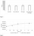
  • Figure 2 shows the dependence of the signal on the amount of FXIII.
  • the amine donor dansylcadaverine was bound to an immobilized FXIII substrate by the transglutaminase FXIII.
  • the binding of dansylcadaverine to the immobilized FXIII substrate results in a spectral change Fluorescence of dansylcadaverine recorded using an imager.
  • the fluorescence increase was measured at an excitation wavelength of 365 nm and an emission wavelength of 505-565 nm.
  • the amount of transglutaminase in international units [IU]
  • the signal is plotted on the y-axis as an intensity change ⁇ I per minute.
  • the change in intensity is the difference between the fluorescence intensity 30 minutes after adding the amine donor and the fluorescence intensity 5 minutes after adding the amine donor.
  • the obtained fluorescence intensity change was divided by the period of 25 minutes to obtain the signal as intensity change ⁇ I per minute.
  • intensity change ⁇ I per minute There is a linear relationship between the amount of transglutaminase (in international units [IU]) and the signal (intensity change ⁇ I per minute).
  • the influence of potential interfering components was examined in comparison to a control without these interfering components.
  • Serum albumin, fibrinogen and a combination of serum albumin and fibrinogen were tested as interfering components. A significant change in the signal due to the potential interfering components was not detected. The measurement is therefore unaffected by the presence of the corresponding components.
  • Figure 4 shows the dependence of the signal on the amount of FXIII.
  • the amine donor Lucifer-Yellow-Cadaverine or the amine donor Lucifer-Yellow-CH were bound to an immobilized FXIII substrate by the transglutaminase FXIII.
  • Lucifer-Yellow-CH has a carbohydrazide chain instead of the cadaverine residue.
  • the increase in fluorescence was measured at an excitation wavelength of 460 nm ⁇ 20 nm and an emission wavelength of 505-565 nm.
  • the amount of transglutaminase in international units [IU]
  • the intensity is plotted on the y-axis.
  • FXIII a binds to the immobilized glutamine donor protein sufficiently tightly to be extracted and enriched from a sample.
  • Example 1 and 2 were carried out in reaction vessels (Eppendorf tubes).
  • Example 3 was carried out on a device with an application zone, a reaction zone and a suction zone.
  • a paper-based, very simple manual lateral flow test (LFT) was suitable for detecting FXIII.
  • test setup and conditions were the same as in Example 3.
  • fluorophores also change their fluorescence intensity depending on the polarity of the environment, e.g. dapoxyl, aminonaphthalimide or NBD fluorophores. Since the reaction with FXIII is successful regardless of the chromophore with many amines, the wavelengths for excitation and emission can be selected accordingly by choosing the fluorophore.
  • Lucifer-Yellow CH and Lucifer-Yellow-Cadaverin are suitable.
  • the reaction was carried out in a lateral flow setup analogous to the method with dansylcadaverine (see Example 3).
  • dansylcadaverine see Example 3
  • the fluorescence of the Lucifer Yellow dye does not change significantly during incorporation, after 20 minutes of exposure, it was washed twice with standard buffer +200 mM CaCl 2 and the fluorescence was then measured on the imager. The results are in Figure 4 shown.
  • the detection therefore works independently of the chromophore.
  • the carbohydrazide in Lucifer YellowCH is also accepted as an amine donor, similar to the cadaverine residue.
  • the advantage of Lucifer Yellow is that it has a longer excitation wavelength than dansylcadaverine.
  • additional washing steps are required to remove the unreacted dye. Measurements using reflectance photometry are also possible. If necessary, dyes with high extinction coefficients and even cadaverine-modified nanogold particles can be used for this purpose.

Landscapes

  • Chemical & Material Sciences (AREA)
  • Health & Medical Sciences (AREA)
  • Life Sciences & Earth Sciences (AREA)
  • Organic Chemistry (AREA)
  • Engineering & Computer Science (AREA)
  • Zoology (AREA)
  • Proteomics, Peptides & Aminoacids (AREA)
  • Wood Science & Technology (AREA)
  • Immunology (AREA)
  • General Health & Medical Sciences (AREA)
  • Analytical Chemistry (AREA)
  • Molecular Biology (AREA)
  • Biochemistry (AREA)
  • Physics & Mathematics (AREA)
  • Biotechnology (AREA)
  • Microbiology (AREA)
  • Hematology (AREA)
  • Bioinformatics & Cheminformatics (AREA)
  • Genetics & Genomics (AREA)
  • General Engineering & Computer Science (AREA)
  • Biophysics (AREA)
  • Biomedical Technology (AREA)
  • Urology & Nephrology (AREA)
  • Clinical Laboratory Science (AREA)
  • Chemical Kinetics & Catalysis (AREA)
  • Neurosurgery (AREA)
  • Cell Biology (AREA)
  • Food Science & Technology (AREA)
  • Medicinal Chemistry (AREA)
  • General Physics & Mathematics (AREA)
  • Pathology (AREA)
  • Measuring Or Testing Involving Enzymes Or Micro-Organisms (AREA)

Abstract

Die vorliegende Erfindung betrifft ein Verfahren und eine Vorrichtung zum Nachweis von Transglutaminasen, insbesondere von Blutgerinnungsfaktor XIII (FXIII). Die Erfindung basiert auf einem schnellen, Point-of-Care-tauglichen Testprinzip für Transglutaminasen und ermöglicht eine genaue Bestimmung von FXIII in kurzer Zeit und mit einfacher Handhabung.

Description

  • Die vorliegende Erfindung betrifft ein Verfahren und eine Vorrichtung zum Nachweis von Transglutaminasen, insbesondere von Blutgerinnungsfaktor XIII (FXIII). Die Erfindung basiert auf einem schnellen, Point-of-Care-tauglichen Testprinzip für Transglutaminasen und ermöglicht eine genaue Bestimmung in kurzer Zeit und mit einfacher Handhabung.
  • Stand der Technik
  • Blut im Körper dient als Transport -und Kommunikationssystem und ist für die Aufrechterhaltung der Körperfunktionen unerlässlich. Kommt es zu einer Gefäßverletzung, so ist ein schneller Wundverschluss nötig, um die Blutverluste so gering wie möglich zu halten. Er erfolgt über das optimale Zusammenspiel zwischen Thrombozyten und Gerinnungsfaktoren. Die Hauptsubstanz des Thrombus bilden zu Polymeren zusammengelagerte Fibrinmonomere. Diese Fibrinmonomere entstehen durch die Abspaltung niedermolekularer Peptide von Fibrinogen durch die Serin-Protease Thrombin. Zur weiteren Stabilisierung des gebildeten Fibrinnetzes katalysiert Blutgerinnungsfaktor XIII, auch fibrinstabilisierender Faktor genannt, eine kovalente Quervernetzung der Fibrinmoleküle untereinander. Durch diese weitere Stabilisierung ist der Thrombus vor vorzeitiger Lyse geschützt. Die genannten Vorgänge stellen die letzten Schritte der komplexen Blutgerinnungskaskade dar.
  • Blutgerinnungsfaktor XIII (auch kurz als "Faktor XIII" oder "FXIII" bezeichnet) ist ein Plasmaprotein mit einem Gewicht von 320 kDa. Er liegt im Blutplasma als inaktives Heterotetramer mit je zwei A- und B-Untereinheiten vor. Das Heterotetramer stellt das Proenzym dar; unter physiologischen Bedingungen wird FXIII durch Thrombin aktiviert. Hierbei werden die N-terminalen Aktivierungspeptide zunächst durch Thrombin abgespalten (FXIII) und das Dissoziationsgleichgewicht wird hin zu den Monomeren verschoben. Geringe Calcium-Konzentrationen reichen dann zur Aktivierung aus (FXIIIa). Auch eine nicht-physiologische, von Thrombin unabhängige Aktivierung durch hohe Calcium-Konzentrationen (> 100 mM) kann den Komplex zu aktiven Monomeren dissoziieren (FXIII0). Aktivierter FXIII verknüpft als Transglutaminase Proteine durch Bildung einer Isopeptidbindung zwischen einem γ-Glutaminrest und dem ε-Aminorest in einer exponierten Lysin-Seitenkette. Bei dieser Reaktion wird Ammoniak frei. In der vorliegenden Offenbarung wird zur Bezeichnung von Aminosäuren oder Aminosäureresten auch der gebräuchliche Dreibuchstabencode verwendet, also beispielsweise "Gln" für Glutamin oder "Lys" für Lysin.
  • Ein Mangel an FXIII kann zu schweren Blutungen, verzögerter Wundheilung, aber auch zu vermehrten Aborten bei Frauen führen. Erblich bedingter FXIII-Mangel wird jedoch meist bereits im Säuglingsalter aufgrund von Nabelstumpfblutungen diagnostiziert. Diese erbliche Form des FXIII-Mangels ist sehr selten. Ein erworbener Mangel tritt häufiger auf und wird beispielsweise durch eine eingeschränkte Synthesekapazität der Leber, chronisch entzündliche DarmErkrankungen, Sepsis oder Verbrauchskoagulopathien ausgelöst. Zum Nachweis eines FXIII-Mangels eigenen sich konventionelle Gerinnungstests wie Thromboplastinzeit oder aktivierte partielle Prothrombinzeit (aPTT) nicht. Thrombozytenzahl und Fibrinogenkonzentration machen ebenfalls keine Aussage über den FXIII.
  • Besonders bei schweren Blutungen, z.B. nach Unfällen oder bei größeren Operationen führt ein Mangel an Gerinnungsfaktoren zu Komplikationen wie z.B. erhöhtem Blutverlust. Die Gabe von Vollblut wird heutzutage mehr und mehr unterlassen, da sie zwar Verluste substituiert, aber auch neue Ungleichgewichte zwischen den Faktoren erzeugt und zusätzliche Komplikationen verursachen kann. Stattdessen versucht man, durch gezielte Gabe von einzelnen Faktoren das physiologische Gleichgewicht wiederherzustellen. Entsprechende Präparate sind für Fibrinogen und auch für FXIII verfügbar.
  • Derzeit kann die Aktivität von FXIII im klinisch-chemischen Labor z.B. photometrisch bestimmt werden. Dazu wird FXIII der Probe durch Thrombin aktiviert und verknüpft ein Glutamin-Donor-Substrat mit einem Amin-Donor, z.B. Glycin-Ethylester, unter Freisetzung von Ammoniak. Der so entstandene Ammoniak kann in einer gekoppelten enzymatischen Reaktion bestimmt werden. NADH, α-Ketoglutarat und freier Ammoniak werden von der Glutamatdehydrogenase zu NAD+ und Glutamat umgewandelt. Die Abnahme von NADH wird über die Extinktion bei 340 nm gemessen und ist direkt proportional zur FXIII-Aktivität.
  • Alternativ kann Faktor XIII durch Antikörper in einem Enzyme-Linked Immunosorbent Assay (ELISA) nachgewiesen werden. Faktor XIII bindet dabei an einen spezifischen Antikörper, der im Testgefäß immobilisiert ist. Nach Auswaschen anderer Substanzen bindet ein zweiter Antikörper an den immobilisierten Faktor XIII. Ein an den Antikörper konjugiertes Enzym setzt das anschließend zugegebene signalgebende Substrat um. Die gebildete Substanz kann photometrisch oder fluorimetrisch quantifiziert und daraus der FXIII-Spiegel bestimmt werden.
  • Beide Tests erfordern einen Transport der Probe ins klinisch-chemische Labor, eine Zentrifugation zur Plasmagewinnung, die Durchführung des eigentlichen Tests sowie eine Auswertung und Rückmeldung an den Auftraggeber. Solche Abläufe unter Einbeziehung des klinischen Labors sind (auch bei anderen Testverfahren) in akuten Fällen zu langsam, um die Notwendigkeit einer Gabe zu beurteilen und die korrekte Dosierung der Präparate zu bestimmen. Alternative Testverfahren sind Thromboelastometrie (eher qualitativ und recht störanfällig) sowie Enzymimmunoassays (sehr aufwändig und teuer, keine Aktivitäts- sondern Proteinbestimmung).
  • Point of Care (POC) Schnelltests könnten vor Ort (z.B. im Operationssaal) angewandt werden und eine korrekte Entscheidung innerhalb weniger Minuten ermöglichen, sind jedoch bislang aus einer Vielzahl von Gründen nicht verfügbar.
  • Blut ist ein hochkomplexes Gemisch aus Zellen (Hauptbestandteil Erythrozyten) und im Serum gelösten Bestandteilen, wie Proteinen und Lipiden. Die starke Lichtabsorption im Hämoglobin macht photometrische oder fluorimetrische Bestimmungen in Vollblut schwierig bis unmöglich. Elektrochemische Bestimmungen werden von den Zellen und von der Viskosität der Blutmatrix ebenfalls stark gestört. Eine (teilweise) Korrektur ist mit Verfahren wie der Impedanzspektroskopie möglich, jedoch ebenfalls mit großem Aufwand verbunden.
  • Eine schnelle Abtrennung der Erythrozyten ist durch Glasfaservliese möglich. Lösliche Blutbestandteile bleiben allerdings im so gewonnenen Blutplasma. Speziell die hohen Konzentrationen von Serum-Albumin und Fibrinogen haben das Potential, Tests für FXIII zu stören. Bekannt ist z.B. der Dansylcadaverin-Test, bei dem ein fluoreszenzmarkiertes Amin in ein glutaminhaltiges Protein, z.B. Dimethyl-Casein, eingebaut wird. Die Blauverschiebung und Verstärkung der Fluoreszenz durch die veränderte chemische Umgebung des Fluorophors kann als Maß für die Aktivität von FXIII herangezogen werden. Serum-Albumin in physiologischen Konzentrationen bindet allerdings das Dansylcadaverin, entzieht es der enzymatischen Reaktion und verschiebt das Fluoreszenzspektrum vorab, so dass die eventuell noch stattfindende Restreaktion nicht mehr sichtbar wird.
  • Fibrinogen ist eine weitere Störkomponente: Die physiologische Aktivierung von FXIII erfordert Spaltung durch Thrombin; alternativ kann mit hohen Ca2+-Konzentrationen gearbeitet werden. In beiden Fällen wird die Gerinnungsreaktion angestoßen; das Gerinnsel behindert durch Lichtstreuung und hohe Viskosität die Nachweisreaktionen.
  • Die einzige bekannte Methode zur Bestimmung von FXIII aus Vollblut ist die Thromboelastografie ( EP 2 474 830 A1 ). In späteren Phasen der Gerinnung (also nach längerer Beobachtungszeit) sorgt FXIII für eine Verfestigung des Gerinnsels, deren Geschwindigkeit und Ausmaß vom Gehalt der Probe an FXIII abhängen. Das Verfahren ist angesichts der längeren Beobachtungszeit verhältnismäßig langsam. Außerdem beeinflussen viele andere Inhaltsstoffe des Blutes (Zellen, Fibrinogen, Serumalbumin, das Blut verdünnende Agenzien) und die einsetzende Fibrinolyse die Ergebnisse, so dass keine genauen FXIII-Bestimmungen möglich sind.
  • Als Ergebnis dieser Probleme bietet der Stand der Technik kein Verfahren, mit dem eine genaue Bestimmung von Transglutaminasen, insbesondere von FXIII in angemessen kurzer Zeit und mit einfacher Handhabung möglich wäre. Aufgabe der vorliegenden Erfindung ist vor diesem Hintergrund die Bereitstellung eines schnellen, POC-tauglichen Testprinzips aus humanem Blut.
  • Beschreibung der Erfindung
  • Das Verfahren der vorliegenden Erfindung eignet sich für alle Transglutaminasen. Die Bestimmung der enzymatischen Aktivität von Blutgerinnungsfaktor XIII (FXIII) in einer Probe ist besonders bevorzugt.
  • Die Bindung erfolgt an einen immobilisierten Glutamindonor. Dieser kann gleichzeitig das zum Nachweis verwendete Substrat sein, muss es aber nicht. Zum Nachweis der Aktivität kann zusätzliches Substrat hinzugegeben werden, entweder der gleiche Glutamindonor oder ein anderer.
  • Der Nachweis kann entweder über Auswaschen von ungebundenem Amindonor und Nachweis des im Reaktivfeld verbleibenden erfolgen oder bevorzugt über eine Spektralverschiebung des Chromophors am Amindonor durch die Bindung. Dafür kann eine Reihe von Fluorophorresten eingesetzt werden, die auf die geänderte Polarität der Umgebung direkt reagieren, z.B. Dansyl, Dapoxyl, 4-Chlor-7-nitrobenzofurazan (NBD), 6-Dimethylamino-2,3-naphthalimid. Solche Fluorophore können unter dem Begriff "spektroskopische Polaritätssensoren" zusammengefasst werden. Die "Menge" an eingebautem Amin kann insbesondere indirekt spektroskopisch bestimmt werden; die Aktivität der Transglutaminase kann insbesondere über die zeitliche Änderung des spektroskopischen Signals bestimmt werden.
  • Als Linker zwischen Chromophor und Glutamin eignen sich insbesondere Diamine, bevorzugt Putrescin, Cadaverin und/oder Carbohydrazid.
  • In einem ersten Aspekt betrifft die Erfindung ein Verfahren zur Bestimmung der enzymatischen Aktivität einer Transglutaminase, insbesondere von Blutgerinnungsfaktor XIII (FXIII), in einer Probe, wobei das Verfahren die folgenden Schritte umfasst:
    1. a) Bereitstellen eines immobilisierten Glutamindonors, z.B. eines FXIII-Substrats, wobei der Glutamindonor ein Peptid ist, insbesondere ein Polypeptid oder Protein, und wobei der Glutamindonor mindestens einen Glutamin-Rest umfasst,
    2. b) Aktivierung der in der Probe vorhandenen Transglutaminase,
    3. c) Inkontaktbringen der Probe mit dem Glutamindonor, so dass in der Probe vorhandene Transglutaminase an den Glutamindonor bindet,
    4. d) Entfernung nicht an den Glutamindonor gebundener Bestandteile der Probe,
    5. e) Inkontaktbringen der an den Glutamindonor gebundenen Transglutaminase mit einem Amindonor, so dass der Amindonor von der Transglutaminase an ein Glutamindonorsubstrat gebunden wird, und
    6. f) Bestimmung der enzymatischen Aktivität der Transglutaminase anhand eines Signals, das durch die Bindung des Amindonors an das Glutamindonorsubstrat erzeugt wird.
  • Bevorzugt ist das Signal proportional zur Geschwindigkeit der von der Transglutaminase katalysierten Bindung des Amindonors an das Glutamindonorsubstrat.
  • Die Schritte werden insbesondere in der angegebenen Reihenfolge a) bis f) durchgeführt.
  • Der immobilisierte Glutamindonor ist bevorzugt das Glutamindonorsubstrat, an das der Amindonor durch die Glutaminase gebunden wird.
  • Die Transglutaminase ist bevorzugt ausgewählt aus der Gruppe der humanen Transglutaminasen. Besonders bevorzugt ist die Transglutaminase Blutgerinnungsfaktor XIII (FXIII).
  • Bevorzugt enthält die Probe genau eine Transglutaminase.
  • Die Probe ist bevorzugt eine Blutprobe, insbesondere eine humane Blutprobe. In anderen Ausführungsformen kann die Probe auch eine Gewebeprobe sein, beispielsweise ein Extrakt aus einem Gewebe.
  • Die Probe enthält Transglutaminase, insbesondere FXIII, bevorzugt in einer Aktivität von 5% bis 200%, beispielsweise von 25% bis 175%, von 50% bis 150%, von 75% bis 125%, oder von 90% bis 110% bezogen auf die Aktivität in Standardplasma. Die Aktivität beträgt bevorzugt mindestens 5%, mindestens 25%, mindestens 50%, mindestens 75%, oder mindestens 90% bezogen auf die Aktivität in Standardplasma. Die Aktivität beträgt bevorzugt höchstens 200%, höchstens 175%, höchstens 150%, höchstens 125%, oder höchstens 110% bezogen auf die Aktivität in Standardplasma.
  • Gemäß Schritt a) des erfindungsgemäßen Verfahrens wird ein immobilisierter Glutamindonor, insbesondere ein immobilisiertes Glutamindonorsubstrat, bevorzugt ein immobilisiertes FXIII-Substrat bereitgestellt. Das (FXIII-)Substrat kann ein physiologisches (FXIII-)Substrat sein. In besonders bevorzugten Ausführungsformen ist das (FXIII-)Substrat kein physiologisches (FXIII-)Substrat. Mit (FXIII-)Substrat ist insbesondere gemeint, dass die Transglutaminase (insbesondere FXIII) die Reaktion eines Glutamin-Rests des Substrats mit der Aminogruppe eines Amindonors katalysieren kann. Wenn der Glutamindonor zwar mindestens einen Glutamin-Rest umfasst, die Transglutaminase (insbesondere FXIII) jedoch keine Bindung zwischen Glutamin-Rest und Amindonor katalysieren kann, ist der Glutamindonor kein (FXIII)Substrat.
  • Der Glutamindonor kann insbesondere ein Peptid sein, bevorzugt ein Polypeptid oder Protein. Der Glutamindonor enthält mindestens einen Glutamin-Rest. Bevorzugt enthält der Glutamindonor mehrere Glutamin-Reste, beispielsweise mindestens zwei, mindestens drei, oder mindestens fünf Glutamin-Reste. Bevorzugt sind Proteine mit einem Molgewicht von 10 bis 300 kDa, beispielsweise von 10 bis 100 kDa oder von 12 bis 30 kDa. Das Molgewicht des Glutamindonors kann beispielsweise mindestens 10 kDa, oder mindestens 12 kDa betragen. Das Molgewicht des Glutamindonors kann beispielsweise höchstens 300 kDa, höchstens 100 kDa oder höchstens 30 kDa betragen. Bevorzugt liegt der Glutamingehalt des Glutamindonors in einem Bereich von 3% bis 15% bezogen auf die Gesamtanzahl an Resten, beispielsweise in einem Bereich von 5% bis 12% oder von 7% bis 10%. Der Glutamingehalt des Glutamindonors kann beispielsweise mindestens 3%, mindestens 5% oder mindestens 7% betragen. Der Glutamingehalt des Glutamindonors kann beispielsweise höchstens 15%, höchstens 12% oder höchstens 10% betragen.
  • Besonders bevorzugt ist der Glutamindonor ein Substrat der Transglutaminase, auch Glutamindonorsubstrat genannt. Dies hat den Vorteil, dass der immobilisierte Glutamindonor als Glutamindonorsubstrat fungieren kann, an das die Transglutaminase den Amindonor bindet. Dadurch kann auf die Zugabe von zusätzlichem Substrat der Transglutaminase verzichtet werden. Es sind jedoch auch Ausführungsformen umfasst, in denen der immobilisierte Glutamindonor nicht mit dem Glutamindonorsubstrat des Schrittes e) des Verfahrens der vorliegenden Erfindung identisch ist. Insbesondere in solchen Ausführungsformen wird zusätzlich zum immobilisierten Glutamindonor noch Glutamindonorsubstrat zugegeben, an das der Amindonor durch die Transglutaminase gebunden werden kann. Es ist beispielsweise möglich, vor Durchführung des Schrittes e) eine Mischung oder Lösung bereitzustellen, die Amindonor und Glutamindonorsubstrat umfasst, und diese in Schritt e) mit der an den Glutamindonor gebundenen Transglutaminase in Kontakt zu bringen, so dass die Bindung zwischen Amindonor und Glutamindonorsubstrat durch die Transglutaminase gebildet werden kann. Die Zugabe von Glutamindonorsubstrat kann auch dann erfolgen, wenn der immobilisierte Glutamindonor selbst bereits ein Glutamindonorsubstrat ist.
  • In der vorliegenden Offenbarung beschreiben die Begriffe "Peptid" oder "Peptide" zusammenfassend Moleküle, die Aminosäuren enthalten, die über Peptidbindungen miteinander verknüpft sind. Dies beinhaltet Oligopeptide (Peptide mit bis zu zehn Aminosäureresten) und Polypeptide (Peptide mit mehr als zehn Aminosäureresten). Polypeptide mit mehr 50 Aminosäureresten werden auch als Proteine bezeichnet. Die Begriffe "Peptid" oder "Peptide", wie in der vorliegenden Offenbarung verwendet, umfassen ausdrücklich auch Proteine.
  • Gemäß Schritt a) des Verfahrens liegt der Glutamindonor, insbesondere das FXIII-Substrat immobilisiert vor. Die Immobilisierung beinhaltet insbesondere die Bindung des Glutamindonors an einen Träger, bevorzugt Sepharose, Cellulose oder Papier, jeweils bevorzugt funktionalisiert mit DEAE (Diethylaminoethyl-Gruppe) oder anderen kationischen Gruppen. Die Immobilisierung kann zusätzlich oder alternativ auch eine Fällung des Glutamindonors, insbesondere des FXIII-Substrats in Gegenwart des Trägers beinhalten, beispielsweise eine Fällung mit Ca2+.
  • Der immobilisierte Glutamindonor, insbesondere der Substrat-Träger-Komplex, kann in verschiedenen Formen vorliegen. Beispielsweise ist es möglich, dass der immobilisierte Glutamindonor in Form einer Suspension vorliegt, insbesondere in einem Reaktionsgefäß. Es ist auch möglich, den Glutamindonor auf einem Festkörper zu immobilisieren, beispielsweise auf einem Papierträger in einem Teststreifen.
  • Die Immobilisierung des Glutamindonors umfasst bevorzugt eine oder mehrere der folgenden Maßnahmen:
    • Fällung und Adsorption
    • Ionische Bindung an DEAE-funktionalisierte Oberflächen
    • Kovalente Bindung durch Formaldehyd, Glutardialdehyd
    • Kovalente Bindung unter Verwendung von EDC/NHS bei Amin- oder COOHfunktionalisierter Trägeroberfläche
    • Kovalente Bindung durch Dimethylsuberimidat, Dimethylpimelimidat, Dimethyladipimidat bei aminfunktionalisierter Trägeroberfläche
    • Kovalente Bindung durch Trägergebundene Epoxide, Isocyanate
    • Kovalente Bindung über Aminogruppen an BrCN-aktivierte Polysaccharide.
  • Der immobilisierte Glutamindonor kann Modifikationen, beispielsweise chemische Modifikationen aufweisen. Besonders bevorzugt sind Modifikationen von Lysin-Resten. Insbesondere kann der Glutamindonor methyliert, acetyliert oder succinyliert sein, beispielsweise dimethyliert, insbesondere N,N-dimethyliert oder auch acetamidiniert.
  • Insbesondere kann der Glutamindonor methyliert sein, beispielsweise dimethyliert, insbesondere N,N-dimethyliert. Die Dimethylierung erfolgt insbesondere durch Umsetzung des Glutamindonors mit Formaldehyd und Natriumborhydrid. Alternativ oder zusätzlich können ein oder mehrere andere Reduktionsmittel eingesetzt werden. Die Reaktion richtet sich nach der Zugänglichkeit der Lysine - gut zugänglich reagiert am schnellsten. Der Dimethylierungsgrad wird durch die Menge der zugegebenen Reagenzien und die Reaktionsdauer gesteuert. Ein möglichst hoher Dimethylierungsgrad der frei zugänglichen Lysine ist bevorzugt, weil das die Quervernetzung innerhalb eines Proteins oder zwischen den Proteinen verhindert. Bevorzugt beträgt der Dimethylierungsgrad mehr als 90%.
  • Zusätzlich oder alternativ zur Methylierung kann der immobilisierte Glutamindonor bevorzugt succinyliert oder acetamidiniert sein.
  • Besonders bevorzugt ist der Glutamindonor ausgewählt aus der Gruppe bestehend aus Casein, insbesondere Dimethylcasein, und Lysozym, insbesondere succinyliertes Lysozym, bevorzugt succinyliertes Hühnereiweißlysozym (Gallus gallus). Casein aus Kuhmilch (Bos taurus) ist bevorzugt. Dimethylcasein ist dem Fachmann bestens bekannt, da es üblicherweise zur Proteasebestimmung über den Nachweis neu gebildeter endständiger Aminogruppen verwendet wird.
  • Gemäß Schritt b) des erfindungsgemäßen Verfahrens wird die in der Probe vorhandene Transglutaminase aktiviert. In Ausführungsformen, in denen die Transglutaminase FXIII ist, kann die Aktivierung durch Thrombin erfolgen. Allerdings sollte in diesem Fall beispielsweise durch Zusatz von Hemmstoffen der Fibrinaggregation, z.B. durch das Tetrapeptid GPRP, die Gerinnung vermieden werden.
  • Bevorzugt werden hohe Ca2+-Konzentrationen zur Aktivierung verwendet. Dies gilt insbesondere, wenn die Transglutaminase FXIII ist, aber auch bei anderen Transglutaminasen. Bei einer Aktivierung mit Ca2+ liegt die Ca2+-Konzentration bevorzugt in einem Bereich von 100 bis 500 mM, beispielsweise von 200 bis 400 mM. Die Ca2+-Konzentration kann beispielsweise mindestens 100 mM oder mindestens 200 mM betragen. Die Ca2+-Konzentration kann beispielsweise höchstens 500 mM oder höchstens 400 mM betragen. Auch niedrigere Ca2+-Konzentrationen, beispielsweise Konzentrationen von 1 bis <100 mM, insbesondere von 1 bis 10 mM sind in Verbindung mit hoher lonenstärke möglich. Eine hohe lonenstärke kann beispielsweise durch NaCI-Konzentration in einem Bereich von 200 bis 1000 mM, insbesondere von 400 bis 700 mM erhalten werden.
  • In Ausführungsformen, in denen die Transglutaminase FXIII ist, liegt FXIII nach der Aktivierung gemäß Schritt b) bevorzugt als FXIIIa (durch Thrombin gespalten) oder als FXIII0 (durch Ca2+-lonen aktiviert) vor.
  • Gemäß Schritt c) des erfindungsgemäßen Verfahrens wird die Probe mit dem immobilisierten Glutamindonor in Kontakt gebracht. Die Einzelheiten des Inkontaktbringens richten sich im Wesentlichen nach der Art der Immobilisierung des Glutamindonors. Wenn der immobilisierte Glutamindonor als Suspension vorliegt, insbesondere in einem Reaktionsgefäß, kann das Inkontaktbringen beispielsweise dadurch erfolgen, dass die Probe zu der Suspension hinzugegeben wird, beispielsweise durch Zugabe der Probe in das Reaktionsgefäß hinzugegeben wird. Wenn der Glutamindonor hingegen immobilisiert auf einem Festkörper, wie beispielsweise einem Teststreifen, vorliegt, kann das Inkontaktbringen erfolgen, indem die Probe und der Festkörper, insbesondere der Teststreifen, in Kontakt gebracht werden. Die Probe kann beispielsweise an der Stelle auf den Festkörper aufgebracht werden, an der auch der Glutamindonor immobilisiert ist. Es ist jedoch auch möglich, die Probe an einer Stelle aufzubringen, an der nicht der Glutamindonor immobilisiert ist. Dies kann insbesondere dann eine bevorzugte Möglichkeit sein, wenn der Festkörper, insbesondere der Teststreifen, derart ausgestaltet ist, dass die Probe, beispielsweise durch Kapillarkräfte, durch den Festkörper wandern und so mit dem immobilisierten Glutamindonor in Kontakt gebracht werden kann.
  • Es ist möglich, die Probe vor dem Inkontaktbringen mit dem immobilisierten Glutamindonor einem Vorbehandlungsschritt, beispielsweise einem Reinigungsschritt, zu unterziehen. Bevorzugt enthält das Verfahren jedoch keinen Schritt der Vorbehandlung der Probe. Es ist ein besonderer Vorteil der vorliegenden Erfindung, dass die Probe keiner Vorbehandlung bedarf. So ist es insbesondere möglich, die Probe, beispielsweise eine (humane) Blutprobe, nach ihrer Entnahme ohne Vorbehandlung mit dem immobilisierten Glutamindonor in Kontakt zu bringen. Calcium kann zugesetzt werden. Im Falle einer Fällung des Glutamindonors mit Ca2+ kann auch das Calcium im gefällten Glutamindonor zur Aktivierung der Transglutaminase beitragen oder die Aktivierung bewirken.
  • Das Inkontaktbringen gemäß Schritt c) des Verfahrens kann einen Inkubationsschritt umfassen. Der Inkubationsschritt ist besonders vorteilhaft, um das Ausmaß der Bindung der Transglutaminase an den Glutamindonor weiter zu erhöhen. Bevorzugt liegt die Inkubationszeit in einem Bereich von 10 Sekunden bis 5 Minuten, beispielsweise von 0,5 bis 5 Minuten oder von 1 bis 3 Minuten. Die Inkubationszeit kann beispielsweise mindestens 10 Sekunden, mindestens 30 Sekunden oder mindestens 60 Sekunden betragen. Die Inkubationszeit kann beispielsweise höchstens 300 Sekunden oder höchstens 180 Sekunden betragen. Die Inkubationszeit bezeichnet insbesondere den Zeitraum vom Beginn des Inkontaktbringens gemäß Schritt c) bis zum Beginn des Entfernens nicht an den Glutamindonor gebundener Bestandteile der Probe gemäß Schritt d).
  • Das Inkontaktbringen der Probe mit dem immobilisierten Glutamindonor gemäß Schritt c) des Verfahrens führt insbesondere zu einer Bindung der Transglutaminase an den immobilisierten Glutamindonor.
  • Glutamin-Reste sind nötig für die Bindung von der Transglutaminase an den immobilisierten Glutamindonor. Transglutaminasen können insbesondere mit einer essentiellen SH-Gruppe an ein Glutamin des Glutamindonors binden, wobei unter Abspaltung von Ammoniak ein Thioester gebildet wird, wie im folgenden Reaktionsschema 1 verdeutlicht.
    Figure imgb0001
  • E-SH beschreibt das Enzym (die Transglutaminase) mit seiner essentiellen SH-Gruppe im aktiven Zentrum. Vom Glutamindonor ist die Seitenkette eines Glutamin-Rests gezeigt. Der übrige Glutamindonor ist als "R" dargestellt. Unter Abspaltung von Ammoniak kommt es zur Ausbildung einer Thioesterbindung, die das Enzym E mit dem Glutamindonor R verknüpft.
  • Diese Reaktion tritt auch physiologisch auf. Transglutaminasen binden jedoch nicht nur an physiologische Glutamindonoren, sondern auch an andere Glutamindonoren, insbesondere solche, die mindestens einen Glutamin-Rest aufweisen.
  • Die Transglutaminase (insbesondere FXIII) bindet an den immobilisierten Glutamindonor, insbesondere an das immobilisierte Glutamindonorsubstrat. Die Bindung zwischen Transglutaminase und Glutamindonor ist bevorzugt eine kovalente Bindung zwischen einer Aminosäure im Aktiven Zentrum der Glutaminase und einem Glutamin-Rest des Glutamindonors. Die Bindung zwischen Transglutaminase und immobilisiertem Glutamindonor ist insbesondere eine Thioesterbindung. Die Thioesterbindung wird bevorzugt durch Reaktion einer SH-Gruppe der Transglutaminase (insbesondere der essentiellen SH-Gruppe) und einem Glutamin-Rest des immobilisierten Glutamindonors gebildet.
  • Gemäß Schritt d) des erfindungsgemäßen Verfahrens werden nicht an den immobilisierten Glutamindonor gebundene Bestandteile der Probe entfernt. Ein Bestandteil der Probe, der an den Glutamindonor bindet, ist insbesondere die Transglutaminase, bevorzugt FXIII. Andere Bestandteile der Probe, insbesondere potentiell den Nachweis der Transglutaminase störende Bestandteile, binden bevorzugt nicht oder nur schwach an den Glutamindonor. Eine geringe Bindung ist insbesondere eine solche, die die Entfernung mit einem oder mehreren Waschschritten erlaubt.
  • Schritt d) des Entfernens nicht an den Glutamindonor gebundener Bestandteile der Probe umfasst bevorzugt einen oder mehrere Waschschritte. Die Anzahl der Waschschritte liegt bevorzugt in einem Bereich von 1 bis 3. Besonders bevorzugt ist das kontinuierliche Waschen, insbesondere durch Überströmen des Trägers des immobilisierten Glutamindonors. Hierbei werden Volumina vom 1- bis 3-fachen des Trägervolumens verwendet. Das Trägervolumen ist das Volumen der im Träger vorhandenen Flüssigkeit.
  • Die Einzelheiten des Entfernens nicht an den Glutamindonor gebundener Bestandteile der Probe richten sich im Wesentlichen nach der Art der Immobilisierung des Glutamindonors.
  • Wenn der immobilisierte Glutamindonor als Suspension vorliegt, insbesondere in einem Reaktionsgefäß, kann das Entfernen beispielsweise dadurch erfolgen, dass ein oder mehrere Waschschritte durchgeführt werden, wobei ein Waschschritt die Trennung der Suspension in feste und flüssige Bestandteile, das Entfernen der flüssigen Bestandteile und die Zugabe von Waschlösung, insbesondere Waschpuffer, umfasst. Die Trennung der Suspension in feste und flüssige Bestandteile kann insbesondere mittels Zentrifugation erfolgen. Bevorzugt ist eine Zentrifugation für 0,5 bis 2 Minuten, wobei sich die erforderliche Beschleunigung nach der Art des Trägers richtet.
  • Wenn das Substrat hingegen immobilisiert auf einem Festkörper, wie beispielsweise dem Papier in einem Teststreifen, vorliegt, kann das Entfernen nicht an den Glutamindonor gebundener Bestandteile der Probe beispielsweise dadurch erfolgen, dass ein oder mehrere Waschschritte durchgeführt werden, wobei ein Waschschritt die Zugabe von Waschlösung zum Festkörper, insbesondere zum Teststreifen, umfasst. Die Waschlösung ist bevorzugt ein Waschpuffer.
  • Die Waschlösung kann beispielsweise an der Stelle auf den Festkörper aufgebracht werden, an der auch der Glutamindonor immobilisiert ist. Es ist jedoch auch möglich, die Waschlösung an einer Stelle aufzubringen, an der nicht der Glutamindonor immobilisiert ist. Dies kann insbesondere dann eine bevorzugte Möglichkeit sein, wenn der Festkörper, insbesondere der Teststreifen, derart ausgestaltet ist, dass die Waschlösung, beispielsweise durch Kapillarkräfte, durch den Festkörper wandern und so mit dem immobilisierten Glutamindonor in Kontakt gebracht werden kann. Dadurch kann erreicht werden, dass nicht an den Glutamindonor gebundene Bestandteile der Probe entfernt werden.
  • Die Waschlösung beziehungsweise der Waschpuffer weist bevorzugt eine in etwa isotonische Zusammensetzung auf, z.B. 100 mM NaCl und 50 mM HEPES. Der pH-Wert liegt bevorzugt in einem Bereich von 6,0 bis 9,0, weiter bevorzugt von 7,0 bis 8,0. Der pH-Wert beträgt bevorzugt mindestens 6,0 oder mindestens 7,0. Der pH-Wert beträgt bevorzugt höchstens 9,0 oder höchstens 8,0. Die Waschlösung beziehungsweise der Waschpuffer kann beispielsweise auch PBS (phosphatgepufferte Salzlösung, englisch: "phosphate-buffered saline") sein, insbesondere 137 mM NaCl, 2,7 mM KCl und 12 mM Phosphat (HPO4 2- und H2PO4 -), pH 7,4. Die Waschlösung beziehungsweise der Waschpuffer kann ein oder mehrere Biozide enthalten, beispielsweise NaN3, bevorzugt in Konzentrationen von 0,01 bis 1,0 mM oder von 0,05 bis 0,5 mM, beispielsweise etwa 0,1 mM.
  • Physiologisch wird der Thioester zwischen Transglutaminase und Glutamindonor von der ε-Aminogruppe eines Lysins angegriffen und es entsteht unter Aminolyse die Isopeptidbindung Gln-Lys. Die essentielle SH-Gruppe wird wieder freigesetzt. Statt Lysin akzeptiert das Enzym eine ganze Reihe von Amindonoren; auch Alkohole werden eingebaut, jedoch weniger bevorzugt. In Abwesenheit solcher Substrate erfolgt Hydrolyse. Aminolyse und Hydrolyse sind schematisch im folgenden Reaktionsschema 2 gezeigt.
    Figure imgb0002
  • Dabei bezeichnet R'-NH2 einen Amindonor. Die Hydrolyse verläuft langsamer als die Aminolyse und ist daher gestrichelt dargestellt.
  • Die Aminolyse kann, wie unten näher beschrieben, zum Nachweis von Transglutaminase verwendet werden. Eine vorzeitige Hydrolyse kann hingegen zu einem verfrühten Freisetzen der Transglutaminase vom immobilisierten Glutamindonor führen, so dass die entsprechende Transglutaminase nicht mehr durch die Aminolyse nachgewiesen werden kann. Für eine möglichst hohe Sensitivität des Nachweises der Transglutaminase sollte also die Hydrolyserate so gering wie möglich sein.
  • Um eine möglichst stabile Bindung der Transglutaminase an den immobilisierten Glutamindonor zu erhalten, ist es vorteilhaft, wenn der Glutamindonor mehr als einen Glutamin-Rest aufweist. Dadurch kann insbesondere erreicht werden, dass die Transglutaminase auch durch mehrere Waschschritte nicht oder nicht nennenswert vom Glutamindonor entfernt wird. So können mehrere Waschschritte durchgeführt und eine verbesserte Entfernung von nicht an den Glutamindonor gebundenen Bestandteilen der Probe erreicht werden, ohne dadurch den Nachweis der Transglutaminase durch ein übermäßiges Auswaschen zu beeinträchtigen. Eine derart feste Bindung der Transglutaminase an einen Glutamindonor, insbesondere an ein Glutamindonorsubstrat ist überraschend. Ohne an eine bestimmte Theorie gebunden wollen zu sein, könnte der Einfluss der Anzahl der Glutamin-Reste auf die Bindung zumindest teilweise wie folgt zu erklären sein.
  • Bei einem Glutamindonor mit diversen Glutaminen bestimmt die Umgebung des Glutamins über Aminolyse- und Hydrolysegeschwindigkeit mit. In so einem System wird die Transglutaminase an eins der Glutamine binden. Ist die Hydrolyse schnell genug, wird das Enzym wieder freigesetzt und kann die Reaktion wiederholen. An einem Glutamin mit langsamer Hydrolyse bleibt es immobilisiert. Das System ist selbstoptimierend. Die Transglutaminase bindet an den Glutamin-Rest mit der langsamsten Hydrolyse, entweder direkt oder nach einigen wegen Hydrolyse nicht erfolgreichen Versuchen an anderen Glutamin-Resten. Dies ist im folgenden Reaktionsschema 3 verdeutlicht.
    Figure imgb0003
    Figure imgb0004
    Figure imgb0005
  • Durch R1, R2 und R3 wird gekennzeichnet, dass es sich um verschiedene Glutamin-Reste des Glutamindonors handelt. Mit k1, k2 und k3 werden die verschiedenen Hydrolysegeschwindigkeitskonstanten bezeichnet. Die Hydrolyserate des dritten Glutamins ist besonders gering und ist daher gestrichelt gezeichnet.
  • Bevorzugt liegen die Glutamin-Reste im molaren Überschuss gegenüber der Transglutaminase vor.
  • Gemäß Schritt e) des erfindungsgemäßen Verfahrens wird die an den Glutamindonor gebundene Transglutaminase mit einem Amindonor in Kontakt gebracht, so dass der Amindonor von der Transglutaminase an ein Glutamindonorsubstrat gebunden wird. Dabei wird insbesondere die Bindung zwischen der Transglutaminase und dem immobilisierten Glutamindonor gelöst. Falls der immobilisierte Glutamindonor selbst ein Glutamindonorsubstrat ist, kann die Transglutaminase die Ausbildung einer Bindung zwischen Amindonor und immobilisiertem Glutamindonorsubstrat katalysieren. Ist der immobilisierte Glutamindonor kein Glutamindonorsubstrat, erfolgt die Bindung des Amindonors nicht an den immobilisierten Glutamindonor, sondern an das zusätzlich vorhandene Glutamindonorsubstrat. Das Glutamindonorsubstrat kann insbesondere in solchen Ausführungsformen beispielsweise separat oder gemeinsam mit dem Amindonor hinzugegeben werden.
  • Der Amindonor kann einen Chromophor beinhalten. Bevorzugt ist insbesondere ein fluoreszenter Amindonor, besonders bevorzugt Dansylcadaverin, Dapoxylcadaverin oder NBD-Cadaverin. "NBD" bezeichnet den Fluorophor 4-Chlor-7-nitro-benzofurazan. Der Amindonor weist bevorzugt mindestens eine fluoreszente Gruppe auf. Der Amindonor kann mindestens einen Farbstoff aufweisen. Ein Amindonor im Sinne der vorliegenden Erfindung ist insbesondere ein Molekül, das mindestens eine primäre Aminogruppe an einer kurzen aliphatischen Kette aufweist. Die primäre Aminogruppe kann beispielsweise die ε-Aminogruppe von Lysin sein oder die freie Aminogruppe eines einseitig substituierten Diamins wie Cadaverin oder Putrescin. Auch eine substituierte Carbohydrazidgruppe kommt in Frage.
  • Im Reaktionsschema 2 ist ein Amindonor beispielhaft als R'-NH2 dargestellt.
  • Insbesondere wird der Amindonor von der an den immobilisierten Glutamindonor gebundenen Transglutaminase an ein Glutamindonorsubstrat gebunden. Dabei löst sich die (Thioester-)Bindung zwischen Transglutaminase und Glutamindonor im Zuge einer Aminolyse. Die Bindung zwischen Amindonor und Glutamindonorsubstrat ist bevorzugt eine Amidbindung, weiter bevorzugt eine Carbonsäureamidbindung.
  • Der Amindonor ist bevorzugt wasserlöslich. Insbesondere weist der Amindonor bevorzugt eine Wasserlöslichkeit von größer als 10 µM auf. Der Amindonor hat bevorzugt ein Molekulargewicht in einem Bereich von 100 bis 1000 Da, beispielsweise von 150 bis 500 Da. Der Amindonor hat bevorzugt ein Molekulargewicht von mindestens 100 Da oder mindestens 150 Da. Der Amindonor hat bevorzugt ein Molekulargewicht von höchstens 1000 Da oder von höchstens 500 Da.
  • Das Ausmaß der Bindung des Amindonors an das Glutamindonorsubstrat ist bevorzugt proportional zur Aktivität der an den immobilisierten Glutamindonor gebundenen Transglutaminase. Die Aktivität der an den immobilisierten Glutamindonor gebundenen Transglutaminase ist wiederum bevorzugt proportional zum Anteil der Transglutaminase in der Probe beziehungsweise zur in der Probe vorhandenen Enzymaktivität der Transglutaminase.
  • Die Einzelheiten des Inkontaktbringens der an den immobilisierten Glutamindonor gebundenen Transglutaminase mit dem Amindonor richten sich im Wesentlichen nach der Art der Immobilisierung des Glutamindonors.
  • Wenn der immobilisierte Glutamindonor als Suspension vorliegt, insbesondere in einem Reaktionsgefäß, kann das Inkontaktbringen beispielsweise dadurch erfolgen, dass der Amindonor zu der Suspension in das Reaktionsgefäß hinzugegeben wird. Wenn der Glutamindonor hingegen immobilisiert auf einem Festkörper, wie beispielsweise einem Teststreifen, vorliegt, kann das Inkontaktbringen erfolgen, indem der Amindonor und der Festkörper, insbesondere der Teststreifen, in Kontakt gebracht werden. Der Amindonor kann beispielsweise an der Stelle auf den Festkörper aufgebracht werden, an der auch der Glutamindonor immobilisiert ist. Es ist jedoch auch möglich, den Amindonor an einer Stelle aufzubringen, an der nicht der Glutamindonor immobilisiert ist. Dies kann insbesondere dann eine bevorzugte Möglichkeit sein, wenn der Festkörper, insbesondere der Teststreifen, derart ausgestaltet ist, dass der Amindonor, beispielsweise durch Kapillarkräfte, durch den Festkörper wandern und so mit dem immobilisierten Glutamindonor in Kontakt gebracht werden kann.
  • Der Amindonor liegt vor und/oder beim Inkontaktbringen bevorzugt in Lösung vor. Bevorzugte Pufferlösungen enthalten in etwa isotonische Konzentrationen von NaCl sowie Puffersubstanzen wie Phosphat oder HEPES insbesondere mit einem pH-Wert in einem Bereich von 6,0 bis 9,0, bevorzugt in einem Bereich von 7,0 bis 8,0. Zu kleine und zu große pH-Werte könnten die Hydrolyse des Thioesters begünstigen und damit das Signal verschlechtern. Der pH-Wert beträgt bevorzugt mindestens 6,0 oder mindestens 7,0. Der pH-Wert beträgt bevorzugt höchstens 9,0 oder höchstens 8,0. Die Pufferlösung kann ein oder mehrere Biozide enthalten, beispielsweise NaN3, bevorzugt in Konzentrationen von 0,01 bis 1,0 mM oder von 0,05 bis 0,5 mM, beispielsweise etwa 0,1 mM.
  • Gemäß Schritt f) des erfindungsgemäßen Verfahrens wird die enzymatischen Aktivität der Transglutaminase anhand eines Signals bestimmt, das durch die Bindung des Amindonors an das Glutamindonorsubstrat erzeugt wird. Das Signal kann insbesondere der Geschwindigkeit der durch die Transglutaminase katalysierten Bindung des Amindonors an das Glutamindonorsubstrat proportional sein. Das Signal kann also proportional zur zeitlichen Zunahme des an das Glutamindonorsubstrat gebundenen Amindonors sein. In solchen Ausführungsformen gilt: Je größer die enzymatische Aktivität der Transglutaminase in der Probe ist, desto größer ist die Geschwindigkeit der Bindung des Amindonors an das Glutamindonorsubstrat und entsprechend das Signal. Das Signal kann insbesondere eine spektrale Änderung je Zeiteinheit sein.
  • Um das Hintergrundsignal zu reduzieren, können vor der Bestimmung der Menge des gebundenen Amindonors ein oder mehrere Waschschritte durchgeführt werden. Insbesondere können ein oder mehrere Waschschritte so durchgeführt werden, wie hinsichtlich der Entfernung nicht an das Substrat gebundener Bestandteile der Probe gemäß Schritt d) beschrieben. Die Anzahl der Waschschritte liegt bevorzugt in einem Bereich von 1 bis 3 oder es wird ein kontinuierliches Überströmen mit Waschpuffer verwendet. Mit den Waschschritten kann insbesondere nicht an das Glutamindonorsubstrat gebundener Amindonor entfernt werden.
  • Bevorzugt wird das Signal optisch bestimmt. Beispielsweise kann das Signal mit Hilfe von Photometrie bestimmt werden. Das Signal kann insbesondere mittels Fluorimetrie bestimmt werden.
  • Die Einzelheiten der Bestimmung des Signals richten sich insbesondere nach dem verwendeten Fluorophor. Einige Fluorophore, beispielsweise Dansyl, ändern beispielsweise ihr Absorptionsmaximum beim Einbau in das Glutamindonorsubstrat (Protein), so dass auch ein Intensitätsverhältnis Emission(eingebaut)/Emission(nicht eingebaut) beziehungsweise dessen zeitliche Änderung gemessen werden kann. Insbesondere in solchen Ausführungsformen kann auf die Durchführung eines oder mehrerer Waschschritte verzichtet werden.
  • Die Zeit vom Inkontaktbringen der Probe mit dem immobilisierten Glutamindonor bis zur Bestimmung der enzymatischen Aktivität der Transglutaminase liegt bevorzugt in einem Bereich von 1 bis 10 Minuten, beispielsweise von 2 bis 5 Minuten. Die Zeit kann beispielsweise mindestens 1 Minuten oder mindestens 2 Minuten betragen. Die Zeit kann beispielsweise höchstens 5 Minuten oder höchstens 10 Minuten betragen.
  • Besonders bevorzugt umfasst das erfindungsgemäße Verfahren die folgenden Schritte:
    1. a) Bereitstellen eines immobilisierten FXIII-Substrats, wobei das FXIII-Substrat ein Peptid ist, insbesondere ein Polypeptid oder Protein, und wobei das FXIII-Substrat mindestens einen Glutamin-Rest umfasst,
    2. b) Aktivierung des in der Probe vorhandenen FXIII,
    3. c) Inkontaktbringen der Probe mit dem FXIII-Substrat, so dass in der Probe vorhandener FXIII an das FXIII-Substrat bindet,
    4. d) Entfernung nicht an das FXIII-Substrat gebundener Bestandteile der Probe,
    5. e) Inkontaktbringen des an das FXIII-Substrat gebundenen FXIII mit einem Amindonor, so dass der Amindonor von FXIII an das FXIII-Substrat gebunden wird, und
    6. f) Bestimmung der enzymatischen Aktivität von FXIII anhand eines Signals, das durch die Bindung des Amindonors an das FXIII-Substrat erzeugt wird.
  • Insbesondere kann das Signal der Geschwindigkeit der durch FXIII katalysierten Bindung des Amindonors an das FXIII-Substrat proportional sein.
  • Die vorliegende Erfindung betrifft auch eine Vorrichtung zur Bestimmung der enzymatischen Aktivität von Transglutaminasen, beispielsweise von Blutgerinnungsfaktor XIII, insbesondere einen Teststreifen, beispielsweise in Form eines Lateral Flow Tests. Die Vorrichtung ist bevorzugt eine Vorrichtung zur Durchführung des erfindungsgemäßen Verfahrens.
  • Die Vorrichtung umfasst mindestens eine Reaktionszone, bevorzugt genau eine Reaktionszone.
  • Die Vorrichtung weist bevorzugt drei oder mehr Zonen auf, insbesondere drei Zonen. Bevorzugt umfasst die Vorrichtung mindestens eine Auftragszone, mindestens eine Reaktionszone und mindestens eine Saugzone. Die Vorrichtung kann einen Träger (beispielsweise aus einem Trägermaterial) umfassen, auf dem die Materialien der verschiedenen Zonen aufgebracht sind. Das Vorhandensein eines Trägers ist optional. In einigen Ausführungsformen der Erfindung weist die Vorrichtung keinen Träger auf.
  • Bevorzugt enthält die Reaktionszone einen immobilisierten Glutamindonor, insbesondere in einer Menge von 0,01 bis 10 mg, beispielsweise von 0,05 bis 5 mg oder von 0,1 bis 2 mg. Die Menge kann beispielsweise mindestens 0,01 mg, mindestens 0,05 mg oder mindestens 0,1 mg betragen. Die Menge kann beispielsweise höchstens 10 mg, höchstens 5 mg oder höchstens 2 mg betragen.
  • Der immobilisierte Glutamindonor ist insbesondere ein Glutamindonorsubstrat, bevorzugt ein FXIII-Substrat. Der immobilisierte Glutamindonor ist insbesondere ein Protein. Bevorzugt sind Proteine mit einem Molgewicht von 10 bis 300 kDa und einem Glutamingehalt von 3 bis 15%. Bevorzugt sind Proteine mit einem Molgewicht von 10 bis 300 kDa, beispielsweise von 10 bis 100 kDa oder von 12 bis 30 kDa. Das Molgewicht des Glutamindonors kann beispielsweise mindestens 10 kDa, oder mindestens 12 kDa betragen. Das Molgewicht des Glutamindonors kann beispielsweise höchstens 300 kDa, höchstens 100 kDa oder höchstens 30 kDa betragen. Bevorzugt liegt der Glutamingehalt des Glutamindonors in einem Bereich von 3% bis 15% bezogen auf die Gesamtanzahl an Resten, beispielsweise in einem Bereich von 5% bis 12% oder von 7% bis 10%. Der Glutamingehalt des Glutamindonors kann beispielsweise mindestens 3%, mindestens 5% oder mindestens 7% betragen. Der Glutamingehalt des Glutamindonors kann beispielsweise höchstens 15%, höchstens 12% oder höchstens 10% betragen.
  • Die Immobilisierung des Glutamindonors umfasst bevorzugt eine oder mehrere der folgenden Maßnahmen:
    • Fällung und Adsorption
    • Ionische Bindung an DEAE-funktionalisierte Oberflächen
    • Kovalente Bindung durch Formaldehyd, Glutardialdehyd
    • Kovalente Bindung unter Verwendung von EDC/NHS bei Amin- oder COOHfunktionalisierter Trägeroberfläche
    • Kovalente Bindung durch Dimethylsuberimidat, Dimethylpimelimidat, Dimethyladipimidat bei aminfunktionalisierter Trägeroberfläche
    • Kovalente Bindung durch trägergebundene Epoxide, Isocyanate
    • Kovalente Bindung über Aminogruppen an BrCN-aktivierte Polysaccharide.
  • Der immobilisierte Glutamindonor kann Modifikationen, beispielsweise chemische Modifikationen aufweisen. Besonders bevorzugt sind Modifikationen von Lysin-Resten. Insbesondere kann der Glutamindonor methyliert, acetyliert oder succinyliert sein, beispielsweise dimethyliert, insbesondere N,N-dimethyliert oder auch acetamidiniert. Besonders bevorzugt ist der Glutamindonor ausgewählt aus der Gruppe bestehend aus Casein und Lysozym, insbesondere Dimethylcasein und succinyliertes Lysozym.
  • Die Reaktionszone ist die Zone der Vorrichtung, in der die Bindung der Transglutaminase an den immobilisierten Glutamindonor stattfindet (insbesondere gemäß Schritt c) des Verfahrens) und in der die Bindung des Amindonors an das Glutamindonorsubstrat stattfindet (insbesondere gemäß Schritt e) des Verfahrens). Die Vorrichtung weist bevorzugt genau eine Reaktionszone auf. Als Alternative können auch zwei oder mehr Reaktionszonen vorgesehen sein.
  • Die Auftragszone, die Reaktionszone und die Saugzone können dieselben Materialien aufweisen oder aus denselben Materialien bestehen. Sie können aber auch unterschiedliche Materialien aufweisen oder aus unterschiedlichen Materialen bestehen. Infrage kommen insbesondere sämtliche poröse Materialien, die einen kapillargetriebenen Transport von Flüssigkeiten und darin gelöster oder suspendierter Stoffen erlauben. Beispiele für solche Materialien sind etwa Nitrocellulosemembranen, Glasfaservließe, Papiere, Filter aus Papier oder hydrophilisierten Polymerfasern (z.B. Polyester, Polypropylen) sowie sämtliche Komposite aus den vorgenannten Materialien. Die Materialien können dabei beispielsweise aus gewebten oder ungewebten Fasern oder Fäden, aber auch aus offenen oder geschlossenen Schäumen bestehen.
  • Bevorzugt weist die Vorrichtung folgende Materialien auf oder besteht daraus:
    • ungewebte Vliese aus Papier-, Polymer- oder Glasfasern
    • Die Vorrichtung kann eine Einhausung aufweisen, beispielsweise eine Einhausung aus Kunststoff.
  • Zusätzlich zu den Materialien der Auftragszone, der Reaktionszone und der Saugzone kann die Vorrichtung ein oder mehrere weitere Materialien umfassen, beispielsweise ein Trägermaterial. Das Trägermaterial kann beispielsweise aus Glas, Plexiglas, einer Polymerfolie oder auch aus einer Beschichtung bestehen, welche eine Barriere für die in der Vorrichtung verwendete Flüssigkeit darstellt. Ein solches Trägermaterial ist aber nicht zwingend nötig, da Luft, die die Vorrichtung umgibt, bereits eine hinreichende (und hydrophobe) Barriere darstellt.
  • Bevorzugt weist die Auftragszone folgende Materialien auf oder besteht daraus:
    • ungewebte Vliese aus Papier-, Polymer- oder Glasfasern
    • Bevorzugt weist die Reaktionszone folgende Materialien auf oder besteht daraus:
      • ungewebte Vliese aus Papier-, Polymer- oder Glasfasern
      • Bevorzugt weist die Saugzone folgende Materialien auf oder besteht daraus: Papier oder Faservlies mit Aufnahmevermögen, das über dem der Auftrags- und Reaktionszone liegt. Dies kann sowohl durch eine höhere Dicke, Länge oder Breite des Fasermaterials der Saugzone erreicht werden. Ausschlaggebend ist das Aufnahmevermögen, nicht die Geometrie.
  • Bevorzugt ist die Dicke oder Länge der Saugzone mindestens doppelt so groß wie die Dicke oder Länge der Auftragszone oder wie die Dicke oder Länge der Reaktionszone. Auftragszone und Reaktionszone haben bevorzugt dieselbe oder im Wesentlichen dieselbe Dicke. Bevorzugt liegt die Dicke der Saugzone in einem Bereich vom 2-Fachen der Dicke der Auftragszone oder der Reaktionszone bis zum 10-Fachen der Dicke der Auftragszone oder der Reaktionszone.
  • Bevorzugt ist das Aufnahmevermögen der Saugzone mindestens doppelt so groß wie das Aufnahmevermögen der Auftragszone oder wie das Aufnahmevermögen der Reaktionszone. Auftragszone und Reaktionszone haben bevorzugt dasselbe oder im Wesentlichen dasselbe Aufnahmevermögen. Bevorzugt liegt das Aufnahmevermögen der Saugzone in einem Bereich vom 2-Fachen des Aufnahmevermögens der Auftragszone oder der Reaktionszone bis zum 10-Fachen des Aufnahmevermögens der Auftragszone oder der Reaktionszone.
  • Die Vorrichtung kann Auftragszone und Reaktionszone in einer kombinierten Auftrags-Reaktionszone enthalten. Bevorzugt befinden sich Auftragszone und Reaktionszone jedoch an räumlich verschiedenen Positionen der Vorrichtung. Dies bedeutet allerdings nicht, dass es keine Verbindung zwischen Auftragszone und Reaktionszone gibt. Ganz im Gegenteil umfasst die Vorrichtung mindestens eine Verbindung, bevorzugt genau eine Verbindung zwischen Auftragszone und Reaktionszone.
  • Die Auftragszone ist die Zone der Vorrichtung, in der bevorzugt die Probe und/oder der Amindonor und/oder Waschlösung aufgebracht wird. Es ist möglich, verschiedene Auftragszonen vorzusehen, beispielsweise mindestens eine Auftragszone zum Aufbringen der Probe, mindestens eine Auftragszone zum Aufbringen des Amindonors und/oder mindestens eine Auftragszone zum Aufbringen der Waschlösung. Bevorzugt ist die Auftragszone der Vorrichtung jedoch eine kombinierte Auftragszone, in der die Probe, der Amindonor und die Waschlösung aufgebracht werden können. Bevorzugt enthält die Vorrichtung genau eine Auftragszone.
  • Die Verbindung von Auftragszone und Reaktionszone ermöglicht es, dass eine in der Auftragszone aufgebrachte Probe und/oder ein in der Auftragszone aufgebrachter Amindonor und/oder in der Auftragszone aufgebrachte Waschlösung von der Auftragszone zur Reaktionszone gelangt. Der Transport aus der Auftragszone in die Reaktionszone kann insbesondere mittels Kapillarkräften geschehen.
  • Bevorzugt enthält die Vorrichtung genau eine Saugzone. Alternativ können auch zwei oder mehr Saugzonen vorgesehen sein. Bevorzugt enthält die Vorrichtung mindestens eine Verbindung, bevorzugt genau eine Verbindung zwischen der Reaktionszone und der Saugzone. Die Saugzone dient insbesondere dazu, nicht an den immobilisierten Glutamindonor gebundene Reagenzien, wie beispielsweise nicht gebundenen Amindonor, und nicht an den immobilisierten Glutamindonor gebundene Probenbestandteile (insbesondere Blutbestandteile) aus der Reaktionszone zu entfernen. Auch überschüssige Waschlösung oder sonstige Lösungen können mit Hilfe der Saugzone aus der Reaktionszone entfernt werden. Der Transport aus der Reaktionszone in die Saugzone kann insbesondere mittels Kapillarkräften geschehen.
  • Die Vorrichtung enthält bevorzugt genau zwei Verbindungen, nämlich eine Verbindung zwischen der Auftragszone und der Reaktionszone sowie eine Verbindung zwischen der Reaktionszone und der Saugzone. Die Verbindungen sind kapillaraktiv, sodass ein ununterbrochener Flüssigkeitstransport gewährleistet ist. Wenn die Zonen aus unterschiedlichen Materialien gefertigt sind, kann die Verbindung zwischen den Zonen aus dem Material einer der beiden Zonen oder aus einer Mischung der Materialien bestehen oder diese umfassen. Es ist auch möglich, dass das Material der Verbindung ein weiteres Material umfasst oder daraus besteht, das nicht Teil einer der beiden verbundenen Zonen ist. Sind die Zonen so gestaltet, dass kein Materialwechsel erfolgt, so sind die Verbindungen bevorzugt aus demselben Material. Zum Beispiel kann die gesamte Vorrichtung auch aus einem Faservlies, z.B. Papier, gefertigt sein.
  • Beschreibung der Figuren
  • Figur 1A zeigt einen schematischen Aufbau einer erfindungsgemäßen Vorrichtung in Form eines Lateral Flow Tests. Die Vorrichtung enthält eine Auftragszone 11, eine Reaktionszone 12 und eine Saugzone 13. Die Auftragszone 11, die Reaktionszone 12 und die Saugzone 13 befinden sich auf einem Träger 14. Die Dicke der Saugzone 13 ist größer als die Dicke der Auftragszone 11 und der Reaktionszone 12, die dieselbe Dicke aufweisen. Es ist beispielsweise möglich, als Material für die Auftragszone 11, die Reaktionszone 12 und die Saugzone 13 jeweils Papier zu verwenden. Die drei Papiere können mit leichter Überlappung auf den Träger 14 (beispielsweise einen Objektträger, bevorzugt aus Plexiglas) aufgelegt werden. Im Gegensatz zur Schemazeichnung legen sich die Papiere in der Praxis in der Regel flexibel an die Oberfläche des Trägers 14 an. Alternativ können die Zonen 11, 12 und 13 auch in einem kontinuierlichen Faservlies, z.B. einem Papier, realisiert werden. Dieser Aufbau ist in Figur 1B dargestellt. Ein Träger ist optional und in Figur 1B nicht gezeigt.
  • Figur 2 zeigt die Abhängigkeit des Signals von der Menge an FXIII. Mit einer wie in Figur 1A gezeigten Vorrichtung wurde der Amindonor Dansylcadaverin von der Transglutaminase FXIII an ein immobilisiertes FXIII-Substrat gebunden. Durch die Bindung von Dansylcadaverin an das immobilisierte FXIII-Substrat kommt es zu einer Änderung spektralen Änderung der Fluoreszenz des Dansylcadaverins, die mittels eines Imagers aufgezeichnet wurde. Die Fluoreszenzzunahme wurde bei einer Anregungswellenlänge von 365 nm und einer Emissionswellenlänge von 505-565 nm gemessen. In Figur 2 ist auf der x-Achse die Menge der Transglutaminase (in Internationalen Einheiten [I.E.]) aufgetragen. Auf der y-Achse ist das Signal als Intensitätsänderung ΔI pro Minute aufgetragen. Die Intensitätsänderung ist die Differenz der Fluoreszenzintensität 30 Minuten nach Zugabe des Amindonors und der Fluoreszenzintensität 5 Minuten nach Zugabe des Amindonors. Die erhaltene Fluoreszenzintensitätsänderung wurde durch den Zeitraum von 25 Minuten dividiert, um das Signal als Intensitätsänderung ΔI pro Minute zu erhalten. Es ergibt sich ein linearer Zusammenhang zwischen der Menge der Transglutaminase (in Internationalen Einheiten [I.E.]) und dem Signal (Intensitätsänderung ΔI pro Minute).
  • Figur 3 zeigt ein Balkendiagramm des Signals (Intensitätsänderung ΔI pro Minute) ± Standardabweichung (n = 3 unabhängige Experimente). Untersucht wurde der Einfluss potentieller Störkomponenten im Vergleich zu einer Kontrolle ohne diese Störkomponenten. Als Störkomponenten wurden Serum-Albumin, Fibrinogen sowie eine Kombination von SerumAlbumin und Fibrinogen getestet. Eine signifikante Änderung des Signals durch die potentiellen Störkomponenten wurde nicht festgestellt. Die Messung ist also unbeeinflusst durch die Gegenwart der entsprechenden Komponenten.
  • Figur 4 zeigt die Abhängigkeit des Signals von der Menge an FXIII. Mit einer wie in Figur 1A gezeigten Vorrichtung wurden der Amindonor Lucifer-Yellow-Cadaverin oder der Amindonor Lucifer-Yellow-CH von der Transglutaminase FXIII an ein immobilisiertes FXIII-Substrat gebunden. Lucifer-Yellow-CH besitzt statt des Cadaverin-Rests eine Carbohydrazid-Kette. Die Fluoreszenzzunahme wurde bei einer Anregungswellenlänge von 460 nm ±20 nm und einer Emissionswellenlänge von 505-565 nm gemessen. In Figur 4 ist auf der x-Achse die Menge der Transglutaminase (in Internationalen Einheiten [I.E.]) aufgetragen. Auf der y-Achse ist die Intensität aufgetragen. Es ergibt sich ein klarer Zusammenhang zwischen der Menge der Transglutaminase (in Internationalen Einheiten [I.E.]) und der gemessenen Fluoreszenzintensität.
  • Beispiele Material und Methoden für sämtliche Beispiele (falls zutreffend) Geräte
    • UV-Lampe 365 nm: neolab
    • Fotoapparat: P 40 Lite (Huawei)
    • Imager (unter anderem zur Anregung bei 460 nm): Fusion FX 7 Edge (Vilber)
    • Puffer und Papiere
    • Standardpuffer: 50 mM HEPES, 100 mM NaCl, 0,1 mM NaN3, pH 7,4
    • Reaktionslösung zur DansCad-Anfärbung von DEAE-Cellulose/Dimethylcasein: Standardpuffer mit Dansyl-Cadaverin (50 µM), DTT (2 mM) und CaCl2 (5mM)
    • Reaktionslösung für LFT (Lateral Flow Test): Wie für Cellulose-Anfärbung, aber 350 µM Dansylcadaverin
    • Papier für Auftrags- und Reaktionszone: Whatman 1 CHR (Chromatographie)
    • Saugpapier: Whatman gel blot paper
    • Enzyme
    • FXIII: Fibrogammin (CSL Behring). Die Menge wird in Internationalen Einheiten [I.E.] angegeben. Diese Einheiten werden vom Hersteller durch Vergleich mit einem standardisierten Normalplasma ermittelt.
    Beispiel 1 (Bindung von FXIII an Dimethylcasein adsorbiert an DEAE-Cellulose)
  • 0,625 I.E. FXIIIa wurden 10 min mit einem immobilisierten Glutamindonorsubstrat (1,25 mg Dimethylcasein an 125 mg DEAE-Cellulose) inkubiert. Dann wurde 0 bis 4 Mal mit Standardpuffer pH 7,4 gewaschen. Anschließend wurde 50 µM Dansylcadaverin als Amindonor zugegeben. Unter UV-Beleuchtung (365 nm) wurde die Fluoreszenz des an Dimethylcasein gebundenen Dansylcadaverin detektiert.
  • Überraschenderweise wurde gefunden, dass FXIIIa an das immobilisierte Glutamin-Donor-Protein genügend fest bindet, um aus einer Probe extrahiert und angereichert zu werden.
  • Ein oder zweimaliges Waschen vermindert die Fluoreszenz nicht sichtbar. Es wird also ein großer Teil des zugesetzten FXIII immobilisiert. Auch nach mehrfachem Waschen lässt sich FXIII noch sehr gut nachweisen.
  • Beispiel 2 (Reversible Bindung von FXIII an Dimethylcasein-DEAE-Cellulose)
  • Gegenüber Beispiel 1 wurden nur die Waschschritte geändert.
    • Beispiel 2A: Reaktion mit Dansylcadaverin nach dreimaligem Waschen mit Standardpuffer
    • Beispiel 2B: Reaktion nach zweimaligem Waschen mit 0,1 M Glycinethylester zur Aminolyse des Intermediats, einmal mit Puffer
    • Beispiel 2C: Zweimaliges Waschen mit 0,1 M Glycinethylester + 10 µM Silbernitrat zum Blockieren von reaktiven Thiolen, einmal mit Puffer
  • Die Bindung von FXIII an Dimethylcasein konnte durch Amindonoren gelöst werden. Eine Blockade der essentiellen -SH-Gruppe durch Ag+-lonen war nicht nötig; in Gegenwart des Amindonors blieb FXIII in Lösung. Sowohl die Bindung als auch die Anfärbung beruhen auf der spezifischen Aktivität des Analyten FXIII.
  • Beispiel 3 (Nachweis von FXIII auf dem LFT im Dansylcadaverintest)
  • Die Beispiele 1 und 2 wurden in Reaktionsgefäßen (Eppendorf-Tubes) durchgeführt. Im Gegensatz dazu wurde das Beispiel 3 an einer Vorrichtung mit einer Auftragszone, einer Reaktionszone und einer Saugzone durchgeführt. Zum Nachweis von FXIII eignete sich ein Papier-basierter, ganz einfacher manuell gefertigter Lateral Flow Test (LFT).
  • Auf die Auftragszone wurden nacheinander Analyt-, Spül- und Reaktionslösung aufgegeben. 50 µl Analyt wurden innerhalb von ca. 20 Sekunden aufgesaugt; danach folgten 100 µL Waschpuffer, um den Analyten in die Reaktionszone zu transportieren. Nach 10minütiger Inkubation zur Bindung von FXIII an das Substrat folgten 100 µL Reaktionslösung zum Auswaschen nicht gebundener Komponenten.
    • Analyt: 0 / 0,313 / 0,625 / 1,25 I.E. FXIII in Standardpuffer mit 200 mM CaCl2.
    • Waschpuffer: Standardpuffer mit 200 mM CaCl2
    • Reaktionslösung : 350 µM Dansylcadaverin, 2 mM Dithiothreitol in Standardpuffer
  • Die Zunahme der Fluoreszenz wurde im Imager bei Raumtemperatur aufgezeichnet. Zwischen 5 und 30 Minuten ergab sich die in der Abbildung gezeigte Abhängigkeit. Die Ergebnisse sind in Figur 2 gezeigt.
  • Da es sich um eine im wesentlichen lineare Zunahme der Intensität handelt, sind auch Messungen nach kürzeren Zeiten möglich.
  • Wurde statt der FXIII-Lösung eine stark fluoreszierende Mischung von Serumalbumin (50 mg/ml) und Dansylcadaverin (350 µM) in Standardpuffer verwendet, dann wurde nach Waschen mit Standardpuffer inklusive 50 µM Dansylcadaverin in der Reaktionszone nur die schwache Fluoreszenz des ungebundenen Dansylcadaverins beobachtet. Die starke Fluoreszenz des Albumin-gebundenen Dansylcadaverins war in der Saugzone sichtbar. Das Protein wurde also aus der Reaktionszone ausgewaschen. Die in Figur 2 gezeigte FXIII-spezifische Reaktion beruhte also auf der Bindung und Retention des Analyten FXIII am Ca2+- gefällten Dimethylcasein.
  • Beispiel 4 (Reaktion in Gegenwart potentieller Störkomponenten)
  • Der Testaufbau und die Bedingungen waren die gleichen wie im Beispiel 3.
  • Die Bindung von FXIII und die Anfärbung mit Dansylcadaverin waren unabhängig von zugesetztem Fibrinogen (5 mg/ml) oder Serumalbumin (50 mg/ml) auch in den hohen physiologisch vorliegenden Konzentrationen (siehe Figur 3). Die Bevorzugung des nichtphysiologischen Substrates war überraschend. Eine Entfernung von Fibrinogen ist hier völlig unnötig.
  • Beispiel 5 (FXIII-Abhängigkeit beim Einbau von Lucifer-Yellow)
  • Es ist bekannt, dass auch andere Fluorophore in Abhängigkeit von der Polarität der Umgebung ihre Fluoreszenzintensität ändern, z.B. Dapoxyl-, Aminonaphthalimid- oder NBD-Fluorophore. Da die Umsetzung mit FXIII unabhängig vom Chromophor mit vielen Aminen gelingt, kann man durch Wahl des Fluorophors die Wellenlängen für Anregung und Emission entsprechend auswählen.
  • Auch ohne diese Fluoreszenzänderung lässt sich FXIII nachweisen. Geeignet sind z.B. Lucifer-Yellow CH und Lucifer-Yellow-Cadaverin. Die Reaktion wurde im Lateral Flow Aufbau analog zum Verfahren mit Dansylcadaverin durchgeführt (siehe Beispiel 3). Da aber die Fluoreszenz des Lucifer-Yellow-Farbstoffs sich beim Einbau nicht wesentlich ändert, wurde nach 20 Minuten Einwirkzeit zweimal mit Standardpuffer +200 mM CaCl2 gewaschen und danach die Fluoreszenz am Imager gemessen. Die Ergebnisse sind in Figur 4 gezeigt.
  • Der Nachweis funktioniert demnach unabhängig vom Chromophor. Auch das Carbohydrazid im Lucifer YellowCH wird ähnlich wie der Cadaverinrest als Amindonor akzeptiert. Vorteilhaft bei Lucifer Yellow ist die gegenüber Dansylcadaverin längere Anregungswellenlänge. Wegen fehlender Polaritätssensitivität sind aber zusätzliche Waschschritte zur Entfernung des unreagierten Farbstoffs erforderlich. Messungen per Remissionsphotometrie sind ebenfalls möglich. Hierfür kann man bei Bedarf Farbstoffe mit hohen Extinktionskoeffizienten bis hin zu Cadaverin-modifizierten Nanogold-Partikeln einsetzen.
  • Bezuqszeichenliste
  • 11
    Auftragszone
    12
    Reaktionszone
    13
    Saugzone
    14
    Träger

Claims (15)

  1. Verfahren zur Bestimmung der enzymatischen Aktivität einer Transglutaminase in einer Probe, wobei das Verfahren die folgenden Schritte umfasst:
    a) Bereitstellen eines immobilisierten Glutamindonors, wobei der Glutamindonor ein Peptid ist, insbesondere ein Polypeptid oder Protein, und wobei der Glutamindonor mindestens einen Glutamin-Rest umfasst,
    b) Aktivierung der in der Probe vorhandenen Transglutaminase,
    c) Inkontaktbringen der Probe mit dem Glutamindonor, so dass in der Probe vorhandene Transglutaminase an den Glutamindonor bindet,
    d) Entfernung nicht an den Glutamindonor gebundener Bestandteile der Probe,
    e) Inkontaktbringen der an den Glutamindonor gebundenen Transglutaminase mit einem Amindonor, so dass der Amindonor von der Transglutaminase an ein Glutamindonorsubstrat gebunden wird, und
    f) Bestimmung der enzymatischen Aktivität der Transglutaminase anhand eines Signals, das durch die Bindung des Amindonors an das Glutamindonorsubstrat erzeugt wird.
  2. Verfahren nach Anspruch 1, wobei die Probe eine Blutprobe ist.
  3. Verfahren nach mindestens einem der vorhergehenden Ansprüche, wobei der Glutamindonor an einen Träger gebunden ist.
  4. Verfahren nach mindestens einem der vorhergehenden Ansprüche, wobei Schritt c) einen Inkubationsschritt umfasst und wobei die Inkubationszeit in einem Bereich von 10 Sekunden bis 5 Minuten liegt.
  5. Verfahren nach mindestens einem der vorhergehenden Ansprüche, wobei der Glutamindonor das Glutamindonorsubstrat ist.
  6. Verfahren nach mindestens einem der vorhergehenden Ansprüche, wobei die Bindung zwischen Transglutaminase und Glutamindonor eine kovalente Bindung zwischen einer Aminosäure im Aktiven Zentrum der Glutaminase und einem Glutamin-Rest des Glutamindonors ist.
  7. Verfahren nach mindestens einem der vorhergehenden Ansprüche, wobei die Bindung zwischen Transglutaminase und immobilisiertem Glutamindonor eine Thioesterbindung ist.
  8. Verfahren nach mindestens einem der vorhergehenden Ansprüche, wobei Schritt d) einen oder mehrere Waschschritte umfasst.
  9. Verfahren nach mindestens einem der vorhergehenden Ansprüche, wobei der Amindonor mindestens eine fluoreszente Gruppe oder mindestens einen Farbstoff aufweist.
  10. Verfahren nach mindestens einem der vorhergehenden Ansprüche, wobei die Transglutaminase Blutgerinnungsfaktor XIII ist.
  11. Verfahren nach mindestens einem der vorhergehenden Ansprüche, wobei das Signal optisch bestimmt wird.
  12. Verfahren nach mindestens einem der vorhergehenden Ansprüche, wobei der Amindonor Dansylcadaverin, Dapoxylcadaverin oder NBD-Cadaverin ist.
  13. Vorrichtung, insbesondere zur Durchführung des Verfahrens nach mindestens einem der Ansprüche 1 bis 12, zur Bestimmung der enzymatischen Aktivität einer Transglutaminase in einer Probe, wobei die Vorrichtung mindestens eine Reaktionszone umfasst und wobei die Reaktionszone immobilisierten Glutamindonor umfasst, insbesondere in einer Menge von 0,01 bis 10 mg.
  14. Vorrichtung nach Anspruch 13, wobei der Glutamindonor ein Protein mit einem Molgewicht von 10 bis 300 kDa ist.
  15. Vorrichtung nach mindestens einem der Ansprüche 13 bis 14, wobei die Vorrichtung mindestens eine Auftragszone umfasst und wobei die Vorrichtung derart eingerichtet ist, dass in der Auftragszone aufgebrachte Probe mittels Kapillarkräften in die Reaktionszone gelangen kann.
EP22176713.0A 2022-06-01 2022-06-01 Verfahren und vorrichtung zum nachweis von transglutaminasen Pending EP4286528A1 (de)

Priority Applications (2)

Application Number Priority Date Filing Date Title
EP22176713.0A EP4286528A1 (de) 2022-06-01 2022-06-01 Verfahren und vorrichtung zum nachweis von transglutaminasen
US18/326,099 US20230392186A1 (en) 2022-06-01 2023-05-31 Method and device for detecting transglutaminases

Applications Claiming Priority (1)

Application Number Priority Date Filing Date Title
EP22176713.0A EP4286528A1 (de) 2022-06-01 2022-06-01 Verfahren und vorrichtung zum nachweis von transglutaminasen

Publications (1)

Publication Number Publication Date
EP4286528A1 true EP4286528A1 (de) 2023-12-06

Family

ID=81877999

Family Applications (1)

Application Number Title Priority Date Filing Date
EP22176713.0A Pending EP4286528A1 (de) 2022-06-01 2022-06-01 Verfahren und vorrichtung zum nachweis von transglutaminasen

Country Status (2)

Country Link
US (1) US20230392186A1 (de)
EP (1) EP4286528A1 (de)

Citations (3)

* Cited by examiner, † Cited by third party
Publication number Priority date Publication date Assignee Title
DE4038899A1 (de) * 1990-12-06 1992-06-11 Dempfle Carl Erik Dr Med Verfahren und reagenzien zur bestimmung der enzymatischen aktivitaet von transglutaminasen (ec 2.3.2.13)
WO2005038045A1 (en) * 2003-10-17 2005-04-28 Peoplebio, Inc. Method and kit for measuring the activity of factor xiii
EP2474830A1 (de) 2005-02-08 2012-07-11 CSL Behring GmbH Methode zur Feststellung von von Faktor XIII-Mangelzuständen unter Einsatz thrombelastographischer Techniken

Patent Citations (3)

* Cited by examiner, † Cited by third party
Publication number Priority date Publication date Assignee Title
DE4038899A1 (de) * 1990-12-06 1992-06-11 Dempfle Carl Erik Dr Med Verfahren und reagenzien zur bestimmung der enzymatischen aktivitaet von transglutaminasen (ec 2.3.2.13)
WO2005038045A1 (en) * 2003-10-17 2005-04-28 Peoplebio, Inc. Method and kit for measuring the activity of factor xiii
EP2474830A1 (de) 2005-02-08 2012-07-11 CSL Behring GmbH Methode zur Feststellung von von Faktor XIII-Mangelzuständen unter Einsatz thrombelastographischer Techniken

Non-Patent Citations (2)

* Cited by examiner, † Cited by third party
Title
KATONA E ET AL: "Measurement of factor XIII activity in plasma", CLINICAL CHEMISTRY AND LABORATORY MEDICINE, vol. 50, no. 7, 23 February 2012 (2012-02-23), pages 1191 - 1202, XP093000891, DOI: 10.1515/cclm-2011-0730 *
WILHELM B ET AL: "Transglutaminases: purification and activity assays", JOURNAL OF CHROMATOGRAPHY B, vol. 684, no. 1, 20 July 1996 (1996-07-20), pages 163 - 177, XP004044279, DOI: 10.1016/0378-4347(95)00562-5 *

Also Published As

Publication number Publication date
US20230392186A1 (en) 2023-12-07

Similar Documents

Publication Publication Date Title
DE69329280T2 (de) Testvorrichtung und verfahren zur durchführung von blutgerinnungstests
US5118428A (en) Method to remove red blood cells from whole blood samples
DE69535336T2 (de) Trockener prothrombin-zeittest
US5580744A (en) Test article and method for performing blood coagulation assays
EP0711838A1 (de) Verfahren zum spezifischen Nachweis eines aktivierten Gerinnungsfaktors V mit einer erhöhten Stabilität gegenüber aktiviertem Protein C
EP1531331A2 (de) Verfahren zur Bestimmung eines Analyten mittels einer Extraktionsschicht
EP2413143B1 (de) Heterogener Gerinnungstest
AT395597B (de) Reagens zur bestimmung von faktor viii-aktivitaet
JP4105768B2 (ja) タンパク質の抽出方法
DE69013730T2 (de) Für ganze Zellen geeigneter Satz und Verfahren zu enzymatischen Bestimmungen.
EP0571939B1 (de) Mittel zur Bestimmung eines Analyts
EP4286528A1 (de) Verfahren und vorrichtung zum nachweis von transglutaminasen
EP2634584A1 (de) Screening-Methode zum Auffinden von Proben mit Antiphospholipid-Antikörpern
EP1484615B1 (de) Nachweisverfahren für Fibrinogen und/oder Fibrinogen-Derivate
DE69022019T2 (de) Faktor-ix chromogenisches assay.
EP0713094B1 (de) Heterogener Immunoassay mittels präzipitierbarer Festphase
KR101416344B1 (ko) 막 분석 방법
EP4239338A1 (de) Globaltest zur feststellung des status des blutgerinnungssystems
Mark et al. Influence of parathyroid hormone, calcitonin, 1, 25 (OH) 2 cholecalciferol, calcium, and the calcium ionophore A23187 on erythrocyte morphology and blood viscosity
EP1801233B1 (de) Diagnostisches Verfahren zur Erkennung von Trägern der Marburg l-Variante der Faktor VII-aktivierenden Protease (FSAP) anhand differenzieller Modulation der FSAP-Aktivität
DE102023133734A1 (de) Schnelle Bestimmung von Analytgehalten
DE3834233A1 (de) Verfahren zur aktivitaetsbestimmung von transglutaminasen
WO2006063789A1 (de) System zur bestimmung von blutkoagulationsparametern
EP1321771A1 (de) Verfahren zur qualitativen und/oder quantitativen Bestimmung von Aggregaten
WO2023014866A9 (en) Reporter labeled annular clot system for diagnosis and research

Legal Events

Date Code Title Description
PUAI Public reference made under article 153(3) epc to a published international application that has entered the european phase

Free format text: ORIGINAL CODE: 0009012

STAA Information on the status of an ep patent application or granted ep patent

Free format text: STATUS: THE APPLICATION HAS BEEN PUBLISHED

AK Designated contracting states

Kind code of ref document: A1

Designated state(s): AL AT BE BG CH CY CZ DE DK EE ES FI FR GB GR HR HU IE IS IT LI LT LU LV MC MK MT NL NO PL PT RO RS SE SI SK SM TR

STAA Information on the status of an ep patent application or granted ep patent

Free format text: STATUS: REQUEST FOR EXAMINATION WAS MADE

17P Request for examination filed

Effective date: 20240404

RBV Designated contracting states (corrected)

Designated state(s): AL AT BE BG CH CY CZ DE DK EE ES FI FR GB GR HR HU IE IS IT LI LT LU LV MC MK MT NL NO PL PT RO RS SE SI SK SM TR