EP4265738A1 - Dna methylation marker combination, use, primer probe for early detection of ovarian cancer and kit - Google Patents
Dna methylation marker combination, use, primer probe for early detection of ovarian cancer and kit Download PDFInfo
- Publication number
- EP4265738A1 EP4265738A1 EP22737702.5A EP22737702A EP4265738A1 EP 4265738 A1 EP4265738 A1 EP 4265738A1 EP 22737702 A EP22737702 A EP 22737702A EP 4265738 A1 EP4265738 A1 EP 4265738A1
- Authority
- EP
- European Patent Office
- Prior art keywords
- seq
- detection
- gene
- chr5
- primers
- Prior art date
- Legal status (The legal status is an assumption and is not a legal conclusion. Google has not performed a legal analysis and makes no representation as to the accuracy of the status listed.)
- Pending
Links
Images

Classifications
-
- C—CHEMISTRY; METALLURGY
- C12—BIOCHEMISTRY; BEER; SPIRITS; WINE; VINEGAR; MICROBIOLOGY; ENZYMOLOGY; MUTATION OR GENETIC ENGINEERING
- C12Q—MEASURING OR TESTING PROCESSES INVOLVING ENZYMES, NUCLEIC ACIDS OR MICROORGANISMS; COMPOSITIONS OR TEST PAPERS THEREFOR; PROCESSES OF PREPARING SUCH COMPOSITIONS; CONDITION-RESPONSIVE CONTROL IN MICROBIOLOGICAL OR ENZYMOLOGICAL PROCESSES
- C12Q1/00—Measuring or testing processes involving enzymes, nucleic acids or microorganisms; Compositions therefor; Processes of preparing such compositions
- C12Q1/68—Measuring or testing processes involving enzymes, nucleic acids or microorganisms; Compositions therefor; Processes of preparing such compositions involving nucleic acids
- C12Q1/6876—Nucleic acid products used in the analysis of nucleic acids, e.g. primers or probes
- C12Q1/6883—Nucleic acid products used in the analysis of nucleic acids, e.g. primers or probes for diseases caused by alterations of genetic material
- C12Q1/6886—Nucleic acid products used in the analysis of nucleic acids, e.g. primers or probes for diseases caused by alterations of genetic material for cancer
-
- C—CHEMISTRY; METALLURGY
- C12—BIOCHEMISTRY; BEER; SPIRITS; WINE; VINEGAR; MICROBIOLOGY; ENZYMOLOGY; MUTATION OR GENETIC ENGINEERING
- C12Q—MEASURING OR TESTING PROCESSES INVOLVING ENZYMES, NUCLEIC ACIDS OR MICROORGANISMS; COMPOSITIONS OR TEST PAPERS THEREFOR; PROCESSES OF PREPARING SUCH COMPOSITIONS; CONDITION-RESPONSIVE CONTROL IN MICROBIOLOGICAL OR ENZYMOLOGICAL PROCESSES
- C12Q1/00—Measuring or testing processes involving enzymes, nucleic acids or microorganisms; Compositions therefor; Processes of preparing such compositions
- C12Q1/68—Measuring or testing processes involving enzymes, nucleic acids or microorganisms; Compositions therefor; Processes of preparing such compositions involving nucleic acids
- C12Q1/6844—Nucleic acid amplification reactions
- C12Q1/6851—Quantitative amplification
-
- C—CHEMISTRY; METALLURGY
- C12—BIOCHEMISTRY; BEER; SPIRITS; WINE; VINEGAR; MICROBIOLOGY; ENZYMOLOGY; MUTATION OR GENETIC ENGINEERING
- C12Q—MEASURING OR TESTING PROCESSES INVOLVING ENZYMES, NUCLEIC ACIDS OR MICROORGANISMS; COMPOSITIONS OR TEST PAPERS THEREFOR; PROCESSES OF PREPARING SUCH COMPOSITIONS; CONDITION-RESPONSIVE CONTROL IN MICROBIOLOGICAL OR ENZYMOLOGICAL PROCESSES
- C12Q2600/00—Oligonucleotides characterized by their use
- C12Q2600/154—Methylation markers
-
- C—CHEMISTRY; METALLURGY
- C12—BIOCHEMISTRY; BEER; SPIRITS; WINE; VINEGAR; MICROBIOLOGY; ENZYMOLOGY; MUTATION OR GENETIC ENGINEERING
- C12Q—MEASURING OR TESTING PROCESSES INVOLVING ENZYMES, NUCLEIC ACIDS OR MICROORGANISMS; COMPOSITIONS OR TEST PAPERS THEREFOR; PROCESSES OF PREPARING SUCH COMPOSITIONS; CONDITION-RESPONSIVE CONTROL IN MICROBIOLOGICAL OR ENZYMOLOGICAL PROCESSES
- C12Q2600/00—Oligonucleotides characterized by their use
- C12Q2600/16—Primer sets for multiplex assays
-
- C—CHEMISTRY; METALLURGY
- C12—BIOCHEMISTRY; BEER; SPIRITS; WINE; VINEGAR; MICROBIOLOGY; ENZYMOLOGY; MUTATION OR GENETIC ENGINEERING
- C12Q—MEASURING OR TESTING PROCESSES INVOLVING ENZYMES, NUCLEIC ACIDS OR MICROORGANISMS; COMPOSITIONS OR TEST PAPERS THEREFOR; PROCESSES OF PREPARING SUCH COMPOSITIONS; CONDITION-RESPONSIVE CONTROL IN MICROBIOLOGICAL OR ENZYMOLOGICAL PROCESSES
- C12Q2600/00—Oligonucleotides characterized by their use
- C12Q2600/166—Oligonucleotides used as internal standards, controls or normalisation probes
Definitions
- the present invention relates to the field of genetic detection technology, in particular to a combination of DNA methylation markers and the use thereof, and primers, probes and a kit for early detection of ovarian cancer which are designed for the markers.
- ovarian cancer In China, the incidence of ovarian cancer ranks third in gynecological malignant tumors, accounting for about 23% of all female reproductive tract tumors, and shows an increasing trend year by year. About 25,000 women die from ovarian cancer every year in China, which ranks first in malignant tumors. Because the ovary is deep in the pelvic cavity, small in size, and lacks typical symptoms, ovarian cancer is difficult to detect early. 60%-70% patients are in the advanced stage as they are diagnosed, and the 5-year survival rate thereof is 20%-30%. During the surgery on the patients with ovarian cancer, less than 30% of the tumor was found to be confined to the ovary, and most of the tumors had spread to the pelvic and abdominal organs. The 5-year survival rate of patients with stage I ovarian cancer may be up to 90%, but early diagnosis thereof is very difficult.
- TVUS transvaginal ultrasound
- CA125 blood test CA125 blood test
- CT contrast-enhanced computed tomography
- MRI magnetic resonance imaging
- TVUS can help identify potential ovarian hyperplasia and determine whether they are solid proliferation or cysts (cysts are noncancerous, fluid-filled sacs) by using ultrasound echoes to transmit imaging images. If a solid mass is found, a doctor may request a biopsy to determine whether the mass is benign or malignant.
- B-ultrasound which is fast, economical, non-invasive and repeatable, is the preferred examination method.
- the morphology, internal structure of small ovarian mass and its relationship with surrounding tissues are often unclear, and it is difficult to detect solid tumors with a diameter of ⁇ 1 cm.
- the CA125 blood test is used to measure the amount of CA125 protein in the blood.
- Many patients with ovarian cancer have an elevated level of CA125 in the blood.
- Not every patient with ovarian cancer has an elevated level of CA125 in the blood.
- Ovarian Cancer Research Fund Alliance Ovarian Cancer Research Fund Alliance (OCRFA)
- approximately 80% of patients with advanced ovarian cancer have a high level of CA125, and 50% of patients have a high level of CA125 in the early stages of the disease.
- patients with other diseases such as pelvic inflammatory disease and endometriosis also have an elevated level of CA125 in the blood.
- a CT scan is used to scan the abdomen with special X-rays.
- the results are processed by a computer, and cross-sectional images are generated so that doctors can see various parts of the abdominal cavity and pelvis.
- pelvic tumors can be located and characterized, and it can be known whether there is metastasis in hepatic, pneumal and retroperitoneal lymph nodes.
- pelvic lymphadenography it can be determined whether ovarian tumors have lymphatic metastasis.
- MRI has a high resolution for soft tissues, allows imaging in multiple planes, and is non-invasive.
- MRI has a large advantage in observing the depth of endometrial lesions invading the myometrium and the boundary between cervical tumors and bladder or rectum tumors, and plays an important role in the diagnosis and differential diagnosis of pelvic lesions caused by ovarian cancer.
- the cost of MRI is also higher than that of CT.
- MRI can only be done after removal of the intrauterine devices.
- Biopsy results are an important factor in the final diagnosis of ovarian cancer. Although the final histopathology is the gold standard, most ovarian cancers by then are in the middle and late stages, which cannot achieve the purpose of early diagnosis.
- Non-invasiveness and low cost are ideal features for early cancer screening.
- DNA methylation is a modification mode of epigenetics.
- DNA methylation mainly occurs in the promoter region of a gene, and is usually closely related to the inactivation of the expression of a tumor suppressor gene.
- the main methods currently used in the detection for gene methylation include: Methylation-specific PCR (MSP), Bisulfite sequencing PCR (BSP) and High Resolution Melting (HRM), etc.
- MSP Methylation-specific PCR
- BSP Bisulfite sequencing PCR
- HRM High Resolution Melting
- Methylation-specific PCR mainly relies on the binding of primers with target templates for PCR amplification to detect methylated sites; the bisulfite sequencing PCR relies on sequencing primers for PCR amplification, and subsequent sequencing is performed on this basis to realize the detection of methylated sites; the high-resolution melting distinguishes between methylated and non-methylated status mainly through the change of the melting temperature caused by the change of the CG content in the sample.
- Each method has its own characteristics.
- the BSP has high accuracy and is easy for intuitive interpretation, but has low sensitivity, relatively more cumbersome operation, and high cost;
- the HRM method has relatively low sensitivity, and has slightly complicated analysis for the results;
- the PCR has high detection sensitivity and relatively low requirements for samples, at the same time, has short detection time, has low cost, and has results easy to interpret.
- the tissue biopsy technology has certain limitations in the process of cancer diagnosis and treatment.
- the major limitations are as follows: tumors are heterogeneous; it is very difficult to obtain tissues from some patients for various reasons; there is also a risk of accelerating tumor metastasis when undergoing a puncture biopsy; and the hysteresis of tissue biopsy is also detrimental to the treatment for patients. Therefore, the tissue biopsy technology has higher requirements for the diagnosis and detection technology of cancers.
- Liquid biopsy is not only a technique, but also a clinical solution.
- the liquid biopsy has the advantages of reducing the harm of tissue biopsy through non-invasive sampling, effectively prolonging the survival period of patients, and being cost-effective.
- tissues are relatively easy to obtain, and this is non-invasive to patients.
- the amount of free DNA in plasma is small and free DNA is prone to degradation. Therefore, it is relatively difficult to detect the methylation of genes in plasma.
- the pretreatment process of the sample is extremely important, including the collection of plasma and the recovery rate and integrity of bis-DNA, but also a high sensitivity of the subsequent PCR amplification detection system is required. Therefore, there are two requirements for the development of such detection kits: the stability of sample during pretreatment and the accuracy of a PCR amplification detection system. Current detection products cannot meet these requirements.
- the purpose of the present invention is to address the above-mentioned shortcomings in the prior art and to provide a combination of DNA methylation markers and the use thereof, primers and a kit for early detection of ovarian cancer.
- the primers and kit have the characteristics of high detection accuracy, and can provide a reliable reference for the clinical diagnosis of ovarian cancer.
- the detection primers and kit can use plasma as the detection sample, and do not cause damage to patients and is more easily accepted by patients.
- the present invention provides a combination of DNA methylation markers, comprising at least one methylated fragment of each of the following four genes, including PCDHB 18P, CDO1, HOXA9 and LYPD5:
- the above four target genes (or referred to as targeted genes, marker genes) and their corresponding functional optimal methylated regions are finally screened out.
- the interpretation threshold can be determined by the complementarity of the methylation results for the methylated region of each target gene.
- the above combination of markers consists of the following methylated fragments:
- the present invention provides the use of the combination of markers in the preparation of a reagent for early screening and diagnosis of ovarian cancer.
- the present invention provides a detection primer set for early screening and diagnosis of ovarian cancer, comprising target gene detection primers for correspondingly detecting the methylated statuses of the methylated fragments in the above combination of markers, wherein each of the target gene detection primers has a nucleotide sequence with the 5' terminus having a sequence with a length of 5-10 bp which is complementary to and paired with the 3' terminus but is not paired with CG bases at the end of the 3' terminus; and the nucleotide sequence of each of the target gene detection primers has a Tm value which is 2-4°C higher than the annealing temperature of a PCR reaction system.
- the nucleotide sequence of the target gene detection primer has a clasp design structure.
- the target gene detection primer preferentially maintains itself to form a clasp structure rather than forming double-strands with other primers during the annealing process, and then a primer dimer does not form, which ensures that the amplification between different target gene detection primers will not cause interference, and there is an obvious advantage for the amplification of multiple primers.
- the clasp structure has a high specificity.
- the binding free energy ⁇ G of the detection primer to the nucleotide sequence of the methylated region of the target gene is greater than the free energy of the hairpin formed by the primer itself by ⁇ G 5-10 kcal mol -1 , and then when the 3' terminus of the detection primer has mismatched bases, the detection primer having the clasp structure is almost difficult to bind to an unmethylated sequence, keeping the primer amplification with a better specificity.
- the nucleotide sequence of the target gene detection primer has a locked nucleic acid modified base.
- the locked nucleic acid modification method is beneficial to increase the binding free energy of the detection primer to the target gene sequence template by 10-20 ⁇ Gkcal mol -1 , improves the capture efficiency of the primer and the target gene template, and increases the detection sensitivity, so that a plasma sample can be used.
- the free DNA in plasma is fragmented (about 150 bp), and the content thereof is lower than 10 ng, which can still be detected.
- nucleotide sequences of the target gene detection primers are shown below:
- the present invention provides a detection kit for early screening and diagnosis of ovarian cancer, comprising any one of the detection primer sets described above.
- the above kit further comprises target gene detection probes for correspondingly detecting the methylated statuses of the methylated fragments in the above combination of markers, wherein the nucleotide sequences of the target gene detection probes are shown below:
- the target gene detection probe also has a clasp structure, and the difference over the target gene detection primer is that a sequence with a length of 5-10 bp which is complementary to and paired with bases at the end near the 3' terminus is added at the 5' terminus of the nucleotide sequence.
- the Tm value of the hairpin structure should be higher than the annealing temperature in the PCR reaction system by 5-6°C, and meanwhile, the locked nucleic acid modified form is added.
- the objective is to increase the binding free energy of the primer to the sequence template by ⁇ G 15-25 kcal mol -1 , and improve the probe to capture low-copy target-methylated regions in the plasma free DNA.
- the target gene detection probe having the clasp structure has another advantage, that is, after labeling with the fluorophore and the quenching group, the fluorophore and the quenching group of the sequence are closer, the mutual absorption effect of fluorescence is better, and no additional background fluorescence is generated. Therefore, the low fluorescence background also does not lead to a primer dimer.
- the target gene detection probe having the clasp structure has a third advantage, that is, it has very high specificity. Because the probe has a clasp design structure, the sequence itself is difficult to bind to unmethylated sequences, which ensures that the probe sequence would only bind to matched methylated sequences.
- the target gene detection probe having the clasp structure has a fourth advantage, that is, it is suitable for multiple amplification reactions.
- the probe sequences are labeled with different fluorophores, so that they can be applied to the combined synchronous detection of multiple genes. Meanwhile, each gene can be independently interpreted to show the methylation status of each gene in different samples, and finally a functional complementary combination detection can be performed.
- the above kit further comprises internal reference gene detection primers and an internal reference gene detection probe for an internal reference gene GAPDH, wherein the internal reference gene detection primers have nucleotide sequences as shown in SEQ ID NOs: 49-50, and the internal reference gene detection probe has a nucleotide sequence as shown in SEQ ID NO: 51.
- the nucleotide sequences of the target gene detection probe and the internal reference gene detection probe are labeled with fluorophores at the 5' terminus and are labeled with quenching groups at the 3' terminus, wherein the labeled fluorophores are different between the target gene detection probes and between the internal reference gene detection probes in the same detection system.
- the fluorophores include, but are not limited to, FAM, ROX, CY5 and HEX, and the quenching groups include but are not limited to BHQ1 and BHQ2.
- the target gene detection probes labeled with the different fluorescence channels can be put in one tube for reaction to ensure the best amplification efficiency of different target genes in the sample.
- the fluorescence curve is a standard S-shaped amplification curve, and the fluorescence curve keeps the same trend with the single amplification curve of each gene.
- the above kit further comprises a PCR reaction solution, wherein each one-person-portion of the PCR reaction solution is composed of 1-1.5 ⁇ L of Taq DNA polymerase with methylation characteristics at a concentration of 1 U/ ⁇ L, 5-8 ⁇ L of dNTPs at a concentration of 10 mM, 3-6 ⁇ L of Mg 2+ at a concentration of 2-5 mM, 5 ⁇ L of 10 ⁇ DNA polymerase buffer and purified water made up to 25 ⁇ L.
- a PCR reaction solution wherein each one-person-portion of the PCR reaction solution is composed of 1-1.5 ⁇ L of Taq DNA polymerase with methylation characteristics at a concentration of 1 U/ ⁇ L, 5-8 ⁇ L of dNTPs at a concentration of 10 mM, 3-6 ⁇ L of Mg 2+ at a concentration of 2-5 mM, 5 ⁇ L of 10 ⁇ DNA polymerase buffer and purified water made up to 25 ⁇ L.
- the present invention has the following beneficial effects:
- the Illumina Infinium MethylationEPIC BeadChip (containing 853,307 CpG sites) was used to screen the methylated genes of ovarian cancer by selecting 20 paraffin-embedded ovarian cancer tissue samples and 20 paraffin-embedded ovarian benign tissue samples.
- the above 40 tissue samples were subjected to the extraction of genomic DNA and the process of DNA bisulfite conversion to obtain qualified, converted bis-DNA for the subsequent methylation chip screening and detection.
- primers were designed and screened according to the specific sequences of the methylation regions to obtain the specific nucleotide sequences of the methylation region detection primers and probes, as shown in Table 1. Table 1.
- PCR amplification system kit Using the primers and probes listed in the above Table 1, a detection kit (PCR amplification system kit) was prepared, the detection kit comprising a PCR reaction solution, a mixture solution of primers and probes, a positive quality control product and a negative quality control product, and the components are listed in Table 2 below: Table 2.
- Table 2 Composition of PCR amplification system kits Components Main ingredients PCR reaction solution 1-1.5 ⁇ L of Taq DNA polymerase with methylation characteristics at a concentration of 1 U/ ⁇ L; 5-8 ⁇ L of dNTPs at a concentration of 10 mM; Mg 2+ 3-6 ⁇ L at a concentration of 2-5 mM; 5 ⁇ L of 10 ⁇ DNA polymerase buffer; Purified water making up to 25 ⁇ L.
- 128 ovarian cancer samples with known and clear pathological information were selected: 62 samples identified as high-grade serous ovarian cancer, 28 samples identified as low-grade serous ovarian cancer, 13 samples identified as mucinous ovarian cancer, 13 samples identified as ovarian endometrioid carcinoma, and 12 samples identified as ovarian clear cell carcinoma; and 110 ovarian benign samples were selected.
- the above samples were obtained from reserved plasma samples.
- Fragments of free DNA in plasma are mostly around 150 bp, and the content thereof is very low (lower than 10 ng). Therefore, sample pretreatment reagents and subsequent PCR reaction solutions are important to determine whether changes in gene methylation in plasma free DNA can be detected.
- the methylation pretreatment reagent for a sample includes a cell genomic DNA extraction reagent and a DNA bisulfite conversion reagent.
- a cell genomic DNA extraction reagent includes a cell genomic DNA extraction reagent and a DNA bisulfite conversion reagent.
- two processes are included, one is the extraction process of plasma free DNA, and the other is the conversion process of bisulfite, and quality control should be well done in each step.
- the important component is the methylation-specific Taq polymerase, which has the following advantages: the template sequence after the bisulfite conversion is amplified, the sequence after the conversion can be specifically recognized, and the amplification efficiency of the primers on the sequence after the conversion is improved. Less Taq enzyme reduces the amplification efficiency, and too much Taq enzyme can easily cause non-specific amplification. Therefore, the selection of the amount of Taq enzyme directly affects the results of PCR amplification. In addition, the proportion of dNTPs, Mg 2+ , and 10 ⁇ DNA polymerase buffer in the system also directly affects the amplification efficiency of the primer-probe combination.
- the PCR reaction system can specifically amplify bis-DNA after the bisulfite conversion, and is used for multiplex primer amplification. Therefore, the amplification ability of the PCR reaction solution is particularly important, and it needs to ensure that the amplification efficiency of the primers and probe of each gene in the PCR reaction system is consistent with that of its corresponding single amplification. Therefore, it is necessary to screen, test and verify methylation-specific Taq polymerases modified by different antibodies and the proportion to dNTPs, Mg 2+ , and 10 ⁇ DNA polymerase buffer components, in order to determine the composition of the final PCR reaction solution and that the amplification efficiency of the PCR reaction system is optimal. Table 4.
- the PCR reaction solution in Table 3 was expanded to 25 ⁇ L, and the PCR amplification reaction system was expanded to 50 ⁇ L.
- the template for amplifying bis-DNA needs to be expanded to 20 ⁇ L to further improve the detection rate of the methylated target gene in plasma free DNA.
- a total of 238 samples were detected using the above kit reaction system, including 128 samples of ovarian cancer and 110 samples of benign ovary.
- the optimal methylated region selected from PCDHB 18P gene was chr5:141235796-141235918
- the optimal methylated region selected from CDO1 gene was Chr5:115816656-115816755
- the optimal methylation region selected from HOXA9 gene was chr7:27165835-27165919
- the optimal methylation region selected from LYPD5 was chr19:43820625-43820694.
- the combined detection of the optimal methylated regions of four target genes including PCDHB18P, CDO1, HOXA9 and LYPD5 (if any one of genes was positive, the methylation detection was interpreted as positive), was analyzed compared with a tumor molecular marker CA125 (a commonly used detection means in clinical practice) detection.
- the results of the comparative analysis are shown in Table 8. It can be seen from Table 8: the positive predictive value (0.984) from the combined detection of the methylation status of the optimal methylated regions of the four genes was significantly higher than that of the tumor molecular marker CA125 (0.736). Table 8.
- DNA methylation has a high positive predictive value for early detection of ovarian cancer, and the detection can be done with plasma samples.
- the present invention uses special primer and probe design skills (for specially capturing low-copy plasma free DNA fragments), and combined detection of multi-target gene methylation regions, which are functionally complementary, and significantly improve the detection of early ovarian cancer.
Landscapes
- Chemical & Material Sciences (AREA)
- Life Sciences & Earth Sciences (AREA)
- Health & Medical Sciences (AREA)
- Organic Chemistry (AREA)
- Proteomics, Peptides & Aminoacids (AREA)
- Engineering & Computer Science (AREA)
- Zoology (AREA)
- Wood Science & Technology (AREA)
- Immunology (AREA)
- Analytical Chemistry (AREA)
- Genetics & Genomics (AREA)
- Pathology (AREA)
- Physics & Mathematics (AREA)
- Biotechnology (AREA)
- Microbiology (AREA)
- Molecular Biology (AREA)
- Biophysics (AREA)
- Biochemistry (AREA)
- Bioinformatics & Cheminformatics (AREA)
- General Engineering & Computer Science (AREA)
- General Health & Medical Sciences (AREA)
- Hospice & Palliative Care (AREA)
- Oncology (AREA)
- Chemical Kinetics & Catalysis (AREA)
- Measuring Or Testing Involving Enzymes Or Micro-Organisms (AREA)
Abstract
Description
- The present invention relates to the field of genetic detection technology, in particular to a combination of DNA methylation markers and the use thereof, and primers, probes and a kit for early detection of ovarian cancer which are designed for the markers.
- In China, the incidence of ovarian cancer ranks third in gynecological malignant tumors, accounting for about 23% of all female reproductive tract tumors, and shows an increasing trend year by year. About 25,000 women die from ovarian cancer every year in China, which ranks first in malignant tumors. Because the ovary is deep in the pelvic cavity, small in size, and lacks typical symptoms, ovarian cancer is difficult to detect early. 60%-70% patients are in the advanced stage as they are diagnosed, and the 5-year survival rate thereof is 20%-30%. During the surgery on the patients with ovarian cancer, less than 30% of the tumor was found to be confined to the ovary, and most of the tumors had spread to the pelvic and abdominal organs. The 5-year survival rate of patients with stage I ovarian cancer may be up to 90%, but early diagnosis thereof is very difficult.
- At present, methods for monitoring the treatment and recurrence of ovarian cancer mainly include transvaginal ultrasound (TVUS), CA125 blood test, CT (scientific name: contrast-enhanced computed tomography), magnetic resonance imaging (MRI), histopathological examination, etc. TVUS can help identify potential ovarian hyperplasia and determine whether they are solid proliferation or cysts (cysts are noncancerous, fluid-filled sacs) by using ultrasound echoes to transmit imaging images. If a solid mass is found, a doctor may request a biopsy to determine whether the mass is benign or malignant. B-ultrasound, which is fast, economical, non-invasive and repeatable, is the preferred examination method. However, the morphology, internal structure of small ovarian mass and its relationship with surrounding tissues are often unclear, and it is difficult to detect solid tumors with a diameter of < 1 cm.
- The CA125 blood test is used to measure the amount of CA125 protein in the blood. Many patients with ovarian cancer have an elevated level of CA125 in the blood. Not every patient with ovarian cancer has an elevated level of CA125 in the blood. According to the data from the Ovarian Cancer Research Fund Alliance (OCRFA), approximately 80% of patients with advanced ovarian cancer have a high level of CA125, and 50% of patients have a high level of CA125 in the early stages of the disease. Moreover, patients with other diseases such as pelvic inflammatory disease and endometriosis also have an elevated level of CA125 in the blood.
- A CT scan is used to scan the abdomen with special X-rays. The results are processed by a computer, and cross-sectional images are generated so that doctors can see various parts of the abdominal cavity and pelvis. Through CT examination, pelvic tumors can be located and characterized, and it can be known whether there is metastasis in hepatic, pneumal and retroperitoneal lymph nodes. In addition, through pelvic lymphadenography, it can be determined whether ovarian tumors have lymphatic metastasis. However, there is no significant difference in CT between primary ovarian tumors and metastatic tumors. When diagnosing ovarian cancer, regardless of the presence or absence of primary foci, it should be differentiated from metastatic tumors, and a comprehensive and detailed investigation should be carried out.
- MRI has a high resolution for soft tissues, allows imaging in multiple planes, and is non-invasive. MRI has a large advantage in observing the depth of endometrial lesions invading the myometrium and the boundary between cervical tumors and bladder or rectum tumors, and plays an important role in the diagnosis and differential diagnosis of pelvic lesions caused by ovarian cancer. However, the cost of MRI is also higher than that of CT. For patients with intrauterine devices, MRI can only be done after removal of the intrauterine devices.
- Abdominal or posterior fornix puncture to take ascites for cytological examination is helpful for the diagnosis of ovarian malignancy. Biopsy results are an important factor in the final diagnosis of ovarian cancer. Although the final histopathology is the gold standard, most ovarian cancers by then are in the middle and late stages, which cannot achieve the purpose of early diagnosis.
- Non-invasiveness and low cost are ideal features for early cancer screening. However, for ovarian cancer, there is currently no effective early screening method in clinical practice. DNA methylation is a modification mode of epigenetics. Research reports that DNA methylation can affect the gene expression and silence of normal mammalian cells; at the same time, it has been found in human tumor research that DNA methylation usually leads to changes of CpG island in the promoter region of a tumor suppressor gene. Hypermethylation or hypomethylation in the promoter region of a tumor suppressor gene may lead to cell transformation, making the DNA methylation status become a potential marker for tumor detection.
- DNA methylation mainly occurs in the promoter region of a gene, and is usually closely related to the inactivation of the expression of a tumor suppressor gene. The main methods currently used in the detection for gene methylation include: Methylation-specific PCR (MSP), Bisulfite sequencing PCR (BSP) and High Resolution Melting (HRM), etc. Methylation-specific PCR mainly relies on the binding of primers with target templates for PCR amplification to detect methylated sites; the bisulfite sequencing PCR relies on sequencing primers for PCR amplification, and subsequent sequencing is performed on this basis to realize the detection of methylated sites; the high-resolution melting distinguishes between methylated and non-methylated status mainly through the change of the melting temperature caused by the change of the CG content in the sample. Each method has its own characteristics. The BSP has high accuracy and is easy for intuitive interpretation, but has low sensitivity, relatively more cumbersome operation, and high cost; the HRM method has relatively low sensitivity, and has slightly complicated analysis for the results; and the PCR has high detection sensitivity and relatively low requirements for samples, at the same time, has short detection time, has low cost, and has results easy to interpret.
- With the deepening of tumor research, it is gradually found that the tissue biopsy technology has certain limitations in the process of cancer diagnosis and treatment. The major limitations are as follows: tumors are heterogeneous; it is very difficult to obtain tissues from some patients for various reasons; there is also a risk of accelerating tumor metastasis when undergoing a puncture biopsy; and the hysteresis of tissue biopsy is also detrimental to the treatment for patients. Therefore, the tissue biopsy technology has higher requirements for the diagnosis and detection technology of cancers.
- The emergence of liquid biopsy technology has solved the above problems and shortened the time to cancer diagnosis. Liquid biopsy is not only a technique, but also a clinical solution. The liquid biopsy has the advantages of reducing the harm of tissue biopsy through non-invasive sampling, effectively prolonging the survival period of patients, and being cost-effective. For the liquid biopsy, tissues are relatively easy to obtain, and this is non-invasive to patients. However, the amount of free DNA in plasma is small and free DNA is prone to degradation. Therefore, it is relatively difficult to detect the methylation of genes in plasma. Not only the pretreatment process of the sample is extremely important, including the collection of plasma and the recovery rate and integrity of bis-DNA, but also a high sensitivity of the subsequent PCR amplification detection system is required. Therefore, there are two requirements for the development of such detection kits: the stability of sample during pretreatment and the accuracy of a PCR amplification detection system. Current detection products cannot meet these requirements.
- The purpose of the present invention is to address the above-mentioned shortcomings in the prior art and to provide a combination of DNA methylation markers and the use thereof, primers and a kit for early detection of ovarian cancer. The primers and kit have the characteristics of high detection accuracy, and can provide a reliable reference for the clinical diagnosis of ovarian cancer. The detection primers and kit can use plasma as the detection sample, and do not cause damage to patients and is more easily accepted by patients.
- The technical scheme of the present invention is described in detail as follows:
In a first aspect, the present invention provides a combination of DNA methylation markers, comprising at least one methylated fragment of each of the following four genes, including PCDHB 18P, CDO1, HOXA9 and LYPD5: - PCDHB 18P gene:
- chr5:141235136-141235232: SEQ ID NO: 1,
- chr5:141235796-141235918: SEQ ID NO: 2,
- chr5:141235982-141236064: SEQ ID NO: 3;
- CDO1 gene:
- Chr5:115816656-115816755: SEQ ID NO: 4,
- Chr5:115816502-115816579: SEQ ID NO: 5,
- Chr5:115816382-115816460: SEQ ID NO: 6;
- HOXA9 gene:
- chr7:27166431-27166523: SEQ ID NO: 7,
- chr7:27166020-27166126: SEQ ID NO: 8,
- chr7:27165835-27165919: SEQ ID NO: 9;
- and LYPD5 gene:
- chr19:43820817-43820923: SEQ ID NO: 10,
- chr19:43820625-43820694: SEQ ID NO: 11,
- chr19:43820271-43820377: SEQ ID NO: 12.
- Through combined screening for the types of ovarian cancer-related genes and the methylated region of each gene, the above four target genes (or referred to as targeted genes, marker genes) and their corresponding functional optimal methylated regions are finally screened out. The interpretation threshold can be determined by the complementarity of the methylation results for the methylated region of each target gene. When the results for each methylated region are combined with each other to be used as markers for early detection of ovarian cancer, the detection result has a high positive predictive value and high accuracy, and can provide clinicians with auxiliary diagnostic reference.
- Optionally or preferably, the above combination of markers consists of the following methylated fragments:
- PCDHB 18P gene: chr5:141235796-141235918: SEQ ID NO: 2;
- CDO1 gene: Chr5:115816656-115816755: SEQ ID NO: 4;
- HOXA9 gene: chr7:27165835-27165919: SEQ ID NO: 9;
- and LYPD5 gene: chr19:43820625-43820694: SEQ ID NO: 11.
- In a second aspect, the present invention provides the use of the combination of markers in the preparation of a reagent for early screening and diagnosis of ovarian cancer.
- In a third aspect, the present invention provides a detection primer set for early screening and diagnosis of ovarian cancer, comprising target gene detection primers for correspondingly detecting the methylated statuses of the methylated fragments in the above combination of markers, wherein each of the target gene detection primers has a nucleotide sequence with the 5' terminus having a sequence with a length of 5-10 bp which is complementary to and paired with the 3' terminus but is not paired with CG bases at the end of the 3' terminus; and the nucleotide sequence of each of the target gene detection primers has a Tm value which is 2-4°C higher than the annealing temperature of a PCR reaction system.
- The nucleotide sequence of the target gene detection primer has a clasp design structure. First, because the Tm value of the hairpin structure is higher than the annealing temperature, the target gene detection primer preferentially maintains itself to form a clasp structure rather than forming double-strands with other primers during the annealing process, and then a primer dimer does not form, which ensures that the amplification between different target gene detection primers will not cause interference, and there is an obvious advantage for the amplification of multiple primers. Second, the clasp structure has a high specificity. Because the binding free energy ΔG of the detection primer to the nucleotide sequence of the methylated region of the target gene is greater than the free energy of the hairpin formed by the primer itself by ΔG 5-10 kcal mol-1, and then when the 3' terminus of the detection primer has mismatched bases, the detection primer having the clasp structure is almost difficult to bind to an unmethylated sequence, keeping the primer amplification with a better specificity.
- Optionally or preferably, in the above detection primer set, the nucleotide sequence of the target gene detection primer has a locked nucleic acid modified base.
- The locked nucleic acid modification method is beneficial to increase the binding free energy of the detection primer to the target gene sequence template by 10-20 ΔGkcal mol-1, improves the capture efficiency of the primer and the target gene template, and increases the detection sensitivity, so that a plasma sample can be used. The free DNA in plasma is fragmented (about 150 bp), and the content thereof is lower than 10 ng, which can still be detected.
- Optionally or preferably, in the above detection primer set, the nucleotide sequences of the target gene detection primers are shown below:
- for PCDHB18P target gene:
- the nucleotide sequences of detection primers for chr5:141235136-141235232: SEQ ID NOs: 13-14,
- the nucleotide sequences of detection primers for chr5:141235796-141235918: SEQ ID NOs: 16-17,
- the nucleotide sequences of detection primers for chr5:141235982-141236064: SEQ ID NOs: 19-20;
- for CDO1 target gene:
- the nucleotide sequences of detection primers for Chr5:115816656-115816755: SEQ ID NOs: 22-23,
- the nucleotide sequences of detection primers for Chr5:115816502-115816579: SEQ ID NOs: 25-26,
- the nucleotide sequences of detection primers for Chr5:115816382-115816460: SEQ ID NOs: 28-29;
- for HOXA9 target gene:
- the nucleotide sequences of detection primers for chr7:27166431-27166523: SEQ ID NOs: 31-32,
- the nucleotide sequences of detection primers for chr7:27166020-27166126: SEQ ID NOs: 34-35,
- the nucleotide sequences of detection primers for chr7:27165835-27165919: SEQ ID NOs: 37-38;
- and for LYPD5 target gene:
- the nucleotide sequences of detection primers for chr19:43820817-43820923: SEQ ID NOs: 40-41,
- the nucleotide sequences of detection primers for chr19:43820625-43820694: SEQ ID NOs: 43-44,
- the nucleotide sequences of detection primers for chr19:43820271-43820377: SEQ ID NOs: 46-47.
- In a fourth aspect, the present invention provides a detection kit for early screening and diagnosis of ovarian cancer, comprising any one of the detection primer sets described above.
- Optionally or preferably, the above kit further comprises target gene detection probes for correspondingly detecting the methylated statuses of the methylated fragments in the above combination of markers, wherein the nucleotide sequences of the target gene detection probes are shown below:
- for PCDHB18P gene:
- the nucleotide sequence of a detection probe for chr5:141235136-141235232: SEQ ID NO: 15,
- the nucleotide sequence of a detection probe for chr5:141235796-141235918: SEQ ID NO: 18,
- the nucleotide sequence of a detection probe for chr5:141235982-141236064: SEQ ID NO: 21;
- for CDO1 gene:
- the nucleotide sequence of a detection probe for Chr5:115816656-115816755: SEQ ID NO: 24,
- the nucleotide sequence of a detection probe for Chr5:115816502-115816579: SEQ ID NO: 27,
- the nucleotide sequence of a detection probe for Chr5:115816382-115816460: SEQ ID NO: 30;
- for HOXA9 gene:
- the nucleotide sequence of a detection probe for chr7:27166431-27166523: SEQ ID NO: 33,
- the nucleotide sequence of a detection probe for chr7:27166020-27166126: SEQ ID NO: 36,
- the nucleotide sequence of a detection probe for chr7:27165835-27165919: SEQ ID NO: 39;
- and for LYPD5 gene:
- the nucleotide sequence of a detection probe for chr19:43820817-43820923: SEQ ID NO: 42,
- the nucleotide sequence of a detection probe for chr19:43820625-43820694: SEQ ID NO: 45,
- the nucleotide sequence of a detection probe for chr19:43820271-43820377: SEQ ID NO: 48.
- The target gene detection probe also has a clasp structure, and the difference over the target gene detection primer is that a sequence with a length of 5-10 bp which is complementary to and paired with bases at the end near the 3' terminus is added at the 5' terminus of the nucleotide sequence. The Tm value of the hairpin structure should be higher than the annealing temperature in the PCR reaction system by 5-6°C, and meanwhile, the locked nucleic acid modified form is added. The objective is to increase the binding free energy of the primer to the sequence template by ΔG 15-25 kcal mol-1, and improve the probe to capture low-copy target-methylated regions in the plasma free DNA. Moreover, the target gene detection probe having the clasp structure has another advantage, that is, after labeling with the fluorophore and the quenching group, the fluorophore and the quenching group of the sequence are closer, the mutual absorption effect of fluorescence is better, and no additional background fluorescence is generated. Therefore, the low fluorescence background also does not lead to a primer dimer. The target gene detection probe having the clasp structure has a third advantage, that is, it has very high specificity. Because the probe has a clasp design structure, the sequence itself is difficult to bind to unmethylated sequences, which ensures that the probe sequence would only bind to matched methylated sequences. The target gene detection probe having the clasp structure has a fourth advantage, that is, it is suitable for multiple amplification reactions. The probe sequences are labeled with different fluorophores, so that they can be applied to the combined synchronous detection of multiple genes. Meanwhile, each gene can be independently interpreted to show the methylation status of each gene in different samples, and finally a functional complementary combination detection can be performed.
- Optionally or preferably, the above kit further comprises internal reference gene detection primers and an internal reference gene detection probe for an internal reference gene GAPDH, wherein the internal reference gene detection primers have nucleotide sequences as shown in SEQ ID NOs: 49-50, and the internal reference gene detection probe has a nucleotide sequence as shown in SEQ ID NO: 51.
- Optionally or preferably, in the above kit, the nucleotide sequences of the target gene detection probe and the internal reference gene detection probe are labeled with fluorophores at the 5' terminus and are labeled with quenching groups at the 3' terminus, wherein the labeled fluorophores are different between the target gene detection probes and between the internal reference gene detection probes in the same detection system.
- The fluorophores include, but are not limited to, FAM, ROX, CY5 and HEX, and the quenching groups include but are not limited to BHQ1 and BHQ2.
- The target gene detection probes labeled with the different fluorescence channels can be put in one tube for reaction to ensure the best amplification efficiency of different target genes in the sample. The fluorescence curve is a standard S-shaped amplification curve, and the fluorescence curve keeps the same trend with the single amplification curve of each gene.
- Optionally or preferably, the above kit further comprises a PCR reaction solution, wherein each one-person-portion of the PCR reaction solution is composed of 1-1.5 µL of Taq DNA polymerase with methylation characteristics at a concentration of 1 U/µL, 5-8 µL of dNTPs at a concentration of 10 mM, 3-6 µL of Mg2+ at a concentration of 2-5 mM, 5 µL of 10 × DNA polymerase buffer and purified water made up to 25 µL.
- Compared with the prior art, the present invention has the following beneficial effects:
- 1. In the present invention, four ovarian cancer-related molecular marker genes, including PCDHB 18P, CDO1, HOXA9 and LYPD5 are screened out, and the optimal methylation region in each gene which can be complementary to each other after combination for early detection of ovarian cancer is screened out and determined. The screened regions include not only the promoter region of the gene, but also the coding region of the gene. Due to the diversity and heterogeneity of ovarian cancer, the detection process is carried out by the sample pretreatment technology and the functional complementary multi-gene methylation combined detection technology. This detection process significantly improves the sensitivity of ovarian cancer detection, and meanwhile maintains a high specificity for normal ovarian cells and benign ovarian tumors. The detection composition detects patients with possible gynecological malignant tumors early through molecular epigenetic methods using methylation detection technology, and the result is very accurate, and can provide clinicians with auxiliary diagnostic reference for early prophylactic treatment.
- 2. For the methylated regions of the four combined target genes, specific primers and probes are designed, all of which have a clasp design structure and a locked nucleic acid modification form, and can double-recognize the methylated templates (the primers and probes capture the methylated region templates simultaneously), thereby enhancing the sensitivity and specificity of the PCR amplification system and reducing the detection error. Especially in the case where plasma is used as the sample, the amount of DNA is small, and the amount of DNA methylation is smaller, it is crucial to improve the sensitivity. The present invention uses a special design form of primers and probes, and the objective is to increase the capture efficiency of the template in the plasma free DNA, and to obtain accurate detection results with a small amount of samples, thereby the present invention is more suitable for clinical application.
All detection systems composed of the primers and probes can also use multi-gene multi-channel fluorescence detection methods, use five fluorescent probe labels and accurately recognize methylated sequences through specific primers and probes, and use the optimal PCR reaction solution, to accurately detect methylated sites in PCDHB 18P, CDO1, HOXA9 and LYPD5 genes and complete detection of methylated sites in multiple genes in batches. The detection method is simple to operate and intuitive to interpret, and results can be obtained within 8 hours. Universal fluorescence quantitative PCR instruments can meet the detection requirements. It is achieved that the experimental process uses a one-stop fully enclosed form, which is easier to operate and avoids the possibility of cross-contamination. - 3. The detection primers and probes and kits can use plasma as a sample, which provides another approach for early detection of ovarian cancer based on plasma free DNA analysis, overcoming the problem of intratumor heterogeneity. In the design of target gene detection primers and probes, the clasp structure is used, which increases the sensitivity and specificity of amplification. In the kit, the PCR reaction solution is expanded to 25 µL, and the total reaction system reaches 50 µL, which enhances the amplification ability of the PCR reaction system and capture efficiency of low-copy methylated templates.
- 4. The kit combines specific primers and probes, sample pretreatment reagents, Taq polymerase in the PCR reaction solution, etc., to ensure that the kit maintains a high sensitivity for low-concentration templates and has very high sensitivity for the detection of early ovarian cancer when used. The high detection sensitivity of this kit is suitable for early detection of ovarian cancer.
-
-
Figure 1 shows the ROC curves of the screening of methylated regions of each of the target genes, including PCDHB 18P, CDO1, HOXA9 and LYPD5. -
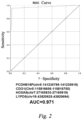
Figure 2 shows the ROC curves of the combined detection of the optimal methylated regions of four target genes, including PCDHB18P, CDO1, HOXA9 and LYPD5. - The technical solutions of the present invention will be explained and described in detail below in conjunction with the accompanying drawings and the preferred specific examples, so that those skilled in the art can better understand and implement the present invention.
- The Illumina Infinium MethylationEPIC BeadChip (containing 853,307 CpG sites) was used to screen the methylated genes of ovarian cancer by selecting 20 paraffin-embedded ovarian cancer tissue samples and 20 paraffin-embedded ovarian benign tissue samples.
- Using commercial nucleic acid extraction or purification reagents and methylation detection sample pretreatment kits, the above 40 tissue samples were subjected to the extraction of genomic DNA and the process of DNA bisulfite conversion to obtain qualified, converted bis-DNA for the subsequent methylation chip screening and detection. Through the significant methylation difference locus algorithm, the results of ovarian cancer samples and ovarian benign samples were compared and analyzed, and the first four genes in deltaBeta (the methylation level of ovarian cancer tissue samples - the methylation level of ovarian benign control samples; PCDHB18P = 0.895, CDO1 = 0.818, HOXA9 = 0.812, and LYPD5 = 0.807) were screened out. Due to the large regional distribution of CpG in each gene, it is required to perform RT-PCR for verification and analysis of the particular methylation position of each gene. Finally, in each gene, three methylated regional fragments were determined, as shown in SEQ ID NOs: 1-2.
- For the methylation regions of four target genes screened in Example 1, primers were designed and screened according to the specific sequences of the methylation regions to obtain the specific nucleotide sequences of the methylation region detection primers and probes, as shown in Table 1.
Table 1. Sequences of detection primers and probes corresponding to different methylated regions of target genes PCDHB 18P-F1 atccaaaATTGGATTAATTTTT+G+GATCG SEQ ID NO: 13 PCDHB 18P-R1 tttaggttCAATATAATC+C+CAAACCTAAACG SEQ ID NO: 14 PCDHB 18P-FP1 FAM-ccgccaTAAC+GTTA+ATA+GTCGT+GGATGGCGG-BHQ1 SEQ ID NO: 15 PCDHB 18P-F2 ataataataaAAAT+G+A+A+T+AT+AAT+ATT+ATTA+TTATCG SEQ ID NO: 16 PCDHB 18P-R2 tttttattttAAA+ATTT+AA+AT+AA+AA+ATAA+A+A+ACG SEQ ID NO: 17 PCDHB 18P-FP2 FAM-ttaacgAATC+GTGTTG+GTTTTC+GAC+GTTAA-B HQ 1 SEQ ID NO: 18 PCDHB 18P-F3 ttaataccAGAT+AG+AG+ATTT+AGGT+ATT+AACG SEQ ID NO: 19 PCDHB 18P-R3 tatttgttCCA+A+A+AA+A+ATA+A+A+A+A+ACAAATACG SEQ ID NO: 20 PCDHB 18P-FP3 FAM-taaaacgTTAT+TCGT+TGT+TGTCGT+TTTA-BHQ1 SEQ ID NO: 21 CDO1-F1 aaaaaccTTT+TGGG+TGGT+TTTTCG SEQ ID NO: 22 CDO1-R1 ggaggatgCT+ATAA+A+ATTCATCC+TCCCG SEQ ID NO: 23 CDO1-FP1 ROX-aacgaccTCGT+TT+TTT+TT+TCGAGGGTCGTT-BHQ2 SEQ ID NO: 24 CDO1-F2 caaaaacGT+TTT+TTGTTGT+TTT+TGCG SEQ ID NO: 25 CDO1-R2 tgtgtgtGCT+AA+ACAA+ACAC+ACACG SEQ ID NO: 26 CDO1-FP2 ROX-aaaatacTTT+TTACGT+TTCGGTAT+TTT-BHQ2 SEQ ID NO: 27 CDO1-F3 ataaaaaGTTCGT+TTAGTT+TTTT+TATCG SEQ ID NO: 28 CDO1-R3 agatggaCTTCG+ATCTAT+TCCATCTCG SEQ ID NO: 29 CDO1-FP3 ROX-taactacgTGAG+ACGCG+CGCGTAGTTA-BHQ2 SEQ ID NO: 30 HOXA9 -F1 cctttctcG+TTAGT+TGA+TGAGA+AAGGCG SEQ ID NO: 31 HOXA9 -R1 tataattaT+T+TT+AA+A+AACCTAA+TT+AA+TTATACG SEQ ID NO: 32 HOXA9 -FP1 CY5-ccacgcaTG+GGCGCG+CGTGTGCGTGG-BHQ2 SEQ ID NO: 33 HOXA9 -F2 aaaacttaTAGT+TT+AATT+TTT+TTAAGTTT+TCG SEQ ID NO: 34 HOXA9 -R2 ttagtttgCA+AAA+ACC+ACACA+AA+CTAACG SEQ ID NO: 35 HOXA9 -FP2 CYS-attcetttaTTA+CGGCG+AGGTA+A+ACGAAT-BHQ2 SEQ ID NO: 36 HOXA9 -F3 aaacccctcTTTA+TGTG+AGGGG+TT+TCG SEQ ID NO: 37 HOXA9 -R3 tgattaattAACTA+TT+TTATA+TCACAAT+TAA+TCACG SEQ ID NO: 38 HOXA9 -FP3 CY5-aacgcgaatTCGTAT+TCGT+ACGCGGG+AT+TCGCGTT-BHQ2 SEQ ID NO: 39 LYPD5-F1 ttttcaatTG+AAA+TTTGA+TAAT+TGA+AAACG SEQ ID NO: 40 LYPD5-R1 ttttttgAA+ATCCCT+T+TA+ACAA+A+AA+ACG SEQ ID NO: 41 LYPD5-FP1 HEX-attaaaaaGTTT+TTATT+TTCGT+ACGTATT+TT+TTAAT-BHQl SEQ ID NO: 42 LYPD5-F2 acccaatTT+T+T+TA+TAGGT+TA+TTGGGTCG SEQ ID NO: 43 LYPD5-R2 tattttagCCCA+AA+AA+TCACT+A+AA+ATACG SEQ ID NO: 44 LYPD5-FP2 HEX-aacgaatAGCG+TGCG+TT+TT+TG+ATTCGTT-BHQ1 SEQ ID NO: 45 LYPD5-F3 cacataatGGTG+TG+TGTAATATTATGTGCG SEQ ID NO: 46 LYPD5-R3 gttgggaCAAA+ACA+TCCC+AACCG SEQ ID NO: 47 LYPD5-FP3 HEX-acgcaacgTCGTGCG+AAT+GGTG+ACGT+TGCGT-BHQ1 SEQ ID NO: 48 GAPDH -F AGGTTAAATATAGTTGTTGA SEQ ID NO: 49 GAPDH -R CAACCCAAACCCCCAAC SEQID NO: 50 GAPDH -FP Joe-TAGTTGGGGGTTTGGGTT-BHQ1 SEQ ID NO: 51 Notes: F stands for a forward detection primer, R stands for a reverse detection primer, and FP stands for a detection probe. In this table, the probe sequences as shown have been labeled with fluorophores and quenching groups. - Using the primers and probes listed in the above Table 1, a detection kit (PCR amplification system kit) was prepared, the detection kit comprising a PCR reaction solution, a mixture solution of primers and probes, a positive quality control product and a negative quality control product, and the components are listed in Table 2 below:
Table 2. Composition of PCR amplification system kits Components Main ingredients PCR reaction solution 1-1.5 µL of Taq DNA polymerase with methylation characteristics at a concentration of 1 U/µL; 5-8 µL of dNTPs at a concentration of 10 mM; Mg2+ 3-6 µL at a concentration of 2-5 mM; 5 µL of 10 × DNA polymerase buffer; Purified water making up to 25 µL. Mixed solution of primers and probes Forward and reverse primers and probes for the methylated region of PCDHB 18P gene, Forward and reverse primers and probes for the methylated region of CDO1 gene, Forward and reverse primers and probes for the methylated region of HOXA9 gene, Forward and reverse primers and probes for the methylated region of LYPD5 gene, Primers and probes for GAPDH gene. Positive quality control Genomic DNA fragments of different malignant tumor cell lines Negative quality control Purified water - 128 ovarian cancer samples with known and clear pathological information were selected: 62 samples identified as high-grade serous ovarian cancer, 28 samples identified as low-grade serous ovarian cancer, 13 samples identified as mucinous ovarian cancer, 13 samples identified as ovarian endometrioid carcinoma, and 12 samples identified as ovarian clear cell carcinoma; and 110 ovarian benign samples were selected. The above samples were obtained from reserved plasma samples.
- Fragments of free DNA in plasma are mostly around 150 bp, and the content thereof is very low (lower than 10 ng). Therefore, sample pretreatment reagents and subsequent PCR reaction solutions are important to determine whether changes in gene methylation in plasma free DNA can be detected.
- The methylation pretreatment reagent for a sample includes a cell genomic DNA extraction reagent and a DNA bisulfite conversion reagent. When using the sample methylation pretreatment reagent, two processes are included, one is the extraction process of plasma free DNA, and the other is the conversion process of bisulfite, and quality control should be well done in each step.
- 1. A nucleic acid extraction or purification reagent in a plasma free DNA extraction kit was used as a plasma free DNA extraction reagent. Free DNA was extracted from the above 238 plasma samples, and meanwhile DNA quality was monitored. DNA with a concentration of above 0.3 ng/ul and a total amount of 6 ng-10 ng were extracted from 2 mL of plasma, and ensure that the quality of the extracted free DNA meets the requirements.
- 2. Using a bisulfite conversion kit (methylation detection sample pretreatment kit) as the DNA bisulfite conversion reagent, bisulfite conversion was performed on the extracted DNA, wherein unmethylated cytosine (C) in DNA was converted to uracil (U), and methylated cytosine (C) remained unchanged, and then the converted bis-DNA was obtained.
- Several important quality control indicators in the conversion process: one is to monitor the amount and integrity of free DNA in the sample, the second is to monitor the recovery rate of the converted bis-DNA, and the third is to monitor the integrity of the converted bis-DNA. So that high-quality bis-DNA were provided for subsequent PCR amplification reactions.
-
Table 3. PCR reaction solution (25 µL/person) Components Adding amount/person (µL) DNA polymerase with methylation characteristics (1 U/µL) 1.5 dNTPs (10 mM) 7 Mg2+ (2-5 mM) 5 10 × DNA polymerase buffer 5 Purified water making up to 25 µL - In the above PCR reaction system, the important component is the methylation-specific Taq polymerase, which has the following advantages: the template sequence after the bisulfite conversion is amplified, the sequence after the conversion can be specifically recognized, and the amplification efficiency of the primers on the sequence after the conversion is improved. Less Taq enzyme reduces the amplification efficiency, and too much Taq enzyme can easily cause non-specific amplification. Therefore, the selection of the amount of Taq enzyme directly affects the results of PCR amplification. In addition, the proportion of dNTPs, Mg2+, and 10 × DNA polymerase buffer in the system also directly affects the amplification efficiency of the primer-probe combination.
- The PCR reaction system can specifically amplify bis-DNA after the bisulfite conversion, and is used for multiplex primer amplification. Therefore, the amplification ability of the PCR reaction solution is particularly important, and it needs to ensure that the amplification efficiency of the primers and probe of each gene in the PCR reaction system is consistent with that of its corresponding single amplification. Therefore, it is necessary to screen, test and verify methylation-specific Taq polymerases modified by different antibodies and the proportion to dNTPs, Mg2+, and 10 × DNA polymerase buffer components, in order to determine the composition of the final PCR reaction solution and that the amplification efficiency of the PCR reaction system is optimal.
Table 4. Mixed solution of primers and probes (5 µL/person) Components Adding amount/person (µL) CDO1/CELF1/HAND2/HS3ST2-F (100µM) 0.2-0.4 CDO1/CELF1/HAND2/HS3ST2-R (100 µM) 0.1-0.3 CDO1/CELF1/HAND2/HS3ST2-FP (100 µM) 0.15-0.25 GAPDH gene-F (100 µM) 0.05 GAPDH gene-R (100 µM) 0.05 GAPDH gene-FP (100 µM) 0.05 Purified water making up to 5 µL - Since the sample used is plasma free DNA and the content thereof is extremely low, the PCR reaction solution in Table 3 was expanded to 25 µL, and the PCR amplification reaction system was expanded to 50 µL. The template for amplifying bis-DNA needs to be expanded to 20 µL to further improve the detection rate of the methylated target gene in plasma free DNA.
- 20 µL of negative quality control, positive quality control and the converted Bis-DNA clinical samples were respectively added to the above formulated system. PCR reaction was carried out and the conditions are: pre-denaturation at 96°C for 5 min; denaturation at 94°C for 15 s, annealing and extension at 60°C for 35 s, 45 cycles; keeping at 25°C for 10 min.
-
- Step 1: pre-denaturation at 96°C for 5 min;
- Step 2: denaturation at 94°C for 15 s, annealing and extension at 60°C for 35 s, 45 cycles;
- Step 3: 25°C, 10 min;
- Signal collection, FAM, HEX, ROX, Joe and CY5 signals were collected at 60°C.
-
- (1) interpretation is performed according to the inflection point of the internal standard curve, and when the internal standard channel has an S-shaped amplification curve and the Ct value ≤ 35.4, the result is valid;
- (2) interpretation is performed according to the inflection point of the internal standard curve, and the amplification Ct values of PCDHB18P, CDO1, HOXA9 and LYPD5 are read successively; and
- (3) the thresholds and performance (including specificity, sensitivity, negative predictive value, and positive predictive value) of multiple methylated regions in the target genes are determined by integrating the Ct values of the above 4 genes and according to the ROC curve, to determine the optimal methylated region and the interpretation way for the 4 target genes.
- A total of 238 samples were detected using the above kit reaction system, including 128 samples of ovarian cancer and 110 samples of benign ovary.
- Comparing the clinicopathological results, in 238 plasma samples,
- the positive rate of PCDHB18P (chr5:141235136-141235232) in ovarian cancer was 63.3% (81/128), the specificity thereof in benign samples was 84.5% (93/110), and the ROC area thereof was 0.739;
- the positive rate of PCDHB18P (chr5:141235796-141235918) in ovarian cancer was 73.4% (94/128), the specificity thereof in benign samples was 99.1% (109/110), and the ROC area thereof was 0.863;
- the positive rate of PCDHB18P (chr5:141235982-141236064) in ovarian cancer was 64.1% (82/128), the specificity thereof in benign samples was 90% (99/110), and the ROC area thereof was 0.770;
- the positive rate of CDO1 (Chr5:115816656-115816755) in ovarian cancer was 71.9% (92/128), the specificity thereof in benign samples was 99.1% (109/110), and the ROC area thereof was 0.855;
- the positive rate of CDO1 (Chr5:115816502-115816579) in ovarian cancer was 60.2% (77/128), the specificity thereof in benign samples was 92.7% (102/110), and the ROC area thereof was 0.764;
- the positive rate of CDO1 (Chr5:115816382-115816460) in ovarian cancer was 56.3% (72/128), the specificity thereof in benign samples was 85.5% (94/110), and the ROC area thereof was 0.709;
- the positive rate of HOXA9 (chr7:27166431-27166523) in ovarian cancer was 60.9% (78/128), the specificity thereof in benign samples was 88.2% (97/110), and the ROC area thereof was 0.746;
- the positive rate of HOXA9 (chr7:27166020-27166126) in ovarian cancer was 57.8% (74/128), the specificity thereof in benign samples was 95.5% (105/110), and the ROC area thereof was 0.766;
- the positive rate of HOXA9 (chr7:27165835-27165919) in ovarian cancer was 69.5% (89/128), the specificity thereof in benign samples was 98.2% (108/110), and the ROC area thereof was 0.839;
- the positive rate of LYPD5 (chr19:43820817-43820923) in ovarian cancer was 63.3% (81/128), the specificity thereof in benign samples was 97.3% (107/110), and the ROC area thereof was 0.803;
- the positive rate of LYPD5 (chr19:43820625-43820694) in ovarian cancer was 71.9% (92/128), the specificity thereof in benign samples was 99.1% (109/110), and the ROC area thereof was 0.855;
- and the positive rate of LYPD5 (chr19:43820271-43820377) in ovarian cancer was 60.2% (77/128), the specificity thereof in benign samples was 89.1% (98/110), and the ROC area thereof was 0.746.
- After comparative analysis of the multiple methylated regions of PCDHB 18P, CDO1, HOXA9 and LYPD5, the optimal methylated region selected from PCDHB 18P gene was chr5:141235796-141235918, the optimal methylated region selected from CDO1 gene was Chr5:115816656-115816755, the optimal methylation region selected from HOXA9 gene was chr7:27165835-27165919, and the optimal methylation region selected from LYPD5 was chr19:43820625-43820694.
- Combined detection of the optimal methylated regions of PCDHB 18P, CDO1, HOXA9 and LYPD5 was carried out, and different gene combinations were compared and interpreted, and finally the optimal interpretation way was obtained. The following results as shown in Table 5 can be obtained:
Table 5. Combined detected performance of optimal methylated regions of two target genes PCDHB18P /CDO1 PCDHB 18P /HOXA9 PCDHB18P /LYPD5 CDO1/ HOXA9 CDO1/ LYPD5 HOXA91 LYPD5 Sensitivity (any one of genes is positive) 82.8% (106/128) 84.4% (108/128) 84.4% (108/128) 85.9% (110/128) 85.9% (110/128) 85.2% (109/128) Specificity (any one of genes is positive) 98.2% (108/110) 98.2% (108/110) 98.2% (108/110) 98.2% (108/110) 99.1% (109/110) 98.2% (108/110) Sensitivity (both genes are positive) 62.5% (80/128) 58.6% (75/128) 60.9% (78/128) 55.5% (71/128) 60.9% (78/128) 56.3% (72/128) Specificity (both genes are positive) 100% (110/110) 99.1% (109/110) 100% (110/110) 99.1% (109/110) 99.1% (109/110) 99.1% (109/110) Table 6. Combined detected performance of optimal methylated regions of three target genes PCDHB18P/ CDO1/HOXA9 PCDHB18P/ CDO1/LYPD5 CDO1/HOXA9/ LYPDS Sensitivity (any one of genes is positive) 89.8% (115/128) 90.6% (116/128) 93% (119/128) Specificity (any one of genes is positive) 98.2% (108/110) 98.2% (108/110) 98.2% (108/110) Sensitivity (any two of genes are positive) 70.3% (90/128) 72.7% (93/128) 68% (87/128) Specificity (any two of genes are positive) 98.2% (108/110) 99.1% (109/110) 99.1% (109/110) Sensitivity (all three genes are positive) 51.6% (66/128) 57.8% (74/128) 52.3% (67/128) Specificity (all three genes are positive) 100% (110/110) 100% (110/110) 99.1% (109/110) Table 7. Combined detected performance of optimal methylated regions of four target genes PCDHB18P/CDO1/HOXA9/LYPD5 Sensitivity (any one of genes is positive) 95.3% (122/128) Specificity (any one of genes is positive) 98.2% (108/110) Sensitivity (any two of genes are positive) 78.1% (100/128) Specificity (any two of genes are positive) 98.2% (108/110) Sensitivity (any three of genes are positive) 63.3% (81/128) Specificity (any three of genes are positive) 99.1% (109/110) Sensitivity (all four genes are positive) 50% (64/128) Specificity (all four genes are positive) 100% (110/110) - In the data of the above three tables, when the optimal methylated regions of four target genes, including PCDHB 18P, CDO1, HOXA9, and LYPD5, were combined detected, and if any one of genes was positive, the sample was interpreted as positive, the sensitivity and specificity in 283 plasma samples were overall optimal, wherein the sensitivity for ovarian cancer was 95.3% (122/128), the specificity for benign samples was 98.2% (108/110), and the detected ROC area was 0.971.
- Meanwhile, the combined detection of the optimal methylated regions of four target genes, including PCDHB18P, CDO1, HOXA9 and LYPD5 (if any one of genes was positive, the methylation detection was interpreted as positive), was analyzed compared with a tumor molecular marker CA125 (a commonly used detection means in clinical practice) detection. The results of the comparative analysis are shown in Table 8. It can be seen from Table 8: the positive predictive value (0.984) from the combined detection of the methylation status of the optimal methylated regions of the four genes was significantly higher than that of the tumor molecular marker CA125 (0.736).
Table 8. Comparative analysis of combined detection of methylated regions and CA125 detection PCDHB18P/CDO1/HOXA9 /LYPD5 Combined methylation detection Molecular markers for tumors CA125 detection Negative Positive Negative Positive Benign ovary (n = 110) 108 2 76 34 High-grade serous ovarian cancer (n = 62) 3 59 16 46 Low-grade serous ovarian cancer (n = 28) 2 26 8 20 Mucinous ovarian cancer (n = 13) 1 12 3 10 Ovarian endometrioid carcinoma (n = 13) 0 13 4 9 Ovarian clear cell carcinoma (n = 12) 0 12 2 10 Sensitivity (ovarian cancer) 95.3% 74.2% Specificity (benign ovary) 98.2% 69.1% Positive predictive value 0.984 0.736 Negative predictive value 0.947 0.697 - Through sample research and test, it is proved that DNA methylation has a high positive predictive value for early detection of ovarian cancer, and the detection can be done with plasma samples. The present invention uses special primer and probe design skills (for specially capturing low-copy plasma free DNA fragments), and combined detection of multi-target gene methylation regions, which are functionally complementary, and significantly improve the detection of early ovarian cancer.
- Herein, specific examples are used to describe the inventive concept in detail, and the description of the above examples is only used to help understand the core idea of the present invention. It should be pointed out that for those of ordinary skill in the art, any obvious modification, equivalent replacement or other improvement made should be included in the protection scope of the present invention without departing from the inventive concept.
Claims (10)
- A combination of DNA methylation markers, wherein the combination comprises at least one methylated fragment of each of the following four genes, including PCDHB18P, CDO1, HOXA9 and LYPD5:PCDHB 18P gene:chr5:141235136-141235232: SEQ ID NO: 1,chr5:141235796-141235918: SEQ ID NO: 2,chr5:141235982-141236064: SEQ ID NO: 3;CDO1 gene:Chr5:115816656-115816755: SEQ ID NO: 4,Chr5:115816502-115816579: SEQ ID NO: 5,Chr5:115816382-115816460: SEQ ID NO: 6;HOXA9 gene:chr7:27166431-27166523: SEQ ID NO: 7,chr7:27166020-27166126: SEQ ID NO: 8,chr7:27165835-27165919: SEQ ID NO: 9;and LYPD5 gene:chr19:43820817-43820923: SEQ ID NO: 10,chr19:43820625-43820694: SEQ ID NO: 11,chr19:43820271-43820377: SEQ ID NO: 12.
- The combination of DNA methylation markers according to claim 1, wherein the combination consists of the following methylated fragments:PCDHB 18P gene: chr5:141235796-141235918: SEQ ID NO: 2;CDO1 gene: Chr5:115816656-115816755: SEQ ID NO: 4;HOXA9 gene: chr7:27165835-27165919: SEQ ID NO: 9;and LYPD5 gene: chr19:43820625-43820694: SEQ ID NO: 11.
- Use of the combination of DNA methylation markers according to claim 1 or 2 in the preparation of a reagent for early screening and diagnosis of ovarian cancer.
- A detection primer set for early screening and diagnosis of ovarian cancer, wherein the detection primer set comprises target gene detection primers for correspondingly detecting the methylated statuses of the methylated fragments in the combination according to claim 1 or 2, wherein each of the target gene detection primers has a nucleotide sequence with the 5' terminus having a sequence with a length of 5-10 bp which is complementary to and paired with the 3' terminus but is not paired with CG bases at the end of the 3' terminus; and the nucleotide sequence of each of the target gene detection primers has a Tm value which is 2-4°C higher than the annealing temperature of a PCR reaction system.
- The detection primer set according to claim 4, wherein the nucleotide sequence of each of the target gene detection primers has a locked nucleic acid modified base.
- The detection primer set according to claim 4, wherein the nucleotide sequences of the target gene detection primers are shown below:for PCDHB18P target gene:the nucleotide sequences of detection primers for chr5:141235136-141235232: SEQ ID NOs: 13-14,the nucleotide sequences of detection primers for chr5:141235796-141235918: SEQ ID NOs: 16-17,the nucleotide sequences of detection primers for chr5:141235982-141236064: SEQ ID NOs: 19-20;for CDO1 target gene:the nucleotide sequences of detection primers for Chr5:115816656-115816755: SEQ ID NOs: 22-23,the nucleotide sequences of detection primers for Chr5:115816502-115816579: SEQ ID NOs: 25-26,the nucleotide sequences of detection primers for Chr5:115816382-115816460: SEQ ID NOs: 28-29;for HOXA9 target gene:the nucleotide sequences of detection primers for chr7:27166431-27166523: SEQ ID NOs: 31-32,the nucleotide sequences of detection primers for chr7:27166020-27166126: SEQ ID NOs: 34-35,the nucleotide sequences of detection primers for chr7:27165835-27165919: SEQ ID NOs: 37-38;and for LYPD5 target gene:the nucleotide sequences of detection primers for chr19:43820817-43820923: SEQ ID NOs: 40-41,the nucleotide sequences of detection primers for chr19:43820625-43820694: SEQ ID NOs: 43-44,the nucleotide sequences of detection primers for chr19:43820271-43820377: SEQ ID NOs: 46-47.
- A detection kit for early screening and diagnosis of ovarian cancer, wherein the detection kit comprises the detection primer set according to any one of claims 4-6.
- The detection kit according to claim 7, wherein the detection kit further comprises target gene detection probes for correspondingly detecting the methylated statuses of the methylated fragments in the combination according to claim 1 or 2, wherein the nucleotide sequence of the target gene detection probe is shown below:for PCDHB18P gene:the nucleotide sequence of a detection probe for chr5:141235136-141235232: SEQ ID NO: 15,the nucleotide sequence of a detection probe for chr5:141235796-141235918: SEQ ID NO: 18,the nucleotide sequence of a detection probe for chr5:141235982-141236064: SEQ ID NO: 21;for CDO1 gene:the nucleotide sequence of a detection probe for Chr5:115816656-115816755: SEQ ID NO: 24,the nucleotide sequence of a detection probe for Chr5:115816502-115816579: SEQ ID NO: 27,the nucleotide sequence of a detection probe for Chr5:115816382-115816460: SEQ ID NO: 30;for HOXA9 gene:the nucleotide sequence of a detection probe for chr7:27166431-27166523: SEQ ID NO: 33,the nucleotide sequence of a detection probe for chr7:27166020-27166126: SEQ ID NO: 36,the nucleotide sequence of a detection probe for chr7:27165835-27165919: SEQ ID NO: 39;and for LYPD5 gene:the nucleotide sequence of a detection probe for chr19:43820817-43820923: SEQ ID NO: 42,the nucleotide sequence of a detection probe for chr19:43820625-43820694: SEQ ID NO: 45,the nucleotide sequence of a detection probe for chr19:43820271-43820377: SEQ ID NO: 48.
- The detection kit according to claim 8, wherein the detection kit further comprises internal reference gene detection primers and an internal reference gene detection probe for an internal reference gene GAPDH, wherein the internal reference gene detection primers have nucleotide sequences as shown in SEQ ID NOs: 49-50, and the internal reference gene detection probe has a nucleotide sequence as shown in SEQ ID NO: 51.
- The detection kit according to claim 9, wherein the nucleotide sequences of the target gene detection probe and the internal reference gene detection probe are labeled with fluorophores at the 5' terminus and are labeled with quenching groups at the 3' terminus, wherein the labeled fluorophores are different between the target gene detection probes and between the internal reference gene detection probes in the same detection system.
Applications Claiming Priority (2)
| Application Number | Priority Date | Filing Date | Title |
|---|---|---|---|
| CN202210208334.2A CN114480655B (en) | 2022-03-03 | 2022-03-03 | DNA methylation marker combination and application thereof, and early detection primer probe and kit for ovarian cancer |
| PCT/CN2022/095437 WO2023165035A1 (en) | 2022-03-03 | 2022-05-27 | Dna methylation marker combination, use, primer probe for early detection of ovarian cancer and kit |
Publications (2)
| Publication Number | Publication Date |
|---|---|
| EP4265738A1 true EP4265738A1 (en) | 2023-10-25 |
| EP4265738A4 EP4265738A4 (en) | 2025-05-07 |
Family
ID=87851182
Family Applications (1)
| Application Number | Title | Priority Date | Filing Date |
|---|---|---|---|
| EP22737702.5A Pending EP4265738A4 (en) | 2022-03-03 | 2022-05-27 | DNA METHYLATION MARKER COMBINATION, USE, PRIMER PROBE FOR EARLY DETECTION OF OVARIAN CANCER AND KIT |
Country Status (2)
| Country | Link |
|---|---|
| US (1) | US11866790B2 (en) |
| EP (1) | EP4265738A4 (en) |
Family Cites Families (8)
| Publication number | Priority date | Publication date | Assignee | Title |
|---|---|---|---|---|
| WO2012031008A2 (en) * | 2010-08-31 | 2012-03-08 | The General Hospital Corporation | Cancer-related biological materials in microvesicles |
| WO2013096661A1 (en) * | 2011-12-22 | 2013-06-27 | Illumina, Inc. | Methylation biomarkers for ovarian cancer |
| TW201632629A (en) | 2015-01-14 | 2016-09-16 | 臺北醫學大學 | Methods for cancer diagnosis and prognosis |
| EP3859015A1 (en) * | 2015-08-06 | 2021-08-04 | The University of Utah Research Foundation | Methods of indentifying male fertility status and embryo quality |
| WO2017201606A1 (en) * | 2016-05-04 | 2017-11-30 | Queen's University At Kingston | Cell-free detection of methylated tumour dna |
| EP3472358B1 (en) * | 2016-06-16 | 2021-08-18 | The Johns Hopkins University | Methods and system for epigenetic analysis |
| TWI648403B (en) | 2016-07-29 | 2019-01-21 | Taipei Medical University (Tmu) | Diagnosis of gynecological tumors |
| CN113337614A (en) | 2021-08-05 | 2021-09-03 | 北京起源聚禾生物科技有限公司 | Marker, primer probe and kit for early screening and diagnosis of endometrial cancer |
-
2022
- 2022-05-27 EP EP22737702.5A patent/EP4265738A4/en active Pending
- 2022-07-22 US US17/871,119 patent/US11866790B2/en active Active
Also Published As
| Publication number | Publication date |
|---|---|
| US11866790B2 (en) | 2024-01-09 |
| EP4265738A4 (en) | 2025-05-07 |
| US20230279497A1 (en) | 2023-09-07 |
Similar Documents
| Publication | Publication Date | Title |
|---|---|---|
| CN113755603B (en) | Marker, primer probe and kit for early screening and diagnosis of endometrial cancer | |
| CN108977543B (en) | An early diagnostic reagent for colorectal cancer based on combined detection of SDC2 and SFRP2 gene methylation levels | |
| CN114480661A (en) | Endometrium benign and malignant lesion combined marker, detection primer probe set and kit | |
| WO2025077120A1 (en) | Kit and method for detecting bladder cancer | |
| WO2023165035A1 (en) | Dna methylation marker combination, use, primer probe for early detection of ovarian cancer and kit | |
| CN112501298B (en) | Composition and kit for methylation noninvasive early detection of ovarian cancer | |
| CN116144782A (en) | A combination marker for lung cancer detection and its application | |
| EP4265738A1 (en) | Dna methylation marker combination, use, primer probe for early detection of ovarian cancer and kit | |
| JP7823936B2 (en) | Cervical cell gene methylation detection kit and detection method | |
| EP4130297A1 (en) | Markers, primers, probes and kit for early screening and diagnosis of endometrial cancer | |
| CN116004831B (en) | A reagent and detection kit for diagnosing or assisting in the diagnosis of bladder cancer | |
| CN118667958A (en) | Marker combination and detection kit for screening endometrial cancer | |
| CN115927610B (en) | Application of reagents for detecting methylation levels of target regions in the FOXO6 gene in the preparation of bladder cancer diagnostic products | |
| CN116463417A (en) | Application of the reagent for detecting the methylation level of the target region in the preparation of liver cancer diagnostic products | |
| CN114645094A (en) | Endometrial cancer biomarker and application thereof | |
| CN115948561B (en) | Reagent and detection kit for esophageal squamous carcinoma diagnosis or auxiliary diagnosis and application thereof | |
| CN115961048B (en) | A gene methylation detection primer combination, reagent and application thereof | |
| CN113943810B (en) | Endometrial cancer detection reagents and kits | |
| CN117187388A (en) | Application of GRIK2 gene as marker in preparation of lung cancer detection kit | |
| CN116814790A (en) | Application of PITX2 gene as a marker in detecting lung cancer | |
| CN116694765A (en) | Kit for detecting endometrial cancer and application thereof | |
| CN117987545A (en) | Methylation detection reagent and kit for auxiliary diagnosis of breast cancer and application | |
| CN117568480A (en) | Reagent and kit for breast cancer or precancerous lesion detection and application | |
| CN117535415A (en) | Nucleic acid composition and kit for breast cancer detection | |
| CN119662824A (en) | Kit for diagnosing/screening endometrial cancer and application thereof |
Legal Events
| Date | Code | Title | Description |
|---|---|---|---|
| STAA | Information on the status of an ep patent application or granted ep patent |
Free format text: STATUS: UNKNOWN |
|
| STAA | Information on the status of an ep patent application or granted ep patent |
Free format text: STATUS: THE INTERNATIONAL PUBLICATION HAS BEEN MADE |
|
| PUAI | Public reference made under article 153(3) epc to a published international application that has entered the european phase |
Free format text: ORIGINAL CODE: 0009012 |
|
| STAA | Information on the status of an ep patent application or granted ep patent |
Free format text: STATUS: REQUEST FOR EXAMINATION WAS MADE |
|
| 17P | Request for examination filed |
Effective date: 20220720 |
|
| AK | Designated contracting states |
Kind code of ref document: A1 Designated state(s): AL AT BE BG CH CY CZ DE DK EE ES FI FR GB GR HR HU IE IS IT LI LT LU LV MC MK MT NL NO PL PT RO RS SE SI SK SM TR |
|
| P01 | Opt-out of the competence of the unified patent court (upc) registered |
Effective date: 20231108 |
|
| A4 | Supplementary search report drawn up and despatched |
Effective date: 20250408 |
|
| RIC1 | Information provided on ipc code assigned before grant |
Ipc: C12N 15/11 20060101ALI20250402BHEP Ipc: C12Q 1/6851 20180101ALI20250402BHEP Ipc: C12Q 1/6886 20180101AFI20250402BHEP |
|
| DAV | Request for validation of the european patent (deleted) | ||
| DAX | Request for extension of the european patent (deleted) | ||
| GRAP | Despatch of communication of intention to grant a patent |
Free format text: ORIGINAL CODE: EPIDOSNIGR1 |
|
| STAA | Information on the status of an ep patent application or granted ep patent |
Free format text: STATUS: GRANT OF PATENT IS INTENDED |
|
| INTG | Intention to grant announced |
Effective date: 20251201 |