EP4252784A2 - Verfahren zur zellmarkierungen und medizinische bildgebung - Google Patents
Verfahren zur zellmarkierungen und medizinische bildgebung Download PDFInfo
- Publication number
- EP4252784A2 EP4252784A2 EP23186316.8A EP23186316A EP4252784A2 EP 4252784 A2 EP4252784 A2 EP 4252784A2 EP 23186316 A EP23186316 A EP 23186316A EP 4252784 A2 EP4252784 A2 EP 4252784A2
- Authority
- EP
- European Patent Office
- Prior art keywords
- cells
- labeling
- labeled
- cell
- imaging
- Prior art date
- Legal status (The legal status is an assumption and is not a legal conclusion. Google has not performed a legal analysis and makes no representation as to the accuracy of the status listed.)
- Pending
Links
Images







Classifications
-
- A—HUMAN NECESSITIES
- A61—MEDICAL OR VETERINARY SCIENCE; HYGIENE
- A61K—PREPARATIONS FOR MEDICAL, DENTAL OR TOILETRY PURPOSES
- A61K51/00—Preparations containing radioactive substances for use in therapy or testing in vivo
- A61K51/02—Preparations containing radioactive substances for use in therapy or testing in vivo characterised by the carrier, i.e. characterised by the agent or material covalently linked or complexing the radioactive nucleus
- A61K51/04—Organic compounds
- A61K51/0474—Organic compounds complexes or complex-forming compounds, i.e. wherein a radioactive metal (e.g. 111In3+) is complexed or chelated by, e.g. a N2S2, N3S, NS3, N4 chelating group
- A61K51/0478—Organic compounds complexes or complex-forming compounds, i.e. wherein a radioactive metal (e.g. 111In3+) is complexed or chelated by, e.g. a N2S2, N3S, NS3, N4 chelating group complexes from non-cyclic ligands, e.g. EDTA, MAG3
-
- A—HUMAN NECESSITIES
- A61—MEDICAL OR VETERINARY SCIENCE; HYGIENE
- A61K—PREPARATIONS FOR MEDICAL, DENTAL OR TOILETRY PURPOSES
- A61K49/00—Preparations for testing in vivo
- A61K49/06—Nuclear magnetic resonance [NMR] contrast preparations; Magnetic resonance imaging [MRI] contrast preparations
- A61K49/08—Nuclear magnetic resonance [NMR] contrast preparations; Magnetic resonance imaging [MRI] contrast preparations characterised by the carrier
- A61K49/10—Organic compounds
- A61K49/101—Organic compounds the carrier being a complex-forming compound able to form MRI-active complexes with paramagnetic metals
- A61K49/103—Organic compounds the carrier being a complex-forming compound able to form MRI-active complexes with paramagnetic metals the complex-forming compound being acyclic, e.g. DTPA
-
- A—HUMAN NECESSITIES
- A61—MEDICAL OR VETERINARY SCIENCE; HYGIENE
- A61K—PREPARATIONS FOR MEDICAL, DENTAL OR TOILETRY PURPOSES
- A61K49/00—Preparations for testing in vivo
- A61K49/06—Nuclear magnetic resonance [NMR] contrast preparations; Magnetic resonance imaging [MRI] contrast preparations
- A61K49/08—Nuclear magnetic resonance [NMR] contrast preparations; Magnetic resonance imaging [MRI] contrast preparations characterised by the carrier
- A61K49/10—Organic compounds
- A61K49/101—Organic compounds the carrier being a complex-forming compound able to form MRI-active complexes with paramagnetic metals
- A61K49/106—Organic compounds the carrier being a complex-forming compound able to form MRI-active complexes with paramagnetic metals the complex-forming compound being cyclic, e.g. DOTA
-
- A—HUMAN NECESSITIES
- A61—MEDICAL OR VETERINARY SCIENCE; HYGIENE
- A61K—PREPARATIONS FOR MEDICAL, DENTAL OR TOILETRY PURPOSES
- A61K49/00—Preparations for testing in vivo
- A61K49/06—Nuclear magnetic resonance [NMR] contrast preparations; Magnetic resonance imaging [MRI] contrast preparations
- A61K49/18—Nuclear magnetic resonance [NMR] contrast preparations; Magnetic resonance imaging [MRI] contrast preparations characterised by a special physical form, e.g. emulsions, microcapsules, liposomes
- A61K49/1896—Nuclear magnetic resonance [NMR] contrast preparations; Magnetic resonance imaging [MRI] contrast preparations characterised by a special physical form, e.g. emulsions, microcapsules, liposomes not provided for elsewhere, e.g. cells, viruses, ghosts, red blood cells, virus capsides
-
- A—HUMAN NECESSITIES
- A61—MEDICAL OR VETERINARY SCIENCE; HYGIENE
- A61K—PREPARATIONS FOR MEDICAL, DENTAL OR TOILETRY PURPOSES
- A61K51/00—Preparations containing radioactive substances for use in therapy or testing in vivo
- A61K51/12—Preparations containing radioactive substances for use in therapy or testing in vivo characterised by a special physical form, e.g. emulsion, microcapsules, liposomes, characterized by a special physical form, e.g. emulsions, dispersions, microcapsules
- A61K51/1203—Preparations containing radioactive substances for use in therapy or testing in vivo characterised by a special physical form, e.g. emulsion, microcapsules, liposomes, characterized by a special physical form, e.g. emulsions, dispersions, microcapsules in a form not provided for by groups A61K51/1206 - A61K51/1296, e.g. cells, cell fragments, viruses, virus capsides, ghosts, red blood cells, viral vectors
Definitions
- This invention relates to methods of ex vivo labeling of a biological material for in vivo imaging, methods of labeling a biological material in vivo, methods for preparing a labeling agent, and methods for in vivo imaging of a subject using a biological material labeled with a labeling agent.
- the biological material is selected from cells.
- SPECT single-photon emission computerized tomography
- PET positron emission tomography
- a PET-based approach would offer superior quantification and imaging sensitivity characteristics over a SPECT-based approach, which are critical for tracking of small numbers of administered cells [Ref. 1].
- 89 Zr has emerged as an attractive PET radionuclide for cell labeling applications due to its high spatial resolution and 78.4 hour half-life that may allow monitoring of administered cells up to a 2-3 week period.
- the invention provides a method of ex vivo labeling of a cell for in vivo imaging.
- the method includes the step of contacting a cell ex vivo with a labeling agent such that the cell becomes labeled for in vivo imaging, wherein the labeling agent comprises a chelating moiety, a conjugation moiety, and at least one of (i) a radionuclide and (ii) a paramagnetic metal ion or compound.
- the labeling agent may include a radionuclide, or a paramagnetic metal ion or compound, or both a radionuclide and a paramagnetic metal ion or compound.
- the invention provides a method of labeling a cell in vivo.
- the method includes the step of administering to a subject a labeling agent thereby labeling a cell in vivo, wherein the labeling agent does not include an antibody, and wherein the labeling agent comprises a chelating moiety, a conjugation moiety, and at least one of (i) a radionuclide and (ii) a paramagnetic metal ion or compound.
- the labeling agent may include a radionuclide, or a paramagnetic metal ion or compound, or both a radionuclide and a paramagnetic metal ion or compound.
- the invention provides a method of ex vivo labeling of a biological material for in vivo imaging.
- the method includes the step of contacting a biological material ex vivo with a labeling agent such that the biological material becomes labeled for in vivo imaging, wherein labeling agent comprises a chelating moiety, a conjugation moiety, and at least one of (i) a radionuclide and (ii) a paramagnetic metal ion or compound, and wherein the biological material is selected from cells, liposomes, DNA aptamers, RNA aptamers, viruses, nanoparticles, microorganisms, antibodies, proteins, peptides, scaffolds, polymers, and nucleic acids.
- the labeling agent may include a radionuclide, or a paramagnetic metal ion or compound, or both a radionuclide and a paramagnetic metal ion or compound.
- the invention provides a method of labeling a biological material in vivo.
- the method includes the step of administering to a subject a labeling agent thereby labeling a biological material in vivo, wherein the labeling agent comprises a chelating moiety, a conjugation moiety, and at least one of (i) a radionuclide and (ii) a paramagnetic metal ion or compound, and wherein the biological material is selected from cells, liposomes, DNA aptamers, RNA aptamers, viruses, nanoparticles, microorganisms, proteins, peptides, scaffolds, polymers, and nucleic acids.
- the labeling agent may include a radionuclide, or a paramagnetic metal ion or compound, or both a radionuclide and a paramagnetic metal ion or compound.
- the chelating moiety may be a hydroxamic acid group, and preferably, the hydroxamic acid group is a desferrioxamine group.
- the conjugation moiety may include an isothiocyanate group.
- the conjugation moiety may include a benzyl group, or a maleimide group, or an NHS ester group, or a combination thereof.
- the labeling agent may be 89 Zrisothiocyanato-benzyl-desferrioxamine.
- the radionuclide may be selected from the group consisting of 11 C, 13 N, 15 O, 18 F, 34m Cl, 38 K, 45 Ti, 51 Mn, 52 Mn, 52m Mn, 52 Fe, 55 Co, 60 Cu, 61 Cu, 62 Cu, 64 Cu, 66 Ga, 68 Ga, 71 As, 72 As, 74 As, 75 Br, 76 Br, 82 Rb, 86 Y, 89 Zr, 90 Nb, 94m Tc, 99m Tc, 110m In, 111 In, 118 Sb, 120 I, 121 I, 122 I, 123 I, and 124 I.
- the radionuclide may be a positron emitter, such as 89 Zr.
- the paramagnetic metal ion may be selected from Gd +3 , Fe +3 , Mn +2 , and Y +3 .
- There are a number of chelators that may be used to chelate the paramagnetic metal ion e.g., Gd +3 , Fe +3 , Mn +2 , and Y +3 ).
- Non-limiting example chelators for the paramagnetic metal ion are diethylenetriaminepentaacetic acid (DTPA), ethylenediaminetetraacetic acid (EDTA), 1,4,7,10-tetraazacyclododecane-1,4,7,10-tetraacetic acid (DOTA), and 1,4,7-triazacyclononane-triacetic acid (NOTA).
- DTPA diethylenetriaminepentaacetic acid
- EDTA ethylenediaminetetraacetic acid
- DOTA 1,4,7,10-tetraazacyclododecane-1,4,7,10-tetraacetic acid
- NOTA 1,4,7-triazacyclononane-triacetic acid
- the paramagnetic compound may be compounds including 19 F, such as a perfluorocarbon.
- the biological material may be cells selected from microorganisms, blood-derived cells, cancer cells, stem cells, and dendritic cells.
- the biological material may be selected from stem cells.
- the biological material may be selected from viruses.
- the invention provides a labeling agent comprising a chelating moiety, a conjugation moiety, and at least one of (i) a radionuclide and (ii) a paramagnetic metal ion or compound, wherein the chelating moiety includes a hydroxamic acid group and the conjugation moiety includes an isothiocyanate group, and wherein the labeling agent is in a reactive form suitable for labeling a biological material selected from cells, liposomes, DNA aptamers, RNA aptamers, viruses, nanoparticles, microorganisms, antibodies, proteins, peptides, scaffolds, polymers, and nucleic acids.
- the chelating moiety may be a hydroxamic acid group, and preferably, the hydroxamic acid group is a desferrioxamine group.
- the conjugation moiety may include an isothiocyanate group.
- the conjugation moiety may include a benzyl group, or a maleimide group, or an NHS ester group, or a combination thereof.
- the labeling agent may be 89 Zr-isothiocyanato-benzyl-desferrioxamine.
- the radionuclide may be selected from the group consisting of 11 C, 13 N, 15 O, 18 F, 34m Cl, 38 K, 45 Ti, 51 Mn, 52 Mn, 52m Mn, 52 Fe, 55 Co, 60 Cu, 61 Cu, 62 Cu, 64 Cu, 66 Ga, 68 Ga, 71 As, 72 As, 74 As, 75 Br, 76 Br, 82 Rb, 86 Y, 89 Zr, 90 Nb, 94m Tc, 99m Tc, 110m In, 111 In, 118 Sb, 120 I, 121 I, 122 I, 123 I, and 124 I.
- the radionuclide may be a positron emitter, such as 89 Zr.
- the paramagnetic metal ion may be selected from Gd +3 , Fe +3 , Mn +2 , and Y +3 .
- the paramagnetic compound may be compounds including 19 F, such as a perfluorocarbon.
- the biological material may be cells that are selected from microorganisms, blood-derived cells, cancer cells, stem cells, and dendritic cells.
- the biological material may be selected from stem cells.
- the biological material may be selected from viruses.
- the invention provides a method for preparing a labeling agent.
- the method includes the steps of: (a) providing a compound including a chelating moiety and a conjugation moiety; and (b) mixing the compound with at least one of (i) a radionuclide and (ii) a paramagnetic metal ion or compound to create the labeling agent before reacting the compound with an additional moiety, wherein the chelating moiety includes a hydroxamic acid group and the conjugation moiety includes an isothiocyanate group.
- the additional moiety may be a lysine residue.
- the additional moiety may be a functional group of at least one of a liposome, DNA aptamer, RNA aptamer, a virus, nanoparticles, microorganisms, antibodies, proteins, peptides, scaffolds, polymers, and nucleic acids.
- the additional moiety may be a functional group of a stem cell.
- the additional moiety may be a functional group of a virus.
- the chelating moiety may be a desferrioxamine group.
- the conjugation moiety may include a benzyl group.
- the labeling agent can be a 89 Zr-isothiocyanato-benzyl-desferrioxamine.
- the invention provides a method for in vivo imaging of a subject.
- the method includes the steps of: (a) administering to the subject a biological material labeled with a labeling agent comprising a chelating moiety, a conjugation moiety, and at least one of (i) a radionuclide and (ii) a paramagnetic metal ion or compound; (b) waiting a time sufficient to allow the labeled cells to accumulate at a tissue site to be imaged; and (c) imaging the tissues with a non-invasive imaging technique such as positron emission tomography imaging, wherein the biological material is selected from cells, liposomes, DNA aptamers, RNA aptamers, viruses, nanoparticles, microorganisms, proteins, peptides, scaffolds, polymers, and nucleic acids.
- a labeling agent comprising a chelating moiety, a conjugation moiety, and at least one of (i) a radionuclide and (ii)
- the invention provides a method of imaging a subject by emission tomography.
- the method comprises: (a) administering to the subject a biological material labeled with a labeling agent comprising a chelating moiety, a conjugation moiety, and at least one of (i) a radionuclide and (ii) a paramagnetic metal ion or compound; (b) using a plurality of detectors to detect gamma rays emitted from the subject and to communicate signals corresponding to the detected gamma rays; and (c) reconstructing from the signals a series of medical images of a region of interest of the subject, wherein the biological material is selected from cells, liposomes, DNA aptamers, RNA aptamers, viruses, nanoparticles, microorganisms, proteins, peptides, scaffolds, polymers, and nucleic acids.
- the invention provides an imaging method comprising acquiring an image of a subject to whom a detectable amount of a biological material labeled with a labeling agent comprising a chelating moiety, a conjugation moiety, and at least one of (i) a radionuclide and (ii) a paramagnetic metal ion or compound has been administered, wherein the biological material is selected from cells, liposomes, DNA aptamers, RNA aptamers, viruses, nanoparticles, microorganisms, proteins, peptides, scaffolds, polymers, and nucleic acids.
- an image is acquired using positron emission tomography imaging, positron emission tomography with computed tomography imaging, or positron emission tomography with magnetic resonance imaging.
- the invention provides an emission tomography system for acquiring a series of medical images of a subject during an imaging process using a radiotracer.
- the system comprises: a plurality of detectors configured to be arranged about the subject to acquire gamma rays emitted from the subject over a time period relative to an administration of the radiotracer to the subject and communicate signals corresponding to acquired gamma rays; a data processing system configured to receive the signals from the plurality of detectors; and a reconstruction system configured to receive the signals from the data processing system and reconstruct therefrom a series of medical images of the subject.
- the radiotracer comprises a biological material labeled with a labeling agent comprising a chelating moiety, a conjugation moiety, and at least one of (i) a radionuclide and (ii) a paramagnetic metal ion or compound.
- the biological material is selected from cells, liposomes, DNA aptamers, RNA aptamers, viruses, nanoparticles, microorganisms, proteins, peptides, scaffolds, polymers, and nucleic acids.
- the image may be acquired using positron emission tomography imaging, positron emission tomography with computed tomography imaging, or positron emission tomography with magnetic resonance imaging.
- the biological material may be selected from cells.
- the chelating moiety may be a hydroxamic acid group.
- the hydroxamic acid group may be a desferrioxamine group.
- the conjugation moiety may include an isothiocyanate group, a benzyl group, or a maleimide group, or an NHS ester group, or a combination thereof.
- the cells may be selected from microorganisms, blood-derived cells, cancer cells, stem cells, and dendritic cells.
- the biological material may be selected from stem cells.
- the biological material may be selected from viruses.
- the radionuclide may be selected from the group consisting of 11 C, 13 N, 15 O, 18 F, 34m Cl, 38 K, 45 Ti, 51 Mn, 52 Mn, 52m Mn, 52 Fe, 55 Co, 60 Cu, 61 Cu, 62 Cu, 64 Cu, 66 Ga, 68 Ga, 71 As, 72 As, 74 As, 75 Br, 76 Br, 82 Rb, 86 Y, 89 Zr, 90 Nb, 94m Tc, 99m Tc, 110m In, 111 In, 118 Sb, 120 I, 121 I, 122 I, 123 I, and 124 I.
- the radionuclide may be a positron emitter, such as 89 Zr.
- the paramagnetic metal ion may be selected Gd +3 , Fe +3 , Mn +2 , and Y +3 .
- the labeling agent may include a metal ion that is both a positron emitter and paramagnetic.
- 52 Mn is a positron emitter which has a half-life similar to 89 Zr (Half-life: 3.26 days).
- 52 Mn is paramagnetic which allows for a dual modality cell labeling reagent (e.g., positron emission tomography with magnetic resonance imaging).
- the paramagnetic compound may be compounds including 19 F, such as a perfluorocarbon.
- the imaging methods and systems and labeling agents of the invention can provide for detection of the radiolabeled biological material in vivo for a time period of one or more days.
- a detection time period of the radiolabeled biological material in vivo can be beyond 24 hours, or beyond 2 days, or beyond 3 days, or beyond 4 days, or beyond 5 days, or beyond 7 days, or beyond 9 days, or beyond 13 days, or beyond 21 days.
- the invention also provides a novel cell labeling strategy that covalently binds a 89 Zr-Desferrioxamine-labeling agent to cell surface proteins independent of cell type.
- Zr-89 DBN may act as a general Zr-89 labeling synthon. It can be produced at a centralized facility and shipped to various sites and labs that need to perform labeling of biological materials such as, without limitation, cells, liposomes, DNA aptamers, RNA aptamers, viruses, nanoparticles, microorganisms, antibodies, proteins, peptides, scaffolds, polymers, and nucleic acids.
- biological materials such as, without limitation, cells, liposomes, DNA aptamers, RNA aptamers, viruses, nanoparticles, microorganisms, antibodies, proteins, peptides, scaffolds, polymers, and nucleic acids.
- imaging based methods should be robust over this time frame to allow evaluation of various interventions for improving cell engraftment.
- the ability to monitor cells in vivo beyond 24 hours is also of high importance for evaluation of infection using radiolabeled leukocytes.
- Conventional infection imaging protocols perform imaging at 1 hour, 4 hours and 24 hours post-injection to differentiate between inflammatory, acute infection and chronic infection loci, however in some patients, 48 hours are necessary for reliable detection of infected lesions.
- a method of this disclosure may employ a two-step method (see Figure 1 ): (1) preparation of 89 Zr-labeled p-isothiocyanato-benzyl-desferrioxamine ( 89 Zr-DBN) and (2) random labeling of primary amines of cell surface proteins with 89 Zr-DBN.
- This labeling strategy has been evaluated in cell types such as mouse melanoma cells (mMCs), human mesenchymal stem cells (hMSCs) and mouse dendritic cells (mDCs).
- the labeled cells were evaluated for 7 days post-labeling for label retention and probable changes in cell proliferation, cell viability and degree of apoptosis in radiolabeled cells as compared to their unlabeled counterparts. Out of these, labeled hMSCs were further tested for imaging characteristics and stability of radiolabel in an in vivo mouse model.
- 89 Zr-desferrioxamine-NCS 89 Zr-DBN
- 89 Zr-DBN 89 Zr-desferrioxamine-NCS
- DFO desferrioxamine
- One could also replace the conjugation moiety from the NCS group of Zr-89 DBN to a maleimide group that would then conjugate thiols on cell surface protein.
- an NHS ester group could be employed to target primary amines, just like the NCS group of 89 Zr-desferrioxamine-NCS.
- an activated group with amine, thiol or hydroxyl moiety could react with the NCS group.
- the generality of the labeling target, along with the multiplicity of primary amines available on the cell surface, also avoids the specific targeting of highly sensitive processes that might affect cellular function or viability.
- FIG. 1 A method for labeling cells with 89 Zr-DBN can be seen in FIG. 1 .
- the method employs two steps: (i) preparation of 89 Zr-labeled p-isothiocyanato-benzyl-desferrioxamine ( 89 Zr-DBN) and (ii) random labeling of primary amines of cell surface proteins with 89 Zr-DBN.
- a method of this disclosure may employ a two-step method (see Figure 1 ): (1) preparation of 89 Zr-labeled p-isothiocyanato-benzyl-desferrioxamine ( 89 Zr-DBN) and (2) random labeling of viruses with 89 Zr-DBN.
- This labeling strategy has been evaluated in viruses such as AAV1, AAV2, AAv8 and AAV9.
- An operator work station 116 including a commercially-available processor running a commercially-available operating system communicates through a communications link 118 with a gantry controller 120 to control operation of the imaging hardware system 110.
- the detector ring assembly 112 is formed of a multitude of radiation detector units 122 that produce a signal responsive to detection of a photon on communications line 124 when an event occurs.
- a set of acquisition circuits 126 receive the signals and produce signals indicating the event coordinates (x, y) and the total energy associated with the photons that caused the event. These signals are sent through a cable 128 to an event locator circuit 130. Each acquisition circuit 126 also produces an event detection pulse that indicates the exact moment the interaction took place. Other systems utilize sophisticated digital electronics that can also obtain this information regarding the precise instant in which the event occurred from the same signals used to obtain energy and event coordinates.
- the event locator circuits 130 in some implementations, form part of a data acquisition processing system 132 that periodically samples the signals produced by the acquisition circuits 126.
- the data acquisition processing system 132 includes a general controller 134 that controls communications on a backplane bus 136 and on the general communications network 118.
- the event locator circuits 130 assemble the information regarding each valid event into a set of numbers that indicate precisely when the event took place and the position in which the event was detected. This event data packet is conveyed to a coincidence detector 138 that is also part of the data acquisition processing system 132.
- the coincidence detector 138 accepts the event data packets from the event locator circuit 130 and determines if any two of them are in coincidence. Coincidence is determined by a number of factors. First, the time markers in each event data packet must be within a predetermined time window, for example, 0.5 nanoseconds or even down to picoseconds. Second, the locations indicated by the two event data packets must lie on a straight line that passes through the field of view in the scanner bore 114. Events that cannot be paired are discarded from consideration by the coincidence detector 138, but coincident event pairs are located and recorded as a coincidence data packet. These coincidence data packets are provided to a sorter 140.
- the function of the sorter in many traditional PET imaging systems is to receive the coincidence data packets and generate memory addresses from the coincidence data packets for the efficient storage of the coincidence data.
- the set of all projection rays that point in the same direction ( ⁇ ) and pass through the scanner's field of view (FOV) is a complete projection, or "view”.
- the distance (R) between a particular projection ray and the center of the FOV locates that projection ray within the FOV.
- the sorter 140 counts all of the events that occur on a given projection ray (R, ⁇ ) during the scan by sorting out the coincidence data packets that indicate an event at the two detectors lying on this projection ray.
- the coincidence counts are organized, for example, as a set of two-dimensional arrays, one for each axial image plane, and each having as one of its dimensions the projection angle ⁇ and the other dimension the distance R.
- This ⁇ by R map of the measured events is call a histogram or, more commonly, a sinogram array. It is these sinograms that are processed to reconstruct images that indicate the number of events that took place at each image pixel location during the scan.
- the sorter 140 counts all events occurring along each projection ray (R, ⁇ ) and organizes them into an image data array.
- the sorter 140 provides image datasets to an image processing / reconstruction system 142, for example, by way of a communications link 144 to be stored in an image array 146.
- the image arrays 146 hold the respective datasets for access by an image processor 148 that reconstructs images.
- the image processing/reconstruction system 142 may communicate with and/or be integrated with the work station 116 or other remote work stations.
- the PET system 100 provides an example emission tomography system for acquiring a series of medical images of a subject during an imaging process after administering a pharmaceutically acceptable composition including labeled cells as described herein.
- the system includes a plurality of detectors configured to be arranged about the subject to acquire gamma rays emitted from the subject over a time period relative to an administration of the composition to the subject and communicate signals corresponding to acquired gamma rays.
- the system also includes a reconstruction system configured to receive the signals and reconstruct therefrom a series of medical images of the subject.
- a second series of medical images is concurrently acquired using an x-ray computed tomography imaging device.
- a second series of medical images is concurrently acquired using a magnetic resonance imaging device.
- Administration to the subject of a pharmaceutical composition including radiolabeled cells for in vivo detection of the accumulated cells in a target region of the subject can be accomplished intravenously, intraarterially, intrathecally, intramuscularly, intradermally, subcutaneously, or intracavitary.
- a "subject” is a mammal, preferably a human.
- sufficient time is allowed after administration of a detectable amount of the radiolabeled cells such that the radiolabeled cells can accumulate in a target region of the subject.
- a "detectable amount” means that the amount of the detectable radiolabeled cells that is administered is sufficient to enable detection of accumulation of the radiolabeled cells in a subject by a medical imaging technique.
- One non-limiting example method of imaging according to the invention involves the use of an intravenous injectable composition including radiolabeled cells or viruses.
- a positron emitting atom of the radiolabeled cells or viruses gives off a positron, which subsequently annihilates and gives off coincident gamma radiation.
- This high energy gamma radiation is detectable outside the body through the use of positron emission tomography imaging, or positron emission tomography concurrent with computed tomography imaging (PET/CT).
- PET/CT computed tomography imaging
- a novel cell-labeling agent 89 Zr-desferrioxamine-NCS ( 89 Zr-DBN) was synthesized.
- Mouse derived melanoma cells mMCs
- dendritic cells mDCs
- human mesenchymal stem cells hMSCs
- the stability of the label on the cell was tested by cell efflux studies for 7 days.
- the effect of labeling on cellular viability was tested by proliferation, trypan blue and cytotoxicity/apoptosis assays.
- the labeling agent 89 Zr-DBN
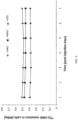
- the cell labeling efficiency was 30-50% after 30 minutes labeling depending on cell type. Radioactivity concentrations of labeled cells of up to 0.5MBq/1 ⁇ 10 6 cells were achieved without a negative effect on cellular viability. Cell efflux studies showed high stability of the radiolabel out to 7 days.
- Myocardially delivered 89 Zr-labeled hMSCs showed retention in the myocardium, as well as redistribution to lung, liver and bone, whereas intravenous delivery of 89 Zr-labeled hMSCs distributed primarily to lung, liver and bone which are expected distribution sites for human MSCs in mouse model.
- 89 Zr(HPO 4 ) 2 distributed to liver and bone with no activity in lung.
- mMCs Mouse melanoma cells
- hMSCs human mesenchymal stem cells
- mDCs JAWSII mouse dendritic cells
- the mMCs and hMSCs were cultured in complete DMEM (DMEM + 10% FBS) and mDCs were cultured in complete alphaMEM (alpha MEM + 4 mM L-glutamine + 1 mM sodium pyruvate + 5 ng/mL murine GM-CSF + 20% FBS).
- the cultures were maintained in a humidified cell culture chamber (21% O 2 , 74% N 2 , 5% CO 2 ) at 37°C.
- 89 Zr 4+ was produced in aqueous solution through the 89 Y( p,n ) 89 Zr nuclear reaction using a solution target containing yttrium nitrate and dilute nitric acid.
- the 89 Zr 4+ was isolated from 89 Y 3+ using a hydroxamate resin based purification method with the exception that the final elution of 89 Zr 4+ off the hydroxamate resin was performed with an appropriate volume of 1.2M K 2 HPO 4 /KH 2 PO 4 buffer (pH 3.5).
- the K 2 HPO 4 /KH 2 PO 4 buffer was allowed to sit on the column for 30 minutes before elution to promote release of 89 Zr as Zirconium hydrogen phosphate, 89 Zr(HPO 4 ) 2 from the column.
- the elution percentage of 89 Zr from the column was ⁇ 89% collected in four fractions of 0.5mL each.
- 89 Zr(HPO 4 ) 2 solution 120 ⁇ l was neutralized to pH 7.8 with ⁇ 100 ⁇ l 1M HEPES-KOH buffer (pH 7.5) and ⁇ 65 ⁇ l 1M K 2 CO 3 .
- 4 ⁇ l 5mM DFO-Bz-NCS in DMSO was added and chelation of 89 Zr 4+ proceeded at 37°C for 1 hour in a thermomixer at 550 rpm.
- Chelation efficiency was determined by silica gel iTLC (Agilent Technologies, Santa Clara, California, USA) with 50 mM DTPA pH 7 as the mobile phase.
- H-HBSS Hanks Balanced Salt solution buffered (ThermoFisher, Atlanta, Georgia, USA) (H-HBSS), pH 7.5.
- the cell labeling reaction was performed with ⁇ 6 ⁇ 10 6 cells in 500ul H-HBSS at pH 7.5. To this, either 100 ⁇ l 89 Zr(HPO 4 ) 2 ( ⁇ 6MBq) or 100ul 89 Zr-DBN ( ⁇ 6MBq) was added and was allowed to incubate at 37°C for 30 minutes on a shaker for cell labeling. After incubation, the cells were washed 4 times with appropriate volume of complete medium. The final labeling efficiency was calculated from the radioactivity bound to cells after all the washes.
- 0.3 ⁇ 10 6 89 Zr-labeled cells were plated into each well of a 6-well culture plate. The medium was replaced with fresh medium daily for 7 days and radioactivity in the replaced medium was counted. For mDCs with mix of adherent and suspension cells, the plate was centrifuged at 1000 rpm for 10 minutes before replacing the medium to avoid loss of unattached 89 Zr-labeled cells.
- the effect of radiolabeling on cellular proliferation was assessed by the CyQUANT DNA content assay (ThermoFisher, Atlanta, Georgia, USA).
- a known number of unlabeled and 89 Zr-labeled cells ( ⁇ 10 4 cells/well) were plated in 21 wells of a 96-well culture plate and maintained at 37°C in a CO 2 incubator. The amount of DNA in each well was quantified from absorbance values as a surrogate marker of the number of cells present. The culture medium was replaced daily.
- the CyQUANT assay was performed for 3 wells per day over 5 days.
- the effect of labeling on cellular viability was assessed using Trypan-Blue exclusion assay test within 1 hour of labeling, 3 rd and 7 th day post-labeling.
- the culture medium was replaced daily and maintained at 37°C in a CO 2 incubator. Unlabeled cells served as control.
- the effect of radiolabeling on cellular viability was also assessed using the ApoTox-Glo viability, cytotoxicity and caspase 3/7 apoptosis assay (Promega Corporation, Madison, Wisconsin, USA). Unlabeled cells served as control. A known number of unlabeled and 89 Zr-labeled cells ( ⁇ 10 4 /well) were plated in a 96-well culture plate. The culture medium was replaced daily and maintained at 37°C in a CO 2 incubator. At day 7, cell viability, cytotoxicity and apoptosis was quantified in triplicate using the ApoTox-Glo assay.
- mice were anesthetized under 1-2% isoflurane and underwent PET imaging using a small animal PET/X-Ray system (Sofie BioSystems Genesys4, Culver City, California, USA).
- mice were sacrificed and tissues were extracted and radioactivity counted using a gamma counter to evaluate the biodistribution of 89 Zr-radioactivity. Images were presented as a whole-body maximal images projection (MIPs) to display sites of uptake.
- MIPs whole-body maximal images projection
- Myocardial reperfusion was confirmed by color change of the left ventricle and electrocardiographic changes.
- 89 Zr-labeled cells (2 ⁇ 10 5 cells with radioactivity concentration ⁇ 0.37 MBq/10 6 cells) were injected at 4 sites within the ischemic region.
- the animals were imaged at day 2, day 5 and day 7 using small animal PET/X-Ray system (Sofie BioSystems Genesys4, Culver City, California, USA). At day 7, the mice were sacrificed and tissues were extracted and radioactivity counted using gamma counter to evaluate cell trafficking.
- 89 Zr hydrogen phosphate was readily chelated by DFO-NCS to form 89 Zr-DBN in a 1.2 M K 2 HPO 4 /KH 2 PO 4 and HEPES buffer system (pH 7.8), with radiolabeling efficiency of 55 ⁇ 5% after 30 minutes. This reaction mixture was then used directly for labeling of cells.
- the cell labeling efficiency using 89 Zr-DBN was ⁇ 30-50% as determined by cell-bound radioactivity. Radioactivity concentrations of 0.50 ⁇ 0.10, 0.47 ⁇ 0.10, and 0.39 ⁇ 0.20 MBq/10 6 cells were achieved when 6 ⁇ 10 6 cells were incubated for 30 minutes with ⁇ 6 MBq 89 Zr-DBN with mMCs, hMSCs and mDCs, respectively. In contrast, no cell labeling was observed using 89 Zr(HPO 4 ) 2 .
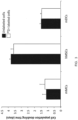
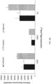
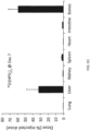
- the ApoTox-Glo assay showed no loss in cellular viability and no increase in cytotoxicity or apoptosis in radiolabeled cells as illustrated in Figures 4A-4C showing an assessment of ( Fig. 4A ) viability, ( Fig. 4B ) cytotoxicity and ( Fig. 4C ) apoptosis in 89 Zr-labeled and unlabeled cells. No statistically significant differences were observed between 89 Zr-labeled and unlabeled cells after 7 days of culture with regard to viability, cytotoxicity or apoptosis. As positive controls, 30 mg/mL digitonin was used for assays ( Fig. 4A ) and ( Fig.
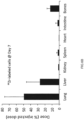
- PET images and biodistribution data of intravenously administered 89 Zr-labeled hMSCs and 89 Zr(HPO 4 ) 2 in healthy mice are in Figures 6A-6C which show representative PET images and biodistribution data of 89 Zr-labeled hMSCs and 89 Zr(HPO 4 ) 2 following intravenous injection.
- 89 Zr-labeled human MSCs (2 ⁇ 10 5 cells with radioactivity concentration ⁇ 0.37 Mbq/10 6 cells) and 89 Zr(HPO 4 ) 2 ( ⁇ 0.074 Mbq radioactivity) were intravenously injected in athymic mice.
- 89 Zr-labeled hMSCs ( ⁇ 19.5 ⁇ 9.5%) were retained for 7 days in heart. See Figures 7A-7B for representative PET images and biodistribution data of 89 Zr-labeled hMSCs following myocardial delivery.
- 18 F-FDG for PET
- 111 In-oxine for SPECT
- Ref. 2-6 111 In-oxine
- 18 F-FDG is useful for assessment of immediate delivery of cells and early fate of cells ( ⁇ first few hours), it is not suited for in vivo cell tracking after 24 hours post-injection due to its short half-life and poor retention in cells. Inability of 18 F-FDG to allow cell tracking after 24 hours limits its utility in cell-based therapies.
- Imaging based methods should be robust over this time frame to allow evaluation of various interventions for improving cell engraftment.
- the ability to monitor cells in vivo beyond 24 hours is also of high importance for evaluation of infection using radiolabeled leukocytes.
- Conventional infection imaging protocols perform imaging at 1 hours, 4 hours and 24 hours post-injection to differentiate between inflammatory, acute infection and chronic infection loci, however in some patients, 48 hours was necessary for reliable detection of infected lesions [Ref. 21].
- Cell labeling with 111 In typically requires a lipophilic carrier molecule (e.g. oxine) for transporting the radiometal into cells [Ref. 2-6]. After entering the cells, the radiometal then dissociates and gets trapped in the cell by binding to non-specific intracellular metal-binding proteins.
- 89 Zr-labeled hMSCs were delivered to the myocardium of athymic mice following an acute myocardial ischemia/reperfusion insult. After 7 days post-delivery, 89 Zr-labeled hMSCs were found in heart (20 ⁇ 7%), lung (40 ⁇ 16%), bone (29 ⁇ 11%) and liver (7 ⁇ 5%). The observed retention in heart is in accordance with previously published work on hMSC engraftment estimated by invasive quantitative PCR method in a similar rodent model [Ref. 34].
- the 89 Zr-DBN labeling agent is shown to be a robust, general, and biostable cell labeling strategy for PET-based non-invasive in vivo cell or virus tracking.
- a new viral labeling agent 89 Zr-desferrioxamine-NCS ( 89 Zr-DBN)
- the adeno-associated viruses AAVs
- the viral radiolabeling yield was quantified, following which cell binding and cell-infection assays were performed using 293T cells to evaluate the transfection potential of purified 89 Zr-DBN-AAV2-GFP.
- in vivo biodistribution of intravenous delivered 89 Zr-DBN-AAV1-Cre was assessed by PET imaging and correlated with transfection profile by bio-luminescence imaging (Lum) in a mouse model system.
- the scAAV1-Cre and AAV2-GFP were labeled with 89 Zr-DBN with viral labeling yield of ⁇ 20mCi/ 10 14 viral particles after 30 minutes of labeling.
- MOI multiplicity of infection
- 7% of the 89 Zr -AAV2-GFP were found to be bound to 293T cells as compared to 0.6% in control group including cells and 89 Zr-hydrogen phosphate ( 89 Zr(HPO 4 ) 2 ).
- the fraction of 293T cells expressing GFP correlated with MOI of 89 Zr-AAV2-GFP.
- the biodistribution of intravenously injected 89 Zr -AAV1-Cre assessed by PET imaging correlated with the tissue transfection profile assessed by bi-luminescence imaging and fluorescence microscopy.
- AAVs were successfully labeled with 89 Zr-DBN.
- the Zr labeling of AAVs did not affect their viral properties like infection potential and tissue tropism.
- 89 Zr-DBN is a promising tool for labeling a wide variety of viruses.
- 89 Zr 4+ was produced in aqueous solution through the 89 Y( p,n ) 89 Zr nuclear reaction using a solution target containing yttrium nitrate and dilute nitric acid.
- the 89 Zr 4+ was isolated from 89 Y 3+ using a hydroxamate resin-based purification method in the form of 89 Zr(HPO 4 ) 2 .
- 89 Zr-DBN was synthesized and scAAV1-Cre produced at the Mayo Clinic and AAV2-GFP was purchased from Cell Biolabs, Inc. The scAAV1-Cre and the AAV2-GFP were labeled with 89 Zr-DBN.
- the 89 Zr-DBN-AAVs were purified from free label and reaction mixture using size exclusion PD-10 desalting columns. The eluted virus in fractions were quantified using protein assay and radioactivity elution pattern was recorded.
- FIG. 8 shows representative data showing cell infection assay.
- Cell infection assay was performed with 2.5 ⁇ 10 5 293T cells with radiolabeled viral particles ( ⁇ 20mCi/10 14 viral particles) at two multiplicity of infection (MOI) ⁇ 2500 and 10,000 in DMEM culture medium + 1% FBS in CO 2 incubator.
- MOI multiplicity of infection
- FIG, 9 shows a representative elution profile of labeled AAVs after size exclusion PD-10 column purification.
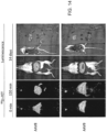
- FIG. 10 shows representative serial PET images and bioluminescence images showing biodistribution and transfection profile of 89 Zr-DBN-AAV1-Cre with 89 Zr(HPO 4 ) 2 and AAV1 as control.
- FIG. 11 shows representative fluorescence images of DAPI counter-stained 10 ⁇ liver sections confirming transfection.
- 89 Zr(HPO 4 ) 2 was readily chelated by DFO-NCS to form 89 Zr-DBN, with radiolabeling efficiency of 55% ⁇ 5% after 1 hour of reaction.
- AAVs were labeled with 89 Zr-DBN and purified using size exclusion PD-10 column.
- the 89 Zr labeling of AAVs did not affect its viral properties in terms of biodistribution and tissue tropism in a mouse model.
- Example 2 illustrates a novel 89 Zr based virus-labeling method for PET imaging of viral trafficking.
- Human MSCs from healthy donors were obtained from the Human Cellular Therapy Laboratory, Mayo Clinic, Rochester, Minnesota, USA. These cells have been characterized with respect to surface markers. Briefly, they are CD73(+), CD90(+), CD105(+), CD44(+), and HLA-ABC(+), and they are being used in several clinical trials.
- Green fluorescent protein (GFP) Transfection MSCs were transfected with GFP lentivirus from Mayo Clinic Rochester Labs, Rochester, Minnesota, USA. MSCs were grown overnight in media containing the GFP lentivirus. The medium was changed to complete growth medium the next day, and cells were checked for fluorescence after 48 hours. Once fluorescence was confirmed, the cells were cultured in complete media that contained 1 ⁇ g puromycin per milliliter. Cells containing the plasmid were expanded in complete growth media.
- GFP Green fluorescent protein
- Noninvasive PET imaging was used to evaluate the biodistribution of MSCs delivered to the adventitia outside the arteriovenous fistula (AVF) in CD1 -Foxn1nu mice.
- MSCs were labeled with 89 Zr-DBN.
- the 89 Zr-labeled MSCs were tracked for 3 weeks by using a small-animal PET/ radiography system (Genesys4; Sofie BioSystems, Culver City, California, USA).
- PET images were normalized to units of standardized uptake value, which was calculated as follows: tissue radioactivity concentration/(injected dose/body weight in grams).
- PET images of mice after adventitial delivery of 89 Zr-labeled MSCs showed that more than 90% of administered 89 Zr radioactivity was retained at the delivery site on day 4 (see Fig. 12A ).
- Fig. 12A Evolutionitial retention of 89 Zr radioactivity cleared slowly from day 4 to day 21, losing approximately 20% over this period (see Fig. 12B ).
- Most 89 Zr radioactivity that was cleared from the adventitia appeared to translocate to bones. This result confirmed results obtained by using confocal microscopy with GFP-labeled cells on day 7.
- PET imaging of 89 Zr-labeled MSCs allowed tracking of cells beyond 7 days, which was not possible with GFP-labeled cells.
- PET imaging of 89 Zr-labeled MSCs showed most (approximately 80%) of the radiolabel to be retained at the adventitial delivery site, even 3 weeks after delivery.
- the use of 89 Zr-labeled MSCs was more sensitive than GFP labeling for detection of cellular location after delivery.
- Example 3 highlights the fact that MSCs can be tracked after delivery by using PET imaging of 89 Zr-labeled MSCs.
- H-HBSS Hanks Balanced Salt Solution
- pH 7.5-9 ThermoFisher, Atlanta, Georgia, USA
- the AAV labeling reaction was performed with 10 10 - 10 15 viral particles in 500ul H-HBSS.
- 100ul 89 Zr-DBN 100ul 89 Zr-DBN ( ⁇ 6MBq) was added and incubated at 37°C for 10-40 minutes on a shaker. After incubation, the Zr-89 labeled virus was purified from free 89 Zr +4 on a PD-10 Sephadex G-25 column.
- Adeno-associated virus serotype 8 (AAV8) and adeno-associated virus serotype 9 (AAV9) were labeled with 89 Zr-DBN as above.
- Example 4 illustrates other 89 Zr based virus-labeling methods for PET imaging of viral trafficking.
- the invention provides methods of ex vivo labeling of a biological material for in vivo imaging, methods of labeling a biological material in vivo, methods for preparing a labeling agent, and methods for in vivo imaging of a subject using a biological material labeled with a labeling agent.
- the biological material is selected from cells.
Landscapes
- Health & Medical Sciences (AREA)
- Life Sciences & Earth Sciences (AREA)
- Chemical & Material Sciences (AREA)
- Animal Behavior & Ethology (AREA)
- Veterinary Medicine (AREA)
- Public Health (AREA)
- General Health & Medical Sciences (AREA)
- Epidemiology (AREA)
- Nuclear Medicine, Radiotherapy & Molecular Imaging (AREA)
- Medicinal Chemistry (AREA)
- Radiology & Medical Imaging (AREA)
- Pharmacology & Pharmacy (AREA)
- Optics & Photonics (AREA)
- Physics & Mathematics (AREA)
- Virology (AREA)
- Proteomics, Peptides & Aminoacids (AREA)
- Dispersion Chemistry (AREA)
- Hematology (AREA)
- Medicines Containing Antibodies Or Antigens For Use As Internal Diagnostic Agents (AREA)
- Magnetic Resonance Imaging Apparatus (AREA)
Applications Claiming Priority (4)
| Application Number | Priority Date | Filing Date | Title |
|---|---|---|---|
| US201562129406P | 2015-03-06 | 2015-03-06 | |
| PCT/US2016/021188 WO2016144873A2 (en) | 2015-03-06 | 2016-03-07 | Methods for cell labeling and medical imaging |
| EP16762295.0A EP3265134B1 (de) | 2015-03-06 | 2016-03-07 | Verfahren zur zellmarkierungen und medizinische bildgebung |
| EP21207036.1A EP3973998B1 (de) | 2015-03-06 | 2016-03-07 | Verfahren zur zellmarkierungen und medizinische bildgebung |
Related Parent Applications (2)
| Application Number | Title | Priority Date | Filing Date |
|---|---|---|---|
| EP21207036.1A Division EP3973998B1 (de) | 2015-03-06 | 2016-03-07 | Verfahren zur zellmarkierungen und medizinische bildgebung |
| EP16762295.0A Division EP3265134B1 (de) | 2015-03-06 | 2016-03-07 | Verfahren zur zellmarkierungen und medizinische bildgebung |
Publications (2)
| Publication Number | Publication Date |
|---|---|
| EP4252784A2 true EP4252784A2 (de) | 2023-10-04 |
| EP4252784A3 EP4252784A3 (de) | 2024-01-10 |
Family
ID=56879259
Family Applications (3)
| Application Number | Title | Priority Date | Filing Date |
|---|---|---|---|
| EP16762295.0A Active EP3265134B1 (de) | 2015-03-06 | 2016-03-07 | Verfahren zur zellmarkierungen und medizinische bildgebung |
| EP23186316.8A Pending EP4252784A3 (de) | 2015-03-06 | 2016-03-07 | Verfahren zur zellmarkierungen und medizinische bildgebung |
| EP21207036.1A Active EP3973998B1 (de) | 2015-03-06 | 2016-03-07 | Verfahren zur zellmarkierungen und medizinische bildgebung |
Family Applications Before (1)
| Application Number | Title | Priority Date | Filing Date |
|---|---|---|---|
| EP16762295.0A Active EP3265134B1 (de) | 2015-03-06 | 2016-03-07 | Verfahren zur zellmarkierungen und medizinische bildgebung |
Family Applications After (1)
| Application Number | Title | Priority Date | Filing Date |
|---|---|---|---|
| EP21207036.1A Active EP3973998B1 (de) | 2015-03-06 | 2016-03-07 | Verfahren zur zellmarkierungen und medizinische bildgebung |
Country Status (3)
| Country | Link |
|---|---|
| US (2) | US11534506B2 (de) |
| EP (3) | EP3265134B1 (de) |
| WO (1) | WO2016144873A2 (de) |
Families Citing this family (12)
| Publication number | Priority date | Publication date | Assignee | Title |
|---|---|---|---|---|
| IL324632A (en) | 2016-12-01 | 2026-01-01 | Regeneron Pharma | Radiolabeled anti-PD-L1 antibodies for immuno-PET imaging |
| US11963839B2 (en) | 2017-01-10 | 2024-04-23 | A. T. Still University | Dental monitoring system |
| CN110650974B (zh) | 2017-02-10 | 2024-04-19 | 瑞泽恩制药公司 | 用于免疫-pet成像的放射性标记的抗-lag3抗体 |
| MY197688A (en) | 2017-07-24 | 2023-07-05 | Regeneron Pharma | Anti-cd8 antibodies and uses thereof |
| JP7750828B2 (ja) | 2019-09-16 | 2025-10-07 | リジェネロン・ファーマシューティカルズ・インコーポレイテッド | 免疫pet撮像のための放射標識されたmet結合タンパク質 |
| EP4034177A1 (de) | 2019-09-27 | 2022-08-03 | Boehringer Ingelheim Veterinary Medicine Belgium | Zellzusammensetzung, radioaktiv markierte mesenchymale stammzellen umfassend, verwendung davon und verfahren zur herstellung radioaktiv markierter mesenchymaler stammzellen |
| CN115461088A (zh) * | 2020-04-28 | 2022-12-09 | 多磁公司 | 细菌和噬菌体的生物医学成像 |
| EP4380628A4 (de) * | 2021-08-06 | 2026-04-15 | Mayo Found Medical Education & Res | Verfahren zur markierung von biologischem material und medizinische bildgebung |
| CN120285240A (zh) * | 2021-09-16 | 2025-07-11 | 南京艾尔普再生医学科技有限公司 | 一种诱导多能干细胞分化的心肌细胞在药动学中的示踪方法 |
| EP4593889A1 (de) * | 2022-09-30 | 2025-08-06 | Mayo Foundation for Medical Education and Research | Pet-bildgebung von bakteriellen infektionen mit einer pet-sonde |
| CN116626300B (zh) * | 2023-06-02 | 2025-01-17 | 昆明理工大学 | 一种利用89Zr成像示踪人源间充质干细胞的方法 |
| CN116999579B (zh) * | 2023-07-19 | 2026-03-03 | 江苏省原子医学研究所 | 一种使用铁螯合剂提高体内细胞示踪准确性的方法 |
Family Cites Families (7)
| Publication number | Priority date | Publication date | Assignee | Title |
|---|---|---|---|---|
| US5059541A (en) * | 1988-04-29 | 1991-10-22 | Neorx Corporation | Minimal derivatization of proteins |
| US20060035946A1 (en) * | 2003-01-22 | 2006-02-16 | The General Hospital Corporation | Amyloid-binding, metal-chelating agents |
| US8545809B2 (en) | 2007-01-11 | 2013-10-01 | Immunomedics, Inc. | Methods and compositions for improved 18F labeling of proteins, peptides and other molecules |
| WO2013138696A1 (en) * | 2012-03-16 | 2013-09-19 | The United States Of America, As Represented By The Secretary, Department Of Health And Human Services | Zirconium-89 complexes, methods of labeling cells, labeled cells, kits, and methods of use thereof |
| DE102012104504B4 (de) | 2012-05-24 | 2021-10-07 | Bundesrepublik Deutschland, vertreten durch das Bundesministerium für Wirtschaft und Technologie, dieses vertreten durch den Präsidenten der BAM, Bundesanstalt für Materialforschung und -prüfung | Polypeptidmarker |
| US10086095B2 (en) * | 2012-10-09 | 2018-10-02 | Case Western Reserve University | Rod-shaped plant virus nanoparticles as imaging agent platforms |
| WO2014164988A1 (en) * | 2013-03-13 | 2014-10-09 | The United States Of America, As Represented By The Secretary, Department Of Health And Human Services | Tetrahydroxamate chelators of zirconium89 and niobium90 for use in diagnostic applications |
-
2016
- 2016-03-07 WO PCT/US2016/021188 patent/WO2016144873A2/en not_active Ceased
- 2016-03-07 US US15/555,775 patent/US11534506B2/en active Active
- 2016-03-07 EP EP16762295.0A patent/EP3265134B1/de active Active
- 2016-03-07 EP EP23186316.8A patent/EP4252784A3/de active Pending
- 2016-03-07 EP EP21207036.1A patent/EP3973998B1/de active Active
-
2021
- 2021-06-25 US US17/358,544 patent/US20210330823A1/en active Pending
Non-Patent Citations (41)
| Title |
|---|
| ADONAI NNGUYEN KNWALSH JIYER MTOYOKUNI TPHELPS ME ET AL.: "Ex vivo cell labeling with Cu-pyruvaldehyde-bis(N4-methylthiosemicarbazone) for imaging cell trafficking in mice with positron-emission tomography", PROC NAT ACAD SCI., vol. 99, 2002, pages 3030 - 5, XP008129268, DOI: 10.1073/pnas.052709599 |
| BOERJESSON PKEJAUW YWSDE BREE RROOS JCCASTELIJNS JALEEMANS CR ET AL.: "Radiation dosimetry of Zr-89-labeled chimeric monoclonal antibody U36 as used for immuno-PET in head and neck cancer patients", J NUCL MED, vol. 50, 2009, pages 1828 - 36 |
| BORJESSON PKEJAUW YWSBOELLAARD RDE BREE RCOMANS EFIROOS JC ET AL.: "Performance of immuno-positron emission tomography with Zirconium-89-labeled chimeric monoclonal antibody U36 in the detection of lymph node metastases in head and neck cancer patients", CLIN CANCER RES., vol. 12, 2006, pages 2133 - 40 |
| BRENNER W, AICHER A, ECKEY T, MASSOUDI S, ZUHAYRA M, KOEHL U: "111In-labeled CD34+ hematopoietic progenitor cells in a rat myocardial infarction model", J NUCL MED, vol. 45, 2004, pages 512 - 8 |
| CHAROENPHUN PMESZAROS LKCHUAMSAAMARKKEE KSHARIF-PAGHALEH EBALLINGER JRFERRIS TJ ET AL.: "Zr]Oxinate for long-term in vivo cell tracking by positron emission tomography", EUR J NUCL MED MOL IMAGING, 2014 |
| DALDRUP-LINK HEM RUDELIUSS METZG PIONTEKB PICHLERM SETTLESU HEINZMANNJ SCHLEGELRA OOSTENDORPEJ RUMMENY: "Cell tracking with gadophrin-2: a bifunctional contrast agent for MR imaging, optical imaging, and fluorescence microscopy", EUR J NUCL MED MOL IMAGING, vol. 31, 2004, pages 1312 - 21 |
| DAVIDSON-MONCADA JSATO NHOYT JR ET AL.: "Proceedings of the 56th Annual Meeting", AMERICAN SOCIETY OF HEMATOLOGY, article "A novel method to study the in vivo trafficking and homing of adoptively transferred NK cells in Rhesus Macaques and Humans" |
| DE VRIES EFROCA MJAMAR FISRAEL OSIGNORE A: "Guidelines for the labelling of leucocytes with Tc-HMPAO. Inflammation/Infection Taskgroup of the European Association of Nuclear Medicine", EUR J NUCL MED MOL IMAGING, vol. 37, 2010, pages 842 - 8 |
| DERI MA, ZEGLIS BM, FRANCESCONI LC, LEWIS JS: "PET imaging with 89Zr: from radiochemistry to the clinic", NUCL MED BIOL, vol. 40, 2013, pages 3 - 14, XP055357231, DOI: 10.1016/j.nucmedbio.2012.08.004 |
| DERI MAPONNALA SZEGLIS BMPOHL GDANNENBERG JJLEWIS JSFRANCESCONI LC: "Alternative chelator for Zr radiopharmaceuticals: radiolabeling and evaluation of 3,4,3-(LI-1,2-HOPO", J MED CHEM., vol. 57, 2014, pages 4849 - 60, XP055385493, DOI: 10.1021/jm500389b |
| DIJKERS ECFKOSTERINK JGWRADEMAKER APPERK LRVAN DONGEN GAMSBART J ET AL.: "Development and characterization of clinical-grade Zr-89-trastuzumab for HER2/neu immunoPET imaging", J NUCL MED, vol. 50, 2009, pages 974 - 81, XP002615664, DOI: 10.2967/JNUMED.108.060392 |
| DIJKERS ECMUNNINK THOKOSTERINK JGBROUWERS AHJAGER PLDE JONG JR ET AL.: "Biodistribution of Zr-89-trastuzumab and PET imaging of HER2-positive lesions in patients with metastatic breast cancer", CLIN PHARMACOL THER, vol. 87, 2010, pages 586 - 92, XP008144583, DOI: 10.1038/clpt.2010.12 |
| DOYLE BKEMP BJCHAREONTHAITAWEE PREED CSCHMECKPEPER JSORAJJA P ET AL.: "Dynamic tracking during intracoronary injection of F-FDG-labeled progenitor cell therapy for acute myocardial infarction", J NUCL MED, vol. 48, 2007, pages 1708 - 14 |
| FISCHER UM, HARTING MT, JIMENEZ F, MONZON-POSADAS WO, XUE H, SAVITZ SI: "Pulmonary passage is a major obstacle for intravenous stem cell delivery: the pulmonary first-pass effect", STEM CELLS DEV, vol. 18, 2009, pages 683 - 92 |
| GAO JJE DENNISRF MUZICM LUNDBERGAI CAPLAN: "The dynamic in vivo distribution of bone marrow-derived mesenchymal stem cells after infusion", CELLS TISSUES ORGANS, vol. 169, 2001, pages 12 - 20, XP009026035, DOI: 10.1159/000047856 |
| GILDEHAUS FJHAASTERS FDROSSE IWAGNER EZACH CMUTSCHLER W ET AL.: "Impact of indium-111 oxine labelling on viability of human mesenchymal stem cells in vitro, and 3D cell-tracking using SPECT/CT in vivo", MOL IMAGING BIOL, vol. 13, 2011, pages 1204 - 14 |
| GLAUDEMANS AWGALLI FPACILIO MSIGNORE A: "Leukocyte and bacteria imaging in prosthetic joint infection", EUR CELL MATER, vol. 25, 2013, pages 61 - 77 |
| HOLLAND JPDIVILOV VBANDER NHSMITH-JONES PMLARSON SMLEWIS JS: "Zr-DFO-J591 for immunoPET of prostate-specific membrane antigen expression in vivo", J NUCL MED, vol. 51, 2010, pages 1293 - 300, XP055145983, DOI: 10.2967/jnumed.110.076174 |
| HOLLAND JPSHEH YLEWIS JS: "Standardized methods for the production of high specific-activity Zirconium-89", NUCL MED BIOL, vol. 36, 2009, pages 729 - 39, XP026545224, DOI: 10.1016/j.nucmedbio.2009.05.007 |
| HUGHES DK: "Nuclear medicine and infection detection: the relative effectiveness of imaging with In-oxine-, Tc-HMPAO-, and Tc-stannous fluoride colloid-labeled leukocytes and with Ga-citrate", J NUCL MED TECH, vol. 31, 2003, pages 196 - 201 |
| KASSIS ALADELSTEIN SJ: "Chemotoxicity of indium-111 oxine in mammalian cells", J NUCL MED, vol. 26, 1985, pages 187 - 90 |
| KEBERLE H: "The Biochemistry of Desferrioxamine and Its Relation to Iron Metabolism", ANNALS N YACAD SCI, vol. 119, 1964, pages 758 - 68 |
| KUYAMA JMCCORMACK AGEORGE AJHEELAN BTOSMAN SBATCHELOR JR ET AL.: "Indium-111 labelled lymphocytes: isotope distribution and cell division", EUR J NUCL MED., vol. 24, 1997, pages 488 - 96 |
| MEIER RPIERT MPIONTEK GRUDELIUS MOOSTENDORP RASENEKOWITSCH-SCHMIDTKE R ET AL.: "Tracking of [18F]FDG-labeled natural killer cells to HER2/neu-positive tumors", NUCL MED BIOL, vol. 35, 2008, pages 579 - 88, XP022851419, DOI: 10.1016/j.nucmedbio.2008.02.006 |
| NAUMOVA AVMODO MMOORE AMURRY CEFRANK JA: "Clinical imaging in regenerative medicine", NAT BIOTECH, vol. 32, 2014, pages 804 - 18 |
| NGUYEN PKRIEGLER JWU JC: "Stem cell imaging: from bench to bedside", CELL STEM CELL, vol. 14, 2014, pages 431 - 44 |
| PANDEY MKENGELBRECHT HPBYRNE JPPACKARD ABDEGRADO TR: "Production of Zr via the Y(p,n)89Zr reaction in aqueous solution: effect of solution composition on in-target chemistry", NUCL MED BIOL, vol. 41, 2014, pages 309 - 16, XP055834403, DOI: 10.1016/j.nucmedbio.2014.01.006 |
| PELLEGRINO DBONAB AADRAGOTAKES SCPITMAN JTMARIANI GCARTER EA: "Inflammation and infection: imaging properties of F-FDG-labeled white blood cells versus F-FDG", J NUCL MED, vol. 46, 2005, pages 1522 - 30 |
| PERK LRVOSJAN MJVISSER GWBUDDE MJUREK PKIEFER GE ET AL.: "p-Isothiocyanatobenzyl-desferrioxamine: a new bifunctional chelate for facile radiolabeling of monoclonal antibodies with Zirconium-89 for immuno-PET imaging", EUR J NUCL MED MOL IMAGING, vol. 37, 2010, pages 250 - 9, XP019778696 |
| RIZVI SNFVISSER OJVOSJAN MJWDVAN LINGEN AHOEKSTRA OSZIJLSTRA JM ET AL.: "Biodistribution, radiation dosimetry and scouting of Y-90-ibritumomab tiuxetan therapy in patients with relapsed B-cell non-Hodgkin's lymphoma using Zr-89-ibritumomab tiuxetan and PET", EUR J NUCL MED MOL IMAGING, vol. 39, 2012, pages 512 - 20 |
| ROCA MDE VRIES EFJAMAR FISRAEL OSIGNORE A: "Guidelines for the labelling of leucocytes with (111)ln-oxine. Inflammation/Infection Taskgroup of the European Association of Nuclear Medicine", EUR J NUCL MED MOL IMAGING, vol. 37, 2010, pages 835 - 41 |
| S. B. M. GAYKEMAA. H. BROUWERSM. N. L. HOOGE ET AL.: "Zr-bevacizumab PET imaging in primary breast cancer", J NUCL MED, vol. 54, 2013, pages 1014 - 18 |
| SATO N, S.L.CHOYKE P: "Cell labeling using Zr-89 - comparison with In-111 oxine", PROCEEDINGS WORLD MOLECULAR IMAGING CONGRESS, vol. 2013, 2013, pages 533 |
| SCHREPFER SDEUSE TREICHENSPURNER HFISCHBEIN MPROBBINS RCPELLETIER MP: "Stem cell transplantation: the lung barrier", TRANSPLANT PROC, vol. 39, 2007, pages 573 - 6, XP005931659, DOI: 10.1016/j.transproceed.2006.12.019 |
| STOJANOV KDE VRIES EFHOEKSTRA DVAN WAARDE ADIERCKX RAZUHORN IS: "F]FDG labeling of neural stem cells for in vivo cell tracking with positron emission tomography: inhibition of tracer release by phloretin", MOL IMAGING, vol. 11, 2012, pages 1 - 12 |
| TAKAGAI YTAKAHASHI AYAMAGUCHI HKUBOTA TIGARASHI S: "Adsorption behaviors of high-valence metal ions on desferrioxamine B immobilization nylon 6,6 chelate fiber under highly acidic conditions", J COLLOID INTERFACE SCI, vol. 313, 2007, pages 359 - 62, XP026043362, DOI: 10.1016/j.jcis.2007.02.079 |
| TARANTAL AFLEE CCKUKIS DLCHERRY SR: "Radiolabeling human peripheral blood stem cells for positron emission tomography (PET) imaging in young rhesus monkeys", PLOS ONE, vol. 8, 2013, pages e77148 |
| TERROVITIS JLAUTAMAKI RBONIOS MFOX JENGLES JMYU J ET AL.: "Noninvasive quantification and optimization of acute cell retention by in vivo positron emission tomography after intramyocardial cardiac-derived stem cell delivery", J AM COLL CARDIOL., vol. 54, 2009, pages 1619 - 26, XP026688278, DOI: 10.1016/j.jacc.2009.04.097 |
| VOSJAN MJPERK LRVISSER GWBUDDE MJUREK PKIEFER GE ET AL.: "Conjugation and radiolabeling of monoclonal antibodies with Zirconium-89 for PET imaging using the bifunctional chelate p-isothiocyanatobenzyl-desferrioxamine", NAT PROTOC, vol. 5, 2010, pages 739 - 43, XP055304936, DOI: 10.1038/nprot.2010.13 |
| YANG BBRAHMBHATT ANIEVES TORRES ETHIELEN BMCCALL DLENGEL SBANSAL APANDEY MKDIETZ ABLEOF EB: "Tracking and Therapeutic Value of Human Adipose Tissue-derived Mesenchymal Stem Cell Transplantation in Reducing Venous Neointimal Hyperplasia Associated with Arteriovenous Fistula", RADIOLOGY, vol. 19, November 2015 (2015-11-01), pages 150947 |
| ZHANG YDASILVA JNHADIZAD TTHORN SKURAITIS DRENAUD JM ET AL.: "F-FDG cell labeling may underestimate transplanted cell homing: more accurate, efficient, and stable cell labeling with hexadecyl-4-[18F]fluorobenzoate for in vivo tracking of transplanted human progenitor cells by positron emission tomography", CELL TRANSPLANT, vol. 21, 2012, pages 1821 - 35 |
Also Published As
| Publication number | Publication date |
|---|---|
| EP3265134A4 (de) | 2019-01-16 |
| EP3265134B1 (de) | 2021-12-15 |
| EP3265134A2 (de) | 2018-01-10 |
| EP3973998B1 (de) | 2023-07-26 |
| US20210330823A1 (en) | 2021-10-28 |
| EP4252784A3 (de) | 2024-01-10 |
| EP3973998A1 (de) | 2022-03-30 |
| US20180043041A1 (en) | 2018-02-15 |
| WO2016144873A2 (en) | 2016-09-15 |
| WO2016144873A3 (en) | 2016-11-03 |
| US11534506B2 (en) | 2022-12-27 |
Similar Documents
| Publication | Publication Date | Title |
|---|---|---|
| US20210330823A1 (en) | Methods for Cell Labeling and Medical Imaging | |
| Bansal et al. | Novel 89Zr cell labeling approach for PET-based cell trafficking studies | |
| Jennewein et al. | Vascular imaging of solid tumors in rats with a radioactive arsenic-labeled antibody that binds exposed phosphatidylserine | |
| US20220040337A1 (en) | Radioligands for pretargeted pet imaging and methods of their therapeutic use | |
| Kalidindi et al. | A simple strategy to reduce the salivary gland and kidney uptake of PSMA-targeting small molecule radiopharmaceuticals | |
| Williams | Tissue distribution studies of protein therapeutics using molecular probes: molecular imaging | |
| RS66467B1 (sr) | Kompleks koji se sastoji iz jedinjenja koje cilja psma vezano sa radionuklidom olova ili torijuma | |
| Price et al. | A comparative evaluation of the chelators H4octapa and CHX-A ″-DTPA with the therapeutic radiometal 90Y | |
| US20240299596A1 (en) | Methods and materials for combining biologics with multiple chelators | |
| US9789211B2 (en) | Methods and compositions for positron emission tomography myocardial perfusion imaging | |
| Wharton et al. | H4picoopa─ Robust Chelate for 225Ac/111In Theranostics | |
| Scott-Robson et al. | Distribution and stability in the rat of a 76Br/125I-labelled polypeptide, epidermal growth factor | |
| Choi et al. | Ga-68-labeled neolactosylated human serum albumin (LSA) for PET imaging of hepatic asialoglycoprotein receptor | |
| US20150174273A1 (en) | Imaging and therapeutic methods for treating parathyroid tumors | |
| US20230293735A1 (en) | Oxine-containing cell radiolabelling agents | |
| US20250135049A1 (en) | Methods for Biological Material Labeling and Medical Imaging | |
| Hasegawa et al. | Development of novel radiotheranostic ligand with positively charged unit targeting prostate-specific membrane antigen | |
| US8846001B2 (en) | Labelled biotin conjugates | |
| EP4327831A1 (de) | Radioaktiver komplex aus anti-cd20-antikörper und radiopharmazeutikum | |
| WO2024138060A2 (en) | Methods and materials for combining biologics with multiple chelators | |
| AU2021361746A9 (en) | Radioactive complexes of anti-her2 antibody, and radiopharmaceutical | |
| CN119751415A (zh) | 双靶向PSMA和FAP-α的前列腺癌诊断或治疗放射性药物 | |
| Larson et al. | Improving cancer treatment with cyclotron produced radionuclides. Comprehensive progress report, February 1, 1992--July 15, 1995 |
Legal Events
| Date | Code | Title | Description |
|---|---|---|---|
| PUAI | Public reference made under article 153(3) epc to a published international application that has entered the european phase |
Free format text: ORIGINAL CODE: 0009012 |
|
| STAA | Information on the status of an ep patent application or granted ep patent |
Free format text: STATUS: THE APPLICATION HAS BEEN PUBLISHED |
|
| AC | Divisional application: reference to earlier application |
Ref document number: 3265134 Country of ref document: EP Kind code of ref document: P Ref document number: 3973998 Country of ref document: EP Kind code of ref document: P |
|
| AK | Designated contracting states |
Kind code of ref document: A2 Designated state(s): AL AT BE BG CH CY CZ DE DK EE ES FI FR GB GR HR HU IE IS IT LI LT LU LV MC MK MT NL NO PL PT RO RS SE SI SK SM TR |
|
| REG | Reference to a national code |
Ref country code: DE Ref legal event code: R079 Free format text: PREVIOUS MAIN CLASS: A61K0049180000 Ipc: A61K0051040000 |
|
| PUAL | Search report despatched |
Free format text: ORIGINAL CODE: 0009013 |
|
| AK | Designated contracting states |
Kind code of ref document: A3 Designated state(s): AL AT BE BG CH CY CZ DE DK EE ES FI FR GB GR HR HU IE IS IT LI LT LU LV MC MK MT NL NO PL PT RO RS SE SI SK SM TR |
|
| RIC1 | Information provided on ipc code assigned before grant |
Ipc: A61K 49/18 20060101ALI20231207BHEP Ipc: A61K 49/10 20060101ALI20231207BHEP Ipc: A61K 51/12 20060101ALI20231207BHEP Ipc: A61K 49/12 20060101ALI20231207BHEP Ipc: A61K 51/04 20060101AFI20231207BHEP |
|
| STAA | Information on the status of an ep patent application or granted ep patent |
Free format text: STATUS: REQUEST FOR EXAMINATION WAS MADE |
|
| 17P | Request for examination filed |
Effective date: 20240709 |
|
| RBV | Designated contracting states (corrected) |
Designated state(s): AL AT BE BG CH CY CZ DE DK EE ES FI FR GB GR HR HU IE IS IT LI LT LU LV MC MK MT NL NO PL PT RO RS SE SI SK SM TR |