EP4235152B1 - Assessing circulatory failure - Google Patents
Assessing circulatory failure Download PDFInfo
- Publication number
- EP4235152B1 EP4235152B1 EP23177112.2A EP23177112A EP4235152B1 EP 4235152 B1 EP4235152 B1 EP 4235152B1 EP 23177112 A EP23177112 A EP 23177112A EP 4235152 B1 EP4235152 B1 EP 4235152B1
- Authority
- EP
- European Patent Office
- Prior art keywords
- heterogeneity
- fcd
- microcirculation
- subject
- patients
- Prior art date
- Legal status (The legal status is an assumption and is not a legal conclusion. Google has not performed a legal analysis and makes no representation as to the accuracy of the status listed.)
- Active
Links
Images


Classifications
-
- A—HUMAN NECESSITIES
- A61—MEDICAL OR VETERINARY SCIENCE; HYGIENE
- A61B—DIAGNOSIS; SURGERY; IDENTIFICATION
- A61B5/00—Measuring for diagnostic purposes; Identification of persons
- A61B5/02—Detecting, measuring or recording for evaluating the cardiovascular system, e.g. pulse, heart rate, blood pressure or blood flow
- A61B5/026—Measuring blood flow
- A61B5/0261—Measuring blood flow using optical means, e.g. infrared light
-
- A—HUMAN NECESSITIES
- A61—MEDICAL OR VETERINARY SCIENCE; HYGIENE
- A61B—DIAGNOSIS; SURGERY; IDENTIFICATION
- A61B3/00—Apparatus for testing the eyes; Instruments for examining the eyes
- A61B3/10—Objective types, i.e. instruments for examining the eyes independent of the patients' perceptions or reactions
- A61B3/13—Ophthalmic microscopes
-
- A—HUMAN NECESSITIES
- A61—MEDICAL OR VETERINARY SCIENCE; HYGIENE
- A61B—DIAGNOSIS; SURGERY; IDENTIFICATION
- A61B5/00—Measuring for diagnostic purposes; Identification of persons
- A61B5/0059—Measuring for diagnostic purposes; Identification of persons using light, e.g. diagnosis by transillumination, diascopy, fluorescence
- A61B5/0075—Measuring for diagnostic purposes; Identification of persons using light, e.g. diagnosis by transillumination, diascopy, fluorescence by spectroscopy, i.e. measuring spectra, e.g. Raman spectroscopy, infrared absorption spectroscopy
-
- A—HUMAN NECESSITIES
- A61—MEDICAL OR VETERINARY SCIENCE; HYGIENE
- A61B—DIAGNOSIS; SURGERY; IDENTIFICATION
- A61B5/00—Measuring for diagnostic purposes; Identification of persons
- A61B5/0059—Measuring for diagnostic purposes; Identification of persons using light, e.g. diagnosis by transillumination, diascopy, fluorescence
- A61B5/0077—Devices for viewing the surface of the body, e.g. camera, magnifying lens
-
- A—HUMAN NECESSITIES
- A61—MEDICAL OR VETERINARY SCIENCE; HYGIENE
- A61B—DIAGNOSIS; SURGERY; IDENTIFICATION
- A61B5/00—Measuring for diagnostic purposes; Identification of persons
- A61B5/02—Detecting, measuring or recording for evaluating the cardiovascular system, e.g. pulse, heart rate, blood pressure or blood flow
- A61B5/02007—Evaluating blood vessel condition, e.g. elasticity, compliance
-
- A—HUMAN NECESSITIES
- A61—MEDICAL OR VETERINARY SCIENCE; HYGIENE
- A61B—DIAGNOSIS; SURGERY; IDENTIFICATION
- A61B5/00—Measuring for diagnostic purposes; Identification of persons
- A61B5/02—Detecting, measuring or recording for evaluating the cardiovascular system, e.g. pulse, heart rate, blood pressure or blood flow
- A61B5/0205—Simultaneously evaluating both cardiovascular conditions and different types of body conditions, e.g. heart and respiratory condition
-
- A—HUMAN NECESSITIES
- A61—MEDICAL OR VETERINARY SCIENCE; HYGIENE
- A61B—DIAGNOSIS; SURGERY; IDENTIFICATION
- A61B5/00—Measuring for diagnostic purposes; Identification of persons
- A61B5/145—Measuring characteristics of blood in vivo, e.g. gas concentration or pH-value ; Measuring characteristics of body fluids or tissues, e.g. interstitial fluid or cerebral tissue
- A61B5/1455—Measuring characteristics of blood in vivo, e.g. gas concentration or pH-value ; Measuring characteristics of body fluids or tissues, e.g. interstitial fluid or cerebral tissue using optical sensors, e.g. spectral photometrical oximeters
- A61B5/14551—Measuring characteristics of blood in vivo, e.g. gas concentration or pH-value ; Measuring characteristics of body fluids or tissues, e.g. interstitial fluid or cerebral tissue using optical sensors, e.g. spectral photometrical oximeters for measuring blood gases
- A61B5/14552—Details of sensors specially adapted therefor
-
- A—HUMAN NECESSITIES
- A61—MEDICAL OR VETERINARY SCIENCE; HYGIENE
- A61B—DIAGNOSIS; SURGERY; IDENTIFICATION
- A61B5/00—Measuring for diagnostic purposes; Identification of persons
- A61B5/145—Measuring characteristics of blood in vivo, e.g. gas concentration or pH-value ; Measuring characteristics of body fluids or tissues, e.g. interstitial fluid or cerebral tissue
- A61B5/1455—Measuring characteristics of blood in vivo, e.g. gas concentration or pH-value ; Measuring characteristics of body fluids or tissues, e.g. interstitial fluid or cerebral tissue using optical sensors, e.g. spectral photometrical oximeters
- A61B5/14558—Measuring characteristics of blood in vivo, e.g. gas concentration or pH-value ; Measuring characteristics of body fluids or tissues, e.g. interstitial fluid or cerebral tissue using optical sensors, e.g. spectral photometrical oximeters by polarisation
-
- A—HUMAN NECESSITIES
- A61—MEDICAL OR VETERINARY SCIENCE; HYGIENE
- A61B—DIAGNOSIS; SURGERY; IDENTIFICATION
- A61B5/00—Measuring for diagnostic purposes; Identification of persons
- A61B5/44—Detecting, measuring or recording for evaluating the integumentary system, e.g. skin, hair or nails
- A61B5/441—Skin evaluation, e.g. for skin disorder diagnosis
- A61B5/445—Evaluating skin irritation or skin trauma, e.g. rash, eczema, wound, bed sore
-
- A—HUMAN NECESSITIES
- A61—MEDICAL OR VETERINARY SCIENCE; HYGIENE
- A61B—DIAGNOSIS; SURGERY; IDENTIFICATION
- A61B5/00—Measuring for diagnostic purposes; Identification of persons
- A61B5/48—Other medical applications
- A61B5/4848—Monitoring or testing the effects of treatment, e.g. of medication
-
- A—HUMAN NECESSITIES
- A61—MEDICAL OR VETERINARY SCIENCE; HYGIENE
- A61B—DIAGNOSIS; SURGERY; IDENTIFICATION
- A61B5/00—Measuring for diagnostic purposes; Identification of persons
- A61B5/48—Other medical applications
- A61B5/4887—Locating particular structures in or on the body
- A61B5/489—Blood vessels
-
- A—HUMAN NECESSITIES
- A61—MEDICAL OR VETERINARY SCIENCE; HYGIENE
- A61B—DIAGNOSIS; SURGERY; IDENTIFICATION
- A61B2503/00—Evaluating a particular growth phase or type of persons or animals
- A61B2503/04—Babies, e.g. for SIDS detection
- A61B2503/045—Newborns, e.g. premature baby monitoring
-
- A—HUMAN NECESSITIES
- A61—MEDICAL OR VETERINARY SCIENCE; HYGIENE
- A61B—DIAGNOSIS; SURGERY; IDENTIFICATION
- A61B2505/00—Evaluating, monitoring or diagnosing in the context of a particular type of medical care
- A61B2505/03—Intensive care
-
- A—HUMAN NECESSITIES
- A61—MEDICAL OR VETERINARY SCIENCE; HYGIENE
- A61B—DIAGNOSIS; SURGERY; IDENTIFICATION
- A61B2576/00—Medical imaging apparatus involving image processing or analysis
-
- A—HUMAN NECESSITIES
- A61—MEDICAL OR VETERINARY SCIENCE; HYGIENE
- A61B—DIAGNOSIS; SURGERY; IDENTIFICATION
- A61B5/00—Measuring for diagnostic purposes; Identification of persons
- A61B5/145—Measuring characteristics of blood in vivo, e.g. gas concentration or pH-value ; Measuring characteristics of body fluids or tissues, e.g. interstitial fluid or cerebral tissue
- A61B5/1455—Measuring characteristics of blood in vivo, e.g. gas concentration or pH-value ; Measuring characteristics of body fluids or tissues, e.g. interstitial fluid or cerebral tissue using optical sensors, e.g. spectral photometrical oximeters
- A61B5/1464—Measuring characteristics of blood in vivo, e.g. gas concentration or pH-value ; Measuring characteristics of body fluids or tissues, e.g. interstitial fluid or cerebral tissue using optical sensors, e.g. spectral photometrical oximeters specially adapted for foetal tissue
Definitions
- the present invention is defined in the appended claims. It relates to the analysis of the microcirculation of a subject, and in particular to methods of, and apparatus for, such analysis and to the use of data obtained thereby, wherein the microcirculation of the conjunctiva is assessed.
- data obtained by means of the present invention may be used to assess the prognosis of subjects presenting with symptoms of circulatory failure, and to assess the effects of treatment of circulatory failure in a patent.
- the methods of the invention can also provide an early warning of circulatory problems prior to a clinical diagnosis thereof.
- Circulatory failure can be defined as the inability of the cardiovascular system to supply sufficient amounts of oxygen to meet the metabolic demands of the cells of the body. In clinical medicine, unfortunately, there is no gold standard for monitoring of tissue oxygenation. ( Arnaldo Dubin. Rev Bras Ter Intensiva. 2011; 23(3):249-251 )
- Circulatory failure can be local or systemic.
- Generalized (i.e. systemic) and clinically evident failure, i.e. shock may be central (e.g. caused by heart failure or hypervolemia) or peripheral (e.g. distributive failure caused by sepsis).
- ECMO Veno-arterial extra-corporeal membrane oxygenation
- ECMO treatment is resource-demanding and the assumption underlying its use, that improved technological solutions and central hemodynamics - i.e. improved blood pressure and cardiac output - translates into improved survival, may not be completely valid.
- Mortality in ECMO patients is most often caused by sepsis, multi-organ failure or bleeding complications.
- Clinical examination of arterial and venous circulation may give valuable information, but conclusions are often wrongly extrapolated to be valid for conclusions of microvascular function.
- a large number of technologies like blood gas analyses and assessment of metabolic products in blood samples, pressure- and cardiac output measurements, as well as imaging techniques, are used to diagnose and guide treatment of circulatory failure. These techniques collect data assessing function of the heart, veins and arteries, as well an average index of the metabolic function of the body.
- measured values within the reference spectrum can coexist with critical systemic or local circulatory failure.
- the challenge is therefore to improve technologies to measure reproducible and relevant microvascular parameters that can be used to assess oxygen delivery to the cells, because if this delivery fails, cells will not function and eventually die.
- MR magnetic resonance
- Tissue perfusion may be assessed, for example through isotope washout, Doppler ultrasound or laser Doppler (LD).
- LD laser Doppler
- metabolic parameters which can also be measured, such as oxygen saturation in the muscle or brain using near infrared spectroscopy or transcutaneous diffusion of oxygen or CO 2 .
- OPS orthogonal polarization spectral
- SDF sidestream dark field
- CAVM computer assisted video microscopy
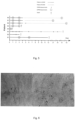
- Figure 6 shows skin microcirculation in fossa tabatière in two ECMO patients. To the left (a) several pericapillary bleedings are seen, and hardly no dot-liked capillaries are present (Patient E1, Proscan (x 200)). To the right (b) circular dark haloes are surrounding capillaries with no flow or extremely slow sluggish flow are seen (Patient E2 Microvision 2100 (x500)).
- CoV coefficient of variation
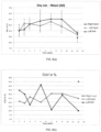
- Figure 5 is an overview over the eight ECMO patients, from start of the ECMO treatment till death or last control measurement. Measurement periods are marked according to measuring technique.
- the patients were grouped according to clinical outcome into two groups: patients dying on ECMO (Group 1) and patients surviving ECMO (Group 2).
- Mean age for group 1 was 44 years, for group 2 it was 58 years. Both sexes were present in each group.
- group 2 one patient had a recovery of heart function and a maintained cerebral function but died from bleeding complications in the intensive care unit.
- one patient (E3) was transferred to another hospital where he died from multi organ failure on day 51 after establishing ECMO. The remaining three were still alive and out of institutions two years after the ECMO treatment.
- CAVM Computer assisted video microscopy
- E1 had numerous pericapillary bleedings in several skin locations (fossa tabatière, volar side of forearm, leg and face) on both examinations, figure 3 (a) . No other patients had any visible pericapillary bleedings. Patients E2 and E4 had circular dark haloes at a distance of 12 ⁇ 1 microns around some skin capillaries (65% in E2 and in 14% in E4), see figure 3 (b) .
- FCD Functional capillary density
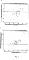
- Figure 4 shows functional capillary density (in capillaries/mm 2 ) for three groups of ECMO patients and eight healthy controls (b) and coefficient of variation of functional capillary density for the same patients (a).
- the first, second and follow-up measurements are given.
- E2 pericapillary dark haloes
- erythrocyte movement in the capillaries was hardly detected in any of the film sequences, although the ECMO circuit gave an output of 4.5 litre/min.
- E3 had a prolonged and complicated stay in the ICU with lung infections, sepsis episodes, and progressive renal failure after ECMO treatment.
- the patient's final microscopy assessments on day 44 showed that FCD was reduced with 30%, CoV was increased with 64% and the mean flow-categorial velocity was reduced to 1.75, a reduction of 34%, compared with the initial measurements.
- the patient was examined during an episode with gram negative sepsis (day 24); at that time the FCD was markedly reduced to 49 capillaries/mm2 (-29%), the CoV was 0.17 (-23%), and 22% of the skin capillaries had brisk flow (category 4). Capillaries in this flow category were not observed for any other patients at any time.
- the controls had a mean flux value of 48 Au (range 22-96) in fossa Tabatière, while the corresponding values at the medial side of the foot were 31.5 (range 17.4-60.1).
- the mean coefficient of variation was 0.20 (range 0.13-0.35) in fossa Tabatière, and 0.25 (range 0.12-0.34) at the medial side of the foot.
- CAVM computer assisted video microscopy
- the patients that died on ECMO had reduced FCD and increased CoV of FCD compared with patients surviving ECMO and healthy controls.
- the patients surviving ECMO had stable FCD and CoV within the reference levels for the controls ( figure 7 ).
- the exception was patient E3, who had a complicated post ECMO course with several septic episodes.
- Freedlander used a microscope to show a decreased capillary density in skin in septic patients. Later studies confirmed Freedlanders findings and demonstrated increased heterogeneity of FCD in different tissues and mammalian species with systemic diseases. In patients with septic and cardiogenic shock a persistent severely reduced FCD for 24 hours in the sublingual area, is associated with increased mortality. Reduced FCD in the rectal mucosa is associated with poor prognosis in patients with severe malaria.
- Figure 7 shows distribution of capillaries in each of five flow categories for patients dying on- (group 1), and patients successfully weaned off ECMO (group 2) as compared to eight healthy controls.
- E2 and E4 Dark haloes were found in two of the patients who died on ECMO (E2 and E4), with the halo edges 12 ⁇ 1 microns from the capillaries. In E2 numerous haloes were present, prominently around capillaries with "no flow”. E4 had fewer haloes, and they were also seen surrounding perfused capillaries. The cause of these haloes is uncertain, but one possibility is that they represent precipitated proteins or erythrocyte degradation products leaking from injured capillaries. Another possibility is that they are caused by pericapillary oedema.
- thermoregulatory plexuses where perfusion is mainly regulated by sympathetic activity. Since perfusion in these plexuses mainly serve a thermoregulatory function, skin nutrition can not be assessed by the LDPM technique. Even though the two patients that died on ECMO (E1 and E4) had the lowest perfusion values of all in both locations and measuring periods, no significant differences were demonstrated du to small numbers and large data variation.
- the number of included patients is small and represents a heterogeneous group with acute heart failure, while the reference data were collected from healthy young male students. Since capillary erythrocyte velocities in healthy subjects are not age dependent, we assume that our control group of young students can be used.
- Microvascular examinations of skin nutritive capillaries in patients on ECMO show major structural and functional pathology in patients dying on ECMO, while patients surviving ECMO have results similar to healthy controls.
- the finding of intact skin microcirculatory morphology and function in survivors early after establishment of ECMO appears to be a robust and clinically useful finding implying a good prognosis.
- Pericapillary bleedings or dark haloes, micro-thrombi/capillaries with "no flow”, low capillary flow velocity and low functional capillary density are associated with poor prognosis.
- Table 3 During a six month period twenty-five healthy term newborns of Caucasian race with healthy mothers were enrolled within the first twenty-four hours after delivery (Table 3).
- CAVM Computer Assisted Video Microscopy
- FCD Functional capillary density
- Microvascular perfusion was assessed with a Moor Blood Flow Monitor (MBF 3D) with Moorsoft (Both Moor instruments, Axminster, Devon, England) for recordings and analyses.
- MPF 3D Moor Blood Flow Monitor
- Moorsoft Both Moor instruments, Axminster, Devon, England
- the output was given in a semi quantitative scale of flux (arbitrary unit, AU) defined as the product of the number of moving blood cells and their mean velocity in the measured volume (approximately 1 mm 3 ). Seven ten-second sequences with no or limited movement artifacts were taken from each ROI.
- Diffuse Reflectance Spectroscopy DRS
- a setup consisting of a spectrometer operating in the visible wavelength region (S2000, Avantes, The Netherlands) and a tungsten halogen light source (AvaLight-HAL, The Netherlands) having an effective spectral range of 450 to 800 nm was used.
- a polytetrafluoretylene tile (WS-2, Avantes, Netherland) enclosed in a black plastic housing was used as reference.
- a custom-built fiber optic probe was used for measurements with a fiber composition of three adjacent illuminating fibers (fiber diameter 400 ⁇ m) and one receiving fiber (fiber diameter 400 ⁇ m) resulting in an emitting-receiving distance for the probe of approximately 800 ⁇ m ( Meglinsky et al. Med Biol Eng Comput, 2001. 39(1): pp. 44-50 ). Twelve spectra were collected from each ROI in 20 neonates.
- Analyses of the spectra were done by adapting a tissue model based on a diffusion approximation ( Farrell et al., Med Phys, 1992. 19(4): pp. 879-88 ; Jacques, IEEE Transactions on Bio-Medical Engineering, 1989. 36(12): pp. 1155-1161 ).
- the model included the chromophores melanin, hemoglobin derivatives , water and a Mie and Rayleigh scattering factor.
- Decomposition of the spectral signature was done by a least square fit of the model to the measured spectra. The decomposition of reflected light spectra was then used to estimate the apparent content of oxy- and deoxy-hemoglobin. Microvascular oxygen saturation was compared with arterial oxygen saturation to estimate oxygen extraction.
- the arterial oxygen saturation was measured using a pulse oxymeter (Masimo Set, Rad-5v, Irvine, USA) with the probe located on the right hand.
- Skin temperature was measured using a surface temperature scanner (Omega Medical, Model no. STS-101-C, USA) attached to the ROI just before measurements with the microvascular techniques. Axillary temperature was taken with an ordinary thermometer (Digitemp, Microlife Asia, Mt1671, Taiwan).
- a transcutaneous bilimeter (Dräger, JM 103, Dräger medical, Lübeck, Made in Japan) was used to estimate bilirubin values, a factor in the DRS analysing algorithm.
- Demographic data are presented as mean with range. All other variables are reported as mean with standard deviation (SD).
- SD standard deviation
- paired t-test was conducted to compare means. P-value ⁇ 0.05 was considered significant.
- Statistical analyses were performed using SPSS for Windows (Statistical Package for the Social Sciences, version 18.0 SPSS Inc., Chicago, IL, USA).
- FCD Functional capillary density
- the dominant capillary flow category was category three (continuous high flow), but flow category two (continuous low flow) was also represented (Table 2, Fig. 4a and b ). Flow categories zero (no flow) and four (brisk flow) were not found.
- the mean flow-categorical velocity (MFCV) was similar both in the chest and in the hand at all the three days, varying between 2.57 (0.10) and 2.71 (0.11).
- the cardio-pulmonary adaptation in neonates with closure of the fetal shunts mainly occurs during the first hours of life, but is not completed until days to months after birth.
- Other adaptive responses such as reduction of total body water accompanied by 4 -7% weight loss, hemolysis of fetal erythrocytes and production of erythrocytes with adult hemoglobin, occurs during the first days to weeks.
- the hematocrit peaks at two hours of age and then decreases steadily over the next weeks.
- Our neonates were examined the first, second and third day of life when many of these adaptive processes affecting the central hemodynamics and hemorheology, and thereby microvascular perfusion, occur.
- MFCV capillary flow-categorical velocity
Landscapes
- Health & Medical Sciences (AREA)
- Life Sciences & Earth Sciences (AREA)
- Physics & Mathematics (AREA)
- Public Health (AREA)
- Animal Behavior & Ethology (AREA)
- Biophysics (AREA)
- Engineering & Computer Science (AREA)
- Biomedical Technology (AREA)
- Heart & Thoracic Surgery (AREA)
- Medical Informatics (AREA)
- Molecular Biology (AREA)
- Surgery (AREA)
- Veterinary Medicine (AREA)
- General Health & Medical Sciences (AREA)
- Pathology (AREA)
- Spectroscopy & Molecular Physics (AREA)
- Cardiology (AREA)
- Physiology (AREA)
- Optics & Photonics (AREA)
- Vascular Medicine (AREA)
- Hematology (AREA)
- Dermatology (AREA)
- Ophthalmology & Optometry (AREA)
- Pulmonology (AREA)
- Investigating Or Analysing Biological Materials (AREA)
- Measuring Pulse, Heart Rate, Blood Pressure Or Blood Flow (AREA)
- Measurement Of The Respiration, Hearing Ability, Form, And Blood Characteristics Of Living Organisms (AREA)
- Measuring And Recording Apparatus For Diagnosis (AREA)
- Liquid Crystal Substances (AREA)
- External Artificial Organs (AREA)
Applications Claiming Priority (4)
| Application Number | Priority Date | Filing Date | Title |
|---|---|---|---|
| GB1301490.7A GB2510176A (en) | 2013-01-28 | 2013-01-28 | Determining patient prognosis by microcirculation analysis |
| GB201312796A GB201312796D0 (en) | 2013-07-17 | 2013-07-17 | Neonatal microcirculation analysis |
| PCT/EP2014/051661 WO2014114814A1 (en) | 2013-01-28 | 2014-01-28 | Assessing circulatory failure |
| EP14701757.8A EP2948042B1 (en) | 2013-01-28 | 2014-01-28 | Assessing circulatory failure |
Related Parent Applications (1)
| Application Number | Title | Priority Date | Filing Date |
|---|---|---|---|
| EP14701757.8A Division EP2948042B1 (en) | 2013-01-28 | 2014-01-28 | Assessing circulatory failure |
Publications (3)
| Publication Number | Publication Date |
|---|---|
| EP4235152A2 EP4235152A2 (en) | 2023-08-30 |
| EP4235152A3 EP4235152A3 (en) | 2023-09-27 |
| EP4235152B1 true EP4235152B1 (en) | 2025-04-02 |
Family
ID=50029038
Family Applications (2)
| Application Number | Title | Priority Date | Filing Date |
|---|---|---|---|
| EP23177112.2A Active EP4235152B1 (en) | 2013-01-28 | 2014-01-28 | Assessing circulatory failure |
| EP14701757.8A Active EP2948042B1 (en) | 2013-01-28 | 2014-01-28 | Assessing circulatory failure |
Family Applications After (1)
| Application Number | Title | Priority Date | Filing Date |
|---|---|---|---|
| EP14701757.8A Active EP2948042B1 (en) | 2013-01-28 | 2014-01-28 | Assessing circulatory failure |
Country Status (5)
| Country | Link |
|---|---|
| US (1) | US10987011B2 (enExample) |
| EP (2) | EP4235152B1 (enExample) |
| JP (2) | JP6542130B2 (enExample) |
| ES (1) | ES2949276T3 (enExample) |
| WO (1) | WO2014114814A1 (enExample) |
Families Citing this family (15)
| Publication number | Priority date | Publication date | Assignee | Title |
|---|---|---|---|---|
| US20210350927A1 (en) * | 2013-01-28 | 2021-11-11 | Oslo Universitetssykehus Hf | Assessing circulatory failure |
| JP6336949B2 (ja) | 2015-01-29 | 2018-06-06 | 富士フイルム株式会社 | 画像処理装置及び画像処理方法、並びに内視鏡システム |
| JP6688164B2 (ja) * | 2016-06-09 | 2020-04-28 | 花王株式会社 | 皮膚毛細血管の観察方法 |
| US11244452B2 (en) * | 2017-10-16 | 2022-02-08 | Massachusetts Institute Of Technology | Systems, devices and methods for non-invasive hematological measurements |
| JP7167132B2 (ja) * | 2018-03-26 | 2022-11-08 | テルモ株式会社 | 支援システム、支援方法、支援プログラム、および支援プログラムを記録した記録媒体 |
| US10485552B1 (en) * | 2018-12-28 | 2019-11-26 | Imad R. Makhoul | Apparatus and method for controlling systemic blood pressure in patients |
| US11754824B2 (en) | 2019-03-26 | 2023-09-12 | Active Medical, BV | Method and apparatus for diagnostic analysis of the function and morphology of microcirculation alterations |
| US20220296108A1 (en) * | 2019-05-24 | 2022-09-22 | Seoul National University Hospital | Method and apparatus for quantitation of microcirculation |
| EP4529839A3 (en) | 2019-07-24 | 2025-04-16 | Massachusetts Institute of Technology | Finger inserts for a nailfold imaging device |
| WO2021081215A1 (en) * | 2019-10-23 | 2021-04-29 | The Johns Hopkins University | Offset illumination capillaroscope |
| GB2589068A (en) | 2019-10-31 | 2021-05-26 | Odi Medical As | Probe |
| US20220369966A1 (en) * | 2019-11-12 | 2022-11-24 | Astem Co., Ltd. | Biological information collection sensor unit, biological information collection device, and biological information processing unit |
| CN111657861B (zh) * | 2020-06-04 | 2022-02-25 | 浙江大学 | 基于双光子显微镜技术的溶栓药效评价方法 |
| CN112587105B (zh) * | 2020-12-11 | 2023-10-27 | 复旦大学附属中山医院 | 一种人体循环灌注状态评估系统 |
| EP4380427A1 (en) | 2021-08-05 | 2024-06-12 | ODI Medical AS | Assessing microcirculation |
Family Cites Families (26)
| Publication number | Priority date | Publication date | Assignee | Title |
|---|---|---|---|---|
| CA2042075C (en) * | 1991-05-08 | 2001-01-23 | Branko Palcic | Endoscopic imaging system |
| US5769792A (en) * | 1991-07-03 | 1998-06-23 | Xillix Technologies Corp. | Endoscopic imaging system for diseased tissue |
| US6420709B1 (en) * | 1992-07-15 | 2002-07-16 | Optix Lp | Methods of minimizing scattering and improving tissue sampling in non-invasive testing and imaging |
| US5699797A (en) * | 1992-10-05 | 1997-12-23 | Dynamics Imaging, Inc. | Method of investigation of microcirculation functional dynamics of physiological liquids in skin and apparatus for its realization |
| US5553613A (en) * | 1994-08-17 | 1996-09-10 | Pfizer Inc. | Non invasive blood analyte sensor |
| US5713364A (en) * | 1995-08-01 | 1998-02-03 | Medispectra, Inc. | Spectral volume microprobe analysis of materials |
| US6104945A (en) * | 1995-08-01 | 2000-08-15 | Medispectra, Inc. | Spectral volume microprobe arrays |
| US20010041843A1 (en) * | 1999-02-02 | 2001-11-15 | Mark Modell | Spectral volume microprobe arrays |
| EP1632173B1 (en) * | 1999-01-26 | 2013-04-03 | HOYA Corporation | Autofluorescence imaging system for endoscopy |
| WO2001022741A2 (en) * | 1999-09-23 | 2001-03-29 | Nadeau Richard G | Medical applications of orthogonal polarization spectral imaging |
| US6902935B2 (en) * | 1999-12-15 | 2005-06-07 | Medispectra, Inc. | Methods of monitoring effects of chemical agents on a sample |
| US6377841B1 (en) * | 2000-03-31 | 2002-04-23 | Vanderbilt University | Tumor demarcation using optical spectroscopy |
| US20020165456A1 (en) * | 2001-03-26 | 2002-11-07 | Murat Canpolat | Estimation of the average size of white light scatterers in normal and cancerous tissue using light scattering spectrum |
| US20050154319A1 (en) * | 2002-01-15 | 2005-07-14 | Xillix Technologies Corporation | Fluorescence endoscopy video systems with no moving parts in the camera |
| US6899675B2 (en) * | 2002-01-15 | 2005-05-31 | Xillix Technologies Corp. | Fluorescence endoscopy video systems with no moving parts in the camera |
| US7706862B2 (en) * | 2003-04-17 | 2010-04-27 | Research Foundation Of The City University Of New York | Detecting human cancer through spectral optical imaging using key water absorption wavelengths |
| US8064976B2 (en) * | 2003-10-03 | 2011-11-22 | Can Ince | Systems and methods for sidesstream dark field imaging |
| US20060241364A1 (en) * | 2004-10-01 | 2006-10-26 | Academisch Medisch Centrum Of The University Van Amsterdam | System and method for imaging the reflectance of a substrate |
| US9131861B2 (en) | 2004-11-30 | 2015-09-15 | Academisch Medisch Centrum | Pulsed lighting imaging systems and methods |
| JP4743486B2 (ja) | 2005-05-24 | 2011-08-10 | 微小循環研究所 有限会社 | 指先の毛細血管血流による医療診断支援システム。 |
| US8214023B2 (en) * | 2006-09-21 | 2012-07-03 | Institute Of Critical Care Medicine | Microcirculation imaging |
| WO2010093503A2 (en) * | 2007-01-05 | 2010-08-19 | Myskin, Inc. | Skin analysis methods |
| US8903247B2 (en) | 2009-10-30 | 2014-12-02 | Cisco Technology, Inc. | Bidirectional multi-mode fiber interface |
| CA2784856C (en) * | 2009-12-18 | 2019-05-07 | University Health Network | System and method for sub-surface fluorescence imaging |
| JP5797921B2 (ja) | 2011-03-30 | 2015-10-21 | 花王株式会社 | 体表評価方法および体表評価装置 |
| GB2510176A (en) * | 2013-01-28 | 2014-07-30 | Univ Oslo Hf | Determining patient prognosis by microcirculation analysis |
-
2014
- 2014-01-28 JP JP2015554205A patent/JP6542130B2/ja active Active
- 2014-01-28 EP EP23177112.2A patent/EP4235152B1/en active Active
- 2014-01-28 WO PCT/EP2014/051661 patent/WO2014114814A1/en not_active Ceased
- 2014-01-28 ES ES14701757T patent/ES2949276T3/es active Active
- 2014-01-28 EP EP14701757.8A patent/EP2948042B1/en active Active
- 2014-01-28 US US14/763,872 patent/US10987011B2/en active Active
-
2019
- 2019-06-12 JP JP2019109448A patent/JP6785339B2/ja active Active
Also Published As
| Publication number | Publication date |
|---|---|
| ES2949276T3 (es) | 2023-09-27 |
| WO2014114814A1 (en) | 2014-07-31 |
| EP4235152A2 (en) | 2023-08-30 |
| EP2948042B1 (en) | 2023-06-07 |
| JP6542130B2 (ja) | 2019-07-10 |
| EP4235152A3 (en) | 2023-09-27 |
| US20150359440A1 (en) | 2015-12-17 |
| EP2948042A1 (en) | 2015-12-02 |
| US10987011B2 (en) | 2021-04-27 |
| JP2016509505A (ja) | 2016-03-31 |
| JP2019188172A (ja) | 2019-10-31 |
| JP6785339B2 (ja) | 2020-11-18 |
Similar Documents
| Publication | Publication Date | Title |
|---|---|---|
| EP4235152B1 (en) | Assessing circulatory failure | |
| Nielsen et al. | The effects of red blood cell transfusion on tissue oxygenation and the microcirculation in the intensive care unit: a systematic review | |
| JP2003510112A (ja) | 直交偏光スペクトルイメージングの医学的応用 | |
| US20210350927A1 (en) | Assessing circulatory failure | |
| Awan et al. | Human microvascular imaging: a review of skin and tongue videomicroscopy techniques and analysing variables | |
| Epstein et al. | Bedside assessment of tissue oxygen saturation monitoring in critically ill adults: an integrative review of the literature | |
| Wester et al. | Skin microvascular morphology and hemodynamics during treatment with veno-arterial extra-corporeal membrane oxygenation | |
| Banerjee et al. | Cerebral blood flow and oximetry response to blood transfusion in relation to chronological age in preterm infants | |
| Jung et al. | Evaluation of the microcirculation during extracorporeal membrane-oxygenation | |
| Kratky et al. | Regional cerebral oxygen saturation in newborn infants in the first 15 min of life after vaginal delivery | |
| Zaman et al. | Free flap pulse oximetry utilizing reflectance photoplethysmography | |
| RU2580895C2 (ru) | Способ оценки регионарного кровообращения, тканевой микроциркуляции и насыщения крови кислородом и устройство для оценки регионарного кровообращения, тканевой микроциркуляции и насыщения крови кислородом | |
| Marcinkevics et al. | Hyperspectral evaluation of skin blood oxygen saturation at baseline and during arterial occlusion | |
| Yao et al. | Hemorrhagic shock assessed by tissue microcirculatory monitoring: a narrative review | |
| GB2510176A (en) | Determining patient prognosis by microcirculation analysis | |
| US20250127412A1 (en) | Assessing microcirculation | |
| Kvernebo | Microcirculation and Tissue Perfusion Assessments for Complex Cardiovascular Disease Care | |
| Lauten et al. | Effect of mechanical ventilation on microvascular perfusion in critical care patients | |
| Weaver | Electrical Cardiometry During Mechanical Ventilation: An Exploratory Study | |
| Wüstenberg et al. | Different swelling mechanisms in nasal septum (Kiesselbach area) and inferior turbinate responses to histamine: an optical rhinometric study | |
| Wong et al. | Assessment of Cerebral Oxygenation Response to Hemodialysis using Near-Infrared Spectroscopy (NIRS): Challenges and Solutions | |
| Elzayat et al. | Role of Perfusion Index on the First Day of Life in prediction of Early Discharge of Newborns | |
| Abbas | Influences of Nursing Procedures and Factors Associated on the Vital Signs of Neonate | |
| Kuryata et al. | Chest pain in patients with diabetes mellitus is associated with severe coronary artery calcification | |
| Müller | The influence of a patent ductus arteriosus on the peripheral muscle oxygenation and perfusion in neonates |
Legal Events
| Date | Code | Title | Description |
|---|---|---|---|
| PUAI | Public reference made under article 153(3) epc to a published international application that has entered the european phase |
Free format text: ORIGINAL CODE: 0009012 |
|
| STAA | Information on the status of an ep patent application or granted ep patent |
Free format text: STATUS: THE APPLICATION HAS BEEN PUBLISHED |
|
| REG | Reference to a national code |
Ref country code: DE Ref legal event code: R079 Free format text: PREVIOUS MAIN CLASS: G01N0021490000 Ipc: A61B0005000000 Ref country code: DE Ref legal event code: R079 Ref document number: 602014091782 Country of ref document: DE Free format text: PREVIOUS MAIN CLASS: G01N0021490000 Ipc: A61B0005000000 |
|
| PUAL | Search report despatched |
Free format text: ORIGINAL CODE: 0009013 |
|
| AC | Divisional application: reference to earlier application |
Ref document number: 2948042 Country of ref document: EP Kind code of ref document: P |
|
| AK | Designated contracting states |
Kind code of ref document: A2 Designated state(s): AL AT BE BG CH CY CZ DE DK EE ES FI FR GB GR HR HU IE IS IT LI LT LU LV MC MK MT NL NO PL PT RO RS SE SI SK SM TR |
|
| AK | Designated contracting states |
Kind code of ref document: A3 Designated state(s): AL AT BE BG CH CY CZ DE DK EE ES FI FR GB GR HR HU IE IS IT LI LT LU LV MC MK MT NL NO PL PT RO RS SE SI SK SM TR |
|
| RIC1 | Information provided on ipc code assigned before grant |
Ipc: G01N 21/49 20060101ALI20230818BHEP Ipc: A61B 5/1455 20060101ALI20230818BHEP Ipc: A61B 5/026 20060101ALI20230818BHEP Ipc: A61B 5/00 20060101AFI20230818BHEP |
|
| STAA | Information on the status of an ep patent application or granted ep patent |
Free format text: STATUS: REQUEST FOR EXAMINATION WAS MADE |
|
| 17P | Request for examination filed |
Effective date: 20240325 |
|
| RBV | Designated contracting states (corrected) |
Designated state(s): AL AT BE BG CH CY CZ DE DK EE ES FI FR GB GR HR HU IE IS IT LI LT LU LV MC MK MT NL NO PL PT RO RS SE SI SK SM TR |
|
| RIC1 | Information provided on ipc code assigned before grant |
Ipc: G01N 21/49 20060101ALI20240410BHEP Ipc: A61B 5/1455 20060101ALI20240410BHEP Ipc: A61B 5/026 20060101ALI20240410BHEP Ipc: A61B 5/00 20060101AFI20240410BHEP |
|
| GRAP | Despatch of communication of intention to grant a patent |
Free format text: ORIGINAL CODE: EPIDOSNIGR1 |
|
| STAA | Information on the status of an ep patent application or granted ep patent |
Free format text: STATUS: GRANT OF PATENT IS INTENDED |
|
| INTG | Intention to grant announced |
Effective date: 20240611 |
|
| GRAJ | Information related to disapproval of communication of intention to grant by the applicant or resumption of examination proceedings by the epo deleted |
Free format text: ORIGINAL CODE: EPIDOSDIGR1 |
|
| STAA | Information on the status of an ep patent application or granted ep patent |
Free format text: STATUS: REQUEST FOR EXAMINATION WAS MADE |
|
| GRAP | Despatch of communication of intention to grant a patent |
Free format text: ORIGINAL CODE: EPIDOSNIGR1 |
|
| STAA | Information on the status of an ep patent application or granted ep patent |
Free format text: STATUS: GRANT OF PATENT IS INTENDED |
|
| INTC | Intention to grant announced (deleted) | ||
| INTG | Intention to grant announced |
Effective date: 20241023 |
|
| GRAS | Grant fee paid |
Free format text: ORIGINAL CODE: EPIDOSNIGR3 |
|
| GRAA | (expected) grant |
Free format text: ORIGINAL CODE: 0009210 |
|
| STAA | Information on the status of an ep patent application or granted ep patent |
Free format text: STATUS: THE PATENT HAS BEEN GRANTED |
|
| AC | Divisional application: reference to earlier application |
Ref document number: 2948042 Country of ref document: EP Kind code of ref document: P |
|
| AK | Designated contracting states |
Kind code of ref document: B1 Designated state(s): AL AT BE BG CH CY CZ DE DK EE ES FI FR GB GR HR HU IE IS IT LI LT LU LV MC MK MT NL NO PL PT RO RS SE SI SK SM TR |
|
| REG | Reference to a national code |
Ref country code: GB Ref legal event code: FG4D |
|
| REG | Reference to a national code |
Ref country code: CH Ref legal event code: EP |
|
| REG | Reference to a national code |
Ref country code: DE Ref legal event code: R096 Ref document number: 602014091782 Country of ref document: DE |
|
| REG | Reference to a national code |
Ref country code: IE Ref legal event code: FG4D |
|
| P01 | Opt-out of the competence of the unified patent court (upc) registered |
Free format text: CASE NUMBER: APP_15807/2025 Effective date: 20250401 |
|
| REG | Reference to a national code |
Ref country code: NL Ref legal event code: MP Effective date: 20250402 |
|
| PG25 | Lapsed in a contracting state [announced via postgrant information from national office to epo] |
Ref country code: NL Free format text: LAPSE BECAUSE OF FAILURE TO SUBMIT A TRANSLATION OF THE DESCRIPTION OR TO PAY THE FEE WITHIN THE PRESCRIBED TIME-LIMIT Effective date: 20250402 |
|
| REG | Reference to a national code |
Ref country code: AT Ref legal event code: MK05 Ref document number: 1780454 Country of ref document: AT Kind code of ref document: T Effective date: 20250402 |
|
| PG25 | Lapsed in a contracting state [announced via postgrant information from national office to epo] |
Ref country code: FI Free format text: LAPSE BECAUSE OF FAILURE TO SUBMIT A TRANSLATION OF THE DESCRIPTION OR TO PAY THE FEE WITHIN THE PRESCRIBED TIME-LIMIT Effective date: 20250402 Ref country code: PT Free format text: LAPSE BECAUSE OF FAILURE TO SUBMIT A TRANSLATION OF THE DESCRIPTION OR TO PAY THE FEE WITHIN THE PRESCRIBED TIME-LIMIT Effective date: 20250804 Ref country code: ES Free format text: LAPSE BECAUSE OF FAILURE TO SUBMIT A TRANSLATION OF THE DESCRIPTION OR TO PAY THE FEE WITHIN THE PRESCRIBED TIME-LIMIT Effective date: 20250402 |
|
| REG | Reference to a national code |
Ref country code: LT Ref legal event code: MG9D |
|
| PG25 | Lapsed in a contracting state [announced via postgrant information from national office to epo] |
Ref country code: GR Free format text: LAPSE BECAUSE OF FAILURE TO SUBMIT A TRANSLATION OF THE DESCRIPTION OR TO PAY THE FEE WITHIN THE PRESCRIBED TIME-LIMIT Effective date: 20250703 Ref country code: NO Free format text: LAPSE BECAUSE OF FAILURE TO SUBMIT A TRANSLATION OF THE DESCRIPTION OR TO PAY THE FEE WITHIN THE PRESCRIBED TIME-LIMIT Effective date: 20250702 |
|
| PG25 | Lapsed in a contracting state [announced via postgrant information from national office to epo] |
Ref country code: PL Free format text: LAPSE BECAUSE OF FAILURE TO SUBMIT A TRANSLATION OF THE DESCRIPTION OR TO PAY THE FEE WITHIN THE PRESCRIBED TIME-LIMIT Effective date: 20250402 |
|
| PG25 | Lapsed in a contracting state [announced via postgrant information from national office to epo] |
Ref country code: BG Free format text: LAPSE BECAUSE OF FAILURE TO SUBMIT A TRANSLATION OF THE DESCRIPTION OR TO PAY THE FEE WITHIN THE PRESCRIBED TIME-LIMIT Effective date: 20250402 |
|
| PG25 | Lapsed in a contracting state [announced via postgrant information from national office to epo] |
Ref country code: HR Free format text: LAPSE BECAUSE OF FAILURE TO SUBMIT A TRANSLATION OF THE DESCRIPTION OR TO PAY THE FEE WITHIN THE PRESCRIBED TIME-LIMIT Effective date: 20250402 |
|
| PG25 | Lapsed in a contracting state [announced via postgrant information from national office to epo] |
Ref country code: AT Free format text: LAPSE BECAUSE OF FAILURE TO SUBMIT A TRANSLATION OF THE DESCRIPTION OR TO PAY THE FEE WITHIN THE PRESCRIBED TIME-LIMIT Effective date: 20250402 |
|
| PG25 | Lapsed in a contracting state [announced via postgrant information from national office to epo] |
Ref country code: RS Free format text: LAPSE BECAUSE OF FAILURE TO SUBMIT A TRANSLATION OF THE DESCRIPTION OR TO PAY THE FEE WITHIN THE PRESCRIBED TIME-LIMIT Effective date: 20250702 |
|
| PG25 | Lapsed in a contracting state [announced via postgrant information from national office to epo] |
Ref country code: IS Free format text: LAPSE BECAUSE OF FAILURE TO SUBMIT A TRANSLATION OF THE DESCRIPTION OR TO PAY THE FEE WITHIN THE PRESCRIBED TIME-LIMIT Effective date: 20250802 |
|
| PG25 | Lapsed in a contracting state [announced via postgrant information from national office to epo] |
Ref country code: LV Free format text: LAPSE BECAUSE OF FAILURE TO SUBMIT A TRANSLATION OF THE DESCRIPTION OR TO PAY THE FEE WITHIN THE PRESCRIBED TIME-LIMIT Effective date: 20250402 |