EP4200591B1 - Biosensor mit partikelbewegung, verwendung und verfahren - Google Patents
Biosensor mit partikelbewegung, verwendung und verfahren Download PDFInfo
- Publication number
- EP4200591B1 EP4200591B1 EP21770323.0A EP21770323A EP4200591B1 EP 4200591 B1 EP4200591 B1 EP 4200591B1 EP 21770323 A EP21770323 A EP 21770323A EP 4200591 B1 EP4200591 B1 EP 4200591B1
- Authority
- EP
- European Patent Office
- Prior art keywords
- particle
- analyte
- moiety
- biosensor device
- state
- Prior art date
- Legal status (The legal status is an assumption and is not a legal conclusion. Google has not performed a legal analysis and makes no representation as to the accuracy of the status listed.)
- Active
Links
Images


Classifications
-
- G—PHYSICS
- G01—MEASURING; TESTING
- G01N—INVESTIGATING OR ANALYSING MATERIALS BY DETERMINING THEIR CHEMICAL OR PHYSICAL PROPERTIES
- G01N15/00—Investigating characteristics of particles; Investigating permeability, pore-volume or surface-area of porous materials
- G01N15/10—Investigating individual particles
- G01N15/1031—Investigating individual particles by measuring electrical or magnetic effects
-
- G—PHYSICS
- G01—MEASURING; TESTING
- G01N—INVESTIGATING OR ANALYSING MATERIALS BY DETERMINING THEIR CHEMICAL OR PHYSICAL PROPERTIES
- G01N15/00—Investigating characteristics of particles; Investigating permeability, pore-volume or surface-area of porous materials
- G01N15/10—Investigating individual particles
- G01N15/14—Optical investigation techniques, e.g. flow cytometry
- G01N15/1429—Signal processing
- G01N15/1433—Signal processing using image recognition
-
- G—PHYSICS
- G01—MEASURING; TESTING
- G01N—INVESTIGATING OR ANALYSING MATERIALS BY DETERMINING THEIR CHEMICAL OR PHYSICAL PROPERTIES
- G01N33/00—Investigating or analysing materials by specific methods not covered by groups G01N1/00 - G01N31/00
- G01N33/48—Biological material, e.g. blood, urine; Haemocytometers
- G01N33/50—Chemical analysis of biological material, e.g. blood, urine; Testing involving biospecific ligand binding methods; Immunological testing
- G01N33/53—Immunoassay; Biospecific binding assay; Materials therefor
- G01N33/543—Immunoassay; Biospecific binding assay; Materials therefor with an insoluble carrier for immobilising immunochemicals
- G01N33/54313—Immunoassay; Biospecific binding assay; Materials therefor with an insoluble carrier for immobilising immunochemicals the carrier being characterised by its particulate form
-
- G—PHYSICS
- G01—MEASURING; TESTING
- G01N—INVESTIGATING OR ANALYSING MATERIALS BY DETERMINING THEIR CHEMICAL OR PHYSICAL PROPERTIES
- G01N33/00—Investigating or analysing materials by specific methods not covered by groups G01N1/00 - G01N31/00
- G01N33/48—Biological material, e.g. blood, urine; Haemocytometers
- G01N33/50—Chemical analysis of biological material, e.g. blood, urine; Testing involving biospecific ligand binding methods; Immunological testing
- G01N33/53—Immunoassay; Biospecific binding assay; Materials therefor
- G01N33/543—Immunoassay; Biospecific binding assay; Materials therefor with an insoluble carrier for immobilising immunochemicals
- G01N33/54366—Apparatus specially adapted for solid-phase testing
- G01N33/54373—Apparatus specially adapted for solid-phase testing involving physiochemical end-point determination, e.g. wave-guides, FETS, gratings
- G01N33/5438—Electrodes
-
- A—HUMAN NECESSITIES
- A61—MEDICAL OR VETERINARY SCIENCE; HYGIENE
- A61M—DEVICES FOR INTRODUCING MEDIA INTO, OR ONTO, THE BODY; DEVICES FOR TRANSDUCING BODY MEDIA OR FOR TAKING MEDIA FROM THE BODY; DEVICES FOR PRODUCING OR ENDING SLEEP OR STUPOR
- A61M2205/00—General characteristics of the apparatus
- A61M2205/33—Controlling, regulating or measuring
- A61M2205/3303—Using a biosensor
-
- G—PHYSICS
- G01—MEASURING; TESTING
- G01N—INVESTIGATING OR ANALYSING MATERIALS BY DETERMINING THEIR CHEMICAL OR PHYSICAL PROPERTIES
- G01N15/00—Investigating characteristics of particles; Investigating permeability, pore-volume or surface-area of porous materials
- G01N15/01—Investigating characteristics of particles; Investigating permeability, pore-volume or surface-area of porous materials specially adapted for biological cells, e.g. blood cells
-
- G—PHYSICS
- G01—MEASURING; TESTING
- G01N—INVESTIGATING OR ANALYSING MATERIALS BY DETERMINING THEIR CHEMICAL OR PHYSICAL PROPERTIES
- G01N15/00—Investigating characteristics of particles; Investigating permeability, pore-volume or surface-area of porous materials
- G01N15/10—Investigating individual particles
- G01N2015/1006—Investigating individual particles for cytology
-
- G—PHYSICS
- G01—MEASURING; TESTING
- G01N—INVESTIGATING OR ANALYSING MATERIALS BY DETERMINING THEIR CHEMICAL OR PHYSICAL PROPERTIES
- G01N15/00—Investigating characteristics of particles; Investigating permeability, pore-volume or surface-area of porous materials
- G01N15/10—Investigating individual particles
- G01N2015/1027—Determining speed or velocity of a particle
Definitions
- the present invention relates to a biosensor device for sensing an analyte over a period of time using particle motion.
- the present invention further relates to a method for sensing an analyte using particle motion, use of the biosensor device of the present invention in a method for sensing an analyte or as a sensor on, in or as part of another device.
- the present invention further relates to the biosensor device of the present invention for use in in vivo biosensing, ex vivo biosensing, or in vitro biosensing.
- Biosensor devices for chemical or biochemical markers have typically been developed for use in in vitro diagnostics, where a sample is taken (e.g. blood, saliva, urine, mucus, sweat or cerebrospinal fluid) and is transferred to an artificial device (e.g. a plastic disposable) outside a living organism.
- a sample is taken (e.g. blood, saliva, urine, mucus, sweat or cerebrospinal fluid) and is transferred to an artificial device (e.g. a plastic disposable) outside a living organism.
- an artificial device e.g. a plastic disposable
- sample pre-treatment steps can be applied (e.g. separation or dilution steps) and multiple reagents can be introduced in the assay (e.g. for target amplification, signal amplification, or washing steps).
- in vitro biosensing assays are: immunoassays, nucleic acid tests, tests for electrolytes and metabolites, electrochemical assays, enzyme activity assays, cell-based assays, and the like.
- immunoassays nucleic acid tests, tests for electrolytes and metabolites
- electrochemical assays electrochemical assays
- enzyme activity assays cell-based assays, and the like.
- in vivo biochemical sensing at least a part of the sensor system remains connected to or is inserted in a living organism, e.g. a human body, e.g. on the skin, in the skin, below the skin, or on, in, or below another part of the body. Due to the contact between the biosensor and the living organism, in vivo biochemical sensing sets high requirements on biocompatibility (e.g. inflammation processes should be minimized) and the sensor system should operate reliably within the complex environment of the living organism. For monitoring applications, the system should be able to perform more than one measurement over time and the system should be robust and easy to handle.
- a living organism e.g. a human body, e.g. on the skin, in the skin, below the skin, or on, in, or below another part of the body. Due to the contact between the biosensor and the living organism, in vivo biochemical sensing sets high requirements on biocompatibility (e.g. inflammation processes should be minimized) and the sensor system should operate reliably within the
- CGM continuous glucose monitoring
- Commercial continuous glucose monitoring devices are based on enzymatic electrochemical sensing (see for example: Heo, Yun Jung, and Shoji Takeuchi; Towards smart tattoos: implantable biosensors for continuous glucose monitoring; Advanced healthcare materials 2(1), 2013: pp. 43-56 ).
- Enzymatic sensing is less generic than affinity-based sensing.
- Commercial systems for in vivo glucose monitoring are available from e.g. Dexcom and Medtronic.
- Bio systems such as cells, multi-cellular systems, organs, organisms, or other systems and materials based on biological molecules or containing biological molecules or cells, exhibit dynamics that are at the most basic level driven by time-dependent changes of bioorganic molecules, such as e.g. small molecules, metabolites, hormones, proteins, or nucleic acids.
- bioorganic molecules such as e.g. small molecules, metabolites, hormones, proteins, or nucleic acids.
- Sensing technologies for the measurement and monitoring of biomolecules will allow studies of dynamic changes in biological systems and control of such systems based on measured responses, e.g. in the fields of healthcare, bio-engineering, and industrial processing. Sensors are available for continuously measuring pH, electrolytes, and metabolites, but not yet for measuring biomolecules at low concentrations.
- TPM tethered particle motion
- Biosensors having functionalized tethers attached to a surface have been developed based on the principle that the motion of particles attached by a tether changes in dependence upon presence of analyte.
- the motion changes are due to changes in the structure of the tether itself due to the presence of the analyte.
- the invention provides hereto a biosensor device according to claim 1 having a surface and a particle, wherein the particle and/or the surface are functionalized, and wherein:
- the biosensor device of the present invention enables continuous molecular biosensing without a fixed tether between the particle and the surface. I.e. the particle remains in proximity of a surface, e.g. due to a field force, e.g. due to the gravitational field.
- the particles of the biosensor device of the present invention show Brownian motion and the motion changes when the particle switches between an associated and a non-associated state (also referred to as the 'dissociated state').
- the motion behaviour and association/dissociated state lifetimes of the particle depend on the concentration of target, i.e. the analyte, in the solution.
- the term 'conjugated' refers to the covalent attachment of a first molecule to a second molecule. Also, the term 'conjugated' refers to the linking of one part of the biosensor device with another part of the biosensor device, e.g. crosslinking the particle of the biosensor device with the surface of the biosensor device via, for example, a linker or tether. As used herein the phrase 'the particle is not conjugated to the surface' refers, in a non-associated state, to a freely movable particle that is not linked to the surface.
- biosensing refers to the identification, testing, characterisation, monitoring, and otherwise measuring an analyte using a biosensor.
- the term 'analyte' refers to a substance being identified, tested, characterized, monitored, or otherwise measured; the analyte can comprise molecules of a single target species (e.g., glucose), or molecules of multiple target species (e.g., glucose and synthetic deoxyribose nucleic acid (DNA)).
- target species e.g., glucose
- DNA synthetic deoxyribose nucleic acid
- analyte examples include latex beads, lipid vesicles, whole chromosomes, nanoparticles, extracellular vesicles, liposomes, viruses, cells, cell fragments, supramolecular objects, protein aggregates, and biomolecules including proteins and nucleic acids, gaseous molecules (e.g., ethylene), metal or semiconductor colloids and clusters, small molecules in the size range of sub-nanometre to 10 nm, metabolites, and other such chemical molecules.
- gaseous molecules e.g., ethylene
- metal or semiconductor colloids and clusters small molecules in the size range of sub-nanometre to 10 nm, metabolites, and other such chemical molecules.
- the term 'particle' can refer to an object with a detectable motion in a fluidic or viscoelastic matrix.
- a fluidic or viscoelastic matrix is often simply referred to as a fluid.
- the particle can consist of e.g. organic material (e.g., polymer, supramolecular system, micelle, nanosome), inorganic material (e.g., oxide, silica, metal), or combinations thereof. It can have different inner and outer shapes and architectures (e.g. spherical, rod-like, hollow, star, bubble, hybrid system, particles inside a matrix, aggregate, regular or irregular). It can have a short axis in the range between 1 nm and 15 ⁇ m, more preferred between 5 nm and 5 ⁇ m, more preferred between 10 nm and 3 ⁇ m.
- the term 'surface' can refer to an object with respect to which a coordinate parameter of a particle can be measured, e.g. a position, a distance, a translation, a displacement, an angle, an orientation, a rotation, a translational or angular velocity.
- a surface can consist of e.g. organic material or inorganic material, or a combination thereof. It can have different shapes (e.g. flat, curved, corrugated) and different inner and outer architectures (e.g. solid, porous, permeable, layered, flexible, viscoelastic).
- the surface may be a supporting structure, such as a planar surface, a surface with concave or convex structure, a chemically and/or physically patterned surface, a particle, a polymer, a porous structure, or a porous matrix. It is emphasized that the surface may also be a three dimensional structure.
- the phrase 'properties of the particle and surface' refer to the parameters of the particle, surface and fluid causing the particle to have a distance between the particle and the surface in the second state being e.g. within the range of 5 nm to 10 ⁇ m.
- particle parameters relevant for providing a biosensor of the present invention may include the size of the particle and density of the particle.
- surface parameters may be a selection of surface material or type of material or design having acoustic or magnetic or transport or mechanical properties or the like.
- the phrase 'properties of the particle and surface' includes the cooperation between the particle, the surface, and the fluid, i.e. the method to confine the particle to the surface, e.g. by weight, by acoustic field, by flow, by mechanical confinement, or the like, and corresponding properties such as density, temperature, applied filed, mechanical design, and the like.
- the term 'biosensor' can refer to any suitable sensor used in biochemical testing, biological testing, chemical testing, electrochemical testing, and the like.
- the phrase 'associated with the surface' refers to a binding or an attachment that is non-covalent and means that the particle of the present invention adheres to, is bonded to, or is electrostatically attached to the surface of the biosensor device, for example.
- the biosensor device of the present invention may contain various amounts of particles. However, preferably the biosensor device of the present invention may comprise at least 10 particles, more preferred at least 100 particles. It was found that by providing a biosensor device comprising more than 10 particles, more preferred more than 100 particles, a robust and reliable biosensing method can be performed. It was further found that the biosensor device may comprise a density of between a few particles to several thousand particles in a 415 ⁇ 415 ⁇ m 2 region. Preferably, the biosensor device may comprise a particle density of between 100 and 100,000 particles in a 415 ⁇ 415 ⁇ m 2 region, more preferably between 500 and 20,000 particles, even more preferably between 1,000 and 10,000 particles in a 415 ⁇ 415 ⁇ m 2 region. The total area where particles are tracked is preferably between 10° and 10 8 ⁇ m 2 , more preferred between 10 3 and 10 7 ⁇ m 2 , more preferred between 10 4 and 10 6 ⁇ m 2 .
- the biosensor device may comprise an optical system having a diffraction limit, wherein the biosensor device comprises particles separated from nearest-neighbour particles by at least the diffraction limit of the optical system.
- the biosensor device of the present invention may implement a binding assay, a competitive assay, a displacement assay, a sandwich assay, an enzymatic assay, an assay with target and/or signal amplification, a multistep assay, or an assay with molecular cascade.
- the term ⁇ functionalized' refers to the state wherein the inert particle and/or inert surface have been transformed into a particle and/or surface having a certain activity.
- the particle and/or the surface may be functionalized with binding sites or binding moieties, such as antibodies, aptamers, nanobodies, molecularly imprinted polymers, organic molecules, and the like.
- the particle of the biosensor device may be functionalized by a first moiety, wherein the first moiety is bound to the particle.
- the biosensor device may comprise a functionalized surface that is functionalized by a second moiety, wherein the second moiety is bound to the surface.
- the moieties used have a binding affinity to the analyte.
- the term 'bound' refers to a binding or an attachment that may be covalent, e.g. by chemically coupling, or non-covalent, e.g. by ionic interactions, hydrophobic interactions, hydrogen bonds, etc.
- Covalent bonds can be, for example, ester, ether, phosphoester, amide, peptide, imide, carbon-sulfur bonds, carbon-phosphorus bonds, and the like.
- the term 'bound' is broader than and includes terms such as 'coupled', ⁇ fused', 'associated', 'linked' and 'attached'.
- both the particle and the surface may be functionalized, i.e. providing a biosensor device of the present invention wherein the particle is functionalized by the first moiety, wherein the first moiety is bound to the particle and wherein the surface is functionalized by the second moiety, wherein the second moiety is bound to the surface.
- both moieties preferably have a binding affinity to each other in dependence on the presence, absence or concentration of the analyte.
- such biosensor device may provide an analyte biosensing method wherein in the presence of the analyte the functionalized particle is in its first state, i.e. associated with the functionalized surface.
- such biosensor device may provide an analyte biosensing method wherein in the absence of the analyte the functionalized particle is in its first state, i.e. associated with the functionalized surface.
- any density may be suitable for providing a biosensor device suitable for use in a method of biosensing an analyte.
- Such surface density may preferably between 10 0 and 10 8 moieties/ ⁇ m 2 .
- the biosensor device may have a density of moieties in the range between 10 1 and 10 7 moieties/ ⁇ m 2 , preferably wherein the moieties bound to the particle or to the surface have a density in the range between 10 1 and 10 7 moieties/ ⁇ m 2 , 10 2 and 10 6 moieties/ ⁇ m 2 or 10 3 and 10 5 moieties/ ⁇ m 2 .
- the first moiety or the second moiety may be selected from the group consisting of a protein, an antibody, a fragment thereof, a recombinant protein, a peptide, a carbohydrate, a saccharide, a molecularly imprinted polymer, a small molecule, a nucleic acid, a DNA molecule, a PNA molecule, an aptamer, a nanobody, a multivalent binder, or a combination thereof.
- the first moiety or the second moiety is selected from the group consisting of a binding molecule for glucose, electrolyte, metabolite, small molecule, bioactive, toxin, lipid, carbohydrate, peptide, hormone, drug, drug metabolite, protein, oligonucleotide, DNA, RNA, nanoparticle, extracellular vesicle, exosome, nanosome, liposome, viral particle, cell, cell fragment, supramolecular object, or protein aggregate.
- the invention relates to the use of the biosensor device according to claim 10 in a method of performing multiplexing, preferably analyte multiplexing, spatial multiplexing (e.g. spot multiplexing or chamber multiplexing), spectroscopic multiplexing, probe functionality multiplexing.
- the present invention relates to the use of the biosensor device according to claim 11 as a sensor on, in or as part of a system for sensing or monitoring, which may include e.g. an endoscope, a tube, a needle, a fiber, a catheter, a patch, a disposable probe, a wearable device, an insidable device, a flow cell, or a disposable cartridge.
- the invention in another aspect of the present invention, relates to a biosensor device according to claim 12 for use in in vivo biosensing, ex vivo biosensing, or in vitro biosensing, such as in in vitro diagnostic testing, point-of-care testing, environmental testing, food testing, process monitoring, process control, forensics, biological, biomedical, and pharmaceutical research, or to monitor assays with live cells, tissue, or an organ.
- the invention relates to a method for sensing an analyte using particle motion according to claim 13, wherein the method comprises the steps of:
- the particle of the biosensor device of the present invention is typically arranged to switch from the first state (i.e. the particle-surface associated state) to the second state (i.e. the particle-surface non-associated state) with an average effective dissociation time. Also the particle of the biosensor device of the present invention is typically arranged to switch from the second state to the first state with an average effective association time.
- method of the present invention may further includes the step wherein in step b) the direction of the flow of the matrix containing the analyte is continuously or intermittently changed. Such change of flow may be subjected to a random flow directional change or to a reversal flow direction change.
- the terms 'average effective dissociation time' and 'average effective association time' refer to the average time needed for the particles to, respectively, dissociate from the surface and associate with the surface. In other words, the average time needed to, respectively, reach the fully particle-surface non-associated state (i.e. the second state) and the particle-surface associated state (i.e. the first state), i.e. any type of associated state, e.g. with a single-molecule bond (monovalent) or with multiple molecular bonds (multivalent).
- step b) of detecting motion characteristics of the particle is performed over a period of time that is longer than the average effective dissociation time and/or the average effective association time.
- the invention describes a biosensor with single-molecule resolution.
- Sensors with single-molecule resolution give signals with digital characteristics, also referred to as levels, states, transitions, switches, or events.
- digital signals obey the fundamental laws of Poisson statistics. It means for example that the coefficient of variation due to stochastics can scale with 1/square-root(N), with N the average number of detected events. Therefore, improving the statistics of detected events, reduces variation and increases precision.
- low concentrations are typically measured by using binding moieties with a high affinity and/or with low dissociation rate constant (low k_off).
- a low dissociation rate constant means slow unbinding characteristics (long bound state lifetime of the analyte), which would not be beneficial if it determines the statistics of binding events of the particles.
- particle state lifetimes should not be too long, otherwise insufficient events are recorded in a given measurement timespan.
- the statistics can be improved by having one binding with relatively low dissociation rate constant (to be able to measure low concentrations), and another binding with relatively high dissociation rate constant (for high N, i.e. good particle event statistics).
- the dissociation rate constants differ by a factor of about 3
- the dissociation of an analyte from the strongest binder takes on average three times as long as the dissociation from the weakest binder (with the highest dissociation rate constant). Due to this time ratio, several particle binding and unbinding events may be observed during the time when the analyte is associated with the strongest binder (e.g.
- the distance between particle and surface in the biosensor of the present invention can depend on the size of the particle.
- the force F can be time and spatially dependent; however, here the force was assumed to be constant (for simplicity reasons).
- the force F can have several origins (e.g. gravitational, acoustic, magnetic, optical, electrical, fluid mechanical) and can depend on the size of the particle.
- Equation 3 shows that the height spread of small particles is much larger than the height spread of large particles.
- a large height spread gives a large average distance between particle and surface, which is disadvantageous for the collision rate or encounter rate between particle and surface, hence hindering the effective association rate between particle and surface.
- the mass density difference ⁇ ⁇ can be positive (i.e. the particle is heavier than the solution; the particle 'sinks' toward the biosensing surface) or negative (i.e. the particle is lighter than the solution; the particle ⁇ floats' toward the biosensing surface).
- the particle may be kept in close vicinity to the surface by a mechanical means, e.g. by a second surface that restricts the height space where the particle can reside, or which limits the accessible distance range between particle and first surface. It can function as a means to keep the particle in proximity of the first surface and hinder the particle from moving away too far from the first surface.
- the second surface may be porous, so that analyte and/or fluid can penetrate into the second surface or permeate through the second surface into and/or out of the region with the particle.
- the first surface may be porous, so that analyte and/or fluid can penetrate into the first surface or permeate through the first surface into and/or out of the region with the particle.
- a large particle can give a small height spread and small effective distances between particle and surface (see above). However, a small height spread and small effective distances can also hinder the reversibility of biomolecular interactions, giving low dissociation rates. Large particles can give steric hindrance, slowing down association and dissociation processes, i.e. the switching of the biosensor device system of the present invention from its first state to its second state and vice versa. Furthermore, large particles can give non-specific interactions between particle and surface including irreversible sticking, even if blocking and antifouling coatings are applied on particle and surface.
- Biosensor devices were prepared comprising particles with a diameter of either 1 ⁇ m or 2.8 ⁇ m. Two types of biosensor devices were prepared by using streptavidin coated 1 ⁇ m particles (Dynabeads MyOne C1) where 10 ⁇ M particle binder biotin-oligo (SEQ ID NO: 1) was coupled via streptavidin to the particle. The remaining part of the particle was blocked using 100 ⁇ M 1 kDa PEG-biotin and 1% BSA.
- the surface was prepared by using a glass substrate comprising 100 ⁇ g/mL neutravidin (physisorption), 500 nM surface biotin-oligo (SEQ ID NO: 4) coupled via neutravidin to the surface and using a detection oligo molecule (SEQ ID NO: 3) coupled via the biotin-oligo to the surface.
- the remaining part of the surface was blocked using 100 ⁇ M 1 kDa PEG-biotin and 1% BSA.
- biosensor device B another surface (biosensor device B) was prepared by using PLL-g-PEG and click-coupled biotin-oligo on a glass substrate.
- Flow cell cartridges with measurement chambers were constructed using a double-side adhesive layer and a top plate with fluid inlet and outlet. Data were collected by replacing fluid in a flow cell, i.e. by consecutively inserting solutions with different analyte concentrations. The fluid was inserted manually, using a pipette. The flow speeds in the experiments were typically on the order of 1-300 microliters per minute.
- an analyte having SEQ ID NO: 2 was used as a target.
- Table 1 Synthetic DNA sequences used SEQ ID Description Sequence NO: 1 Particle binder biotin-3' AGCATGGCACT 5' NO: 2 Analyte 5' TCGTACCGTGAGTAATAATGCG 3' NO: 3 Detecting 3' CATTATTACAAGCTAAGCTCTTGCACTGACG 5' NO: 4 Surface binder 5' CGATTCCAGAACGTGACTGCTTTTT 3'-biotin
- the data for the biosensor device A show that the diffusion coefficient histograms and the measured state lifetimes depend on analyte concentration provided into the flow cell. State lifetimes could be extracted because reversible switches were observed between associated and dissociated states. State lifetimes show short-lived and long-lived states, which can be attributed to interactions of different types, e.g. different valencies.
- the examples for the biosensor device B are shown of measured diffusion coefficient histograms as a function of the analyte concentration provided into the flow cell; motion traces at 10 pM concentration (free, single-bond, and multiple bond states are visible); and motion traces at 50 pM concentration (single-bond and multiple-bond states are visible).
- transition rates and state lifetimes can be measured, which depend on the captured amount of target on the particle and on the surface, and therefore depend on the concentration of target in solution. For example, when a particle is observed in a monovalent bond state or with a single-molecular bond, an additional bond can be formed. This gives lifetimes and transition rates corresponding to the formation of the first and the additional bond, which depend on the concentration, but in a different magnitude. This results in multiple parameters that can be extracted and used to improve the biosensing performance.
- biosensing performance aspects of a biosensor are e.g. sensitivity, specificity, speed, reversibility, precision, accuracy, dynamic range, robustness, stability, multiplexing.
- a particle mobility assay without a fixed tether can be used for continuous biomarker monitoring. Due to reversible interactions, increases as well as decreases of analyte concentration can be followed.
- changing the direction of flow in the flow cell can help to compensate displacements and minimize net distances that particles are displaced, which minimizes loss of particles and enables long measurement sequences and measurements over long timespans.
- the preferred distance is in the range between 5 nm and 10 ⁇ m.
- the lower limit (5 nm) is determined by the fact that molecules and reversible biomolecular interactions are used, which operate typically on the length scale of a few nanometres; sufficient space is needed between particle and surface to be able to achieve an unbound state.
- the upper limit (10 ⁇ m) is determined by the fact that a sufficiently high collision rate between particle and surface is needed to achieve an effective association rate, so that sufficient transitions can be observed from unbound to bound states.
- the biosensor of the present invention is a system that may contain multiple components, e.g. a component for sampling analyte from a system of interest (e.g. a biological system, an environmental system (e.g. river, pond, sea, lake, source), a channel, a pipe, a pool, a well, an exhaust, a process, a reactor, a fermenter, a flow, an organism, a reservoir, a patient, an animal, an organoid), for pre-treating the sample (e.g.
- a system of interest e.g. a biological system, an environmental system (e.g. river, pond, sea, lake, source), a channel, a pipe, a pool, a well, an exhaust, a process, a reactor, a fermenter, a flow, an organism, a reservoir, a patient, an animal, an organoid)
- a system of interest e.g. a biological system, an environmental system (e.g. river
- dilution, filtration, heating, enzymatic processing, separation for leading the sample to the sensing particles, for illuminating the particles, for collecting radiation from the particles, for imaging the particles, for determining spatial coordinate parameters of the particles at different time points, for determining displacements or translations or rotations or motion parameters of the particles, for determining states and binding and unbinding events in particle time traces, for processing histograms and distributions of parameters, for translating processed parameters (e.g. amplitude, state, diffusivity, diffusion constant, lifetime, rate, population in distributions, fractional occupation, switching activity, event frequency, time delay) into analytical parameters (e.g. concentration, precision, accuracy, time profile), for translating analytical parameters into control actions (e.g. warning signal or closed-loop control parameter), for controlling the different components in the system (e.g. a computer with software), or for communicating with an external component (e.g. larger control system, a database, an internet system, an information system, or a cloud system).
- an external component e.g.
- the biosensor of the present invention may contain a reader system (with e.g. an optical component, a component for data and signal processing, a component for interfacing and data communication), a fluidic system (with e.g. a method to bring fluid or particles into motion, a pump, a method to apply underpressure or overpressure, a method to effectuate dilution, a method to effectuate mixing, a vent, a tube, a valve, a filter, a switch, a flow sensor, a pressure sensor, a gas sensor, a gas handling method, a degassing unit, a flow regulator, a pressure regulator), or a cartridge or another container device (with e.g.
- a reader system with e.g. an optical component, a component for data and signal processing, a component for interfacing and data communication
- a fluidic system with e.g. a method to bring fluid or particles into motion, a pump, a method to apply underpressure or overpressure, a method to effect
- the system can have containers for reagents (e.g. buffer, particles, pre-treatment reagents) or for collection of fluids (e.g. a waste reservoir).
- reagents e.g. buffer, particles, pre-treatment reagents
- fluids e.g. a waste reservoir
- the sensor system can contain wet reagents or dry reagents (e.g. dried-in or lyophilized).
- a sensor particle may be positioned near a first surface, with fluid transport or molecular transport or analyte transport in different directions with respect to the surface, e.g in a direction along the surface and/or a direction perpendicular to the surface; this includes transport through the surface.
- a particle may be positioned between a first surface and a second surface; the transport of fluid, molecules, or analyte may occur in different directions, e.g. along or through the different surfaces.
- Surfaces may be biofunctionalized in order to effectuate binding between particle and surface.
- Cartridge and other components may be produced by patterning techniques (e.g., lithography, contact printing, microcontact printing, non-contact printing, self-assembly), additive manufacturing (e.g., 3D printing), joining (e.g., gluing, welding, adhesives, adhesive tape), assembly, lamination, automated placement, molding, over-molding, drop casting, curing (e.g., optical, thermal).
- patterning techniques e.g., lithography, contact printing, microcontact printing, non-contact printing, self-assembly
- additive manufacturing e.g., 3D printing
- joining e.g., gluing, welding, adhesives, adhesive tape
- assembly e.g., lamination
- automated placement e.g., molding, over-molding, drop casting, curing (e.g., optical, thermal).
- Other possible manufacturing techniques are e.g. bio-patterning, bio-deposition, bio-conjugation, physisorption, drying, freeze
- Conditioning the sample or sample stream by chemical, biochemical, or physical means may improve the analytical performance of the sensor, e.g. by stabilizing the pH, the temperature, the mass density of the solution (which is e.g. relevant for ⁇ in equation 3), the composition of the solution (e.g. absence of disturbing molecular or cellular aggregates), etc.
- the particle detection and tracking can involve radiation, waves, electromagnetic principles, acoustics, scattering, fluorescence, absorbance, interference, plasmonic sensing, spectroscopic sensing, imaging, etc.
- the detection may allow reliable tracking of individual particles.
- the cartridge and optical components may contain optically transparent materials, e.g., glass or polymer.
- the height tolerance of the method to track coordinate parameters is preferably compatible with the height fluctuations of the particles (see for example equation 2), so that a reliable tracking algorithm can be developed and so the probability to lose track of a particle due to height fluctuations is acceptable with respect to other sources of error.
- the state of the particle can be determined with precision and/or accuracy. For example, if the amount of time that a particle can be tracked is longer than the time needed to determine an effective spatial coordinate parameter or a motion parameter, then an effective spatial coordinate parameter or a motion parameter can be determined with precision and/or accuracy. For example, if a sufficiently high fraction of particles that interact with a surface are tracked, then a bound fraction, an unbound fraction, and/or a bound-to-unbound ratio can be determined with precision and/or accuracy. For example, if the amount of time that a particle can be tracked is longer than a characteristic state lifetime of a particle, then a characteristic state lifetime can be determined with precision and/or accuracy.
- the biosensor of the present invention may be prepared for immediate use, or rapid use, or plug-and-play, e.g. by incorporating particles stored in a fluid, or particles stored inside a dissolvable matrix that is dispersed upon wetting and activated for sensing functionality in the measurement chamber.
- the biosensor of the present invention may be used with a variety of binders, e.g. molecules, molecular constructs, and materials; e.g. with oligonucleotides, proteins, peptides, polymers, aptamers, small molecules, sugars, molecularly imprinted polymers, etc.
- binders e.g. molecules, molecular constructs, and materials
- Association and dissociation state lifetimes in the system can be tuned by choice of e.g. binders, binder densities, blocking methods, buffer conditions, etc.
- the average tracking time of an individual particle is longer than the average association state lifetime and/or the average dissociation state lifetime of the particle, then multiple (un)binding events can be measured per particle. This is advantageous for the statistics and for the precision of derived parameters.
- the biosensor of the present invention may comprise a component and method for dissociation or removal of particles from the sensor, e.g. by applying a fluid mechanical drag force (e.g. flow pulse), an interfacial tension (e.g. gas/liquid interface, gas bubble), a field force (e.g. magnetic field, acoustic force, optical field), a thermal excitation, or another directional or random force on particles or fluid.
- a fluid mechanical drag force e.g. flow pulse
- an interfacial tension e.g. gas/liquid interface, gas bubble
- a field force e.g. magnetic field, acoustic force, optical field
- thermal excitation e.g. magnetic field, acoustic force, optical field
- the biosensor of the present invention may also comprise a component or method for supply or addition of particles, e.g. by flowing a fluid containing dispersed particles into the measurement chamber, or by another force on particles or fluid. Removing and/or adding can be helpful for optimizing the
- Removing particles can be helpful when particles are no longer suited for sensing, e.g. have become inactive, non-responsive, static, or saturated. Adding or replacing particles can be helpful to supply particles with good sensing properties, or with different sensing properties, e.g. for sequentially measuring different analytes, or for sequentially sensing the same analyte at different time points (particularly relevant if relaxation times are long), or for sensing the same analyte with particles having different response properties (e.g. different sensitivity or specificity).
- the biosensor of the present invention may have mixed sensing particles, e.g. particles with a fixed tether and particles without a fixed tether, or particles with different optical and/or sensing properties (e.g. for multiplexing).
- mixed sensing particles e.g. particles with a fixed tether and particles without a fixed tether, or particles with different optical and/or sensing properties (e.g. for multiplexing).
- the biosensor of the present invention may be used to measure affinity parameters and distributions of affinity parameters, of molecules and/or of particle-surface combinations.
- the biosensor of the present invention may be used for continuous monitoring, intermittent testing, as well as for end-point measurements, e.g. for use at a point-of-need or for use in a laboratory setting.
- the biosensor of the present invention can be used for e.g. industrial process monitoring, life science applications, medical applications, fermentations, bioreactors, patient care, clinical trials, pharmaceutical applications, environmental monitoring, testing in field settings, monitoring in home settings, extra-terrestrial testing, air quality monitoring, vapour testing, breath fluid testing, water monitoring, chemical monitoring, closed loop control, real time monitoring, early warning systems, etc.
- Figure 1 shows a schematic view of a biosensor device of the present invention wherein both the particle and the surface are functionalized by a first moiety and a second moiety.
- the analyte of interest is visualized in figure 1 as well.
- the biosensor device as depicted in figure 1 is in its second, dissociated state: the functionalized particle is not associated with the functionalized surface.
- Figure 2 shows a schematic view of a biosensor device of the present invention wherein sensing of the analyte of interest is measured by using a sandwich assay, wherein the analyte of interest is sandwiched (see: figure 2A ) between the first moiety of the particle and the second moiety of the surface bringing the particle in association with the surface (i.e. the first state of the invention).
- Figure 2B shows a schematic view of the biosensor device wherein the analyte is not sensed by the biosensor.
- Figure 3 shows a schematic view of a biosensor device of the present invention wherein sensing of the analyte of interest is measured by using a competition assay, wherein either the first moiety of the particle binds to the second moiety of the surface ( figure 3A ) or the analyte of interest binds to the moiety of the surface ( figure 3B ).
- FIG. 4 shows an example of a flow cell cartridge suitable for use as the biosensor device of the present invention.
- the flow cell cartridge comprises an inlet, flow channels and an outlet.
- Figure 5 shows the results of a measurement of a particle with 1 ⁇ m diameter, in an oligonucleotide-based sandwich assay at an ssDNA target concentration of 125 pM.
- the panel on the left of figure 5 shows a 2D motion pattern reconstructed from the xy-trajectory data.
- the panel in the middle of figure 5 shows a diffusion coefficient over time, showing free Brownian motion and two instances of confined Brownian motion caused by target-induced sandwich formation between the particle and the substrate.
- the panel on the right of figure 5 shows a histogram of calculated diffusion coefficient values showing a Gaussian-like distribution in the unbound state, and a peak below the threshold for the bound state.
- 500 nM incubation concentration of substrate-side binder and 10 ⁇ M incubation concentration of particle-side binder with 125 pM target, about 15% of all particles show single-molecule binding. At 250 pM target concentration this increases to about 30%. It is further noted that the measurements were started 2 minutes after addition of target (the analyte to be sensed by the biosensor).
- Figure 6 shows various diffusion coefficient histograms of an oligonucleotide-based sandwich assay with 1 ⁇ m diameter particles.
- PBS buffer
- a Gaussian-like curve is observed, with a mean D of about 0.25 ⁇ m 2 /s.
- particles can bind to the substrate in a sandwich format, and therefore the diffusion coefficient decreases. This is reflected in the histograms by the appearance of a peak at D ⁇ 0.15 ⁇ m 2 /s. The prominence of the peak increases with the target concentration.
- Figure 7 shows bound state lifetime survival curves of the same experimental data as presented in Figure 6 .
- the graphs show the lifetimes (x-axis, lin scale) and their surviving fraction (y-axis, log scale) at different target concentrations.
- the cumulative distribution function (CDF) of all bound state lifetimes is determined, and the surviving fraction is defined as 1-CDF (dots in graphs).
- Characteristic bound state lifetimes are extracted based on double exponential fitting of the state lifetime survival curves (solid lines in the graphs).
- the first exponent represents short bound state lifetimes, which are attributed to the single-molecule binding mode ( ⁇ sm ). This characteristic lifetime remains relatively constant upon addition of target, because the lifetime is dependent only on the affinity binder properties.
- the second exponent represents the longer-lived bound states, attributed to multivalent binding ( ⁇ mv ).
- the fraction of multivalent binding as well as the characteristic lifetime observed in this experiment increases with increasing target concentration.
- Figure 8 shows unbound state lifetime survival curves of the same experimental data as presented in Figure 6 .
- the graphs show the lifetimes (x-axis, lin scale) and their surviving fraction (y-axis, log scale) at different target concentrations.
- the characteristic unbound state lifetimes are extracted based on double exponential fitting of the state lifetimes survival curves (solid lines in the graphs).
- the first exponent ( ⁇ 1 ) represents short unbound state lifetimes ( ⁇ 10 s), which are attributed to non-specific interactions as well as measurement and analysis artefacts; these are independent of the target concentration.
- the second exponent ( ⁇ 2 ) is attributed to molecular binding related unbound state lifetimes, which are inversely dependent on the target concentration. With increasing target concentration, the particles bind more often to the substrate and the time between binding events shortens. This is reflected in a decrease in the characteristic unbound state lifetime.
- Figure 9 shows a sketch of a ssDNA sandwich assay experiment with PLL-PEG functionalization.
- Particles were functionalized with particle-side binders having 11 bp complementary to the ssDNA target.
- the DBCO-tagged substrate-side binders were coupled to the physisorbed PLL-g-PEG polymer via the integrated azide groups, using second generation click chemistry.
- the reversible 9 bp hybridization between substrate-side binders and the ssDNA target results in transient binding of particles.
- particles can bind to the surface due to the target-induced sandwich bond and can switch from unbound states (left) to single-bound or double-bound states (right).
- Figure 10 shows the results of a single-stranded DNA target with a concentration of 1 pM, 10 pM and 100 pM added sequentially to the sensor to perform the DNA sandwich assay.
- the positions of particles were tracked over a duration of 10 minutes with a frame rate of 60Hz.
- Diffusion coefficient histograms of an ensemble of particles were plotted for each concentration, showing unbound state and bound state populations in dependence of the target concentration.
- Figure 11 shows examples of single particle trajectories and the corresponding evolution of diffusion coefficient.
- Single-stranded DNA target with a concentration of 10 pM was added to perform the DNA sandwich assay explained in Figure 9 .
- the positions of particles were tracked over a duration of 10 minutes and particle trajectories (inset graphs) can be reconstructed.
- the diffusion coefficient of every particle is calculated as a function of time and the binding/unbinding events are detected for all the particles in the field of view.
- Figure 12 shows examples of single particle trajectories and the corresponding evolution of diffusion coefficient.
- Single-stranded DNA target with a concentration of 50 pM was added to the system shown in Figure 9 , followed by a five-minute measurement.
- particles mainly switched between single-bound states and double-bound states.
- Time traces with the two bound states are shown, corresponding to the timespan marked by different grayscale colours in the insets.
- Particles show pancake-like motion pattern at single bound state and stripelike or dot-like motion patterns at double-bound states.
- Figure 13 shows the basic principle of the monitoring biosensor based on measuring free long-range diffusional motion of biofunctionalized particles having reversible molecular binding with a substrate.
- Figure 13A shows microparticles that are functionalized with particle-side binders. The particles diffuse in the vicinity of a substrate functionalized with substrate-side binders. The binders have a specific affinity to target molecules. Target-induced sandwich complexes are reversibly formed and cause the particle to switch between unbound and bound states. The particle exhibits free Brownian motion in the unbound state and confined Brownian motion in the bound state.
- the right panel of Figure 13A shows a microscopy image of about 500 particles in a field of view of approximately 500 ⁇ m ⁇ 500 ⁇ m.
- Figure 13B shows the experimental data for a sandwich system with oligonucleotide binders and target.
- the left column of figure 13B shows trajectories of single particles in absence (top) and presence (bottom) of target molecules in solution.
- the black spots in the bottom panel indicate bound states caused by target-induced sandwich bonds.
- the right column of figure 13B shows the diffusion parameter D calculated as a function of time based on the in-plane displacements derived from the particle trajectories. In the absence of analyte (top) the particles typically exhibit free Brownian motion.
- FIG. 13C shows the distributions of measured D of about 500 particles showing unbound state (gray) and bound state (black) populations in dependence of the target concentration.
- Figure 14 shows the mobility time-traces and state lifetimes for particles with a diameter of 1 ⁇ m and 2.8 ⁇ m.
- Figure 14A shows the diffusion coefficient.
- Figure 14D shows the values measured over a 5-minute period showing unbound states (gray) and bound states (black).
- Figure 14B shows the distributions of D derived from the single-particle traces in panel A, illustrating the difference between 1 and 2.8 ⁇ m particles.
- Figure 14C shows the D distributions for hundreds of particles.
- Figure 14D shows the distribution of unbound state lifetimes plotted as survival curves, for 1 ⁇ m and 2.8 ⁇ m particles with similar biofunctionalization and target concentration. Larger particles show shorter unbound state lifetimes than smaller particles in comparable conditions.
- the inset shows the same data on lin-lin scales.
- Figure 14E shows the survival plot as in panel D, here for bound state lifetimes. Curve segments are attributed to short-lived monovalent bonds and longer-lived multivalent bonds.
- Figure 15 shows a DNA-based sandwich assay using 2.8 ⁇ m particles.
- Figure 15A shows the survival curves of the characteristic unbound state lifetimes showing a dependency on the target concentration. With increasing DNA sandwich target concentration, the survival curves become steeper (black arrow), reflecting shorter times between binding events.
- figure 15B shows characteristic unbound state lifetimes (circles) dependent on the target concentration in a range of 30-500 pM, and the dashed line scales as about [T] -1.6 ⁇ 0.1 .
- the characteristic bound state lifetimes (triangles) are independent of the target concentration, with an average of 13 ⁇ 2 seconds (dashed line). Lifetimes of the blank and 15 pM target samples are not reported because the fitted lifetimes are much longer than the measurement time due to low background.
- Error bars are the standard deviations of the lifetime fits and are typically smaller than the symbol size.
- the inset shows the Neutravidin substrate functionalized with ssDNA binders, combined with 2.8 ⁇ m particles functionalized with different ssDNA binders. The ssDNA target strand is also depicted.
- Figure 15C shows the dose-response curve expressed as the activity fitted with a Hill equation and the EC50 is 65 ⁇ 4 pM.
- the inset shows the response in the bound fraction, with an EC50 of 240 ⁇ 40 pM. Dashed lines indicate the 95% confidence interval of the Hill equation fit.
- Figure 15D demonstrates the continuous monitoring of various target concentrations and reversibility of the sensor (fitted with exponential decay function, solid line).
- the bottom panel of figure 15D shows sandwich target concentrations applied over time in a stepwise fashion, followed by washing with buffer.
- the top panel of figure 15D shows the switching activity measured over time increases with increasing target concentrations, and reversibility is demonstrated within 90 minutes. Sensor functionality is retained after the washing steps with buffer.
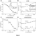
- Figure 16 shows the response to target concentration of a sensor with 1 ⁇ m particles, for a ssDNA competition assay in PBS and in filtered undiluted blood plasma.
- 1 ⁇ m particles were functionalized with particle-side binders via biotin-streptavidin interactions and DNA hybridization.
- the DBCO-tagged substrate-side binders were coupled to the PLL-g-PEG polymer via the integrated azide groups, using second generation click chemistry.
- the reversible 9 bp hybridization between substrate-side binders (which also functions as ssDNA analogues) and particle binders results in transient binding of particles.
- Figure 16A shows sensor response curves, i.e., the bound fraction and the switching activity as a function of target concentration, fitted with a Hill equation. The black and gray curves represent two consecutively measured dose-response curves with decreasing concentration series, which demonstrates the reversibility of the sensor and its suitability for monitoring applications.
- Figure 16B shows characteristic unbound state lifetimes dependent on the target concentration in a range of 10 to 2000 nM.
- Figure 16C shows the switching activity measured for the ssDNA target in 50 kDa spin-filtered bovine blood plasma.
- Figure 16D shows characteristic unbound and bound state lifetimes measured in filtered bovine blood plasma.
- Figure 17 shows a reversible sensor for the detection of sepsis biomarker procalcitonin (PCT) was demonstrated using an antibody sandwich immunoassay. Data are shown for two sensor devices. Glass substrates were functionalized with 100 nM capture antibody (c-Ab) through physisorption and subsequently blocked with 1 % BSA in PBS (blocking buffer). Streptavidin coated 2.8 ⁇ m Dynabeads were functionalized with 100 nM biotinylated detection antibody (dAb), blocked with 100 ⁇ M biotinylated PEG (1 kDa) and blocked with blocking buffer.
- dAb biotinylated detection antibody
- the d-Ab functionalized microparticles were diluted to 66 ⁇ g/mL in PBS with 0.1 % BSA (assay buffer) and injected into the c-Ab functionalized sensor surface.
- Analyte PCT was spiked in assay buffer and 30 ⁇ L solution was injected into the sensor flow chamber. Each injection was carried out with flow reversal, i.e. alternating supply in inlet or outlet, to minimize the loss of particles at the sensor active field-of-view. Washing was carried out with assay buffer injection, identical to PCT measurement, for minimum of three washes to reach baseline bound particle fraction (open symbols). Particle motion was tracked for 10 minutes at 60 Hz under brightfield illumination. The data clearly shows the monitoring functionality of the sensor, i.e. a sensor response to PCT concentration and a reversibility of the sensor.
- the biosensor may not be in direct contact with a system of interest, or may be in direct contact with a system of interest.
- the biosensor may be embedded or integrated or implanted in a system of interest.
- the biosensor can be placed at a distance from the system of interest.
- the biosensor may be located near the system of interest, on the system, wirelessly integrated, or the like. Samples can be put in a container and then transported to the biosensing system (sometimes called at-line or off-line operation), samples can be taken and automatically transported to the biosensing system (sometimes called on-line operation), or the biosensing system can be fully integrated with the system of interest (sometimes called in-line operation or bypass operation).
- the device or method may be connected to or integrated in an industrial system or process, a fermentor, a bioreactor, an on-body device, a catheter, an in-body device, a wearable device, or an insidable device.
- time-dependent samples can be taken, measurement data may be recorded, and a time profile may be established of analyte concentration as a function of time.
- a biosensor may be configured to receive a series of samples (from the same or from different sources) where the series of samples are serially measured on the biosensor and result in time-dependent data that relate to different samples that have been supplied to the biosensor.
- the device or method may be combined with a method or device module for sample pre-treatment or analyte pre-treatment, e.g. reagent addition, dilution, filtration, extraction, enrichment, purification, separation, amplification, change of buffer condition, stabilization, (dis)aggregation, or removal, modification, or addition of a chemical group or a biochemical domain or residue or moiety.
- a method or device module for sample pre-treatment or analyte pre-treatment e.g. reagent addition, dilution, filtration, extraction, enrichment, purification, separation, amplification, change of buffer condition, stabilization, (dis)aggregation, or removal, modification, or addition of a chemical group or a biochemical domain or residue or moiety.
- the device or method may be combined with a method or device module for optimization or control of operation, e.g. temperature, humidity, pressure, light conditions, vibration conditions, sound conditions, sterility, hygiene, ingress protection, cleaning, parts replacement, easy maintenance, calibration, and the like.
- a method or device module for optimization or control of operation, e.g. temperature, humidity, pressure, light conditions, vibration conditions, sound conditions, sterility, hygiene, ingress protection, cleaning, parts replacement, easy maintenance, calibration, and the like.
Landscapes
- Health & Medical Sciences (AREA)
- Chemical & Material Sciences (AREA)
- Life Sciences & Earth Sciences (AREA)
- Immunology (AREA)
- Engineering & Computer Science (AREA)
- Physics & Mathematics (AREA)
- Analytical Chemistry (AREA)
- Biochemistry (AREA)
- General Health & Medical Sciences (AREA)
- General Physics & Mathematics (AREA)
- Pathology (AREA)
- Dispersion Chemistry (AREA)
- Molecular Biology (AREA)
- Biomedical Technology (AREA)
- Hematology (AREA)
- Urology & Nephrology (AREA)
- Signal Processing (AREA)
- Biotechnology (AREA)
- Cell Biology (AREA)
- Microbiology (AREA)
- Food Science & Technology (AREA)
- Medicinal Chemistry (AREA)
- Measuring Or Testing Involving Enzymes Or Micro-Organisms (AREA)
- Apparatus Associated With Microorganisms And Enzymes (AREA)
Claims (15)
- Biosensorvorrichtung zum Wahrnehmen eines Analyten über einen Zeitraum unter Verwendung von Partikelbewegung, wobei die Biosensorvorrichtung eine Oberfläche und ein Partikel aufweist, wobei das Partikel und/oder die Oberfläche funktionalisiert sind/ist und wobei:- die Biosensorvorrichtung einen ersten Zustand, in dem das Partikel mit der Oberfläche assoziiert ist, und einen zweiten Zustand, in dem das Partikel nicht mit der Oberfläche assoziiert ist, aufweist und- Wechseln zwischen dem ersten und dem zweiten Zustand vom Vorliegen, Fehlen und/oder der Konzentration des Analyten abhängt,womit die Bewegungscharakteristiken des Partikels je nach Vorliegen, Fehlen und/oder Konzentration des Analyten veränderbar sind, wodurch Wahrnehmen des Analyten über das Messen von Änderungen eines Raumkoordinatenparameters des Partikels relativ zur Oberfläche gestattet wird,wobei die Eigenschaften des Partikels bzw. der Oberfläche so ausgewählt sind, dass sich das Partikel im zweiten Zustand in der Nähe der Oberfläche befindet, so dass der Biosensor Änderungen eines Raumkoordinatenparameters des Partikels relativ zur Oberfläche messen kann, vorzugsweise wobei der Abstand zwischen dem Partikel und der Oberfläche im zweiten Zustand im Bereich von 5 nm bis 10 µm liegt, undwobei das Partikel nicht an die Oberfläche konjugiert ist.
- Biosensorvorrichtung nach Anspruch 1, wobei der erste Zustand, in dem das Partikel mit der Oberfläche assoziiert ist, einen ersten Assoziationszustand und einen zweiten Assoziationszustand umfasst, wobei:- der erste Assoziationszustand eine Einzelmolekülbindung zwischen Partikel und Oberfläche enthält und- der zweite Assoziationszustand zwei oder mehr als zwei Einzelmolekülbindungen zwischen Partikel und Oberfläche enthält.
- Biosensorvorrichtung nach Anspruch 1 oder 2, wobei die Biosensorvorrichtung Folgendes umfasst:- wenigstens 10 Partikel, bevorzugter wenigstens 100 Partikel;- eine Dichte zwischen einigen wenigen und mehreren tausend Partikeln in einem Bereich von 415 × 415 µm2 und/oder- ein optisches System mit einer Beugungsgrenze, wobei die Biosensorvorrichtung Partikel umfasst, die von den nächstgelegenen Partikeln um wenigstens die Beugungsgrenze des optischen Systems getrennt sind.
- Biosensorvorrichtung nach einem der vorangehenden Ansprüche, wobei von der Biosensorvorrichtung ein Bindungs-Assay, ein kompetitiver Assay, ein Verdrängungs-Assay, ein Sandwich-Assay, ein Enzym-Assay, ein Assay mit Ziel- und/oder Signalamplifikation, ein mehrstufiger Assay oder ein Assay mit Molekülkaskade implementiert wird.
- Biosensorvorrichtung nach einem der vorhergehenden Ansprüche, wobei:- das Partikel durch eine erste Gruppierung funktionalisiert ist, wobei die erste Gruppierung an das Partikel gebunden ist; oder- die Oberfläche durch eine zweite Gruppierung funktionalisiert ist, wobei die zweite Gruppierung an die Oberfläche gebunden ist,wobei die Gruppierungen eine Bindungsaffinität zum Analyten aufweisen.
- Biosensorvorrichtung nach einem der Ansprüche 1-4, wobei:- das Partikel durch eine erste Gruppierung funktionalisiert ist, wobei die erste Gruppierung an das Partikel gebunden ist; und- die Oberfläche durch eine zweite Gruppierung funktionalisiert ist, wobei die zweite Gruppierung an die Oberfläche gebunden ist,wobei die Gruppierungen eine Bindungsaffinität zueinander je nach Vorliegen, Fehlen oder Konzentration des Analyten aufweisen.
- Biosensorvorrichtung nach Anspruch 5 oder 6, wobei sich die Dissoziationsgeschwindigkeitskonstante:- des Analyten und der ersten Gruppierung bezogen auf die des Analyten und der zweiten Gruppierung um einen Faktor von wenigstens 3, bevorzugt um einen Faktor von wenigstens 5 unterscheidet; oder- der ersten Gruppierung und der zweiten Gruppierung bezogen auf die des Analyten und der ersten Gruppierung und/oder bezogen auf die des Analyten und der zweiten Gruppierung um einen Faktor von wenigstens 3, bevorzugt um einen Faktor von wenigstens 5 unterscheidet.
- Biosensorvorrichtung nach einem der Ansprüche 5-7, wobei die Biosensorvorrichtung eine Gruppierungsdichte im Bereich zwischen 100 und 108 Gruppierungen/µm2 aufweist, vorzugsweise wobei die an das Partikel oder an die Oberfläche gebundenen Gruppierungen eine Dichte im Bereich zwischen 101 und 107 Gruppierungen/µm2, 102 und 106 Gruppierungen/µm2 oder 103 und 105 Gruppierungen/µm2 aufweisen.
- Biosensorvorrichtung nach einem der Ansprüche 5-8, wobei es sich bei der ersten Gruppierung oder der zweiten Gruppierung um ein Protein, einen Antikörper, ein Fragment davon, ein rekombinantes Protein, ein Peptid, ein Kohlenhydrat, ein Saccharid, ein molekular geprägtes Polymer, ein Kleinmolekül, eine Nukleinsäure, ein DNA-Molekül, ein PNA-Molekül, ein Aptamer, ein Nanobody, einen multivalenten Binder oder eine Kombination davon handelt, vorzugsweise wobei es sich bei der ersten Gruppierung oder der zweiten Gruppierung um ein Bindemolekül für Glucose, Elektrolyt, Metabolit, Kleinmolekül, Lipid, Kohlenhydrat, Peptid, Hormon, Arzneistoff, Arzneistoffmetabolit, Protein, Oligonukleotid, DNA, RNA, Nanopartikel, extrazelluläres Vesikel, Exosom, Nanosom, Liposom, Viruspartikel, Zelle, Zellfragment, supramolekulares Objekt oder Proteinaggregat handelt.
- Verwendung der Biosensorvorrichtung nach einem der vorhergehenden Ansprüche bei einem Verfahren zum Durchführen von Multiplexing, vorzugsweise Analyt-Multiplexing, Raum-Multiplexing, Spektroskopie-Multiplexing, Sondenfunktionalität-Multiplexing.
- Verwendung der Biosensorvorrichtung nach einem der Ansprüche 1-9 als Sensor auf oder in oder als Teil von einem System zur Wahrnehmung oder Überwachung, das ein Endoskop, einen Schlauch, eine Nadel, eine Faser, einen Katheter, ein Pflaster, eine Einwegsonde, eine Durchflusszelle oder eine Einwegkartusche enthält.
- Biosensorvorrichtung nach einem der Ansprüche 1 bis 9 zur Verwendung in der In-vivo-, Ex-vivo- oder In-vitro-Biosensorik, wie bei In-vitro-Diagnosetests, Point-of-Care-Tests, Umwelttests, Lebensmitteltests, Prozessüberwachung, Prozesskontrolle, Forensik, biologischer, biomedizinischer und pharmazeutischer Forschung, oder zur Überwachung von Assays mit lebenden Zellen, Gewebe oder einem Organ.
- Verfahren zum Wahrnehmen eines Analyten unter Verwendung von Partikelbewegung, wobei das Verfahren Folgendes umfasst:a) In-Kontakt-Bringen einer Matrix, die den Analyten enthält, mit der Biosensorvorrichtung nach einem der Ansprüche 1-9; undb) Nachweisen von Bewegungscharakteristiken des Partikels, die sich je nach Vorliegen, Fehlen und/oder Konzentration des Analyten ändern;wobei die Bewegungscharakteristiken einen Raumkoordinatenparameter des Partikels relativ zur Oberfläche umfassen.
- Verfahren nach Anspruch 13, wobei das Partikel:- so angeordnet ist, dass der Wechsel vom ersten zum zweiten Zustand mit einer mittleren effektiven Dissoziationszeit erfolgt; und- so angeordnet ist, dass der Wechsel vom zweiten zum ersten Zustand mit einer mittleren effektiven Assoziationszeit erfolgt, undwobei Schritt b) des Nachweisens von Bewegungscharakteristiken des Partikels über einen Zeitraum durchgeführt wird, der länger ist als die mittlere effektive Dissoziationszeit und/oder die mittlere effektive Assoziationszeit.
- Verfahren nach Anspruch 13 oder 14, wobei in Schritt b) die Richtung des Flusses der Matrix, die den Analyten enthält, ständig oder periodisch geändert wird, gegebenenfalls wobei die Flussänderung einer zufälligen Flussrichtungsänderung oder einer Flussrichtungsumkehränderung unterliegt.
Applications Claiming Priority (2)
| Application Number | Priority Date | Filing Date | Title |
|---|---|---|---|
| NL2026320 | 2020-08-21 | ||
| PCT/NL2021/050510 WO2022039594A1 (en) | 2020-08-21 | 2021-08-17 | Biosensor using particle motion |
Publications (3)
| Publication Number | Publication Date |
|---|---|
| EP4200591A1 EP4200591A1 (de) | 2023-06-28 |
| EP4200591B1 true EP4200591B1 (de) | 2024-11-20 |
| EP4200591C0 EP4200591C0 (de) | 2024-11-20 |
Family
ID=77774959
Family Applications (1)
| Application Number | Title | Priority Date | Filing Date |
|---|---|---|---|
| EP21770323.0A Active EP4200591B1 (de) | 2020-08-21 | 2021-08-17 | Biosensor mit partikelbewegung, verwendung und verfahren |
Country Status (4)
| Country | Link |
|---|---|
| EP (1) | EP4200591B1 (de) |
| JP (1) | JP7795283B2 (de) |
| CN (1) | CN116438439A (de) |
| WO (1) | WO2022039594A1 (de) |
Families Citing this family (1)
| Publication number | Priority date | Publication date | Assignee | Title |
|---|---|---|---|---|
| JP7145517B2 (ja) | 2017-03-08 | 2022-10-03 | ザ リージェンツ オブ ザ ユニバーシティ オブ ミシガン | 分析物の検出 |
Family Cites Families (7)
| Publication number | Priority date | Publication date | Assignee | Title |
|---|---|---|---|---|
| US6836559B2 (en) * | 2000-03-09 | 2004-12-28 | The Regents Of The University Of California | Automated video-microscopic imaging and data acquisition system for colloid deposition measurements |
| JP2011221009A (ja) * | 2010-03-25 | 2011-11-04 | Fujifilm Corp | 生体物質検出装置 |
| JP2013531787A (ja) * | 2010-05-25 | 2013-08-08 | アリックス インコーポレイテッド | 粒子の運動度および/または細胞の分散を求めるためのホログラフィック変動顕微鏡装置および方法 |
| EP3508853A1 (de) * | 2014-11-12 | 2019-07-10 | Technische Universiteit Eindhoven | Biosensor mit dynamischer umschaltung |
| JP6712999B2 (ja) * | 2014-12-16 | 2020-06-24 | セルダイナミクス アイ エス アール エル | 流体中の懸濁粒子のリアルタイム分析装置および該粒子の分析方法 |
| US10519486B2 (en) | 2014-12-16 | 2019-12-31 | Technische Universiteit Eindhoven | Biosensor based on a tethered particle |
| JP7203532B2 (ja) * | 2018-08-10 | 2023-01-13 | シスメックス株式会社 | 被検物質の検出方法 |
-
2021
- 2021-08-17 CN CN202180051285.XA patent/CN116438439A/zh active Pending
- 2021-08-17 EP EP21770323.0A patent/EP4200591B1/de active Active
- 2021-08-17 JP JP2023512696A patent/JP7795283B2/ja active Active
- 2021-08-17 WO PCT/NL2021/050510 patent/WO2022039594A1/en not_active Ceased
Also Published As
| Publication number | Publication date |
|---|---|
| EP4200591A1 (de) | 2023-06-28 |
| WO2022039594A1 (en) | 2022-02-24 |
| US20240044770A1 (en) | 2024-02-08 |
| JP7795283B2 (ja) | 2026-01-07 |
| JP2023539473A (ja) | 2023-09-14 |
| CN116438439A (zh) | 2023-07-14 |
| EP4200591C0 (de) | 2024-11-20 |
Similar Documents
| Publication | Publication Date | Title |
|---|---|---|
| Bashir | BioMEMS: state-of-the-art in detection, opportunities and prospects | |
| EP3523640B1 (de) | Vorrichtungen zur probenanalyse | |
| Patel et al. | Biosensors in health care: the milestones achieved in their development towards lab‐on‐chip‐analysis | |
| CN109863396B (zh) | 用于样品分析的装置和方法 | |
| Bhattacharya et al. | BioMEMS and nanotechnology‐based approaches for rapid detection of biological entities | |
| CN101438142B (zh) | 快速磁生物传感器 | |
| Morrison et al. | Clinical applications of micro-and nanoscale biosensors | |
| Shoji et al. | Spatially resolved chemical detection with a nanoneedle-probe-supported biological nanopore | |
| Rajpoot | Recent advances and applications of biosensors in novel technology | |
| Liu | Biosensors | |
| US20130130243A1 (en) | Method and device for detecting and quantifying an analyte with recycling of the reagents | |
| Halpin et al. | Direct plate-reader measurement of nitric oxide released from hypoxic erythrocytes flowing through a microfluidic device | |
| EP4200591B1 (de) | Biosensor mit partikelbewegung, verwendung und verfahren | |
| Arora et al. | Biosensors: way of diagnosis | |
| Kurbanoglu et al. | Nanobiodevices for electrochemical biosensing of pharmaceuticals | |
| US12553814B2 (en) | Biosensor using particle motion | |
| CN108414745A (zh) | 一种简单高效的可视化生物传感信号放大方法 | |
| EP1974211B1 (de) | Molekulare identifizierung über membrankonstruierte zellen | |
| Daştan et al. | Biosensors | |
| Pawar et al. | Polymers in Diagnostics | |
| Sett | Threading Precision: Recent Advances and Emerging Trends in Aptamer-Based Nanopore Sensing | |
| Dastan et al. | Biosensors 20 | |
| Marimuthu et al. | Microfluidics-integrated biosensor platform for modern clinical analysis | |
| Goli-Malekabadi¹ et al. | Biosensors for drug detection | |
| TR2023019410A1 (tr) | Elektroki̇myasal sensör |
Legal Events
| Date | Code | Title | Description |
|---|---|---|---|
| STAA | Information on the status of an ep patent application or granted ep patent |
Free format text: STATUS: UNKNOWN |
|
| STAA | Information on the status of an ep patent application or granted ep patent |
Free format text: STATUS: THE INTERNATIONAL PUBLICATION HAS BEEN MADE |
|
| PUAI | Public reference made under article 153(3) epc to a published international application that has entered the european phase |
Free format text: ORIGINAL CODE: 0009012 |
|
| STAA | Information on the status of an ep patent application or granted ep patent |
Free format text: STATUS: REQUEST FOR EXAMINATION WAS MADE |
|
| 17P | Request for examination filed |
Effective date: 20230130 |
|
| AK | Designated contracting states |
Kind code of ref document: A1 Designated state(s): AL AT BE BG CH CY CZ DE DK EE ES FI FR GB GR HR HU IE IS IT LI LT LU LV MC MK MT NL NO PL PT RO RS SE SI SK SM TR |
|
| DAV | Request for validation of the european patent (deleted) | ||
| DAX | Request for extension of the european patent (deleted) | ||
| REG | Reference to a national code |
Free format text: PREVIOUS MAIN CLASS: G01N0015100000 Ref country code: DE Ref legal event code: R079 Ref document number: 602021022137 Country of ref document: DE Free format text: PREVIOUS MAIN CLASS: G01N0015100000 Ipc: G01N0015103100 |
|
| GRAP | Despatch of communication of intention to grant a patent |
Free format text: ORIGINAL CODE: EPIDOSNIGR1 |
|
| STAA | Information on the status of an ep patent application or granted ep patent |
Free format text: STATUS: GRANT OF PATENT IS INTENDED |
|
| RIC1 | Information provided on ipc code assigned before grant |
Ipc: G01N 33/543 20060101ALI20240830BHEP Ipc: G01N 15/10 20060101ALI20240830BHEP Ipc: G01N 15/1433 20240101ALI20240830BHEP Ipc: G01N 15/1031 20240101AFI20240830BHEP |
|
| GRAS | Grant fee paid |
Free format text: ORIGINAL CODE: EPIDOSNIGR3 |
|
| INTG | Intention to grant announced |
Effective date: 20240911 |
|
| GRAA | (expected) grant |
Free format text: ORIGINAL CODE: 0009210 |
|
| STAA | Information on the status of an ep patent application or granted ep patent |
Free format text: STATUS: THE PATENT HAS BEEN GRANTED |
|
| AK | Designated contracting states |
Kind code of ref document: B1 Designated state(s): AL AT BE BG CH CY CZ DE DK EE ES FI FR GB GR HR HU IE IS IT LI LT LU LV MC MK MT NL NO PL PT RO RS SE SI SK SM TR |
|
| REG | Reference to a national code |
Ref country code: GB Ref legal event code: FG4D |
|
| REG | Reference to a national code |
Ref country code: CH Ref legal event code: EP |
|
| REG | Reference to a national code |
Ref country code: DE Ref legal event code: R096 Ref document number: 602021022137 Country of ref document: DE |
|
| REG | Reference to a national code |
Ref country code: IE Ref legal event code: FG4D |
|
| U01 | Request for unitary effect filed |
Effective date: 20241125 |
|
| U07 | Unitary effect registered |
Designated state(s): AT BE BG DE DK EE FI FR IT LT LU LV MT NL PT RO SE SI Effective date: 20241202 |
|
| PG25 | Lapsed in a contracting state [announced via postgrant information from national office to epo] |
Ref country code: IS Free format text: LAPSE BECAUSE OF FAILURE TO SUBMIT A TRANSLATION OF THE DESCRIPTION OR TO PAY THE FEE WITHIN THE PRESCRIBED TIME-LIMIT Effective date: 20250320 Ref country code: HR Free format text: LAPSE BECAUSE OF FAILURE TO SUBMIT A TRANSLATION OF THE DESCRIPTION OR TO PAY THE FEE WITHIN THE PRESCRIBED TIME-LIMIT Effective date: 20241120 |
|
| PG25 | Lapsed in a contracting state [announced via postgrant information from national office to epo] |
Ref country code: ES Free format text: LAPSE BECAUSE OF FAILURE TO SUBMIT A TRANSLATION OF THE DESCRIPTION OR TO PAY THE FEE WITHIN THE PRESCRIBED TIME-LIMIT Effective date: 20241120 |
|
| PG25 | Lapsed in a contracting state [announced via postgrant information from national office to epo] |
Ref country code: NO Free format text: LAPSE BECAUSE OF FAILURE TO SUBMIT A TRANSLATION OF THE DESCRIPTION OR TO PAY THE FEE WITHIN THE PRESCRIBED TIME-LIMIT Effective date: 20250220 |
|
| PG25 | Lapsed in a contracting state [announced via postgrant information from national office to epo] |
Ref country code: GR Free format text: LAPSE BECAUSE OF FAILURE TO SUBMIT A TRANSLATION OF THE DESCRIPTION OR TO PAY THE FEE WITHIN THE PRESCRIBED TIME-LIMIT Effective date: 20250221 |
|
| PG25 | Lapsed in a contracting state [announced via postgrant information from national office to epo] |
Ref country code: PL Free format text: LAPSE BECAUSE OF FAILURE TO SUBMIT A TRANSLATION OF THE DESCRIPTION OR TO PAY THE FEE WITHIN THE PRESCRIBED TIME-LIMIT Effective date: 20241120 |
|
| PG25 | Lapsed in a contracting state [announced via postgrant information from national office to epo] |
Ref country code: RS Free format text: LAPSE BECAUSE OF FAILURE TO SUBMIT A TRANSLATION OF THE DESCRIPTION OR TO PAY THE FEE WITHIN THE PRESCRIBED TIME-LIMIT Effective date: 20250220 |
|
| PG25 | Lapsed in a contracting state [announced via postgrant information from national office to epo] |
Ref country code: SM Free format text: LAPSE BECAUSE OF FAILURE TO SUBMIT A TRANSLATION OF THE DESCRIPTION OR TO PAY THE FEE WITHIN THE PRESCRIBED TIME-LIMIT Effective date: 20241120 |
|
| PG25 | Lapsed in a contracting state [announced via postgrant information from national office to epo] |
Ref country code: SK Free format text: LAPSE BECAUSE OF FAILURE TO SUBMIT A TRANSLATION OF THE DESCRIPTION OR TO PAY THE FEE WITHIN THE PRESCRIBED TIME-LIMIT Effective date: 20241120 |
|
| PG25 | Lapsed in a contracting state [announced via postgrant information from national office to epo] |
Ref country code: CZ Free format text: LAPSE BECAUSE OF FAILURE TO SUBMIT A TRANSLATION OF THE DESCRIPTION OR TO PAY THE FEE WITHIN THE PRESCRIBED TIME-LIMIT Effective date: 20241120 |
|
| PLBE | No opposition filed within time limit |
Free format text: ORIGINAL CODE: 0009261 |
|
| STAA | Information on the status of an ep patent application or granted ep patent |
Free format text: STATUS: NO OPPOSITION FILED WITHIN TIME LIMIT |
|
| U20 | Renewal fee for the european patent with unitary effect paid |
Year of fee payment: 5 Effective date: 20250827 |
|
| 26N | No opposition filed |
Effective date: 20250821 |
|
| GRAT | Correction requested after decision to grant or after decision to maintain patent in amended form |
Free format text: ORIGINAL CODE: EPIDOSNCDEC |
|
| REG | Reference to a national code |
Ref country code: CH Ref legal event code: Q17 Free format text: ST27 STATUS EVENT CODE: U-0-0-Q10-Q17 (AS PROVIDED BY THE NATIONAL OFFICE) Effective date: 20260211 |