EP4153038B1 - System zur muskelstimulation und/oder impedanzmessung zur überprüfung der korrekten platzierung des schlauches - Google Patents
System zur muskelstimulation und/oder impedanzmessung zur überprüfung der korrekten platzierung des schlauches Download PDFInfo
- Publication number
- EP4153038B1 EP4153038B1 EP21731335.2A EP21731335A EP4153038B1 EP 4153038 B1 EP4153038 B1 EP 4153038B1 EP 21731335 A EP21731335 A EP 21731335A EP 4153038 B1 EP4153038 B1 EP 4153038B1
- Authority
- EP
- European Patent Office
- Prior art keywords
- catheter
- electrode assembly
- stimulation
- recording electrode
- tissue
- Prior art date
- Legal status (The legal status is an assumption and is not a legal conclusion. Google has not performed a legal analysis and makes no representation as to the accuracy of the status listed.)
- Active
Links
Images


Classifications
-
- A—HUMAN NECESSITIES
- A61—MEDICAL OR VETERINARY SCIENCE; HYGIENE
- A61B—DIAGNOSIS; SURGERY; IDENTIFICATION
- A61B5/00—Measuring for diagnostic purposes; Identification of persons
- A61B5/06—Devices, other than using radiation, for detecting or locating foreign bodies ; Determining position of diagnostic devices within or on the body of the patient
- A61B5/065—Determining position of the probe employing exclusively positioning means located on or in the probe, e.g. using position sensors arranged on the probe
- A61B5/068—Determining position of the probe employing exclusively positioning means located on or in the probe, e.g. using position sensors arranged on the probe using impedance sensors
-
- A—HUMAN NECESSITIES
- A61—MEDICAL OR VETERINARY SCIENCE; HYGIENE
- A61B—DIAGNOSIS; SURGERY; IDENTIFICATION
- A61B5/00—Measuring for diagnostic purposes; Identification of persons
- A61B5/05—Detecting, measuring or recording for diagnosis by means of electric currents or magnetic fields; Measuring using microwaves or radio waves
- A61B5/053—Measuring electrical impedance or conductance of a portion of the body
- A61B5/0538—Measuring electrical impedance or conductance of a portion of the body invasively, e.g. using a catheter
-
- A—HUMAN NECESSITIES
- A61—MEDICAL OR VETERINARY SCIENCE; HYGIENE
- A61B—DIAGNOSIS; SURGERY; IDENTIFICATION
- A61B5/00—Measuring for diagnostic purposes; Identification of persons
- A61B5/06—Devices, other than using radiation, for detecting or locating foreign bodies ; Determining position of diagnostic devices within or on the body of the patient
- A61B5/065—Determining position of the probe employing exclusively positioning means located on or in the probe, e.g. using position sensors arranged on the probe
-
- A—HUMAN NECESSITIES
- A61—MEDICAL OR VETERINARY SCIENCE; HYGIENE
- A61B—DIAGNOSIS; SURGERY; IDENTIFICATION
- A61B5/00—Measuring for diagnostic purposes; Identification of persons
- A61B5/24—Detecting, measuring or recording bioelectric or biomagnetic signals of the body or parts thereof
- A61B5/316—Modalities, i.e. specific diagnostic methods
- A61B5/389—Electromyography [EMG]
- A61B5/392—Detecting gastrointestinal contractions
-
- A—HUMAN NECESSITIES
- A61—MEDICAL OR VETERINARY SCIENCE; HYGIENE
- A61B—DIAGNOSIS; SURGERY; IDENTIFICATION
- A61B5/00—Measuring for diagnostic purposes; Identification of persons
- A61B5/68—Arrangements of detecting, measuring or recording means, e.g. sensors, in relation to patient
- A61B5/6846—Arrangements of detecting, measuring or recording means, e.g. sensors, in relation to patient specially adapted to be brought in contact with an internal body part, i.e. invasive
- A61B5/6847—Arrangements of detecting, measuring or recording means, e.g. sensors, in relation to patient specially adapted to be brought in contact with an internal body part, i.e. invasive mounted on an invasive device
- A61B5/6852—Catheters
-
- A—HUMAN NECESSITIES
- A61—MEDICAL OR VETERINARY SCIENCE; HYGIENE
- A61J—CONTAINERS SPECIALLY ADAPTED FOR MEDICAL OR PHARMACEUTICAL PURPOSES; DEVICES OR METHODS SPECIALLY ADAPTED FOR BRINGING PHARMACEUTICAL PRODUCTS INTO PARTICULAR PHYSICAL OR ADMINISTERING FORMS; DEVICES FOR ADMINISTERING FOOD OR MEDICINES ORALLY; BABY COMFORTERS; DEVICES FOR RECEIVING SPITTLE
- A61J15/00—Feeding-tubes for therapeutic purposes
- A61J15/0003—Nasal or oral feeding-tubes, e.g. tube entering body through nose or mouth
-
- A—HUMAN NECESSITIES
- A61—MEDICAL OR VETERINARY SCIENCE; HYGIENE
- A61J—CONTAINERS SPECIALLY ADAPTED FOR MEDICAL OR PHARMACEUTICAL PURPOSES; DEVICES OR METHODS SPECIALLY ADAPTED FOR BRINGING PHARMACEUTICAL PRODUCTS INTO PARTICULAR PHYSICAL OR ADMINISTERING FORMS; DEVICES FOR ADMINISTERING FOOD OR MEDICINES ORALLY; BABY COMFORTERS; DEVICES FOR RECEIVING SPITTLE
- A61J15/00—Feeding-tubes for therapeutic purposes
- A61J15/0026—Parts, details or accessories for feeding-tubes
- A61J15/0069—Tubes feeding directly to the intestines, e.g. to the jejunum
-
- A—HUMAN NECESSITIES
- A61—MEDICAL OR VETERINARY SCIENCE; HYGIENE
- A61J—CONTAINERS SPECIALLY ADAPTED FOR MEDICAL OR PHARMACEUTICAL PURPOSES; DEVICES OR METHODS SPECIALLY ADAPTED FOR BRINGING PHARMACEUTICAL PRODUCTS INTO PARTICULAR PHYSICAL OR ADMINISTERING FORMS; DEVICES FOR ADMINISTERING FOOD OR MEDICINES ORALLY; BABY COMFORTERS; DEVICES FOR RECEIVING SPITTLE
- A61J15/00—Feeding-tubes for therapeutic purposes
- A61J15/0026—Parts, details or accessories for feeding-tubes
- A61J15/008—Sensor means, e.g. for sensing reflux, acidity or pressure
- A61J15/0084—Sensor means, e.g. for sensing reflux, acidity or pressure for sensing parameters related to the patient
-
- A—HUMAN NECESSITIES
- A61—MEDICAL OR VETERINARY SCIENCE; HYGIENE
- A61J—CONTAINERS SPECIALLY ADAPTED FOR MEDICAL OR PHARMACEUTICAL PURPOSES; DEVICES OR METHODS SPECIALLY ADAPTED FOR BRINGING PHARMACEUTICAL PRODUCTS INTO PARTICULAR PHYSICAL OR ADMINISTERING FORMS; DEVICES FOR ADMINISTERING FOOD OR MEDICINES ORALLY; BABY COMFORTERS; DEVICES FOR RECEIVING SPITTLE
- A61J15/00—Feeding-tubes for therapeutic purposes
- A61J15/0026—Parts, details or accessories for feeding-tubes
- A61J15/008—Sensor means, e.g. for sensing reflux, acidity or pressure
- A61J15/0088—Sensor means, e.g. for sensing reflux, acidity or pressure for sensing parameters related to the device
-
- A—HUMAN NECESSITIES
- A61—MEDICAL OR VETERINARY SCIENCE; HYGIENE
- A61J—CONTAINERS SPECIALLY ADAPTED FOR MEDICAL OR PHARMACEUTICAL PURPOSES; DEVICES OR METHODS SPECIALLY ADAPTED FOR BRINGING PHARMACEUTICAL PRODUCTS INTO PARTICULAR PHYSICAL OR ADMINISTERING FORMS; DEVICES FOR ADMINISTERING FOOD OR MEDICINES ORALLY; BABY COMFORTERS; DEVICES FOR RECEIVING SPITTLE
- A61J15/00—Feeding-tubes for therapeutic purposes
- A61J15/0026—Parts, details or accessories for feeding-tubes
- A61J15/0092—Valves on feeding tubes
-
- A—HUMAN NECESSITIES
- A61—MEDICAL OR VETERINARY SCIENCE; HYGIENE
- A61M—DEVICES FOR INTRODUCING MEDIA INTO, OR ONTO, THE BODY; DEVICES FOR TRANSDUCING BODY MEDIA OR FOR TAKING MEDIA FROM THE BODY; DEVICES FOR PRODUCING OR ENDING SLEEP OR STUPOR
- A61M25/00—Catheters; Hollow probes
- A61M25/01—Introducing, guiding, advancing, emplacing or holding catheters
- A61M25/0105—Steering means as part of the catheter or advancing means; Markers for positioning
-
- A—HUMAN NECESSITIES
- A61—MEDICAL OR VETERINARY SCIENCE; HYGIENE
- A61N—ELECTROTHERAPY; MAGNETOTHERAPY; RADIATION THERAPY; ULTRASOUND THERAPY
- A61N1/00—Electrotherapy; Circuits therefor
- A61N1/02—Details
- A61N1/04—Electrodes
- A61N1/05—Electrodes for implantation or insertion into the body, e.g. heart electrode
- A61N1/0507—Electrodes for the digestive system
-
- A—HUMAN NECESSITIES
- A61—MEDICAL OR VETERINARY SCIENCE; HYGIENE
- A61N—ELECTROTHERAPY; MAGNETOTHERAPY; RADIATION THERAPY; ULTRASOUND THERAPY
- A61N1/00—Electrotherapy; Circuits therefor
- A61N1/18—Applying electric currents by contact electrodes
- A61N1/32—Applying electric currents by contact electrodes alternating or intermittent currents
- A61N1/36—Applying electric currents by contact electrodes alternating or intermittent currents for stimulation
- A61N1/372—Arrangements in connection with the implantation of stimulators
- A61N1/37211—Means for communicating with stimulators
- A61N1/37235—Aspects of the external programmer
- A61N1/37247—User interfaces, e.g. input or presentation means
-
- A—HUMAN NECESSITIES
- A61—MEDICAL OR VETERINARY SCIENCE; HYGIENE
- A61N—ELECTROTHERAPY; MAGNETOTHERAPY; RADIATION THERAPY; ULTRASOUND THERAPY
- A61N1/00—Electrotherapy; Circuits therefor
- A61N1/18—Applying electric currents by contact electrodes
- A61N1/32—Applying electric currents by contact electrodes alternating or intermittent currents
- A61N1/36—Applying electric currents by contact electrodes alternating or intermittent currents for stimulation
- A61N1/372—Arrangements in connection with the implantation of stimulators
- A61N1/37211—Means for communicating with stimulators
- A61N1/37252—Details of algorithms or data aspects of communication system, e.g. handshaking, transmitting specific data or segmenting data
- A61N1/37258—Alerting the patient
-
- A—HUMAN NECESSITIES
- A61—MEDICAL OR VETERINARY SCIENCE; HYGIENE
- A61N—ELECTROTHERAPY; MAGNETOTHERAPY; RADIATION THERAPY; ULTRASOUND THERAPY
- A61N1/00—Electrotherapy; Circuits therefor
- A61N1/18—Applying electric currents by contact electrodes
- A61N1/32—Applying electric currents by contact electrodes alternating or intermittent currents
- A61N1/36—Applying electric currents by contact electrodes alternating or intermittent currents for stimulation
- A61N1/372—Arrangements in connection with the implantation of stimulators
- A61N1/375—Constructional arrangements, e.g. casings
- A61N1/3752—Details of casing-lead connections
- A61N1/3754—Feedthroughs
-
- A—HUMAN NECESSITIES
- A61—MEDICAL OR VETERINARY SCIENCE; HYGIENE
- A61N—ELECTROTHERAPY; MAGNETOTHERAPY; RADIATION THERAPY; ULTRASOUND THERAPY
- A61N1/00—Electrotherapy; Circuits therefor
- A61N1/18—Applying electric currents by contact electrodes
- A61N1/32—Applying electric currents by contact electrodes alternating or intermittent currents
- A61N1/36—Applying electric currents by contact electrodes alternating or intermittent currents for stimulation
- A61N1/372—Arrangements in connection with the implantation of stimulators
- A61N1/378—Electrical supply
-
- G—PHYSICS
- G16—INFORMATION AND COMMUNICATION TECHNOLOGY [ICT] SPECIALLY ADAPTED FOR SPECIFIC APPLICATION FIELDS
- G16H—HEALTHCARE INFORMATICS, i.e. INFORMATION AND COMMUNICATION TECHNOLOGY [ICT] SPECIALLY ADAPTED FOR THE HANDLING OR PROCESSING OF MEDICAL OR HEALTHCARE DATA
- G16H20/00—ICT specially adapted for therapies or health-improving plans, e.g. for handling prescriptions, for steering therapy or for monitoring patient compliance
- G16H20/10—ICT specially adapted for therapies or health-improving plans, e.g. for handling prescriptions, for steering therapy or for monitoring patient compliance relating to drugs or medications, e.g. for ensuring correct administration to patients
- G16H20/17—ICT specially adapted for therapies or health-improving plans, e.g. for handling prescriptions, for steering therapy or for monitoring patient compliance relating to drugs or medications, e.g. for ensuring correct administration to patients delivered via infusion or injection
-
- G—PHYSICS
- G16—INFORMATION AND COMMUNICATION TECHNOLOGY [ICT] SPECIALLY ADAPTED FOR SPECIFIC APPLICATION FIELDS
- G16H—HEALTHCARE INFORMATICS, i.e. INFORMATION AND COMMUNICATION TECHNOLOGY [ICT] SPECIALLY ADAPTED FOR THE HANDLING OR PROCESSING OF MEDICAL OR HEALTHCARE DATA
- G16H20/00—ICT specially adapted for therapies or health-improving plans, e.g. for handling prescriptions, for steering therapy or for monitoring patient compliance
- G16H20/30—ICT specially adapted for therapies or health-improving plans, e.g. for handling prescriptions, for steering therapy or for monitoring patient compliance relating to physical therapies or activities, e.g. physiotherapy, acupressure or exercising
-
- G—PHYSICS
- G16—INFORMATION AND COMMUNICATION TECHNOLOGY [ICT] SPECIALLY ADAPTED FOR SPECIFIC APPLICATION FIELDS
- G16H—HEALTHCARE INFORMATICS, i.e. INFORMATION AND COMMUNICATION TECHNOLOGY [ICT] SPECIALLY ADAPTED FOR THE HANDLING OR PROCESSING OF MEDICAL OR HEALTHCARE DATA
- G16H40/00—ICT specially adapted for the management or administration of healthcare resources or facilities; ICT specially adapted for the management or operation of medical equipment or devices
- G16H40/60—ICT specially adapted for the management or administration of healthcare resources or facilities; ICT specially adapted for the management or operation of medical equipment or devices for the operation of medical equipment or devices
- G16H40/63—ICT specially adapted for the management or administration of healthcare resources or facilities; ICT specially adapted for the management or operation of medical equipment or devices for the operation of medical equipment or devices for local operation
-
- A—HUMAN NECESSITIES
- A61—MEDICAL OR VETERINARY SCIENCE; HYGIENE
- A61B—DIAGNOSIS; SURGERY; IDENTIFICATION
- A61B5/00—Measuring for diagnostic purposes; Identification of persons
- A61B5/24—Detecting, measuring or recording bioelectric or biomagnetic signals of the body or parts thereof
- A61B5/316—Modalities, i.e. specific diagnostic methods
- A61B5/389—Electromyography [EMG]
- A61B5/395—Details of stimulation, e.g. nerve stimulation to elicit EMG response
-
- A—HUMAN NECESSITIES
- A61—MEDICAL OR VETERINARY SCIENCE; HYGIENE
- A61M—DEVICES FOR INTRODUCING MEDIA INTO, OR ONTO, THE BODY; DEVICES FOR TRANSDUCING BODY MEDIA OR FOR TAKING MEDIA FROM THE BODY; DEVICES FOR PRODUCING OR ENDING SLEEP OR STUPOR
- A61M25/00—Catheters; Hollow probes
- A61M25/01—Introducing, guiding, advancing, emplacing or holding catheters
- A61M25/0105—Steering means as part of the catheter or advancing means; Markers for positioning
- A61M2025/0166—Sensors, electrodes or the like for guiding the catheter to a target zone, e.g. image guided or magnetically guided
-
- A—HUMAN NECESSITIES
- A61—MEDICAL OR VETERINARY SCIENCE; HYGIENE
- A61M—DEVICES FOR INTRODUCING MEDIA INTO, OR ONTO, THE BODY; DEVICES FOR TRANSDUCING BODY MEDIA OR FOR TAKING MEDIA FROM THE BODY; DEVICES FOR PRODUCING OR ENDING SLEEP OR STUPOR
- A61M2210/00—Anatomical parts of the body
- A61M2210/10—Trunk
-
- A—HUMAN NECESSITIES
- A61—MEDICAL OR VETERINARY SCIENCE; HYGIENE
- A61N—ELECTROTHERAPY; MAGNETOTHERAPY; RADIATION THERAPY; ULTRASOUND THERAPY
- A61N1/00—Electrotherapy; Circuits therefor
- A61N1/02—Details
- A61N1/08—Arrangements or circuits for monitoring, protecting, controlling or indicating
- A61N2001/083—Monitoring integrity of contacts, e.g. by impedance measurement
Definitions
- catheters include a tube which is inserted into the human body. Certain catheters are inserted through the patient's nose or mouth for treating the digestive or gastrointestinal tract. These catheters, sometimes referred to as enteral catheters, typically include feeding tubes. The feeding tube lies in the stomach or intestines, and a feeding bag delivers liquid nutrient, liquid medicine or a combination of the two to the patient.
- X-ray machines In some cases, health care providers use X-ray machines to gather information about the location of the catheters within the body. There are several disadvantages with using X-ray machines. For example, X-ray machines are relatively large and heavy, consume a relatively large amount of energy and may expose the patient to a relatively high degree of radiation. Also, these machines are typically not readily accessible for use because, due to their size, they are usually installed in a special X-ray room. This room can be far away from the patient's room. Therefore, health care providers may find it inconvenient to use these machines for their catheter procedures. In addition, using X-ray technology is expensive and is a time-consuming task that can create unnecessary delays in delivering critical nutrients to the patient.
- WO2014105759 discloses stimulating one or more esophageal muscle contractions to evoke motion and/or restore function in one or more organs located distal to the lower esophageal sphincter.
- the present invention is directed to a tubing assembly.
- the tubing assembly includes a catheter having a proximal end and a distal end and extending in a longitudinal direction, wherein the proximal end and the distal end define a lumen therebetween, and wherein the catheter is configured for placement within a digestive tract of a patient; a stimulation electrode assembly, wherein the stimulation electrode assembly is configured to deliver an electrical stimulation to tissue; and an electrical connection for delivering a stimulation waveform to the stimulation electrode assembly.
- the stimulation electrode assembly can include an anode and a cathode, wherein the anode and the cathode are disposed on an outer wall of the distal end of the catheter.
- the stimulation electrode assembly can include a first electrode disposed on an outer wall of the catheter and a second electrode configured for placement on a surface of skin.
- the tubing assembly can also include a recording electrode assembly.
- the recording electrode assembly can include an active recording electrode, an inactive recording electrode, and a reference electrode. Further, the active recording electrode, the inactive recording electrode, and the reference electrode can be disposed on an outer wall of the distal end of the catheter. Alternatively, the active recording electrode, the inactive recording electrode, and the reference electrode can be configured for placement on a surface of skin.
- the recording electrode assembly can be configured to monitor for electrical activity elicited by the tissue in response to the stimulation waveform and communicate the electrical activity elicited by the tissue to a processor in real-time.
- the stimulation electrode assembly can be configured for a wired connection or a wireless connection to the processor.
- the wired connection can include a wire or a printed conduit.
- the present invention is directed to a catheter guidance system.
- the catheter guidance system includes (a) a processor; (b) a power source; (c) a stimulator; and (d) a tubing assembly.
- the tubing assembly includes a catheter having a proximal end and a distal end and extending in a longitudinal direction, wherein the proximal end and the distal end define a lumen therebetween; a stimulation electrode assembly, wherein the stimulation electrode assembly is configured to deliver an electrical stimulation to tissue; and a recording electrode assembly, wherein the recording electrode assembly is configured to monitor for electrical activity elicited by the tissue in response to the stimulation waveform, further wherein the recording electrode assembly communicates the electrical activity elicited by the tissue to the processor in real-time; wherein the catheter guidance system alerts a user as to correct placement of the catheter in a digestive tract of a patient or alerts the user as to incorrect placement of the catheter in a respiratory tract of the patient.
- the system can also include a display device, wherein the display device is coupled to the processor and displays a graph of the electrical activity elicited by the tissue and communicated to the processor by the recording electrode assembly.
- the system can also include a memory device storing instructions which, when executed by the processor, cause the processor to (i) interpret the electrical activity elicited by the tissue and communicated by the recording electrode assembly and (ii) cause the catheter guidance system to alert the user as to correct placement of the catheter in the digestive tract of the patient or alert the user as to incorrect placement of the catheter in the respiratory tract of the patient based on the interpretation of the electrical activity elicited by the tissue.
- the stimulation electrode assembly can include an anode and a cathode, wherein the anode and the cathode are disposed on an outer wall of the distal end of the catheter.
- the stimulation electrode assembly can include first electrode disposed on an outer wall of the catheter and a second electrode configured for placement on a surface of skin.
- the recording electrode assembly can include an active recording electrode, an inactive recording electrode, and a reference electrode. Further, the active recording electrode, the inactive recording electrode, and the reference electrode can be disposed on an outer wall of the distal end of the catheter. Alternatively, the active recording electrode, the inactive recording electrode, and the reference electrode can be configured for placement on a surface of skin.
- the terms "about,” “approximately,” or “generally,” when used to modify a value, indicates that the value can be raised or lowered by 5% and remain within the disclosed embodiment.
- any combination of a minimum value and a maximum value described in the plurality of ranges are contemplated by the present invention. For example, if ranges of “from about 20% to about 80%” and “from about 30% to about 70%” are described, a range of "from about 20% to about 70%” or a range of "from about 30% to about 80%” are also contemplated by the present invention.
- the present invention is directed to a tubing assembly that includes a catheter having a proximal end and a distal end and extending in a longitudinal direction, where the proximal end and the distal end define a lumen therebetween. Further, the catheter is configured for placement within a digestive tract of a patient.
- the tubing assembly also includes a stimulation electrode assembly that is configured to deliver an electrical stimulation to tissue (e.g., esophageal tissue such as striated muscle or tissue in the trachea such as cartilage, connective tissue, and the trachealis muscle), as well as an electrical connection for delivering a stimulation waveform to the stimulation electrode assembly.
- tissue e.g., esophageal tissue such as striated muscle or tissue in the trachea such as cartilage, connective tissue, and the trachealis muscle
- a catheter guidance system and method for accurately placing a catheter in the digestive tract are also provided.
- the tubing assembly contemplated by the present invention can also include a recording electrode assembly.
- the recording electrode assembly is configured to monitor for electrical activity elicited by the tissue in response to the stimulation waveform and communicate the electrical activity elicited by the tissue to a processor in real-time. For example, if the electrical activity elicited by the tissue in response to a stimulation waveform (e.g., a monophasic or biphasic square wave pulse) is in the form of one or more evoked potentials, then it can be confirmed that the catheter has been placed or inserted into the digestive tract.
- a stimulation waveform e.g., a monophasic or biphasic square wave pulse
- the electrical activity elicited by the tissue is not in the form of one or more evoked potentials, then it can be confirmed that the catheter is not placed in the digestive tract.
- the electrical activity elicited by the tissue is monitored by measuring the impedance of the tissue in response to a stimulation waveform (e.g., an electrical noise signal, a single frequency waveform, multiple frequency waveforms superimposed on top of one another, etc.), a first level of impedance can indicate that the catheter is placed in the digestive tract, while a second level of impedance can indicated that the catheter is not placed in the digestive tract.
- a stimulation waveform e.g., an electrical noise signal, a single frequency waveform, multiple frequency waveforms superimposed on top of one another, etc.
- tubing assembly catheter guidance system, and method described in more detail herein allow for the electrical activity elicited by tissue in response to a stimulation waveform delivered by the stimulation electrode assembly and captured in real-time via the recording electrode assembly can be used to determine if the distal end of the catheter is accurately placed within the digestive tract (e.g., the epiglottis, esophagus, stomach, intestines, etc.) rather than erroneously placed within the respiratory system (e.g., the trachea, bronchi, lungs, etc.), where such placement could be harmful and even fatal to a patient.
- the digestive tract e.g., the epiglottis, esophagus, stomach, intestines, etc.
- respiratory system e.g., the trachea, bronchi, lungs, etc.
- the present inventors have found that because the recording electrode assembly can obtain measurements and communicate those measurements to processor and ultimately a display device or other communication device (e.g., a phone, pager, etc.) in real time, the correct placement of the catheter can be confirmed within seconds of a catheter placement procedure, which can save valuable time, resources, and cost while at the same time limit patient risk in the event of the erroneous placement of the catheter.
- a display device or other communication device e.g., a phone, pager, etc.
- the present inventors have found that capturing and monitoring electrical activity elicited by tissue at a distal end of a catheter in response to a stimulation waveform in real-time, where the catheter is to be placed in a predetermined location along the digestive tract (e.g., esophagus, stomach, intestines, etc.), which is facilitated by the stimulation electrode assembly and the recording electrode assembly of the catheter guidance system of the present invention, allows for the efficient and accurate placement of the catheter within the digestive tract at a low cost.
- a predetermined location along the digestive tract e.g., esophagus, stomach, intestines, etc.
- the recording electrode assembly of the tubing assembly can capture data associated with electrical activity elicited by tissue in response to stimulation in the form an electrical pulse waveform or random noise signal waveform within the catheter as it is being directed by a health care provider in to the body of a patient, where the recorded electrical activity (e.g., in the form of evoked potentials, impedance of tissue measured between electrical contacts, etc.) can then be transmitted to a display device via a processor.
- the health care provider can then view the electrical activity elicited by the tissue on the display device to determine if the catheter has been accurately placed in the digestive tract or erroneously placed in an anatomical region of the respiratory system (e.g., the trachea, bronchi, lungs, etc.).
- a memory device that can include machine readable instructions and one or more computer programs (which, for example, may include a plurality of algorithms) can be used by the processor to process the data from the recording electrode assembly, where the display device can then indicate the catheter information to the health care provider in the form of a signal as to whether the catheter is accurately placed in the digestive tract or erroneously placed within, for instance, a portion of the respiratory system.
- a green check mark or the word "Yes” can be displayed on the screen to indicate accurate placement of the catheter within the digestive or gastrointestinal tract, while a red circle with a diagonal line through it, an "X", or the word "No” can be displayed on the screen for erroneous placement, such as placement within the respiratory system.
- the catheter guidance system 2 contemplated by the present invention includes: (a) an apparatus 10 having a housing 18 which supports a controller or processor 20 and a display device 22; (b) a power cord 27 that couples the apparatus 10 to a power source 25; (c) a printer 28 coupled to the apparatus 10 for printing out paper having graphics which indicate catheter location information; (d) an optional non-invasive movable receiver-transmitter or transceiver 32 electronically coupled to the processor 20 by a wire, cable, signal data connection or signal carrier 63; and (e) an invasive electronic catheter unit 12 in communication with and operatively coupled to the apparatus 10 by a wire, cable, cord or electrical extension 34, which, in turn, is operatively coupled to the processor 20, where the electronic catheter unit 12 includes a tubing assembly 14 that includes a catheter 50; a stimulation electrode assembly 46; a recording electrode assembly 48; a stimulator 128; and an optional signal generator 58 when the system 2 includes the optional non-
- the system 2 in one embodiment, includes: (a) a plurality of input devices 17 for providing input signals to the system 2 such as one or more control buttons 29, a touch screen 31, and the optional transceiver 32; (b) a stimulation electrode assembly 46; (c) a recording electrode assembly 48 that can continuously capture electrical activity elicited by tissue near the catheter 50 of the tubing assembly 14 in real-time; (d) a stimulator 128 which sends electrical signals in the form of a waveform via the stimulation electrode assembly 46; (e) an optional signal generator 58 which produces or generates electronic signals that are received by the transceiver 32; (f) a memory device 21 including machine readable instructions and one or more computer programs (which, for example, may include a plurality of algorithms 23) which are used by the processor 20 to process the electrical activity data captured by the recording electrode assembly 48 as well as to process the signal data produced by the signal generator 58 and transmitted by the transceiver 32 if present; (g) a data acquisition system
- the memory device 21 can store instructions which, when executed by the processor 20, cause the processor 20 to (i) interpret catheter 50 location and/or position information as determined and communicated by the recording electrode assembly 48 based on the tissue's response to stimulation delivered from the stimulator 128 via the stimulation electrode assembly 46 and the optional signal generating assembly 16 (including the signal generator 58 and the non-invasive transceiver 32), and (ii) cause the processor 20 to then instruct the system 2 to alert the health care provider either via the display device 22, auditory signals, etc. as to the accurate or inaccurate placement of the catheter 50.
- Health care providers can use the system 2 in a variety of catheter applications.
- the system 2 is used in an enteral application.
- a portion of the electronic catheter unit 12 is placed through an orifice 72 of the patient, such as the patient's nose or mouth.
- the distal end or tip 60 of the electronic catheter unit 12 can ultimately by positioned in the stomach 74.
- the stimulator 128 can be activated to deliver a stimulation waveform to the stimulation electrode assembly 46 while the recording electrode assembly 48 can continuously monitor for electrical activity elicited by tissue near the distal end 60 of the catheter 50 where at least one electrode in the stimulation electrode assembly 46 is positioned as the catheter 50 is inserted by the health care provider, as shown in Figs. 1 and 4 .
- the stimulation electrode assembly 46 and the recording electrode assembly 48 can each include a plurality of electrodes as will be discussed in more detail with respect to Figs.
- the display device 22 and/or the printer 28 can indicate information related to the location of the portion of the electronic catheter unit 12 within the body 78 based on the electrical activity data acquired by the recording electrode assembly 48, as well as information related to the shape of the pathway taken by the catheter unit 12 if the system includes a signal generating assembly 16 that utilizes the signal generator 58 and the associated non-invasive transceiver 32. It should be appreciated that the system 2 need not indicate the exact location or path of the catheter unit 12 to provide assistance to the health care provider.
- the electronic catheter unit 12 includes a tubing assembly 14, which includes the catheter 50, at least a portion of the stimulation electrode assembly 46, and optionally at least a portion of the recording electrode assembly 48.
- the catheter 50 includes a lumen 70 defined between a proximal end 162 and a distal end 164 to define a catheter body 160, where the catheter 50 can generally extend in the longitudinal direction L.
- at least a portion of the stimulation electrode assembly 46 and at least a portion of the recording electrode assembly 48 can be disposed on an outer wall 52 of the catheter, at a distal end 164 of the catheter 50 or tip 60 of the electronic catheter unit 12, as shown in Fig. 4 .
- a portion of the stimulation electrode assembly 46 and all or a portion of the recording electrode assembly 48 can be positioned on a surface of skin 98 so long as the stimulation waveform delivered to an area of tissue by the stimulation electrode assembly 46 can reach the area of tissue at levels sufficient to elicit an electrical response and the electrical activity elicited by tissue in response to the stimulation waveform can be adequately recorded by the recording electrode assembly 48.
- a surface electrode 46c of the stimulation electrode assembly 46 and one or more electrodes of the recording electrode assembly 48 can be located on a surface of skin 98.
- the surface electrode 46c can be used when the electrical stimulation waveform is delivered by the stimulation electrode assembly 46 in monopolar fashion, while a cathode 46a and an anode 46b disposed on an outer wall 52 of the catheter 50 can be used in the electrical stimulation waveform delivered by the stimulation electrode assembly 46 is delivered in bipolar fashion.
- the recording electrode assembly 48 can include an active recording electrode 48a, an inactive recording electrode 48b, and a reference electrode 46c, each of which can also be disposed on the outer wall 52 of the catheter anywhere along its length or can be disposed on a surface of skin 98.
- the electrodes when any of the electrodes or located on the outer wall 52 of the catheter 50, the electrodes (e.g., the stimulation electrode assembly 46 and/or recording electrode assembly 48) can be electrically connected to the apparatus 10 via an electrode electrical connection 68, which can be in the form of a wire or printed conduit.
- the connection can be wireless.
- the electrodes when any of the electrodes are located on a surface of skin 98 for transcutaneous stimulation and/or recording, the electrodes (e.g., the stimulation electrode assembly 46 and/or recording electrode assembly 48) can be electrically connected to the apparatus 10 via a wire 56.
- the connection can be wireless.
- the stimulation electrode assembly 46 can be disposed on the outer wall 52 of the catheter 50 at distal end 164 of the catheter 50, while the recording electrode assembly 48 can also be disposed at the distal end 164 or can be disposed anywhere along the length of the catheter 50 between the proximal end 162 and the distal end 164.
- the cathode 46a and the anode 46b of the stimulation electrode assembly 46 and the active recording electrode 48a, the inactive recording electrode 48b, and the reference electrode 48c of the recording electrode assembly 48 can each have a width W ranging from about 0.1 millimeters to about 10 millimeters, such as from about 0.25 millimeters to about 5 millimeters, such as from about 0.5 millimeters to about 2.5 millimeters and can encircle all or a portion of the catheter wall 52 in the form of a ring, a stent-like embodiment, an expandable balloon, etc.
- the cathode 46a and the anode 46b can be separated by a distance D1 ranging from about 1 millimeter to about 10 millimeters, such as from about 2 millimeters to about 8 millimeters, such as from about 4 millimeters to about 6 millimeters.
- the stimulation electrode assembly 46 and the recording electrode assembly can be separated by a distance D2 ranging from about 2 millimeters to about 20 millimeters, such as from about 4 millimeters to about 16 millimeters, such as from about 8 millimeters to about 12 millimeters.
- the active recording electrode 48a and the reference electrode 48c as well as the inactive recording electrode 48b and the reference electrode 48c, can be separated by a distance D3 ranging from about 1 millimeter to about 10 millimeters, such as from about 2 millimeters to about 8 millimeters, such as from about 4 millimeters to about 6 millimeters.
- the components of the stimulation electrode assembly 46, the recording electrode assembly 48, and any other electrical components can be formed from MRI compatible materials such that the catheter guidance system 2 can be used in patients undergoing MRI or other diagnostic testing where magnetic components cannot be used.
- MRI compatible materials such that the catheter guidance system 2 can be used in patients undergoing MRI or other diagnostic testing where magnetic components cannot be used.
- carbon or any other non-magnetic materials can be used.
- the tubing assembly 14 can also include (a) a tube or an electrical tubular insulator 40; (b) a mid-connector or union device 42 which receives the tubular insulator 40; (c) a multi-port connector or y-port connector 44 attachable to the union device 42; (d) a catheter 50, such as a feeding tube, connected to the y-port connector 44; and (e) a distal end or tip 60 of the catheter 50, where at least a portion of the stimulation electrode assembly 46 and optionally the recording electrode assembly 48 can be located on an outer wall 52 of the catheter 50 at the distal end
- the tubular insulator 40 includes a tube having a proximal end 100 attachable to an attachment member or neck 108 of a controller coupler or electrical connector 36 and a distal end 102 receivable by the union device 42; and an internal diameter which is substantially equal to or greater than an external diameter of a wire assembly 62 and the electrode electrical connection 68, which can serve as the hard wired electrical connection between the portions of the stimulation electrode assembly 46 and the recording electrode assembly 48 present on the outer wall 52 of the catheter 50 signal generator 58 and the processor 20, so as to slide over the electrical electrode connection 68 and the wire assembly 62.
- the tubular insulator 40 may fit relatively tightly over the electrical electrode connection 68 and the wire assembly 62.
- the union device 42 includes: (a) a proximal end 116; (b) a distal end 118; (c) a position adjuster, extender or elongated neck 120 positioned between the proximal end 116 and the distal end 118; (d) a grasp or gripping member 122 positioned adjacent to the distal end 118 so as to assist users in grasping and manipulating the union device 42; and (e) an insert 124 positioned adjacent to the gripping member 122 which is received by the y-port connector 44.
- the proximal end 116 of the union device 42 is coupled to the distal end 102 of the tubular insulator 40.
- the multi-port or y-port connector 44 includes: (a) a body 140; (b) a liquid delivery branch, medicine delivery branch or medicine branch 142 attached to the body 140 for distributing drugs, medicine or other medicinal liquids to the patient; (c) a nutrient delivery branch or feeding branch 144 attached to the body 140 and sized to receive the insert 124 of the union device 42; (d) a catheter or feeding tube connection branch 146 attached to the catheter 50; (e) a flexible or movable arm 148 attached to the body 140; and (f) a flexible or movable arm 150 attached to the body 140.
- y-port connector 44 includes additional branches for administering various nutrients or medicines to the body 78.
- the y-port connector 44 includes only a feeding branch 144 and a connection branch 146.
- the arm 148 has a stopper 152
- the arm 150 has a stopper 154.
- the stoppers 152 and 154 are sized to prevent fluid from passing through the branches 142 and 144 after such branches 142 and 144 are plugged with stoppers 152 and 154, respectively.
- the arm 150 includes a fastener 155 which secures a tube-size adapter 156 to the arm 150.
- the tube-size adapter 156 enables fluid delivery tubes (not shown) having various diameters to connect to the feeding branch 144 of the y-port connector 44.
- the catheter 50 includes a feeding tube or catheter 50 with a body 160 having a proximal end 162 attached to the catheter connection branch 146 of the y-port connector 44 and a distal end 164.
- the proximal end 162 is insertable into the catheter connection branch 146 of the y-port connector 44 so as to bring the catheter 50 into fluid communication with the y-port connector 44.
- the end member, bolus or tip 60 is attached to the distal end 164 of the catheter 50.
- the tip 60 includes a body 172 having a collar 174 and an end member 176.
- the body 172 defines a passage 178 and an opening 180.
- the opening 180 is positioned between the collar 174 and the end member 176.
- a portion 177 of the end member 176 can have a rounded shape.
- the shape of the passage 178 and opening 180 of the tip 60 is configured to facilitate the flow of fluid from the catheter 50 into the patient's body while decreasing the likelihood that the opening 180 will become clogged.
- the tubular connector 40, union device 42, y-port connector 44, catheter 50, and tip 60 can be made from any suitable polymer or plastic material including, but not limited to, polyamide, polyethylene, polypropylene, polyurethane, silicone and polyacrylonitrile.
- a controller coupler or an electrical connector 36 can be operatively connected to the electrical extension 34 and an electrode electrical connection 68 can be operatively coupled to the electrical connector 36 to form a wired connection between the electrode assemblies and the processor 20 and any other electrical components of the system 2.
- an elongated wire assembly 62 can be operatively coupled to the electrical connector 36 to form a wired connection between the signal generator 58 and the processor 20, although it is to be understood that all of the electrical connections can be wireless.
- a wire or elongated stiffener 39 can be attached to the connector 36 and can serve as a support for the wire assembly 62 when it is inserted into the body 160 of the catheter 50.
- the tubular insulator 40 described above can cover a portion 41 of the wire assembly 62 and the electrode electrical connection 48 positioned adjacent to the connector 36.
- the electrical connector or controller coupler 36 can provide the electrical connection between the apparatus 10 and the stimulation electrode assembly 46 as well as the recording electrode assembly 48 when the assemblies are hard wired to the catheter guidance system 2 via the electrode electrical connection 68.
- the catheter body 160 can have a plurality of markings 112 uniformly spaced along its external surface that can be used in conjunction with the stimulation electrode assembly 46 and the recording electrode assembly 48 to determine accurate placement of the catheter 50 and/or to determine the appropriate time during which to deliver the stimulation waveform from the stimulator 128 via the stimulation electrode assembly 46 and to initiate recording of the electrical activity elicited by tissue near the esophagus 91 of a patient via the recording electrode assembly 48.
- These markings 112 can function as placement markers which assist the user in assessing the depth that the catheter 50 is placed within the patient's body 78.
- the markings 112 can be present from the distal end 60 of the electronic catheter unit 12 or the distal end 164 of the catheter 50 to a point 126 on the catheter 50 that spans a distance that can correspond with the average distance between the trachea 92 and nostril 87 in a typical patient.
- the user can be alerted to start delivering an electrical stimulation waveform from the stimulator 128 via the stimulating electrode assembly 46 and to start monitoring the graphs 37 on the display device 22 to observe the data recorded by the recording electrode assembly 48 related to the electrical activity elicited by tissue in response to the stimulation or to start monitoring for a visual indication, auditory indication, or both that the catheter 50 has be inserted into the correct (e.g., digestive tract) or incorrect location (e.g., respiratory tract).
- the correct e.g., digestive tract
- incorrect location e.g., respiratory tract
- these markings 112 can assist the user in measuring the flow or distribution of liquid to or from the patient.
- the method for determining if the catheter 50 is accurately placed within a digestive tract includes inserting a distal end of the tubing assembly 14 (e.g., the distal end 164 of the catheter 50 or tip 60 of the electronic catheter unit 12) into an orifice 72 of the body 78, such as a nostril 87 of the patient's nose.
- the tubing assembly 14 can include the catheter 50, at least a portion of the stimulation electrode assembly 46 (part of which can be placed transcutaneously on a surface of skin 98), the recording electrode assembly 48 (or, alternatively all or a portion of the recording electrode assembly 48 can be placed transcutaneously on a surface of skin 98) and the optional signal generator 58.
- the stimulation electrode assembly 46, the recording electrode assembly 48, and the optional signal generator 58 can be electrically connected to a processor 20 via a wired connection, such as the electrode electrical connection 68 and the wire assembly 62, although a wireless connection is also contemplated by the present invention such that no electrode electrical connection 68, wire assembly, 62 or controller coupler 36 is required.
- a wired connection such as the electrode electrical connection 68 and the wire assembly 62
- a wireless connection is also contemplated by the present invention such that no electrode electrical connection 68, wire assembly, 62 or controller coupler 36 is required.
- the stimulation electrode assembly 46 is activated, such as by providing power to stimulator 128 connected to the stimulation electrode assembly 46, and the stimulation electrode assembly 46 then begins to deliver a stimulation waveform to tissue near the distal end 164 of the catheter/the tip or distal end 60 of the electronic catheter unit 12 via the stimulation electrode assembly 46.
- the recording electrode assembly 48 can be activated to begin to record the electrical activity (e.g., muscle activity or twitches in the form of action potentials, impedance measurements, etc.) elicited by the tissue in response to the stimulation waveform.
- the recording electrode assembly 48 then communicates with the processor 20 via the wired connection (e.g., connection 68) or the wireless connection to deliver the acquired electrical activity data to the processor 20 in real-time.
- the stimulation waveform can have various features depending on the electrical activity to be monitored. For instance, when monitoring for electrical muscle activity in response to stimulation, where the muscle activity is in the form of evoked potentials, indicating activation of muscle tissue associated with the presence of the distal end 60 of the electronic catheter unit 12 in the esophagus 91, which contains striated muscle, or where there is insufficient activation of muscle tissue, indicating that the distal end 60 of the electronic catheter unit 12 has been inserted into the trachea, which contains cartilage such that no evoked potential is present, the stimulation waveform can be a constant-current or constant voltage square waveform (monophasic or biphasic).
- the stimulation waveform can have a stimulation amplitude ranging from greater than 1 volt to less than 150 volts, such as from about 1.01 volts to about 100 volts, such as from about 1.05 volts to about 50 volts, such as from 1.1 volts to about 25 volts.
- the stimulation waveform can have a stimulation waveform of less than 50 milliamps, such as from about 0.5 milliamps to about 50 milliamps, such as from about 1 milliamp to about 25 milliamps, such as from about 1.5 milliamps to about 10 milliamps.
- the stimulation waveform can have a frequency ranging from about 0.01 hertz to about 100 hertz, such as from about 0.1 hertz to about 75 hertz, such as from about 0.5 hertz to about 50 hertz.
- the stimulation waveform can have a pulse width of less than 25 milliseconds, such as from about 0.5 milliseconds to about 20 milliseconds, such as from about 1 millisecond to about 15 milliseconds, such as from about 5 milliseconds to about 10 milliseconds.
- the stimulation waveform can be in the form of a noise signal having a frequency ranging from greater than 0 hertz to about 500 kilohertz, such as from about 1 kilohertz to about 300 kilohertz, such as from about 5 kilohertz to about 150 kilohertz, such as from about 10 kilohertz to about 125 kilohertz, and with the same stimulation amplitudes as described above.
- the impedance of esophageal tissue and tissue in the trachea have distinct characteristics, it can then be determined if the distal end 164 of the catheter 50/the distal end or tip 60 of the electronic catheter unit 12 is positioned in the esophagus or trachea.
- a display device 22 can be coupled to the processor 20 and displays the electrical activity data communicated to the processor 20 by the recording electrode assembly 48 for a health care provider to use during the catheter insertion procedure, where the data may first pass through an amplifier 134 to amplify the frequencies of interest and through a data acquisition system 132 to digitize the recorded signals. The data can then be presented on the display device 22, where differences in the responses recorded by the recording electrode assembly 48 in response to the stimulation waveforms delivered by the stimulation electrode assembly 46 associated with catheter insertion into the digestive tract and into the respiratory tract can be easily identified by the health care provider via the graphs 37 on the display device 22.
- the memory device 21 can store instructions which, when executed by the processor 20, cause the processor 20 to interpret catheter 50 location and/or position information as determined and communicated by the optional signal generating assembly 16 and the non-invasive transceiver 32 and cause the processor 20 to then instruct the system 2 to alert the health care provider either via the display device 22, auditory signals, etc. as to the accurate or inaccurate placement of the catheter 50.
- the appearance of evoked potentials on an amplitude versus time graph 37 that can be shown on the display device 22 can indicate placement of the catheter 50 in the digestive tract, where the evoked potentials are associated with activation of the muscle in the esophagus 91 near the distal end 60 of the electronic catheter unit 12.
- the lack of appearance of such evoked potentials upon delivery of the square-wave pulsed waveform on an amplitude versus time graph 37 shown on the display device 22 can indicate erroneous placement of the catheter in the respiratory system (e.g., the trachea 92, bronchi 93, lungs 94, etc., or other anatomical region of the respiratory tract of the patient) at which time the insertion procedure should be stopped immediately and the tubing assembly 14 be removed from the respiratory tract to avoid potential harm to the patient.
- the catheter in the respiratory system e.g., the trachea 92, bronchi 93, lungs 94, etc., or other anatomical region of the respiratory tract of the patient
- a memory device 21 stores instructions which, when executed by the processor 20, cause the processor 20 to (i) interpret the data communicated by the recording electrode assembly 48 and (ii) cause the display device 22 to communicate whether or not the catheter 50 is accurately placed within the digestive tract of the patient based on the interpretation of the electrical activity data.
- the impedance measurements shown on an impedance versus time graph 37 on the display device 22 can indicate accurate placement of the catheter 50 in the digestive tract (e.g., the esophagus 91) or erroneous placement in the respiratory system (e.g., the trachea 92, bronchi 93, lungs 94, etc., or other anatomical region of the respiratory tract of the patient) at which time the insertion procedure should be stopped immediately and the tubing assembly 14 be removed from the respiratory tract to avoid potential harm to the patient.
- the digestive tract e.g., the esophagus 91
- erroneous placement in the respiratory system e.g., the trachea 92, bronchi 93, lungs 94, etc., or other anatomical region of the respiratory tract of the patient
- a memory device 21 stores instructions which, when executed by the processor 20, cause the processor 20 to (i) interpret the data communicated by the recording electrode assembly 48 and (ii) cause the display device 22 to communicate whether or not the catheter 50 is accurately placed within the digestive tract of the patient based on the interpretation of the electrical activity data.
- the present inventors have found that the distinctions between the electrical activity elicited by tissue (e.g., electrical activity elicited by muscle in the esophagus versus electrical activity elicited by cartilage and/or muscle in the trachea) propagating from the opening 180 at the distal end 60 of the electronic catheter unit 12 and catheter 50 when the distal end or tip 60 of the electronic catheter unit 12 or catheter 50 is placed within the digestive tract or respiratory system are allow for an efficient and possibly life-saving determination of accurate enteral feeding catheter 50 placement in the digestive tract, where erroneously placing the catheter in the respiratory system would deliver fluid into the lungs, which can have fatal consequences.
- tissue e.g., electrical activity elicited by muscle in the esophagus
- cartilage and/or muscle in the trachea electrical activity propagating from the opening 180 at the distal end 60 of the electronic catheter unit 12 and catheter 50 when the distal end or tip 60 of the electronic catheter unit 12 or catheter 50 is placed within the digestive tract or respiratory system
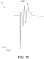
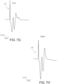
- Figs. 7A through 7H when the distal end or tip 60 of the electronic catheter unit 12 and catheter 50 is inserted into the nostril 87 of the patient and is advanced through the nasal cavity 88, past the nasopharynx 89, and into the esophagus 91 just past the epiglottis 90, as the recording electrode assembly 48 is continuously receiving, recording, and/or processing electrical activity elicited by tissue near the distal end 60 of the electronic catheter unit 12 and catheter 50 where the stimulation electrode assembly 46 is positioned or disposed, the amplitude versus time spectrogram graph 37 (Fig.
- the processor 20 may show the presence of evoked potentials as the distal end or tip 60 of the electronic catheter unit 12 or catheter 50 travels into the digestive tract and not into the respiratory system, so long as the stimulation waveform amplitude is above a certain threshold.
- the stimulation amplitude of 1 volt in Fig. 7B was insufficient to elicit an evoked potential by the muscle in the esophagus 91 in response to the stimulation waveform, although the stimulation amplitudes of 1.1 volts ( Fig. 7C ), 1.25 volts ( Fig. 7D ), 1.5 volts ( Fig. 7E ), 2.25 volts ( Fig.
- Figs. 8A and 8B when the distal end or tip 60 of the catheter 50 is inserted into the nostril 87 of the patient and is advanced through the nasal cavity 88, past the nasopharynx 89, and into the trachea 92 just past the epiglottis 90, and then into the bronchi 93 or lungs 94, as the recording electrode assembly 48 is continuously receiving, recording, and/or processing electrical activity elicited by tissue near the distal end 60 of the electronic catheter unit 12 and catheter 50 where the stimulation electrode assembly 46 is positioned or disposed, the amplitude versus time spectrogram graph 37 ( Fig.
- the stimulation amplitude of 7.25 volts in Fig. 8B was insufficient to elicit an evoked potential in response to the stimulation waveform, although the stimulation amplitudes of 1.1 volts ( Fig. 7C ), 1.25 volts ( Fig. 7D ), 1.5 volts ( Fig. 7E ), 2.25 volts ( Fig. 7F ), 4.5 volts, reversed polarity ( Fig.
- the present inventors have also found that the distinctions between the impedance measurements of tissue collected by the various electrical electrodes on the catheter (e.g., impedance data collected from electrodes placed in the esophagus versus impedance data collected from electrodes placed in the trachea) allow for an efficient and possibly life-saving determination of accurate enteral feeding catheter 50 placement in the digestive tract, where erroneously placing the catheter in the respiratory system would deliver fluid into the lungs, which can have fatal consequences.
- Fig. 9A is a graphical view of the impedance of tissue measured at an electrical contact (e.g., any of the electrodes that are part of the stimulation electrode assembly 46 or the recording electrode assembly 48) when placed on the electronic catheter unit 12 or the catheter 50 at various frequencies as the electronic catheter unit 12 and catheter 50 of Fig. 7A is inserted past the epiglottis in real-time, where the characteristics of the impedance values measured are in response to a noise signal that is generated by the stimulator and delivered through the contacts onto the catheter and are indicative of the placement of the catheter within the esophagus.
- an electrical contact e.g., any of the electrodes that are part of the stimulation electrode assembly 46 or the recording electrode assembly 48
- the impedance values (in Ohms) over a range of frequencies ranging from greater than 0 hertz to about 50,000 hertz show an impedance signature indicative of various peaks and valleys characteristic of esophageal tissue.
- Fig. 9A shows the impedance values measured when a noise signal is delivered to the tissue, it is also to be understood that that impedance can be measured at a single frequency (e.g., 5 kilohertz, 10 kilohertz, 25 kilohertz, 50 kilohertz, etc.), or can be measured when multiple frequencies are superimposed and delivered as one waveform (e.g., multiple sine waves delivered from the stimulator 128).
- Fig. 9B is a graphical view of the impedance measured at an electrical contact (e.g., any of the electrodes that are part of the stimulation electrode assembly 46 or the recording electrode assembly 48) when placed on the electronic catheter unit 12 or the catheter 50 at various frequencies as the electronic catheter unit 12 and catheter 50 of Fig. 8A is inserted past the epiglottis in real-time, where the characteristics of the impedance values measured are in response to a noise signal that is generated by the stimulator and delivered through the contacts onto the catheter and are indicative of erroneous placement of the catheter in the trachea.
- an electrical contact e.g., any of the electrodes that are part of the stimulation electrode assembly 46 or the recording electrode assembly 48
- the impedance values in Ohms
- Fig. 9B shows the impedance values measured when a noise signal is delivered to the tissue
- that impedance can be measured at a single frequency (e.g., 5 kilohertz, 10 kilohertz, 25 kilohertz, 50 kilohertz, etc.), or can be measured when multiple frequencies are superimposed and delivered as one waveform (e.g., multiple sine waves delivered from the stimulator 128).
- the impedance values at various electrical contacts can vary depending on the location of the catheter 50.
- tissue e.g., esophageal tissue or tracheal tissue
- the impedance can be between 500 ohms and 600 ohms.
- the impedance when the impedance value is determined between the active recording electrode 48a and the reference electrode 48c of the recording electrode assembly 48, the impedance can be between 900 ohms and 1000 ohms. Further, when the impedance value is determined between the inactive recording electrode 48b and the reference electrode 48c, the impedance can be between 950 ohms and 1050 ohms. In addition, when the impedance value is determined between the active recording electrode 48a and the inactive recording electrode 48b, the impedance can be between about 1000 ohms and 1100 ohms. Thus, regardless of the particular electrical contacts used when determining the impedance of tissue in the esophagus (e.g., epithelium, muscle, etc.), the impedance of the esophageal tissue is generally consistent.
- the impedance value is determined between either between the cathode 46a and the anode 46b of the stimulation electrode assembly 46, between the active recording electrode 48a and the reference electrode 48c of the recording electrode assembly 48, between the inactive recording electrode 48b and the reference electrode 48c, or between the active recording electrode 48a and the inactive recording electrode 48b, the impedance is around 2000 ohms and is intermittent. This illustrates that the impedance signature collected from electrodes placed in the esophagus is distinct from the impedance signature collected from electrodes placed in the trachea, such that impedance measurements in response to a stimulation waveform can be utilized to determine the proper placement of a catheter in the respiratory tract.
- the present inventors have found that the impedance signature of tissue in the trachea is minimal or exponentially decaying due to lack of contact with excitable tissue since the trachea is rigid due to the presence of cartilaginous tissue. Meanwhile, the impedance of esophageal tissue is generally consistent and has increased values due to the excitability of tissue in the esophagus (e.g., muscle capable of contracting).
- the ratio of the impedance collected in the trachea to the impedance collected in the esophagus can be at least about 1.1;1, such as from about 1.25:1 to about 5:1, such as from about 1.5:1 to 4:1, such as from about 1.75:1 to about 3:1.
- the health care provider can also verify accurate placement of the catheter 50 in the esophagus 91 rather than the trachea 92 by observing for the presence or absence of a plurality of markings 112 uniformly spaced along the external surface of the catheter 50.
- markings 112 can be used in conjunction with the stimulation electrode assembly 46 and the recording electrode assembly 48 to determine accurate placement of the catheter 50.
- These markings 112 can function as placement markers which assist the user in assessing the depth that the catheter 50 is placed within the body 78.
- the markings 112 can be present from the distal end 60 of the catheter 50 to a point 126 on the catheter 50 that spans a distance that can correspond with the average distance between the trachea 92 and nostril 87 in a typical patient.
- the health care provider can be alerted to start monitoring the graphs 37 on the display device 22 to observe the amplitude or impedance versus time plotted from electrical activity data measured by the recording electrode assembly 48 in response to stimulation delivered by the stimulation electrode assembly 46 or to start monitoring for a visual indication, auditory indication, or both that the catheter 50 has be inserted into the correct (e.g., digestive tract) or incorrect location (e.g., respiratory tract).
- correct e.g., digestive tract
- respiratory tract e.g., respiratory tract
- the user will be able to determine that the catheter 50 has been improperly inserted into the trachea 92 instead of the esophagus 91, and the catheter 50 should be immediately retracted.
- the health care provider can then attach medicine and nutritional delivery tubes to the y-port connector 44 for introducing fluids into the body (e.g., digestive tract) for medical treatment.
- the system 2 also contemplates the use of an optional signal generator 58 and associated transceiver 32 that can be used to track the position of the distal end 60 of the catheter 50 as it is being inserted into the patient's body 78.
- the signal generator 58 which is located at the distal end 60 of the electronic catheter unit 12 and can be connected to the apparatus 10 via the controller coupler/electrical connected 36 and the wire assembly 62 (see Figs. 1 , 3 , and 4 ), can be formed through a plurality of spirals or coils of wires.
- the apparatus 10 can be configured to transmit electrical current through the wires such that the current travels in a circular path defined by the coils. This circular motion of current produces an electromagnetic field.
- the apparatus 10 sends electrical current to the coils of the signal generator 58, the coils then transmit a signal or electromagnetic field capable of being detected by the non-invasive transceiver 32.
- the transceiver 32 detects the electromagnetic field or signal generated by the signal generator 58 inside the patient's body 78 and the system 2 analyzes the resulting information to cause the display device 22 and the printer 28 to produce additional graphics 37 which can assist the health care provider in a catheter placement procedure in conjunction with electrical activity data acquired by the recording electrode assembly 48.
- the system 2 can include a memory device 21 including machine readable instructions and one or more computer programs (which, for example, may include a plurality of algorithms 23) which are used by the processor 20 to process the signal data produced by the signal generator and transmitted by the transceiver 32, after which the processed data is displayed in graphical format on the display device 22 corresponding to the location of the distal end 60 of the catheter 50 within the patient's body 78.
- a memory device 21 including machine readable instructions and one or more computer programs (which, for example, may include a plurality of algorithms 23) which are used by the processor 20 to process the signal data produced by the signal generator and transmitted by the transceiver 32, after which the processed data is displayed in graphical format on the display device 22 corresponding to the location of the distal end 60 of the catheter 50 within the patient's body 78.
- the transceiver 32 can be used to determine the distance the signal generator 58 is from the transceiver 32 and its dept in the patient's body 78 can communicate with the display device 22 via the processor 20 to show a reference image of a non-subject body and an image of the signal generator 58 located on the display device 22 with the reference image.
- tubing assembly, electronic catheter unit and catheter position guidance system of the present invention can be used in a variety of catheter procedures and applications. These procedures may involve the treatment of the digestive or gastrointestinal tract or other portions of the human body. These procedures may involve treatment of humans by physicians, physician assistants, nurses or other health care providers. In addition, these procedures may involve treatment of other mammals and animals by veterinarians, researchers and others.
Landscapes
- Health & Medical Sciences (AREA)
- Life Sciences & Earth Sciences (AREA)
- Engineering & Computer Science (AREA)
- General Health & Medical Sciences (AREA)
- Public Health (AREA)
- Veterinary Medicine (AREA)
- Animal Behavior & Ethology (AREA)
- Biomedical Technology (AREA)
- Heart & Thoracic Surgery (AREA)
- Nuclear Medicine, Radiotherapy & Molecular Imaging (AREA)
- Radiology & Medical Imaging (AREA)
- Medical Informatics (AREA)
- Biophysics (AREA)
- Surgery (AREA)
- Molecular Biology (AREA)
- Pathology (AREA)
- Physics & Mathematics (AREA)
- Human Computer Interaction (AREA)
- Primary Health Care (AREA)
- Epidemiology (AREA)
- Pulmonology (AREA)
- Cardiology (AREA)
- Hematology (AREA)
- Anesthesiology (AREA)
- Physical Education & Sports Medicine (AREA)
- Business, Economics & Management (AREA)
- General Business, Economics & Management (AREA)
- Otolaryngology (AREA)
- Gastroenterology & Hepatology (AREA)
- Medicinal Chemistry (AREA)
- Bioinformatics & Cheminformatics (AREA)
- Chemical & Material Sciences (AREA)
- Measurement And Recording Of Electrical Phenomena And Electrical Characteristics Of The Living Body (AREA)
- Electrotherapy Devices (AREA)
- Media Introduction/Drainage Providing Device (AREA)
Claims (15)
- Schlauchanordnung (14), umfassend:einen Katheter (50), der ein proximales Ende (162) und ein distales Ende (164) aufweist und sich in einer Längsrichtung erstreckt, wobei das proximale Ende (162) und das distale Ende (164) ein Lumen (70) dazwischen definieren und wobei der Katheter (50) für die Platzierung in einem Verdauungstrakt eines Patienten konfiguriert ist;eine Stimulationselektrodenanordnung (46), wobei die Stimulationselektrodenanordnung (46) so konfiguriert ist, dass sie eine elektrische Stimulation an das Gewebe abgibt;eine elektrische Verbindung, um eine Stimulationswellenform an die Stimulationselektrodenanordnung (46) abzugeben; undeine Aufzeichnungselektrodenanordnung (48),dadurch gekennzeichnet, dass die Aufzeichnungselektrodenanordnung (48) konfiguriert ist zum:(i) Überwachen der elektrischen Aktivität, die durch das Gewebe als Reaktion auf die Stimulationswellenform hervorgerufen wird, und Übermitteln der elektrischen Aktivität, die durch das Gewebe hervorgerufen wird, an einen Prozessor (20) in Echtzeit, wobei, wenn die elektrische Aktivität, die durch das Gewebe hervorgerufen wird, in Form eines oder mehrerer evozierter Potentiale vorliegt, bestätigt wird, dass der Katheter (50) im Verdauungstrakt platziert ist, und wenn die elektrische Aktivität, die durch das Gewebe hervorgerufen wird, nicht in Form eines oder mehrerer evozierter Potentiale vorliegt, der Katheter (50) nicht im Verdauungstrakt platziert ist; oder(ii) Überwachen der durch das Gewebe hervorgerufenen elektrischen Aktivität durch Messen eines Impedanzpegels des Gewebes über einen oder mehrere elektrische Kontakte in der Stimulationselektrodenanordnung (46) oder der Aufzeichnungselektrodenanordnung (48), wobei ein erster Impedanzpegel anzeigt, dass der Katheter (50) im Verdauungstrakt platziert ist, und ein zweiter Impedanzpegel anzeigt, dass der Katheter (50) nicht im Verdauungstrakt platziert ist; undÜbermitteln die vom Gewebe hervorgerufene elektrische Aktivität in Echtzeit an einen Prozessor (20).
- Schlauchanordnung nach Anspruch 1, wobei die Stimulationselektrodenanordnung (46) eine Anode (46b) und eine Kathode (46a) umfasst, wobei die Anode (46b) und die Kathode (46a) an einer Außenwand (52) des distalen Endes (164) des Katheters (50) angeordnet sind.
- Schlauchanordnung nach Anspruch 1, wobei die Stimulationselektrodenanordnung (46) eine erste Elektrode (46a, b), die an einer Außenwand (52) des Katheters (50) angeordnet ist, und eine zweite Elektrode umfasst, die für die Platzierung auf einer Hautoberfläche (46c) konfiguriert ist.
- Schlauchanordnung nach Anspruch 1, wobei die Aufzeichnungselektrodenanordnung (48) eine aktive Aufzeichnungselektrode (48a), eine inaktive Aufzeichnungselektrode (48b) und eine Referenzelektrode (48c) umfasst.
- Schlauchanordnung nach Anspruch 4, wobei die aktive Aufzeichnungselektrode (48a), die inaktive Aufzeichnungselektrode (48b) und die Referenzelektrode (48c) an einer Außenwand des distalen Endes des Katheters (50) angeordnet sind.
- Schlauchanordnung nach Anspruch 4, wobei die aktive Aufzeichnungselektrode (48a), die inaktive Aufzeichnungselektrode (48b) und die Referenzelektrode (48c) für die Platzierung auf einer Hautoberfläche konfiguriert sind.
- Schlauchanordnung nach Anspruch 1, wobei die Stimulationselektrodenanordnung (46) für eine Kabel- oder drahtlose Verbindung mit dem Prozessor (20) konfiguriert ist.
- Schlauchanordnung nach Anspruch 7, wobei die Kabelverbindung einen Draht oder ein gedrucktes Rohr umfasst.
- Katheterführungssystem (2), umfassend:(a) einen Prozessor (20);(b) eine Stromquelle (25);(c) einen Stimulator (128); und(d) die Schlauchanordnung nach Anspruch 1,wobei das Katheterführungssystem (2) so konfiguriert ist, dass es einen Benutzer über die korrekte Platzierung des Katheters (50) in einem Verdauungstrakt eines Patienten benachrichtigt oder den Benutzer über die falsche Platzierung des Katheters (50) in einem Atemtrakt des Patienten benachrichtigt.
- Katheterführungssystem nach Anspruch 9, das weiter eine Anzeigevorrichtung (22) umfasst, wobei die Anzeigevorrichtung (22) mit dem Prozessor (20) gekoppelt ist und ein Diagramm der elektrischen Aktivität anzeigt, die durch das Gewebe hervorgerufen und von der Aufzeichnungselektrodenanordnung (48) an den Prozessor (20) übermittelt wird.
- Katheterführungssystem nach Anspruch 9, das weiter eine Speichervorrichtung (21) umfasst, in der Anweisungen gespeichert sind, die, wenn sie vom Prozessor (20) ausgeführt werden, den Prozessor (20) veranlassen, (i) die vom Gewebe hervorgerufene und von der Aufzeichnungselektrodenanordnung (48) übermittelte elektrische Aktivität zu interpretieren und (ii) das Katheterführungssystem (2) zu veranlassen, den Benutzer auf der Grundlage der Interpretation der vom Gewebe hervorgerufenen elektrischen Aktivität über die richtige Platzierung des Katheters (50) im Verdauungstrakt des Patienten zu benachrichtigen oder den Benutzer über die falsche Platzierung des Katheters (50) im Atmungstrakt des Patienten zu benachrichtigen.
- Katheterführungssystem nach Anspruch 9, wobei die Stimulationselektrodenanordnung (46) eine Anode (46b) und eine Kathode (46a) umfasst, wobei die Anode (46b) und die Kathode (48a) an einer Außenwand des distalen Endes (164) des Katheters (50) angeordnet sind.
- Katheterführungssystem nach Anspruch 9, wobei die Stimulationselektrodenanordnung (46) eine erste Elektrode (46a, b), die an einer Außenwand des Katheters angeordnet ist, und eine zweite Elektrode (46c) umfasst, die für die Platzierung auf einer Hautoberfläche konfiguriert ist.
- Katheterführungssystem nach Anspruch 9, wobei die Aufzeichnungselektrodenanordnung eine aktive Aufzeichnungselektrode (48a), eine inaktive Aufzeichnungselektrode (48b) und eine Referenzelektrode (48c) umfasst.
- Katheterführungssystem nach Anspruch 14, wobei die aktive Aufzeichnungselektrode (48a), die inaktive Aufzeichnungselektrode (48b) und die Referenzelektrode (48c) an einer Außenwand des distalen Endes (164) des Katheters (50) angeordnet sind, oder wobei die aktive Aufzeichnungselektrode (48a), die inaktive Aufzeichnungselektrode (48b) und die Referenzelektrode (48c) für die Platzierung auf einer Hautoberfläche konfiguriert sind.
Priority Applications (1)
| Application Number | Priority Date | Filing Date | Title |
|---|---|---|---|
| EP25157512.2A EP4541268A3 (de) | 2020-05-22 | 2021-05-11 | System zur muskelstimulation und/oder impedanzmessung zur überprüfung der korrekten platzierung des schlauches |
Applications Claiming Priority (2)
| Application Number | Priority Date | Filing Date | Title |
|---|---|---|---|
| US16/881,223 US12233254B2 (en) | 2020-05-22 | 2020-05-22 | System and method for muscle stimulation and/or impedance measurement to verify proper tube placement |
| PCT/US2021/031755 WO2021236375A1 (en) | 2020-05-22 | 2021-05-11 | System for muscle stimulation and/or impedance measurement to verify proper tube placement |
Related Child Applications (1)
| Application Number | Title | Priority Date | Filing Date |
|---|---|---|---|
| EP25157512.2A Division EP4541268A3 (de) | 2020-05-22 | 2021-05-11 | System zur muskelstimulation und/oder impedanzmessung zur überprüfung der korrekten platzierung des schlauches |
Publications (2)
| Publication Number | Publication Date |
|---|---|
| EP4153038A1 EP4153038A1 (de) | 2023-03-29 |
| EP4153038B1 true EP4153038B1 (de) | 2025-02-19 |
Family
ID=76355571
Family Applications (2)
| Application Number | Title | Priority Date | Filing Date |
|---|---|---|---|
| EP25157512.2A Pending EP4541268A3 (de) | 2020-05-22 | 2021-05-11 | System zur muskelstimulation und/oder impedanzmessung zur überprüfung der korrekten platzierung des schlauches |
| EP21731335.2A Active EP4153038B1 (de) | 2020-05-22 | 2021-05-11 | System zur muskelstimulation und/oder impedanzmessung zur überprüfung der korrekten platzierung des schlauches |
Family Applications Before (1)
| Application Number | Title | Priority Date | Filing Date |
|---|---|---|---|
| EP25157512.2A Pending EP4541268A3 (de) | 2020-05-22 | 2021-05-11 | System zur muskelstimulation und/oder impedanzmessung zur überprüfung der korrekten platzierung des schlauches |
Country Status (6)
| Country | Link |
|---|---|
| US (2) | US12233254B2 (de) |
| EP (2) | EP4541268A3 (de) |
| JP (1) | JP2023526202A (de) |
| AU (1) | AU2021275738A1 (de) |
| MX (1) | MX2022013732A (de) |
| WO (1) | WO2021236375A1 (de) |
Family Cites Families (49)
| Publication number | Priority date | Publication date | Assignee | Title |
|---|---|---|---|---|
| US4921481A (en) | 1988-03-23 | 1990-05-01 | Abbott Laboratories | Enteral feeding system utilizing gastrointestinal myoelectrography |
| US5556425A (en) | 1994-11-28 | 1996-09-17 | Brunswick Biomedical Technologies, Inc. | Esophageal/stomach placement electrode |
| AR004329A1 (es) | 1995-11-28 | 1998-11-04 | Abbott Lab | Disposicion para alimentacion enteral que comprende un tubo de alimentacion que tiene un extremo proximal y un extremo distal. |
| US6532388B1 (en) | 1996-04-30 | 2003-03-11 | Medtronic, Inc. | Method and system for endotracheal/esophageal stimulation prior to and during a medical procedure |
| IL143418A (en) | 2000-05-31 | 2004-09-27 | Given Imaging Ltd | Measurement of electrical properties of tissue |
| US6754536B2 (en) | 2001-01-31 | 2004-06-22 | Medtronic, Inc | Implantable medical device affixed internally within the gastrointestinal tract |
| NO20022960D0 (no) | 2002-06-19 | 2002-06-19 | Medinnova Sf | Apparat og metode for intubasjonsovervåkning |
| CA2455287A1 (en) | 2004-01-16 | 2005-07-16 | Francois Bellemare | Catheter for transdiaphragmatic pressure and diaphragm electromyogram recording using helicoidal electrodes |
| GB0419238D0 (en) | 2004-08-28 | 2004-09-29 | Univ Manchester | Dysphagia recovery |
| US20060116564A1 (en) | 2004-10-14 | 2006-06-01 | Mintchev Martin P | Esophageal diagnostic sensor |
| US8355801B2 (en) | 2005-09-26 | 2013-01-15 | Biosense Webster, Inc. | System and method for measuring esophagus proximity |
| US8295926B2 (en) * | 2006-06-02 | 2012-10-23 | Advanced Neuromodulation Systems, Inc. | Dynamic nerve stimulation in combination with other eating disorder treatment modalities |
| DE602006017366D1 (de) | 2006-08-25 | 2010-11-18 | Pulsion Medical Sys Ag | Katheter für die enterale Ernährung und Vorrichtung zur Determinierung des intraabdominalen Druckes |
| US20150224310A1 (en) | 2006-10-09 | 2015-08-13 | Endostim, Inc. | Device and Implantation System for Electrical Stimulation of Biological Systems |
| JP5295119B2 (ja) | 2006-11-16 | 2013-09-18 | コーニンクレッカ フィリップス エレクトロニクス エヌ ヴィ | 特に完全非経口の栄養投与及び/又は医薬投与のための栄養管 |
| WO2008072150A1 (en) | 2006-12-13 | 2008-06-19 | Koninklijke Philips Electronics N. V. | Feeding tube |
| US7734351B2 (en) | 2006-12-15 | 2010-06-08 | Medtronic Xomed, Inc. | Method and apparatus for assisting deglutition |
| US8301239B2 (en) | 2007-01-18 | 2012-10-30 | Cardiac Pacemakers, Inc. | Systems, devices and methods for acute autonomic stimulation |
| EP2148712B1 (de) | 2007-04-27 | 2018-10-31 | Maquet Critical Care AB | Steuereinheit und anzeigeeinheit für ein emg-gesteuertes beatmungsgerät |
| US8032222B2 (en) | 2007-06-19 | 2011-10-04 | Loushin Michael K H | Device for electrically and mechanically stimulating a compartment in a body |
| US8485980B2 (en) | 2007-09-28 | 2013-07-16 | Maquet Critical Care Ab | Electrode positioning |
| RU2548138C2 (ru) | 2008-08-28 | 2015-04-10 | Конинклейке Филипс Электроникс Н.В. | Приспособление, устройство и способ получения физиологических сигналов посредством питательной трубки |
| US8280498B2 (en) | 2008-11-13 | 2012-10-02 | Maquet Critical Care Ab | Method, ventilator and control unit for determining a position of an esophageal catheter |
| WO2010071520A1 (en) | 2008-12-18 | 2010-06-24 | Maquet Critical Care Ab | A method of determining a position of an oesophageal catheter, a control unit and a computer program product |
| US8448636B2 (en) | 2008-12-31 | 2013-05-28 | Manu B. Singh | Methods and apparatus for safe application of an intubation device |
| CN102316787A (zh) | 2009-02-17 | 2012-01-11 | 西门子公司 | 胃镜 |
| US9226878B2 (en) | 2010-01-28 | 2016-01-05 | Art Healthcare Ltd. | Method and device of detecting and/or blocking reflux |
| GB2496074B (en) | 2010-07-13 | 2016-07-20 | Sandhill Scient Inc | Apparatus and method for detecting and measuring condition of esophageal mucosa and indications of gastroesophageal reflux disease |
| US9999767B2 (en) * | 2011-06-27 | 2018-06-19 | E-Motion Medical, Ltd. | Esophageal stimulation system |
| US9037245B2 (en) | 2011-09-02 | 2015-05-19 | Endostim, Inc. | Endoscopic lead implantation method |
| WO2013184841A1 (en) * | 2012-06-05 | 2013-12-12 | Texas Heart Institute | Location determining endotracheal tube and methods |
| CN107398013A (zh) | 2012-12-24 | 2017-11-28 | 电子运动医疗有限公司 | 用于唤起胃肠器官中连续的肌肉收缩的装置 |
| WO2015017837A1 (en) | 2013-08-01 | 2015-02-05 | The Regents Of The University Of California | Estimating gastroenterology tract parameters |
| EP3077037B1 (de) * | 2013-12-04 | 2021-11-17 | ReShape Lifesciences Inc. | System zur lokalisierung intragastrischer vorrichtungen |
| JP6456389B2 (ja) | 2013-12-18 | 2019-01-23 | コーニンクレッカ フィリップス エヌ ヴェKoninklijke Philips N.V. | 食道温度プローブ及び栄養チューブにおける誤アラームを抑制又は阻止する装置及び方法 |
| CA2943608C (en) | 2014-03-13 | 2023-05-02 | Art Healthcare Ltd. | Combined measure positioning and/or monitoring of a naso/orogastric feeding tube |
| EP3197348B1 (de) | 2014-09-23 | 2021-08-25 | SIDAM S.r.l. | Gerät zur enteralen ernährung |
| US10278616B2 (en) | 2015-05-12 | 2019-05-07 | Navix International Limited | Systems and methods for tracking an intrabody catheter |
| EP4335373A3 (de) * | 2015-05-20 | 2024-05-29 | Gravitas Medical, Inc. | Verfahren und vorrichtung zur führung der medizinischen versorgung auf basis von sensordaten des magen-darm-trakts |
| WO2017151576A1 (en) | 2016-02-29 | 2017-09-08 | The Methodist Hospital System | System and method using cardiac-esophageal impedance mapping to predict and detect esophageal injury during cardiac ablation procedures |
| WO2017210672A1 (en) * | 2016-06-03 | 2017-12-07 | The Cleveland Clinic Foundation | Systems and methods for monitoring a depth of neuromuscular blockade |
| US10835178B2 (en) | 2016-12-06 | 2020-11-17 | ART MEDICAL Ltd. | Systems and methods for sensing lung fluid and functionality |
| US10940308B2 (en) | 2017-08-04 | 2021-03-09 | Lungpacer Medical Inc. | Systems and methods for trans-esophageal sympathetic ganglion recruitment |
| US10864337B2 (en) | 2017-09-20 | 2020-12-15 | ART MEDICAL Ltd. | Systems and methods for tracking spontaneous breathing in a mechanically ventilated patient |
| DE102017010318B8 (de) | 2017-11-07 | 2019-09-12 | Hochschule Offenburg | Ösophaguselektrodensonde und Vorrichtung zur kardiologischen Behandlung und/oder Diagnose |
| US11291382B2 (en) | 2018-06-01 | 2022-04-05 | Diversatek Healthcare, Inc. | System and method for detecting and measuring the condition of intraluminal esophageal mucosa |
| WO2020069171A1 (en) | 2018-09-26 | 2020-04-02 | Gravitas Medical, Inc. | Methods and apparatus for accessing and monitoring the gastrointestinal tract |
| US12350238B2 (en) | 2019-03-26 | 2025-07-08 | Avent, Inc. | System and method for real-time humidity and temperature sensing to verify proper nasogastric tube placement |
| US12171722B2 (en) | 2019-07-30 | 2024-12-24 | Avent, Inc. | System and method for acoustic sensing to verify proper nasogastric tube placement |
-
2020
- 2020-05-22 US US16/881,223 patent/US12233254B2/en active Active
-
2021
- 2021-05-11 EP EP25157512.2A patent/EP4541268A3/de active Pending
- 2021-05-11 AU AU2021275738A patent/AU2021275738A1/en active Pending
- 2021-05-11 JP JP2022568426A patent/JP2023526202A/ja active Pending
- 2021-05-11 WO PCT/US2021/031755 patent/WO2021236375A1/en not_active Ceased
- 2021-05-11 EP EP21731335.2A patent/EP4153038B1/de active Active
- 2021-05-11 MX MX2022013732A patent/MX2022013732A/es unknown
-
2025
- 2025-01-09 US US19/014,962 patent/US20250144408A1/en active Pending
Non-Patent Citations (1)
| Title |
|---|
| CHEN PO-TING ET AL: "Fluoroscopic-guided nasogastric tube placement in patients with advanced head and neck cancers", JOURNAL OF THE FORMOSAN MEDICAL ASSOCIATION, EXCERPTA MEDICA ASIA, HONG KONG, HK, vol. 119, no. 8, 21 November 2019 (2019-11-21), pages 1320 - 1324, XP086230722, ISSN: 0929-6646, [retrieved on 20191121], DOI: 10.1016/J.JFMA.2019.11.003 * |
Also Published As
| Publication number | Publication date |
|---|---|
| EP4541268A3 (de) | 2025-06-18 |
| JP2023526202A (ja) | 2023-06-21 |
| WO2021236375A1 (en) | 2021-11-25 |
| MX2022013732A (es) | 2022-11-16 |
| US20250144408A1 (en) | 2025-05-08 |
| AU2021275738A1 (en) | 2022-12-01 |
| US12233254B2 (en) | 2025-02-25 |
| EP4153038A1 (de) | 2023-03-29 |
| US20210361934A1 (en) | 2021-11-25 |
| EP4541268A2 (de) | 2025-04-23 |
Similar Documents
| Publication | Publication Date | Title |
|---|---|---|
| US7020531B1 (en) | Gastric device and suction assisted method for implanting a device on a stomach wall | |
| US12426960B2 (en) | Medical device position notification system | |
| US9918907B2 (en) | Method for electromagnetic guidance of feeding and suctioning tube assembly | |
| US20080065169A1 (en) | Endoscopic Instrument for Engaging a Device | |
| US20080249507A1 (en) | Emergency Electrode on Medical Tube | |
| US20170189110A1 (en) | Multi-functional catheter | |
| US12350238B2 (en) | System and method for real-time humidity and temperature sensing to verify proper nasogastric tube placement | |
| EP4247479B1 (de) | Vorrichtungen zur behandlung von krankheiten mittels elektrischer stimulation | |
| US20130289363A1 (en) | Method for diagnosing the function of intrinsic sphincters | |
| CA3046988C (en) | Method and system for assessing laryngeal and vagus nerve integrity in patients under general anesthesia | |
| WO2013163267A1 (en) | Apparatus to diagnose internal sphincters | |
| US12171722B2 (en) | System and method for acoustic sensing to verify proper nasogastric tube placement | |
| WO2006060458A1 (en) | Emergency electrode on medical tube | |
| EP4203893A1 (de) | System und verfahren zur echtzeit-kohlendioxid- und druckmessung zur überprüfung der platzierung eines tubus in der luftröhre oder in der speiseröhre | |
| EP4153038B1 (de) | System zur muskelstimulation und/oder impedanzmessung zur überprüfung der korrekten platzierung des schlauches | |
| US20250009325A1 (en) | Airway detection using acoustic signals | |
| US12220544B2 (en) | Airway detection using ultrasound | |
| US20130204313A1 (en) | System to diagnose the function of intrinsic sphincters | |
| US20210030628A1 (en) | Medical Device Position Notification System | |
| CN121534317A (zh) | 一种咽喉电刺激装置及方法 | |
| CN119454037A (zh) | 一种可视可控消化道电位监测多功能导管 |
Legal Events
| Date | Code | Title | Description |
|---|---|---|---|
| STAA | Information on the status of an ep patent application or granted ep patent |
Free format text: STATUS: UNKNOWN |
|
| STAA | Information on the status of an ep patent application or granted ep patent |
Free format text: STATUS: THE INTERNATIONAL PUBLICATION HAS BEEN MADE |
|
| PUAI | Public reference made under article 153(3) epc to a published international application that has entered the european phase |
Free format text: ORIGINAL CODE: 0009012 |
|
| STAA | Information on the status of an ep patent application or granted ep patent |
Free format text: STATUS: REQUEST FOR EXAMINATION WAS MADE |
|
| 17P | Request for examination filed |
Effective date: 20221222 |
|
| AK | Designated contracting states |
Kind code of ref document: A1 Designated state(s): AL AT BE BG CH CY CZ DE DK EE ES FI FR GB GR HR HU IE IS IT LI LT LU LV MC MK MT NL NO PL PT RO RS SE SI SK SM TR |
|
| P01 | Opt-out of the competence of the unified patent court (upc) registered |
Effective date: 20230518 |
|
| DAV | Request for validation of the european patent (deleted) | ||
| DAX | Request for extension of the european patent (deleted) | ||
| GRAP | Despatch of communication of intention to grant a patent |
Free format text: ORIGINAL CODE: EPIDOSNIGR1 |
|
| STAA | Information on the status of an ep patent application or granted ep patent |
Free format text: STATUS: GRANT OF PATENT IS INTENDED |
|
| RIC1 | Information provided on ipc code assigned before grant |
Ipc: A61B 5/01 20060101AFI20240229BHEP |
|
| INTG | Intention to grant announced |
Effective date: 20240328 |
|
| GRAJ | Information related to disapproval of communication of intention to grant by the applicant or resumption of examination proceedings by the epo deleted |
Free format text: ORIGINAL CODE: EPIDOSDIGR1 |
|
| STAA | Information on the status of an ep patent application or granted ep patent |
Free format text: STATUS: REQUEST FOR EXAMINATION WAS MADE |
|
| INTC | Intention to grant announced (deleted) | ||
| GRAP | Despatch of communication of intention to grant a patent |
Free format text: ORIGINAL CODE: EPIDOSNIGR1 |
|
| STAA | Information on the status of an ep patent application or granted ep patent |
Free format text: STATUS: GRANT OF PATENT IS INTENDED |
|
| INTG | Intention to grant announced |
Effective date: 20240911 |
|
| GRAS | Grant fee paid |
Free format text: ORIGINAL CODE: EPIDOSNIGR3 |
|
| GRAA | (expected) grant |
Free format text: ORIGINAL CODE: 0009210 |
|
| STAA | Information on the status of an ep patent application or granted ep patent |
Free format text: STATUS: THE PATENT HAS BEEN GRANTED |
|
| AK | Designated contracting states |
Kind code of ref document: B1 Designated state(s): AL AT BE BG CH CY CZ DE DK EE ES FI FR GB GR HR HU IE IS IT LI LT LU LV MC MK MT NL NO PL PT RO RS SE SI SK SM TR |
|
| REG | Reference to a national code |
Ref country code: GB Ref legal event code: FG4D |
|
| REG | Reference to a national code |
Ref country code: CH Ref legal event code: EP |
|
| REG | Reference to a national code |
Ref country code: IE Ref legal event code: FG4D |
|
| REG | Reference to a national code |
Ref country code: DE Ref legal event code: R096 Ref document number: 602021026384 Country of ref document: DE |
|
| REG | Reference to a national code |
Ref country code: NL Ref legal event code: MP Effective date: 20250219 |
|
| PG25 | Lapsed in a contracting state [announced via postgrant information from national office to epo] |
Ref country code: RS Free format text: LAPSE BECAUSE OF FAILURE TO SUBMIT A TRANSLATION OF THE DESCRIPTION OR TO PAY THE FEE WITHIN THE PRESCRIBED TIME-LIMIT Effective date: 20250519 |
|
| PG25 | Lapsed in a contracting state [announced via postgrant information from national office to epo] |
Ref country code: FI Free format text: LAPSE BECAUSE OF FAILURE TO SUBMIT A TRANSLATION OF THE DESCRIPTION OR TO PAY THE FEE WITHIN THE PRESCRIBED TIME-LIMIT Effective date: 20250219 |
|
| PG25 | Lapsed in a contracting state [announced via postgrant information from national office to epo] |
Ref country code: PL Free format text: LAPSE BECAUSE OF FAILURE TO SUBMIT A TRANSLATION OF THE DESCRIPTION OR TO PAY THE FEE WITHIN THE PRESCRIBED TIME-LIMIT Effective date: 20250219 |
|
| PGFP | Annual fee paid to national office [announced via postgrant information from national office to epo] |
Ref country code: DE Payment date: 20250429 Year of fee payment: 5 |
|
| PG25 | Lapsed in a contracting state [announced via postgrant information from national office to epo] |
Ref country code: ES Free format text: LAPSE BECAUSE OF FAILURE TO SUBMIT A TRANSLATION OF THE DESCRIPTION OR TO PAY THE FEE WITHIN THE PRESCRIBED TIME-LIMIT Effective date: 20250219 |
|
| PGFP | Annual fee paid to national office [announced via postgrant information from national office to epo] |
Ref country code: GB Payment date: 20250501 Year of fee payment: 5 |
|
| REG | Reference to a national code |
Ref country code: LT Ref legal event code: MG9D |
|
| PG25 | Lapsed in a contracting state [announced via postgrant information from national office to epo] |
Ref country code: NO Free format text: LAPSE BECAUSE OF FAILURE TO SUBMIT A TRANSLATION OF THE DESCRIPTION OR TO PAY THE FEE WITHIN THE PRESCRIBED TIME-LIMIT Effective date: 20250519 Ref country code: IS Free format text: LAPSE BECAUSE OF FAILURE TO SUBMIT A TRANSLATION OF THE DESCRIPTION OR TO PAY THE FEE WITHIN THE PRESCRIBED TIME-LIMIT Effective date: 20250619 |
|
| PG25 | Lapsed in a contracting state [announced via postgrant information from national office to epo] |
Ref country code: NL Free format text: LAPSE BECAUSE OF FAILURE TO SUBMIT A TRANSLATION OF THE DESCRIPTION OR TO PAY THE FEE WITHIN THE PRESCRIBED TIME-LIMIT Effective date: 20250219 |
|
| PG25 | Lapsed in a contracting state [announced via postgrant information from national office to epo] |
Ref country code: HR Free format text: LAPSE BECAUSE OF FAILURE TO SUBMIT A TRANSLATION OF THE DESCRIPTION OR TO PAY THE FEE WITHIN THE PRESCRIBED TIME-LIMIT Effective date: 20250219 |
|
| PG25 | Lapsed in a contracting state [announced via postgrant information from national office to epo] |
Ref country code: PT Free format text: LAPSE BECAUSE OF FAILURE TO SUBMIT A TRANSLATION OF THE DESCRIPTION OR TO PAY THE FEE WITHIN THE PRESCRIBED TIME-LIMIT Effective date: 20250620 Ref country code: LV Free format text: LAPSE BECAUSE OF FAILURE TO SUBMIT A TRANSLATION OF THE DESCRIPTION OR TO PAY THE FEE WITHIN THE PRESCRIBED TIME-LIMIT Effective date: 20250219 |
|
| PG25 | Lapsed in a contracting state [announced via postgrant information from national office to epo] |
Ref country code: BG Free format text: LAPSE BECAUSE OF FAILURE TO SUBMIT A TRANSLATION OF THE DESCRIPTION OR TO PAY THE FEE WITHIN THE PRESCRIBED TIME-LIMIT Effective date: 20250219 Ref country code: GR Free format text: LAPSE BECAUSE OF FAILURE TO SUBMIT A TRANSLATION OF THE DESCRIPTION OR TO PAY THE FEE WITHIN THE PRESCRIBED TIME-LIMIT Effective date: 20250520 |
|
| REG | Reference to a national code |
Ref country code: AT Ref legal event code: MK05 Ref document number: 1767512 Country of ref document: AT Kind code of ref document: T Effective date: 20250219 |
|
| PG25 | Lapsed in a contracting state [announced via postgrant information from national office to epo] |
Ref country code: SE Free format text: LAPSE BECAUSE OF FAILURE TO SUBMIT A TRANSLATION OF THE DESCRIPTION OR TO PAY THE FEE WITHIN THE PRESCRIBED TIME-LIMIT Effective date: 20250219 |
|
| PG25 | Lapsed in a contracting state [announced via postgrant information from national office to epo] |
Ref country code: SM Free format text: LAPSE BECAUSE OF FAILURE TO SUBMIT A TRANSLATION OF THE DESCRIPTION OR TO PAY THE FEE WITHIN THE PRESCRIBED TIME-LIMIT Effective date: 20250219 |
|
| PG25 | Lapsed in a contracting state [announced via postgrant information from national office to epo] |
Ref country code: DK Free format text: LAPSE BECAUSE OF FAILURE TO SUBMIT A TRANSLATION OF THE DESCRIPTION OR TO PAY THE FEE WITHIN THE PRESCRIBED TIME-LIMIT Effective date: 20250219 |
|
| PG25 | Lapsed in a contracting state [announced via postgrant information from national office to epo] |
Ref country code: IT Free format text: LAPSE BECAUSE OF FAILURE TO SUBMIT A TRANSLATION OF THE DESCRIPTION OR TO PAY THE FEE WITHIN THE PRESCRIBED TIME-LIMIT Effective date: 20250219 |
|
| PG25 | Lapsed in a contracting state [announced via postgrant information from national office to epo] |
Ref country code: AT Free format text: LAPSE BECAUSE OF FAILURE TO SUBMIT A TRANSLATION OF THE DESCRIPTION OR TO PAY THE FEE WITHIN THE PRESCRIBED TIME-LIMIT Effective date: 20250219 |
|
| PG25 | Lapsed in a contracting state [announced via postgrant information from national office to epo] |
Ref country code: EE Free format text: LAPSE BECAUSE OF FAILURE TO SUBMIT A TRANSLATION OF THE DESCRIPTION OR TO PAY THE FEE WITHIN THE PRESCRIBED TIME-LIMIT Effective date: 20250219 Ref country code: CZ Free format text: LAPSE BECAUSE OF FAILURE TO SUBMIT A TRANSLATION OF THE DESCRIPTION OR TO PAY THE FEE WITHIN THE PRESCRIBED TIME-LIMIT Effective date: 20250219 |
|
| PG25 | Lapsed in a contracting state [announced via postgrant information from national office to epo] |
Ref country code: RO Free format text: LAPSE BECAUSE OF FAILURE TO SUBMIT A TRANSLATION OF THE DESCRIPTION OR TO PAY THE FEE WITHIN THE PRESCRIBED TIME-LIMIT Effective date: 20250219 |
|
| PG25 | Lapsed in a contracting state [announced via postgrant information from national office to epo] |
Ref country code: SK Free format text: LAPSE BECAUSE OF FAILURE TO SUBMIT A TRANSLATION OF THE DESCRIPTION OR TO PAY THE FEE WITHIN THE PRESCRIBED TIME-LIMIT Effective date: 20250219 |
|
| REG | Reference to a national code |
Ref country code: DE Ref legal event code: R097 Ref document number: 602021026384 Country of ref document: DE |
|
| REG | Reference to a national code |
Ref country code: CH Ref legal event code: H13 Free format text: ST27 STATUS EVENT CODE: U-0-0-H10-H13 (AS PROVIDED BY THE NATIONAL OFFICE) Effective date: 20251223 |
|
| PLBE | No opposition filed within time limit |
Free format text: ORIGINAL CODE: 0009261 |
|
| STAA | Information on the status of an ep patent application or granted ep patent |
Free format text: STATUS: NO OPPOSITION FILED WITHIN TIME LIMIT |
|
| PG25 | Lapsed in a contracting state [announced via postgrant information from national office to epo] |
Ref country code: LU Free format text: LAPSE BECAUSE OF NON-PAYMENT OF DUE FEES Effective date: 20250511 |
|
| PG25 | Lapsed in a contracting state [announced via postgrant information from national office to epo] |
Ref country code: CH Free format text: LAPSE BECAUSE OF NON-PAYMENT OF DUE FEES Effective date: 20250531 |
|
| 26N | No opposition filed |
Effective date: 20251120 |
|
| PG25 | Lapsed in a contracting state [announced via postgrant information from national office to epo] |
Ref country code: MC Free format text: LAPSE BECAUSE OF FAILURE TO SUBMIT A TRANSLATION OF THE DESCRIPTION OR TO PAY THE FEE WITHIN THE PRESCRIBED TIME-LIMIT Effective date: 20250219 |