EP4140544A1 - Agent for preventing and/or improving photoaging and/or dermal pigmentation, cosmetic method using same, and cosmetic device to be applied in said method - Google Patents
Agent for preventing and/or improving photoaging and/or dermal pigmentation, cosmetic method using same, and cosmetic device to be applied in said method Download PDFInfo
- Publication number
- EP4140544A1 EP4140544A1 EP21792866.2A EP21792866A EP4140544A1 EP 4140544 A1 EP4140544 A1 EP 4140544A1 EP 21792866 A EP21792866 A EP 21792866A EP 4140544 A1 EP4140544 A1 EP 4140544A1
- Authority
- EP
- European Patent Office
- Prior art keywords
- skin
- macrophages
- experiment
- stimulation
- subject
- Prior art date
- Legal status (The legal status is an assumption and is not a legal conclusion. Google has not performed a legal analysis and makes no representation as to the accuracy of the status listed.)
- Pending
Links
- 238000000034 method Methods 0.000 title claims abstract description 101
- 230000019612 pigmentation Effects 0.000 title claims abstract description 54
- 239000002537 cosmetic Substances 0.000 title claims abstract description 48
- 206010051246 Photodermatosis Diseases 0.000 title claims abstract description 44
- 230000008845 photoaging Effects 0.000 title claims abstract description 42
- 230000002500 effect on skin Effects 0.000 title claims abstract description 39
- 239000003795 chemical substances by application Substances 0.000 title claims abstract description 36
- 239000000284 extract Substances 0.000 claims abstract description 49
- 239000004480 active ingredient Substances 0.000 claims abstract description 9
- 210000003491 skin Anatomy 0.000 claims description 149
- 230000000638 stimulation Effects 0.000 claims description 98
- 230000008569 process Effects 0.000 claims description 15
- 210000002615 epidermis Anatomy 0.000 claims description 14
- 230000033228 biological regulation Effects 0.000 claims description 11
- 239000011159 matrix material Substances 0.000 claims description 2
- 210000000582 semen Anatomy 0.000 abstract 1
- 238000002474 experimental method Methods 0.000 description 109
- 210000004322 M2 macrophage Anatomy 0.000 description 96
- 210000003690 classically activated macrophage Anatomy 0.000 description 91
- 210000004027 cell Anatomy 0.000 description 83
- XUMBMVFBXHLACL-UHFFFAOYSA-N Melanin Chemical compound O=C1C(=O)C(C2=CNC3=C(C(C(=O)C4=C32)=O)C)=C2C4=CNC2=C1C XUMBMVFBXHLACL-UHFFFAOYSA-N 0.000 description 76
- 210000002950 fibroblast Anatomy 0.000 description 70
- 230000000694 effects Effects 0.000 description 69
- 210000002540 macrophage Anatomy 0.000 description 64
- 239000006228 supernatant Substances 0.000 description 48
- 230000014509 gene expression Effects 0.000 description 28
- 108010035532 Collagen Proteins 0.000 description 26
- 102000008186 Collagen Human genes 0.000 description 26
- 229920001436 collagen Polymers 0.000 description 26
- 230000003834 intracellular effect Effects 0.000 description 25
- 239000010410 layer Substances 0.000 description 25
- 108020004999 messenger RNA Proteins 0.000 description 21
- 108010005774 beta-Galactosidase Proteins 0.000 description 20
- 238000012258 culturing Methods 0.000 description 19
- 210000004207 dermis Anatomy 0.000 description 19
- 230000032683 aging Effects 0.000 description 18
- WQZGKKKJIJFFOK-FPRJBGLDSA-N beta-D-galactose Chemical compound OC[C@H]1O[C@@H](O)[C@H](O)[C@@H](O)[C@H]1O WQZGKKKJIJFFOK-FPRJBGLDSA-N 0.000 description 18
- 238000000605 extraction Methods 0.000 description 18
- 230000001965 increasing effect Effects 0.000 description 16
- FWBHETKCLVMNFS-UHFFFAOYSA-N 4',6-Diamino-2-phenylindol Chemical compound C1=CC(C(=N)N)=CC=C1C1=CC2=CC=C(C(N)=N)C=C2N1 FWBHETKCLVMNFS-UHFFFAOYSA-N 0.000 description 15
- 230000004907 flux Effects 0.000 description 15
- 230000006540 mitochondrial respiration Effects 0.000 description 15
- 239000000203 mixture Substances 0.000 description 15
- 239000000523 sample Substances 0.000 description 15
- 101000576894 Homo sapiens Macrophage mannose receptor 1 Proteins 0.000 description 14
- 230000004069 differentiation Effects 0.000 description 14
- 210000001519 tissue Anatomy 0.000 description 14
- 102100025354 Macrophage mannose receptor 1 Human genes 0.000 description 13
- 238000005259 measurement Methods 0.000 description 13
- 239000002904 solvent Substances 0.000 description 13
- CSCPPACGZOOCGX-UHFFFAOYSA-N Acetone Chemical compound CC(C)=O CSCPPACGZOOCGX-UHFFFAOYSA-N 0.000 description 12
- HEMHJVSKTPXQMS-UHFFFAOYSA-M Sodium hydroxide Chemical compound [OH-].[Na+] HEMHJVSKTPXQMS-UHFFFAOYSA-M 0.000 description 12
- 239000003513 alkali Substances 0.000 description 12
- 230000002401 inhibitory effect Effects 0.000 description 12
- 108010008951 Chemokine CXCL12 Proteins 0.000 description 11
- 102000006573 Chemokine CXCL12 Human genes 0.000 description 11
- 238000010186 staining Methods 0.000 description 11
- 230000037319 collagen production Effects 0.000 description 10
- 239000000243 solution Substances 0.000 description 10
- 239000000126 substance Substances 0.000 description 10
- 235000021122 unsaturated fatty acids Nutrition 0.000 description 10
- 150000004670 unsaturated fatty acids Chemical class 0.000 description 10
- 102000004127 Cytokines Human genes 0.000 description 9
- 108090000695 Cytokines Proteins 0.000 description 9
- 230000006872 improvement Effects 0.000 description 9
- 239000002609 medium Substances 0.000 description 9
- VLKZOEOYAKHREP-UHFFFAOYSA-N n-Hexane Chemical compound CCCCCC VLKZOEOYAKHREP-UHFFFAOYSA-N 0.000 description 9
- XEKOWRVHYACXOJ-UHFFFAOYSA-N Ethyl acetate Natural products CCOC(C)=O XEKOWRVHYACXOJ-UHFFFAOYSA-N 0.000 description 8
- 101001046686 Homo sapiens Integrin alpha-M Proteins 0.000 description 8
- 101000934372 Homo sapiens Macrosialin Proteins 0.000 description 8
- 102100022338 Integrin alpha-M Human genes 0.000 description 8
- 102100025136 Macrosialin Human genes 0.000 description 8
- 108010050808 Procollagen Proteins 0.000 description 8
- 102100021669 Stromal cell-derived factor 1 Human genes 0.000 description 8
- 230000002829 reductive effect Effects 0.000 description 8
- XLYOFNOQVPJJNP-UHFFFAOYSA-N water Substances O XLYOFNOQVPJJNP-UHFFFAOYSA-N 0.000 description 8
- 101000617130 Homo sapiens Stromal cell-derived factor 1 Proteins 0.000 description 7
- 241000283973 Oryctolagus cuniculus Species 0.000 description 7
- 230000001747 exhibiting effect Effects 0.000 description 7
- 238000002360 preparation method Methods 0.000 description 7
- 230000001105 regulatory effect Effects 0.000 description 7
- MZOFCQQQCNRIBI-VMXHOPILSA-N (3s)-4-[[(2s)-1-[[(2s)-1-[[(1s)-1-carboxy-2-hydroxyethyl]amino]-4-methyl-1-oxopentan-2-yl]amino]-5-(diaminomethylideneamino)-1-oxopentan-2-yl]amino]-3-[[2-[[(2s)-2,6-diaminohexanoyl]amino]acetyl]amino]-4-oxobutanoic acid Chemical compound OC[C@@H](C(O)=O)NC(=O)[C@H](CC(C)C)NC(=O)[C@H](CCCN=C(N)N)NC(=O)[C@H](CC(O)=O)NC(=O)CNC(=O)[C@@H](N)CCCCN MZOFCQQQCNRIBI-VMXHOPILSA-N 0.000 description 6
- 241000196324 Embryophyta Species 0.000 description 6
- 108090000174 Interleukin-10 Proteins 0.000 description 6
- 108060008682 Tumor Necrosis Factor Proteins 0.000 description 6
- 102000000852 Tumor Necrosis Factor-alpha Human genes 0.000 description 6
- 230000006835 compression Effects 0.000 description 6
- 238000007906 compression Methods 0.000 description 6
- 229940079593 drug Drugs 0.000 description 6
- 239000003814 drug Substances 0.000 description 6
- 230000036284 oxygen consumption Effects 0.000 description 6
- 230000001737 promoting effect Effects 0.000 description 6
- 238000011084 recovery Methods 0.000 description 6
- 230000004936 stimulating effect Effects 0.000 description 6
- 238000003756 stirring Methods 0.000 description 6
- 102100031181 Glyceraldehyde-3-phosphate dehydrogenase Human genes 0.000 description 5
- 206010061218 Inflammation Diseases 0.000 description 5
- 102000003814 Interleukin-10 Human genes 0.000 description 5
- 206010057249 Phagocytosis Diseases 0.000 description 5
- 240000002924 Platycladus orientalis Species 0.000 description 5
- 230000004103 aerobic respiration Effects 0.000 description 5
- 238000003483 aging Methods 0.000 description 5
- DTOSIQBPPRVQHS-PDBXOOCHSA-N alpha-linolenic acid Chemical compound CC\C=C/C\C=C/C\C=C/CCCCCCCC(O)=O DTOSIQBPPRVQHS-PDBXOOCHSA-N 0.000 description 5
- 235000020661 alpha-linolenic acid Nutrition 0.000 description 5
- 230000003110 anti-inflammatory effect Effects 0.000 description 5
- 230000030833 cell death Effects 0.000 description 5
- 230000036569 collagen breakdown Effects 0.000 description 5
- 239000006071 cream Substances 0.000 description 5
- 238000012137 double-staining Methods 0.000 description 5
- 210000001339 epidermal cell Anatomy 0.000 description 5
- -1 ethyl acetate ester Chemical class 0.000 description 5
- 239000000499 gel Substances 0.000 description 5
- 108020004445 glyceraldehyde-3-phosphate dehydrogenase Proteins 0.000 description 5
- 239000000413 hydrolysate Substances 0.000 description 5
- 230000002757 inflammatory effect Effects 0.000 description 5
- 230000004054 inflammatory process Effects 0.000 description 5
- 229960004488 linolenic acid Drugs 0.000 description 5
- KQQKGWQCNNTQJW-UHFFFAOYSA-N linolenic acid Natural products CC=CCCC=CCC=CCCCCCCCC(O)=O KQQKGWQCNNTQJW-UHFFFAOYSA-N 0.000 description 5
- 238000004519 manufacturing process Methods 0.000 description 5
- 239000000463 material Substances 0.000 description 5
- 230000035800 maturation Effects 0.000 description 5
- 230000008099 melanin synthesis Effects 0.000 description 5
- 210000004379 membrane Anatomy 0.000 description 5
- 239000012528 membrane Substances 0.000 description 5
- 239000000049 pigment Substances 0.000 description 5
- 238000003753 real-time PCR Methods 0.000 description 5
- KIUKXJAPPMFGSW-DNGZLQJQSA-N (2S,3S,4S,5R,6R)-6-[(2S,3R,4R,5S,6R)-3-Acetamido-2-[(2S,3S,4R,5R,6R)-6-[(2R,3R,4R,5S,6R)-3-acetamido-2,5-dihydroxy-6-(hydroxymethyl)oxan-4-yl]oxy-2-carboxy-4,5-dihydroxyoxan-3-yl]oxy-5-hydroxy-6-(hydroxymethyl)oxan-4-yl]oxy-3,4,5-trihydroxyoxane-2-carboxylic acid Chemical compound CC(=O)N[C@H]1[C@H](O)O[C@H](CO)[C@@H](O)[C@@H]1O[C@H]1[C@H](O)[C@@H](O)[C@H](O[C@H]2[C@@H]([C@@H](O[C@H]3[C@@H]([C@@H](O)[C@H](O)[C@H](O3)C(O)=O)O)[C@H](O)[C@@H](CO)O2)NC(C)=O)[C@@H](C(O)=O)O1 KIUKXJAPPMFGSW-DNGZLQJQSA-N 0.000 description 4
- JDKIKEYFSJUYJZ-OUJQXAOTSA-N (5Z,11Z,14Z,17Z)-icosatetraenoic acid Chemical compound CC\C=C/C\C=C/C\C=C/CCCC\C=C/CCCC(O)=O JDKIKEYFSJUYJZ-OUJQXAOTSA-N 0.000 description 4
- CIWBSHSKHKDKBQ-JLAZNSOCSA-N Ascorbic acid Chemical compound OC[C@H](O)[C@H]1OC(=O)C(O)=C1O CIWBSHSKHKDKBQ-JLAZNSOCSA-N 0.000 description 4
- 108010009992 CD163 antigen Proteins 0.000 description 4
- CURLTUGMZLYLDI-UHFFFAOYSA-N Carbon dioxide Chemical compound O=C=O CURLTUGMZLYLDI-UHFFFAOYSA-N 0.000 description 4
- 239000006144 Dulbecco’s modified Eagle's medium Substances 0.000 description 4
- LFQSCWFLJHTTHZ-UHFFFAOYSA-N Ethanol Chemical compound CCO LFQSCWFLJHTTHZ-UHFFFAOYSA-N 0.000 description 4
- 102100021866 Hepatocyte growth factor Human genes 0.000 description 4
- 101000898034 Homo sapiens Hepatocyte growth factor Proteins 0.000 description 4
- 101001076408 Homo sapiens Interleukin-6 Proteins 0.000 description 4
- 101000868152 Homo sapiens Son of sevenless homolog 1 Proteins 0.000 description 4
- 102000003777 Interleukin-1 beta Human genes 0.000 description 4
- 108090000193 Interleukin-1 beta Proteins 0.000 description 4
- 102100020880 Kit ligand Human genes 0.000 description 4
- 101710177504 Kit ligand Proteins 0.000 description 4
- 241001465754 Metazoa Species 0.000 description 4
- 239000012980 RPMI-1640 medium Substances 0.000 description 4
- 239000006146 Roswell Park Memorial Institute medium Substances 0.000 description 4
- 102100025831 Scavenger receptor cysteine-rich type 1 protein M130 Human genes 0.000 description 4
- 210000003855 cell nucleus Anatomy 0.000 description 4
- 239000002552 dosage form Substances 0.000 description 4
- 230000006539 extracellular acidification Effects 0.000 description 4
- 230000001815 facial effect Effects 0.000 description 4
- 229920002674 hyaluronan Polymers 0.000 description 4
- 229960003160 hyaluronic acid Drugs 0.000 description 4
- 230000001939 inductive effect Effects 0.000 description 4
- 210000003470 mitochondria Anatomy 0.000 description 4
- 230000008782 phagocytosis Effects 0.000 description 4
- 108090000623 proteins and genes Proteins 0.000 description 4
- 238000011160 research Methods 0.000 description 4
- OYHQOLUKZRVURQ-NTGFUMLPSA-N (9Z,12Z)-9,10,12,13-tetratritiooctadeca-9,12-dienoic acid Chemical compound C(CCCCCCC\C(=C(/C\C(=C(/CCCCC)\[3H])\[3H])\[3H])\[3H])(=O)O OYHQOLUKZRVURQ-NTGFUMLPSA-N 0.000 description 3
- 102000003780 Clusterin Human genes 0.000 description 3
- 108090000197 Clusterin Proteins 0.000 description 3
- 102100033902 Endothelin-1 Human genes 0.000 description 3
- 102100024785 Fibroblast growth factor 2 Human genes 0.000 description 3
- 108090000379 Fibroblast growth factor 2 Proteins 0.000 description 3
- 241000282412 Homo Species 0.000 description 3
- 101000925493 Homo sapiens Endothelin-1 Proteins 0.000 description 3
- OKKJLVBELUTLKV-UHFFFAOYSA-N Methanol Chemical compound OC OKKJLVBELUTLKV-UHFFFAOYSA-N 0.000 description 3
- 150000001298 alcohols Chemical class 0.000 description 3
- 230000008859 change Effects 0.000 description 3
- 238000005516 engineering process Methods 0.000 description 3
- 238000001914 filtration Methods 0.000 description 3
- 230000034659 glycolysis Effects 0.000 description 3
- 230000001976 improved effect Effects 0.000 description 3
- 238000011534 incubation Methods 0.000 description 3
- 239000003112 inhibitor Substances 0.000 description 3
- 210000002752 melanocyte Anatomy 0.000 description 3
- 239000000843 powder Substances 0.000 description 3
- 238000010298 pulverizing process Methods 0.000 description 3
- 238000007665 sagging Methods 0.000 description 3
- 230000037303 wrinkles Effects 0.000 description 3
- 102100026802 72 kDa type IV collagenase Human genes 0.000 description 2
- 101710151806 72 kDa type IV collagenase Proteins 0.000 description 2
- 101710100366 A disintegrin and metalloproteinase with thrombospondin motifs 2 Proteins 0.000 description 2
- 102100027400 A disintegrin and metalloproteinase with thrombospondin motifs 4 Human genes 0.000 description 2
- 101100055072 Agrocybe aegerita Agr1 gene Proteins 0.000 description 2
- 241000283707 Capra Species 0.000 description 2
- 102100033601 Collagen alpha-1(I) chain Human genes 0.000 description 2
- 102100036213 Collagen alpha-2(I) chain Human genes 0.000 description 2
- 208000034656 Contusions Diseases 0.000 description 2
- 238000008157 ELISA kit Methods 0.000 description 2
- 241001559542 Hippocampus hippocampus Species 0.000 description 2
- 101000875067 Homo sapiens Collagen alpha-2(I) chain Proteins 0.000 description 2
- 101000836383 Homo sapiens Serpin H1 Proteins 0.000 description 2
- 101000914484 Homo sapiens T-lymphocyte activation antigen CD80 Proteins 0.000 description 2
- 102000004125 Interleukin-1alpha Human genes 0.000 description 2
- 108010082786 Interleukin-1alpha Proteins 0.000 description 2
- MIJPAVRNWPDMOR-ZAFYKAAXSA-N L-ascorbic acid 2-phosphate Chemical compound OC[C@H](O)[C@H]1OC(=O)C(OP(O)(O)=O)=C1O MIJPAVRNWPDMOR-ZAFYKAAXSA-N 0.000 description 2
- 102000000380 Matrix Metalloproteinase 1 Human genes 0.000 description 2
- 108010016113 Matrix Metalloproteinase 1 Proteins 0.000 description 2
- 102100029438 Nitric oxide synthase, inducible Human genes 0.000 description 2
- 101710089543 Nitric oxide synthase, inducible Proteins 0.000 description 2
- 238000003559 RNA-seq method Methods 0.000 description 2
- 102100027287 Serpin H1 Human genes 0.000 description 2
- 238000000692 Student's t-test Methods 0.000 description 2
- 102100027222 T-lymphocyte activation antigen CD80 Human genes 0.000 description 2
- 239000002253 acid Substances 0.000 description 2
- 230000002411 adverse Effects 0.000 description 2
- 108010029483 alpha 1 Chain Collagen Type I Proteins 0.000 description 2
- 230000003712 anti-aging effect Effects 0.000 description 2
- 239000003963 antioxidant agent Substances 0.000 description 2
- 235000006708 antioxidants Nutrition 0.000 description 2
- 229960005070 ascorbic acid Drugs 0.000 description 2
- 235000010323 ascorbic acid Nutrition 0.000 description 2
- 239000011668 ascorbic acid Substances 0.000 description 2
- 238000003556 assay Methods 0.000 description 2
- 210000002469 basement membrane Anatomy 0.000 description 2
- 102000005936 beta-Galactosidase Human genes 0.000 description 2
- 239000001569 carbon dioxide Substances 0.000 description 2
- 229910002092 carbon dioxide Inorganic materials 0.000 description 2
- 238000004113 cell culture Methods 0.000 description 2
- 239000000512 collagen gel Substances 0.000 description 2
- 239000012228 culture supernatant Substances 0.000 description 2
- 230000006378 damage Effects 0.000 description 2
- 238000004332 deodorization Methods 0.000 description 2
- 239000003085 diluting agent Substances 0.000 description 2
- 238000004821 distillation Methods 0.000 description 2
- 239000007789 gas Substances 0.000 description 2
- 230000007062 hydrolysis Effects 0.000 description 2
- 238000006460 hydrolysis reaction Methods 0.000 description 2
- 230000006698 induction Effects 0.000 description 2
- 230000005764 inhibitory process Effects 0.000 description 2
- 239000004816 latex Substances 0.000 description 2
- 229920000126 latex Polymers 0.000 description 2
- 239000007788 liquid Substances 0.000 description 2
- 230000007246 mechanism Effects 0.000 description 2
- 230000036564 melanin content Effects 0.000 description 2
- 230000003061 melanogenesis Effects 0.000 description 2
- 230000002438 mitochondrial effect Effects 0.000 description 2
- 238000002156 mixing Methods 0.000 description 2
- 210000004940 nucleus Anatomy 0.000 description 2
- 235000019645 odor Nutrition 0.000 description 2
- 239000003921 oil Substances 0.000 description 2
- NRNCYVBFPDDJNE-UHFFFAOYSA-N pemoline Chemical compound O1C(N)=NC(=O)C1C1=CC=CC=C1 NRNCYVBFPDDJNE-UHFFFAOYSA-N 0.000 description 2
- 230000000737 periodic effect Effects 0.000 description 2
- 239000000546 pharmaceutical excipient Substances 0.000 description 2
- 239000000419 plant extract Substances 0.000 description 2
- 238000003825 pressing Methods 0.000 description 2
- 230000035755 proliferation Effects 0.000 description 2
- 238000011002 quantification Methods 0.000 description 2
- 230000009467 reduction Effects 0.000 description 2
- 230000028327 secretion Effects 0.000 description 2
- 210000001626 skin fibroblast Anatomy 0.000 description 2
- 230000007306 turnover Effects 0.000 description 2
- 238000005406 washing Methods 0.000 description 2
- MNULEGDCPYONBU-WMBHJXFZSA-N (1r,4s,5e,5'r,6'r,7e,10s,11r,12s,14r,15s,16s,18r,19s,20r,21e,25s,26r,27s,29s)-4-ethyl-11,12,15,19-tetrahydroxy-6'-[(2s)-2-hydroxypropyl]-5',10,12,14,16,18,20,26,29-nonamethylspiro[24,28-dioxabicyclo[23.3.1]nonacosa-5,7,21-triene-27,2'-oxane]-13,17,23-trio Polymers O([C@@H]1CC[C@@H](/C=C/C=C/C[C@H](C)[C@@H](O)[C@](C)(O)C(=O)[C@H](C)[C@@H](O)[C@H](C)C(=O)[C@H](C)[C@@H](O)[C@H](C)/C=C/C(=O)O[C@H]([C@H]2C)[C@H]1C)CC)[C@]12CC[C@@H](C)[C@@H](C[C@H](C)O)O1 MNULEGDCPYONBU-WMBHJXFZSA-N 0.000 description 1
- MNULEGDCPYONBU-DJRUDOHVSA-N (1s,4r,5z,5'r,6'r,7e,10s,11r,12s,14r,15s,18r,19r,20s,21e,26r,27s)-4-ethyl-11,12,15,19-tetrahydroxy-6'-(2-hydroxypropyl)-5',10,12,14,16,18,20,26,29-nonamethylspiro[24,28-dioxabicyclo[23.3.1]nonacosa-5,7,21-triene-27,2'-oxane]-13,17,23-trione Polymers O([C@H]1CC[C@H](\C=C/C=C/C[C@H](C)[C@@H](O)[C@](C)(O)C(=O)[C@H](C)[C@@H](O)C(C)C(=O)[C@H](C)[C@H](O)[C@@H](C)/C=C/C(=O)OC([C@H]2C)C1C)CC)[C@]12CC[C@@H](C)[C@@H](CC(C)O)O1 MNULEGDCPYONBU-DJRUDOHVSA-N 0.000 description 1
- MNULEGDCPYONBU-YNZHUHFTSA-N (4Z,18Z,20Z)-22-ethyl-7,11,14,15-tetrahydroxy-6'-(2-hydroxypropyl)-5',6,8,10,12,14,16,28,29-nonamethylspiro[2,26-dioxabicyclo[23.3.1]nonacosa-4,18,20-triene-27,2'-oxane]-3,9,13-trione Polymers CC1C(C2C)OC(=O)\C=C/C(C)C(O)C(C)C(=O)C(C)C(O)C(C)C(=O)C(C)(O)C(O)C(C)C\C=C/C=C\C(CC)CCC2OC21CCC(C)C(CC(C)O)O2 MNULEGDCPYONBU-YNZHUHFTSA-N 0.000 description 1
- MNULEGDCPYONBU-VVXVDZGXSA-N (5e,5'r,7e,10s,11r,12s,14s,15r,16r,18r,19s,20r,21e,26r,29s)-4-ethyl-11,12,15,19-tetrahydroxy-6'-[(2s)-2-hydroxypropyl]-5',10,12,14,16,18,20,26,29-nonamethylspiro[24,28-dioxabicyclo[23.3.1]nonacosa-5,7,21-triene-27,2'-oxane]-13,17,23-trione Polymers C([C@H](C)[C@@H](O)[C@](C)(O)C(=O)[C@@H](C)[C@H](O)[C@@H](C)C(=O)[C@H](C)[C@@H](O)[C@H](C)/C=C/C(=O)OC([C@H]1C)[C@H]2C)\C=C\C=C\C(CC)CCC2OC21CC[C@@H](C)C(C[C@H](C)O)O2 MNULEGDCPYONBU-VVXVDZGXSA-N 0.000 description 1
- 108091032973 (ribonucleotides)n+m Proteins 0.000 description 1
- MNULEGDCPYONBU-UHFFFAOYSA-N 4-ethyl-11,12,15,19-tetrahydroxy-6'-(2-hydroxypropyl)-5',10,12,14,16,18,20,26,29-nonamethylspiro[24,28-dioxabicyclo[23.3.1]nonacosa-5,7,21-triene-27,2'-oxane]-13,17,23-trione Polymers CC1C(C2C)OC(=O)C=CC(C)C(O)C(C)C(=O)C(C)C(O)C(C)C(=O)C(C)(O)C(O)C(C)CC=CC=CC(CC)CCC2OC21CCC(C)C(CC(C)O)O2 MNULEGDCPYONBU-UHFFFAOYSA-N 0.000 description 1
- 125000004172 4-methoxyphenyl group Chemical group [H]C1=C([H])C(OC([H])([H])[H])=C([H])C([H])=C1* 0.000 description 1
- 229940126565 ATP-synthase inhibitor Drugs 0.000 description 1
- 102000011690 Adiponectin Human genes 0.000 description 1
- 108010076365 Adiponectin Proteins 0.000 description 1
- UIFFUZWRFRDZJC-UHFFFAOYSA-N Antimycin A1 Natural products CC1OC(=O)C(CCCCCC)C(OC(=O)CC(C)C)C(C)OC(=O)C1NC(=O)C1=CC=CC(NC=O)=C1O UIFFUZWRFRDZJC-UHFFFAOYSA-N 0.000 description 1
- NQWZLRAORXLWDN-UHFFFAOYSA-N Antimycin-A Natural products CCCCCCC(=O)OC1C(C)OC(=O)C(NC(=O)c2ccc(NC=O)cc2O)C(C)OC(=O)C1CCCC NQWZLRAORXLWDN-UHFFFAOYSA-N 0.000 description 1
- 206010003210 Arteriosclerosis Diseases 0.000 description 1
- 102100036301 C-C chemokine receptor type 7 Human genes 0.000 description 1
- 101150001151 CD86 gene Proteins 0.000 description 1
- BMZRVOVNUMQTIN-UHFFFAOYSA-N Carbonyl Cyanide para-Trifluoromethoxyphenylhydrazone Chemical compound FC(F)(F)OC1=CC=C(NN=C(C#N)C#N)C=C1 BMZRVOVNUMQTIN-UHFFFAOYSA-N 0.000 description 1
- 108010022452 Collagen Type I Proteins 0.000 description 1
- 102000012422 Collagen Type I Human genes 0.000 description 1
- 241000218691 Cupressaceae Species 0.000 description 1
- 238000000116 DAPI staining Methods 0.000 description 1
- 230000005778 DNA damage Effects 0.000 description 1
- 231100000277 DNA damage Toxicity 0.000 description 1
- 102100030074 Dickkopf-related protein 1 Human genes 0.000 description 1
- 229920002595 Dielectric elastomer Polymers 0.000 description 1
- 102000016942 Elastin Human genes 0.000 description 1
- 108010014258 Elastin Proteins 0.000 description 1
- 102000015782 Electron Transport Complex III Human genes 0.000 description 1
- 108010024882 Electron Transport Complex III Proteins 0.000 description 1
- 102000004190 Enzymes Human genes 0.000 description 1
- 108090000790 Enzymes Proteins 0.000 description 1
- 238000001134 F-test Methods 0.000 description 1
- 229940122588 Heparanase inhibitor Drugs 0.000 description 1
- 101000716065 Homo sapiens C-C chemokine receptor type 7 Proteins 0.000 description 1
- 101000864646 Homo sapiens Dickkopf-related protein 1 Proteins 0.000 description 1
- VEXZGXHMUGYJMC-UHFFFAOYSA-N Hydrochloric acid Chemical compound Cl VEXZGXHMUGYJMC-UHFFFAOYSA-N 0.000 description 1
- 108090000176 Interleukin-13 Proteins 0.000 description 1
- 102000003816 Interleukin-13 Human genes 0.000 description 1
- 108090000978 Interleukin-4 Proteins 0.000 description 1
- 108010028275 Leukocyte Elastase Proteins 0.000 description 1
- 102000016799 Leukocyte elastase Human genes 0.000 description 1
- OYHQOLUKZRVURQ-HZJYTTRNSA-N Linoleic acid Chemical compound CCCCC\C=C/C\C=C/CCCCCCCC(O)=O OYHQOLUKZRVURQ-HZJYTTRNSA-N 0.000 description 1
- 241000045552 Longidorus orientalis Species 0.000 description 1
- 229940124761 MMP inhibitor Drugs 0.000 description 1
- FYYHWMGAXLPEAU-UHFFFAOYSA-N Magnesium Chemical compound [Mg] FYYHWMGAXLPEAU-UHFFFAOYSA-N 0.000 description 1
- 239000004677 Nylon Substances 0.000 description 1
- 208000008589 Obesity Diseases 0.000 description 1
- 102100026450 POU domain, class 3, transcription factor 4 Human genes 0.000 description 1
- 101710133389 POU domain, class 3, transcription factor 4 Proteins 0.000 description 1
- 241000241627 Pfaffia Species 0.000 description 1
- 108010009736 Protein Hydrolysates Proteins 0.000 description 1
- LCTONWCANYUPML-UHFFFAOYSA-M Pyruvate Chemical compound CC(=O)C([O-])=O LCTONWCANYUPML-UHFFFAOYSA-M 0.000 description 1
- ISRUGXGCCGIOQO-UHFFFAOYSA-N Rhoden Chemical compound CNC(=O)OC1=CC=CC=C1OC(C)C ISRUGXGCCGIOQO-UHFFFAOYSA-N 0.000 description 1
- 241000872198 Serjania polyphylla Species 0.000 description 1
- 101710088580 Stromal cell-derived factor 1 Proteins 0.000 description 1
- QAOWNCQODCNURD-UHFFFAOYSA-N Sulfuric acid Chemical compound OS(O)(=O)=O QAOWNCQODCNURD-UHFFFAOYSA-N 0.000 description 1
- 238000002835 absorbance Methods 0.000 description 1
- 238000009825 accumulation Methods 0.000 description 1
- 230000004913 activation Effects 0.000 description 1
- 239000008186 active pharmaceutical agent Substances 0.000 description 1
- 239000000443 aerosol Substances 0.000 description 1
- 230000004099 anaerobic respiration Effects 0.000 description 1
- 238000004458 analytical method Methods 0.000 description 1
- 229940121363 anti-inflammatory agent Drugs 0.000 description 1
- 239000002260 anti-inflammatory agent Substances 0.000 description 1
- UIFFUZWRFRDZJC-SBOOETFBSA-N antimycin A Chemical compound C[C@H]1OC(=O)[C@H](CCCCCC)[C@@H](OC(=O)CC(C)C)[C@H](C)OC(=O)[C@H]1NC(=O)C1=CC=CC(NC=O)=C1O UIFFUZWRFRDZJC-SBOOETFBSA-N 0.000 description 1
- PVEVXUMVNWSNIG-UHFFFAOYSA-N antimycin A3 Natural products CC1OC(=O)C(CCCC)C(OC(=O)CC(C)C)C(C)OC(=O)C1NC(=O)C1=CC=CC(NC=O)=C1O PVEVXUMVNWSNIG-UHFFFAOYSA-N 0.000 description 1
- 230000003078 antioxidant effect Effects 0.000 description 1
- 239000007864 aqueous solution Substances 0.000 description 1
- 208000011775 arteriosclerosis disease Diseases 0.000 description 1
- QVGXLLKOCUKJST-UHFFFAOYSA-N atomic oxygen Chemical compound [O] QVGXLLKOCUKJST-UHFFFAOYSA-N 0.000 description 1
- 230000004900 autophagic degradation Effects 0.000 description 1
- 230000003796 beauty Effects 0.000 description 1
- 235000013361 beverage Nutrition 0.000 description 1
- 230000015572 biosynthetic process Effects 0.000 description 1
- 210000004204 blood vessel Anatomy 0.000 description 1
- 230000001925 catabolic effect Effects 0.000 description 1
- 230000015556 catabolic process Effects 0.000 description 1
- 230000004663 cell proliferation Effects 0.000 description 1
- 230000001413 cellular effect Effects 0.000 description 1
- 230000006567 cellular energy metabolism Effects 0.000 description 1
- 239000003638 chemical reducing agent Substances 0.000 description 1
- 238000004040 coloring Methods 0.000 description 1
- 239000013065 commercial product Substances 0.000 description 1
- 230000000052 comparative effect Effects 0.000 description 1
- 150000001875 compounds Chemical class 0.000 description 1
- 229920001940 conductive polymer Polymers 0.000 description 1
- 238000012790 confirmation Methods 0.000 description 1
- 239000000470 constituent Substances 0.000 description 1
- 238000010276 construction Methods 0.000 description 1
- 210000004748 cultured cell Anatomy 0.000 description 1
- 230000009089 cytolysis Effects 0.000 description 1
- 230000003247 decreasing effect Effects 0.000 description 1
- 230000007850 degeneration Effects 0.000 description 1
- 230000001419 dependent effect Effects 0.000 description 1
- 230000008021 deposition Effects 0.000 description 1
- 238000011033 desalting Methods 0.000 description 1
- 238000013461 design Methods 0.000 description 1
- 238000001514 detection method Methods 0.000 description 1
- 238000011161 development Methods 0.000 description 1
- 230000018109 developmental process Effects 0.000 description 1
- 238000010586 diagram Methods 0.000 description 1
- 238000006073 displacement reaction Methods 0.000 description 1
- 238000001035 drying Methods 0.000 description 1
- 210000004177 elastic tissue Anatomy 0.000 description 1
- 229920002549 elastin Polymers 0.000 description 1
- 230000006571 energy metabolism pathway Effects 0.000 description 1
- 210000005175 epidermal keratinocyte Anatomy 0.000 description 1
- 239000000686 essence Substances 0.000 description 1
- 150000002170 ethers Chemical class 0.000 description 1
- 238000011156 evaluation Methods 0.000 description 1
- 230000003203 everyday effect Effects 0.000 description 1
- 239000000835 fiber Substances 0.000 description 1
- 239000000706 filtrate Substances 0.000 description 1
- 235000013305 food Nutrition 0.000 description 1
- 230000037406 food intake Effects 0.000 description 1
- 238000009472 formulation Methods 0.000 description 1
- 239000012634 fragment Substances 0.000 description 1
- 230000006870 function Effects 0.000 description 1
- 238000003633 gene expression assay Methods 0.000 description 1
- 239000011521 glass Substances 0.000 description 1
- ZDXPYRJPNDTMRX-UHFFFAOYSA-N glutamine Natural products OC(=O)C(N)CCC(N)=O ZDXPYRJPNDTMRX-UHFFFAOYSA-N 0.000 description 1
- 230000002414 glycolytic effect Effects 0.000 description 1
- 239000008269 hand cream Substances 0.000 description 1
- 230000036541 health Effects 0.000 description 1
- 230000013632 homeostatic process Effects 0.000 description 1
- 230000028993 immune response Effects 0.000 description 1
- 239000012535 impurity Substances 0.000 description 1
- 238000000338 in vitro Methods 0.000 description 1
- 239000000411 inducer Substances 0.000 description 1
- 238000007689 inspection Methods 0.000 description 1
- 238000007918 intramuscular administration Methods 0.000 description 1
- 230000001678 irradiating effect Effects 0.000 description 1
- 235000020778 linoleic acid Nutrition 0.000 description 1
- OYHQOLUKZRVURQ-IXWMQOLASA-N linoleic acid Natural products CCCCC\C=C/C\C=C\CCCCCCCC(O)=O OYHQOLUKZRVURQ-IXWMQOLASA-N 0.000 description 1
- 230000004807 localization Effects 0.000 description 1
- 239000006210 lotion Substances 0.000 description 1
- 230000002934 lysing effect Effects 0.000 description 1
- 229910052749 magnesium Inorganic materials 0.000 description 1
- 239000011777 magnesium Substances 0.000 description 1
- 239000003550 marker Substances 0.000 description 1
- 201000001441 melanoma Diseases 0.000 description 1
- 210000001440 melanophage Anatomy 0.000 description 1
- 230000006686 mitochondrial oxygen consumption Effects 0.000 description 1
- 238000004264 monolayer culture Methods 0.000 description 1
- 210000003205 muscle Anatomy 0.000 description 1
- 230000007935 neutral effect Effects 0.000 description 1
- IJGRMHOSHXDMSA-UHFFFAOYSA-N nitrogen Substances N#N IJGRMHOSHXDMSA-UHFFFAOYSA-N 0.000 description 1
- 229910052757 nitrogen Inorganic materials 0.000 description 1
- QJGQUHMNIGDVPM-UHFFFAOYSA-N nitrogen group Chemical group [N] QJGQUHMNIGDVPM-UHFFFAOYSA-N 0.000 description 1
- 235000015097 nutrients Nutrition 0.000 description 1
- 229920001778 nylon Polymers 0.000 description 1
- 235000020824 obesity Nutrition 0.000 description 1
- 239000002674 ointment Substances 0.000 description 1
- 229930191479 oligomycin Natural products 0.000 description 1
- MNULEGDCPYONBU-AWJDAWNUSA-N oligomycin A Polymers O([C@H]1CC[C@H](/C=C/C=C/C[C@@H](C)[C@H](O)[C@@](C)(O)C(=O)[C@@H](C)[C@H](O)[C@@H](C)C(=O)[C@@H](C)[C@H](O)[C@@H](C)/C=C/C(=O)O[C@@H]([C@@H]2C)[C@@H]1C)CC)[C@@]12CC[C@H](C)[C@H](C[C@@H](C)O)O1 MNULEGDCPYONBU-AWJDAWNUSA-N 0.000 description 1
- 235000020660 omega-3 fatty acid Nutrition 0.000 description 1
- 229940012843 omega-3 fatty acid Drugs 0.000 description 1
- 239000006014 omega-3 oil Substances 0.000 description 1
- 235000020665 omega-6 fatty acid Nutrition 0.000 description 1
- 229940033080 omega-6 fatty acid Drugs 0.000 description 1
- 230000003287 optical effect Effects 0.000 description 1
- 238000012261 overproduction Methods 0.000 description 1
- 230000001590 oxidative effect Effects 0.000 description 1
- 239000001301 oxygen Substances 0.000 description 1
- 229910052760 oxygen Inorganic materials 0.000 description 1
- 244000052769 pathogen Species 0.000 description 1
- 230000037361 pathway Effects 0.000 description 1
- 230000035515 penetration Effects 0.000 description 1
- 230000002399 phagocytotic effect Effects 0.000 description 1
- 210000004694 pigment cell Anatomy 0.000 description 1
- 239000003495 polar organic solvent Substances 0.000 description 1
- 239000011148 porous material Substances 0.000 description 1
- 125000002924 primary amino group Chemical group [H]N([H])* 0.000 description 1
- 108090000765 processed proteins & peptides Proteins 0.000 description 1
- 239000003223 protective agent Substances 0.000 description 1
- 102000004169 proteins and genes Human genes 0.000 description 1
- 230000003716 rejuvenation Effects 0.000 description 1
- 230000008439 repair process Effects 0.000 description 1
- 230000029058 respiratory gaseous exchange Effects 0.000 description 1
- 229940080817 rotenone Drugs 0.000 description 1
- JUVIOZPCNVVQFO-UHFFFAOYSA-N rotenone Natural products O1C2=C3CC(C(C)=C)OC3=CC=C2C(=O)C2C1COC1=C2C=C(OC)C(OC)=C1 JUVIOZPCNVVQFO-UHFFFAOYSA-N 0.000 description 1
- 238000010079 rubber tapping Methods 0.000 description 1
- 150000003839 salts Chemical class 0.000 description 1
- 238000012216 screening Methods 0.000 description 1
- 230000009758 senescence Effects 0.000 description 1
- 238000000926 separation method Methods 0.000 description 1
- 239000002453 shampoo Substances 0.000 description 1
- 230000035939 shock Effects 0.000 description 1
- 210000004927 skin cell Anatomy 0.000 description 1
- 230000036555 skin type Effects 0.000 description 1
- 210000000130 stem cell Anatomy 0.000 description 1
- 238000007920 subcutaneous administration Methods 0.000 description 1
- 210000004003 subcutaneous fat Anatomy 0.000 description 1
- 125000000472 sulfonyl group Chemical group *S(*)(=O)=O 0.000 description 1
- 230000000475 sunscreen effect Effects 0.000 description 1
- 239000000516 sunscreening agent Substances 0.000 description 1
- 230000008093 supporting effect Effects 0.000 description 1
- 239000002344 surface layer Substances 0.000 description 1
- 239000004094 surface-active agent Substances 0.000 description 1
- 238000012360 testing method Methods 0.000 description 1
- 238000002560 therapeutic procedure Methods 0.000 description 1
- 239000002562 thickening agent Substances 0.000 description 1
- 208000001072 type 2 diabetes mellitus Diseases 0.000 description 1
- 239000004061 uncoupling agent Substances 0.000 description 1
- 230000000007 visual effect Effects 0.000 description 1
- 230000002087 whitening effect Effects 0.000 description 1
Images


Classifications
-
- A—HUMAN NECESSITIES
- A61—MEDICAL OR VETERINARY SCIENCE; HYGIENE
- A61K—PREPARATIONS FOR MEDICAL, DENTAL OR TOILETRY PURPOSES
- A61K8/00—Cosmetics or similar toiletry preparations
- A61K8/18—Cosmetics or similar toiletry preparations characterised by the composition
- A61K8/96—Cosmetics or similar toiletry preparations characterised by the composition containing materials, or derivatives thereof of undetermined constitution
- A61K8/97—Cosmetics or similar toiletry preparations characterised by the composition containing materials, or derivatives thereof of undetermined constitution from algae, fungi, lichens or plants; from derivatives thereof
- A61K8/9755—Gymnosperms [Coniferophyta]
- A61K8/9761—Cupressaceae [Cypress family], e.g. juniper or cypress
-
- A—HUMAN NECESSITIES
- A45—HAND OR TRAVELLING ARTICLES
- A45D—HAIRDRESSING OR SHAVING EQUIPMENT; EQUIPMENT FOR COSMETICS OR COSMETIC TREATMENTS, e.g. FOR MANICURING OR PEDICURING
- A45D44/00—Other cosmetic or toiletry articles, e.g. for hairdressers' rooms
- A45D44/22—Face shaping devices, e.g. chin straps; Wrinkle removers, e.g. stretching the skin
-
- A—HUMAN NECESSITIES
- A61—MEDICAL OR VETERINARY SCIENCE; HYGIENE
- A61Q—SPECIFIC USE OF COSMETICS OR SIMILAR TOILETRY PREPARATIONS
- A61Q19/00—Preparations for care of the skin
- A61Q19/02—Preparations for care of the skin for chemically bleaching or whitening the skin
-
- A—HUMAN NECESSITIES
- A61—MEDICAL OR VETERINARY SCIENCE; HYGIENE
- A61Q—SPECIFIC USE OF COSMETICS OR SIMILAR TOILETRY PREPARATIONS
- A61Q19/00—Preparations for care of the skin
- A61Q19/08—Anti-ageing preparations
-
- A—HUMAN NECESSITIES
- A61—MEDICAL OR VETERINARY SCIENCE; HYGIENE
- A61H—PHYSICAL THERAPY APPARATUS, e.g. DEVICES FOR LOCATING OR STIMULATING REFLEX POINTS IN THE BODY; ARTIFICIAL RESPIRATION; MASSAGE; BATHING DEVICES FOR SPECIAL THERAPEUTIC OR HYGIENIC PURPOSES OR SPECIFIC PARTS OF THE BODY
- A61H2201/00—Characteristics of apparatus not provided for in the preceding codes
- A61H2201/10—Characteristics of apparatus not provided for in the preceding codes with further special therapeutic means, e.g. electrotherapy, magneto therapy or radiation therapy, chromo therapy, infrared or ultraviolet therapy
-
- A—HUMAN NECESSITIES
- A61—MEDICAL OR VETERINARY SCIENCE; HYGIENE
- A61H—PHYSICAL THERAPY APPARATUS, e.g. DEVICES FOR LOCATING OR STIMULATING REFLEX POINTS IN THE BODY; ARTIFICIAL RESPIRATION; MASSAGE; BATHING DEVICES FOR SPECIAL THERAPEUTIC OR HYGIENIC PURPOSES OR SPECIFIC PARTS OF THE BODY
- A61H7/00—Devices for suction-kneading massage; Devices for massaging the skin by rubbing or brushing not otherwise provided for
- A61H7/002—Devices for suction-kneading massage; Devices for massaging the skin by rubbing or brushing not otherwise provided for by rubbing or brushing
- A61H7/004—Devices for suction-kneading massage; Devices for massaging the skin by rubbing or brushing not otherwise provided for by rubbing or brushing power-driven, e.g. electrical
- A61H7/005—Devices for suction-kneading massage; Devices for massaging the skin by rubbing or brushing not otherwise provided for by rubbing or brushing power-driven, e.g. electrical hand-held
Definitions
- the present invention relates to an agent for preventing and/or improving photoaging and/or dermal pigmentation, to a cosmetic method using it, and to a cosmetic device to be applied in the method.
- Photoaging is skin-specific, occurring in a site-specific manner as a result of exposure to light. In photoaging, it is believed that UV rays elicit production of active oxygen and DNA damage in cells, leading to damage to the fibrous tissue of skin and the resulting in appearance of wrinkles and sagging.
- Some of the phenomena associated with photoaging of skin are reduction in collagen fibers (composed of collagen) and degeneration of elastic fibers (composed of elastin). Melanocytes also suffer damage, resulting in overproduction of melanin pigment which can lead to skin spots.
- Pigmentation occurs due to accumulation of melanin produced by melanocytes in the basal lamina of the epidermis.
- Melanin is normally present in the epidermis and basal lamina, but the melanin tends to be discharged due to the relatively high turnover of the epidermis.
- Melanin is often also present in the dermis layer as it accrues in the dermis through gaps in the basal membrane. Because the turnover cycle for dermal cells is much slower than the epidermis, the melanin usually accumulates without being discharged. Consequently it is extremely difficult to ameliorate dermal pigmentation.
- PTL 1 discloses an agent for preventing or inhibiting skin photoaging by preventing inhibition of leukocyte elastase.
- PTL 2 discloses a photoaging inhibitor composition comprising a plant extract of Amaranthaceae Pfaffia , which has a collagen synthesis-promoting effect, and an animal-derived collagen peptide.
- NPL 13 proposes a cosmetic method for improving pigmentation of the dermis, wherein the basal membrane is reinforced, to prevent sinking of melanin into the dermis.
- PTL 3 discloses a cosmetic composition for anti-aging that includes a compound that elicits increased expression of adiponectin and activation of autophagy. It has been suggested to utilize the phagocytotic effect of macrophages to improve dermal pigmentation, and PTL 8 discloses an agent for preventing or ameliorating dermal blemishes by attracting macrophages to fibroblasts that have taken up melanin and fallen into the dermis.
- hakushinin e.g. Thuja orientalis L.
- the present invention has been developed on the basis of this finding. Specifically, the invention encompasses the following.
- Application of the present invention can regulate the M1/M2 balance of macrophages, thereby preventing and/or ameliorating photoaging and/or dermal pigmentation.
- Macrophages are cells that are localized in various tissues of the body and elicit an immune response against foreign matter and pathogens, and they are known to be associated with inflammation. Macrophages differentiate from undifferentiated M0 macrophages (M0) into M1 and M2 macrophages. M1 macrophages (M1) are the inflammatory type, while M2 macrophages (M2) are the repair (anti-inflammatory) form. Imbalance between M1 macrophages and M2 macrophages has been reported to be associated with conditions such as obesity, type II diabetes and arteriosclerosis (PTLs 4 to 6, NPLs 1 to 5). However, the relationship between M1/M2 balance and skin photoaging or pigmentation is still unclear.
- the present inventors have found that the balance between M1 macrophages and M2 macrophages (M1/M2 balance) is specifically disturbed at light-exposed skin or sites where pigmentation has occurred, and that regulating the M1/M2 balance is extremely important for preventing and ameliorating photoaging or pigmentation of the dermis.
- the present inventors therefore searched for substances that can prevent or ameliorate photoaging and/or dermal pigmentation, using M1/M2 balance as the index, and having found that hakushinin regulates M1/M2 balance, have developed the present invention which prevents and ameliorates photoaging and/or pigmentation in the skin, and particularly the dermis.
- hakushinin acts on fibroblasts causing proliferation of fibroblasts, and has effects of promoting collagen and hyaluronic acid production, and of inhibiting melanin production (PTLs 9 and 10).
- PTLs 9 and 10 melanin production
- M1/M2 balance can be regulated by applying specific physical stimulation to the skin of a subject. More specifically, it has been found that even in photoaged skin or skin with pigmentation, M1/M2 balance is regulated and improved by applying weak physical stimulation with a specific stretching rate and a vibrational frequency of 60 Hz or lower, such as 1 Hz or lower or 10 Hz or lower, for example.
- the invention therefore provides an agent that prevents and/or improves photoaging and/or dermal pigmentation by adjusting M1/M2 balance, as well as a cosmetic method using the same and a cosmetic device to be applied in the method.
- the method of the invention will often be for the purpose of beautifying, instead of treatment by a doctor or health care professional.
- pigmentation refers to deposition of pigments in the dermis and epidermis, and it includes not only pigmentation by melanin as a result of photoaging, but also artificially injected pigments (such as tattoos).
- the present invention is effective for both the dermis and the epidermis, but it is particularly promising as a countermeasure against dermal pigmentation in light of limitations of improvement methods other than phagocytosis by melanophages.
- Application of the present invention improves skin spots, loss of skin clarity and eye circles associated with pigmentation. It is also effective for removing tattoos that are difficult to remove after pigments have been injected into the dermis layer.
- M1 macrophages can be measured by markers such as CD86, CD80 and iNOS.
- M2 macrophages can be measured by markers such as CD206, CD163 and Agr1.
- CD11b and CD68 are general markers for macrophages including M1 and M2. Measurement may also be by quantifying M1-specific cytokines such as IL-1 beta and TNF-alpha, or M2-specific cytokines such as IL-10, either in addition to or together with the above. However, the markers used are not limited to these so long as they allow measurement of M1/M2 balance.
- M1/M2 balance may refer to the proportion of the number of M1 macrophages and the number of M2 macrophages, or it may refer to the ratio of the mRNA level of M1 macrophage markers (such as CD86, CD80 or iNOS) to the mRNA level of M2 macrophage markers (such as CD206, CD163 or Agr1).
- M1/M2 balance may be achieved by an increase in the proportion of M2 with respect to M1 (number of M2/number of M1, and/or mRNA level of M2 markers/mRNA level of M1 markers).
- Such an increase may be, for example, an increase in the statistically significant difference (in a Student's t test, for example) at a significance level of 5%, and/or, an increase of 1% or greater, 5% or greater, 10% or greater, 20% or greater, 30% or greater, 40% or greater, 50% or greater, 60% or greater, 70% or greater, 80% or greater, 90% or greater or 100%, for example.
- regulation or improvement of M1/M2 balance may be bringing the ratio of M2 to M1 (number of M2/number of M1, and/or mRNA level of M2 markers/mRNA level of M1 markers) to within a specified range of, e.g., from about 4/6 to about 9/1, about 5/5 to about 8/2 or about 5/5 to about 7/3, or approaching such a range, or maintaining such a range, or supporting homeostasis centered around such a range.
- M1/M2 balance may also refer to the balance between intracellular activity of M1 macrophages and intracellular activity of M2 macrophages, and may be represented using, for example, an index of intracellular activity with the most common state of use of M1 macrophages (in the glycolysis state, for example), and/or intracellular activity with the most common state of use of M2 macrophages (in a state of aerobic respiration (mitochondrial respiration), for example).
- Intracellular activity may be evaluated, for example, by measuring the oxygen consumption rate (OCR value), extracellular acidification rate (ECAR value) or OCR/ECAR value of cells.
- an "increase in the ratio of M2 to M1" may be an increase in the intracellular mitochondria activity for the most common use of M2 macrophages, and for example, it may be an increase in aerobic respiration (mitochondrial respiration) in a group including M1 macrophages and M2 macrophages (for example, aerobic respiration (mitochondrial respiration) being greater in a group including M1 macrophages and M2 macrophages), and/or it may be a decrease in glycolytic activity as anaerobic respiration in a group including M1 macrophages and M2 macrophages, such as an increase in the OCR value in a group including M1 macrophages and M2 macrophages, and/or a decrease in the ECAR value in a group including M1 macrophages and M2 macrophages, and/or an increase in the OCR/ECAR value in a group including M1 macrophages and M2 macrophages.
- Intracellular activity may be measured by non-invasive and highly sensitive periodic measurement of cells to determine their state of glycolysis and mitochondrial aerobic respiration, as the main energy metabolism pathway of the cells, using an XFe24 extracellular flux analyzer (24-well) by Agilent Technologies (previously Seahorse Bioscience) as a non-limitative example, with the understanding that any device and method may be used for measurement and evaluation of the intracellular activity.
- Photoaged skin has a high proportion of M1 and a low proportion of M2, and since M2 is associated with higher phagocytosis of melanin, regulation or improvement of M1/M2 balance may be achieved by increasing the proportion of M2 intracellular activity with respect to M1, such as increasing the OCR value in a group including M1 macrophages and M2 macrophages, and/or reducing the ECAR value in a group including M1 macrophages and M2 macrophages.
- increase or decrease may be increase or decrease in the statistically significant difference (in a Student's t test, for example) at a significance level of 5%, and/or increase or decrease of 1% or greater, 5% or greater, 10% or greater, 20% or greater, 30% or greater, 40% or greater, 50% or greater, 60% or greater, 70% or greater, 80% or greater, 90% or greater or 100%, for example.
- Hakushinin (or hakushijin) is a galenical obtained by drying seeds of Platycladus orientalis Franco, Thuja orientalis L. or Biota orientalis Endl. of the family Cupressaceae.
- the hakushinin to be used for the invention is preferably from the seeds (endosperm part), but the whole seeds may also be used since the active ingredient is also included in the whole seeds.
- the hakushinin may be produced by a common method, or a commercial product may be used.
- the method for preparing the hakushinin extract to be used for the invention may be produced with reference to publicly known methods (such as PTLs 9 and 10), although this is not limitative.
- the method for preparing hakushinin extract to be used for the invention may be a method of alkali treatment of a plant extract obtained by extraction from the plant seeds (hereunder referred to as "first method"), and a method of pulverizing the plant seeds and extraction after maturation for 1 week or longer at a temperature of 10 to 35°C and a relative humidity of 20 to 90% (hereunder referred to as "second method").
- the extraction solvent used in the first method and second method may be any volatile solvent commonly used for extraction, and particularly polar or non-polar organic solvents including alcohols such as methanol and ethanol, water-containing alcohols, acetone, ethyl acetate ester, hexane and ethers may be used, either alone or in combinations, but acetone, ethyl acetate and hexane are especially preferred because of they are inexpensive and are easily concentrated under reduced pressure. Extraction may also be accomplished using supercritical carbon dioxide gas. Extraction solvents containing large amounts of water, on the other hand, are not preferred because they inhibit extraction of active ingredients.
- Extraction is carried out for a fixed period with a solvent at a mass of about 1 to 100 times and preferably 1 to 30 times the mass of the seeds.
- the seeds used may be pulverized as necessary and appropriate, but they may also be used without pulverizing when clogging can be problematic during filtration. Repeated extraction with small amounts of solvent is also possible, and a Soxhlet recycler may be used.
- the extraction may be carried out under generally employed conditions of near 40°C and 20 to 40 MPa.
- the extraction solvent is removed from the extract prior to alkali treatment for hydrolysis of the extract.
- the alkali treatment is addition of a concentrated aqueous alkali solution in a 0.2- to 2-fold volume to the solvent-removed extract, followed by thorough stirring and mixing, and ageing for about 30 minutes to several days.
- the temperature during mixing is preferably in the range of room temperature to 70°C, and more preferably in the range of 30 to 60°C.
- Appropriate stirring is preferably carried out during the ageing to avoid separation of the hydrophilic components and lipophilic components.
- the concentrated aqueous alkali solution may be a 1 to 10 N aqueous solution of NaOH or KOH.
- oily substances are highly safe for skin while also exhibiting a very high effect against dermal pigmentation.
- Alcohol, acetone or the like may also be added as appropriate after the alkali treatment in order to promote recovery of the oily substances, and procedures such as decoloration, deodorization, desalting and distillation may also be carried out afterwards as necessary.
- the seeds are appropriately pulverized or pressed at an early stage of extraction, and allowed to age for a fixed period at near room temperature.
- the ageing period is not particularly specified so long as it is a range allowing the desired effect to be obtained, but if the ageing period is as short as less than a week it will be difficult to obtain an adequate whitening effect, and conversely if it is as long as several years, problems such as decay and oxidative odor production will occur, and therefore neither case is desirable.
- antioxidant addition or nitrogen exchange may be carried out during the ageing to inhibit production of odors.
- Extraction from the aged seeds is carried out using the aforementioned extraction solvent and extraction method.
- the extraction may also be followed by solvent removal, decoloration, deodorization and distillation, as necessary.
- the extract obtained by pulverizing of specific seeds, aging and extraction in this manner is highly safe for skin while also exhibiting an effect against dermal pigmentation, similar to the extract obtained by the aforementioned alkali treatment.
- an agent for preventing and/or improving photoaging and/or dermal pigmentation comprising an extract prepared from plant seeds by the first method or second method, which is plant-based, safe and has an excellent dermal pigmentation effect, as described above, as well as an external preparation for skin comprising the agent.
- the agent of the invention may also be used in combination with any publicly known substances that are known to regulate or improve M1/M2 balance, such as the substances described in PTLs 4 to 7, for example.
- the route of administration may be selected from among transdermal administration, oral administration, subcutaneous administration, transmucosal administration and intramuscular administration, with transdermal administration being preferred since it allows administration to a specific site of skin for preventing and/or improvement of dermal pigmentation. For pigmentation occurring in the epidermis or dermis, transdermal administration is also sometimes preferred to allow penetration from the skin to the epidermis or dermis. Regulation or improvement of M1/M2 balance may be inducing differentiation to M2 macrophages, or it may be any other method of regulating and improving M1/M2 balance.
- an agent or composition used for the invention may regulate or improve M1/M2 balance, resulting in inhibition of photoaging and/or dermal pigmentation.
- the regulation or improvement in M1/M2 balance, the anti-photoaging agent, the pigmentation inhibiting agent and the agent against dermal pigmentation according to the invention may comprise any one of the aforementioned active ingredients alone, or it may comprise any combination of two or more of them in any proportion.
- the agent of the invention may be prepared as a composition comprising the aforementioned active ingredient in combination with one or more other components, such as an excipient, carrier and/or diluent.
- the constitution and form of the composition may be as desired, and it may be appropriately selected depending on conditions including the active ingredient and the purpose of use.
- the composition may be prepared by a common method as a formulation in an appropriate combination with an excipient, carrier and/or diluent or other components, depending on the dosage form.
- the agent of the invention may also be combined in a cosmetic or the like for humans or animals, or administered as a medical preparation to humans or animals. Different types of foods and beverages or feeds may also be added for ingestion by humans and animals.
- the content (dry weight) of the plant body or its extract may be appropriately determined according to the type of plant, the purpose, the form and the method of use.
- hakushinin may be added at 0.00001% to 50% (in terms of dry mass for an extract or galenical) of the total cosmetic.
- components that are commonly used in external preparations for skin such as cosmetics, drugs or quasi drugs, including antioxidants, oils, ultraviolet protecting agents, surfactants, thickeners, alcohols, powder constituents, coloring materials, aqueous components, water and skin nutrient preparations, in ranges that do not interfere with the effect of the invention.
- An external preparation for skin according to the invention is not particularly restricted so long as can be used as a cosmetic to be applied to the outer skin, or a quasi drug, and most preferably as a cosmetic, and the dosage form is also not restricted if it can be applied to skin and may be a dosage form such as a solvent-based, solubilized, emulsified or powder-dispersed form, or a water-oil two-layer system, water-oil-powder three-layer system, ointment, cosmetic water, gel or aerosol.
- a dosage form such as a solvent-based, solubilized, emulsified or powder-dispersed form, or a water-oil two-layer system, water-oil-powder three-layer system, ointment, cosmetic water, gel or aerosol.
- an agent of the invention when used as a cosmetic, it may be used in the form of a cosmetic water, latex, foundation, lipstick, lip cream, cleansing cream, massage cream, pack, hand cream, hand powder, body, shampoo, body, lotion, body, cream or bath cosmetic.
- agent and composition of the invention are not limited to the dosage forms mentioned above, however.
- agent or composition of the invention may also be used in conjunction with the device or method of the invention, or with another device or method.
- the method, device, agent and composition of the invention may be used for a subject with objective or subjective skin photoaging and/or pigmentation (such as dermal pigmentation), or for a subject desiring to prevent pigmentation.
- a subject judged to have disrupted M1/M2 balance may be used for a subject judged to have disrupted M1/M2 balance.
- the subject may be a subject judged to have a high degree of pigmentation (such as high dermal pigmentation) using M1/M2 balance in skin as the index.
- the subject may be a subject concerned with a phenotype specific to photoaging of skin, such as skin spots, wrinkles or sagging, or a subject concerned with epidermis or pigmentation, such as skin spots, loss of skin clarity, bruises or tattoo remnants. Skin spots, wrinkles, sagging, loss of skin clarity, bruises and tattoo remnants can be identified using visual assessment or a publicly known index.
- One embodiment of the invention provides a cosmetic method for preventing and/or ameliorating photoaging and/or dermal pigmentation in a subject, wherein the method includes:
- the cosmetic method according to one embodiment of the invention may also be carried out by applying to the skin a process in which weak physical stimulation is applied to skin, such as weak physical stimulation such as stretching stimulation, pressing stimulation or massaging.
- the process in which weak physical stimulation is applied to skin may be carrying out a cycle that includes (b-1) stretching skin to a stretching rate of 0.1% to 50.0%; and (c-1) restoring it from the stretched state; at a vibrational frequency of 60 Hz or lower.
- the stretching rate is calculated by Formula 1 above.
- the physical stimulation may be carried out to a stretching rate of 0.001% to 80.0%, 0.01% to 60.0% or 0.1% to 50.0%, and preferably 0.1% to 50.0%.
- a stretching rate in any arbitrary range may be employed, such as 0.1% to 1.0%, 0.1% to 5.0%, 0.1% to 10.0%, 0.1% to 20.0%, 0.1% to 30.0%, 1.0% to 5.0%, 1.0% to 10.0%, 1.0% to 20.0%, 1.0% to 30.0%, 1.0% to 50.0%, 10.0% to 20.0% or 10.0% to 30.0%, for example.
- the stretching speed is the speed (%/s) at which the maximum stretching rate (%) is reached during one cycle.
- the recovery speed is the speed (%/s) at which it returns to a non-stretched state from the maximum stretching rate.
- the stretching speed and recovery speed may be any arbitrary speed such as 0.010%/s to 40%/s, 0.05%/s to 30%/s, 0.10%/s to 20%/s, 0.2%/s to 15%/s or 0.3%/s to 10%/s.
- the stretching speed and recovery speed may be the same or different.
- the process in which weak physical stimulation is applied to skin may be carrying out a cycle that includes, for example, (b-2) compressing the skin of a subject by 1 ⁇ m to 1000 ⁇ m; and (c-2) restoring the skin of the subject from the compressed state; at a vibrational frequency of 60 Hz or lower.
- the phrase "compressing the skin by 1 ⁇ m to 1000 ⁇ m” means that the skin is compressed to a depth of 1 ⁇ m to 1000 ⁇ m from the outer surface of the skin.
- the depth of compression may be set as desired, such as 1 ⁇ m to 1000 ⁇ m, 10 ⁇ m to 1000 ⁇ m, 10 ⁇ m to 300 ⁇ m or 10 ⁇ m to 100 ⁇ m from the outer surface of the skin.
- the vibrational frequency is the number of cycles per second, where one cycle is a cycle from the start of stretching or compression to recovery to a non-stretched or non-compressed state.
- One cycle may also include maintaining a stretched or compressed state for a fixed time period, and/or pausing in a non-stretched or non-compressed state.
- one cycle may also include: maintaining a stretched or compressed state for 0 seconds to 30 minutes, 1 second to 20 minutes, 5 seconds to 10 minutes or 10 seconds to 5 minutes, either after (b-1) and before (c-1), or after (b-2) and before (c-2); and/or: pausing at the non-stretched or non-compressed state for 0 seconds to 30 seconds, 0 seconds to 20 seconds, 0 seconds to 10 seconds, 1 second to 10 seconds, 1 second to 20 seconds or 1 second to 10 seconds, either after (c-1) and before (b-1) in the next cycle, or after (b-2) and before (c-2).
- the vibrational frequency may be 0.0000001 Hz to 10 kHz, 0.000001 Hz to 1 kHz or 0.00001 Hz to 100 Hz, and is preferably 0.0001 Hz to 60 Hz or 0.0001 Hz to 10 Hz.
- vibrational frequencies in the range of the present invention, such as 60 Hz or lower, 10 Hz or lower or 1 Hz or lower, which are vibrational frequencies of "below 60 Hz", considered to be the lower limit for cosmetic equipment in the prior art.
- vibrational frequencies are "too low” to exhibit the effect of the invention, for which reason almost no research has been done in this regard.
- the present inventors nevertheless attempted to actually subject skin to physical stimulation using vibrational frequencies that are extremely low in terms of the common technical knowledge of the prior art and found, surprisingly, that a satisfactory effect was exhibited even with gentle stimulation at such low vibrational frequencies.
- low-to-medium frequency devices are commercially available, such as EMS devices, they are specially designed to act in the deep layers such as muscle or subcutaneous fat, and it is unclear what effects they have on the skin surface layer, as according to the present invention.
- Such devices often produce prickly stimulation even when a low-frequency current flows, and therefore differ from the present invention which applies more gentle stimulation to skin.
- the present invention can provide a simple cosmetic method of applying stretching stimulation directly to the skin, without application of energy such as ultrasonic waves, electrical current or a magnetic field.
- stretching stimulation at such a vibrational frequency is gentle, it produces an effect of regulation or improvement of M1/M2 balance, as illustrated in the Examples.
- Using the method/device of the invention is therefore expected to provide an effect of preventing and/or ameliorating photoaging and/or dermal pigmentation without adverse effects on the skin.
- Physical stimulation may be massaging using an instrument such as a facial massager, or an experimental device, or a human hand or instrument, or facial exercising, and it may be either with or without contact.
- mechanically generated physical stimulation can be applied to skin using a device that comprises a stimulation generator that produces physical stimulation, and a stimulation applicator that applies the physical stimulation either with or without contact.
- the physical stimulation may be carried out by contact, such as tension, compression, tapping, pinching or suction of the skin, and/or by non-contact such as causing displacement by application of shock waves to the skin using ultrasonic waves, air pressure or water pressure, for example. Exercising of the face may be inflation of the cheeks or wide opening of the eyes.
- Massaging may be performed using the hand of the subject undergoing the operation or of an operator such as a cosmetologist, or using an instrument such as a roller. This is not limitative, however, so long as it is within the scope of physical stimulation according to the invention.
- the device to be used for the invention may be a cosmetic device comprising a skin-contacting part that contacts with the skin of the user to apply weak physical stimulation according to the invention. It may also comprise a gripping part and a skin stretching part or skin compressing part.
- the device shown at left in Fig. 13 is designed so that the skin-contacting part contacts with skin, thus stretching the skin with a specific vibrational frequency and stretching rate.
- the device of the invention may comprise a power source, a stimulation generator and a skin stimulating part, the power source generating an electrical signal, the stimulation generator converting the electrical signal from the power source to physical stimulation and providing the physical stimulation, and the skin stimulating part receiving the physical stimulation generated by the stimulation generator and applying the physical stimulation to the skin of the user.
- the device shown at left in Fig. 13 comprises a gripping part, a power source, a controller that controls the physical stimulation, a stimulation generator, and a skin-contacting part that includes a skin stimulating part and a skin fixing part.
- the design is such that the user holds the gripping part with the skin-contacting part against the skin and fixes the skin with the skin fixing part, operating the controller to convert electrical signals from the power source to physical stimulation by the stimulation generator, with the physical stimulation being transmitted to the skin stimulating part and causing the skin to be stretched at the specified vibrational frequency and stretching rate by the skin stimulating part while it is fixed by the skin fixing part.
- the stimulation generator may be driven by a motor to convert electrical signals to physical stimulation.
- the skin stimulating part shown at left in Fig. 13 applies stretching stimulation to skin, but it may also apply compression stimulation to skin.
- the device of the invention may be a cosmetic device comprising a power source, a controller that controls physical stimulation, a stimulation generator, and a skin-contacting part that includes a skin contact surface made of a sheet-like material.
- a cosmetic device comprising a power source, a controller that controls physical stimulation, a stimulation generator, and a skin-contacting part that includes a skin contact surface made of a sheet-like material.
- An example is the skin-contacting part of the cosmetic device shown at right in Fig. 13 .
- the sheet-like material may be one that allows flow of current to convert electrical signals from a power source into physical stimulation.
- Such sheet-like materials include Dielectric Elastomer Actuators (DEA), conductive polymers, IPMC, PVC gels and McKinnen-type materials.
- the power source of the device of the invention may be an internal power source or external power source, and may also be rechargeable.
- the device of the invention may use data stored in a cellular phone or cloud, for example, and may be remotely operated in a wireless manner.
- the physical stimulation may be such as to cause stretching of the skin by application of the physical stimulation in a contact or non-contact manner as described above.
- the physical stimulation may be applied parallel to the skin surface, i.e. horizontally, or perpendicular to the skin surface, i.e. vertically, or in any other direction such as diagonally or in a twisted direction.
- the number of cycles for the physical stimulation is not restricted. Any number of cycles such as 10 to 500 cycles, 20 to 400 cycles, 30 to 300 cycles, 40 to 200 cycles or 50 to 100 cycles may be carried out, for example. It will often be sufficient to carry out 27 cycles as described in the Examples.
- an arbitrary number of sets such as 1 to 100 sets, 2 to 50 sets or 3 to 10 sets may be carried out.
- the time period for the physical stimulation is also not restricted.
- the cycles may be repeated, with or without a pause period, for a fixed period of 5 minutes to 3 hours, 10 minutes to 2 hours or 30 minutes to 1 hour.
- stretching or compression stimulation may be carried out in a single set or multiple sets, with one or more sets being carried out once every day, or every 2 days, 3 days, 4 days, 5 days, 6 days or 7 days, or once every week or every 2, 3 or 4 weeks, either continuously or intermittently, and regularly or irregularly.
- the vibrational frequency, stretching rate, number of cycles and frequency of use are not restricted so long as sufficient stimulation is provided to exhibit an inhibiting effect against photoaging and/or pigmentation of skin.
- the waveform for physical stimulation may also be set as desired, as a square wave, sine wave, triangular wave or sawtooth wave, for example.
- the cosmetic treatment is not particularly restricted and may include any treatment thought to be effective for inhibiting photoaging and/or pigmentation, such as application of a cosmetic containing the agent of the invention and other components, for example.
- Cosmetic refers to a cosmetic to be applied to skin, such as cosmetic water or a latex, essence, cream, foundation or the like, but there is no limitation to these, and it includes all substances that are not directly for the purpose of improving skin condition but are to be applied onto skin, which also include sunscreens, for example.
- the cosmetic treatment may be application of physical stimulation such as stretching stimulation, pressing stimulation and massaging, onto skin.
- the cosmetic treatment may be a single treatment, or it may be continuous treatment conducted over several days or several weeks.
- the cosmetic treatment may be carried out personally, or at a beauty parlor, cosmetic sales outlet or esthetic salon.
- Frozen sections of skin were taken from the eye corners of young (20-30 year old) and photoaged elderly (60-70 year old) Caucasians, having the ages shown in Fig. 1a , and after preparing thin slices, the slices were stained with the following macrophage markers.
- the antibody double-positive cells in (1) to (3) up to 200 ⁇ m directly under the epidermis were counted as the number for each macrophage type ( Fig. 1d ).
- double staining was carried out with goat anti-human CD86 antibody (R&D) and rat anti-procollagen antibody (Millipore), or mouse anti-human CD206 antibody (BD) and rat anti-procollagen antibody (Millipore) ( Fig. 1e , left).
- Fig. 1b and 1c The tissue staining of (1) to (3) are shown in Fig. 1b and 1c , a graph of the counted results is shown in Fig. 1d , and the results of double staining with M1 and M2 macrophage antibody and anti-procollagen antibody are shown in Fig. 1e , left.
- Fig. 1b , 1c and 1d more M1 macrophages were observable and M2 macrophages were reduced in the elderly which had photoaging. More specifically, as shown in Fig. 1b , M1 macrophages were present only around the blood vessels in the young, but were dispersed throughout the tissue in the elderly.
- Fig. 1b M1 macrophages were present only around the blood vessels in the young, but were dispersed throughout the tissue in the elderly.
- M2 macrophages were present throughout the tissue in the young, but the numbers were reduced in the elderly.
- the total number of macrophages (M1 + M2) was the same in the young and elderly, the only difference being the M1/M2 balance.
- M2/M1 the ratio of M2 to M1
- M2/M1 the ratio of M2 to M1 was drastically reduced, resulting in a large change in the M1/M2 balance which differed significantly from that of the young subjects.
- THP-1 human established cell line THP-1 was used to induce differentiation to M1 and M2 macrophages.
- THP-1 was cultured with addition of 1 mM Na Pyruvate (Nakalai), 2 mM L Glutamine (Nakalai) and 10% FBS to RPMI 1640 (Nakalai), by the method shown in Fig. 2a .
- 100 nM PMA (Abcam) was further added prior to 24 hours of stimulation to cause differentiation to macrophages.
- IL-1 beta, TNF-alpha and IL-10 probes were used for realtime PCR using a TaqMan Gene expression assay, to quantify the expression levels ( Fig. 2c , top).
- PCR was likewise conducted using the same CD86 antibody (R&D) and CD206 antibody (BD) used in Experiment 1 for quantification of the expression levels ( Fig. 2c , bottom). The values were each calibrated as GAPDH mRNA expression levels.
- the macrophages differentiated by the method described in Experiment 2 produced M1-associated inflammatory cytokines (IL-1 beta and TNF-alpha) and an M2-associated anti-inflammatory cytokine (IL-10).
- the differentiation-induced macrophages all had increased expression of the M1 and M2 macrophage surface markers CD86 and CD206.
- M1 and M2 macrophages differentiated by the method of Experiment 2 and undifferentiated M0 macrophages were therefore used for the following Experiments 3 and 4.
- the supernatant was removed from THP-1 differentiated to M1 or M2 by the same method as Experiment 2, or undifferentiated as M0, and washed once with PBS, after which medium was added and culturing was carried out for 48 hours.
- the supernatants containing the M1 or M2 secretions (inflammatory or anti-inflammatory cytokines) were added to neonatal fibroblasts.
- Cells with added RPMI 1640 (Nakalai) were used as the control.
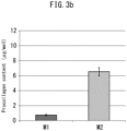
- the fibroblasts were cultured for 72 hours and the amount of procollagen in each supernatant was quantified with a PIP ELISA kit (TAKARA) ( Fig. 3b ).
- the cell fraction was stained with rabbit anti-human collagen antibody (Cederlane) and biotinylated hyaluronic acid bonded protein (Hokudo) ( Fig. 3c ).
- ⁇ -gal as an index of aging, the cell nuclei were stained with DAPI and then the intracellular ⁇ -gal was stained using a Senescence Detection Kit (Abcam) ( Fig. 3d ), and the number of ⁇ -gal-positive cells and DAPI-positive cells were counted ( Fig. 3e ).
- the macrophage supernatants were each added to fibroblasts and culturing was carried out, in the same manner as above.
- the fibroblasts were collected, the mRNA was recovered, and HGF, ET1, bFGF, IL-1 alpha, SCF and clusterin probes were used in real time PCR for quantification of the mRNA expression levels ( Fig. 3f ).
- Figs. 3b to 3f show procollagen amounts after addition of each macrophage (M1 or M2) supernatant to fibroblasts, and culturing for 72 hours.
- Fig. 3c shows localization of collagen and hyaluronic acid in fibroblasts after addition of each macrophage (M1 or M2) supernatant and culturing for 72 hours. From Figs. 3b and 3c it is seen that M1 markedly inhibits collagen production.
- Fig. 3d shows intracellular ⁇ -gal in fibroblasts after addition of each macrophage (M1 or M2) supernatant and culturing for 72 hours.
- Fig. 3b shows procollagen amounts after addition of each macrophage (M1 or M2) supernatant to fibroblasts, and culturing for 72 hours.
- Fig. 3c shows localization of collagen and hyaluronic acid in fibroblasts after addition of each macrophage (M1 or M2) supernatant and cul
- Fig. 3d suggests that ⁇ -gal-positive cells increase with M1, thereby promoting aging, whereas M2 inhibits aging.
- Fig. 3e shows a graph with the count of ⁇ -gal-positive cells and DAPI-positive cells. At right are shown the total counts of DAPI-positive cells per well. The graph at right in Fig. 3e suggests that M2 not only inhibits aging of cells but also promotes proliferation of cells.
- At left in Fig. 3e is a graph showing the proportion (%) of ⁇ -gal-positive cells to total DAPI-positive cells, which suggests that M1 has an effect of promoting aging and cell death, while M2 has an effect of inhibiting aging and promoting cell proliferation. As shown in Fig.
- M1 tended to increase melanin production while M2 tended to inhibit melanin production.
- fibroblasts are known to secrete factors such as SCF and HGF under light stimulation by UV and the like, thus leading to cell death.
- fibroblasts also secrete factors such as HGF, ET1, bFGF, SCF and clusterin under light stimulation, acting either directly or indirectly on melanocytes to produce melanin. It is therefore suggested that cell death and melanin production induced by light stimulation is a result of disturbance in the M1/M2 balance.
- Experiment 4 Experiment adding M1 and M2 macrophage supernatants to young and elderly fibroblasts
- M1 and M2 macrophage supernatants were added to young and elderly cells, and the difference in effects of the macrophage supernatants was examined based on the difference in fibroblast age.
- each macrophage supernatant collected by the same method as Experiment 3 was added to neonatal human prepuce fibroblasts (young fibroblasts) and 68-year-old human fibroblasts (elderly fibroblasts), and culturing was carried out for 72 hours. After subsequent ⁇ -gal and DAPI staining by the same method as Experiment 3, the numbers of ⁇ -gal-positive cells and DAPI-positive cells were counted ( Figs. 4a and 4b ).
- M1 and M2 supernatants were added to the young and elderly fibroblasts in the same manner as Experiment 3, the fibroblasts were cultured for 72 hours, and the procollagen levels in the supernatants were quantified with a PIP ELISA kit (Takara) ( Fig. 4c ), and stained with rabbit anti-human collagen antibody (Cederlane) and DAPI ( Fig. 4d ).
- Fig. 4a shows ⁇ -gal staining images.
- Fig. 4b shows graphs with the results of the numbers of ⁇ -gal-positive cells and the total numbers of cells per well, for the young fibroblasts and elderly fibroblasts.
- Fig. 4a shows that even with the elderly cells, addition of M2 supernatant markedly reduced ⁇ -gal-positive cells and inhibited aging.
- Fig. 4b shows that, regardless of age, aging was promoted by M1 but inhibited by M2. This is also supported by the results shown in Figs. 4c and d , wherein regardless of age, cells to which M1 supernatant was added had markedly lower collagen production compared to M2 supernatant.
- Experiment 5 Experiment with co-culturing of M1 macrophages, M2 macrophages and neonatal prepuce fibroblasts
- Human dermal fibroblasts (0.2 ⁇ 10 6 ) were seeded in a cell culture insert (12 mm ⁇ , porous membrane mean pore size: 0.4 ⁇ m), and 200 ⁇ M of magnesium ascorbate-2-phosphate (APM) and 10% FBS-DMEM were used for one week of culturing with medium exchange once every two days.
- APM magnesium ascorbate-2-phosphate
- FBS-DMEM FBS-DMEM
- each of the collagen gels was formed on the human dermal fibroblasts and culturing was carried out for 1 to 5 days.
- Epidermal keratinocytes dispersed in Humedia-KG2 medium were seeded on collagen gel at 5 ⁇ 10 5 /well, medium comprising a mixture of Humedia-KG2 and 10% FBS-DMEM at 1:1 with addition of 200 ⁇ M APM was added outside the insert to the same liquid level height as the inner side, and culturing was carried out for 3 days.
- skin model medium (with mixture of 10% FBS-DMEM and Humedia-KG2 EGF (-) at 1:1 and prepared to 1.8 mM Ca), with 200 ⁇ M APM, 10 ⁇ M N-hydroxy-2-[[(4-methoxyphenyl)sulfonyl]3-picolyl)amino]-3-methylbutaneamide hydrochloride (CGS27023A (MMP inhibitor)) and 10 ⁇ M BIPBIPU (heparanase inhibitor), was added on the outer side to the height of the bottom of the insert, and gas-liquid boundary culturing was carried out with the insert interior exposed to air. Culturing was carried out for 2 weeks with medium exchange once every 2 to 3 days.
- Experiment 7 Experiment irradiating sunlight onto ex vivo model using human skin
- NativeSkin (skin taken from 38-year-old female) by Genoskin was cultured for 1 day after arrival with the included culture solution.
- an optical filter was placed in a 1000 W solar simulator by Oriel and UVA and UVB alone were irradiated at 11.5 J/cm 2 , after which culturing was continued with the included culture solution (irradiation (+)).
- a sample without irradiation was used as the control (irradiation (-)).
- the sample was collected five days after irradiation, M1 macrophages, M2 macrophages and total macrophages were stained in the same manner as Experiment 1, and the numbers were plotted on a graph.
- Experiment 8 Experiment of stretching stimulation on ex vivo model using human skin
- Stretching conditions A stretching instrument was constructed, having a gripping part for gripping both ends of the skin in the wells, as shown in Fig. 11 , and causing stretching of the skin by pulling of the gripping part.
- the wells containing the tissue strips were placed horizontal, the gripping part was operated to stretch the skin from both ends, and stretching was carried out at a speed of 10%/s to a 10% stretching rate as shown in Fig. 10 , after which the samples were restored to their original non-stretched state at a recovery speed of 10%/s.
- This procedure as one cycle, a total of 90 cycles were carried out over a period of 30 minutes.
- 90 cycles as one set a total of 3 sets (total of 270 cycles) were carried out over a period of 3 hours with a pause period of 30 minutes to 1 hour between sets.
- a sample without stretching stimulation was used as the control.
- each macrophage supernatant was added to fibroblasts and cultured by the same method as Experiment 3, and the mRNA was collected after 72 hours.
- MMP-1, MMP-2 and IL-1 ⁇ probes (Taqman probes by Applied Biosystems) were used as indexes of collagen breakdown, and real time PCR was carried out to quantify the mRNA expression level for each ( Fig. 14a ).
- each macrophage supernatant was added to fibroblasts and cultured by the same method as Experiment 3, and the mRNA was collected after 72 hours.
- COL1A1, COL1A2, HSP47 and ADAMTS-2 probes (Taqman probes by Applied Biosystems) were used as indexes of collagen production and maturation, and real time PCR was carried out to quantify the mRNA expression level for each ( Fig. 14b ).
- Figs. 14a and 14b confirmed that M1 contributes to collagen breakdown while M2 contributes to collagen production and maturation.
- Figs. 15b , 15c and 15d The results are shown in Figs. 15b , 15c and 15d .
- the proportion of M1 macrophages was higher and the proportion of M2 macrophages was lower in the elderly group compared to the young group. Regardless of age, however, the number of macrophages exhibiting 3/4 collagen positivity tended to be greater compared to fibroblasts exhibiting 3/4 collagen positivity, and the number of M2 macrophages exhibiting 3/4 collagen positivity tended to be greater than M1 macrophages exhibiting 3/4 collagen positivity.
- THP-1 cells were differentiated to M1 macrophages and M2 macrophages by the same method as Experiment 3.
- Melanin solution (Sigma Corp., Melanin-BioReagent, Synthetic, suitable for cell culture) was dissolved in PBS and adjusted to 0.02% W/V, and added to the differentiated M1 macrophages, M2 macrophages and fibroblasts (Kurabo Industries, Ltd.). After 24 hours, the cells were washed with PBS and photographed with a microscope, and the cells were collected. The number of collected cells was counted with Alamar Blue (Life Technologies) and the melanin content was calculated and quantified in the following manner.
- Alamar Blue was added to each cell medium (macrophages: RPMI 1640, fibroblasts: 1DMEM) adjusted to 1:10, and then culturing was carried out at 37°C for 30 minutes. The supernatant was then collected at 100 ⁇ l/well, and the fluorescence was measured with an Ascent (Thermo) with excitement/emission: 544 nm/590 nm. After measurement, washing was performed with PBS and 1 M NaOH was added, after which the mixture was incubated at room temperature for 3 hours for complete lysis. The cell solution was measured using a POWERSCAN HT (DS Pharma Biomedical) at OD475.
- the results are shown in Figs. 16a to 16c .
- the "melanin/almar blue" units in the graph represent a relative value ((2)/(1)), where (1) is the intensity for cultured cells stained with Alamar Blue and with measurement of the supernatant using fluorescence at 544 nm/590 nm, and (2) is the intensity after lysing the cells, decomposing the melanin and measuring at an absorbance of 475 nm, and it represents the melanin content per cell.
- Experiment 12 Experiment ex vivo showing uptake of melanin by dermis
- hakushinin was found to have a powerful M2-inducing effect.
- the hakushininin extract used here (non-hydrolyzed or hydrolyzed) was prepared according to Japanese Patent Publication No. 4781842 (PTL 9) or International Patent Publication No. WO2012/0571243 (PTL 10), by the following procedure. It is to be understood, however, that the procedure for preparing hakushinin extract to be used for the invention is not limited to the following.
- Hakushinin seeds (non-pulverized, outer shells removed as necessary) were immersed in a 5-fold volume (v/w) of acetone, ethyl acetate or hexane and extracted for 10 days at room temperature. The residue was removed beforehand with a nylon mesh (100 mesh), and filtration was carried out with filter paper. After removing the solvent from the filtrate using a rotary evaporator, 0.5 to 15 N NaOH was added as NaOH in a range of 100 to 300 g/l of extract, while stirring with a stirring blade (temperature: 50°C). Treatment was carried out for 5 hours while stirring at 200 rpm. After then gradually adding 5N H 2 SO 4 to lower the pH to near 1 while stirring, the separated oily substance was collected.
- THP-1 cells were used in the M0 undifferentiated state and cultured overnight at 37°C.
- the non-hydrolyzed (non-alkali treated) hakushinin (control) dissolved in a solvent (DMSO) was added at 3 ppm, or the hakushinin hydrolysate was added at 0.3 ppm, 1 ppm or 3 ppm, and culturing was carried out for two nights at 37°C.
- the cells were recovered and the RNA was extracted, and the expression levels of CD86, CCR7, TNF-alpha, CD206, CD163, IL-10 and GAPDH were quantified by real time PCR, dividing the expression level of each gene by the expression level of GAPDH.
- the hakushinin hydrolysate lowered CD86 gene and TNF- ⁇ gene expression levels while increasing CD206 gene and IL-10 gene levels, in a concentration-dependent manner. This indicated that hakushinin hydrolysate has an effect of inhibiting M1 induction and promoting M2 induction ( Fig. 18 ).
- Non-hydrolyzed (non-alkali treated) hakushinin (control) dissolved in a solvent (DMSO) was added at 3 ppm, or hakushinin hydrolysate was added at 1.0 ppm or 3.0 ppm, to immature (M0) THP-1 cells. Culturing was carried out at 37°C, melanin solution (Sigma Corp.) was added after 48 hours, and the mixture was observed with a microscope (Bio Station CT (Nikon)).
- Figs. 19a and 19b The results are shown in Figs. 19a and 19b .
- the hakushinin-added macrophages began to take up melanin about 30 minutes after addition of melanin (black arrows in Fig. 19b ). After another 3 hours, the control still had many cells that had not taken up melanin, whereas all of the cells of the hakushinin hydrolysate-added sample had taken up melanin.
- M0 macrophages, M1 macrophages and M2 macrophages induced to differentiate from THP-1 by the same method as Experiment 2 were measured for intracellular activity using an XFe24 (24-well) extracellular flux analyzer by Agilent Technologies (previously Seahorse Bioscience) (hereunder referred to as "flux analyzer"), with reference to Nat Immunol, 2014. 15(9): pp846-855 (NPL 14).
- a flux analyzer is a device that allows non-invasive, highly sensitive periodic measurement of cells to determine the state of glycolysis as the main cellular energy metabolism pathway, and aerobic respiration by mitochondria.
- oligomycin ATP synthase inhibitor
- FCCP uncoupling agent
- rotenone mitochondriachondrial complex I inhibitor + antimycin A (mitochondria complex III inhibitor)
- OCR oxygen consumption rate
- ECAR extracellular acidification rate
- M0, M1 and M2 differentiated from THP-1 were prepared by the same method as Experiment 2, and hakushinin extract (hakushinin hydrolysate) prepared by the same method as Experiment 13 was added to the M0 (at 0.3 ppm, 1 ppm or 3 ppm).
- OCR oxygen consumption rate
- Hakushinin extract is known to contain unsaturated fatty acids such as the ⁇ 3 fatty acids linolenic acid and juniperonic acid, and the ⁇ 6 fatty acid linoleic acid. It has been reported that unsaturated fatty acids (EPA, DHA) act on M1 inflammation related genes, thus inhibiting inflammation (NPL 15 ( Cell Metab. 2017 Feb 7; 25(2):412-427 )). The effects of the unsaturated fatty acids in hakushinin extract on the intracellular activity of macrophages was investigated.
- unsaturated fatty acids such as the ⁇ 3 fatty acids linolenic acid and juniperonic acid, and the ⁇ 6 fatty acid linoleic acid. It has been reported that unsaturated fatty acids (EPA, DHA) act on M1 inflammation related genes, thus inhibiting inflammation (NPL 15 ( Cell Metab. 2017 Feb 7; 25(2):412-427 )). The effects of the unsaturated fatty acids in hakushinin extract on
- M0 and M2 differentiated from THP-1 were prepared by the same method as Experiment 2, linoleic acid, linolenic acid or juniperonic acid (3 ppm) was added to M0, and after 48 hours the oxygen consumption rate (OCR) was measured with a flux analyzer by the same method as Experiment 15. Each measured value was calibrated by the number of cells determined by counting the cell nuclei stained by addition of Hoechst.
- the Basal OCR and mitochondrial respiration values for M0 were examined with the incubation time changed to 24 hours after addition of the hakushinin extract or unsaturated fatty acid.
- the experiment method was carried out by the same method as described in Experiment 17, except that the incubation time was changed to 24 hours after addition of the hakushinin extract or unsaturated fatty acid.
- a flux analyzer was used to analyze the change in intracellular activity with addition of hakushinin extract or an unsaturated fatty acid (linoleic acid, linolenic acid or juniperonic acid) in the step of further inducing differentiation to M1 from M0 cells differentiated from THP-1 by the same method as Experiment 2 ( Fig. 25 ).
- OCR value oxygen consumption rate
- ECAR value extracellular acidification rate
- Mitochondrial respiration in macrophages during M1 differentiation was significantly increased by hakushinin extract, but no significant increase was found using only the unsaturated fatty acids that are present in hakushinin extract, suggesting that the effect of increasing mitochondrial respiration may be a specific effect attributable to mixture of the different components in hakushinin extract.
- SDF-1 ⁇ (CXCL12) expression levels are lower in fibroblasts present below blemished areas of skin, and that the reduction in SDF-1 levels may therefore be related to formation of blemishes in skin (NPL 16 ( Theranostics. 2018 Sep 9; 8(17):4620-4632 )).
- NPL 16 Theranostics. 2018 Sep 9; 8(17):4620-4632 .
- the expression levels of SDF-1 ⁇ (CXCL12) in M1/M2 macrophages themselves were therefore examined.
- the M2 supernatant was shown to have more SDF-1 ⁇ (CXCL12) than the M1 supernatant ( Fig. 27(B) ).
- Experiment 21 Experiment adding M1 and M2 macrophage supernatants to fibroblasts (2)
- RNA-seq lead was mapped to the human genome (hg19) using STAR. After filtering out the low expression genes, the obtained count data were corrected for sample size and converted to log2.
- the log2 (CPM) values calculated for each of the samples by the method described above were compared between the two groups by quasi-likelihood F-test, using a glmQLFit and glmQLFTest as R package edgeR functions, and were then corrected by multiple comparison test using the Benjamini-Hochberg method to calculate the q-value.
- M1/M2 balance can prevent and/or ameliorate photoaging and/or dermal pigmentation.
- Regulation or improvement in M1/M2 balance is achieved by applying stretching stimulation or compression stimulation, or by applying hakushinin, whereby it is expected that photoaging and/or dermal pigmentation can be prevented and/or improved.
Landscapes
- Health & Medical Sciences (AREA)
- Life Sciences & Earth Sciences (AREA)
- Veterinary Medicine (AREA)
- Public Health (AREA)
- General Health & Medical Sciences (AREA)
- Animal Behavior & Ethology (AREA)
- Dermatology (AREA)
- Microbiology (AREA)
- Epidemiology (AREA)
- Birds (AREA)
- Mycology (AREA)
- Botany (AREA)
- Biotechnology (AREA)
- Gerontology & Geriatric Medicine (AREA)
- Engineering & Computer Science (AREA)
- Medicines Containing Plant Substances (AREA)
- Cosmetics (AREA)
Abstract
Description
- The present invention relates to an agent for preventing and/or improving photoaging and/or dermal pigmentation, to a cosmetic method using it, and to a cosmetic device to be applied in the method.
- Aging of human skin is generally classified into the categories of "natural aging" and "photoaging". Photoaging is skin-specific, occurring in a site-specific manner as a result of exposure to light. In photoaging, it is believed that UV rays elicit production of active oxygen and DNA damage in cells, leading to damage to the fibrous tissue of skin and the resulting in appearance of wrinkles and sagging. Some of the phenomena associated with photoaging of skin are reduction in collagen fibers (composed of collagen) and degeneration of elastic fibers (composed of elastin). Melanocytes also suffer damage, resulting in overproduction of melanin pigment which can lead to skin spots.
- Pigmentation, including skin spots and loss of skin clarity, occurs due to accumulation of melanin produced by melanocytes in the basal lamina of the epidermis. Melanin is normally present in the epidermis and basal lamina, but the melanin tends to be discharged due to the relatively high turnover of the epidermis. Melanin is often also present in the dermis layer as it accrues in the dermis through gaps in the basal membrane. Because the turnover cycle for dermal cells is much slower than the epidermis, the melanin usually accumulates without being discharged. Consequently it is extremely difficult to ameliorate dermal pigmentation.
- As one cosmetic method for anti-photoaging,
PTL 1 discloses an agent for preventing or inhibiting skin photoaging by preventing inhibition of leukocyte elastase.PTL 2 discloses a photoaging inhibitor composition comprising a plant extract of Amaranthaceae Pfaffia, which has a collagen synthesis-promoting effect, and an animal-derived collagen peptide. - NPL 13 proposes a cosmetic method for improving pigmentation of the dermis, wherein the basal membrane is reinforced, to prevent sinking of melanin into the dermis.
- Inflammation has also been suggested as one factor in the photoaging phenomenon of skin, and numerous anti-inflammatory agents have been developed.
PTL 3 discloses a cosmetic composition for anti-aging that includes a compound that elicits increased expression of adiponectin and activation of autophagy. It has been suggested to utilize the phagocytotic effect of macrophages to improve dermal pigmentation, andPTL 8 discloses an agent for preventing or ameliorating dermal blemishes by attracting macrophages to fibroblasts that have taken up melanin and fallen into the dermis. -
- [PTL 1]
Japanese Patent Publication No. 5657723 - [PTL 2]
Japanese Unexamined Patent Publication No. 2017-203004 - [PTL 3]
Japanese Unexamined Patent Publication No. 2018-177805 - [PTL 4]
Japanese Patent Public Inspection No. 2014-504629 - [PTL 5]
Japanese Unexamined Patent Publication No. 2015-140334 - [PTL 6]
Japanese Patent Publication No. 6178088 - [PTL 7]
Japanese Patent Publication No. 6273304 - [PTL 8]
Japanese Unexamined Patent Publication No. 2018-072098 - [PTL 9]
Japanese Patent Publication No. 4781842 - [PTL 10] International Patent Publication No.
WO2012/057123 -
- [NPL 1] Journal of the American College of Cardiology, Vol. 62, No. 20, 2013, November 12, 2013:1890-901
- [NPL 2] Experimental & Molecular Medicine (2014) 46, e70; doi:10.1038/emm.2013.135
- [NPL 3] Nature, Vol. 495, 28 March 2013, pp524-530, doi:10.1038/nature11930
- [NPL 4] Journal of Investigative Dermatology, 2009 April; 129(4):1016-25. Epub 2008 Oct 9.
- [NPL 5] Stem Cell Research & Therapy (2018) 9:88, https://doi.org/10.1186/s13287-018-0821-5
- [NPL 6] Journal of the European Academy of Dermatology and Venereology 2011 European Academy of Dermatology and Venereology, 2012, 26, 1577-1580
- [NPL 7] British Journal of Dermatology, 2005, 153, pp733-739
- [NPL 8] Pigment Cell Melanoma Research, Vol. 27,
- [NPL 9] Ann Dermatol Vol. 28, No. 3, pp279-289, 2016
- [NPL 10] Endocrinology, 2011. 152(10): pp3779-90
- [NPL 11] British Journal of Dermatology, 2005. 153 Suppl 2: pp37-46
- [NPL 12] Experimental Dermatology, 2011. 20(11): pp953-5
- [NPL 13] Fujifilm Research & Development (No. 55-2010):pp33-37
- [NPL 14] Nat Immunol, 2014. 15(9): pp846-855
- [NPL 15] Cell Metab. 2017 Feb 7; 25(2):412-427
- [NPL 16] Theranostics. 2018 Sep 9; 8(17):4620-4632
- It is an object of the present invention to provide an agent for preventing and/or improving photoaging and/or dermal pigmentation, to a cosmetic method using it, and to a cosmetic device to be applied in the method.
- As a result of avidly searching for an agent for preventing and/or ameliorating photoaging and/or dermal pigmentation, the present inventors have found that hakushinin (e.g. Thuja orientalis L.) extract regulates the M1/M2 balance of macrophages and thereby exhibits an effect of preventing and/or ameliorating photoaging and/or dermal pigmentation. The present invention has been developed on the basis of this finding. Specifically, the invention encompasses the following.
- [1] An agent for preventing and/or improving photoaging and/or dermal pigmentation, which contains hakushinin (e.g. Thuja orientalis L.) extract as an active ingredient.
- [2] The agent according to [1] above, which prevents and/or ameliorates photoaging and/or dermal pigmentation by regulation of M1/M2 balance.
- [3] The agent according to [2] above, wherein the regulation of M1/M2 balance is increase in the ratio of M2 with respect to M1.
- [4] A cosmetic method for preventing and/or ameliorating photoaging and/or dermal pigmentation of a subject, wherein the method includes:
- (a) a process in which an agent according to any one of [1] to [3] is applied to the skin of a subject.
- [5] The cosmetic method according to [4] above, which further includes a process of applying weak physical stimulation to the skin, the process of applying physical stimulation comprising application of physical stimulation to the skin of the subject in which a cycle that includes, for example:
- (b-1) stretching the skin of the subject to a stretching rate of 0.1% to 50.0%, where the stretching rate is calculated by:
[Mathematical Formula 1]
- (c-1) restoring the skin of the subject from the stretched state; and/or
- (b-2) compressing the skin of the subject by 1 µm to 1000 µm; and
- (c-2) restoring the skin of the subject from the compressed state;
- (b-1) stretching the skin of the subject to a stretching rate of 0.1% to 50.0%, where the stretching rate is calculated by:
- [6] A cosmetic device for application in the cosmetic method according to [5] above, wherein the device comprises:
- a stimulation generator that produces physical stimulation, and
- a stimulation applicator that applies the physical stimulation generated by the stimulation generator to skin,
- the device serving to carry out a process in which weak physical stimulation is applied to skin, and
- the process carries out a cycle that includes, for example:
- (i-1) stretching the skin to a stretching rate of 0.1% to 50.0%; and
- (ii-1) restoring the skin of the subject from the stretched state; and/or
- (i-2) compressing the skin of the subject by 1 µm to 1000 µm; and
- (ii-2) restoring the skin of the subject from the compressed state;
- where the stretching rate is calculated by
Formula 1 above.
- Application of the present invention can regulate the M1/M2 balance of macrophages, thereby preventing and/or ameliorating photoaging and/or dermal pigmentation.
-
-
Fig. 1a shows the types of macrophage markers used inExperiment 1, and the ages, skin types and genders of subjects. -
Fig. 1b is a photomicrograph showing macrophages (CD11b: red) and M1 macrophages (CD86: green) in skin tissue of young and elderly persons. -
Fig. 1c is a photomicrograph showing macrophages (CD11b: red) and M2 macrophages (CD206: green) in skin tissue of young and elderly persons. -
Fig. 1d is a graph showing the number of M1 and M2 macrophages and the total number of all macrophages (cells/mm2), in skin tissue of young and elderly persons. -
Fig. 1e (left) is a set of photomicrographs showing co-staining of M1 macrophages (CD86: red) or M2 macrophages (CD206: red) and procollagen (green), in skin tissue of young and elderly persons. The rectangular sections in each of the 4 panels at left are magnified portions of particular sections.Fig. 1e (right) is a pair of diagrams schematically illustrating the relationship between M1, M2, fibroblasts and collagen production/breakdown. -
Fig. 2a shows in overview the method used inExperiment 2. -
Fig. 2b is a photomicrograph of M0, M1 and M2 macrophages derived inExperiment 2. -
Fig. 2c is a set of graphs showing gene expression levels of cytokines (IL-1 beta, TNF-alpha and IL-10) produced by M0, M1 and M2 macrophages derived inExperiment 2. The lower part ofFig. 2c shows graphs for expression levels of mRNA of cell surface markers on M1 and M2 macrophages (M1: CD86, M2: CD206). Shown are relative values (%) calibrated against GAPDH mRNA expression level, with respect to M0 as 100%. -
Fig. 3a shows in overview the method used inExperiment 3. -
Fig. 3b shows procollagen amounts (µg/well) in fibroblasts to which M1 or M2 macrophage supernatant was added inExperiment 3. -
Fig. 3c is a set of photomicrographs showing collagen (red) and hyaluronic acid (green) in fibroblasts to which M1 or M2 macrophage supernatant was added inExperiment 3. -
Fig. 3d is a set of photomicrographs showing β-galactosidase (β-Gal) in fibroblasts to which M1 or M2 macrophage supernatant was added inExperiment 3. -
Fig. 3e is a pair of graphs showing the results of age-related β-galactosidase (SA β-Gal) assay (left: percentage (%) of SA β-gal-positive cells with respect to total number of DAPI-positive cells) in fibroblasts to which M1 or M2 macrophage supernatant was added inExperiment 3, and the total number of DAPI-positive cells per well (right). -
Fig. 3f is a set of graphs showing mRNA expression levels of different melanogenesis-promoting factors (HGF, ET1, bFGF, IL-1 alpha, SCF) and melanogenesis-inhibiting factors (clusterin, DKK1), in fibroblasts to which M1 or M2 macrophage supernatant was added inExperiment 3. -
Fig. 4a is a set of photomicrographs showing β-Gal in young and elderly fibroblasts to which M1 or M2 macrophage supernatant was added inExperiment 4. -
Fig. 4b is a set of graphs showing the results of SA β-Gal assay (percentage (%) of SA β-gal-positive cells with respect to total number of DAPI-positive cells) and the total number of DAPI-positive cells per well, for young (top) and elderly (bottom) fibroblasts to which M1 or M2 macrophage supernatant was added inExperiment 4. -
Fig. 4c shows procollagen amounts (µg/well) in young and elderly fibroblasts to which M1 or M2 macrophage supernatant was added inExperiment 4. -
Fig. 4d is a set of photomicrographs showing collagen (red) and DAPI-stained nuclei (blue) in young and elderly fibroblasts to which M1 or M2 macrophage supernatant was added inExperiment 3. -
Fig. 5 is a set of photomicrographs showing type I collagen (red) and macrophages (CD68: green) in neonatal prepuce-derived fibroblasts to which M1 or M2 macrophage supernatant was added inExperiment 5. The photograph at top left shows fibroblasts cultured without addition of macrophage supernatant. -
Fig. 6 is a set of photomicrographs showing macrophages (CD68: red) and M1 macrophages (CD86: green) or M2 macrophages (CD206: green) in a 3D model prepared according toExperiment 6. The triangles indicate portions double stained with CD68 and CD86 or with CD68 and CD206. -
Fig. 7 is a set of photomicrographs showing p21 (red) and DAPI-stained nuclei (blue) in a 3D model prepared according toExperiment 6. -
Fig. 8 is a pair of graphs showing percentage (%) of number of p21-positive cells with respect to total number of cells in each layer of a 3D model prepared according to Experiment 6 (left: upper layer = epidermal cell layer, right: lower layer = fibroblast layer). -
Fig. 9 is a graph showing the number of all macrophages and the numbers of M1 and M2 macrophages in an ex vivo skin model irradiated with a solar simulator inExperiment 7. Shown are bars for each number of macrophages without irradiation as 100% (left bar: irradiation (-)), and bars at right as the relative values (%) for the number of macrophages with irradiation (irradiation (+)). -
Fig. 10 shows setting of stretching stimulation forExperiment 8. -
Fig. 11 shows the device and skin sample used inExperiment 8. -
Fig. 12 shows a comparison of M1 and M2 macrophage numbers and total macrophage numbers, forExperiment 8 with stretching stimulation (stretch) applied to an ex vivo skin model and without (control). At left is a graph showing the number of macrophages per 1 mm2 of skin sample (cells/1 mm2). At right is a graph showing the percentage (%) of each macrophage type (M1, M2) with respect to the total number of macrophages. -
Fig. 13 shows an example of a device of the invention. -
Fig. 14a is a set of graphs showing mRNA expression levels of collagen catabolic enzymes (MMP-1, MMP-2) and an inflammatory cytokine (IL-1β) in fibroblasts to which M1 or M2 macrophage supernatant was added inExperiment 9, as relative values (%) with respect to 100% as the results with M1. -
Fig. 14b is a set of graphs showing mRNA expression levels of collagen production and maturation factors (COL1A1, COL1A2, HSP47 and ADAMTS-2) in fibroblasts to which M1 or M2 macrophage supernatant was added inExperiment 9, as relative values (%) with respect to 100% as the results with M1. -
Fig. 15a shows ages of subjects in a young group and an elderly group from which samples were taken forExperiment 10. -
Fig. 15b is a pair of graphs showing the number of M1 and M2 macrophages (cells/mm2) in skin tissue of young and elderly persons (50 µm directly under the basal membrane) forExperiment 10. -
Fig. 15c is a pair of graphs showing the number of M1 and M2 macrophages (cells/mm2) in skin tissue of young and elderly persons (200 µm directly under the basal membrane) forExperiment 10. -
Fig. 15d is a set of graphs showing the number of CD68-negative cells and M1 and M2 macrophages exhibiting 3/4 collagen positivity (cells/mm2) in skin tissue of young and elderly persons forExperiment 10. -
Fig. 16a shows the state of fibroblasts, M1 macrophages andM2 macrophages 24 hours after melanin addition inExperiment 11. The melanin-phagocytic cells had circular forms, as shown at right. -
Fig. 16b shows the melanin amounts (melanin/almar blue) per cell, taken up by fibroblasts, M1 macrophages andM2 macrophages 24 hours after melanin addition inExperiment 11. -
Fig. 16c shows the state of M1 macrophages andM2 macrophages 5 days after melanin addition inExperiment 11. -
Fig. 17 (top) is a graph showing numbers of melanin-phagocytic M1 macrophages and M2 macrophages in the dermis layer, for different age groups, as counted inExperiment 12.Fig. 17 (bottom) shows the average numbers of melanin-phagocytic M1 macrophages and M2 macrophages for all of the subjects. -
Fig. 18 shows the results of screening inExperiment 13. The bars in the graphs indicate expression levels of M1 and M2 markers (marker/GAPDH) with hakushinin extract added at different concentrations (0.3 ppm, 1.0 ppm and 3.0 ppm), represented as relative values (%) with respect to 100% for the control (no hakushinin hydrolysis). -
Fig. 19a shows the state of immature (M0) THP-1 cells with addition of 3 ppm of non-hydrolyzed hakushinin (control) or 1.0 ppm or 3.0 ppm of hakushinin hydrolysate, 30 minutes after melanin addition, inExperiment 14. -
Fig. 19b shows magnified views ofFig. 19a . The black arrows indicate macrophages that have taken up melanin. -
Fig. 20 shows the results for (A) oxygen consumption rate (OCR), (B) extracellular acidification rate (ECAR) and (C) OCR/ECAR, based on analysis of intracellular activity of M0 macrophages, M1 macrophages and M2 macrophages using a flux analyzer, inExperiment 15. -
Fig. 21 shows the results of using a flux analyzer to analyze the effects on intracellular activity ofM0 macrophages 48 hours after addition of hakushinin extract, inExperiment 16. (A) normalized OCR, (B) Basal OCR (OCR value at 3rd A measurement). -
Fig. 22 shows the results of using a flux analyzer to analyze the effects on intracellular activity ofM0 macrophages 48 hours after addition of hakushinin extract, inExperiment 16. (A) normalized OCR, (B) mitochondrial respiration OCR (difference between 3rd and 12th A measurement). -
Fig. 23 shows the results of using a flux analyzer to analyze the effects on intracellular activity ofM0 macrophages 48 hours after addition of only unsaturated fatty acids typically present in hakushinin extract, inExperiment 17. (A) Basal OCR, (B) mitochondrial respiration OCR. -
Fig. 24 shows the results of using a flux analyzer to analyze the effects on intracellular activity ofM0 macrophages 24 hours after addition of different components present in hakushinin extract, inExperiment 18. (A) Basal OCR, (B) mitochondrial respiration OCR. -
Fig. 25 shows in overview the method used inExperiment 19. -
Fig. 26 shows the results of using a flux analyzer to analyze the effect of hakushinin extract or different components present in hakushinin extract on intracellular activity of M1 macrophages during differentiation, inExperiment 19. (A) Basal OCR, (B) mitochondrial respiration OCR, (C) OCR/ECAR. -
Fig. 27 shows expression levels of SDF-1α in M1 and M2 macrophage supernatants. (A) Overview ofExperiment 20, (B) amount of SDF-1α in M1 macrophage or M2 macrophage culture supernatant. -
Fig. 28 shows effects on SDF-1α expression by skin fibroblasts cultured with addition of M1 or M2 macrophage supernatant, in Experiment 21. (A) Overview of Experiment 21, (B) amount of SDF-1α incultured fibroblasts 3 days after addition of M1 macrophage or M2 macrophage culture supernatant. - Macrophages are cells that are localized in various tissues of the body and elicit an immune response against foreign matter and pathogens, and they are known to be associated with inflammation. Macrophages differentiate from undifferentiated M0 macrophages (M0) into M1 and M2 macrophages. M1 macrophages (M1) are the inflammatory type, while M2 macrophages (M2) are the repair (anti-inflammatory) form. Imbalance between M1 macrophages and M2 macrophages has been reported to be associated with conditions such as obesity, type II diabetes and arteriosclerosis (
PTLs 4 to 6,NPLs 1 to 5). However, the relationship between M1/M2 balance and skin photoaging or pigmentation is still unclear. - The present inventors have found that the balance between M1 macrophages and M2 macrophages (M1/M2 balance) is specifically disturbed at light-exposed skin or sites where pigmentation has occurred, and that regulating the M1/M2 balance is extremely important for preventing and ameliorating photoaging or pigmentation of the dermis. The present inventors therefore searched for substances that can prevent or ameliorate photoaging and/or dermal pigmentation, using M1/M2 balance as the index, and having found that hakushinin regulates M1/M2 balance, have developed the present invention which prevents and ameliorates photoaging and/or pigmentation in the skin, and particularly the dermis. It has been reported that hakushinin acts on fibroblasts causing proliferation of fibroblasts, and has effects of promoting collagen and hyaluronic acid production, and of inhibiting melanin production (PTLs 9 and 10). However, the present inventors are the first to have found that hakushinin has an effect of regulating and improving M1/M2 balance.
- The present inventors also found that M1/M2 balance can be regulated by applying specific physical stimulation to the skin of a subject. More specifically, it has been found that even in photoaged skin or skin with pigmentation, M1/M2 balance is regulated and improved by applying weak physical stimulation with a specific stretching rate and a vibrational frequency of 60 Hz or lower, such as 1 Hz or lower or 10 Hz or lower, for example.
- The invention therefore provides an agent that prevents and/or improves photoaging and/or dermal pigmentation by adjusting M1/M2 balance, as well as a cosmetic method using the same and a cosmetic device to be applied in the method. The method of the invention will often be for the purpose of beautifying, instead of treatment by a doctor or health care professional.
- Throughout the present specification, the term "pigmentation" refers to deposition of pigments in the dermis and epidermis, and it includes not only pigmentation by melanin as a result of photoaging, but also artificially injected pigments (such as tattoos). The present invention is effective for both the dermis and the epidermis, but it is particularly promising as a countermeasure against dermal pigmentation in light of limitations of improvement methods other than phagocytosis by melanophages. Application of the present invention improves skin spots, loss of skin clarity and eye circles associated with pigmentation. It is also effective for removing tattoos that are difficult to remove after pigments have been injected into the dermis layer.
- M1 macrophages can be measured by markers such as CD86, CD80 and iNOS. M2 macrophages can be measured by markers such as CD206, CD163 and Agr1. CD11b and CD68 are general markers for macrophages including M1 and M2. Measurement may also be by quantifying M1-specific cytokines such as IL-1 beta and TNF-alpha, or M2-specific cytokines such as IL-10, either in addition to or together with the above. However, the markers used are not limited to these so long as they allow measurement of M1/M2 balance.
- Throughout the present specification, "M1/M2 balance" may refer to the proportion of the number of M1 macrophages and the number of M2 macrophages, or it may refer to the ratio of the mRNA level of M1 macrophage markers (such as CD86, CD80 or iNOS) to the mRNA level of M2 macrophage markers (such as CD206, CD163 or Agr1). Since the ratio of M1 is high and the proportion of M2 is low in photoaged skin, and M2 macrophages have high melanin phagocytosis activity, regulation and improvement in M1/M2 balance may be achieved by an increase in the proportion of M2 with respect to M1 (number of M2/number of M1, and/or mRNA level of M2 markers/mRNA level of M1 markers). Such an increase may be, for example, an increase in the statistically significant difference (in a Student's t test, for example) at a significance level of 5%, and/or, an increase of 1% or greater, 5% or greater, 10% or greater, 20% or greater, 30% or greater, 40% or greater, 50% or greater, 60% or greater, 70% or greater, 80% or greater, 90% or greater or 100%, for example. Alternatively, regulation or improvement of M1/M2 balance may be bringing the ratio of M2 to M1 (number of M2/number of M1, and/or mRNA level of M2 markers/mRNA level of M1 markers) to within a specified range of, e.g., from about 4/6 to about 9/1, about 5/5 to about 8/2 or about 5/5 to about 7/3, or approaching such a range, or maintaining such a range, or supporting homeostasis centered around such a range.
- Throughout the present specification, "M1/M2 balance" may also refer to the balance between intracellular activity of M1 macrophages and intracellular activity of M2 macrophages, and may be represented using, for example, an index of intracellular activity with the most common state of use of M1 macrophages (in the glycolysis state, for example), and/or intracellular activity with the most common state of use of M2 macrophages (in a state of aerobic respiration (mitochondrial respiration), for example). Intracellular activity may be evaluated, for example, by measuring the oxygen consumption rate (OCR value), extracellular acidification rate (ECAR value) or OCR/ECAR value of cells. As used herein, an "increase in the ratio of M2 to M1" may be an increase in the intracellular mitochondria activity for the most common use of M2 macrophages, and for example, it may be an increase in aerobic respiration (mitochondrial respiration) in a group including M1 macrophages and M2 macrophages (for example, aerobic respiration (mitochondrial respiration) being greater in a group including M1 macrophages and M2 macrophages), and/or it may be a decrease in glycolytic activity as anaerobic respiration in a group including M1 macrophages and M2 macrophages, such as an increase in the OCR value in a group including M1 macrophages and M2 macrophages, and/or a decrease in the ECAR value in a group including M1 macrophages and M2 macrophages, and/or an increase in the OCR/ECAR value in a group including M1 macrophages and M2 macrophages. Intracellular activity may be measured by non-invasive and highly sensitive periodic measurement of cells to determine their state of glycolysis and mitochondrial aerobic respiration, as the main energy metabolism pathway of the cells, using an XFe24 extracellular flux analyzer (24-well) by Agilent Technologies (previously Seahorse Bioscience) as a non-limitative example, with the understanding that any device and method may be used for measurement and evaluation of the intracellular activity.
- Photoaged skin has a high proportion of M1 and a low proportion of M2, and since M2 is associated with higher phagocytosis of melanin, regulation or improvement of M1/M2 balance may be achieved by increasing the proportion of M2 intracellular activity with respect to M1, such as increasing the OCR value in a group including M1 macrophages and M2 macrophages, and/or reducing the ECAR value in a group including M1 macrophages and M2 macrophages. As used herein, "increase or decrease" may be increase or decrease in the statistically significant difference (in a Student's t test, for example) at a significance level of 5%, and/or increase or decrease of 1% or greater, 5% or greater, 10% or greater, 20% or greater, 30% or greater, 40% or greater, 50% or greater, 60% or greater, 70% or greater, 80% or greater, 90% or greater or 100%, for example.
- Hakushinin (or hakushijin) is a galenical obtained by drying seeds of Platycladus orientalis Franco, Thuja orientalis L. or Biota orientalis Endl. of the family Cupressaceae. The hakushinin to be used for the invention is preferably from the seeds (endosperm part), but the whole seeds may also be used since the active ingredient is also included in the whole seeds. The hakushinin may be produced by a common method, or a commercial product may be used.
- The method for preparing the hakushinin extract to be used for the invention may be produced with reference to publicly known methods (such as
PTLs 9 and 10), although this is not limitative. For example, the method for preparing hakushinin extract to be used for the invention may be a method of alkali treatment of a plant extract obtained by extraction from the plant seeds (hereunder referred to as "first method"), and a method of pulverizing the plant seeds and extraction after maturation for 1 week or longer at a temperature of 10 to 35°C and a relative humidity of 20 to 90% (hereunder referred to as "second method"). - The extraction solvent used in the first method and second method may be any volatile solvent commonly used for extraction, and particularly polar or non-polar organic solvents including alcohols such as methanol and ethanol, water-containing alcohols, acetone, ethyl acetate ester, hexane and ethers may be used, either alone or in combinations, but acetone, ethyl acetate and hexane are especially preferred because of they are inexpensive and are easily concentrated under reduced pressure. Extraction may also be accomplished using supercritical carbon dioxide gas. Extraction solvents containing large amounts of water, on the other hand, are not preferred because they inhibit extraction of active ingredients.
- Extraction is carried out for a fixed period with a solvent at a mass of about 1 to 100 times and preferably 1 to 30 times the mass of the seeds. The seeds used may be pulverized as necessary and appropriate, but they may also be used without pulverizing when clogging can be problematic during filtration. Repeated extraction with small amounts of solvent is also possible, and a Soxhlet recycler may be used. When extraction is with supercritical carbon dioxide gas, the extraction may be carried out under generally employed conditions of near 40°C and 20 to 40 MPa.
- In the first method, the extraction solvent is removed from the extract prior to alkali treatment for hydrolysis of the extract. The alkali treatment is addition of a concentrated aqueous alkali solution in a 0.2- to 2-fold volume to the solvent-removed extract, followed by thorough stirring and mixing, and ageing for about 30 minutes to several days. The temperature during mixing is preferably in the range of room temperature to 70°C, and more preferably in the range of 30 to 60°C. Appropriate stirring is preferably carried out during the ageing to avoid separation of the hydrophilic components and lipophilic components. The concentrated aqueous alkali solution may be a 1 to 10 N aqueous solution of NaOH or KOH.
- If the hydrolyzed extract obtained by the alkali treatment is further neutralized with an acid to near neutral, the oily substances will separate out. Oily substances are highly safe for skin while also exhibiting a very high effect against dermal pigmentation. Alcohol, acetone or the like may also be added as appropriate after the alkali treatment in order to promote recovery of the oily substances, and procedures such as decoloration, deodorization, desalting and distillation may also be carried out afterwards as necessary.
- In the second method for preparing the hakushinin extract, the seeds are appropriately pulverized or pressed at an early stage of extraction, and allowed to age for a fixed period at near room temperature. The ageing period is not particularly specified so long as it is a range allowing the desired effect to be obtained, but if the ageing period is as short as less than a week it will be difficult to obtain an adequate whitening effect, and conversely if it is as long as several years, problems such as decay and oxidative odor production will occur, and therefore neither case is desirable. If necessary, antioxidant addition or nitrogen exchange may be carried out during the ageing to inhibit production of odors.
- Extraction from the aged seeds is carried out using the aforementioned extraction solvent and extraction method. The extraction may also be followed by solvent removal, decoloration, deodorization and distillation, as necessary. The extract obtained by pulverizing of specific seeds, aging and extraction in this manner is highly safe for skin while also exhibiting an effect against dermal pigmentation, similar to the extract obtained by the aforementioned alkali treatment.
- According to the invention it is possible to provide an agent for preventing and/or improving photoaging and/or dermal pigmentation comprising an extract prepared from plant seeds by the first method or second method, which is plant-based, safe and has an excellent dermal pigmentation effect, as described above, as well as an external preparation for skin comprising the agent.
- The agent of the invention may also be used in combination with any publicly known substances that are known to regulate or improve M1/M2 balance, such as the substances described in
PTLs 4 to 7, for example. The route of administration may be selected from among transdermal administration, oral administration, subcutaneous administration, transmucosal administration and intramuscular administration, with transdermal administration being preferred since it allows administration to a specific site of skin for preventing and/or improvement of dermal pigmentation. For pigmentation occurring in the epidermis or dermis, transdermal administration is also sometimes preferred to allow penetration from the skin to the epidermis or dermis. Regulation or improvement of M1/M2 balance may be inducing differentiation to M2 macrophages, or it may be any other method of regulating and improving M1/M2 balance. - For example, an agent or composition used for the invention may regulate or improve M1/M2 balance, resulting in inhibition of photoaging and/or dermal pigmentation. The regulation or improvement in M1/M2 balance, the anti-photoaging agent, the pigmentation inhibiting agent and the agent against dermal pigmentation according to the invention (hereunder these may also be referred to collectively as "agent of the invention") may comprise any one of the aforementioned active ingredients alone, or it may comprise any combination of two or more of them in any proportion.
- The agent of the invention may be prepared as a composition comprising the aforementioned active ingredient in combination with one or more other components, such as an excipient, carrier and/or diluent. The constitution and form of the composition may be as desired, and it may be appropriately selected depending on conditions including the active ingredient and the purpose of use. The composition may be prepared by a common method as a formulation in an appropriate combination with an excipient, carrier and/or diluent or other components, depending on the dosage form.
- The agent of the invention may also be combined in a cosmetic or the like for humans or animals, or administered as a medical preparation to humans or animals. Different types of foods and beverages or feeds may also be added for ingestion by humans and animals.
- When an agent of the invention is to be added to an external preparation for skin such as a cosmetic, drug or quasi drug, the content (dry weight) of the plant body or its extract may be appropriately determined according to the type of plant, the purpose, the form and the method of use. For example, hakushinin may be added at 0.00001% to 50% (in terms of dry mass for an extract or galenical) of the total cosmetic.
- In addition to the aforementioned components, there may also be appropriately added, as necessary, components that are commonly used in external preparations for skin such as cosmetics, drugs or quasi drugs, including antioxidants, oils, ultraviolet protecting agents, surfactants, thickeners, alcohols, powder constituents, coloring materials, aqueous components, water and skin nutrient preparations, in ranges that do not interfere with the effect of the invention.
- An external preparation for skin according to the invention is not particularly restricted so long as can be used as a cosmetic to be applied to the outer skin, or a quasi drug, and most preferably as a cosmetic, and the dosage form is also not restricted if it can be applied to skin and may be a dosage form such as a solvent-based, solubilized, emulsified or powder-dispersed form, or a water-oil two-layer system, water-oil-powder three-layer system, ointment, cosmetic water, gel or aerosol.
- When an agent of the invention is to be used as a cosmetic, it may be used in the form of a cosmetic water, latex, foundation, lipstick, lip cream, cleansing cream, massage cream, pack, hand cream, hand powder, body, shampoo, body, lotion, body, cream or bath cosmetic.
- The form of the agent and composition of the invention are not limited to the dosage forms mentioned above, however. The agent or composition of the invention may also be used in conjunction with the device or method of the invention, or with another device or method.
- The method, device, agent and composition of the invention may be used for a subject with objective or subjective skin photoaging and/or pigmentation (such as dermal pigmentation), or for a subject desiring to prevent pigmentation. For example, it may be used for a subject judged to have disrupted M1/M2 balance. According to one embodiment, the subject may be a subject judged to have a high degree of pigmentation (such as high dermal pigmentation) using M1/M2 balance in skin as the index. Alternatively, the subject may be a subject concerned with a phenotype specific to photoaging of skin, such as skin spots, wrinkles or sagging, or a subject concerned with epidermis or pigmentation, such as skin spots, loss of skin clarity, bruises or tattoo remnants. Skin spots, wrinkles, sagging, loss of skin clarity, bruises and tattoo remnants can be identified using visual assessment or a publicly known index.
- One embodiment of the invention provides a cosmetic method for preventing and/or ameliorating photoaging and/or dermal pigmentation in a subject, wherein the method includes:
- (a) a process in which an agent for preventing and/or improving photoaging and/or dermal pigmentation that contains hakushinin extract as an active ingredient is applied to the skin of a subject.
- In addition to process (a) above, the cosmetic method according to one embodiment of the invention may also be carried out by applying to the skin a process in which weak physical stimulation is applied to skin, such as weak physical stimulation such as stretching stimulation, pressing stimulation or massaging. For example, the process in which weak physical stimulation is applied to skin may be carrying out a cycle that includes (b-1) stretching skin to a stretching rate of 0.1% to 50.0%; and (c-1) restoring it from the stretched state; at a vibrational frequency of 60 Hz or lower.
- The stretching rate is calculated by
Formula 1 above. The physical stimulation may be carried out to a stretching rate of 0.001% to 80.0%, 0.01% to 60.0% or 0.1% to 50.0%, and preferably 0.1% to 50.0%. A stretching rate in any arbitrary range may be employed, such as 0.1% to 1.0%, 0.1% to 5.0%, 0.1% to 10.0%, 0.1% to 20.0%, 0.1% to 30.0%, 1.0% to 5.0%, 1.0% to 10.0%, 1.0% to 20.0%, 1.0% to 30.0%, 1.0% to 50.0%, 10.0% to 20.0% or 10.0% to 30.0%, for example. - The stretching speed is the speed (%/s) at which the maximum stretching rate (%) is reached during one cycle. The recovery speed is the speed (%/s) at which it returns to a non-stretched state from the maximum stretching rate. The stretching speed and recovery speed may be any arbitrary speed such as 0.010%/s to 40%/s, 0.05%/s to 30%/s, 0.10%/s to 20%/s, 0.2%/s to 15%/s or 0.3%/s to 10%/s. The stretching speed and recovery speed may be the same or different.
- The process in which weak physical stimulation is applied to skin may be carrying out a cycle that includes, for example, (b-2) compressing the skin of a subject by 1 µm to 1000 µm; and (c-2) restoring the skin of the subject from the compressed state; at a vibrational frequency of 60 Hz or lower.
- The phrase "compressing the skin by 1 µm to 1000 µm" means that the skin is compressed to a depth of 1 µm to 1000 µm from the outer surface of the skin. The depth of compression may be set as desired, such as 1 µm to 1000 µm, 10 µm to 1000 µm, 10 µm to 300 µm or 10 µm to 100 µm from the outer surface of the skin.
- The vibrational frequency is the number of cycles per second, where one cycle is a cycle from the start of stretching or compression to recovery to a non-stretched or non-compressed state. One cycle may also include maintaining a stretched or compressed state for a fixed time period, and/or pausing in a non-stretched or non-compressed state. For example, one cycle may also include: maintaining a stretched or compressed state for 0 seconds to 30 minutes, 1 second to 20 minutes, 5 seconds to 10 minutes or 10 seconds to 5 minutes, either after (b-1) and before (c-1), or after (b-2) and before (c-2); and/or: pausing at the non-stretched or non-compressed state for 0 seconds to 30 seconds, 0 seconds to 20 seconds, 0 seconds to 10 seconds, 1 second to 10 seconds, 1 second to 20 seconds or 1 second to 10 seconds, either after (c-1) and before (b-1) in the next cycle, or after (b-2) and before (c-2). The vibrational frequency may be 0.0000001 Hz to 10 kHz, 0.000001 Hz to 1 kHz or 0.00001 Hz to 100 Hz, and is preferably 0.0001 Hz to 60 Hz or 0.0001 Hz to 10 Hz. For example, a vibrational frequency in any arbitrary range such as 0.001 Hz to 60 Hz, 0.01 Hz to 60 Hz, 0.001 Hz to 10 Hz, 0.01 Hz to 10 Hz, 0.1 Hz to 60 Hz, 0.1 Hz to 10 Hz, 0.5 Hz to 60 Hz, 0.5 Hz to 50 Hz, 0.5 Hz to 10 Hz, 0.5 Hz to 5 Hz, 0.5 Hz to 1 Hz, 0.001 Hz to 0.01 Hz, 0.001 Hz to 0.1 Hz, 0.001 Hz to 1 Hz, 0.01 Hz to 1 Hz, 0.1 Hz to 1 Hz, 1 Hz to 60 Hz, 1 Hz to 10 Hz or 1 Hz to 5 Hz may be employed.
- Commercially available facial massagers and other massage equipment use electromagnetic waves with a frequency of about 0.3 to 300 MHz (RF waves), or ultrasonic waves with a frequency of about 1 MHz to 7 MHz. The vibrational frequency used by the method/device of the invention is extremely low compared to these frequency/vibrational frequency ranges. When a strong vibrational frequency is applied to skin as with a conventional facial massager, it can potentially produce adverse effects on skin such as redness, pressure marks, scratches, pain or inflammation, but application in the vibrational frequency range of the invention reduces this risk and allows noninvasive physical stimulation. The present inventors have found that an excessively high stretching rate or vibrational frequency causes overly strong stimulation, and that it is therefore preferred to adjust the values to appropriate ranges to stimulate skin in a more gentle manner.
- It has been technical knowledge to those skilled in the art that with the types of cosmetic devices conventionally used in the field, which employ common motor mechanisms, it is only possible to select vibrational frequencies above 60 Hz due to the mechanical mechanisms of the motors. Construction of special machinery has been required to employ vibrational frequencies in the range of the present invention, such as 60 Hz or lower, 10 Hz or lower or 1 Hz or lower, which are vibrational frequencies of "below 60 Hz", considered to be the lower limit for cosmetic equipment in the prior art. In addition, it has been firmly accepted that such low vibrational frequencies are "too low" to exhibit the effect of the invention, for which reason almost no research has been done in this regard. The present inventors nevertheless attempted to actually subject skin to physical stimulation using vibrational frequencies that are extremely low in terms of the common technical knowledge of the prior art and found, surprisingly, that a satisfactory effect was exhibited even with gentle stimulation at such low vibrational frequencies.
- While low-to-medium frequency devices are commercially available, such as EMS devices, they are specially designed to act in the deep layers such as muscle or subcutaneous fat, and it is unclear what effects they have on the skin surface layer, as according to the present invention. Such devices often produce prickly stimulation even when a low-frequency current flows, and therefore differ from the present invention which applies more gentle stimulation to skin. The present invention can provide a simple cosmetic method of applying stretching stimulation directly to the skin, without application of energy such as ultrasonic waves, electrical current or a magnetic field. In addition, while stretching stimulation at such a vibrational frequency is gentle, it produces an effect of regulation or improvement of M1/M2 balance, as illustrated in the Examples. Using the method/device of the invention is therefore expected to provide an effect of preventing and/or ameliorating photoaging and/or dermal pigmentation without adverse effects on the skin.
- Physical stimulation may be massaging using an instrument such as a facial massager, or an experimental device, or a human hand or instrument, or facial exercising, and it may be either with or without contact. According to one aspect, mechanically generated physical stimulation can be applied to skin using a device that comprises a stimulation generator that produces physical stimulation, and a stimulation applicator that applies the physical stimulation either with or without contact. The physical stimulation may be carried out by contact, such as tension, compression, tapping, pinching or suction of the skin, and/or by non-contact such as causing displacement by application of shock waves to the skin using ultrasonic waves, air pressure or water pressure, for example. Exercising of the face may be inflation of the cheeks or wide opening of the eyes. Massaging may be performed using the hand of the subject undergoing the operation or of an operator such as a cosmetologist, or using an instrument such as a roller. This is not limitative, however, so long as it is within the scope of physical stimulation according to the invention.
- For example, the device to be used for the invention may be a cosmetic device comprising a skin-contacting part that contacts with the skin of the user to apply weak physical stimulation according to the invention. It may also comprise a gripping part and a skin stretching part or skin compressing part. For example, the device shown at left in
Fig. 13 is designed so that the skin-contacting part contacts with skin, thus stretching the skin with a specific vibrational frequency and stretching rate. - Alternatively, for example, the device of the invention may comprise a power source, a stimulation generator and a skin stimulating part, the power source generating an electrical signal, the stimulation generator converting the electrical signal from the power source to physical stimulation and providing the physical stimulation, and the skin stimulating part receiving the physical stimulation generated by the stimulation generator and applying the physical stimulation to the skin of the user.
- The device shown at left in
Fig. 13 , for example, comprises a gripping part, a power source, a controller that controls the physical stimulation, a stimulation generator, and a skin-contacting part that includes a skin stimulating part and a skin fixing part. The design is such that the user holds the gripping part with the skin-contacting part against the skin and fixes the skin with the skin fixing part, operating the controller to convert electrical signals from the power source to physical stimulation by the stimulation generator, with the physical stimulation being transmitted to the skin stimulating part and causing the skin to be stretched at the specified vibrational frequency and stretching rate by the skin stimulating part while it is fixed by the skin fixing part. For example, the stimulation generator may be driven by a motor to convert electrical signals to physical stimulation. The skin stimulating part shown at left inFig. 13 applies stretching stimulation to skin, but it may also apply compression stimulation to skin. - Alternatively, the device of the invention may be a cosmetic device comprising a power source, a controller that controls physical stimulation, a stimulation generator, and a skin-contacting part that includes a skin contact surface made of a sheet-like material. An example is the skin-contacting part of the cosmetic device shown at right in
Fig. 13 . The sheet-like material may be one that allows flow of current to convert electrical signals from a power source into physical stimulation. Such sheet-like materials include Dielectric Elastomer Actuators (DEA), conductive polymers, IPMC, PVC gels and McKinnen-type materials. - The power source of the device of the invention may be an internal power source or external power source, and may also be rechargeable. The device of the invention may use data stored in a cellular phone or cloud, for example, and may be remotely operated in a wireless manner.
- The physical stimulation may be such as to cause stretching of the skin by application of the physical stimulation in a contact or non-contact manner as described above. The physical stimulation may be applied parallel to the skin surface, i.e. horizontally, or perpendicular to the skin surface, i.e. vertically, or in any other direction such as diagonally or in a twisted direction.
- The number of cycles for the physical stimulation is not restricted. Any number of cycles such as 10 to 500 cycles, 20 to 400 cycles, 30 to 300 cycles, 40 to 200 cycles or 50 to 100 cycles may be carried out, for example. It will often be sufficient to carry out 27 cycles as described in the Examples.
- With an arbitrary number of cycles defined as 1 set, an arbitrary number of sets, such as 1 to 100 sets, 2 to 50 sets or 3 to 10 sets may be carried out.
- The time period for the physical stimulation is also not restricted. For example, the cycles may be repeated, with or without a pause period, for a fixed period of 5 minutes to 3 hours, 10 minutes to 2 hours or 30 minutes to 1 hour.
- The time interval between cycles or between sets is not restricted. For example, stretching or compression stimulation may be carried out in a single set or multiple sets, with one or more sets being carried out once every day, or every 2 days, 3 days, 4 days, 5 days, 6 days or 7 days, or once every week or every 2, 3 or 4 weeks, either continuously or intermittently, and regularly or irregularly.
- However, the vibrational frequency, stretching rate, number of cycles and frequency of use are not restricted so long as sufficient stimulation is provided to exhibit an inhibiting effect against photoaging and/or pigmentation of skin. The waveform for physical stimulation may also be set as desired, as a square wave, sine wave, triangular wave or sawtooth wave, for example.
- The cosmetic treatment is not particularly restricted and may include any treatment thought to be effective for inhibiting photoaging and/or pigmentation, such as application of a cosmetic containing the agent of the invention and other components, for example. The term "cosmetic" refers to a cosmetic to be applied to skin, such as cosmetic water or a latex, essence, cream, foundation or the like, but there is no limitation to these, and it includes all substances that are not directly for the purpose of improving skin condition but are to be applied onto skin, which also include sunscreens, for example. Alternatively, the cosmetic treatment may be application of physical stimulation such as stretching stimulation, pressing stimulation and massaging, onto skin. The cosmetic treatment may be a single treatment, or it may be continuous treatment conducted over several days or several weeks. The cosmetic treatment may be carried out personally, or at a beauty parlor, cosmetic sales outlet or esthetic salon.
- The present invention will now be explained in greater detail by examples. However, the invention is in no way limited by the Examples.
- Frozen sections of skin were taken from the eye corners of young (20-30 year old) and photoaged elderly (60-70 year old) Caucasians, having the ages shown in
Fig. 1a , and after preparing thin slices, the slices were stained with the following macrophage markers. - (1) M1 macrophage staining: Double staining with goat anti-human CD86 antibody (R&D) and rabbit anti-human CD11b antibody (Abcam) (
Fig. 1b ). - (2) M2 macrophage staining: Double staining with mouse anti-human CD206 antibody (BD) or mouse anti-human CD163 antibody (Leica), and rabbit anti-human CD11b antibody (Abcam) (
Fig. 1c ). - (3) Total macrophage staining: Double staining with mouse anti-human CD68 antibody (Abcam) and rabbit anti-human CD11b antibody (Abcam).
- The antibody double-positive cells in (1) to (3) up to 200 µm directly under the epidermis were counted as the number for each macrophage type (
Fig. 1d ). In order to examine the relationship between M1 and M2 macrophages and collagen production, double staining was carried out with goat anti-human CD86 antibody (R&D) and rat anti-procollagen antibody (Millipore), or mouse anti-human CD206 antibody (BD) and rat anti-procollagen antibody (Millipore) (Fig. 1e , left). - The tissue staining of (1) to (3) are shown in
Fig. 1b and1c , a graph of the counted results is shown inFig. 1d , and the results of double staining with M1 and M2 macrophage antibody and anti-procollagen antibody are shown inFig. 1e , left. As seen inFig. 1b ,1c and1d , more M1 macrophages were observable and M2 macrophages were reduced in the elderly which had photoaging. More specifically, as shown inFig. 1b , M1 macrophages were present only around the blood vessels in the young, but were dispersed throughout the tissue in the elderly. As shown inFig. 1c , M2 macrophages were present throughout the tissue in the young, but the numbers were reduced in the elderly. As shown inFig. 1d , the total number of macrophages (M1 + M2) was the same in the young and elderly, the only difference being the M1/M2 balance. In the young subjects, the ratio of M2 to M1 (M2/M1) was in the range of approximately 5/5 to 7/3, whereas in the elderly subjects the ratio of M2 to M1 was drastically reduced, resulting in a large change in the M1/M2 balance which differed significantly from that of the young subjects. - In the two photographs at top left of
Fig. 1e , both the young and elderly subjects show a shift in the location of M1 macrophages and procollagen, but the two photographs at bottom left show the same locations for M2 macrophages and procollagen. This suggests the possibility that M1 promotes breakdown of collagen and M2 acts on fibroblasts to promote collagen production, as shown schematically at right inFig. 1e . - Following the method described in
NPL 1, the human established cell line THP-1 was used to induce differentiation to M1 and M2 macrophages. Specifically, THP-1 was cultured with addition of 1 mM Na Pyruvate (Nakalai), 2 mM L Glutamine (Nakalai) and 10% FBS to RPMI 1640 (Nakalai), by the method shown inFig. 2a . Next, 100 nM PMA (Abcam) was further added prior to 24 hours of stimulation to cause differentiation to macrophages. For differentiation to M1, 100 ng/mL LPS (Sigma) and 20 ng/mL IFNγ (R&D) were further added prior to 24 hours of stimulation, and for differentiation to M2, 20 ng/ml IL-4 (R&D) and 20 ng/mL IL13 (R&D) were further added prior to 24 hours of stimulation. Observation with a microscope confirmed that the forms had changed as shown inFig. 2b . - After extracting mRNA from the differentiated or undifferentiated cells, IL-1 beta, TNF-alpha and IL-10 probes (Applied Biosystems) were used for realtime PCR using a TaqMan Gene expression assay, to quantify the expression levels (
Fig. 2c , top). PCR was likewise conducted using the same CD86 antibody (R&D) and CD206 antibody (BD) used inExperiment 1 for quantification of the expression levels (Fig. 2c , bottom). The values were each calibrated as GAPDH mRNA expression levels. - As shown in
Fig. 2c , the macrophages differentiated by the method described inExperiment 2 produced M1-associated inflammatory cytokines (IL-1 beta and TNF-alpha) and an M2-associated anti-inflammatory cytokine (IL-10). The differentiation-induced macrophages all had increased expression of the M1 and M2 macrophage surface markers CD86 and CD206. The results confirmed that differentiation had been successfully induced. M1 and M2 macrophages differentiated by the method ofExperiment 2 and undifferentiated M0 macrophages were therefore used for the followingExperiments - As illustrated in
Fig. 3a , the supernatant was removed from THP-1 differentiated to M1 or M2 by the same method asExperiment 2, or undifferentiated as M0, and washed once with PBS, after which medium was added and culturing was carried out for 48 hours. The supernatants containing the M1 or M2 secretions (inflammatory or anti-inflammatory cytokines) were added to neonatal fibroblasts. Cells with added RPMI 1640 (Nakalai) were used as the control. After addition of the supernatant, the fibroblasts were cultured for 72 hours and the amount of procollagen in each supernatant was quantified with a PIP ELISA kit (TAKARA) (Fig. 3b ). The cell fraction was stained with rabbit anti-human collagen antibody (Cederlane) and biotinylated hyaluronic acid bonded protein (Hokudo) (Fig. 3c ). Using β-gal as an index of aging, the cell nuclei were stained with DAPI and then the intracellular β-gal was stained using a Senescence Detection Kit (Abcam) (Fig. 3d ), and the number of β-gal-positive cells and DAPI-positive cells were counted (Fig. 3e ). - In order to examine the contribution of M1 macrophages and M2 macrophages to melanin production, the macrophage supernatants were each added to fibroblasts and culturing was carried out, in the same manner as above. The fibroblasts were collected, the mRNA was recovered, and HGF, ET1, bFGF, IL-1 alpha, SCF and clusterin probes were used in real time PCR for quantification of the mRNA expression levels (
Fig. 3f ). - The results are shown in
Figs. 3b to 3f .Fig. 3b shows procollagen amounts after addition of each macrophage (M1 or M2) supernatant to fibroblasts, and culturing for 72 hours.Fig. 3c shows localization of collagen and hyaluronic acid in fibroblasts after addition of each macrophage (M1 or M2) supernatant and culturing for 72 hours. FromFigs. 3b and3c it is seen that M1 markedly inhibits collagen production.Fig. 3d shows intracellular β-gal in fibroblasts after addition of each macrophage (M1 or M2) supernatant and culturing for 72 hours.Fig. 3d suggests that β-gal-positive cells increase with M1, thereby promoting aging, whereas M2 inhibits aging.Fig. 3e shows a graph with the count of β-gal-positive cells and DAPI-positive cells. At right are shown the total counts of DAPI-positive cells per well. The graph at right inFig. 3e suggests that M2 not only inhibits aging of cells but also promotes proliferation of cells. At left inFig. 3e is a graph showing the proportion (%) of β-gal-positive cells to total DAPI-positive cells, which suggests that M1 has an effect of promoting aging and cell death, while M2 has an effect of inhibiting aging and promoting cell proliferation. As shown inFig. 3f , in terms of mRNA expression of melanogenesis-associated factors, M1 tended to increase melanin production while M2 tended to inhibit melanin production. As reported inNPL 6, fibroblasts are known to secrete factors such as SCF and HGF under light stimulation by UV and the like, thus leading to cell death. As reported inNPLs 7 to 9, fibroblasts also secrete factors such as HGF, ET1, bFGF, SCF and clusterin under light stimulation, acting either directly or indirectly on melanocytes to produce melanin. It is therefore suggested that cell death and melanin production induced by light stimulation is a result of disturbance in the M1/M2 balance. - In order to confirm whether the anti-aging effect of M2 macrophages is useful for fibroblasts, i.e. whether it results in rejuvenation, M1 and M2 macrophage supernatants were added to young and elderly cells, and the difference in effects of the macrophage supernatants was examined based on the difference in fibroblast age.
- Specifically, each macrophage supernatant collected by the same method as
Experiment 3 was added to neonatal human prepuce fibroblasts (young fibroblasts) and 68-year-old human fibroblasts (elderly fibroblasts), and culturing was carried out for 72 hours. After subsequent β-gal and DAPI staining by the same method asExperiment 3, the numbers of β-gal-positive cells and DAPI-positive cells were counted (Figs. 4a and4b ). In addition, M1 and M2 supernatants were added to the young and elderly fibroblasts in the same manner asExperiment 3, the fibroblasts were cultured for 72 hours, and the procollagen levels in the supernatants were quantified with a PIP ELISA kit (Takara) (Fig. 4c ), and stained with rabbit anti-human collagen antibody (Cederlane) and DAPI (Fig. 4d ). -
Fig. 4a shows β-gal staining images.Fig. 4b shows graphs with the results of the numbers of β-gal-positive cells and the total numbers of cells per well, for the young fibroblasts and elderly fibroblasts.Fig. 4a (bottom) shows that even with the elderly cells, addition of M2 supernatant markedly reduced β-gal-positive cells and inhibited aging.Fig. 4b shows that, regardless of age, aging was promoted by M1 but inhibited by M2. This is also supported by the results shown inFigs. 4c andd , wherein regardless of age, cells to which M1 supernatant was added had markedly lower collagen production compared to M2 supernatant. - A comparison was made between the amount of collagen produced when neonatal prepuce fibroblasts were cultured in RPMI 1640 (Nakalai) with the same number of M1 or M2 macrophages (prepared by the method of Experiment 2), and when neonatal prepuce fibroblasts were cultured in RPMI with half the number of M1 or M2 macrophages, based on staining with rabbit anti-human collagen antibody (Cederlane). The macrophages were visualized by simultaneous staining with mouse anti-human CD68 antibody (Abcam).
- The results are shown in
Fig. 5 . In the neonatal prepuce fibroblasts as well, addition of M1 supernatant reduced collagen while addition of M2 resulted in increased collagen. In other words it was found that disturbance in M1/M2 balance also affects collagen in young cells. - In order to examine the effects of M1/M2 macrophages on epidermal cells and fibroblasts, three types of 3D skin models with prepared, as shown in the following table.
[Table 1] M1 model M2 model Control model Upper layer Epidermal cell layer Intermediate layer Fibroblast + gel + M 1 layerFibroblast + gel + M2 layer Fibroblast + gel layer Lower layer Fibroblast layer Experiment 2, to the solution, as M1 or M2 model, each of the collagen gels was formed on the human dermal fibroblasts and culturing was carried out for 1 to 5 days. - Epidermal keratinocytes dispersed in Humedia-KG2 medium (Kurabo Industries, Ltd.) were seeded on collagen gel at 5 × 10⁵/well, medium comprising a mixture of Humedia-KG2 and 10% FBS-DMEM at 1:1 with addition of 200 µM APM was added outside the insert to the same liquid level height as the inner side, and culturing was carried out for 3 days.
- After then removing the insert or the medium inside the glass ring, skin model medium (with mixture of 10% FBS-DMEM and Humedia-KG2 EGF (-) at 1:1 and prepared to 1.8 mM Ca), with 200 µM APM, 10 µM N-hydroxy-2-[[(4-methoxyphenyl)sulfonyl]3-picolyl)amino]-3-methylbutaneamide hydrochloride (CGS27023A (MMP inhibitor)) and 10 µM BIPBIPU (heparanase inhibitor), was added on the outer side to the height of the bottom of the insert, and gas-liquid boundary culturing was carried out with the insert interior exposed to air. Culturing was carried out for 2 weeks with medium exchange once every 2 to 3 days.
- After fixing the cultured skin model with 4% PFA/PBS (Nakarai), it was stained with anti-CD206 antibody (Abcam), anti-CD68 antibody (Abcam) and anti-CD86 antibody (Abcam) by the same method as
Experiment 1, and the presence of M1 and M2 macrophages was confirmed (Fig. 6 ). After then staining with anti-p21 antibody (Abcam) and DAPI (Vector), the number of cells stained with DAPI in the epidermal cell layer and the fibroblast layer were counted as the total number of cells, among which the number of cells also stained with anti-p21 antibody were counted as the number of p21-positive cells. The percentage of the number of p21-positive cells with respect to the total number of cells was calculated by the following formula.
- The results are shown in
Figs. 7 and8 . As shown in the photographs, in the M1 model (M1), the p21-positive cells were increased with respect to the control model (cont) in both the upper epidermal cell layer and the lower fibroblast layer in contact with the intermediate layer, whereas they were decreased in the M2 model (M2). This tendency matched the results ofExperiment 3 which used monolayer culture. This suggests that even in a 3D skin model used to more closely approximate human skin, M1 macrophages have an effect of promoting aging and cell death while M2 macrophages have an effect of inhibiting aging and cell death. - NativeSkin (skin taken from 38-year-old female) by Genoskin was cultured for 1 day after arrival with the included culture solution. On the following day, an optical filter was placed in a 1000 W solar simulator by Oriel and UVA and UVB alone were irradiated at 11.5 J/cm2, after which culturing was continued with the included culture solution (irradiation (+)). A sample without irradiation was used as the control (irradiation (-)). The sample was collected five days after irradiation, M1 macrophages, M2 macrophages and total macrophages were stained in the same manner as
Experiment 1, and the numbers were plotted on a graph. - The results are shown in
Fig. 9 . The M1 count increased most with irradiation using a solar simulator. The increase in M2 count, however, was much lower than M1. In other words, light stimulation disrupted the M1/M2 balance causing an increased proportion of M1. - A method of regulating or improving M1/M2 balance was investigated next.
- Sample: NativeSkin (skin taken from 38-year-old female) by Genoskin was used (6-well size, diameter: about 2 to 2.5 cm).
- Stretching conditions: A stretching instrument was constructed, having a gripping part for gripping both ends of the skin in the wells, as shown in
Fig. 11 , and causing stretching of the skin by pulling of the gripping part. The wells containing the tissue strips were placed horizontal, the gripping part was operated to stretch the skin from both ends, and stretching was carried out at a speed of 10%/s to a 10% stretching rate as shown inFig. 10 , after which the samples were restored to their original non-stretched state at a recovery speed of 10%/s. With this procedure as one cycle, a total of 90 cycles were carried out over a period of 30 minutes. With 90 cycles as one set, a total of 3 sets (total of 270 cycles) were carried out over a period of 3 hours with a pause period of 30 minutes to 1 hour between sets. A sample without stretching stimulation was used as the control. - Observation method: Skin samples with and without stretching stimulation were stained for M1 macrophages, M2 macrophages and total macrophages by the same method as
Experiment 1, except that staining for total macrophages was carried out using only rabbit anti-human CD11b antibody (Abcam), and the counts were measured. The percentage (%) of each macrophage type (M1, M2) with respect to the total number of macrophages was determined and plotted on a graph. - The results are shown in
Fig. 12 . In comparison to the control, the sample with stretching stimulation had very little change in total macrophage count and M1 count, but a highly increased M2 count. In other words, stretching stimulation improved the M1/M2 balance. - In order to examine the contribution of M1 macrophages and M2 macrophages to collagen breakdown in addition to
Experiment 3, each macrophage supernatant was added to fibroblasts and cultured by the same method asExperiment 3, and the mRNA was collected after 72 hours. MMP-1, MMP-2 and IL-1β probes (Taqman probes by Applied Biosystems) were used as indexes of collagen breakdown, and real time PCR was carried out to quantify the mRNA expression level for each (Fig. 14a ). - In order to examine the contribution of M1 macrophages and M2 macrophages to collagen production and maturation, each macrophage supernatant was added to fibroblasts and cultured by the same method as
Experiment 3, and the mRNA was collected after 72 hours. COL1A1, COL1A2, HSP47 and ADAMTS-2 probes (Taqman probes by Applied Biosystems) were used as indexes of collagen production and maturation, and real time PCR was carried out to quantify the mRNA expression level for each (Fig. 14b ). -
Figs. 14a and14b confirmed that M1 contributes to collagen breakdown while M2 contributes to collagen production and maturation. - Skin from the temple of young (
age 20 to 30) and elderly (aged 60 to 80) Caucasians, having the ages listed inFig. 15a , was cut into frozen sections and sliced in the same manner asExperiment 1, and then anti-procollagen antibody (Millipore), which stains the 3/4 collagen fragment, anti-fragmented collagen antibody (AdipoGen), and the same CD68, CD11b, CD206 and CD86 antibodies used inExperiment 1 were used to count positive cells by the same method asExperiment 1. - The results are shown in
Figs. 15b ,15c and15d . The proportion of M1 macrophages was higher and the proportion of M2 macrophages was lower in the elderly group compared to the young group. Regardless of age, however, the number of macrophages exhibiting 3/4 collagen positivity tended to be greater compared to fibroblasts exhibiting 3/4 collagen positivity, and the number of M2 macrophages exhibiting 3/4 collagen positivity tended to be greater than M1 macrophages exhibiting 3/4 collagen positivity. - THP-1 cells were differentiated to M1 macrophages and M2 macrophages by the same method as
Experiment 3. Melanin solution (Sigma Corp., Melanin-BioReagent, Synthetic, suitable for cell culture) was dissolved in PBS and adjusted to 0.02% W/V, and added to the differentiated M1 macrophages, M2 macrophages and fibroblasts (Kurabo Industries, Ltd.). After 24 hours, the cells were washed with PBS and photographed with a microscope, and the cells were collected. The number of collected cells was counted with Alamar Blue (Life Technologies) and the melanin content was calculated and quantified in the following manner. - Alamar Blue was added to each cell medium (macrophages: RPMI 1640, fibroblasts: 1DMEM) adjusted to 1:10, and then culturing was carried out at 37°C for 30 minutes. The supernatant was then collected at 100 µl/well, and the fluorescence was measured with an Ascent (Thermo) with excitement/emission: 544 nm/590 nm. After measurement, washing was performed with PBS and 1 M NaOH was added, after which the mixture was incubated at room temperature for 3 hours for complete lysis. The cell solution was measured using a POWERSCAN HT (DS Pharma Biomedical) at OD475.
- The results are shown in
Figs. 16a to 16c . The "melanin/almar blue" units in the graph represent a relative value ((2)/(1)), where (1) is the intensity for cultured cells stained with Alamar Blue and with measurement of the supernatant using fluorescence at 544 nm/590 nm, and (2) is the intensity after lysing the cells, decomposing the melanin and measuring at an absorbance of 475 nm, and it represents the melanin content per cell. These show that M2 cells phagocytose very large amounts of melanin. This tendency was already seen after 24 hours, but the difference became even more apparently observable after continuing cell culturing to more than 5 days (Fig. 16c ). - Since
Experiment 11 showed that M2 macrophages phagocytose larger amounts of melanin than fibroblasts or M1 macrophages, an LSM880 (Carl Zeiss) was used for ex vivo observation of the dermis layer of human skin counted inExperiment 11. Both the elderly and young M2 macrophages were stained black with melanin as observed in a bright field, but melanin was not present near the M1 macrophages. This serves as ex vivo indication that M2 macrophages in the dermis take up more melanin than fibroblasts or M1 macrophages, and their numbers were therefore counted and plotted on a graph (Fig. 17 ). - As seen in
Fig. 17 , the numbers of M2 macrophages taking up melanin were greater than the numbers of M1 macrophages, both in elderly and young, and therefore little difference in age was observed. These results suggest that uptake of melanin in the dermis is by M2 macrophages, and that consequently increasing the proportion of M2 macrophages can rectify M1/M2 balance to prevent and/or ameliorate photoaging and/or dermal pigmentation. - Upon using the different markers shown in
Fig. 18 to regulate or improve M1/M2 balance and searching for agents that can prevent and/or ameliorate photoaging and/or dermal pigmentation, hakushinin was found to have a powerful M2-inducing effect. The hakushinin extract used here (non-hydrolyzed or hydrolyzed) was prepared according toJapanese Patent Publication No. 4781842 WO2012/0571243 - Hakushinin seeds (non-pulverized, outer shells removed as necessary) were immersed in a 5-fold volume (v/w) of acetone, ethyl acetate or hexane and extracted for 10 days at room temperature. The residue was removed beforehand with a nylon mesh (100 mesh), and filtration was carried out with filter paper. After removing the solvent from the filtrate using a rotary evaporator, 0.5 to 15 N NaOH was added as NaOH in a range of 100 to 300 g/l of extract, while stirring with a stirring blade (temperature: 50°C). Treatment was carried out for 5 hours while stirring at 200 rpm. After then gradually adding 5N H2SO4 to lower the pH to near 1 while stirring, the separated oily substance was collected. An equivolume of water was added to the collected oily substance for washing to remove the impurities, salts and excess acid. The oily substance was dried under reduced pressure and subjected to alkali treatment (for hydrolyzed hakushinin extract). As a comparative control, the extract prior to alkali treatment was used (for non-hydrolyzed hakushinin extract).
- THP-1 cells were used in the M0 undifferentiated state and cultured overnight at 37°C. Next, the non-hydrolyzed (non-alkali treated) hakushinin (control) dissolved in a solvent (DMSO) was added at 3 ppm, or the hakushinin hydrolysate was added at 0.3 ppm, 1 ppm or 3 ppm, and culturing was carried out for two nights at 37°C. The cells were recovered and the RNA was extracted, and the expression levels of CD86, CCR7, TNF-alpha, CD206, CD163, IL-10 and GAPDH were quantified by real time PCR, dividing the expression level of each gene by the expression level of GAPDH. As a result, the hakushinin hydrolysate lowered CD86 gene and TNF-α gene expression levels while increasing CD206 gene and IL-10 gene levels, in a concentration-dependent manner. This indicated that hakushinin hydrolysate has an effect of inhibiting M1 induction and promoting M2 induction (
Fig. 18 ). - Non-hydrolyzed (non-alkali treated) hakushinin (control) dissolved in a solvent (DMSO) was added at 3 ppm, or hakushinin hydrolysate was added at 1.0 ppm or 3.0 ppm, to immature (M0) THP-1 cells. Culturing was carried out at 37°C, melanin solution (Sigma Corp.) was added after 48 hours, and the mixture was observed with a microscope (Bio Station CT (Nikon)).
- The results are shown in
Figs. 19a and19b . The hakushinin-added macrophages began to take up melanin about 30 minutes after addition of melanin (black arrows inFig. 19b ). After another 3 hours, the control still had many cells that had not taken up melanin, whereas all of the cells of the hakushinin hydrolysate-added sample had taken up melanin. - Since photoaging was shown to increase the proportion of M1 (Experiment 7), and a higher proportion of M2 tended to result in a greater amount of skin collagen (
Experiments 9 and 10) and a higher pigment phagocytosis effect (Experiments 11 and 12), this suggests that hakushinin which was screened as a M2-inducing agent, together with weak physical stimulation according to the invention which has an effect of improving M1/M2 balance, has a high effect of inhibiting pigmentation in the dermis. - M0 macrophages, M1 macrophages and M2 macrophages induced to differentiate from THP-1 by the same method as
Experiment 2 were measured for intracellular activity using an XFe24 (24-well) extracellular flux analyzer by Agilent Technologies (previously Seahorse Bioscience) (hereunder referred to as "flux analyzer"), with reference to Nat Immunol, 2014. 15(9): pp846-855 (NPL 14). A flux analyzer is a device that allows non-invasive, highly sensitive periodic measurement of cells to determine the state of glycolysis as the main cellular energy metabolism pathway, and aerobic respiration by mitochondria. - Using a flux analyzer, oligomycin (ATP synthase inhibitor) ("Oligo"), FCCP (uncoupling agent) and rotenone (mitochondrial complex I inhibitor) + antimycin A (mitochondria complex III inhibitor) ("Rot + Ant") were added at predetermined times and the oxygen consumption rate (OCR) and extracellular acidification rate (ECAR) were measured. The ECAR and OCR values used were the basal OCR and basal ECAR values at 18.3 minutes after measurement. Each measured value was calibrated by the number of cells determined by counting the cell nuclei stained by addition of Hoechst.
- The results showed that respiration activity by mitochondria was higher in the order: M2 macrophages, M0 macrophages, M1 macrophages (
Fig. 20 ). - M0, M1 and M2 differentiated from THP-1 were prepared by the same method as
Experiment 2, and hakushinin extract (hakushinin hydrolysate) prepared by the same method asExperiment 13 was added to the M0 (at 0.3 ppm, 1 ppm or 3 ppm). At 48 hours after incubation, the oxygen consumption rate (OCR) was measured using a flux analyzer. Each measured value was calibrated by the number of cells determined by counting the cell nuclei stained by addition of Hoechst (Fig. 21 ). - As a result it was found that addition of hakushinin extract significantly increased the Basal OCR value 18.3 minutes after the start of measurement (measured value for 3 points) (
Fig. 21 ), and the mitochondrial respiration value (the value of the Basal OCR at 18.3 minutes after the start of measurement minus non-mitochondrial respiration 95.1 minutes after the start of measurement (non-mitochondrial oxygen consumption)) (Fig. 22 ). - Hakushinin extract is known to contain unsaturated fatty acids such as the ω3 fatty acids linolenic acid and juniperonic acid, and the ω6 fatty acid linoleic acid. It has been reported that unsaturated fatty acids (EPA, DHA) act on M1 inflammation related genes, thus inhibiting inflammation (NPL 15 (Cell Metab. 2017 )). The effects of the unsaturated fatty acids in hakushinin extract on the intracellular activity of macrophages was investigated.
- M0 and M2 differentiated from THP-1 were prepared by the same method as
Experiment 2, linoleic acid, linolenic acid or juniperonic acid (3 ppm) was added to M0, and after 48 hours the oxygen consumption rate (OCR) was measured with a flux analyzer by the same method asExperiment 15. Each measured value was calibrated by the number of cells determined by counting the cell nuclei stained by addition of Hoechst. - As a result, the values for Basal OCR and mitochondrial respiration for the M0 macrophages by addition of linoleic acid, linolenic acid and juniperonic acid in the hakushinin extract were not significantly different from the control which had addition of the solvent (DMSO) alone (
Fig. 23 ). - The Basal OCR and mitochondrial respiration values for M0 were examined with the incubation time changed to 24 hours after addition of the hakushinin extract or unsaturated fatty acid. The experiment method was carried out by the same method as described in
Experiment 17, except that the incubation time was changed to 24 hours after addition of the hakushinin extract or unsaturated fatty acid. - As a result, no increase was seen in the Basal OCR and mitochondrial respiration values 24 hours after addition of the hakushinin extract and each unsaturated fatty acid (
Fig. 24 ). - A flux analyzer was used to analyze the change in intracellular activity with addition of hakushinin extract or an unsaturated fatty acid (linoleic acid, linolenic acid or juniperonic acid) in the step of further inducing differentiation to M1 from M0 cells differentiated from THP-1 by the same method as Experiment 2 (
Fig. 25 ). - The oxygen consumption rate (OCR value) and extracellular acidification rate (ECAR value) were measured by the same method shown in
Fig. 15 using a flux analyzer, 24 hours after addition of hakushinin extract or an unsaturated fatty acid during further differentiation to M1 from M0 cells differentiated from THP-1. - As a result, Basal OCR and mitochondrial respiration were only significantly increased when hakushinin extract was added (
Figs. 26(A) and (B) ). A strong inhibiting effect on the ECAR value, and thus increased OCR/ECAR value, was observed only with linolenic acid, which is reported to have a powerful anti-inflammatory effect. - Mitochondrial respiration in macrophages during M1 differentiation was significantly increased by hakushinin extract, but no significant increase was found using only the unsaturated fatty acids that are present in hakushinin extract, suggesting that the effect of increasing mitochondrial respiration may be a specific effect attributable to mixture of the different components in hakushinin extract.
- It has been reported that SDF-1α (CXCL12) expression levels are lower in fibroblasts present below blemished areas of skin, and that the reduction in SDF-1 levels may therefore be related to formation of blemishes in skin (NPL 16 (Theranostics. 2018 )). The expression levels of SDF-1α (CXCL12) in M1/M2 macrophages themselves were therefore examined.
- After differentiation of THP-1 to M1 and M2 by the same method as
Experiment 2, the supernatants were removed and washed once with PBS, after which RPMI (0.5% FBS) was added and culturing was carried out for 24 hours, and the SDF-1α (CXCL12) levels in the supernatants were determined using a cytokine array (RayBiotech) (Fig. 27(A) ). - As a result, the M2 supernatant was shown to have more SDF-1α (CXCL12) than the M1 supernatant (
Fig. 27(B) ). - The effects of factors produced by M1 and M2 macrophages on expression of SDF-1α (CXCL12) in fibroblasts were examined (
Fig. 28(A) ). - After differentiation of THP-1 to M1 and M2 by the same method as
Experiment 2, the supernatants were removed and washed once with PBS, and then RPMI (10% FBS, 500 µM ascorbic acid) was added and culturing was carried out for 48 hours. The supernatants containing the M1 or M2 secretions (inflammatory or anti-inflammatory cytokines) were added to skin fibroblasts. Cells with only RPMI (10% FBS, 500 µM ascorbic acid) added were used as the control. After addition of supernatant, the fibroblasts were cultured for 72 hours. The fibroblasts were then collected and the mRNA was recovered and analyzed for SDF-1α (CXCL12) expression level using RNA-seq (Fig. 28(A) ). - The RNA-seq lead was mapped to the human genome (hg19) using STAR. After filtering out the low expression genes, the obtained count data were corrected for sample size and converted to log2. The log2 (CPM) values calculated for each of the samples by the method described above were compared between the two groups by quasi-likelihood F-test, using a glmQLFit and glmQLFTest as R package edgeR functions, and were then corrected by multiple comparison test using the Benjamini-Hochberg method to calculate the q-value.
- The results showed that the M1 supernatant-added fibroblasts had notably reduced SDF-1a expression level (
Fig. 28(B) ). - The results described above suggest that regulating or ameliorating M1/M2 balance can prevent and/or ameliorate photoaging and/or dermal pigmentation. Regulation or improvement in M1/M2 balance is achieved by applying stretching stimulation or compression stimulation, or by applying hakushinin, whereby it is expected that photoaging and/or dermal pigmentation can be prevented and/or improved.
Claims (6)
- An agent for preventing and/or improving photoaging and/or dermal pigmentation, which contains hakushinin extract as an active ingredient.
- The agent according to claim 1, which prevents and/or ameliorates photoaging and/or dermal pigmentation by regulation of M1/M2 balance.
- The agent according to claim 2, wherein the regulation of M1/M2 balance is increase in the ratio of M2 with respect to M1.
- A cosmetic method for preventing and/or ameliorating photoaging and/or dermal pigmentation of a subject, wherein the method includes:(a) a process in which an agent according to any one of claims 1 to 3 is applied to the skin of a subject.
- The cosmetic method according to claim 4, which further includes a process in which weak physical stimulation is applied to the skin, the process of applying physical stimulation comprising application of physical stimulation to the skin of the subject in which a cycle that includes, for example:(b-1) stretching the skin of the subject to a stretching rate of 0.1% to 50.0%, where the stretching rate is calculated by:
[Mathematical Formula 1] (c-1) restoring the skin of the subject from the stretched state; and/or(b-2) compressing the skin of the subject by 1 µm to 1000 µm; and(c-2) restoring the skin of the subject from the compressed state;wherein the cycle of (b-1) and (c-1), and/or (b-2) and (c-2), is carried out at a vibrational frequency of 60 Hz or lower.
 - A cosmetic device for application in the cosmetic method according to claim 5, wherein the device comprises:a stimulation generator that produces physical stimulation, anda stimulation applicator that applies the physical stimulation generated by the stimulation generator to skin,the device serving to carry out a process in which weak physical stimulation is applied to skin, andthe process carries out a cycle that includes, for example:(i-1) stretching the skin to a stretching rate of 0.1% to 50.0%; and(ii-1) restoring the skin of the subject from the stretched state; and/or(i-2) compressing the skin of the subject by 1 µm to 1000 µm; and(ii-2) restoring the skin of the subject from the compressed state;at a vibrational frequency of 60 Hz or lower,where the stretching rate is calculated by Formula 1 above.
Applications Claiming Priority (2)
| Application Number | Priority Date | Filing Date | Title |
|---|---|---|---|
| JP2020075019 | 2020-04-20 | ||
| PCT/JP2021/015919 WO2021215409A1 (en) | 2020-04-20 | 2021-04-19 | Agent for preventing and/or improving photoaging and/or dermal pigmentation, cosmetic method using same, and cosmetic device to be applied in said method |
Publications (2)
| Publication Number | Publication Date |
|---|---|
| EP4140544A1 true EP4140544A1 (en) | 2023-03-01 |
| EP4140544A4 EP4140544A4 (en) | 2024-05-29 |
Family
ID=78269421
Family Applications (1)
| Application Number | Title | Priority Date | Filing Date |
|---|---|---|---|
| EP21792866.2A Pending EP4140544A4 (en) | 2020-04-20 | 2021-04-19 | Agent for preventing and/or improving photoaging and/or dermal pigmentation, cosmetic method using same, and cosmetic device to be applied in said method |
Country Status (6)
| Country | Link |
|---|---|
| US (1) | US20230172837A1 (en) |
| EP (1) | EP4140544A4 (en) |
| JP (1) | JPWO2021215409A1 (en) |
| CN (1) | CN115315247A (en) |
| TW (1) | TW202203897A (en) |
| WO (1) | WO2021215409A1 (en) |
Family Cites Families (34)
| Publication number | Priority date | Publication date | Assignee | Title |
|---|---|---|---|---|
| JPH05345719A (en) * | 1992-02-03 | 1993-12-27 | Nippon Shinyaku Co Ltd | Whitening cosmetic composition |
| JPH0725746A (en) * | 1993-07-13 | 1995-01-27 | Suntory Ltd | Whitening cosmetic composition |
| CA2451144A1 (en) * | 2001-06-21 | 2003-01-03 | Kyowa Hakko Kogyo Co., Ltd. | Process for producing a plant extract containing plant powder |
| EP1627662B1 (en) * | 2004-06-10 | 2011-03-02 | Candela Corporation | Apparatus for vacuum-assisted light-based treatments of the skin |
| US7320691B2 (en) * | 2003-01-15 | 2008-01-22 | Pacific Bioscience Laboratories, Inc. | Apparatus and method for acoustic/mechanical treatment of early stage acne |
| JP2005179226A (en) * | 2003-12-18 | 2005-07-07 | Shiseido Co Ltd | Method for extracting skin-whitening ingredient from hakushinin |
| US20050148907A1 (en) * | 2003-12-24 | 2005-07-07 | Gregory Skover | Treatment of skin using a benefit agent and an apparatus |
| JP2005281205A (en) * | 2004-03-30 | 2005-10-13 | Naris Cosmetics Co Ltd | Macrophage activator |
| JP2006289098A (en) * | 2005-04-12 | 2006-10-26 | Inolase 2002 Ltd | Apparatus for vacuum-assisted light-based treatment of skin |
| US20120010603A1 (en) * | 2005-08-12 | 2012-01-12 | Dermalucent, LLC | Tissue optical clearing devices for subsurface light-induced phase-change and method of use |
| JP4781842B2 (en) * | 2006-02-23 | 2011-09-28 | 株式会社 資生堂 | Method for producing vegetable whitening agent, plant whitening agent and skin external preparation for whitening |
| WO2007117580A2 (en) * | 2006-04-06 | 2007-10-18 | Palomar Medical Technologies, Inc. | Apparatus and method for skin treatment with compression and decompression |
| WO2008008971A1 (en) * | 2006-07-13 | 2008-01-17 | Candela Corporation | Compact, handheld device for home-based acne treatment |
| US10441764B2 (en) * | 2008-06-09 | 2019-10-15 | Robert E. Akridge | Sonic applicator for skin formulations |
| JP5550839B2 (en) * | 2009-02-09 | 2014-07-16 | 株式会社 資生堂 | Whitening agent |
| JP5913118B2 (en) | 2010-10-27 | 2016-04-27 | 株式会社 資生堂 | Collagen production promoter, hyaluronic acid production promoter, fibroblast proliferation promoter, and anti-wrinkle agent |
| CA2826506C (en) | 2011-02-04 | 2017-07-25 | Biocopea Limited | Compositions and methods for treating chronic inflammation and inflammatory diseases |
| KR20120115436A (en) * | 2011-04-05 | 2012-10-18 | 서울향료주식회사 | Composition for skin wrinkle improvement comprising extracts of biota orientalis [l.] endl. as an active ingredient |
| JP5657723B2 (en) | 2013-03-08 | 2015-01-21 | 株式会社エヌ・ティー・エイチ | Prevention or suppression of skin photoaging |
| JP6178088B2 (en) | 2013-03-18 | 2017-08-09 | 株式会社明治 | M2 macrophage differentiation inducer |
| EP2873850A1 (en) | 2013-11-14 | 2015-05-20 | Delphi Automotive Systems Luxembourg SA | Method and apparatus to control a multi spark ignition system for an internal combustion engine |
| JP2015140334A (en) | 2014-01-30 | 2015-08-03 | テルモ株式会社 | Inducer of differentiation to tissue repairing macrophage and method of producing tissue repairing macrophage |
| US10772473B2 (en) * | 2014-08-13 | 2020-09-15 | Nse Products, Inc. | Device and method for cleansing and treating skin |
| CN106998970A (en) * | 2014-08-13 | 2017-08-01 | Nse 产品公司 | For cleaning the apparatus and method with nursing skin |
| KR20160054679A (en) * | 2014-11-06 | 2016-05-17 | 주식회사 엘지생활건강 | Composition for promoting synthesis of hyaluronic acid comprising Thujae orientalis folium extracts and the use thereof |
| US10105280B2 (en) * | 2014-12-17 | 2018-10-23 | Koninklijke Philips N.V. | Massaging device |
| JP2016120107A (en) * | 2014-12-25 | 2016-07-07 | コーニンクレッカ フィリップス エヌ ヴェKoninklijke Philips N.V. | Massage device |
| JP2017203004A (en) | 2016-05-12 | 2017-11-16 | 松浦薬業株式会社 | Photo-aging inhibitor composition |
| JP6824689B2 (en) | 2016-10-27 | 2021-02-03 | 株式会社ナリス化粧品 | Screening method for dermis stain preventive / ameliorating agent and / or macrophage attractant |
| KR102006241B1 (en) | 2017-04-20 | 2019-08-01 | 주식회사 인코스팜 | cosmetic composition for anti-aging or alleviating skin redness, and medicinal composition for preventing and treating skin inflammatory diseases, containing compound inducing increase in expression of adiponectin |
| JP7247100B2 (en) * | 2017-11-17 | 2023-03-28 | 株式会社 資生堂 | Screening method for substance having firmness-improving action |
| CN110680776A (en) * | 2018-07-05 | 2020-01-14 | 伽蓝(集团)股份有限公司 | Application of arborvitae |
| US20200237607A1 (en) * | 2018-10-10 | 2020-07-30 | ResMed Pty Ltd | Compression apparatus and systems for circulatory-related disorders |
| JP7526167B2 (en) * | 2019-04-19 | 2024-07-31 | 株式会社 資生堂 | Method and apparatus for skin anti-aging |
-
2021
- 2021-04-19 WO PCT/JP2021/015919 patent/WO2021215409A1/en unknown
- 2021-04-19 US US17/919,954 patent/US20230172837A1/en active Pending
- 2021-04-19 JP JP2022517040A patent/JPWO2021215409A1/ja active Pending
- 2021-04-19 EP EP21792866.2A patent/EP4140544A4/en active Pending
- 2021-04-19 CN CN202180022858.6A patent/CN115315247A/en active Pending
- 2021-04-20 TW TW110114120A patent/TW202203897A/en unknown
Also Published As
| Publication number | Publication date |
|---|---|
| EP4140544A4 (en) | 2024-05-29 |
| US20230172837A1 (en) | 2023-06-08 |
| WO2021215409A1 (en) | 2021-10-28 |
| CN115315247A (en) | 2022-11-08 |
| JPWO2021215409A1 (en) | 2021-10-28 |
| TW202203897A (en) | 2022-02-01 |
Similar Documents
| Publication | Publication Date | Title |
|---|---|---|
| KR101695002B1 (en) | Composition of skin external application containing propanoid derivatives | |
| JP5628797B2 (en) | Skin external preparation composition containing ginseng flower or ginseng seed extract | |
| CN106456676A (en) | Hair growth-promoting function of culture medium of stimulated stem cells and use thereof | |
| KR101438367B1 (en) | Composition of skin external application containing ginseng fruit extracts | |
| JP6823014B2 (en) | Topical skin composition containing thioredoxin | |
| JP6387219B2 (en) | Skin external preparation composition containing tangeretin and EGCG | |
| JP5746687B2 (en) | Skin external preparation composition containing hinoki polysaccharide | |
| KR101858095B1 (en) | Cosmetic composition comprising Curcumae Longae Rhizoma extract comprising curcumin as an active ingredient for skin lightening, reducing wrinkle formation, and alleviating pruritus | |
| JP2024037928A (en) | Method, device, and anti-photoaging and/or dermal pigmentation inhibitor for preventing and/or improving photoaging and/or dermal pigmentation, screening method for anti-photoaging and/or dermal pigmentation inhibitors, evaluation method for anti-photoaging and/or dermal pigmentation-inhibiting cosmetic treatments, and evaluation method for photoaging and/or dermal pigmentation level | |
| JP2018162325A (en) | Cosmetic compositions comprising marine plants | |
| KR20120003178A (en) | Composition with skin lightening and moisturizing effects containing bee venom extracts | |
| US20070116789A1 (en) | Extracts of durian fruit for use in skin care compositions | |
| KR20170055172A (en) | Cosmetic Composition Comprising Extracts of Centipeda minima | |
| US20220192347A1 (en) | Method and device for anti-aging of skin | |
| EP4140544A1 (en) | Agent for preventing and/or improving photoaging and/or dermal pigmentation, cosmetic method using same, and cosmetic device to be applied in said method | |
| JP2024528993A (en) | Methods for determining skin aging | |
| KR20160008862A (en) | Composition for skin irritant alleviation, skin barrier enhancement and anti-inflammatory with the ethyl acetate extract of Rose davurica | |
| WO2023063118A1 (en) | Inducer or activator of m2 or m2-like macrophages, method for inducing or activating m2 or m2-like macrophages, composition for preventing and/or ameliorating dermal pigmentation, and method for preventing and/or ameliorating dermal pigmentation | |
| CN111166688A (en) | Application of arteannuin in cosmetics as moisture-keeping, wrinkle-resisting and anti-aging component | |
| CN106456529A (en) | Whitening ability of small stem cell and use thereof | |
| KR101460672B1 (en) | Composition for preventing hair damage containing the extract of mycoleptodonoides aitchisonii fruit body | |
| KR102504878B1 (en) | cosmetic composition having anti-inflammatory and skin soothing and effects and improvement of skin itching containing fermented extracts of Campsis grandiflora, Plum blossom, Quercus acuta THUNB | |
| US20220323536A1 (en) | Use of beta-l-aspartyl-l-arginine on senescent skin | |
| KR20180108254A (en) | Composition for skin improvement containing parishin A | |
| KR20070109886A (en) | Cosmetic or pharmaceutical product for external use based on papaya |
Legal Events
| Date | Code | Title | Description |
|---|---|---|---|
| STAA | Information on the status of an ep patent application or granted ep patent |
Free format text: STATUS: THE INTERNATIONAL PUBLICATION HAS BEEN MADE |
|
| PUAI | Public reference made under article 153(3) epc to a published international application that has entered the european phase |
Free format text: ORIGINAL CODE: 0009012 |
|
| STAA | Information on the status of an ep patent application or granted ep patent |
Free format text: STATUS: REQUEST FOR EXAMINATION WAS MADE |
|
| 17P | Request for examination filed |
Effective date: 20221017 |
|
| AK | Designated contracting states |
Kind code of ref document: A1 Designated state(s): AL AT BE BG CH CY CZ DE DK EE ES FI FR GB GR HR HU IE IS IT LI LT LU LV MC MK MT NL NO PL PT RO RS SE SI SK SM TR |
|
| DAV | Request for validation of the european patent (deleted) | ||
| DAX | Request for extension of the european patent (deleted) | ||
| A4 | Supplementary search report drawn up and despatched |
Effective date: 20240502 |
|
| RIC1 | Information provided on ipc code assigned before grant |
Ipc: A61Q 19/02 20060101ALI20240424BHEP Ipc: A61K 8/9761 20170101ALI20240424BHEP Ipc: A61H 23/02 20060101ALI20240424BHEP Ipc: A61Q 19/08 20060101ALI20240424BHEP Ipc: A61Q 19/00 20060101ALI20240424BHEP Ipc: A61Q 17/04 20060101AFI20240424BHEP |