EP4127719B1 - Verfahren zur detektion von cd16b - Google Patents
Verfahren zur detektion von cd16b Download PDFInfo
- Publication number
- EP4127719B1 EP4127719B1 EP21717178.4A EP21717178A EP4127719B1 EP 4127719 B1 EP4127719 B1 EP 4127719B1 EP 21717178 A EP21717178 A EP 21717178A EP 4127719 B1 EP4127719 B1 EP 4127719B1
- Authority
- EP
- European Patent Office
- Prior art keywords
- cd16b
- sample
- cells
- test
- level
- Prior art date
- Legal status (The legal status is an assumption and is not a legal conclusion. Google has not performed a legal analysis and makes no representation as to the accuracy of the status listed.)
- Active
Links
Images


Classifications
-
- G—PHYSICS
- G01—MEASURING; TESTING
- G01N—INVESTIGATING OR ANALYSING MATERIALS BY DETERMINING THEIR CHEMICAL OR PHYSICAL PROPERTIES
- G01N33/00—Investigating or analysing materials by specific methods not covered by groups G01N1/00 - G01N31/00
- G01N33/48—Biological material, e.g. blood, urine; Haemocytometers
- G01N33/50—Chemical analysis of biological material, e.g. blood, urine; Testing involving biospecific ligand binding methods; Immunological testing
- G01N33/53—Immunoassay; Biospecific binding assay; Materials therefor
- G01N33/543—Immunoassay; Biospecific binding assay; Materials therefor with an insoluble carrier for immobilising immunochemicals
- G01N33/54366—Apparatus specially adapted for solid-phase testing
- G01N33/54386—Analytical elements
- G01N33/54387—Immunochromatographic test strips
- G01N33/54388—Immunochromatographic test strips based on lateral flow
-
- G—PHYSICS
- G01—MEASURING; TESTING
- G01N—INVESTIGATING OR ANALYSING MATERIALS BY DETERMINING THEIR CHEMICAL OR PHYSICAL PROPERTIES
- G01N33/00—Investigating or analysing materials by specific methods not covered by groups G01N1/00 - G01N31/00
- G01N33/48—Biological material, e.g. blood, urine; Haemocytometers
- G01N33/50—Chemical analysis of biological material, e.g. blood, urine; Testing involving biospecific ligand binding methods; Immunological testing
- G01N33/53—Immunoassay; Biospecific binding assay; Materials therefor
- G01N33/569—Immunoassay; Biospecific binding assay; Materials therefor for microorganisms, e.g. protozoa, bacteria, viruses
- G01N33/56966—Animal cells
- G01N33/56972—White blood cells
-
- G—PHYSICS
- G01—MEASURING; TESTING
- G01N—INVESTIGATING OR ANALYSING MATERIALS BY DETERMINING THEIR CHEMICAL OR PHYSICAL PROPERTIES
- G01N2333/00—Assays involving biological materials from specific organisms or of a specific nature
- G01N2333/435—Assays involving biological materials from specific organisms or of a specific nature from animals; from humans
- G01N2333/705—Assays involving receptors, cell surface antigens or cell surface determinants
- G01N2333/70503—Immunoglobulin superfamily, e.g. VCAMs, PECAM, LFA-3
- G01N2333/70535—Fc-receptors, e.g. CD16, CD32, CD64 (CD2314/705F)
-
- G—PHYSICS
- G01—MEASURING; TESTING
- G01N—INVESTIGATING OR ANALYSING MATERIALS BY DETERMINING THEIR CHEMICAL OR PHYSICAL PROPERTIES
- G01N2800/00—Detection or diagnosis of diseases
- G01N2800/26—Infectious diseases, e.g. generalised sepsis
Definitions
- the present invention relates to detecting CD16b and the use thereof in determining neutrophil levels, particularly via a lateral flow assay.
- the invention provides methods for determining the neutrophil level in a sample and the use of such methods for diagnosing neutropenia and/or sepsis.
- Various other aspects of the invention provide devices and kits for carrying out such methods.
- Neutrophils are the most abundant white blood cell in humans and mice. They are 12-1 ⁇ m in diameter and have a half-life in the peripheral blood circulation of 6-8 hours. Neutrophils make up 40-80% of the total white blood cell count and their normal range is around 1500 to 7500 cells/ ⁇ L. Neutrophils represent the first line of defence in response to invading microbes, by phagocytosis of pathogens and/or release of antimicrobial factors contained in specialised granules.
- Chemotherapy is an example of a treatment that reduces the production of white blood cells, including neutrophils by the bone marrow, resulting in a drop in circulating neutrophil cell numbers.
- neutrophil levels drop to below 1500 cells/ ⁇ L blood
- the subject has neutropenia.
- neutrophils act as the first line of defence against infection, dramatically reduced levels leave patients susceptible to microbial attack.
- Neutropenia at levels below 1000 cells/ ⁇ L is deemed to be particularly clinically relevant in terms of reducing a patient's ability to fight infections. As such, neutropenia puts patients at risk of neutropenic sepsis.
- Neutropenic sepsis is a potentially life-threatening complication of, for example, cancer chemotherapy and immunosuppressive treatments. It is defined by symptoms of sepsis (including a temperature above 38°C) along with a neutrophil count of 0.5 ⁇ 10 9 /L or lower (National Institute for Health and Care Excellence (NICE) 2012). It is a relatively common condition, causing the death of about 1 in 500 patients diagnosed with cancer.
- Sepsis is a potentially life-threatening condition caused by the body's response to an infection. Whilst NS is associated with low neutrophil counts, in non-neutropenic subjects (i.e. most of the general population) sepsis is frequently associated with increased neutrophil counts of over 7500 cells/ ⁇ L blood. Sepsis is a leading cause of death in the United Kingdom, with a mortality of 25-30% for severe cases.
- Neutrophil counts can be determined using, for example, flow cytometry, which requires the use of a flow cytometer.
- US2013/130286 A1 describes methods of determining the onset or susceptibility of an immunological disease.
- WO02/33408 A1 describes a method for rapid detection of at least one inflammatory indicator contained in a body fluid sample.
- WO2018/018095 A1 describes an immunoassay for assisting in the diagnosis of sepsis or sever infection in a patient.
- the present inventors provide in vitro methods for detecting CD16b as a neutrophil cell marker.
- CD16 (a cluster of differentiation molecule found on the surface of certain white blood cells) is an IgG cell surface receptor. It is a type III Fcy receptor (fragment, crystallisable). In humans it exists in two relatively homologous forms - CD16a and CD16b (also known as Fc ⁇ RIIIa and FcyRlllb respectively).
- CD16a is a transmembrane protein
- CD16b is anchored by a glycosyl-phosphatidylinositol (GPI) linker to the plasma membrane (Zhang et al., 2000).
- GPI glycosyl-phosphatidylinositol
- CD16 proteins are expressed on the surface of neutrophils in over 99% of the population. CD16 functions in phagocytosis, degranulation, and oxidative burst. CD16b also specifically functions in removal of soluble immune complexes from blood vasculature.
- CD16a is expressed by neutrophils, T cells, monocyte/macrophages, NK cells, mast cells and dendritic cells. They are most abundantly found on neutrophils (i.e. only 20% of monocytes express CD16).
- CD16b is expressed on neutrophils and to a far lesser extent on basophils and activated eosinophils, which comprise only 1-5% of all white blood cells.
- CD16b Each neutrophil cell is estimated typically to express between 100,000 and 200,000 copies of CD16b.
- CD16b together with FcyRlla (CD32a) activates phagocytosis, neutrophil degranulation, and oxidative burst, to tackle pathogens (Jianga et al 2016).
- CD16b is shed from neutrophils via protease action during activation and apoptosis, resulting in a soluble fraction in blood. However, it is also rapidly replenished to the cell surface by intracellular stores.
- CD16b shedding involves cleavage of the stalk region of Cd16b by a protease, believed to be ADAM17. The cleavage site is believed to be between Ala195 and Val196.
- the inventors have determined that there is a good positive correlation between the levels of CD16b and neutrophil counts.
- the inventors have isolated white blood cells, including neutrophils, from blood samples and split the resulting cell solutions into 2 halves. Neutrophil counts of one half were determined using flow cytometry. The white blood cells (including neutrophils) present in the other half were lysed and CD16b levels were determined. A good positive correlation between neutrophil counts and CD16b levels was determined.
- the inventors have determined that there is a good positive correlation between the levels of intracellular and membrane-anchored CD16b (i.e. cell-associated CD16b) and neutrophil counts.
- the correlation between intracellular and membrane-anchored CD16b and neutrophil counts is much better than the correlation between the soluble CD16b and neutrophil counts.
- the detected CD16b or determined CD16b level includes the intracellular form of CD16b, and more preferably also the membrane-anchored form of CD16b and even more preferably excludes at least a proportion of the soluble form of CD16b that was present in the subject's sample.
- any reference herein to the 'detection of CD16b' or 'detected CD16b', or to the 'determination of the levels of CD16b' or ⁇ CD16b levels' should be understood to include that the CD16b includes the intracellular form of CD16b, and more preferably also the membrane-anchored form of CD16b and even more preferably excludes at least a proportion of the soluble form of CD16b that was present in the subject's sample.
- CD16b as a marker for neutrophils in a sample. Any discussion herein to a "marker” should therefore be understood to encompass the embodiment that the marker is CD16b, unless specified otherwise.
- the inventors have developed a lateral flow assay for the detection of CD16b.
- the assay may be used to determine the level of CD16b in a sample.
- the invention provides an in vitro method of determining the neutrophil level in a sample by determining the level of CD16b, wherein the CD16b comprises the intracellular form of CD16b.
- the invention also provides an in vitro method for predicting, diagnosing, excluding and/or monitoring neutropenia, neutropenic sepsis and/or non-neutropenic sepsis in a subject, the method comprising determining the level of CD16b, in a sample, wherein the CD16b comprises the intracellular form of CD16b.
- the invention also provides a method of predicting responsiveness of a subject to treatment with an antibiotic and/or selecting a subject for treatment with an antibiotic comprising performing the method of any preceding claim and predicting responsiveness and/or selecting the subject for treatment where neutropenia and/or neutropenic sepsis, or (non-neutropenic) sepsis, is predicted or diagnosed on the basis of the determined neutrophil level.
- any of the methods or uses provided herein employ a lateral flow assay for the detection of CD16b and/or the determination of the level of CD16b.
- the presence and/or level of neutrophils may be determined on the basis of the presence or level of the detected CD16b.
- the (detected) CD16b comprises the intracellular form of CD16b. More preferably still, the (detected) CD16b comprises, consists essentially of, or consists of the membrane-anchored form of CD16b and the intracellular form of CD16b.
- Blood lactate in circulation can be used as a marker for systemic tissue hypoperfusion and cellular dysfunction in sepsis patients.
- the method may further comprise a step of detecting lactate in a sample.
- the method may further comprise detecting one or more different markers indicative of the presence of (neutropenic or non-neutropenic) sepsis in the sample.
- the method may further comprise a step of detecting lactate, and/or determining the level of lactate, in the sample.
- the step of detecting lactate, and/or determining the level of lactate may be carried out (i) before determining the level of CD16b, (ii) before any step of cell lysis, (iii) on a solution comprising soluble components of the sample, and/or (iv) via a lateral flow assay.
- the method of detecting neutrophils and/or determining the neutrophil level in a sample may comprise:
- lactate is detected and/or the level of lactate is determined using lactate dehydrogenase and a reagent, preferably wherein the reagent is a coloured reagent, more preferably wherein the reagent changes the colour upon the action of lactate dehydrogenase in the presence of lactate.
- the method may comprise the steps of:
- the lysis reagent may be a surfactant, which may be ionic or non-ionic, such as Triton X-100.
- the lysis reagent should be a membrane-solubilizing reagent.
- the labelled detection moiety which binds to CD16b may be an anti-CD16b antibody conjugated to a detectable label, such as a gold particle.
- the further detection moiety may be an anti-CD16b antibody, preferably a polyclonal antibody.
- the step of adding the sample to the first application zone may be followed by a step of cell wash.
- the filter may be a membrane suitable for trapping neutrophils. In one embodiment, transferring the membrane with trapped neutrophils is done manually or automatically.
- the method may further comprise detecting lactate, and/or determining the level of lactate, in the sample.
- the invention also provides a testing device, or a testing kit comprising:
- the invention also provides a testing device, or a testing kit comprising:
- the invention also provides a system or test kit for diagnosing or monitoring a subject, comprising:
- CD16b is meant the CD16b protein.
- the CD16b may comprise, consist essentially of, or consist of the membrane-anchored form of CD16b (more particularly the glycosylphosphatidylinisotol (GPI) anchored form of CD16b) and/or the intracellular form of CD16b.
- the method may comprise a step of cell lysis, or be carried out on a sample that was pre-treated to achieve cell lysis. The step of cell lysis should lyse at least a proportion of the neutrophils present in the sample.
- the cell lysis step results in lysis of at least 50, 60, 70, 80, 90, 95, 96, 97, 98, or 99%, or substantially all of the neutrophils.
- the cell lysis may be full or partial.
- the cell lysis at least partially, preferably substantially, solubilises the neutrophil cell membrane.
- the cell lysis at least partially, preferably substantially, solubilises the neutrophil intracellular membranes.
- the term "cell lysis” is used herein to denote that the cell membrane is at least partially disrupted; bursting of the cell is not necessarily required.
- the cell lysis may release the intracellular form of CD16b, or otherwise make it accessible to the detection moiety, such as an anti-CD16b antibody.
- Cell lysis may, for example, be achieved through the use of a suitable surfactant, which may be ionic or non-ionic, for example Triton x100.
- the step of cell lysis may preferably be performed for at least 10, 20, 30, 40, or 50 seconds and/or no more than 30, 20 or 10 minutes; about 1-5 minutes or about 3-5 minutes is preferred.
- the step of cell lysis may be carried out:
- the method may further comprise a step of removing at least a proportion, and preferably most or all, of the soluble form of CD16b from the sample prior to determining the level of CD16b.
- the method may be performed on a sample from which at least a proportion, and preferably most or all, of the soluble form of CD16b has previously been removed.
- the step of removing at least a proportion of the soluble form of CD16b may be a filtration and/or wash step, wherein the filtration and/or wash step preferably yields:
- GPI anchored form of CD16b which is herein used interchangeably with “membrane-anchored form of CD16b” or “membrane-anchored CD16b” is meant CD16b that is anchored to the cell membrane in an intact neutrophil. This term should be understood to encompass CD16b that was anchored to the cell membrane in an intact neutrophil prior to subjecting the neutrophil to a step of cell lysis. Thus, as a result of cell lysis, the GPI anchored form of CD16b may be anchored to a cell membrane fraction, or released from the cell membrane, but should nevertheless be considered to be the "GPI anchored form of CD16b".
- intracellular form of CD16b is meant CD16b that is present inside of the cell in an intact neutrophil.
- Neutrophils typically contain intracellular pools of CD16b, which can be translocated to the membrane, for example to replace membrane-anchored CD16b that was shed from the membrane (i.e. soluble CD16b).
- intracellular form of CD16b encompasses such CD16b pools.
- CD16b that was intracellular in an intact neutrophil prior to subjecting the neutrophil to a step of cell lysis.
- the intracellular form of CD16b may be released from the cell, but should nevertheless be considered to be the "intracellular form of CD16b".
- Chemoattractants may stimulate the translocation of CD16b from intracellular pools to the surface of neutrophils.
- CD16b may transition from "intracellular CD16b" to "GPI anchored CD16b".
- Both the "GPI anchored form of CD16b” and the “intracellular form of CD16b” may be considered to be a form of CD16b that was not actively shed by a neutrophil, or was not actively shed prior to the sample being taken from the subject.
- both the "GPI anchored form of CD16b” and the “intracellular form of CD16b” typically have not been cleaved by a protease such as ADAM17 and therefore typically have an intact stalk region. They may also be referred to as “intact” or “non-truncated” or “non-soluble” CD16b.
- the CD16b comprises, consists essentially or consists of a combination of the "GPI anchored form of CD16b" and the "intracellular form of CD16b".
- it preferably comprises, consists essentially or consists of "intact" CD16b.
- the CD16b comprises the soluble form of CD16b. In other embodiments, the CD16b does not comprise the soluble form of CD16b.
- the method may comprise a step of removing the soluble form of CD16b from the sample, or be carried out on a sample that was pre-treated to remove the soluble form of CD16b.
- soluble form of CD16b CD16b that has been actively shed by neutrophils. As mentioned above, shedding involved cleavage in the stalk region, so the soluble form typically lacks the C-terminal amino acids, e.g. lacks the C-terminal region up to Val196 or Ala195. The soluble form may therefore be referred to as “truncated”, particularly “C-terminally truncated”.
- removing is meant in this context that at least a proportion of the soluble form of CD16b is removed, e.g. at least 20, 30, 40 or 50%, preferably at least 60, 70, 80, 85, 90, 95 or 99%.
- Neutrophils may continuously shed at least some CD16b, so it will be understood that a complete removal of all the soluble form of CD16b from a sample may not be possible, because any neutrophil-containing sample will typically contain some freshly shed CD16b.
- Sample handling conditions such as temperature and pH may influence the level of shedding and the method may comprise a step using or adjusting such conditions to minimise shedding.
- the removal increases the ratio of neutrophils to the soluble form of CD16b, for example increases it 2, 3, 4 or 5 or 10 fold.
- the removal ensures that the level of any the soluble form of CD16b detected by the assay does not impact on the determination of the neutrophil level. For example, after removal, the level of any the soluble form of CD16b may be insignificant, or, due to any fresh shedding, show a positive correlation with neutrophil levels.
- Such removal may, for example, involve a step of forming a cell pellet, for example by centrifugation, and removing at least some of the fluid, such as the supernatant.
- One or more wash steps may be employed.
- such removal may involve a step of filtering the sample to separate soluble CD16b from neutrophils. It will be understood that such a step would be carried out before the remaining step(s) is/are carried out to detect CD16b, for example before any cell lysis step, if used.
- the sample containing white blood cells may be deposited on a filter, for example a suitable membrane which acts as a filter, wherein white blood cells (e.g. neutrophils) from the sample get trapped in or on the filter.
- a filter for example a suitable membrane which acts as a filter, wherein white blood cells (e.g. neutrophils) from the sample get trapped in or on the filter.
- the filter should be suitable for, e.g. configured to (i) trap cells including neutrophils; or (ii) specifically trap neutrophils.
- the filter may thus "trap” neutrophils.
- “tapping" neutrophils is meant that neutrophils are retained on the surface and/or within the filter, whereas other components of the sample, particularly components such as soluble CD16b and preferably also red blood cells are not trapped by the filter.
- the filter may not trap 100% of the neutrophils present in the sample, but it should trap the majority or a significant proportion of the neutrophils, preferably at least or about 70, 80, 85, 90, 92, 94, 95, 96, 97, 98 or 99% of the neutrophils present in the sample.
- other cell types present in the sample may also be trapped by the filter, but in other embodiments, other cells types are not trapped.
- red blood cells are not trapped by the filter.
- the filter is configured selectively to trap white blood cells, particularly neutrophils.
- the sample may be filtered using a suitable filter that allows the separation of neutrophils from soluble CD16b, for example by size exclusion and/or adsorption.
- the filter may include a suitable matrix, such as Leukosorb TM , for example as part of the Acrodisc ® White Blood Cell Syringe Filter, both available from Pall Life Sciences, 600 South Wagner Road Ann Arbor, MI 48103-9019, USA.
- the filter may preferably be or comprise a membrane, such as a microporous membrane. Blood separation membranes are known.
- a filter suitable for use in the kits, devices and methods of the present invention includes any filter suitable for trapping neutrophils, for example, FR1 or FR2 blood separator pad (available from Mdi Membrane Technologies, 20-21 Industrial Area, Ambala Cantt 133 006, INDIA)), such as FR1 R2990/998/1).
- the filter may be an integral part of the detection device; be a modular part of the detection device; or be separate from the detection device.
- the filter and the detection device may be provided as part of a kit.
- the detection device is preferably a lateral flow device.
- the filter may act as a sample application pad and/or be in a sample application zone.
- the sample may contain sufficient fluid to allow neutrophils to become trapped by the filter whilst allowing other sample components, particularly soluble CD16b and preferably also red blood cells, to pass through the filter.
- a suitable fluid is applied to the filter to remove sample components other than white blood cells, particularly soluble CD16b and preferably also red blood cells.
- non-white-blood-cell components may be flushed off the filter.
- the solution may, for example, be a buffer.
- Such a step of removing sample components other than white blood cells may be referred to as a 'wash' step.
- the filtration step may preferably include a wash step and any reference to ⁇ filtration' should be understood to encompass the preferred embodiment of filtration including a wash step, unless otherwise stated.
- the fluid is preferably a buffer and should be selected to ensure that the neutrophils remain intact and preferably viable (non-apoptotic).
- non-white blood cell (e.g. non-neutrophil) components of the sample are removed with a solution.
- all non-white blood cell (e.g. non-neutrophil) components of the sample are removed from the filter.
- the filtration step may yield a solution comprising soluble components of the sample; wherein the solution is preferably white blood cell-free or at least neutrophil-free but may comprise red blood cells. This solution may be assayed, for example to determine the level of lactate.
- the method may involve a step of bringing the filter into fluid communication with a vessel or the/a sample pad or sample application zone of the lateral flow device.
- this may include transferring the filter with trapped white blood cells (e.g. neutrophils) from a first sample application zone to a second sample application zone.
- the transfer may be done manually or automatically. This may involve contacting the filter and vessel or sample pad or sample application zone; or using a fluid to flush the white blood cells (e.g. neutrophils) from the filter into the vessel or into the sample pad or sample application zone.
- the manual or automatic transfer may be done with use of forceps or by flipping the filter from a first position, such as initial first assay zone or compartment, to a second position, such as the zone or compartment of CD16b detection.
- a sample comprising neutrophils may be added to a filter, e.g. a membrane in a first sample application zone of a first test strip.
- the filter with trapped neutrophils may be transferred to a second sample application zone of a second test strip.
- the filter traps neutrophils and is washed prior to transfer to the second sample application zone.
- the sample may be filtered to capture the white blood cells.
- the filtrate may be collected separately.
- the white blood cells e.g. neutrophils
- the filter may subsequently be lysed whilst in contact with the filter, for example using a lysis buffer (e.g. containing a surfactant).
- the cells may be eluted from the filter, for example into a vessel or onto a device or test strip.
- the filtration step should be performed under conditions that keep neutrophils intact and preferably non-apoptotic (i.e. neutrophils are kept live).
- white blood cells e.g. neutrophils
- white blood cells (e.g. neutrophils) Preferably, more than 50%, 60%, 70, 80, 90%, 95% of white blood cells (e.g. neutrophils) have not been lysed or undergone apoptosis during the filtration step.
- white blood cell (e.g. neutrophil) lysis should (substantially) only take place after the filtration step.
- the white blood cells e.g. neutrophils
- a cell lysis reagent such as a surfactant
- the test device may comprise a cell lysis reagent.
- cells preferably white blood cells (e.g. neutrophils)
- a volume of this cell suspension may, for example, be contacted with a lysis buffer and the lysed sample may then be applied to a test device or test strip.
- (intact) cells preferably white blood cells (e.g. neutrophils)
- the sample pad of a lateral flow device may be impregnated with a cell lysis reagent.
- removal of the soluble form of CD16b may involve the use of a binding reagent, such as an antibody, that is highly selective for the soluble form of CD16b, for example that specifically recognises the C-terminal region of the soluble form of CD16b that is generated through protease cleavage during the shedding process.
- a binding reagent such as an antibody
- a reference is used to correct for the presence of the soluble form of CD16b, thereby allowing a determination of the level of the membrane-anchored form of CD16b (more particularly glycosylphosphatidylinisotol (GPI) anchored CD16b) and/or the intracellular form of CD16b.
- a reference is used to correct for the presence of the soluble form of CD16b, thereby allowing a determination of the level of the membrane-anchored form of CD16b (more particularly glycosylphosphatidylinisotol (GPI) anchored CD16b) and/or the intracellular form of CD16b.
- GPI glycosylphosphatidylinisotol
- the CD16b level is indicative of the neutrophil level in the sample.
- neutrophil levels below a certain threshold are indicative of neutropenia.
- Subjects with neutropenia are at an increased risk of neutropenic sepsis.
- the CD16b level is indicative of neutropenia and may be used to determine the risk of neutropenic sepsis or to diagnose neutropenic sepsis, particularly if the subject has one or more symptoms of infection, such as a temperature of above 38 °C.
- the sample may preferably be taken form the subject no more than 24 hours prior to determining the level of CD16b and optionally one or more further markers, such as lactate, preferably no more than 4, 3, 2 or 1 hours or 30, 25, 20, 15, 10 or 5minutes prior to determining the level of CD16b and optionally one or more further markers, such as lactate.
- further markers such as lactate
- the levels of CD16b (and optionally other markers, such as lactate) and the corresponding neutrophil levels can be determined in less than 60, 45, 30, or 15 minutes.
- the levels of CD16b (and optionally other markers, such as lactate) can be determined in less than 15 min.
- the method of the present invention preferably does not require a step of incubating the sample, for example with exogenously added any cell-signalling molecules, prior to performing the method steps disclosed herein.
- Neutrophil levels above a certain threshold are indicative of sepsis.
- the term "non-neutropenic" sepsis may be used.
- an in vitro method for predicting, diagnosing, excluding and/or monitoring neutropenia and/or neutropenic sepsis, or (non-neutropenic) sepsis, in a subject comprising detecting CD16b, and/or determining the level of CD16b, in a sample, wherein the CD16b comprises the intracellular form of CD16b.
- the CD16b also comprises the GPI-anchored form of CD16b and preferably excludes at least a proportion of the soluble form of CD16b.
- a determination of the neutrophil level in the sample may be made on the basis of the determined CD16b level (which, as discussed elsewhere herein comprises the intracellular form of CD16b, and preferably also the GPI-anchored form of CD16b and preferably excludes at least a proportion of the soluble form of CD16b.
- the CD16b level may, alternatively, conveniently be used as an indirect maker of neutropenia and/or neutropenic sepsis, or (non-neutropenic) sepsis.
- the determination of the CD16b level and/or neutrophil level may, for example, be semi-quantitative or quantitative.
- the normal range of neutrophils in a human blood sample is around 1500 to 7500 cells/ ⁇ L.
- Serial dilutions of samples with known neutrophil counts may be used to determine how to translate determined CD16b levels into neutrophil counts.
- the CD16b level and/or neutrophil level may be determined to be above or below a predetermined threshold.
- Threshold levels may be defined from population studies or be specific to the individual. Where specific to the individual, the levels may reflect those taken at one or more earlier time points. Details of suitable thresholds are discussed below. It should naturally be understood that the "marker" in these passages may be CD16b, but the discussion is also applicable to any other suitable marker.
- the threshold level of the marker is set by determining the levels of the marker in blood samples taken from the subject at one or more earlier time points.
- the invention may rely upon a simple comparison between the test sample and the level of the marker in the previously taken sample (i.e. a single earlier time point).
- the earlier time points may comprise at least two, and possibly at least 3, 4, 5, 6, 7, 8, 9, or 10 earlier measurements preceding the determination of the level of the marker in the current sample. Those earlier measurements may be taken over a period of days or weeks, such as 1, 2, 3, 4, 5 or 6 weeks or longer.
- the baseline may be set during a period of healthy (non-neutropenic and non-septic) status to determine the initial thresholds against which future changes in levels are measured.
- Healthy (non-neutropenic and non-septic) status may initially be identified by routine methods, such as neutrophil counts via flow cytometry.
- the threshold may be set with reference to a sliding window within which levels of the markers have been measured to provide a baseline. The threshold level is thus "learned" by the system.
- Threshold levels may be set with reference to a training data set comprising samples defined in relation to neutropenia and/or neutropenic sepsis or (non-neutropenic) sepsis status.
- the threshold levels may vary according to the measuring technique adopted. Example population thresholds are set forth herein.
- a semi-quantitative determination may involve a determination whether or not the neutrophil level is "normal".
- a "normal” neutrophil level which may also be referred to as a “healthy”, “non-neutropenic” and/or “non-infected” neutrophil level, may be defined as between about 1500 to 7500 neutrophil cells/ ⁇ L of blood.
- a "normal” CD16b which may also be referred to as a “healthy”, “non-neutropenic” and/or “non-infected” CD16b level, may be defined as the level typically found in a sample comprising between about 1500 to 7500 neutrophil cells/ ⁇ L of blood. This may, for example, be equated to about 5-25ng/ml CD16b which may, for example correspond to a cube unit reading of 25-80 CU.
- a "normal” lactate level which may also be referred to as a "healthy”, “non-neutropenic” and/or “non-infected” lactate level, may be defined as the level typically found in a sample comprising between about 1500 to 7500 neutrophil cells/ ⁇ L of blood.
- a semi-quantitative determination may involve a determination whether or not the neutrophil level is "neutropenic".
- a “neutropenic" neutrophil level may be defined as less than 1500 neutrophil cells/ ⁇ L blood, preferably less than 1400, 1300, 1200, 1100 or 1000 cells/ ⁇ L blood.
- a “neutropenic" CD16b level may be defined as the level typically found in a sample comprising less than 1500 neutrophil cells/ ⁇ L blood, preferably less than 1400, 1300, 1200, 1100 or 1000 neutrophil cells/ ⁇ L blood.
- This may, for example, be equated to about ⁇ 5ng/mL CD16b which may, for example correspond to a cube unit reading of ⁇ 25 CU.
- a "neutropenic" lactate level may be defined as the level typically found in a sample comprising less than 1500 neutrophil cells/ ⁇ L blood, preferably less than 1400, 1300, 1200, 1100 or 1000 neutrophil cells/ ⁇ L blood.
- a semi-quantitative determination may involve a determination whether or not the neutrophil level is "severely neutropenic".
- a “severely neutropenic” neutrophil level which is a subset of the "neutropenic” level, may be defined as less than 1000 cells/ ⁇ L blood, preferably less than 900, 800, 700, 600, 500, 400 or 300 cells/ ⁇ L blood.
- a “severely neutropenic" CD16b level may be defined as the level typically found in a sample comprising less than 1000 neutrophil cells/ ⁇ L blood, preferably less than 900, 800, 700, 600, 500, 400 or 300 neutrophil cells/ ⁇ L blood.
- a "severely neutropenic" lactate level may be defined as the level typically found in a sample comprising less than 1000 neutrophil cells/ ⁇ L blood, preferably less than 900, 800, 700, 600, 500, 400 or 300 neutrophil cells/ ⁇ L blood.
- a semi-quantitative determination may involve a determination whether or not the neutrophil level is "aberrantly high".
- An "aberrantly high” neutrophil level may be defined as more than 7500 neutrophil cells/ ⁇ L blood, preferably more than 7600, 7700, 7800, 7900, 8000, 8100, 8200, 8300, 8400, 8500 or 9000 cells/ ⁇ L blood.
- An "aberrantly high” CD16b level may be defined as the level typically found in a sample comprising more than 7500 neutrophil cells/ ⁇ L blood, preferably more than 7600, 7700, 7800, 7900, 8000, 8100, 8200, 8300, 8400, 8500 or 9000 cells/ ⁇ L blood.
- An "aberrantly high" lactate level may be defined as the level typically found in a sample comprising more than 7500 neutrophil cells/ ⁇ L blood, preferably more than 7600, 7700, 7800, 7900, 8000, 8100, 8200, 8300, 8400, 8500 or 9000 cells/ ⁇ L blood.
- the lactate level is typically about 1.0-1.5 mmol/L of blood.
- a semi-quantitative determination of the lactate level may involve a determination whether or not the lactate level is "normal”, “elevated” or “aberrantly high", wherein a "normal” lactate level may be defined as about 1.0- 1.5 mmol/L, an "elevated” lactate level may be defined as about 1.5- 2.0 mmol/L and an "aberrantly high” lactate level may be defined as more than about 2.0 mmol/L, for example more than 2.5, 2.7, 3, 3.5, 3.7 or 4.0 mmol/L.
- a CD16b level or neutrophil level below a predetermined threshold such as a "neutropenic” or “severely neutropenic” level
- a predetermined threshold such as a "neutropenic” or “severely neutropenic” level
- a CD16b level or neutrophil level above a predetermined threshold such as a "neutropenic” level, may be indicative of the absence, or an insignificant risk, of the presence or development of neutropenia and/or neutropenic sepsis.
- a CD16b level or neutrophil level above a predetermined threshold may be indicative of (non-neutropenic) sepsis and/or a significant risk of developing (non-neutropenic) sepsis.
- One or more appropriate thresholds may be used, preferably at least or exactly 2 or 3.
- the CD16b level or neutrophil level may be used to predict, diagnose or exclude that the subject has, or is at significant risk of developing, neutropenia and/or neutropenic sepsis, or non-neutropenic sepsis.
- the method may comprise determining that the CD16b level is at or below the neutropenic or severely neutropenic threshold and, as a consequence of that determination, determining that the subject should receive an appropriate treatment for neutropenia and/or neutropenic sepsis.
- the method may further comprise a step of administering or adjusting a suitable treatment. Suitable treatments are discussed elsewhere herein, but in some circumstances, an appropriate treatment may be, or include, the reduction or discontinuation (which may be temporary) of a treatment that is causing neutropenia, such as chemotherapy.
- the method may comprise determining that the CD16b level is at or around the normal level, i.e. above the "neutropenic” threshold but below the “aberrantly high” threshold, and, as a consequence of that determination, determining that the subject does not require any treatment for neutropenia, neutropenic sepsis or (non-neutropenic) sepsis.
- the method may comprise determining that the CD16b level is at or below the neutropenic threshold but above the severely neutropenic threshold and, as a consequence of that determination, determining that the subject should be monitored, or continue to be monitored, for neutropenia and/or neutropenic sepsis.
- the method may comprise determining that the CD16b level is at or above the aberrantly high neutrophil threshold and, as a consequence of that determination, determining that the subject should receive an appropriate treatment for (non-neutropenic) sepsis.
- the method may further comprise a step of administering or adjusting a suitable treatment. Suitable treatments are discussed elsewhere herein.
- the methods, systems, devices, testing kits or testing compositions of matter of the invention may be used in conjunction with monitoring other markers or indicators (such as symptoms) of neutropenia, neutropenic sepsis and/or (non-neutropenic) sepsis.
- the subject is a mammalian subject, typically a human.
- the subject may, for example, be at significant risk of neutropenia, neutropenic sepsis and/or (non-neutropenic) sepsis; or be a subject suspected of suffering from neutropenia, neutropenic sepsis and/or (non-neutropenic) sepsis; or be a subject who has not been diagnosed as having, and/or is not known to have, neutropenia, neutropenic sepsis and/or (non-neutropenic) sepsis.
- the subject may have cancer.
- the subject may be selected from a subject that (i) has been exposed to radiation; (ii) has received or is receiving a drug capable of causing neutropenia, such as an anti-psychotic drug or a thyroid drug; (iii) is suffering from HIV, hepatitis and/or an autoimmune disorder such as rheumatoid arthritis; (iv) has received or is receiving chemotherapy and/or radiotherapy; (v) is displaying or experiencing symptoms of an infection; and/or (vi) has recently undergone surgery.
- Any such exposure, condition or treatment is preferably ongoing or took place within the last 1, 2, 3, 4, 5, or 6 weeks or months.
- the subject has cancer and has received or is receiving chemotherapy and/or radiotherapy and the method is carried out to detect, diagnose or monitor neutropenia and/or neutropenic sepsis.
- the invention is performed in vitro based upon isolated samples, which may or may not have been pre-treated.
- the methods of the invention therefore typically do not (although they may) include steps of obtaining a sample for testing and are instead carried out on a provided sample.
- the sample may advantageously be obtained, or provided from, a fingerstick. This is particularly advantageous from a compliance perspective and provides adequate volume for the various testing devices described herein, in particular the lateral flow formats.
- the systems and test kits include suitable vessels for receiving a sample.
- the container may be coloured to protect any light sensitive analytes.
- the blood sample may be a capillary or intravenous blood sample, preferably capillary, such as a blood sample obtained via a fingerstick (i.e. a small prick to the finger to draw one or a few drops of blood).
- the sample is fresh, for example the sample may be analysed within 1, 2, 3, 4, 5, 10, 20, 30, 40, 40 or 60 minutes of the sample being taken, most preferably within 20 minutes or less, 10 minutes or less, 5 minutes or less or 1 minute or less.
- a point-of-care (POC) measurement of status of a subject with regard to neutropenia, neutropenic sepsis and/or non-neutropenic sepsis using a fingerstick test and lateral flow assay The determination of CD16b conveniently avoids the need to perform a neutrophil count which is technically challenging and not well-suited for a point-of-care diagnostic test.
- the subject may be hospitalised or non-hospitalised.
- any of the provided methods are preferably implemented in a system, device, composition of matter or test kit for a point of care setting, to be used e.g. by the patient, a nurse, clinician or other care provider.
- the determination of the presence or level of a marker, such as CD16b may rely upon a detection moiety, which may, e.g., be a binding reagent that binds specifically to the marker of interest (e.g. CD16b).
- a detection moiety which may, e.g., be a binding reagent that binds specifically to the marker of interest (e.g. CD16b).
- the level of CD16b is determined using one or more CD16b detection moieties, preferably labelled binding reagents, preferably one or more CD16b-binding antibodies.
- the binding reagent may for example be an antibody or aptamer.
- the antibody may be of monoclonal or polyclonal origin.
- the antibody is a polyclonal antibody.
- Fragments and derivative antibodies may also be utilised, to include without limitation Fab fragments, scFv, single domain antibodies, nanoantibodies, heavy chain antibodies, aptamers etc. which retain specific binding function and these are included in the definition of "antibody”. Such antibodies are useful in the methods, systems and test kits of the invention.
- CD16b specific antibodies are commercially available, such as anti-CD16b antibody [MM0272-5L11] (ab89207) from Abcam Pic of Discovery Drive, Cambridge Biomedical Campus, Cambridge, CB2 0AX, UK. Most preferably, the antibody is anti-CD16 Human Fc gamma RIIIA/B (CD16b) Antibody (Polyclonal Goat IgG) from R&D Systems (Product code: AF 1597) or anti-Human CD16b Rabbit Antibody from Stratech Scientific Ltd (Product code: 11046-RP02). The antibody may be an anti-Human CD16 Rabbit Antibody.
- Antibodies may be of human or non-human origin (e.g. rodent, such as rat or mouse) and be humanized and/or otherwise modified according to known techniques ( Jones et al., Nature (1986) May 29-Jun. 4;321(6069):522-5 ; Roguska et al., Protein Engineering, 1996, 9(10):895-904 ; and Studnicka et al., Humanizing Mouse Antibody Frameworks While Preserving 3-D Structure. Protein Engineering, 1994, Vol.7, pg 805 ).
- rodent such as rat or mouse
- the level of a marker is determined using an antibody or aptamer conjugated to a label.
- label is meant a component that permits detection, directly or indirectly.
- the label may be an enzyme, optionally a peroxidase, or a fluorophore.
- Coloured particles, such as gold labels may be utilised, e.g. in the form of colloidal gold.
- anti-CD16b binding antibodies may be conjugated to biotin.
- Biotinylated antibodies can bind to a polystreptavidin test line in a test device immobilising CD16b (if present).
- These antibodies can, optionally, further or alternatively comprise a detectable label, e.g. a gold label.
- the detectable, e.g. gold, label facilitates detection of CD16b on the test strip.
- the detection method employs two different anti-CD16b antibodies. In this embodiment, one of the anti-CD16b antibodies is conjugated to biotin, whereas the other anti-CD16b antibody is conjugated to a detectable label, e.g. gold label.
- Biotin and streptavidin may alternatively be reversed, such that the anti-CD16b agent is conjugated to streptavidin and the test line comprises biotin.
- Coloured particles such as gold labels, may be detected optically and typically do not require the use of a detection agent.
- the detection of a label may require the use of a detection agent.
- the detection agent may comprise a chemical composition such that the enzyme catalyses a chemical reaction to produce a detectable product.
- the products of reactions catalysed by appropriate enzymes can be, without limitation, fluorescent, luminescent, or radioactive or they may absorb or reflect visible or ultraviolet light.
- detectors suitable for detecting such detectable labels include, without limitation, x-ray film, radioactivity counters, scintillation counters, spectrophotometers, colorimeters, fluorometers, luminometers, photodetectors and densitometers.
- the detection agent may comprise a secondary antibody. The marker level is then determined using an unlabelled primary antibody that binds to the target protein, such as CD16b, and a secondary antibody conjugated to a label, wherein the secondary antibody binds to the primary antibody.
- Additional techniques for determining expression level at the level of protein and/or the amount and/or concentration of a marker include, for example, Western blot, immunoprecipitation, immunocytochemistry, mass spectrometry, ELISA and others.
- monoclonal antibodies are often used because of their specific epitope recognition.
- Polyclonal antibodies have also been successfully used in various immunoassays because of their increased affinity for the target as compared to monoclonal antibodies.
- Levels of protein may be detected using a lateral flow assay in preferred embodiments.
- sample in or from which the CD16b is detected may be blood or a blood derivative containing neutrophils, such as plasma or buffy coat, preferably blood.
- the sample may have been pre-treated, for example through the addition of a stabiliser and/or anticoagulant, but in some embodiments the sample is not pre-treated.
- the method may include a step of removing one or more non-target cell types, or it may be carried out on a sample from which one or more non-target cell types have been removed.
- Exemplary non-target cells are erythrocytes.
- Non-target cells may be removed through physical means, such as a histopaque TM gradient, or through other means, such as the use of binding reagents that specifically bind to a target molecule expressed on the surface of the non-target cells (and preferably not expressed, or not expressed in significant amounts on the surface of neutrophils).
- the method may include a step of removing the soluble form of CD16b or it may be carried out on a sample from which the soluble form of CD16b has been removed.
- the method may include a step of lysing the neutrophils, or it may be carried out on a sample in which the neutrophil cells have been lysed.
- the invention may be performed in lateral flow or vertical flow devices in certain embodiments. Generally, therefore, the invention (or one or more detection devices) may rely upon some form of solid support.
- the solid support may define a liquid flow path for the sample.
- the solid support comprises a chromatographic medium or a capillary flow device.
- the invention may be provided in a test strip format in some embodiments.
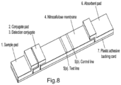
- a suitable lateral flow configuration is shown schematically in Figure 8 . It comprises a sample pad (1) to which the sample may be added; a conjugate pad (2) which may contain detection moieties (3) such as labelled binding reagents; a suitable membrane, such as nitrocellulose, comprising a test capture line (5a) and optionally a control capture line (5b); and optionally an absorbent pad (6). These components may be assembled on a backing material, such as a plastic adhesive backing card.
- the sample comprises filtered and/or washed cells (e.g. neutrophils).
- a suitable lateral flow device may comprise:
- the first test strip may comprise a zone in which lactate is detected and/or the level of lactate is determined.
- An exemplary format is shown in Figure 12 .
- the first test strip may be configured to perform a lactate assay that may be based on the enzymatic activity of lactate dehydrogenase.
- the lactate assay may rely on lactate dehydrogenase enzyme a dye such as a yellow tetrazole (3-(4,5-dimethylthiazol-2-yl)-2, 5-diphenyltetrazolium bromide (MTT)) which can be converted to a purple precipitate called Formazan ((E, Z)-5-(4, 5-dimethylthiazol-2-yl)-1, 3-diphenylformazan) in the presence of lactate through the reduction of NAD+ to NADH during its conversion to pyruvate.
- MTT 5-diphenyltetrazolium bromide
- Dye formation may be assessed visually or measured via other means.
- the test strip may comprise, e.g. in immobilised form, lactate dehydrogenase and a dye in the form of a yellow tetrazole (3-(4,5-dimethylthiazol-2-yl)-2, 5-diphenyltetrazolium bromide (MTT)).
- lactate dehydrogenase lactate dehydrogenase
- a dye in the form of a yellow tetrazole (3-(4,5-dimethylthiazol-2-yl)-2, 5-diphenyltetrazolium bromide (MTT) e.g. in immobilised form, lactate dehydrogenase and a dye in the form of a yellow tetrazole (3-(4,5-dimethylthiazol-2-yl)-2, 5-diphenyltetrazolium bromide (MTT)
- the sample Prior to assaying the lactate concentration, the sample may be filtered to remove red blood cells.
- the sample application pad may trap red blood cells and allow components such as lactate to travel to the detection zone.
- the sample may be applied to a first filter to trap white blood cells; the components not tapped by the first filter may be applied to a second filter to trap red blood cells, such that the lactate levels may be determined in a sample that is free of any cells.
- the sample containing unknown concentrations of analyte may be applied to the sample pad and travel to the conjugate pad where the analyte binds to the detection moiety and travels up the nitrocellulose membrane towards the capture lines.
- the test line comprises an immobilised binding reagent specific for the target analyte. If present in the sample, the analyte (bound to the detection moiety) will form a complex with immobilised binding reagent resulting in a visible test line.
- the control line if present, comprises an immobilised binding reagent specific for a labelled control reagent. A visible label at the control line confirms that the test has run successfully.
- the presence and amount of label at the capture line(s) may be determined by eye or using a suitable lateral flow device reader such as the Cube (Optricon, Germany).
- the testing device, kit, system or composition of matter can be of any form suitable for home use or use in a primary care setting (e.g. clinic).
- the testing device, kit, system or composition of matter may comprise a disposable single use device to which the sample is applied.
- it may comprise a sample application zone to which the sample is added.
- the sample application zone can receive an appropriate volume of sample, for example the volume of one or more drops of blood.
- the device, kit, system or composition of matter typically also incorporates a solid support which defines a liquid/capillary flow path for the sample once applied to the sample application zone. This may be a microfluidic flowpath.
- the sample application zone may be an integral part of the solid support.
- the solid support may comprise a chromatographic medium, such as a membrane material, which may preferably be nitrocellulose.
- a blood sample applied to the sample application zone will typically rehydrate the necessary reagents to detect the marker (such as CD16b).
- a chase fluid (diluent) may also be applied depending on the viscosity of the sample.
- the reagents may include a detection moiety such as a binding reagent which specifically interacts with the marker, such as with CD16b, or a substrate for effector molecules where activity is measured.
- a further reagent may be immobilized further along the flow path. This reagent may bind to the complex of marker and binding reagent.
- the binding reagent is typically labelled to provide a signal at the site of immobilization of the complex of marker and binding reagent (through binding to the further reagent).
- Suitable labels include fluorescent labels, magnetic labels, latex or gold as would be readily understood by one skilled in the art.
- the binding reagent and further reagent are typically antibodies (as defined herein).
- the device, kit, system or composition of matter may comprise a lateral flow test strip.
- the device, kit, system or composition of matter may also include a control zone to confirm sample has passed through the device satisfactorily. In the absence of confirmation by the control zone, the device, kit, system or composition of matter may indicate an invalid result to the user, for example via the display.
- the device, kit, system or composition of matter may act as a competitive or sandwich assay, as discussed herein.
- ELISA enzyme linked immunosorbent assay
- ELISA enzyme linked immunosorbent assay
- typically all reagents to detect the levels of the one or more markers are pre-loaded onto the device, kit, system or composition of matter such that they can interact with the blood sample once added to the device. This minimizes intervention and thus error caused by the subject.
- device, kit, system or composition of matter may only require the user to apply the sample and subsequently observe the output of the assay.
- the device, kit, system or composition of matter may incorporate a suitable reader to provide a quantitative output (in conjunction with a processor and storage medium). This output can be an absolute or a relative output.
- Suitable readers may incorporate an illuminator to expose the device to a specific wavelength or wavelengths of light and a suitable detector for the reflected or emitted light.
- the device, kit, system or composition of matter may also incorporate a suitable processor and computer application to output the determined CD16b or neutrophil level based upon the detected signal.
- the processor running the computer application will be in operable connection with the reader.
- operable connection is meant a functional connection that permits the exchange of a signal or information between the elements.
- the suitable processor and computer application to output the determined CD16b or neutrophil level based upon the detected signal may be incorporated on a remote computing device (e.g. tablet, phone or computer), which is in operable connection with the processor/computer application housed in the reader. This may take the form of a connectivity platform based, for instance, on cloud-based computing services.
- a suitable display module e.g.
- the processor/computer application housed in the reader may be configured to determine the levels of the markers on the device, kit, system or composition of matter and transmit the data to a remote computing device (e.g. tablet, phone or computer) which is configured to analyse the data and output the determined levels.
- the data may be transmitted to a remote computing device (e.g. tablet, phone or computer) via a cloud-based computing service.
- the device, kit, system or composition of matter may comprise one or more specific binding reagents to bind to the marker whose level is detected in the blood sample.
- the reagent may comprise an antibody (to include derivatives, fragments and aptamers).
- the methods provided herein may comprise a step of determining (e.g. on the basis of the CD16b levels) that the subject is in need of a suitable treatment and/or a step of administering a suitable treatment.
- a suitable treatment may comprise or consist of one or more antibiotics.
- the antibiotic is preferably a broad-spectrum antibiotic, or a mixture of 2 or more antibiotics.
- Suitable antibiotics include macrolides (e.g. azithromycin, clarithromycin), cephalosporins (e.g. cefuroxime, cefpodoxime, cefdinir), ketolides (e.g. telithromycin), fluoroquinolones (e.g.
- moxifloxacin gemifloxacin, levofloxacin
- doxycycline trimethoprim/sulfamethoxazole
- amoxicillin/clavunate may be combined with one or more other treatments as appropriate.
- the inventors have demonstrated that a successful assay could be established for the measurement of recombinant CD16b in buffer when a combination of polyclonal antibody AF1597 was deposited on the Nitrocellulose at 1 mg/mL and also conjugated to gold at 30 ⁇ g/mL. Through optimisation of the system, an assay range of 10 -1,000 ng/mL was achieved.
- test system was then progressed to evaluate matrix effects using plasma to see if the assay would spike and recover in a simpler matrix than whole blood.
- results showed that the test system could measure CD16b in plasma and gave a curve from 50-1,000 ng/mL.
- Buffy coat preparations were then investigated using a histopaque gradient separation method, whereby blood is separated into erythrocytes and a plasma/cells mix which could be further processed in order to gain a reproducible white blood cell preparation. These cells were investigated neat and with the addition of lysis/solubilisation buffer and it was shown that the assay signal would increase with lysis indicating the release of "intracellular" CD16b from the cells.
- the buffer type, molarity and pH can affect the antibody binding to the gold particles because the binding is largely due to the charge and hydrophobic interactions.
- 20 mM buffers were made up at the pH ranging from 6.7 to 9.3
- the antibody was loaded at 15 ⁇ g/mL for all buffers investigated. A volume of 1.5 ⁇ L antibody was added to the bottom of a clean 96 plate well, 5 ⁇ L of conjugation buffer was then added and finally 100 ⁇ L of gold OD5. These are incubated for 10 minutes at room temperature whilst stationary. The plate was then read to measure absorbance using a plate reader at 550 and 600nm; the ratio of 550/600 was then calculated to give an aggregation ratio. If the aggregation ratio was ⁇ 3.5 then the buffer was deemed suitable for conjugation.
- the optimal loading of the antibody was determined.
- the loading concentrations 0 ⁇ g/mL to 30 ⁇ g/mL in 5 ⁇ g/mL increments were investigated in the buffer of choice.
- the respective volume of antibody (0, 0.5, 1.0 ⁇ L etc...) were added to the bottom of a clean 96 well plate, 5 ⁇ L of conjugation buffer was added followed by 100 ⁇ L of OD5 gold. Again, these were incubated for 10 minutes at room temperature. Once incubated, 10 ⁇ L of 1M NaCl was placed into each well. If the gold was not sufficiently coated, then the concentrated salt causes the gold conjugate to become unstable and aggregate, indicated by a colour change from bright pink to purple/grey.
- the plate was read on a plate reader and, again, an absorbance of 550 and 600 nm was measured. The ratio of 550/600 was taken and if the conjugate again met an aggregation ratio of ⁇ 3.5 then it was deemed suitable.
- the conjugation of the antibody at 15 ⁇ g/mL was achieved by the following method:
- the conjugation of the antibody at 20 ⁇ g/mL was achieved by the following method:
- the conjugation of the antibody at 30 ⁇ g/mL was achieved by the following method:
- NCs Three different NCs were plotted (CN95, CN140 and CN180) using the CD16b antibody of interest, these have differing pore sizes and hence run at different speeds.
- the antibody was prepared in PBS + 1% Sucrose to a final concentration of between 0.5 mg/mL-1 mg/mL.
- the antibody was plotted in 10 cm bands and then dried at 37 °C for a minimum of 30 minutes.
- Strip Construction The antibody plotted onto NC bands were mounted on adhesive backing card at the base of the card.
- the absorbent pad/sink pad was mounted at the top of the backing card with a 7mm overlap on the NC. These bands were then cut into 3mm wide strips using the Biodot Guillotine Cutter.
- the first experiments performed involved the conjugation of selected antibodies to gold nanoparticles with a buffer screen (at a set 15 ⁇ g/mL loading). 8 buffers were chosen to give the widest range of pH and buffer type. The antibodies that were tested were AF1597, Mab 1597 and NBP-2.
- the absorbance for the gold particles was read at 550 and 600 nm, a ratio of 550/600nm was taken; if the ratio was found to be ⁇ 3.5 then it met the criteria required to be suitable for conjugation. The results for this can be found in Table 1.
- NC nitrocellulose
- PBS + 1% sucrose a solution of PBS + 1% sucrose
- Table 2 Raw data obtained for hand deposited antibodies (in PBS + 1% sucrose) on CN140 strips with varying gold conjugations at different concentrations of CD16b analyte.
- the non-specifics for the AF1597 pair system are low for all three golds tested and all show a specific test line signal over the range tested.
- the 100 and 1,000 ng/mL gave specific signal; these signals were comparable when tested on the Isoflow plotted antibody system.
- NC Concentration Antibody loading on gold 15 ng/mL 20 ng/mL 30 ⁇ g/mL AF1597 on CN95 0 ng/mL 1.3 1.7 1.3 10 ng/mL 5.4 6.2 15 50 ng/mL 47 54 41 100 ng/mL 96 86 84 200 ng/mL 94 110 100 1000 ng/mL 98 122 137
- the 20 ⁇ g/mL gold conjugate showed an increase in signal for all concentrations tested when compared to the 15 ⁇ g/mL coat; the lowest increase in specific signal was seen between 200 and 1,000 ng/mL. This suggested the higher gold loading gave an improvement in the assay performance by giving a larger range to the assay. This was more apparent when the 30 ⁇ g/mL coat was tested, with a specific signal gain at both the low end (10 ng/mL) and an enhanced specific signal gain between 200 and 1,000 ng/mL. This suggested that the best gold conjugate loading concentration at this stage was 30 ⁇ g/mL.
- NC membrane Another change that can improve assay sensitivity and range of a lateral flow assay is the NC membrane used.
- Table 8 Raw data obtained for AF1597 antibody deposited on CN95, CN140 and CN180 (in PBS + 1% sucrose) with AF1597 conjugated to gold with varying concentrations of analyte.
- the system taken forward for testing in plasma and blood samples was CN95 with 1 mg/mL AF1597 antibody deposited in PBS + 1% sucrose and AF1597 loaded at 30 ⁇ g/mL on gold.
- CD16b could be spiked and recovered in a simpler matrix than blood.
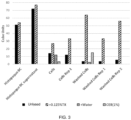
- the matrix chosen for this was plasma, with whole blood being drawn and collected in EDTA and lithium heparin tubes. Once collected, the blood was spun down at 1,228x g for 10 minutes, the blood separates into three distinct layers at this point; plasma, buffy coat and erythrocytes. The plasma was collected from the top layer and placed into polypropylene tubes. The analyte (CD16b) was then spiked in the plasma and run on the strips to see if it could be recovered successfully; the results for this experiment can be found in Table 9 and Fig.1 . Table 9.
- a histopaque gradient separation column method was used. This involved carefully layering 2 mL of blood over 1 mL of histopaque and leaving it at room temperature for 145 mins. This then gently separates the erythrocytes from the plasma and white blood cells to leave a plasma/cells mix that can be removed without disturbance of the erythrocytes. See Fig. 2 .
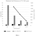
- Example 3 To confirm the results of Example 3 that the signal obtained from the lysed cells correlated to the number of cells present, five further blood donors were investigated.
- the cells were prepared and washed as described above. The cells were prepared at different dilutions to gain a better idea of how the cell number changes affect the assay.
- the dilutions prepared for the first blood sample were 4x, 2x, neat and 1/2 cells.
- Blood donors 1113, 1114, 1117 and 1118 were prepared at 2x, neat and 1/2 cells.
- 800 ⁇ L plasma/cell mix was centrifuged, the cells were resuspended in 400 ⁇ L PBS.
- results show a good reproducibility of results for 0, 2 and 3.5 hour time points. At 24 hours, the results began to plateau suggesting that the blood needed to be processed or sampled prior to this time. From this data it can be determined that the blood can be stored at ambient temperature for 3.5 hours without impact.
- RP02 A new antibody (RP02) was identified for testing as it was cheaper than the AF1597 antibody. To determine RP02's suitability, a gold optimisation test was performed with the antibody (as was described in Example 1). The results for the aggregation ratio testing (which informs which buffer is most suitable for conjugation) can be found in Figure 9 .
- RP02 was deposited onto CN140 nitrocellulose in 1% sucrose and tested against AF1597 AuC (at 30 ⁇ g/mL). The initial results showed that the use of RP02 on the test line gave an increase in non-specific binding (NSB) whilst giving a good specific signal increase.
- NBS non-specific binding
- the running buffer was changed from PBS to PBST.
- the system was also tested using two different CD16b antigens (sourced from Sinobio and R&D systems) which were used to produce the two test antibodies, RP02 and AF1597.
- RP02/AF1597 test strips using CN95 NC with lysed washed cells
- two blood donors were obtained and the white blood cells processed as described in Example 3.
- the RP02/AF1597 system was then compared to the AF1597 self-pair to ensure good comparability between the two systems.
- the various processed components of the blood were also measured to include, plasma (from spun blood), plasma (buffy coat after Histopaque treatment) and different concentrations of the washed cells.
- the cube units obtained for the washed cells at the different concentrations gave very comparable results for both systems. Differences were observed when measuring the different plasmas for the two donors.
- the AF self-pair system gave very comparable results for the spun and buffy coat plasmas when both donors were measured.
- the RP02 system showed differences between the types of plasma, with the spun plasma giving a much lower value when compared to the buffy coat plasma.
- the reagents were dried down. Initially, the volume of the gold conjugate (2.4, 4 and 6 ⁇ L) to be dried down was investigated to determine the best specific signal gain.
- the CD16b standards (Sinobio) were initially spiked into PBST and were also spiked into PBS for comparison.
- the 200 ng/ml standard gave very comparable results for all volume of gold conjugate.
- the 500 ng/ml standard was found to be highest on the 3 ⁇ L deposit which was then lower but comparable for both the 4 and 5 ⁇ L deposits.
- the 1000 ng/ml standard was very comparable for the 3 and 4 ⁇ L deposits which then decreased at 5 ⁇ L of deposit.
- the best condition was seen with 4 ⁇ L of gold deposit as there was good specific signal difference for all concentrations of CD16b tested, comparable to a high concentration of analyte with wet gold conjugate at the same volume.
- the AF1597 AuC conjugate was sprayed onto the two pads using an Isoflow plotter.
- polystreptavidin-biotin system was evaluated; this can also help reduce costs as antibodies are typically more expensive.
- polystreptavidin was plotted on the test line of CN140 NC and the antibodies RP02 or AF1597 conjugated to biotin and AF1597-gold were combined and dotted onto the conjugate pad.
- the biotinylated and gold antibodies form a sandwich with CD16b and the resulting complex will then bind to the polystreptavidin test line.
- a slower CN140 NC was used in this set up to allow more time for the sandwich to form before reaching the test line.
- the antibodies RP02 and AF1597 were biotinylated using a Biotinylation Lightning Link Kit (@ 2 mg/ml) and then diluted 1 in 100 to a final concentration of 16.7 ⁇ g/ml.
- 4 ⁇ L of the biotin conjugate was mixed with 4 ⁇ L of the gold conjugate and 20 ⁇ L of CD16b analyte in PBS.
- the biotin system was dried down to assess its performance and suitability as a system to be taken forward. As the dry down requires two components (gold and biotin conjugate), it was decided to investigate if the two conjugates could be dried down mixed together or if they needed to be separated on the pad.
- Example 9 Trapping and lysis of white blood cells within the CD16b lateral flow device
- washed white blood cells The trapping and lysis of washed white blood cells was demonstrated within the CD16b lateral flow assay utilising a filter in the form of a sample pad membrane under the sample application window to trap the cells. Washed white blood cells from EDTA blood were used. It was determined that a range of filters may be used to trap neutrophils, for example PES10 (2 orientations), MLRF-NANO-1 (LF1) and MLRF-NANO-2 (LF2) or no cell membrane (filter). It was determined that a range of surfactants may be used for the cell lysis step, for example 0.03%TX-100, 0.03125%SDS or 0.0125% Sarkosyl.
- PES10 2 orientations
- MLRF-NANO-1 MLRF-NANO-1
- LF2 MLRF-NANO-2
- surfactants may be used for the cell lysis step, for example 0.03%TX-100, 0.03125%SDS or 0.0125% Sarkosyl.
- the test strip was prepared using a CD16b antibody-gold conjugate sprayed and dried onto two different conjugate pads (8951 & 8980; Ahlstrom Munksjo). Dry strips were constructed using conjugate pads at the base, overlapping with a CN95 nitrocellulose membrane (Sartorius) plotted with an anti CD16b antibody (11046-RP02-SiB; Stratech) as a test line, then a sink pad at the top.
- CD16b standards (Stratech) were spiked into PBS at a range of concentrations from 10-1000ng/mL. 50 ⁇ L of each standard was run up the dry strips followed by 30 ⁇ L of PBS and then the test read @ 10minutes on a cube reader.
- Table 15 Raw data obtained for varying concentrations of CD16b in PBS with dried sprayed gold conjugate on two conjugate pads (8951 and 8980) with a 10 minute read time. Standard 10 min read CD16b ng/mL 8951(CP) 8980(CP) 0 5.6 9.6 10 9.8 25 50 32 41 100 55 71 200 65 83 500 95 134 1000 152 157
- the format of the Lactate assay is based on the enzymatic activity of lactate dehydrogenase. This was achieved by immobilisation of the lactate dehydrogenase enzyme on a nitrocellulose membrane and the inclusion of a dye in the form of a yellow tetrazole (3-(4,5-dimethylthiazol-2-yl)-2, 5-diphenyltetrazolium bromide (MTT)) which can be converted to a purple precipitate called Formazan ((E, Z)-5-(4, 5-dimethylthiazol-2-yl)-1, 3-diphenylformazan) in the presence of lactate through the reduction of NAD+ to NADH during its conversion to pyruvate. (NB: other reagents can be used here to form different formazans).
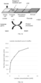
- the overall proposed format can be found in Figure 12 .
- a dry lateral flow assay was constructed using a dye mix (containing MTT) deposited and dried onto a conjugate pad (8951; Ahlstrom), overlapping with a CN95 nitrocellulose membrane with an LDH test line plotted at 1mg/mL.
- Sodium Lactate was spiked into PBS at a range of concentrations from 0.1-10mM and 80 ⁇ L of each standard was run up the dry strips (in triplicate) and read at 10minutes on a cube reader. The results for this testing can be found in Table 16 and Figure 13 .
- Table 16 Raw data obtained for varying concentrations of Sodium Lactate, dye mix dried on conjugate pad & triplicate LDH test line signal measured in Cube units.
- the strip design for whole blood requires a blood separator to retain the cells and stop them being lysed prior to running the CD16b portion of the assay. This blood separator will allow the plasma to be run through the strip which will give the lactate assay result.
- the initial volume of whole blood to be tested was 20 ⁇ L with an additional 50 ⁇ L of PBS to potentially fully wash all the plasma from the blood (and therefore from the sample pad). It was hoped that the higher volume of blood would give a higher volume of plasma, and thus, would give a better lactate response.
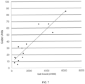
- Example 12 Comparative data for correlation of the levels of soluble CD16b vs. membrane-anchored and intracellular CD16b with the number of neutrophils in a sample
- the correlation of membrane-anchored and intracellular CD16b with the number of neutrophils is substantially better than the correlation of soluble CD16b with the number of neutrophils.
- detection of the levels of membrane-anchored and intracellular CD16b provides a more accurate method for determining the number of neutrophils in the sample.
- strips were constructed using a CN95 nitrocellulose membrane (Sartorius) at the base, plotted with an anti CD16b antibody (AF1597) as a test line, then a sink pad at the top.
- CD16b standards R&D Systems ) were spiked into PBS at a range of concentrations from 5-200ng/mL. 20 ⁇ L of each standard was mixed with 2.4uL of AF Gold conjugate mixed and run up the wet strips followed by 30 ⁇ L of PBS and then the test read @ 10minutes on a cube reader. The results for this testing can be found in Table 17 and Figure 1 .
- Table 17 Raw data obtained for varying concentrations of CD16b in PBS in a wet assay format with a 10 minute read time.
Landscapes
- Health & Medical Sciences (AREA)
- Life Sciences & Earth Sciences (AREA)
- Immunology (AREA)
- Engineering & Computer Science (AREA)
- Hematology (AREA)
- Molecular Biology (AREA)
- Chemical & Material Sciences (AREA)
- Urology & Nephrology (AREA)
- Biomedical Technology (AREA)
- Cell Biology (AREA)
- Analytical Chemistry (AREA)
- Biochemistry (AREA)
- Biotechnology (AREA)
- Pathology (AREA)
- General Physics & Mathematics (AREA)
- Food Science & Technology (AREA)
- Medicinal Chemistry (AREA)
- Physics & Mathematics (AREA)
- General Health & Medical Sciences (AREA)
- Microbiology (AREA)
- Zoology (AREA)
- Tropical Medicine & Parasitology (AREA)
- Virology (AREA)
- Investigating Or Analysing Biological Materials (AREA)
- Electrical Discharge Machining, Electrochemical Machining, And Combined Machining (AREA)
- Crystals, And After-Treatments Of Crystals (AREA)
- Spectrometry And Color Measurement (AREA)
Claims (18)
- In-vitro-Verfahren zum Bestimmen des Neutrophilenspiegels in einer Probe durch Bestimmen des Spiegels von CD16b, wobei das CD16b die intrazelluläre Form von CD16b umfasst.
- In-vitro-Verfahren zum Vorhersagen, Diagnostizieren, Ausschließen und/oder Monitoring von Neutropenie, neutropenischer Sepsis und/oder nicht-neutropenischer Sepsis in einem Individuum, wobei das Verfahren Bestimmen des Spiegels von CD16b in einer Probe umfasst, wobei das CD16b die intrazelluläre Form von CD16b umfasst.
- Verfahren nach einem der vorhergehenden Ansprüche, wobei das CD16b die membranverankerte Form von CD16b und die intrazelluläre Form von CD16 umfasst, im Wesentlichen daraus besteht oder daraus besteht.
- Verfahren nach einem der vorhergehenden Ansprüche, wobei(i) das Verfahren ferner einen Schritt des Entfernens mindestens eines Anteils und vorzugsweise das meiste oder alles der löslichen Form von CD16b aus der Probe vor dem Bestimmen des Spiegels von CD16b umfasst;(ii) das Verfahren an einer Probe durchgeführt wird, aus welcher zuvor mindestens ein Teil und vorzugsweise das meiste oder alles der löslichen Form von CD16b entfernt wurde,
wobei optional der Schritt des Entfernens mindestens eines Teils der löslichen Form von CD16b aus der Probe unter Bedingungen durchgeführt wird, welche mindestens einen großen Teil oder im Wesentlichen alle der Neutrophilen, welche in der Probe vorhanden sind, intakt halten. - Verfahren nach Anspruch 4, wobei der Schritt des Entfernens mindestens eines Teils der löslichen Form von CD16b ein Filtrationsschritt ist, wobei der Filtrationsschritt ergibt:(i) eine gefilterte Probe, umfassend weiße Blutkörperchen, wobei die gefilterte Probe an den löslichen Komponenten der Probe verarmt ist; und(ii) eine Lösung, umfassend die lösliche Form von CD16b; wobei die Lösung vorzugsweise frei von weißen Blutkörperchen ist, aber vorzugsweise rote Blutkörperchen enthält, wobei optional der Filtrationsschritt unter Bedingungen durchgeführt wird, welche mindestens einen großen Teil oder im Wesentlichen alle der Neutrophilen, welche in der Probe vorhanden sind, intakt halten.
- Verfahren nach einem der vorhergehenden Ansprüche, wobei der Spiegel von CD16b unter Verwendung eines oder mehrerer CD16b-bindender Antikörper bestimmt wird.
- Verfahren nach einem der vorhergehenden Ansprüche, wobei der Neutrophilenspiegel als über oder unter einem oder mehreren vorbestimmten Schwellenwerten liegend bestimmt wird, wobei:(i) ein Neutrophilenspiegel unter einem vorbestimmten Schwellenwert indikativ für Neutropenie, neutropenische Sepsis, ein signifikantes Risiko des Entwickelns von Neutropenie und/oder ein signifikantes Risiko des Entwickelns von neutropenischer Sepsis ist;(ii) ein Neutrophilenspiegel über einem vorgegebenen Schwellenwert indikativ für das Fehlen oder ein unbedeutendes Risiko der Anwesenheit oder Entwicklung von Neutropenie und/oder neutropenischer Sepsis ist;(iii) ein Neutrophilenspiegel über einem vorgegebenen Schwellenwert indikativ für nicht-neutropenische Sepsis oder ein signifikantes Risiko des Entwickelns einer nicht-neutropenischen Sepsis ist; oder(iv) ein Neutrophilenspiegel unterhalb eines vorbestimmten Schwellenwerts indikativ für das Fehlen oder ein unbedeutendes Risiko der Anwesenheit oder Entwicklung von nicht-neutropenischer Sepsis ist, wobei optional ein oder mehrere Schwellenwerte verwendet werden, wobei der eine oder mehrere Schwellenwerte ausgewählt sind aus:(a) weniger als 1000 neutrophile Zellen/µl Blut, was indikativ für Neutropenie, neutropenische Sepsis, ein signifikantes Risiko des Entwickelns von Neutropenie und/oder ein signifikantes Risiko des Entwickelns von neutropenischen Sepsis ist;(b) 1000-1500 neutrophile Zellen/µl Blut, was indikativ für eine Notwendigkeit ist, das Individuum weiter auf die Entwicklung von Neutropenie und/oder neutropenischer Sepsis zu überwachen;(c) mehr als 1500 neutrophile Zellen/µl Blut, was indikativ für das Fehlen oder ein unbedeutendes Risiko der Anwesenheit oder Entwicklung von Neutropenie und/oder neutropenischer Sepsis ist;(d) mehr als 7500 neutrophile Zellen/µl Blut, was indikativ für nicht-neutropenische Sepsis oder ein signifikantes Risiko des Entwickelns nicht-neutropenischen Sepsis ist; und/oder(e) weniger als 7500 neutrophile Zellen/µl Blut, was indikativ für das Fehlen von nicht-neutropenischen Sepsis oder ein unbedeutendes Risiko der Anwesenheit oder Entwicklung von nicht-neutropenischer Sepsis ist.
- Verfahren nach einem der vorhergehenden Ansprüche, wobei die Probe von einem Individuum stammt, welches (i) Strahlung ausgesetzt wurde; (ii) ein Medikament erhalten hat oder erhält, welches in der Lage ist, Neutropenie zu verursachen; (iii) an HIV, Hepatitis und/oder einer Autoimmunerkrankung leidet; (iv) an Krebs leidet und/oder eine Chemotherapie und/oder Strahlentherapie erhalten hat oder erhält; (v) Symptome einer Infektion zeigt oder erlebt; und/oder (vi) sich kürzlich einer Operation unterzogen hat.
- Verfahren nach einem der vorhergehenden Ansprüche, wobei das Verfahren umfasst:(a) einen Schritt der Zelllyse vor Bestimmen des Spiegels von CD16, wobei optional der Schritt der Zelllyse durchgeführt wird:(i) nach einem Schritt des Entfernens mindestens eines Teils des löslichen CD16b; oder(ii) an einer Probe, aus der zuvor mindestens ein Teil des löslichen CD16b entfernt wurde; und/oder(b) Detektieren eines oder mehrerer verschiedener Marker, welche indikativ für die Anwesenheit von neutropenischer oder nicht-neutropenischer Sepsis in der Probe sind.
- Verfahren nach einem der vorhergehenden Ansprüche, wobei der Spiegel von CD16b durch einen Lateral-Flow-Assay bestimmt wird.
- Verfahren nach einem der vorhergehenden Ansprüche, bei dem mindestens 2, 3, 4, 5, 6, 7, 8, 9 oder 10 oder mehr Proben von dem Individuum zu unterschiedlichen Zeiten entnommen werden und die Spiegel von CD16b und optional ein oder mehrere weitere Marker bestimmt werden und wobei die Proben alle 6 bis 24 Stunden, täglich oder alle 2, 3, 4, 5, 6, 7 oder 14 Tage entnommen werden.
- Verfahren zum Vorhersagen der Ansprechbarkeit eines Individuums auf Behandlung mit einem Antibiotikum und/oder Selektieren eines Individuums zur Behandlung mit einem Antibiotikum, umfassend Durchführen des Verfahrens nach einem der vorhergehenden Ansprüche und Vorhersagen der Ansprechbarkeit und/oder Selektieren des Individuums zur Behandlung, wobei Neutropenie und/oder neutropenische Sepsis oder nicht-neutropenische Sepsis auf der Basis des bestimmten Neutrophilenspiegels vorhergesagt oder diagnostiziert wird.
- Verfahren nach einem der Ansprüche 1-12, umfassend die Schritte:i. Zugeben einer Probe zu einer ersten Probeanwendungszone eines ersten Teststreifens, um jegliche Neutrophile auf einem Filter einzufangen, welcher in dem ersten Teststreifen enthalten ist;ii. Transferieren des Filters mit eingefangenen Neutrophilen zu einer zweiten Probeanwendungszone eines zweiten Teststreifens;iii. Zugeben eines Lysereagens zu dem Filter, um die Neutrophile zu lysieren;
undiv. Bestimmen des Spiegels von CD16b, wobei optional der Schritt des Bestimmens des Spiegels von CD16b umfasst:i. Kontaktieren einer Lösung der lysierten Neutrophile mit einer Konjugatzone, umfassend mindestens eine markierte Detektionsgruppe, welche an CD16b bindet;ii. Kontaktieren der Mischung der lysierten Neutrophile und der mindestens einen markierten Detektionsgruppe, welche an CD16b bindet, mit:(i) einer Testlinie für mindestens CD16b, wobei die Testlinie eine immobilisierte weitere Detektionsgruppe umfasst, welche ebenfalls an CD16b bindet, wodurch das CD16b an der Testlinie immobilisiert wird, um über die markierte Detektionsgruppe, welche ebenfalls an dem CD16b gebunden ist, ein Signal zu erzeugen; oder(ii) einer Testlinie für mindestens CD16b, wobei die Testlinie eine immobilisierte weitere Detektionsgruppe umfasst, welche an die CD16b-gebundene markierte Detektionsgruppe von Schritt ii. bindet, wodurch diese an der Testlinie immobilisiert wird, und ein Signal erzeugt. - Verfahren nach Anspruch 13, wobei(a) das Lysereagenz ein Tensid ist, welches ionisch oder nicht-ionisch sein kann;(b) die markierte Detektionsgruppe, welche an CD16b bindet, ein Anti-CD16b-Antikörper ist, welcher an Goldpartikel konjugiert ist;(c) die weitere Detektionsgruppe ein Anti-CD16b-Antikörper ist, vorzugsweise ein polyklonaler Antikörper;(d) der Schritt des Zugebens der Probe zu der ersten Anwendungszone von einem Schritt des Zellwaschens gefolgt wird; und/oder(e) der Filter eine Membran ist, welche zum Einfangen von Neutrophilen geeignet ist, wie beispielsweise eine mikroporöse Membran.
- Testvorrichtung oder Testkit, umfassend:a. eine Probenaufnahmezone, zu der eine Probe von einem Individuum zugegeben wird, wobei die Probenaufnahmezone einen Filter umfasst, welcher Neutrophile einfängt, und wobei die Probenaufnahmezone stromaufwärts einer Konjugatzone liegt;b. die Konjugatzone, umfassend mindestens eine markierte Detektionsgruppe, welche spezifisch an CD16b bindet;c. einen festen Träger, welcher einen Flüssigkeitsströmungspfad für die Probe definiert und umfassend(i) eine Testlinie für mindestens CD16b, wobei die Testlinie eine immobilisierte weitere Detektionsgruppe umfasst, welche ebenfalls spezifisch an CD16b bindet, wodurch das CD16b an der Testlinie immobilisiert wird, um über die markierte Detektionsgruppe, welche auch an das CD16b gebunden ist, ein Signal zu erzeugen; oder(ii) eine Testlinie für mindestens CD16b, wobei die Testlinie eine immobilisierte weitere Detektionsgruppe umfasst, welche spezifisch an die an CD16b gebundene markierte Detektionsgruppe von Schritt (b) bindet, wodurch diese an der Testlinie immobilisiert wird, und ein Signal erzeugt; und optional ferner umfassend:d. mindestens ein markiertes Kontrollbindungsreagenz, welches an einen Bindungspartner bindet, welcher an einer Kontrolllinie stromabwärts der Testlinie für das CD16b immobilisiert ist, und somit bestätigt, dass der Test erfolgreich abgeschlossen wurde; und optional ferner umfassend:e. ein absorbierendes Material stromabwärts der Test- (und Kontroll-, falls vorhanden) Linien, um überschüssige Probe zu absorbieren.
- Testvorrichtung oder Testkit nach Anspruch 15, wobei (i) der feste Träger ein chromatographisches Medium und/oder eine Kapillarflussvorrichtung umfasst; und/oder (ii) die Testvorrichtung oder das Testkit ein Teststreifen sind.
- Eine Testvorrichtung oder ein Testkit, umfassend:a. einen ersten Teststreifen, umfassend eine erste Probenanwendungszone, zu welcher eine Probe von einem Individuum zugegeben wird, wobei die erste Probenanwendungszone einen Filter umfasst, welcher die weißen Blutkörperchen einfängt;b. einen zweiten Teststreifen, umfassend:i. eine zweite Probenanwendungszone, zu welcher der Filter mit eingefangenen weißen Blutkörperchen zugegeben wird;ii. eine Konjugatzone, umfassend mindestens eine markierte Detektionsgruppe, welche spezifisch an CD16b bindet;iii. einen festen Träger, welcher einen Flüssigkeitsströmungspfad für die Probe definiert und umfassend (i) eine Testlinie für mindestens CD16b, wobei die Testlinie eine immobilisierte weitere Detektionsgruppe umfasst, welche ebenfalls spezifisch an CD16b bindet, wodurch das CD16b an der Testlinie immobilisiert wird, um über die markierte Detektionsgruppe, welche auch an das CD16b gebunden ist, ein Signal zu erzeugen; oder (ii) eine Testlinie für mindestens CD16b, wobei die Testlinie eine immobilisierte weitere Detektionsgruppe umfasst, welche spezifisch an die CD16b-gebundene markierte Detektionsgruppe von Schritt ii. bindet, wodurch diese an der Testlinie immobilisiert wird, und ein Signal erzeugt; und optional ferner umfassend:iv. mindestens ein markiertes Kontrollbindungsreagens, welches an einen Bindungspartner bindet, welcher an einer Kontrolllinie stromabwärts der Testlinie für das CD16b immobilisiert ist, und somit bestätigt, dass der Test erfolgreich abgeschlossen wurde; und optional ferner umfassend:v. ein absorbierendes Material stromabwärts der Test- (und Kontroll-, falls vorhanden) Linien, um überschüssige Probe zu absorbieren.
- System oder Testkit zum Diagnostizieren oder Monitoring eines Individuums, umfassend:a. eine Testvorrichtung zum Bestimmen von Spiegeln von mindestens CD16b in einer Probe nach einem der Ansprüche 15-17;b. einen Prozessor; undc. ein Speichermedium, umfassend eine Computerapplikation, welche, wenn sie vom Prozessor ausgeführt wird, dazu eingerichtet ist:i. auf die bestimmten Spiegel von mindestens CD16b in der Probe auf der Testvorrichtung zuzugreifen oder zu berechnen;ii. ein Testergebnis aus den Spiegeln von mindestens CD16b in der Probe zu berechnen, einschließlich eines Vergleichs der Spiegel mit einem oder mehreren Schwellenwerten und/oder CD16b-Spiegeln, welche zu einem oder mehreren früheren Zeitpunkten bestimmt wurden, um dadurch Neutroprenie und/oder neutroprenische Sepsis vorherzusagen oder zu diagnostizieren; undiii. das vorhergesagte oder diagnostiziere Ergebnis für das Individuum von dem Prozessor auszugeben.
Applications Claiming Priority (2)
| Application Number | Priority Date | Filing Date | Title |
|---|---|---|---|
| GBGB2004972.2A GB202004972D0 (en) | 2020-04-03 | 2020-04-03 | Blood analysis method |
| PCT/GB2021/050813 WO2021198693A1 (en) | 2020-04-03 | 2021-04-01 | Method for detection of cd16b |
Publications (3)
| Publication Number | Publication Date |
|---|---|
| EP4127719A1 EP4127719A1 (de) | 2023-02-08 |
| EP4127719B1 true EP4127719B1 (de) | 2024-11-13 |
| EP4127719B8 EP4127719B8 (de) | 2024-12-18 |
Family
ID=70768817
Family Applications (1)
| Application Number | Title | Priority Date | Filing Date |
|---|---|---|---|
| EP21717178.4A Active EP4127719B8 (de) | 2020-04-03 | 2021-04-01 | Verfahren zur detektion von cd16b |
Country Status (8)
| Country | Link |
|---|---|
| US (1) | US20230138656A1 (de) |
| EP (1) | EP4127719B8 (de) |
| CN (1) | CN115485557A (de) |
| AU (1) | AU2021250702A1 (de) |
| CA (1) | CA3173438A1 (de) |
| ES (1) | ES3003911T3 (de) |
| GB (1) | GB202004972D0 (de) |
| WO (1) | WO2021198693A1 (de) |
Families Citing this family (1)
| Publication number | Priority date | Publication date | Assignee | Title |
|---|---|---|---|---|
| GB2611355B (en) * | 2021-10-04 | 2025-07-30 | 52 North Health Ltd | Assay device for a suspension |
Family Cites Families (4)
| Publication number | Priority date | Publication date | Assignee | Title |
|---|---|---|---|---|
| WO2002033408A1 (en) * | 2000-10-17 | 2002-04-25 | Besst-Test Aps | Assay for directly detecting an inflammatory indicator in a body fluid sample |
| WO2011060203A1 (en) * | 2009-11-11 | 2011-05-19 | The Trustees Of Columbia University In The City Of New York | Methods for assessing the immune system in a patient |
| US9097712B2 (en) * | 2013-03-15 | 2015-08-04 | Allied Innovative Systems Llc | Flow-through cell counting assay |
| IL264431B2 (en) * | 2016-07-28 | 2024-06-01 | Macfarlane Burnet Institute For Medical Res And Public Health Limited | Estimating cellular populations |
-
2020
- 2020-04-03 GB GBGB2004972.2A patent/GB202004972D0/en not_active Ceased
-
2021
- 2021-04-01 CN CN202180032891.7A patent/CN115485557A/zh active Pending
- 2021-04-01 AU AU2021250702A patent/AU2021250702A1/en active Pending
- 2021-04-01 CA CA3173438A patent/CA3173438A1/en active Pending
- 2021-04-01 EP EP21717178.4A patent/EP4127719B8/de active Active
- 2021-04-01 WO PCT/GB2021/050813 patent/WO2021198693A1/en not_active Ceased
- 2021-04-01 US US17/916,415 patent/US20230138656A1/en active Pending
- 2021-04-01 ES ES21717178T patent/ES3003911T3/es active Active
Also Published As
| Publication number | Publication date |
|---|---|
| CA3173438A1 (en) | 2021-10-07 |
| ES3003911T3 (en) | 2025-03-11 |
| EP4127719B8 (de) | 2024-12-18 |
| US20230138656A1 (en) | 2023-05-04 |
| AU2021250702A1 (en) | 2022-11-03 |
| EP4127719A1 (de) | 2023-02-08 |
| CN115485557A (zh) | 2022-12-16 |
| WO2021198693A1 (en) | 2021-10-07 |
| GB202004972D0 (en) | 2020-05-20 |
Similar Documents
| Publication | Publication Date | Title |
|---|---|---|
| EP0615129B1 (de) | Verfahren zum selektiven Nachweis von perinuklearen anti-neutrophilen cytoplasmischen Antikörpern bei ulzerativen Kolitis oder primärer sclerotischer Cholangitis | |
| EP2783216B1 (de) | Immunoassays mit signalverstärkung | |
| US20090030054A1 (en) | Cytoplasmic antigens for detection of candida | |
| EP2495565B1 (de) | Verfahren und kit zur Bestimmung einer zur überprüfenden Komponente in einer Probe. | |
| US20050227223A1 (en) | Method of judging viral infection | |
| JP3887340B2 (ja) | ノロウイルス又はサポウイルス検体用希釈液及びウイルス検出試薬 | |
| US20050069958A1 (en) | Method for simultaneous evaluation of a sample containing a cellular target and a soluble analyte | |
| NO177444B (no) | Fremgangsmåte for immunometrisk bestemmelse av et antigen, samt utstyrspakke for slik bestemmelse | |
| EP3264085A1 (de) | Immunoassay-verfahren und in besagtem verfahren verwendete testreagenz | |
| EP4127719B1 (de) | Verfahren zur detektion von cd16b | |
| EP3564673B1 (de) | L-fabp-immunoassay-verfahren und in besagtem verfahren verwendetes testreagens | |
| JP6797427B2 (ja) | 被検者が膵臓癌に罹患している可能性を試験する方法 | |
| EP3458853B1 (de) | Verbesserte verfahren zur überwachung des immunstatus einer person | |
| NO872095L (no) | Immunologisk maalemetode og -sett. | |
| US20120238037A1 (en) | Immunological assay and immunological assay kit | |
| Harinath et al. | Field evaluation of ELISA using Wuchereria bancrofti mf ES antigen for bancroftian filariasis | |
| EP0756173A1 (de) | Reagenz zum nachweis von substanzen und verfahren zur diagnose der rheumatoiden arthritis | |
| EP1393076B1 (de) | Verfahren zur vorhersage eines transplantatabstossungsrisikos und immunologischer testkit | |
| US9658227B2 (en) | Recombinant GRA antigens and the use of same for early diagnosis of toxoplasmosis | |
| JPH0972906A (ja) | 腎・尿路系疾患の鑑別診断用キット | |
| JP6873460B2 (ja) | 被検者が膵臓癌に罹患している可能性を試験する方法 | |
| RU2197736C1 (ru) | Реагент для иммуноферментного анализа и способ иммуноферментного тестирования специфических антител при описторхозе, вызываемом печеночной трематодой opisthorhis felineus | |
| JP2010286375A (ja) | 重症薬疹易罹患性診断用マーカー、重症薬疹易罹患性の診断方法および重症薬疹易罹患性診断用キット | |
| US20240210402A1 (en) | Method for detecting oral neoplastic lesion, test reagent, test kit and therapeutic composition | |
| JP3309900B2 (ja) | 抗fkbp12自己抗体の検出方法 |
Legal Events
| Date | Code | Title | Description |
|---|---|---|---|
| STAA | Information on the status of an ep patent application or granted ep patent |
Free format text: STATUS: UNKNOWN |
|
| STAA | Information on the status of an ep patent application or granted ep patent |
Free format text: STATUS: THE INTERNATIONAL PUBLICATION HAS BEEN MADE |
|
| PUAI | Public reference made under article 153(3) epc to a published international application that has entered the european phase |
Free format text: ORIGINAL CODE: 0009012 |
|
| STAA | Information on the status of an ep patent application or granted ep patent |
Free format text: STATUS: REQUEST FOR EXAMINATION WAS MADE |
|
| 17P | Request for examination filed |
Effective date: 20221021 |
|
| AK | Designated contracting states |
Kind code of ref document: A1 Designated state(s): AL AT BE BG CH CY CZ DE DK EE ES FI FR GB GR HR HU IE IS IT LI LT LU LV MC MK MT NL NO PL PT RO RS SE SI SK SM TR |
|
| DAV | Request for validation of the european patent (deleted) | ||
| DAX | Request for extension of the european patent (deleted) | ||
| STAA | Information on the status of an ep patent application or granted ep patent |
Free format text: STATUS: EXAMINATION IS IN PROGRESS |
|
| 17Q | First examination report despatched |
Effective date: 20231016 |
|
| GRAP | Despatch of communication of intention to grant a patent |
Free format text: ORIGINAL CODE: EPIDOSNIGR1 |
|
| STAA | Information on the status of an ep patent application or granted ep patent |
Free format text: STATUS: GRANT OF PATENT IS INTENDED |
|
| INTG | Intention to grant announced |
Effective date: 20240607 |
|
| GRAS | Grant fee paid |
Free format text: ORIGINAL CODE: EPIDOSNIGR3 |
|
| GRAA | (expected) grant |
Free format text: ORIGINAL CODE: 0009210 |
|
| STAA | Information on the status of an ep patent application or granted ep patent |
Free format text: STATUS: THE PATENT HAS BEEN GRANTED |
|
| GRAT | Correction requested after decision to grant or after decision to maintain patent in amended form |
Free format text: ORIGINAL CODE: EPIDOSNCDEC |
|
| AK | Designated contracting states |
Kind code of ref document: B1 Designated state(s): AL AT BE BG CH CY CZ DE DK EE ES FI FR GB GR HR HU IE IS IT LI LT LU LV MC MK MT NL NO PL PT RO RS SE SI SK SM TR |
|
| REG | Reference to a national code |
Ref country code: GB Ref legal event code: FG4D |
|
| REG | Reference to a national code |
Ref country code: CH Ref legal event code: EP |
|
| REG | Reference to a national code |
Ref country code: DE Ref legal event code: R096 Ref document number: 602021021776 Country of ref document: DE |
|
| REG | Reference to a national code |
Ref country code: CH Ref legal event code: PK Free format text: BERICHTIGUNG B8 |
|
| RAP4 | Party data changed (patent owner data changed or rights of a patent transferred) |
Owner name: 52 NORTH HEALTH LTD |
|
| REG | Reference to a national code |
Ref country code: IE Ref legal event code: FG4D |
|
| P01 | Opt-out of the competence of the unified patent court (upc) registered |
Free format text: CASE NUMBER: APP_59405/2024 Effective date: 20241031 |
|
| REG | Reference to a national code |
Ref country code: LT Ref legal event code: MG9D |
|
| REG | Reference to a national code |
Ref country code: ES Ref legal event code: FG2A Ref document number: 3003911 Country of ref document: ES Kind code of ref document: T3 Effective date: 20250311 |
|
| REG | Reference to a national code |
Ref country code: NL Ref legal event code: MP Effective date: 20241113 |
|
| PG25 | Lapsed in a contracting state [announced via postgrant information from national office to epo] |
Ref country code: PT Free format text: LAPSE BECAUSE OF FAILURE TO SUBMIT A TRANSLATION OF THE DESCRIPTION OR TO PAY THE FEE WITHIN THE PRESCRIBED TIME-LIMIT Effective date: 20250313 Ref country code: IS Free format text: LAPSE BECAUSE OF FAILURE TO SUBMIT A TRANSLATION OF THE DESCRIPTION OR TO PAY THE FEE WITHIN THE PRESCRIBED TIME-LIMIT Effective date: 20250313 Ref country code: HR Free format text: LAPSE BECAUSE OF FAILURE TO SUBMIT A TRANSLATION OF THE DESCRIPTION OR TO PAY THE FEE WITHIN THE PRESCRIBED TIME-LIMIT Effective date: 20241113 |
|
| PG25 | Lapsed in a contracting state [announced via postgrant information from national office to epo] |
Ref country code: FI Free format text: LAPSE BECAUSE OF FAILURE TO SUBMIT A TRANSLATION OF THE DESCRIPTION OR TO PAY THE FEE WITHIN THE PRESCRIBED TIME-LIMIT Effective date: 20241113 Ref country code: NL Free format text: LAPSE BECAUSE OF FAILURE TO SUBMIT A TRANSLATION OF THE DESCRIPTION OR TO PAY THE FEE WITHIN THE PRESCRIBED TIME-LIMIT Effective date: 20241113 |
|
| REG | Reference to a national code |
Ref country code: AT Ref legal event code: MK05 Ref document number: 1742053 Country of ref document: AT Kind code of ref document: T Effective date: 20241113 |
|
| PG25 | Lapsed in a contracting state [announced via postgrant information from national office to epo] |
Ref country code: BG Free format text: LAPSE BECAUSE OF FAILURE TO SUBMIT A TRANSLATION OF THE DESCRIPTION OR TO PAY THE FEE WITHIN THE PRESCRIBED TIME-LIMIT Effective date: 20241113 |
|
| PG25 | Lapsed in a contracting state [announced via postgrant information from national office to epo] |
Ref country code: NO Free format text: LAPSE BECAUSE OF FAILURE TO SUBMIT A TRANSLATION OF THE DESCRIPTION OR TO PAY THE FEE WITHIN THE PRESCRIBED TIME-LIMIT Effective date: 20250213 |
|
| PG25 | Lapsed in a contracting state [announced via postgrant information from national office to epo] |
Ref country code: LV Free format text: LAPSE BECAUSE OF FAILURE TO SUBMIT A TRANSLATION OF THE DESCRIPTION OR TO PAY THE FEE WITHIN THE PRESCRIBED TIME-LIMIT Effective date: 20241113 Ref country code: GR Free format text: LAPSE BECAUSE OF FAILURE TO SUBMIT A TRANSLATION OF THE DESCRIPTION OR TO PAY THE FEE WITHIN THE PRESCRIBED TIME-LIMIT Effective date: 20250214 Ref country code: AT Free format text: LAPSE BECAUSE OF FAILURE TO SUBMIT A TRANSLATION OF THE DESCRIPTION OR TO PAY THE FEE WITHIN THE PRESCRIBED TIME-LIMIT Effective date: 20241113 |
|
| PG25 | Lapsed in a contracting state [announced via postgrant information from national office to epo] |
Ref country code: PL Free format text: LAPSE BECAUSE OF FAILURE TO SUBMIT A TRANSLATION OF THE DESCRIPTION OR TO PAY THE FEE WITHIN THE PRESCRIBED TIME-LIMIT Effective date: 20241113 |
|
| PG25 | Lapsed in a contracting state [announced via postgrant information from national office to epo] |
Ref country code: RS Free format text: LAPSE BECAUSE OF FAILURE TO SUBMIT A TRANSLATION OF THE DESCRIPTION OR TO PAY THE FEE WITHIN THE PRESCRIBED TIME-LIMIT Effective date: 20250213 |
|
| PG25 | Lapsed in a contracting state [announced via postgrant information from national office to epo] |
Ref country code: SM Free format text: LAPSE BECAUSE OF FAILURE TO SUBMIT A TRANSLATION OF THE DESCRIPTION OR TO PAY THE FEE WITHIN THE PRESCRIBED TIME-LIMIT Effective date: 20241113 |
|
| PG25 | Lapsed in a contracting state [announced via postgrant information from national office to epo] |
Ref country code: DK Free format text: LAPSE BECAUSE OF FAILURE TO SUBMIT A TRANSLATION OF THE DESCRIPTION OR TO PAY THE FEE WITHIN THE PRESCRIBED TIME-LIMIT Effective date: 20241113 |
|
| PGFP | Annual fee paid to national office [announced via postgrant information from national office to epo] |
Ref country code: GB Payment date: 20250630 Year of fee payment: 5 |
|
| PG25 | Lapsed in a contracting state [announced via postgrant information from national office to epo] |
Ref country code: RO Free format text: LAPSE BECAUSE OF FAILURE TO SUBMIT A TRANSLATION OF THE DESCRIPTION OR TO PAY THE FEE WITHIN THE PRESCRIBED TIME-LIMIT Effective date: 20241113 |
|
| PG25 | Lapsed in a contracting state [announced via postgrant information from national office to epo] |
Ref country code: SK Free format text: LAPSE BECAUSE OF FAILURE TO SUBMIT A TRANSLATION OF THE DESCRIPTION OR TO PAY THE FEE WITHIN THE PRESCRIBED TIME-LIMIT Effective date: 20241113 |
|
| PG25 | Lapsed in a contracting state [announced via postgrant information from national office to epo] |
Ref country code: CZ Free format text: LAPSE BECAUSE OF FAILURE TO SUBMIT A TRANSLATION OF THE DESCRIPTION OR TO PAY THE FEE WITHIN THE PRESCRIBED TIME-LIMIT Effective date: 20241113 |
|
| REG | Reference to a national code |
Ref country code: DE Ref legal event code: R097 Ref document number: 602021021776 Country of ref document: DE |
|
| PG25 | Lapsed in a contracting state [announced via postgrant information from national office to epo] |
Ref country code: SE Free format text: LAPSE BECAUSE OF FAILURE TO SUBMIT A TRANSLATION OF THE DESCRIPTION OR TO PAY THE FEE WITHIN THE PRESCRIBED TIME-LIMIT Effective date: 20241113 |
|
| PLBE | No opposition filed within time limit |
Free format text: ORIGINAL CODE: 0009261 |
|
| STAA | Information on the status of an ep patent application or granted ep patent |
Free format text: STATUS: NO OPPOSITION FILED WITHIN TIME LIMIT |
|
| PGFP | Annual fee paid to national office [announced via postgrant information from national office to epo] |
Ref country code: ES Payment date: 20250722 Year of fee payment: 5 |
|
| PGFP | Annual fee paid to national office [announced via postgrant information from national office to epo] |
Ref country code: DE Payment date: 20250721 Year of fee payment: 5 |
|
| PGFP | Annual fee paid to national office [announced via postgrant information from national office to epo] |
Ref country code: IT Payment date: 20250728 Year of fee payment: 5 |
|
| PGFP | Annual fee paid to national office [announced via postgrant information from national office to epo] |
Ref country code: FR Payment date: 20250716 Year of fee payment: 5 |
|
| PGFP | Annual fee paid to national office [announced via postgrant information from national office to epo] |
Ref country code: CH Payment date: 20250717 Year of fee payment: 5 |
|
| 26N | No opposition filed |
Effective date: 20250814 |
|
| PG25 | Lapsed in a contracting state [announced via postgrant information from national office to epo] |
Ref country code: LU Free format text: LAPSE BECAUSE OF NON-PAYMENT OF DUE FEES Effective date: 20250401 |
|
| PG25 | Lapsed in a contracting state [announced via postgrant information from national office to epo] |
Ref country code: MC Free format text: LAPSE BECAUSE OF FAILURE TO SUBMIT A TRANSLATION OF THE DESCRIPTION OR TO PAY THE FEE WITHIN THE PRESCRIBED TIME-LIMIT Effective date: 20241113 |
|
| REG | Reference to a national code |
Ref country code: BE Ref legal event code: MM Effective date: 20250430 |
|
| PG25 | Lapsed in a contracting state [announced via postgrant information from national office to epo] |
Ref country code: BE Free format text: LAPSE BECAUSE OF NON-PAYMENT OF DUE FEES Effective date: 20250430 |