EP4117667B1 - Sequential treatment of cancers using 6-thio-dg and checkpoint inhibitors - Google Patents
Sequential treatment of cancers using 6-thio-dg and checkpoint inhibitors Download PDFInfo
- Publication number
- EP4117667B1 EP4117667B1 EP21767125.4A EP21767125A EP4117667B1 EP 4117667 B1 EP4117667 B1 EP 4117667B1 EP 21767125 A EP21767125 A EP 21767125A EP 4117667 B1 EP4117667 B1 EP 4117667B1
- Authority
- EP
- European Patent Office
- Prior art keywords
- thio
- cancer
- cells
- tumor
- checkpoint inhibitor
- Prior art date
- Legal status (The legal status is an assumption and is not a legal conclusion. Google has not performed a legal analysis and makes no representation as to the accuracy of the status listed.)
- Active
Links
Images


Classifications
-
- A—HUMAN NECESSITIES
- A61—MEDICAL OR VETERINARY SCIENCE; HYGIENE
- A61K—PREPARATIONS FOR MEDICAL, DENTAL OR TOILETRY PURPOSES
- A61K31/00—Medicinal preparations containing organic active ingredients
- A61K31/33—Heterocyclic compounds
- A61K31/395—Heterocyclic compounds having nitrogen as a ring hetero atom, e.g. guanethidine or rifamycins
- A61K31/435—Heterocyclic compounds having nitrogen as a ring hetero atom, e.g. guanethidine or rifamycins having six-membered rings with one nitrogen as the only ring hetero atom
- A61K31/44—Non condensed pyridines; Hydrogenated derivatives thereof
- A61K31/445—Non condensed piperidines, e.g. piperocaine
- A61K31/4523—Non condensed piperidines, e.g. piperocaine containing further heterocyclic ring systems
- A61K31/4545—Non condensed piperidines, e.g. piperocaine containing further heterocyclic ring systems containing a six-membered ring with nitrogen as a ring hetero atom, e.g. pipamperone, anabasine
-
- A—HUMAN NECESSITIES
- A61—MEDICAL OR VETERINARY SCIENCE; HYGIENE
- A61K—PREPARATIONS FOR MEDICAL, DENTAL OR TOILETRY PURPOSES
- A61K31/00—Medicinal preparations containing organic active ingredients
- A61K31/70—Carbohydrates; Sugars; Derivatives thereof
- A61K31/7042—Compounds having saccharide radicals and heterocyclic rings
- A61K31/7052—Compounds having saccharide radicals and heterocyclic rings having nitrogen as a ring hetero atom, e.g. nucleosides, nucleotides
- A61K31/706—Compounds having saccharide radicals and heterocyclic rings having nitrogen as a ring hetero atom, e.g. nucleosides, nucleotides containing six-membered rings with nitrogen as a ring hetero atom
- A61K31/7064—Compounds having saccharide radicals and heterocyclic rings having nitrogen as a ring hetero atom, e.g. nucleosides, nucleotides containing six-membered rings with nitrogen as a ring hetero atom containing condensed or non-condensed pyrimidines
- A61K31/7076—Compounds having saccharide radicals and heterocyclic rings having nitrogen as a ring hetero atom, e.g. nucleosides, nucleotides containing six-membered rings with nitrogen as a ring hetero atom containing condensed or non-condensed pyrimidines containing purines, e.g. adenosine, adenylic acid
-
- A—HUMAN NECESSITIES
- A61—MEDICAL OR VETERINARY SCIENCE; HYGIENE
- A61K—PREPARATIONS FOR MEDICAL, DENTAL OR TOILETRY PURPOSES
- A61K39/00—Medicinal preparations containing antigens or antibodies
- A61K39/395—Antibodies; Immunoglobulins; Immune serum, e.g. antilymphocytic serum
- A61K39/39533—Antibodies; Immunoglobulins; Immune serum, e.g. antilymphocytic serum against materials from animals
- A61K39/3955—Antibodies; Immunoglobulins; Immune serum, e.g. antilymphocytic serum against materials from animals against proteinaceous materials, e.g. enzymes, hormones, lymphokines
-
- A—HUMAN NECESSITIES
- A61—MEDICAL OR VETERINARY SCIENCE; HYGIENE
- A61K—PREPARATIONS FOR MEDICAL, DENTAL OR TOILETRY PURPOSES
- A61K41/00—Medicinal preparations obtained by treating materials with wave energy or particle radiation ; Therapies using these preparations
- A61K41/0038—Radiosensitizing, i.e. administration of pharmaceutical agents that enhance the effect of radiotherapy
-
- A—HUMAN NECESSITIES
- A61—MEDICAL OR VETERINARY SCIENCE; HYGIENE
- A61K—PREPARATIONS FOR MEDICAL, DENTAL OR TOILETRY PURPOSES
- A61K45/00—Medicinal preparations containing active ingredients not provided for in groups A61K31/00 - A61K41/00
- A61K45/06—Mixtures of active ingredients without chemical characterisation, e.g. antiphlogistics and cardiaca
-
- A—HUMAN NECESSITIES
- A61—MEDICAL OR VETERINARY SCIENCE; HYGIENE
- A61N—ELECTROTHERAPY; MAGNETOTHERAPY; RADIATION THERAPY; ULTRASOUND THERAPY
- A61N5/00—Radiation therapy
-
- A—HUMAN NECESSITIES
- A61—MEDICAL OR VETERINARY SCIENCE; HYGIENE
- A61P—SPECIFIC THERAPEUTIC ACTIVITY OF CHEMICAL COMPOUNDS OR MEDICINAL PREPARATIONS
- A61P35/00—Antineoplastic agents
-
- C—CHEMISTRY; METALLURGY
- C07—ORGANIC CHEMISTRY
- C07K—PEPTIDES
- C07K16/00—Immunoglobulins [IG], e.g. monoclonal or polyclonal antibodies
- C07K16/18—Immunoglobulins [IG], e.g. monoclonal or polyclonal antibodies against material from animals or humans
- C07K16/28—Immunoglobulins [IG], e.g. monoclonal or polyclonal antibodies against material from animals or humans against receptors, cell surface antigens or cell surface determinants
- C07K16/2803—Immunoglobulins [IG], e.g. monoclonal or polyclonal antibodies against material from animals or humans against receptors, cell surface antigens or cell surface determinants against the immunoglobulin superfamily
- C07K16/2818—Immunoglobulins [IG], e.g. monoclonal or polyclonal antibodies against material from animals or humans against receptors, cell surface antigens or cell surface determinants against the immunoglobulin superfamily against CD28 or CD152
-
- C—CHEMISTRY; METALLURGY
- C07—ORGANIC CHEMISTRY
- C07K—PEPTIDES
- C07K16/00—Immunoglobulins [IG], e.g. monoclonal or polyclonal antibodies
- C07K16/18—Immunoglobulins [IG], e.g. monoclonal or polyclonal antibodies against material from animals or humans
- C07K16/28—Immunoglobulins [IG], e.g. monoclonal or polyclonal antibodies against material from animals or humans against receptors, cell surface antigens or cell surface determinants
- C07K16/2803—Immunoglobulins [IG], e.g. monoclonal or polyclonal antibodies against material from animals or humans against receptors, cell surface antigens or cell surface determinants against the immunoglobulin superfamily
- C07K16/2827—Immunoglobulins [IG], e.g. monoclonal or polyclonal antibodies against material from animals or humans against receptors, cell surface antigens or cell surface determinants against the immunoglobulin superfamily against B7 molecules, e.g. CD80, CD86
-
- A—HUMAN NECESSITIES
- A61—MEDICAL OR VETERINARY SCIENCE; HYGIENE
- A61K—PREPARATIONS FOR MEDICAL, DENTAL OR TOILETRY PURPOSES
- A61K39/00—Medicinal preparations containing antigens or antibodies
- A61K2039/505—Medicinal preparations containing antigens or antibodies comprising antibodies
-
- A—HUMAN NECESSITIES
- A61—MEDICAL OR VETERINARY SCIENCE; HYGIENE
- A61K—PREPARATIONS FOR MEDICAL, DENTAL OR TOILETRY PURPOSES
- A61K2300/00—Mixtures or combinations of active ingredients, wherein at least one active ingredient is fully defined in groups A61K31/00 - A61K41/00
-
- A—HUMAN NECESSITIES
- A61—MEDICAL OR VETERINARY SCIENCE; HYGIENE
- A61N—ELECTROTHERAPY; MAGNETOTHERAPY; RADIATION THERAPY; ULTRASOUND THERAPY
- A61N5/00—Radiation therapy
- A61N5/10—X-ray therapy; Gamma-ray therapy; Particle-irradiation therapy
- A61N2005/1092—Details
- A61N2005/1098—Enhancing the effect of the particle by an injected agent or implanted device
-
- C—CHEMISTRY; METALLURGY
- C07—ORGANIC CHEMISTRY
- C07K—PEPTIDES
- C07K2317/00—Immunoglobulins specific features
- C07K2317/20—Immunoglobulins specific features characterized by taxonomic origin
- C07K2317/21—Immunoglobulins specific features characterized by taxonomic origin from primates, e.g. man
-
- C—CHEMISTRY; METALLURGY
- C07—ORGANIC CHEMISTRY
- C07K—PEPTIDES
- C07K2317/00—Immunoglobulins specific features
- C07K2317/20—Immunoglobulins specific features characterized by taxonomic origin
- C07K2317/24—Immunoglobulins specific features characterized by taxonomic origin containing regions, domains or residues from different species, e.g. chimeric, humanized or veneered
-
- C—CHEMISTRY; METALLURGY
- C07—ORGANIC CHEMISTRY
- C07K—PEPTIDES
- C07K2317/00—Immunoglobulins specific features
- C07K2317/70—Immunoglobulins specific features characterized by effect upon binding to a cell or to an antigen
- C07K2317/76—Antagonist effect on antigen, e.g. neutralization or inhibition of binding
Definitions
- the present disclosure relates to the fields of medicine, pharmacology, molecular biology and oncology. More particular, the disclosure relates to methods and compositions for treating cancers using a sequential therapy of 6-thio-dG, a checkpoint inhibitor and/or radiation therapy.
- Immunotherapies have revolutionized the treatment of many cancers in the immuno-oncology field (Brahmer et al. , 2012; Hodi et al., 2010; Ribas and Wolchok, 2018; Topalian et al. , 2012).
- the most commonly used immunotherapies are PD-L1/PD-1 checkpoint blockades that have been approved by the FDA for advanced cancers such as melanoma, non-small cell lung cancer, breast cancer, cervical cancer, colon cancer, head and neck cancer, Hodgkin lymphoma, liver, cancer, lung cancer, renal cell cancer, stomach cancer, rectal cancer, and any solid tumor that is not able to repair errors in its DNA that occur during replication (Garon et al., 2015; Ribas et al., 2016; Rizvi et al., 2015b; Socinski et al., 2018; National Cancer Institute).
- advanced cancers such as melanoma, non-small cell lung cancer, breast cancer, cervical cancer, colon cancer, head and neck cancer, Hodgkin lymphoma, liver, cancer, lung cancer, renal cell cancer, stomach cancer, rectal cancer, and any solid tumor that is not able to repair errors in its DNA that occur during replication (Garon et al., 2015; Ribas et al., 2016
- Micronuclei are the products of chromosome damage as a result of genotoxic stress and chromosome mis-segregation during cell division (Fenech et al., 2011).
- the cytosolic DNA sensor cGAS recognizes micronuclei and converts GTP (guanosine triphosphate) and ATP (adenosine triphosphate) into second messenger cGAMP (cyclic GMP-AMP) (Wu et al., 2013). Then the adaptor protein Stimulator of IFN Gene (STING) binds to cGAMP (Ablasser et al., 2013; Diner et al., 2013; Gao et al., 2013; Zhang et al. , 2013).
- TANK-binding kinase 1 TNK1
- IRF3 IFN regulatory factor 3
- telomeres Eukaryotic linear chromosomes are capped by special structures called telomeres (TTAGGG), which are essential to maintain chromosomal stability (reviewed in (Blackbum, 1991)). Telomeres constitute the final ⁇ 10 kb of all human chromosomes and the final 12-80 kb of all mouse chromosomes (Lansdorp et al., 1996; Zijlmans et al., 1997). In all somatic human cells, telomeres shorten with each cell division due to the end replication problem and the absence of a telomere maintenance mechanism (reviewed in (Greider, 1996)).
- telomeres are a reverse transcriptase enzyme that elongates telomeres by adding TTAGGG repeats to the ends of chromosomes and is expressed in -90% of human tumors, but not in most normal cells (Shay and Bacchetti, 1997). Therefore, telomerase is an attractive target to develop anti-cancer therapies.
- the nucleoside analogue, 6-thio-2'-deoxyguanosine (6-thio-dG), is a new and an effective therapeutic approach in the cancer field. Its incorporation into de novo synthesized telomeres by telomerase is known to induce damage on telomeric DNA (Mender et al., 2015a). This results in rapid tumor shrinkage or growth arrest in many tumor-derived xenograft models with minimal side effects (Mender et al., 2018; Sengupta et al., 2018; Zhang et al. , 2018). The most important advantage of this telomere-targeted therapy over direct telomerase inhibitors is that 6-thio-dG does not have a long lag period for tumor killing effects.
- telomere does not directly inhibit telomerase but is preferentially recognized by telomerase over other polymerases and incorporated into the telomeres resulting in an immediate DNA chain termination. Importantly, its effect is independent of initial telomere length by hijacking tumor telomerase to make unstable telomeres (Mender et al., 2015b).
- WO 2017/205756 relates to a method of treating a subject with melanoma comprising administering to said subject a therapeutically effective amount of 6-thio-2'-deoxyguanosine (6-thio-dG), wherein melanoma is resistant to an immunotherapy and/or MAPKi therapy.
- 6-thio-dG 6-thio-2'-deoxyguanosine
- melanoma is resistant to an immunotherapy and/or MAPKi therapy.
- 6-thio-dG 6-thio-2'-deoxyguanosine
- 6-thio-2'-deoxyguanosine for use in a method of treating a cancer, in a subject, said method comprising administering 6-thio-dG to said subject followed by treatment with an immune checkpoint inhibitor, wherein the cancer is selected from the group consisting of pancreatic, lung, stomach, liver, bladder, head & neck, oral, nasopharyngeal, colon, prostate, ovarian, cervical, testicular, lymphoma, leukemia, and skin, wherein the immune checkpoint inhibitor is atezolizumab.
- 6-thio-2'-deoxyguanosine for use in a method of treating a cancer, in a subject, said method comprising administering 6-thio-dG to said subject followed by treatment with an immune checkpoint inhibitor, wherein the cancer is selected from the group consisting of pancreatic, lung, mesothelioma, stomach, esophagus, liver, biliary tract, bladder, head & neck, oral, nasopharyngeal, colon, rectum, colorectal, prostate, ovarian, cervical, uterine, testicular, lymphoma, leukemia, skin, breast, kidney, neuroblastoma, Merkel cell carcinoma, myelodysplastic syndrome, myelofibrosis, and multiple myeloma, wherein the immune checkpoint inhibitor is cemiplimab or pembrolizumab.
- an immune checkpoint inhibitor for use in a method of treating a cancer, in a subject, said method comprising administering the immune checkpoint inhibitor to said subject after treatment with thio-2'-deoxyguanosine (6-thio-dG), wherein the cancer is selected from the group consisting of pancreatic, lung, stomach, liver, bladder, head & neck, oral, nasopharyngeal, colon, prostate, ovarian, cervical, testicular, lymphoma, leukemia, and skin, wherein the immune checkpoint inhibitor is atezolizumab.
- an immune checkpoint inhibitor for use in a method of treating a cancer, in a subject, said method comprising administering the immune checkpoint inhibitor to said subject after treatment with thio-2'-deoxyguanosine (6-thio-dG), wherein the cancer is selected from the group consisting of pancreatic, lung, mesothelioma, stomach, esophagus, liver, biliary tract, bladder, head & neck, oral, nasopharyngeal, colon, rectum, colorectal, prostate, ovarian, cervical, uterine, testicular, lymphoma, leukemia, skin, breast, kidney, neuroblastoma, Merkel cell carcinoma, myelodysplastic syndrome, myelofibrosis, and multiple myeloma, wherein the immune checkpoint inhibitor is cemiplimab or pembrolizumab.
- the 6-thio-dG is administered for about 1 to about 5 days per therapeutic cycle. In some embodiments, the checkpoint inhibitor is administered for about 1 to about 3 days per therapeutic cycle.
- therapeutic cycle means about 1 to about 12 weeks between administration of therapies.
- the 6-thio-dG and the checkpoint inhibitor are administered in combination with a chemotherapeutic agent, a hormonal therapy, a toxin therapy or surgery.
- a cancer in a subject needing treatment, comprising administering to said subject 6-thio-dG followed by treatment with cemiplimab (Libtayo ® ), wherein the cancer is selected from one or more of the group consisting of pancreatic, lung, mesothelioma, stomach, esophagus, liver, biliary tract, bladder, head & neck, oral, nasopharyngeal, adult brain, colon, rectum, colorectal, prostate, ovarian, cervical, uterine, testicular, lymphoma, leukemia, skin, breast, kidney, neuroblastoma, Merkel cell carcinoma, myelodysplastic syndrome, myelofibrosis, and multiple myeloma.
- cemiplimab Libtayo ®
- the 6-thio-dG is administered for about 1 to about 5 days per therapeutic cycle. In some embodiments of the method, cemiplimab is administered for about 1 to about 3 days per therapeutic cycle. In one embodiment of the method, the 6-thio-dG and cemiplimab are administered in combination with a chemotherapeutic agent, a hormonal therapy, a toxin therapy or surgery.
- a cancer in a subject comprising administering to said subject 6-thio-dG followed by treatment with atezolizumab, wherein the cancer is selected from one or more of the group consisting of pancreatic, lung, stomach, liver, bladder, head & neck, oral, nasopharyngeal, colon, prostate, ovarian, cervical, testicular, lymphoma, leukemia, skin.
- the 6-thio-dG is administered for about 1 to about 5 days per therapeutic cycle.
- atezolizumab is administered for about 1 to about 3 days per therapeutic cycle.
- the 6-thio-dG and atezolizumab are administered in combination with a chemotherapeutic agent, a hormonal therapy, a toxin therapy or surgery.
- the radiation therapy is administered first followed by the check point inhibitors.
- the radiation therapy is administered after the administration one or more check point inhibitors.
- the cancer treated is lung, colorectal, liver, melanoma, pancreatic, ovarian, or brain (adult).
- the cancer treated is pancreatic cancer, lung cancer, stomach cancer, liver cancer, bladder cancer, head & neck cancer, oral cancer, nasopharyngeal cancer, colon cancer, prostate cancer, ovarian cancer, cervical cancer, testicular cancer, lymphoma, leukemia, skin cancer, or breast cancer
- the total dosage of 6-thio-dG administered over about 1-5 days of therapy is about 10-2000 mg or about 15-2000 mg or about 20-2000 mg or about 10-4800 mg per therapeutic cycle.
- the cancer treated is metastatic.
- the cancer treated is recurrent or relapsed.
- the cancer treated is therapy resistant.
- the therapy resistant cancer is checkpoint inhibitor therapy resistant.
- the therapy resistant cancer is resistant to one or more of PD-1, PD-L1, and/or CTLA-4 inhibitors.
- the cancer is resistant to a tyrosine kinase inhibitor such as, without limitation, erlotinib.
- the subject treated was previously treated with a checkpoint inhibitor therapy.
- the subject was previously treated with one or more of a PD-1, PD-L1, or CTLA-4.
- the subject was previously treated with a tyrosine kinase inhibitor therapy.
- the administration of 6-thio-dG followed by treatment with the checkpoint inhibitor is repeated at least once.
- the 6-thio-dG and the checkpoint inhibitor are administered systemically. In other embodiments, the 6-thio-dG and the checkpoint inhibitor are administered locally or regionally to a tumor site. In one embodiment, the 6-thio-dG is administered locally or regionally to a tumor site and the checkpoint inhibitor is administered systemically.
- administering results in inhibition of tumor growth.
- administering results in remission of the cancer treated.
- administering results in reduction in tumor burden.
- administering results in inhibition of cancer cell metastasis.
- the administration of 6-thio-dG and one or more checkpoint inhibitors results in tumor eradication.
- the administration of 6-thio-dG and radiation therapy is repeated at least once.
- the cancer may exhibit telomerase activity.
- the 6-thio-dG and atezolizumab, pembrolizumab or cemiplimab may be administered in combination with a chemotherapeutic agent, a radiotherapy, a hormonal therapy, a toxin therapy or surgery.
- the daily dosage of 6-thio-dG administered may be about 0.15 mg/kg to about 70 mg/kg.
- the gap between 6-thio-dG administration and the PD-L1 or PD-1 inhibitor administration may be about 1-14 days, such as about 1-4 days, or about 2-4 days, or about 2-5 days, or about 2-6 days, or about 2-7 days, or about 2-8 days, or about 2-9 days, or about 2-10 days or about 2-11 days or about 2-12 days or about 2-13 days.
- the administration of 6-thio-dG and the PD-1 or PD-L1 inhibitor may result in inhibition of tumor growth, remission of said cancer, reduction in tumor burden, inhibition of cancer cell metastasis, or in tumor eradication.
- the cancer may be pancreatic cancer, lung cancer, stomach cancer, liver cancer, bladder cancer, head & neck cancer, oral cancer, nasopharyngeal cancer, brain cancer, colon cancer, prostate cancer, ovarian cancer, cervical cancer, testicular cancer, lymphoma, leukemia, or skin cancer.
- the cancer may be metastatic and/or recurrent and/or therapy resistant.
- the therapy resistant cancer may be checkpoint inhibitor therapy resistant, such as PD-L1, PD-1 and/or CTLA-4 resistant.
- the subject may have been previously treated with a checkpoint inhibitor therapy, such as a PD-L1, PD-1 and/or CTLA-4 therapy.
- the administration of 6-thio-dG followed by treatment with the PD-1 or PD-L1 inhibitor is repeated at least once.
- 6-thio-dG and the PD-1 or PD-L1 inhibitor may be administered systemically or administered local or regional to a tumor site.
- 6-thio-dG may be administered in the same or a different route than the PD-1, PD-L1 and/or CTLA4 inhibitor.
- telomerase is almost universally expressed in tumor cells.
- the telomerase-mediated telomere-targeted drug, 6-thio-dG reduces the lag time between the initial treatment and response to therapy by directly inducing telomere damage in telomerase-positive cancer cells but not in normal telomerase silent cells.
- the inventors aimed to explore whether 6-thio-dG that induces telomere stress in telomerase-positive cancer cells could initiate rapid DNA damage for innate sensing. They used syngeneic wild-type and genetic deficient mice to evaluate how 6-thio-dG triggers innate sensing and how it contributes to host anti-tumor immunity.
- 6-thio-dG overcomes PD-L1 blockade resistance in advanced tumors.
- 6-thio-dG induced DNA mediated innate sensing and activation of immune responses in a host STING-dependent manner, leading to improved anti-tumor efficacy.
- 6-thio-dG sequentially followed by anti-PD-L1 therapy can completely eliminate advanced tumors.
- 6-thio-dG is a tumor-targeting and immune-stimulating drug that can benefit telomerase-positive and PD-L1 resistant cancer patients in the clinic.
- telomere located at the end of its "arms.” Telomeres are controlled by the presence of the enzyme telomerase.
- a telomere is a repeating DNA sequence (for example, TTAGGG) at the end of the body's chromosomes.
- the telomere can reach a length of 15,000 base pairs.
- Telomeres function by preventing chromosomes from losing base pair sequences at their ends. They also stop chromosomes from fusing to each other. However, each time a cell divides, some of the telomere is lost (usually 25-200 base pairs per division). When the telomere becomes too short, the chromosome reaches a "critical length" and can no longer replicate. This means that a cell becomes old and dies by a process called apoptosis or undergoes senescence.
- Telomere activity is controlled by two mechanisms: erosion and addition. Erosion, as mentioned, occurs each time a cell divides due to the failure of lagging strand DNA synthesis to be completed all the way to the end. Addition is determined by the activity of telomerase.
- Telomerase also called telomere terminal transferase, is an enzyme made of protein and RNA subunits that elongates chromosomes by adding TTAGGG sequences to the end of existing chromosomes. Telomerase is found in fetal tissues, adult germ cells, and also tumor cells. Telomerase activity is regulated during development and has a very low, almost undetectable activity in somatic (body) cells. Because these somatic cells do not regularly use telomerase, they age. The result of aging cells is an aging body. If telomerase is activated in a cell, the cell will continue to grow and divide. This "immortal cell" theory is important in two areas of research: aging and cancer.
- Cellular aging is the process by which a cell becomes old and stops growing or dies. It is due to the shortening of chromosomal telomeres to the point that the chromosome reaches a critical length.
- Cellular aging is analogous to a wind-up clock. If the clock stays wound, a cell becomes immortal and constantly produces new cells. If the clock winds down, the cell stops producing new cells and undergoes what is termed replicative senescence or dies.
- Cells are constantly aging. Being able to make the body's cells extend their replication ability certainly creates some exciting possibilities especially for disease associated with genetic inheritance of short telomeres (termed telomeropathies or telomere spectrum disorders). Telomerase research could therefore yield important discoveries related to the aging process.
- telomeres Cancer cells have escaped the normal short telomere aging phenomenon and become malignant cells. The malignant cells multiply until they form a tumor that grows uncontrollably and spreads to distant tissue throughout the human body. Telomerase has been detected in almost all human cancer cells. This provides a selective growth advantage to many types of tumors. If telomerase activity was to be turned off, then telomeres in cancer cells would progressively shorten, just like they do in normal body cells. This would prevent the cancer cells from dividing uncontrollably in their early stages of development. In the event that a tumor has already thoroughly developed, it may be removed and anti-telomerase therapy could be administered to prevent relapse. In essence, preventing telomerase from performing its function would change cancer cells from immortal to mortal.
- telomerase inhibitors require a lag period from initiation of treatment until tumor shrinkage occurs and have not progressed well in clinical development due to increased toxicities.
- the present invention provides methods to reduce the lag period but require telomerase activity to be effective and potentially reduce side effects.
- Atezolizumab (trade name Tecentriq ® ) is a fully humanized, engineered monoclonal antibody of IgG1 isotype against the protein programmed cell death-ligand 1 (PD-L1).
- P-L1 protein programmed cell death-ligand 1
- NSCLC metastatic non-small cell lung cancer
- Atezolizumab prolongs survival in extensive stage small cell lung cancer treatment, according to study results presented at the 19th World Conference on Lung Cancer (WCLC) in Toronto, Canada.
- WCLC World Conference on Lung Cancer
- Atezolizumab blocks the interaction of PD-L1 with programmed cell death protein 1 (PD-1) and CD80 receptors (B7-1Rs).
- PD-L1 can be highly expressed on certain tumors, which is thought to lead to reduced activation of immune cells (cytotoxic T-cells in particular) that might otherwise recognize and attack the cancer. Inhibition of PD-L1 by Atezolizumab can remove this inhibitor effect and thereby engender an anti-tumor response. It is one of several ways to block inhibitory signals related to T-cell activation, a more general strategy known as immune checkpoint inhibition. For some cancers (notably bladder) the probability of benefit is related to PD-L1 expression, but most cancers with PD-L1 expression still do not respond, and some (about 15%) without PD-L1 expression do respond.
- Avelumab (Bavencio ® ) (not part of the invention) is a fully human IgG1 antibody developed by Merck Serono and Pfizer. Avelumab is FDA approved for the treatment of metastatic Merkel-cell carcinoma. It failed phase III clinical trials for gastric cancer.
- Durvalumab (Imfinzi ® ) (not part of the invention) is a fully human IgG1 antibody developed by AstraZeneca. Durvalumab is FDA approved for the treatment of urothelial carcinoma and unresectable non-small cell lung cancer after chemoradiation.
- KN035 (not part of the invention) is the only PD-L1 antibody with subcutaneous formulation currently under clinical evaluations in the US, China, and Japan.
- AUNP12 (not part of the invention) is a 29-mer peptide as the first peptic PD-1/PD-L1 inhibitor developed by Aurigene and Laboratoires Pierre Fabre that is being evaluated in clinical trials for treating cancer.
- CA-170 (not part of the invention), discovered by Aurigene/Curis as a PD-L1 and VISTA antagonist is currently under phase I clinical trial for treatment of mesothelioma.
- the PD-1 inhibitor is cemiplimab or pembrolizumab.
- Cemiplimab sold under the brand name Libtayo ® , is a monoclonal antibody medication for the treatment of squamous cell skin cancer, basal cell carcinoma skin cancer, and Non-small Cell Lung Cancer.
- Cemiplimab belongs to a class of drugs that binds to the programmed death receptor-1 (PD-1), blocking the PD-1/PD-L1 pathway. In September 2018, it was approved by the U.S. Food and Drug Administration (FDA) for treating people with metastatic cutaneous squamous cell carcinoma (CSCC) or locally advanced CSCC who are not candidates for curative surgery or curative radiation.
- CSCC metastatic cutaneous squamous cell carcinoma
- Cemiplimab is being investigated for the treatment of melanoma cervical cancer, brain cancer, head and neck cancer, renal cell carcinoma, and Hodgkin's lymphoma.
- Pembrolizumab (formerly lambrolizumab, sold under the brand name Keytruda ® ) is a humanized antibody used in cancer immunotherapy. Pembrolizumab was approved for medical use in the United States in 2014. In 2017, the US Food and Drug Administration (FDA) approved it for any unresectable or metastatic solid tumor with certain genetic anomalies (mismatch repair deficiency or microsatellite instability). Approved indication for Keytruda ® presently include metastatic melanoma, NSCLC, head and neck cancer, Hodgkin's lymphoma, and metastatic esophageal squamous cell carcinoma among other indications. Pembrolizumab is administered by slow injection into a vein.
- FDA US Food and Drug Administration
- 6-thioguanine and 6-mercaptopurine are currently used as anti-inflammatory, antileukemic, and immunosuppressive agents in clinical practice.
- 6-thioguanine is converted to 6-thioguanosine monophosphate by the hypoxanthine guanine phosphoribosyltransferase (HPRT) enzyme.
- HPRT hypoxanthine guanine phosphoribosyltransferase
- 6-thioguanosine monophosphate is further metabolized to 6-thio-2'-deoxyguanosine 5'-triphosphate by kinases and RNA reductases, which may eventually be incorporated into DNA strands during DNA replication.
- DNA-incorporated 6-thioguanine may also generate reactive oxygen species, which may cause additional damage to DNA, proteins, and other cellular macromolecules, and thus block cellular replication.
- thiopurines are in clinical use for the treatment of some types of leukemia, their utility for solid tumor treatment has been limited, in part, due to increased toxicities and the development of other therapies.
- 6-thio-dG One particular thiopurine is 6-thio-dG.
- This compound is a nucleoside analog and has proven to be a telomerase-mediated telomere disrupting compound.
- cancer cells are very sensitive to 6-thio-dG with observed IC 50 values ranging from 0.7-2.9 ⁇ M, depending on cell type, even including therapy resistant cancers (Mender et al., 2018).
- the structure is shown below:
- the present disclosure provides for sequential treatment of cancers using 6-thio-dG treatment followed by the PD-L1 or PD-1 therapy as defined in the claims.
- the periods for each treatment may vary and it is contemplated that short gap between treatments will be advantageous.
- the 6-thio-dG treatment may be as little as 2 days but may be 3, 4 or more days, including 2-4 days.
- the gap prior to PD-L1 or PD-1 treatment should be at least one day and may be up 14 days, such as 2-4 days.
- An overlap between 6-thio-dG and PD-L1 or PD-1 should be avoided due to potentially detrimental effects of 6-thio-dG on activated effector T cells.
- the daily dosage of 6-thio-dG will be between 0.5 mg/kg and 10 mg/kg, preferably intravenous or oral.
- the dose of PD-L1 or PD-1 will be between be consistent with approved current dosing schedules.
- Telomerase-positive cancers are far more susceptible to the methods of the present disclosure than are telomerase-negative cancers. Therefore, testing a biopsy to determine whether the cancer is or is not telomerase-positive is highly useful, though not required.
- telomerase activity The most common methods for detecting telomerase activity are telomeric repeat amplification protocols (TRAPs), which allow one to perform semi-quantitative and quantitative analyses, using some of their modifications (called ddTRAP for droplet digital TRAP). Among these modifications are the scintillation proximity assay, hybridization protection assay, transcription amplification assay, and the magnetic bead-based extraction assay.
- TRAPs telomeric repeat amplification protocols
- ddTRAP droplet digital TRAP
- the telomeric repeat amplification protocol can be subdivided into three main stages: primer elongation, amplification of telomerase-synthesized DNA, and finally its detection.
- telomeric repeats are added to the telomere-imitating oligonucleotide by telomerase present in the cell extract.
- PCR-amplification of telomerase-synthesized DNA is carried out with telomere-imitating and reverse primers. Different labels can be incorporated into the telomerase-synthesized DNA.
- This stage is then followed by detection (e.g ., electrophoretic separation and imaging of PCR products).
- Still other methods involve the quantitative isolation of telomerase, and the subsequent measurement of the overall activity of the telomerase from a given cell quantity, which can be compared to appropriate standards.
- a wide variety of labeling and detection methodologies can be employed once telomerase has been isolated and tested in vitro.
- Antineoplastic resistance is the resistance of neoplastic (cancerous) cells, or the ability of cancer cells to survive and grow despite anti-cancer therapies.
- cancers can evolve resistance to multiple drugs, called multiple drug resistance.
- antineoplastic therapy failure There are two general causes of antineoplastic therapy failure: Inherent genetic characteristics, giving cancer cells their resistance and acquired resistance after drug exposure, which is rooted in the concept of cancer cell heterogeneity. Characteristics of resistant cells include altered membrane transport, enhanced DNA repair, apoptotic pathway defects, alteration of target molecules, protein and pathway mechanisms, such as enzymatic deactivation. Since cancer is a genetic disease, two genomic events underlie acquired drug resistance: Genome alterations (e.g., gene amplification and deletion) and epigenetic modifications. Cancer cells are constantly using a variety of tools, involving genes, proteins, and altered pathways, to ensure their survival against antineoplastic drugs.
- Antineoplastic resistance synonymous with chemotherapy resistance, is the ability of cancer cells to survive and grow despite different anti-cancer therapies, i.e. , their multiple drug resistance.
- Genome alterations e.g ., gene amplification and deletion
- epigenetic modifications e.g., epigenetic modifications
- Gene amplification is the increase in copy number of a region of a chromosome, which occur frequently in solid tumors and can contribute to tumor evolution through altered gene expression.
- DHFR amplification occurs in response to methotrexate
- TYMS involved in DNA synthesis
- BCR-ABL amplification occurs in response to imatinib mesylate. Determining areas of gene amplification in cells from cancer patients has huge clinical implications. Gene deletion is the opposite of gene amplification, where a region of a chromosome is lost and drug resistance occurs by losing tumor suppressor genes such as TP53.
- Genomic instability can occur when the replication fork is disturbed or stalled in its migration. This can occur with replication fork barriers, proteins such as PTIP, CHD4 and PARP1, which are normally cleared by the cell's DNA damage sensors, surveyors, and responders BRCA1 and BRCA2.
- DNA methylation is the process of adding methyl groups to DNA, usually in the upstream promoter regions, which stops DNA transcription at the region and effectively silences individual genes.
- Histone modifications such as deacetylation, alters chromatin formation and silence large chromosomal regions.
- the oncogenes are activated via hypomethylation and tumor suppressors are silenced via hypermethylation.
- drug resistance development it has been suggested that epigenetic modifications can result in the activation and overexpression of pro-drug resistance genes.
- hypomethylation loss of methylation
- Cancer cells can become resistant to multiple drugs by altered membrane transport, enhanced DNA repair, apoptotic pathway defects, alteration of target molecules, protein and pathway mechanisms, such as enzymatic deactivation.
- the p-glycoprotein (P-gp), or the multiple drug resistance protein is a phosphorylated and glycosylated membrane transporter that can shuttle drugs out of the cell, thereby decreasing or ablating drug efficacy.
- This transporter protein is encoded by the MDR1 gene and is also called the ATP-binding cassette (ABC) protein.
- MDR1 has promiscuous substrate specificity, allowing it to transport many structurally diverse compounds across the cell membrane, mainly hydrophobic compounds. Studies have found that the MDR1 gene can be activated and overexpressed in response to pharmaceutical drugs, thus forming the basis for resistance to many drugs. Overexpression of the MDR1 gene in cancer cells is used to keep intracellular levels of antineoplastic drugs below cell-killing levels.
- the antibiotic rifampicin has been found to induce MDR1 expression.
- Experiments in different drug resistant cell lines and patient DNA revealed gene rearrangements which had initiated the activation or overexpression of MDR1.
- a C3435T polymorphism in exon 226 of MDR1 has also been strongly correlated with p-glycoprotein activities.
- MDR1 is activated through NF- ⁇ B, a protein complex which acts as a transcription factor.
- NF- ⁇ B a protein complex which acts as a transcription factor.
- an NF- ⁇ B binding site is adjacent to the mdr1b gene, NF- ⁇ B can be active in tumour cells because its mutated NF- ⁇ B gene or its inhibitory I ⁇ B gene mutated under chemotherapy.
- inhibition of NF- ⁇ B or MDR1 caused increased apoptosis in response to a chemotherapeutic agent.
- Enhanced DNA repair plays an important role in the ability for cancer cells to overcome drug-induced DNA damages.
- Platinum-based chemotherapies target tumor cells by cross-linking their DNA strands, causing mutation and damage. Such damage will trigger programmed cell death (e.g., apoptosis) in cancer cells.
- Cisplatin resistance occurs when cancer cells develop an enhanced ability to reverse such damage by removing the cisplatin from DNA and repairing any damage done.
- the cisplatin-resistant cells upregulate expression of the excision repair cross-complementing (ERCC1) gene and protein.
- chemotherapies are alkylating agents meaning they attach an alkyl group to DNA to stop it from being read.
- O6-methylguanine DNA methyltransferase MGMT
- MGMT O6-methylguanine DNA methyltransferase
- MGMT expression is upregulated in many cancer cells, which protects them from alkylating agents. Increased MGMT expression has been found in colon cancer, lung cancer, non-Hodgkin's lymphoma, breast cancer, gliomas, myeloma and pancreatic cancer.
- TP53 is a tumor suppressor gene encoding the p53 protein, which responds to DNA damage either by DNA repair, cell cycle arrest, or apoptosis. Losing TP53 via gene deletion can allow cells to continuously replicate despite DNA damage. The tolerance of DNA damage can grant cancer cells a method of resistance to those drugs which normally induce apoptosis through DNA damage.
- h-ras genes involved in the apoptotic pathway related drug resistance include h-ras and bcl-2 / bax.
- Oncogenic h-ras has been found to increase expression of ERCC1, resulting in enhanced DNA repair (see above).
- Inhibition of h-ras was found to increase cisplatin sensitivity in glioblastoma cells.
- Upregulated expression of Bcl-2 in leukemic cells non-Hodgkin's lymphoma
- Bcl-2 is a pro-survival oncogene.
- ER estrogen receptor
- PR progesterone receptor
- HER2 human epidermal growth factor receptor 2
- CML chronic myeloid leukemia
- a tyrosine kinase inhibitor that targets the BCR / ABL fusion gene called imatinib.
- the BCR / ABL gene is reactivated or amplified, or a single point mutation has occurred on the gene. These point mutations enhance autophosphorylation of the BCR-ABL protein, resulting in the stabilization of the ATP-binding site into its active form, which cannot be bound by imatinib for proper drug activation.
- Topoisomerase is a lucrative target for cancer therapy due to its critical role as an enzyme in DNA replication, and many topoisomerase inhibitors have been made. Resistance can occur when topoisomerase levels are decreased, or when different isoforms of topoisomerase are differentially distributed within the cell. Mutant enzymes have also been reported in patient leukemic cells, as well as mutations in other cancers that confer resistance to topoisomerase inhibitors.
- antineoplastic resistance is over-expression of drug-metabolizing enzymes or carrier molecules.
- drugs are more rapidly converted to drug conjugates or inactive forms that can then be excreted.
- increased expression of glutathione promotes drug resistance, as the electrophilic properties of glutathione allow it to react with cytotoxic agents, inactivating them.
- decreased expression or loss of expression of drug-metabolizing enzymes confers resistance, as the enzymes are needed to process a drug from an inactive form to an active form.
- Arabinoside a commonly used chemotherapy for leukemia and lymphomas, is converted into cytosine arabinoside triphosphate by deoxycytidine kinase. Mutation of deoxycytidine kinase or loss of expression results in resistance to arabinoside. This is a form of enzymatic deactivation.
- IL-6 activates the CCAAT enhancer-binding protein transcription factors which activate MDR1 gene expression.
- antineoplastic resistance is resistance to checkpoint inhibitors.
- Primary resistance to immune checkpoint blockade occurs in approximately 40% to 65% of patients with melanoma treated with anti-PD-1 based therapy. This clinical problem occurs when there is failure to induce an effective antitumor immune response at any of the three stages of the cancer immune cycle.
- the factors that have been associated with primary resistance include elevated levels of baseline serum LDH, increased baseline tumor burden, lack of PD-L1 expression in baseline melanoma tissue samples, lack of T-cell infiltration, the absence of PD-1 T cells and PD-L1 macrophages in melanoma biopsies taken early during treatment, insufficient neoantigens and low mutational burden, the presence of an innate anti-PD-1 resistance signature (IPRES) transcriptional signature, or absence of an interferon signature.
- IPRES innate anti-PD-1 resistance signature
- Loss-of-function mutations in the genes encoding JAK1 or JAK2 were found in relapsed tumors following whole-exome sequencing of baseline and progression biopsies; all patients had an objective response to treatment with pembrolizumab and then progressed.
- acquired resistance can also occur on the level of the individual cells, where tumor cells alter their gene expression in response to immune molecules within the tumor microenvironment.
- PD-L1 can be upregulated by tumor cells in response to immune cytokines, such as IFN- ⁇ released by T cells, hence limiting T-cell function, and can occur in both primary and acquired resistance.
- compositions will be prepared in a form appropriate for the intended application. Generally, this will entail preparing compositions that are essentially free of pyrogens, as well as other impurities that could be harmful to humans or animals.
- Aqueous compositions of the present disclosure comprise an effective amount of the drug dissolved or dispersed in a pharmaceutically acceptable carrier or aqueous medium.
- pharmaceutically acceptable carrier includes solvents, buffers, solutions, dispersion media, coatings, antibacterial and antifungal agents, isotonic and absorption delaying agents and the like acceptable for use in formulating pharmaceuticals, such as pharmaceuticals suitable for administration to humans.
- compositions of the present disclosure may include classic pharmaceutical preparations. Administration of these compositions according to the present disclosure may be via any common route so long as the target tissue is available via that route, but generally including systemic administration. This includes oral, nasal, or buccal. Alternatively, administration may be by intradermal, subcutaneous, intramuscular, intraperitoneal or intravenous injection, or intratumoral or regional to a tumor, such as in the tumor vasculature. Such compositions would normally be administered as pharmaceutically acceptable compositions, as described supra.
- the active compounds may also be administered parenterally or intraperitoneally.
- solutions of the active compounds as free base or pharmacologically acceptable salts can be prepared in water suitably mixed with a surfactant, such as hydroxypropylcellulose.
- Dispersions can also be prepared in glycerol, liquid polyethylene glycols, and mixtures thereof and in oils. Under ordinary conditions of storage and use, these preparations generally contain a preservative to prevent the growth of microorganisms.
- the pharmaceutical forms suitable for injectable use include, for example, sterile aqueous solutions or dispersions and sterile powders for the extemporaneous preparation of sterile injectable solutions or dispersions.
- these preparations are sterile and fluid to the extent that easy injectability exists.
- Preparations should be stable under the conditions of manufacture and storage and should be preserved against the contaminating action of microorganisms, such as bacteria and fungi.
- Appropriate solvents or dispersion media may contain, for example, water, ethanol, polyol (for example, glycerol, propylene glycol, and liquid polyethylene glycol, and the like), suitable mixtures thereof, and vegetable oils.
- the proper fluidity can be maintained, for example, by the use of a coating, such as lecithin, by the maintenance of the required particle size in the case of dispersion and by the use of surfactants.
- a coating such as lecithin
- surfactants for example, sodium sulfate, sodium sulfate, sodium sulfate, sodium sulfate, sodium sulfate, sodium sulfate, sodium sulfate, sodium sorbic acid, thimerosal, and the like.
- isotonic agents for example, sugars or sodium chloride.
- Prolonged absorption of the injectable compositions can be brought about by the use in the compositions of agents delaying absorption, for example, aluminum monostearate and gelatin.
- Sterile injectable solutions may be prepared by incorporating the active compounds in an appropriate amount into a solvent along with any other ingredients (for example as enumerated above) as desired, followed by filtered sterilization.
- dispersions are prepared by incorporating the various sterilized active ingredients into a sterile vehicle which contains the basic dispersion medium and the desired other ingredients, e.g., as enumerated above.
- the preferred methods of preparation include vacuum-drying and freeze-drying techniques which yield a powder of the active ingredient(s) plus any additional desired ingredient from a previously sterile-filtered solution thereof.
- compositions of the present disclosure generally may be formulated in a neutral or salt form.
- Pharmaceutically acceptable salts include, for example, acid addition salts (formed with the free amino groups of the protein) derived from inorganic acids (e.g ., hydrochloric or phosphoric acids, or from organic acids ( e.g ., acetic, oxalic, tartaric, mandelic, and the like. Salts formed with the free carboxyl groups of the protein can also be derived from inorganic bases (e.g ., sodium, potassium, ammonium, calcium, or ferric hydroxides) or from organic bases (e.g ., isopropylamine, trimethylamine, histidine, procaine and the like.
- inorganic acids e.g ., hydrochloric or phosphoric acids
- organic acids e.g acetic, oxalic, tartaric, mandelic, and the like.
- Salts formed with the free carboxyl groups of the protein can also be
- solutions are preferably administered in a manner compatible with the dosage formulation and in such amount as is therapeutically effective.
- the formulations may easily be administered in a variety of dosage forms such as injectable solutions, drug release capsules and the like.
- aqueous solution for example, the solution generally is suitably buffered and the liquid diluent first rendered isotonic for example with sufficient saline or glucose.
- aqueous solutions may be used, for example, for intravenous, intramuscular, subcutaneous and intraperitoneal administration.
- sterile aqueous media are employed as is known to those of skill in the art, particularly in light of the present disclosure.
- a single dose may be dissolved in 1 ml of isotonic NaCl solution and either added to 1000 ml of hypodermoclysis fluid or injected at the proposed site of infusion, (see for example, " Remington's Pharmaceutical Sciences” 15th Edition, pages 1035-1038 and 1570-1580 ).
- Some variation in dosage will necessarily occur depending on the condition of the subject being treated.
- the person responsible for administration will, in any event, determine the appropriate dose for the individual subject.
- preparations should meet sterility, pyrogenicity, general safety and purity standards as required by FDA Office of Biologics standards.
- 6-thio-dG/atezolizumab anti PD-L1
- 6-thio-dG/anti PD-1 such as Libtayo ®
- 6-thio-dG anti PD-L1 or anti PD-1 could be used in conjunction with chemo- or radiotherapeutic intervention, or other treatments. It also may prove effective, in particular, to combine 6-thio-dG anti PD-L1 or anti PD-1 with other therapies that target different aspects of cancer cell function.
- compositions of the present disclosure To kill cells, inhibit cell growth, inhibit metastasis, inhibit angiogenesis or otherwise reverse or reduce the malignant phenotype of tumor cells, using the methods and compositions of the present disclosure, one would generally contact a "target" cell with 6-thio-dG and at least one other agent. These compositions would be provided in a sequential or combined amount effective to kill or inhibit proliferation of the cell. This process may involve contacting the cells with 6-thio-dG/ anti PD-L1 or anti PD-1 and the other agent(s) or factor(s) at the same time.
- This may be achieved by contacting the cell with a single composition or pharmacological formulation that includes both agents, or by contacting the cell with two distinct compositions or formulations, at the same time, wherein one composition includes the interferon prodrugs according to the present disclosure and the other includes the other agent.
- the 6-thio-dG/ anti PD-L1 or anti PD-1 therapy may precede or follow the other agent treatment by intervals ranging from minutes to weeks.
- the other agent and the interferon prodrugs are applied separately to the cell, one would generally ensure that a significant period of time did not expire between each delivery, such that the agent and expression construct would still be able to exert an advantageously combined effect on the cell.
- Agents or factors suitable for cancer therapy include any chemical compound or treatment method that induces DNA damage when applied to a cell.
- agents and factors include radiation and waves that induce DNA damage such as, irradiation, microwaves, electronic emissions, and the like.
- a variety of chemical compounds, also described as “chemotherapeutic” or “genotoxic agents,” may be used. This may be achieved by irradiating the localized tumor site; alternatively, the tumor cells may be contacted with the agent by administering to the subject a therapeutically effective amount of a pharmaceutical composition.
- chemotherapeutic agents include selective estrogen receptor antagonists ("SERMs"), such as Tamoxifen, 4-hydroxy Tamoxifen (Afimoxfene), Falsodex, Raloxifene, Bazedoxifene, Clomifene, Femarelle, Lasofoxifene, Ormeloxifene, and Toremifene.
- SERMs selective estrogen receptor antagonists
- the agents camptothecin, actinomycin-D, and mitomycin C are commonly used chemotherapeutic drugs.
- the disclosure also encompasses the use of a combination of one or more DNA damaging agents, whether radiation-based or actual compounds, such as the use of X-rays with cisplatin or the use of cisplatin with etoposide.
- the agent may be prepared and used as a combined therapeutic composition.
- Heat shock protein 90 is a regulatory protein found in many eukaryotic cells. HSP90 inhibitors have been shown to be useful in the treatment of cancer. Such inhibitors include Geldanamycin, 17-(Allylamino)-17-demethoxygeldanamycin, PU-H71 and Rifabutin.
- Agents that directly cross-link DNA or form adducts are also envisaged. Agents such as cisplatin, and other DNA alkylating agents may be used. Cisplatin has been widely used to treat cancer, with efficacious doses used in clinical applications of 20 mg/m 2 for 5 days every three weeks for a total of three courses. Cisplatin is not absorbed orally and must therefore be delivered via injection intravenously, subcutaneously, intratumorally or intraperitoneally.
- Agents that damage DNA also include compounds that interfere with DNA replication, mitosis and chromosomal segregation.
- chemotherapeutic compounds include adriamycin, also known as doxorubicin, etoposide, verapamil, podophyllotoxin, and the like. Widely used in a clinical setting for the treatment of neoplasms, these compounds are administered through bolus injections intravenously at doses ranging from 25-75 mg/m 2 at 21-day intervals for doxorubicin, to 35-50 mg/m 2 for etoposide intravenously or double the intravenous dose orally.
- Microtubule inhibitors such as taxanes, also are contemplated. These molecules are diterpenes produced by the plants of the genus Taxus and include paclitaxel and docetaxel.
- Epidermal growth factor receptor inhibitors such as Iressa, mTOR, the mammalian target of rapamycin (also known as FK506-binding protein 12-rapamycin associated protein 1 (FRAP1)), is a serine/threonine protein kinase that regulates cell growth, cell proliferation, cell motility, cell survival, protein synthesis, and transcription. Rapamycin and analogs thereof (“rapalogs”) are therefore contemplated for use in cancer therapy in accordance with the present disclosure.
- Another EGFR inhibitor of particular utility here is Gefitinib.
- TNF- ⁇ tumor necrosis factor-alpha
- TNF- ⁇ tumor necrosis factor-alpha
- cytokine involved in systemic inflammation
- cytokines that stimulate the acute phase reaction.
- the primary role of TNF is in the regulation of immune cells. TNF is also able to induce apoptotic cell death, to induce inflammation, and to inhibit tumorigenesis and viral replication.
- nucleic acid precursors and subunits also lead to DNA damage.
- nucleic acid precursors have been developed.
- agents that have undergone extensive testing and are readily available are particularly useful.
- agents such as 5-fluorouracil (5-FU) are preferentially used by neoplastic tissue, making this agent particularly useful for targeting to neoplastic cells.
- 5-FU is applicable in a wide range of carriers, including topical, however intravenous administration with doses ranging from 3 to 15 mg/kg/day being commonly used.
- Dosage ranges for x-rays range from daily doses of 50 to 200 roentgens for prolonged periods of time (3 to 4 weeks), to single doses of 2000 to 6000 roentgens.
- Dosage ranges for radioisotopes vary widely, and depend on the half-life of the isotope, the strength and type of radiation emitted, and the uptake by the neoplastic cells.
- mice Female C57BL/6J, BALB/c, Myd88- / -, Tmem173 -/-, Batf3- / - and OT-1 CD8+ T cell receptor transgeic mice in the C57BL/6J background and NSG-SMG3 mice were purchased from The Jackson Laboratory.
- MC38, CT26, LLC A375 and HCT116 cells were purchased from ATCC.
- MC38-OVA cells were made by lenti-viral transduction of OVA gene. All cell lines were routinely tested using mycoplasma con-tamination kit (R&D) and cultured in Dulbecco's modified Eagle's medium supplemented with 10% heat-inactivated fetal bovine serum, 100 U/ml penicillin, and 100 U/ml streptomycin under 5% CO 2 at 37 °C.
- R&D mycoplasma con-tamination kit
- Anti-CD4 (GK1.5), anti-NK1.1 (PK136), anti-CD8 (53-5.8) and anti-CSF1R (AFS98) mAbs were purchased from BioXCell.
- Anti-PD-L1 (Atezolizumab) and anti-CTLA-4 (Ipilimumab) were kindly provided by UT Southwestern Simmons Cancer Center Pharmacy.
- 6-thio-dG was purchased from Metkinen Oy. For in vitro studies, 6-thio-dG was dissolved in DMSO/water (1:1) to prepare 10mM stock solutions. For in vivo studies, 3 mg/kg 6-thio-dG was prepared in 5% DMSO (in 1xPBS) for intraperitoneal injection. Drugs were kept frozen at -20°C until use.
- MC38 cells were seeded in three different concentrations on six well plates (1000-4000 cells/ well) and treated with various drug concentrations every 3-4 days. Following 13 days treatment, cells were fixed and stained with 6% glutaraldehyde (Fisher Scientific) plus 0.5% crystal violet (Sigma) solution. After washing with tap water, cells were air-dried and images captured using a G-BOX (Syngene, model: G-BOX F3).
- TIF Telomere dysfunction Induced Foci
- micronuclei assays Telomere dysfunction Induced Foci (TIF) and micronuclei assays.
- the TIF assay is based on the co-localization detection of DNA damage by an antibody against DNA damage response factors such as gamma-H2AX, 53BP1, and antibody against telomeric proteins or telomeres using telomere sequence-specific peptide nucleic acid (PNA) probe (Mender and Shay, 2015). Briefly, cells were seeded into 4-well chamber slides. Next day, cells were treated with 1 ⁇ M 6-thio-dG for 24hrs (for TIF assay) or 1-3 ⁇ M 6-thio-dG for 48hrs (for micronuclei assay).
- PNA telomere sequence-specific peptide nucleic acid
- the slides were sequentially dehydrated with 70%, 90%, 100% ethanol followed by denaturation with hybridization buffer containing FAM-conjugated telomere sequence (C-rich)-specific PNA probe, 70 % formamide, 30% 2 ⁇ SSC, 10% (w/v) MgCl 2 .6H 2 O (Fisher Sci), 0.25% (w/v) blocking reagent for nucleic acid hybridization and detection (Roche) for 7 mins at 80 °C on a heat block, followed by overnight incubation at RT.
- hybridization buffer containing FAM-conjugated telomere sequence (C-rich)-specific PNA probe, 70 % formamide, 30% 2 ⁇ SSC, 10% (w/v) MgCl 2 .6H 2 O (Fisher Sci), 0.25% (w/v) blocking reagent for nucleic acid hybridization and detection (Roche) for 7 mins at 80 °C on a heat block, followed by overnight incubation at RT.
- tissue sections were deparaffinized with xylene (2 ⁇ 5 mins), 100% ethanol (2 ⁇ 2 mins), 95% ethanol (1 ⁇ 2 mins), 75% ethanol (1 ⁇ 2 mins), and 50% ethanol (1 ⁇ 2 mins) and then washed with tap water (2 ⁇ 3 mins).
- Denaturation was conducted with hybridization buffer (70 % formamide, 30% 2 ⁇ SSC, 10% (w/v) MgCl 2 .6H 2 O (Fisher Sci), 0.25% (w/v) blocking reagent (Roche)) containing FITC-conjugated telomere sequence (TTAGGG)s-specific PNA probe for 7 mins at 80°C on a heat block.
- hybridization buffer 70 % formamide, 30% 2 ⁇ SSC, 10% (w/v) MgCl 2 .6H 2 O (Fisher Sci), 0.25% (w/v) blocking reagent (Roche)) containing FITC-conjugated telomere sequence (TTAGGG)s-specific PNA probe for 7 mins at 80°C on a heat block.
- Sections were washed sequentially with PBST (3 ⁇ 5 mins) and PBS (1 ⁇ 5 mins). The slides were mounted with Vectashield mounting medium with DAPI. Images were captured with a fluorescein microscope using a 100X objective. TIFs were quantified using Image J.
- Tumor growth and treatment A total of 5 ⁇ 10 5 MC38, 5 ⁇ 10 5 CT26 or 1 ⁇ 10 6 LLC cells were inoculated subcutaneously into right dorsal flanks of the mice in 100 ⁇ L phosphate buffered saline (PBS). Tumor-bearing mice were randomly grouped into treatment groups when tumors grew to around 100 mm 3 .
- PBS phosphate buffered saline
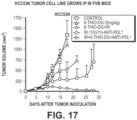
- 6-thio-dG single treatment 3 mg/kg 6-thio-dG was intraperitoneally given on days 7, 8 and 9 in MC38 tumor and LLC tumor and on days 5, 6, 7 for CT26 tumor.
- NK1.1, CD4+and CD8+ T cell depletion 200 ⁇ g of antibodies were intraperitoneally injected 1 day before treatment initiation and then twice a week for 2 weeks.
- 6-thio-dG was given on day 10 and day 11
- 50 ⁇ g PD-L1 was intraperitoneally injected on day 13 and 17.
- 6-thio-dG was given on day 4
- 5, 6, 10 and 11 200 ⁇ g PD-L1 was intraperitoneally injected on day 8 and 13.
- Humanized mouse tumor models Humanized mouse reconstitution was previously described (Qiao et al. , 2019). Briefly, four-week-old NSG-SGM3 female mice were irradiated with 100 cGy (X-ray irradiation with X-RAD 320 irradiator) one day prior to human CD34+ cells transfer. Cord blood was obtained from UT Southwestern Parkland Hospital. Human CD34+ cells were purified from cord blood by density gradient centrifugation (Ficoll ® Paque Plus, GE healthcare) followed by positive immunomagnetic selection with anti-human CD34 microbeads (Stemcell). 1 ⁇ 10 5 CD34+ cells were intravenously injected into each recipient mouse.
- mice with over 50 % human CD45+ cells reconstitution and age and sex matched non-humanized mice were inoculated with 1 ⁇ 10 6 HCT116 tumor cells subcutaneously on the right flank. 3 mg/kg 6-thio-dG was intraperitoneally given on days 7, 8 and 9. Tumor volumes were measured twice a week. Experiments were performed in compliance with UTSW Human Investigation Committee protocol and UTSW Institutional Animal Care and Use Committee.
- Tmem173 and Mb21d1 knockout MC38 cell line Tmem173 and Mb21d1 knockout MC38 cell line.
- Tmem173 and Mb21d1 genes in MC38 cells were knocked out by CRISPR/Cas9 technology.
- the guide sequence 5'-CACCTAGCCTCGCACGAACT-3' (SEQ ID NO: 1) for Tmem173 and 5'-CGCAAAGGGGGGCTCGATCG -3' (SEQ ID NO: 2) for Mb21d1 were cloned into px458 plasmids (non-integrating plasmid with GFP selecting marker), and then were transiently transfected into tumor cells using lipofectamine 2000 (Thermo Fisher). 24 hrs later, GFP positive cells were sorted and cultured for another one week.
- IFN- ⁇ enzyme-linked immunosorbent spot assay ELISPOT.
- MC38 tumors were injected subcutaneously on the right flank of C57BL/6.
- 6-thio-dG single treatment 3mg/kg 6-thio-dG was intraperitoneally given on days 7, 8 and 9;
- PD-L1 blockade combination therapy in MC38 model 3mg/kg 6-thio-dG was given on day 10 and day 11, 50 ⁇ g PD-L1 was intraperitoneally injected on day 11.
- 7 days after last treatment tumor draining lymphoid and spleen from tumor-bearing mice were collected and single-cell suspension was prepared.
- Irradiated MC38 tumor cells and control LLC tumor cells were used to re-stimulate the tumor-specific T cells.
- 1.5 ⁇ 10 5 draining lymph nodes cells or splenocytes and 7.5 ⁇ 10 4 irradiated tumor cells were co-cultured for 48hrs, and ELISPOT assay was performed using the IFN- ⁇ ELISPOT kit (BD Bioscience) according to the manufacturer's instructions.
- IFN- ⁇ spots were enumerated with the CTL-ImmunoSpot ® S6 Analyzer (Cellular Technology Limited).
- BMDC bone marrow dendritic cells
- T cells Single-cell suspensions of bone marrow (BM) cells were collected from tibias and femurs of C57BL/6 mice. The BM cells were placed in 10 cm dish and cultured with complete RPMI 1640 medium containing 20 ng/mL recombinant mouse GM-CSF (BioLegend). Fresh medium with was added into the culture on day 3 and day 6. The BMDCs were harvested Day 7.
- CD8+ T cells were isolated from lymph nodes and spleens of OT-1 transgenic mice with a negative CD8+ T cell isolation kit (Stemcell). MC38-OVA cells pretreated with 200 nM 6-thio-dG for 4hrs.
- BMDCs were sorted with CD11c+ positive selection kit (Stemcell) and co-cultured with OT-1 CD8+T cells for 48 hrs. Supernatants were collected and IFN- ⁇ was measured by cytometric bead array assay (BD Biosciences).
- HCT116 cells were pretreated with 500 nM 6-thio-dG for 4 hrs. Then drug was washed out, tumor cells were continued to culture for 72 hrs and harvest on the same day as BMDC harvest. Then HCT116 cells were mixed 1: 1 with 1 ⁇ 10 6 BMDC for 4 hrs. BMDC was purified and divided into two equal aliquots. One aliquot was extracted for total genomic DNA with Purelink Genomic DNA kit (Invitrogen) and served as normalization control.
- Purelink Genomic DNA kit Invitrogen
- cytosolic extract buffer containing 150 mM NaCl, 50 mM HEPES and 25 mg/mL digitonin (Sigma) and incubated for 10 mins at RT for plasma membrane permeabilization(West et al., 2015). Then cells were centrifuged to pellet intact cells. The cytosolic supernatants were collected and centrifuged at 12000g for 10 mins to pellet the remaining cellular debris. Then cytosolic DNA was extracted with Purelink Genomic DNA kit (Invitrogen). Quantitative PCR was performed on both whole-cell extracts and cytosolic fractions using human DNA primers and mouse DNA primers (Xu et al. , 2017).
- Tumor digestion Tumor tissues were excised and digested with 1 mg/mL Collagenase I (Sigma) and 0.5 mg/mL DNase I (Roche) in the 37°C for 30mins, tumor was then passed through a 70 ⁇ m cell strainer to remove large pieces of undigested tumor. Tumor infiltrating cells were washed twice with PBS containing 2 mM EDTA.
- Real-time PCR was performed with SsoAdvanced TM Universal SYBR ® Green Supermix (Bio-Rad) according to the manufacturer's instructions with different primer sets (human MT-CO1, forward primer 5'- CGCCACACTCCACGGAAGCA-3' (SEQ ID NO: 3), reverse primer 5'- CGGGGCATTCCG GATAGGCC-3' (SEQ ID NO: 4); human18s rRNA, forward primer 5'- ACCGATTGGATGGTTTAGTGAG-3' (SEQ ID NO: 5), reverse primer 5'- CCTACGGAAACCTTGTTACGAC-3' (SEQ ID NO: 6); mouse IFN- ⁇ , forward primer 5'- ATGAGTGGTGGTTGCAGGC-3' (SEQ ID NO: 7), reverse primer 5'-TGACCTTTCAAATGCAGTAGATTCA-3' (SEQ ID NO: 8); mouse GAPDH, forward primer 5'- CATCAAGAA GGTGGTGAAGC-3' (SEQ ID NO:
- BMDC and MC38 treatment were same as " In vitro co-culture of bone marrow dendritic cells”. 6hrs after co-culture, DC was isolated with CD11c+ positive selection kit (Stemcell). Protein sample preparation and immunoblot procedures were performed as previously described (Liu et al., 2019). Proteins were detected with rabbit monoclonal antibodies for pSTING (Cell signaling, 72971), STING (Cell signaling, 50494), pTBK1 (Cell signaling, 5483), TBK1 (Cell signaling, 3504). Protein loading was determined with antibodies against with Cyclophilin A (Cell signaling, 2175).
- Anti-rabbit (1:2000 in 5%BSA) was used for secondary antibody (Cell signaling, 7074).
- X-ray film (GeneMate, F-9024-8X10) was used to develop the membranes. Clarity Max Western ECL Substrate (Biorad, 1705062) or Supersignal West PicoPlus Chemiluminescent Substrate (Thermoscientific, 34577) was used for chemiluminescent western blot.
- 6-thio-dG depends on CD8+ T cells. All previous studies with xenograft models showed that intensive daily treatment with 6-thio-dG over 10 days could partially control tumor growth in many tumor models (Mender et al., 2015a; Mender et al., 2018; Zhang et al., 2018). However, the potential role of this drug on interaction between tumors and the adaptive immune system is unknown.
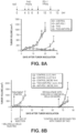
- the inventors In order to explore whether 6-thio-dG induces telomere-based DNA sensing for T cell responses, the inventors first determined the inhibition of cell viability by 6-thio-dG on telomerase-positive murine colon cancer cells (MC38) in immunocompetent host.
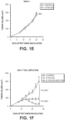
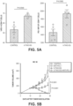
- MC38 tumor cells are sensitive to 6-thio-dG with an IC 50 concentration of 370nM ( FIG. 1A ). They also confirmed 6-thio-dG sensitivity in MC38 cells by a separate colony formation assay. MC38 cells treated with 6-thio-dG every three days for 13 days, resulted in less than 50% of the cells forming colonies with 0.5 ⁇ M 6-thio-dG treatment ( FIGS. 1B and 1C ). To evaluate whether 6-thio-dG reduces tumor burden in syngeneic mouse models in vivo, the inventors subcutaneously inoculated MC38 cell into immunocompetent wild-type (WT) C57BL/6 mice.
- WT wild-type
- FIG. 1D Seven days after tumor inoculation (when the tumor volume was ⁇ 100mm 3 ), 3mg/kg 6-thio-dG was administrated daily for only three days and tumor growth was significantly reduced ( FIG. 1D ) compared to the control tumor.
- 6-thio-dG might have an immune stimulatory role in vivo. Therefore, they inoculated tumors on Rag1 knock out mice that cannot generate mature T and B cells. Indeed, the therapeutic effect of 6-thio-dG was completely diminished ( FIG. 1E ), indicating adaptive immune cells are largely required for tumor control in vivo.
- 6-thio-dG treatment increases tumor-specific T cell response.
- the inventors reasoned that 6-thio-dG treatment might change immune cell expansion in the tumor microenvironment.
- TILs tumor infiltrating lymphocytes
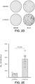
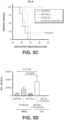
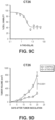
- the inventors found an increase in the frequency of CD3+ T cells and CD8+T cells in TILs after 6-thio-dG treatment ( FIGS. 2A , 10A and 10B ). They also observed a significant upregulation of CD8+ T cell proliferation indicated by elevated Ki67 expression ( FIG. 2B ), but no significant changes of Treg cells ( FIG. 10C ).
- NK cells are not essential but CD8+ T cell responses are required in 6-thio-dG mediated anti-tumor effects.
- the inventors further tested the antigen-specific T cell response after 6-thio-dG treatment by using the MC38-OVA tumor model, which allows tracking of antigen specific T cells in the tumor tissue. Indeed, they observed increased tumor-specific CD8+ T cells in tumors 6 days after 6-thio-dG treatment ( FIG. 2C ). They also observed enhanced tumor-specific cytotoxic T cell responses in the MC38 tumor model by measuring IFN- ⁇ producing T cells after 6-thio-dG treatment ( FIGS. 2D and 2E ). To directly assess the capacity of T cells to produce IFN- ⁇ in vivo, the inventors utilized IFN- ⁇ YFP reporter mice that allow tracking of IFN- ⁇ producing T cells with YFP expression (Reinhardt et al., 2009).
- 6-thio-dG treatment significantly increased YFP+ T cells in the tumor, suggesting enhanced IFN- ⁇ production ability of T cells ( FIGS. 2F and 10F ).
- the hallmark of an adaptive immune response is the formation of memory that initiates a rapid recall response when the same antigen appears.
- 6-thio-dG treatment induces a memory response, mice with completely regressed tumors after 6-thio-dG treatment were rested for 5 weeks and re-challenged with the same MC38 tumor but with 10 times more tumor cells on the opposite flank (left flank), and LLC tumor cells were inoculated as control on the right flank.
- naive mice When naive mice (never exposed to MC38 cells or 6-thio-dG) were injected with the same number of MC38 cells, the tumors grew aggressively. Remarkably, all cured mice by 6-thio-dG treatment spontaneously rejected re-challenged MC38 tumors.
- 6-thio-dG treatment enhances the cross-priming capacity of dendritic cells.
- Antigen cross-presentation by antigen presenting cells (APCs) such as DCs or macrophages accounts for the tumor-specific CD8+ T cell activation.
- APCs antigen presenting cells
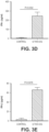
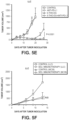
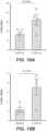
- the inventors first used anti-CSF1R antibody to deplete macrophages. They found that 6-thio-dG worked even better in macrophage depleted group ( FIG. 3A ), which can be explained by the additive effect of the removal of immune suppressive tumor associated macrophages.
- BATF3 basic leucine zipper ATF-like transcription factor 3
- AATF3-dependent DCs are critical for the priming of antigen-specific CD8+ T cells (Broz et al., 2014; Edelson et al. , 2010).
- 6-thio-dG treatment in Batf3 deficient mice partially delayed tumor growth but was significantly less effective compared with WT mice ( FIG. 3B ). Noticeably, 60% of WT mice were completely tumor free but none of mice were tumor free in Batf3- / - mice ( FIG. 3C ), suggesting an important role of BATF3-dependent DCs in the therapeutic effect of 6-thio-dG.
- 6-thio-dG treatment enhances cross-priming capacity of DCs
- the inventors co-cultured 6-thio-dG pretreated MC38-OVA tumor cells with bone marrow derived DCs (BMDCs) overnight. Then the DCs were purified and co-cultured with naive OT-1 transgenic CD8+ T cells that express the TCR with the specificity to recognize the OVA 257-264 epitope. They observed a significant increase of IFN- ⁇ production by CD8+ T cells in the 6-thio-dG treatment group ( FIG. 3D ), which indicates an increased cross-priming capacity of DCs after 6-thio-dG treatment.
- IFN-I signaling promotes the cross-priming capacity of DCs (Diamond et al. , 2011; Le Bon et al., 2003; Sanchez-Paulete et al., 2017)
- the inventors tested the production of IFN- ⁇ by DCs after co-culturing them with 6-thio-dG treated tumor cells. Indeed, IFN- ⁇ production significantly increased in the 6-thio-dG treatment group, indicating increased innate sensing of DCs ( FIG. 3E ). They further investigated whether the IFN-I pathway is essential for 6-thio-dG-mediated anti-tumor effect.
- STING signaling in the host is required for 6-thio-dG induced innate sensing.
- Tumor cells under stress might release danger-associated molecular patterns (DAMPs) to engage TLR/Myd88 pathways in APCs and initiate IFN-I signaling.
- DAMPs danger-associated molecular patterns
- Tumor-derived DNAs can also trigger the cytosolic DNA sensing cGAS/STING pathway and activate IFN-I pathways (Deng et al. , 2014; Li et al., 2019).
- the inventors inoculated MC38 tumors into Myd88 -/- and Tmem173 -/- ( Tmem173 encodes STING) mice.
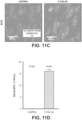
- 6-thio-dG treatment activates the host STING/IFN-I pathway They observed an increase of TBK1 phosphorylation in DCs after co-culture with 6-thio-dG pre-treated tumor cells and the phosphorylation was completely diminished in Tmem173 DCs ( FIG. 11A ).
- 6-thio-dG treatment induced IFN- ⁇ production in DCs in a STING-dependent manner FIG. 11B ).
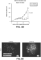
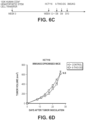
- tumor-intrinsic STING signaling is critical in innate-sensing inducing cancer therapies (Sen et al. , 2019; Vanpouille-Box et al., 2017), the inventors tested whether tumor-intrinsic STING signaling also contributes to 6-thio-dG treatment efficacy. They used CRISPR/Cas9 to knock out Tmem173 and Mb21d1 ( Mb21d1 encodes cGAS) in MC38 tumor cells. In contrast to other studies, tumor-intrinsic STING signaling played a non-essential role, as 6-thio-dG treatment still controlled tumor growth in mice bearing Tmem173 KO and Mb21d1 KO tumor cells ( FIGS. 4C and 4D ).
- the inventors then sought to determine how 6-thio-dG treated tumor cells trigger innate sensing in DCs. Since 6-thio-dG is a telomere-targeting drug, 6-thio-dG induced telomere stress might contribute to innate sensing of DCs by releasing DNAs. Therefore, the inventors first analyzed telomere stress by the TIF (Telomere dysfunction Induced Foci) assay and showed that 6-thio-dG induced telomere damages in MC38 cells ( FIGS. 4E and 4F ). Since telomeres are only a small fraction of genomic DNA (-1/6000) any co-localization of telomeres with DNA damage is significant.
- TIF Telomere dysfunction Induced Foci
- 6-thio-dG also induced interphase bridges between the two daughter cells during telophase and since many contained telomere sequences, this may explain why many micronuclei containing telomere signals when cells re-entered interphase after mitosis ( FIG. 11E ). These cytosolic fragments formed micronuclei with fragile nuclear envelopes ( FIGS. 11E and 11F ), which can be eventually recognized as a danger signal. These DNA fragments are released from the cells and can be taken up by DCs.
- HCT116 a human colon cancer cell line
- 6-thio-dG co-cultured them with mouse BMDC for 4hrs
- the short-time co-culture of a human tumor cell line with mouse BMDCs allowed us to distinguish DNAs from different origins. They found an increase of human DNAs (MT-CO1 and human 18S) in the cytosol of mouse DCs after 6-thio-dG treatment, which suggests that DNAs from the tumors enter the host DCs ( FIG. 4G ).
- 6-thio-dG overcomes PD-L1 blockade resistance in advanced tumors. While 6-thio-dG treatment activated CD8+ T cells, it also upregulated PD-1 expression in the frequency of total CD8+ T cells and on per cell basis ( FIG. 5A ). PD-1 is a co-inhibitory molecule that limits T cell activation. The elevated PD-1 expression might eventually inhibit the cytotoxic CD8+ T cell function after 6-thio-dG treatment. Therefore, the inventors reasoned that combination of 6-thio-dG with PD-1/PD-L1 blockade might augment the overall anti-tumor immune response, especially in the advanced tumor setting which harbors a more immune suppressive microenvironment containing multiple resistance mechanisms that limit single treatment efficacy.
- 6-thio-dG single treatment was only effective in relatively small tumor sizes -100 mm 3
- the inventors let the tumor sizes reach to 150-200mm 3 and then treated with 6-thio-dG and/or anti-PD-L1 treatment.
- tumor volume is difficult to control with two daily treatments with 6-thio-dG or by two treatments with anti-PD-L1 ( FIG. 5B ).
- sequential administration of 6-thio-dG and anti-PD-L1 completely inhibited the tumor growth ( FIG. 5B ).
- only mice in the combination treatment group achieved a 100% survival rate ( FIG. 5C ), showing a synergistic effect of 6-thio-dG treatment with PD-L1 blockade.
- mice did not observe any body weight loss of mice in the combination treatment group ( FIG. 12 ). They further analyzed the tumor-specific T cell response in draining lymph nodes (dLNs) and found that the anti-PD-L1 treatment had little effect on T cell activation in advanced tumors. In contrast, combination therapy significantly increased IFN- ⁇ production compared to other groups. The immune response was MC38 tumor specific as there were almost no IFN- ⁇ spots in the control LLC tumor stimulation group ( FIG. 5D ).
- MC38 is known to be an immunogenic tumor model.
- the inventors employed the mouse LLC tumor model that has been reported to be resistant to PD-L1 blockade (Bullock et al., 2019; Li et al., 2017). Consistent with previous reports, single treatment with anti-PD-L1 had no therapeutic effect ( FIG. 5E ). Notably, combination of 6-thio-dG with anti-PD-L1 significantly reduced mouse tumor burden and 40% of mice eventually completely rejected tumors ( FIG. 5E ). The inventors re-challenged tumor-free mice 6 weeks after tumor regression to check the memory response.
- mice spontaneously rejected LLC tumors but not MC38 tumors, suggesting a long-lasting tumor-specific immune memory ( FIG. 5F ). Based on these results, 6-thio-dG treatment overcomes PD-L1 blockade resistance in advanced tumors. This will potentially benefit PD-1/PD-L1 blockade resistant patients in the clinic.
- 6-thio-dG reduces human colon cancer burden in a humanized mouse model.
- TERT the catalytic subunit of the telomerase
- the inventors thus analyzed colorectal adenocarcinoma patients from the TCGA database and found patients with abnormal high expression of TERT had significantly worse overall survival rates compared to the colon cancer patients with low TERT expression ( FIG. 6A ).
- the inventors developed a humanized mouse model with NSG-SGM3 mouse which has human SCF-1, GM-CSF and IL-3 transgenic expression that support the better development of human myeloid cells. They reconstituted the human immune system in NSG-SGM3 mice with human CD34+ hematopoietic stem cells (HSCs). 12 weeks after HSCs transfer, the humanized mice had an average of over 60% human CD45+ cells and over 20% human T cells among human CD45+ cells in circulation ( FIGS. 13A-13C ).
- HCT116 a human colon cancer cell line that is sensitive to 6-thio-dG treatment with an IC 50 of 0.73 ⁇ M
- FIG. 6B The control group of humanized mice had similar constitution of human immune cells with 6-thio-dG treated group before treatment started ( FIGS. 13B and 13C ).
- FIGS. 13D After three doses of 6-thio-dG treatment, immunocompromised mice did not have significant difference compared to control group ( FIG. 6D ).
- the humanized mice significantly delayed tumor growth with 6-thio-dG treatment ( FIGS. 6C and 6E ).
- the inventors then tested a human melanoma cell line A375 that is sensitive to 6-thio-dG in vitro ( FIG. 13D ). They did not observe any effect in immunocompromised NSG-SGM3 mice since they provided a relatively short-time 6-thio-dG treatment ( FIG. 13E ). Notably, they found a treatment with two doses of 6-thio-dG partially delayed tumor growth in humanized mice. In addition, combination with checkpoint blockades further reduced tumor burden, suggesting pretreatment with 6-thio-dG sensitizes human tumors to checkpoint blockades ( FIG. 13F ). Given that humanized mice only partially restore human immunity due to missing some immune cells and limited number of human T cells, it is not surprising that the inventors did not observe complete tumor regression.
- FIGS. 8A-B The inventors have demonstrated ( FIGS. 8A-B ) that three days treatment with 6-thio-dG followed two days later with anti-PD-L1 results in complete tumor remissions in the lewis lung carcinoma. This is a very aggressive tumor type as is illustrated in FIG. 8A . Within 20 day the tumor injected subcutaneously reached to over 1000 m 3 . They observed the same rapid tumor growth with just treating with anti-PD-L1. However, just three treatments with 6-thio-dG (THIO) results in significant tumor control for 20 days. Surprisingly, treatment with THIO followed by anti-PDL-L1 (Atezolizumab) resulted in complete tumor regression.
- THIO 6-thio-dG
- the inventors maintained the cured mice for 5 additional weeks and rechallenged the same mice with five time more LLC tumor and no tumor growth was observed. However, if they injected the LLC cured mice with MC38 the tumors grew and anti-PD-L1 had no affect ( FIG. 8B ). In control mice that were never treated with THIO, control LLC tumors grew similarly to MC38. This suggests tumor specific immune memory.
- 6-thio-dG induces telomerase-dependent DNA damage and increases tumor DNAs taken up by DCs.
- the increased cytosolic DNAs trigger the DC-intrinsic STING/IFN-I pathway, resulting in enhanced cross-priming capacity of DCs and subsequent tumor-specific T cell activation.
- 6-thio-dG overcomes PD-L1 blockade resistance in advanced tumors. This study identifies 6-thio-dG as a novel immune stimulatory drug that will potentially benefit a wide population of cancer patients in the clinic.
- telomerase dependent telomere targeting therapy (6-thio-dG) in inducing anti-tumor immune responses in syngeneic colon and lung mouse models and humanized mouse cancer models.
- This effect is mediated through triggering the cytosolic DNA sensing STING/IFN-I pathway in DCs, which ultimately enhances the cross-priming capacity of DCs and subsequent tumor specific T cell activation.
- telomerase is a universal tumor marker and it can potentially be applied to many other telomerase positive cancers.
- sequential administration of 6-thio-dG and anti-PD-L1 overcomes PD-L1 resistance in PD-L1 blockade resistant tumors, suggesting the combination therapy can benefit PD-L1 resistant patients in the clinic.
- 6-thio-dG is an immune stimulatory drug might allow the design of better combinational treatments including immunotherapy to amplify initial immunity.
- 6-thio-dG treatment triggered innate sensing is host STING signaling dependent as 6-thio-dG completely lost its efficacy in Tmem173 deficient mice but not in Tmem173 deficient tumors. Since STING signaling in MC38 tumors is active, one explanation is after 6-thio-dG treatment, tumor intrinsic STING was activated but most tumor cells died, so little type IIFN can be produced. Another possibility is that there might be an intrinsic mechanism that limits STING activation in tumor cells, which still remains poorly defined.
- 6-thio-dG Compared to the general DNA damage induction approaches, for example, radiation therapy or chemotherapy that non-selectively induce DNA damage in all proliferating cells, one unique feature of 6-thio-dG is the specific induction of telomere-associated DNA damage in telomerase expressing cells, primarily tumor cells, but not affecting immune cells and other telomerase-silent somatic cells.
- 6-thio-dG can be preferentially incorporated into de novo -synthesized telomeres and causes rapid tumor shrinkage.
- direct telomerase inhibitors function through the inhibition of telomerase activity and rely on the progressive shortening of telomeres.
- 6-thio-dG takes effect rapidly regardless of the initial telomere length.
- telomere dysfunction induced foci Telomeres are only -1/6000th of genomic DNA so any TIF is highly significant. Moreover, some TIFs are taken up by DCs and further trigger STING-dependent IFN I signaling.
- 6-thio-dG with PD-L1 blockade should augment overall anti-tumor immunity responses. Indeed, this study showed sequential administration of 6-thio-dG and anti-PD-L1 have synergistic effect in advanced tumors and in PD-L1 blockade resistant tumors. Further studies should be carried out regarding optimal combination regimens.
- 6-thio-dG a telomerase-dependent telomere targeting small molecule drug, in potentiating anti-tumor immune responses.
- 6-thio-dG induces telomere dysfunction and increases cytosolic DNA release.
- these telomeric DNA fragments are taken up by DCs and activate the DC intrinsic STING/IFN pathway, resulting in enhanced cross-priming capacity of DCs and subsequent tumor specific T cell activation.
- this study showing the remarkable efficacy of sequential administration of 6-thio-dG and anti-PD-L1 in advanced tumors and PD-L1 blockade resistant tumors provides a strong scientific rationale for propelling combination therapy into clinical trials. The inventors expect that these findings will be translated in the near future and benefit more patients in the clinic.
Landscapes
- Health & Medical Sciences (AREA)
- Chemical & Material Sciences (AREA)
- Immunology (AREA)
- Life Sciences & Earth Sciences (AREA)
- Medicinal Chemistry (AREA)
- General Health & Medical Sciences (AREA)
- Organic Chemistry (AREA)
- Animal Behavior & Ethology (AREA)
- Veterinary Medicine (AREA)
- Public Health (AREA)
- Pharmacology & Pharmacy (AREA)
- Molecular Biology (AREA)
- Epidemiology (AREA)
- Biophysics (AREA)
- Biochemistry (AREA)
- Proteomics, Peptides & Aminoacids (AREA)
- Genetics & Genomics (AREA)
- Engineering & Computer Science (AREA)
- Nuclear Medicine, Radiotherapy & Molecular Imaging (AREA)
- Biomedical Technology (AREA)
- General Chemical & Material Sciences (AREA)
- Chemical Kinetics & Catalysis (AREA)
- Endocrinology (AREA)
- Microbiology (AREA)
- Mycology (AREA)
- Bioinformatics & Cheminformatics (AREA)
- Pathology (AREA)
- Radiology & Medical Imaging (AREA)
- Pharmaceuticals Containing Other Organic And Inorganic Compounds (AREA)
- Medicines That Contain Protein Lipid Enzymes And Other Medicines (AREA)
- Medicines Containing Antibodies Or Antigens For Use As Internal Diagnostic Agents (AREA)
- Saccharide Compounds (AREA)
Applications Claiming Priority (2)
| Application Number | Priority Date | Filing Date | Title |
|---|---|---|---|
| US202062989041P | 2020-03-13 | 2020-03-13 | |
| PCT/US2021/022090 WO2021183873A1 (en) | 2020-03-13 | 2021-03-12 | Sequential treatment of cancers using 6-thio-dg, checkpoint inhibitors and radiation therapy |
Publications (3)
| Publication Number | Publication Date |
|---|---|
| EP4117667A1 EP4117667A1 (en) | 2023-01-18 |
| EP4117667A4 EP4117667A4 (en) | 2023-11-15 |
| EP4117667B1 true EP4117667B1 (en) | 2024-12-18 |
Family
ID=77671975
Family Applications (1)
| Application Number | Title | Priority Date | Filing Date |
|---|---|---|---|
| EP21767125.4A Active EP4117667B1 (en) | 2020-03-13 | 2021-03-12 | Sequential treatment of cancers using 6-thio-dg and checkpoint inhibitors |
Country Status (16)
| Country | Link |
|---|---|
| US (2) | US12097213B2 (https=) |
| EP (1) | EP4117667B1 (https=) |
| JP (1) | JP7829491B2 (https=) |
| KR (1) | KR20220154134A (https=) |
| CN (1) | CN115397428A (https=) |
| BR (1) | BR112022018238A2 (https=) |
| CA (1) | CA3170930A1 (https=) |
| DK (1) | DK4117667T3 (https=) |
| ES (1) | ES3014683T3 (https=) |
| FI (1) | FI4117667T3 (https=) |
| HU (1) | HUE070513T2 (https=) |
| IL (1) | IL296403A (https=) |
| MX (1) | MX2022011331A (https=) |
| PL (1) | PL4117667T3 (https=) |
| PT (1) | PT4117667T (https=) |
| WO (1) | WO2021183873A1 (https=) |
Families Citing this family (2)
| Publication number | Priority date | Publication date | Assignee | Title |
|---|---|---|---|---|
| US20210023107A1 (en) | 2018-03-22 | 2021-01-28 | The Board Of Regents Of The University Of Texas System | USE OF 6-THIO-dG TO TREAT THERAPY-RESISTANT TELOMERASEPOSITIVE PEDIATRIC BRAIN TUMORS |
| WO2025090539A1 (en) * | 2023-10-22 | 2025-05-01 | Regeneron Pharmaceuticals, Inc. | Treatment of non-small cell lung cancer with 6-thio-dg and cemiplimab |
Family Cites Families (11)
| Publication number | Priority date | Publication date | Assignee | Title |
|---|---|---|---|---|
| EP2630255B1 (en) * | 2010-10-22 | 2014-09-10 | Roche Diagnostics GmbH | A conjugate between a thiophilic solid phase and an oligonucleotide comprising a thiooxonucleotide |
| NZ713498A (en) | 2013-04-08 | 2017-07-28 | Univ Texas | Mercaptopurine ribonucleoside analogues for altering telomerase mediated telomere |
| CA2991628C (en) * | 2015-07-16 | 2020-04-07 | Bioxcel Therapeutics, Inc. | A novel approach for treatment of cancer using immunomodulation |
| US20180036331A1 (en) | 2016-03-24 | 2018-02-08 | The Board Of Regents Of The University Of Texas System | Treatment of drug resistant proliferative diseases with telomerase mediated telomere altering compounds |
| WO2017205756A1 (en) | 2016-05-27 | 2017-11-30 | The Board Of Regents Of The University Of Texas System | 6-thio-2'-deoxyguanosine (6-thio-dg) results in telomerase dependent telomere dysfunction and cell death in therapy-resistant cancer cells |
| WO2018049474A1 (en) | 2016-09-16 | 2018-03-22 | Bionomics Limited | Antibody and checkpoint inhibitor combination therapy |
| MA47604A (fr) | 2017-02-21 | 2020-01-01 | Regeneron Pharma | Anticorps anti-pd-1 pour le traitement du cancer du poumon |
| US11571459B2 (en) | 2017-04-03 | 2023-02-07 | Oncxerna Therapeutics, Inc. | Methods for treating cancer using PS-targeting antibodies with immuno-oncology agents |
| EP3746119A4 (en) * | 2018-02-01 | 2021-11-10 | Merck Sharp & Dohme Corp. | METHOD OF TREATMENT FOR CANCER OR INFECTION USING A COMBINATION OF ANTI-PD-1 ANTIBODY, ANTI-LAG3 ANTIBODY, AND ANTI-TIGITE ANTIBODY |
| US20210113602A1 (en) | 2018-02-28 | 2021-04-22 | The Board Of Regents Of The University Of Texas System | USE OF 6-THIO-2'-DEOXYGUANOSINE (6-THIO-dG) TO TREAT MELANOMA |
| US20210023107A1 (en) | 2018-03-22 | 2021-01-28 | The Board Of Regents Of The University Of Texas System | USE OF 6-THIO-dG TO TREAT THERAPY-RESISTANT TELOMERASEPOSITIVE PEDIATRIC BRAIN TUMORS |
-
2021
- 2021-03-12 ES ES21767125T patent/ES3014683T3/es active Active
- 2021-03-12 CN CN202180028952.2A patent/CN115397428A/zh active Pending
- 2021-03-12 PT PT217671254T patent/PT4117667T/pt unknown
- 2021-03-12 MX MX2022011331A patent/MX2022011331A/es unknown
- 2021-03-12 KR KR1020227033906A patent/KR20220154134A/ko active Pending
- 2021-03-12 CA CA3170930A patent/CA3170930A1/en active Pending
- 2021-03-12 HU HUE21767125A patent/HUE070513T2/hu unknown
- 2021-03-12 DK DK21767125.4T patent/DK4117667T3/da active
- 2021-03-12 BR BR112022018238A patent/BR112022018238A2/pt unknown
- 2021-03-12 WO PCT/US2021/022090 patent/WO2021183873A1/en not_active Ceased
- 2021-03-12 IL IL296403A patent/IL296403A/en unknown
- 2021-03-12 JP JP2022554769A patent/JP7829491B2/ja active Active
- 2021-03-12 EP EP21767125.4A patent/EP4117667B1/en active Active
- 2021-03-12 US US17/200,539 patent/US12097213B2/en active Active
- 2021-03-12 PL PL21767125.4T patent/PL4117667T3/pl unknown
- 2021-03-12 FI FIEP21767125.4T patent/FI4117667T3/fi active
-
2024
- 2024-09-09 US US18/829,162 patent/US20240424013A1/en active Pending
Also Published As
| Publication number | Publication date |
|---|---|
| WO2021183873A1 (en) | 2021-09-16 |
| PL4117667T3 (pl) | 2025-04-07 |
| IL296403A (en) | 2022-11-01 |
| US12097213B2 (en) | 2024-09-24 |
| US20210290652A1 (en) | 2021-09-23 |
| EP4117667A4 (en) | 2023-11-15 |
| CA3170930A1 (en) | 2021-09-16 |
| JP2023517671A (ja) | 2023-04-26 |
| DK4117667T3 (da) | 2025-02-10 |
| FI4117667T3 (fi) | 2025-02-10 |
| JP7829491B2 (ja) | 2026-03-13 |
| EP4117667A1 (en) | 2023-01-18 |
| AU2021233023B2 (en) | 2026-04-16 |
| PT4117667T (pt) | 2025-02-06 |
| ES3014683T3 (en) | 2025-04-24 |
| AU2021233023A1 (en) | 2022-11-03 |
| HUE070513T2 (hu) | 2025-06-28 |
| MX2022011331A (es) | 2022-10-27 |
| BR112022018238A2 (pt) | 2022-10-25 |
| CN115397428A (zh) | 2022-11-25 |
| KR20220154134A (ko) | 2022-11-21 |
| US20240424013A1 (en) | 2024-12-26 |
Similar Documents
| Publication | Publication Date | Title |
|---|---|---|
| Mender et al. | Telomere stress potentiates STING-dependent anti-tumor immunity | |
| US20240424013A1 (en) | SEQUENTIAL TREATMENT OF CANCERS USING 6-THIO-dG, CHECKPOINT INHIBITORS AND RADIATION THERAPY | |
| Luo et al. | Necroptosis-dependent immunogenicity of cisplatin: implications for enhancing the radiation-induced abscopal effect | |
| US20190077856A1 (en) | Method of treating diseases using kinase modulators | |
| US20190091229A1 (en) | Therapeutic methods relating to hsp90 inhibitors | |
| Groelly et al. | Anti‐tumoural activity of the G‐quadruplex ligand pyridostatin against BRCA1/2‐deficient tumours | |
| JP2019502753A (ja) | ナンセンス変異依存mRNA分解機構の抑制による免疫応答の誘導法 | |
| KR20230059792A (ko) | 암 치료를 위한 조합 | |
| US20240009223A1 (en) | 6-thio-2'-deoxyguanosine (6-thio-dg) results in telomerase dependent telomere dysfunction and cell death in various models of therapy-resistant cancer cells | |
| Franzese et al. | Tumor immunotherapy: drug-induced neoantigens (xenogenization) and immune checkpoint inhibitors | |
| JP2023531522A (ja) | デオキシウリジントリホスファターゼ阻害剤との併用療法 | |
| Mender et al. | Activating an adaptive immune response with a telomerase-mediated telomere targeting therapeutic in hepatocellular carcinoma | |
| KR20250008931A (ko) | 기성품 면역치료요법을 위한 nk 세포 생성물의 냉동보존 | |
| US20190365719A1 (en) | Combination treatments of hsp90 inhibitors for enhancing tumor immunogenicity and methods of use thereof | |
| AU2021233023B9 (en) | Sequential treatment of cancers using 6-thio-dg, checkpoint inhibitors and radiation therapy | |
| RU2833475C1 (ru) | ПОСЛЕДОВАТЕЛЬНОЕ ЛЕЧЕНИЕ РАКА С ИСПОЛЬЗОВАНИЕМ 6-ТИО-dG, ИНГИБИТОРОВ КОНТРОЛЬНЫХ ТОЧЕК И ЛУЧЕВОЙ ТЕРАПИИ | |
| HK40084657B (en) | Sequential treatment of cancers using 6-thio-dg and checkpoint inhibitors | |
| HK40084657A (en) | Sequential treatment of cancers using 6-thio-dg and checkpoint inhibitors | |
| Li et al. | Implications of DNA damage response and immunotherapy in tumor therapy | |
| CA3025522C (en) | 6-thio-2'-deoxyguanosine (6-thio-dg) results in telomerase dependent telomere dysfunction and cell death in various models of therapy-resistant cancer cells | |
| Ravelli | Novel therapeutic approaches for the treatment of solid tumors | |
| JP2024503513A (ja) | がんを処置するための併用療法スケジュール | |
| WO2025090539A1 (en) | Treatment of non-small cell lung cancer with 6-thio-dg and cemiplimab | |
| Aquino | Tumor immunotherapy: drug-induced neoantigens (xenogenization) and immune checkpoint inhibitors | |
| Krishnan et al. | Hematopoietic cell therapy for human immunodeficiency virus infection |
Legal Events
| Date | Code | Title | Description |
|---|---|---|---|
| STAA | Information on the status of an ep patent application or granted ep patent |
Free format text: STATUS: THE INTERNATIONAL PUBLICATION HAS BEEN MADE |
|
| PUAI | Public reference made under article 153(3) epc to a published international application that has entered the european phase |
Free format text: ORIGINAL CODE: 0009012 |
|
| STAA | Information on the status of an ep patent application or granted ep patent |
Free format text: STATUS: REQUEST FOR EXAMINATION WAS MADE |
|
| 17P | Request for examination filed |
Effective date: 20221012 |
|
| AK | Designated contracting states |
Kind code of ref document: A1 Designated state(s): AL AT BE BG CH CY CZ DE DK EE ES FI FR GB GR HR HU IE IS IT LI LT LU LV MC MK MT NL NO PL PT RO RS SE SI SK SM TR |
|
| DAV | Request for validation of the european patent (deleted) | ||
| DAX | Request for extension of the european patent (deleted) | ||
| REG | Reference to a national code |
Ref country code: HK Ref legal event code: DE Ref document number: 40084657 Country of ref document: HK |
|
| A4 | Supplementary search report drawn up and despatched |
Effective date: 20231012 |
|
| RIC1 | Information provided on ipc code assigned before grant |
Ipc: A61K 41/00 20200101ALI20231006BHEP Ipc: C07K 16/28 20060101ALI20231006BHEP Ipc: A61N 5/10 20060101ALI20231006BHEP Ipc: A61P 35/00 20060101ALI20231006BHEP Ipc: A61K 39/395 20060101ALI20231006BHEP Ipc: A61K 31/4545 20060101ALI20231006BHEP Ipc: A61K 31/708 20060101ALI20231006BHEP Ipc: A61K 31/7076 20060101ALI20231006BHEP Ipc: A61K 31/522 20060101AFI20231006BHEP |
|
| GRAP | Despatch of communication of intention to grant a patent |
Free format text: ORIGINAL CODE: EPIDOSNIGR1 |
|
| STAA | Information on the status of an ep patent application or granted ep patent |
Free format text: STATUS: GRANT OF PATENT IS INTENDED |
|
| INTG | Intention to grant announced |
Effective date: 20240715 |
|
| GRAS | Grant fee paid |
Free format text: ORIGINAL CODE: EPIDOSNIGR3 |
|
| GRAA | (expected) grant |
Free format text: ORIGINAL CODE: 0009210 |
|
| STAA | Information on the status of an ep patent application or granted ep patent |
Free format text: STATUS: THE PATENT HAS BEEN GRANTED |
|
| AK | Designated contracting states |
Kind code of ref document: B1 Designated state(s): AL AT BE BG CH CY CZ DE DK EE ES FI FR GB GR HR HU IE IS IT LI LT LU LV MC MK MT NL NO PL PT RO RS SE SI SK SM TR |
|
| REG | Reference to a national code |
Ref country code: CH Ref legal event code: EP |
|
| REG | Reference to a national code |
Ref country code: DE Ref legal event code: R096 Ref document number: 602021023612 Country of ref document: DE |
|
| P01 | Opt-out of the competence of the unified patent court (upc) registered |
Free format text: CASE NUMBER: APP_63487/2024 Effective date: 20241129 |
|
| REG | Reference to a national code |
Ref country code: IE Ref legal event code: FG4D |
|
| REG | Reference to a national code |
Ref country code: PT Ref legal event code: SC4A Ref document number: 4117667 Country of ref document: PT Date of ref document: 20250206 Kind code of ref document: T Free format text: AVAILABILITY OF NATIONAL TRANSLATION Effective date: 20250130 |
|
| REG | Reference to a national code |
Ref country code: FI Ref legal event code: FGE Ref country code: DK Ref legal event code: T3 Effective date: 20250203 |
|
| REG | Reference to a national code |
Ref country code: SE Ref legal event code: TRGR |
|
| REG | Reference to a national code |
Ref country code: NL Ref legal event code: FP |
|
| REG | Reference to a national code |
Ref country code: GR Ref legal event code: EP Ref document number: 20250400280 Country of ref document: GR Effective date: 20250314 |
|
| REG | Reference to a national code |
Ref country code: LT Ref legal event code: MG9D |
|
| PG25 | Lapsed in a contracting state [announced via postgrant information from national office to epo] |
Ref country code: HR Free format text: LAPSE BECAUSE OF FAILURE TO SUBMIT A TRANSLATION OF THE DESCRIPTION OR TO PAY THE FEE WITHIN THE PRESCRIBED TIME-LIMIT Effective date: 20241218 |
|
| PGFP | Annual fee paid to national office [announced via postgrant information from national office to epo] |
Ref country code: PT Payment date: 20250303 Year of fee payment: 5 |
|
| PGFP | Annual fee paid to national office [announced via postgrant information from national office to epo] |
Ref country code: BG Payment date: 20250123 Year of fee payment: 5 |
|
| PG25 | Lapsed in a contracting state [announced via postgrant information from national office to epo] |
Ref country code: LV Free format text: LAPSE BECAUSE OF FAILURE TO SUBMIT A TRANSLATION OF THE DESCRIPTION OR TO PAY THE FEE WITHIN THE PRESCRIBED TIME-LIMIT Effective date: 20241218 |
|
| REG | Reference to a national code |
Ref country code: ES Ref legal event code: FG2A Ref document number: 3014683 Country of ref document: ES Kind code of ref document: T3 Effective date: 20250424 |
|
| PGFP | Annual fee paid to national office [announced via postgrant information from national office to epo] |
Ref country code: PL Payment date: 20250226 Year of fee payment: 5 |
|
| PG25 | Lapsed in a contracting state [announced via postgrant information from national office to epo] |
Ref country code: RS Free format text: LAPSE BECAUSE OF FAILURE TO SUBMIT A TRANSLATION OF THE DESCRIPTION OR TO PAY THE FEE WITHIN THE PRESCRIBED TIME-LIMIT Effective date: 20250318 |
|
| REG | Reference to a national code |
Ref country code: HU Ref legal event code: AG4A Ref document number: E070513 Country of ref document: HU |
|
| PG25 | Lapsed in a contracting state [announced via postgrant information from national office to epo] |
Ref country code: SM Free format text: LAPSE BECAUSE OF FAILURE TO SUBMIT A TRANSLATION OF THE DESCRIPTION OR TO PAY THE FEE WITHIN THE PRESCRIBED TIME-LIMIT Effective date: 20241218 |
|
| PGFP | Annual fee paid to national office [announced via postgrant information from national office to epo] |
Ref country code: ES Payment date: 20250404 Year of fee payment: 5 |
|
| PG25 | Lapsed in a contracting state [announced via postgrant information from national office to epo] |
Ref country code: IS Free format text: LAPSE BECAUSE OF FAILURE TO SUBMIT A TRANSLATION OF THE DESCRIPTION OR TO PAY THE FEE WITHIN THE PRESCRIBED TIME-LIMIT Effective date: 20250418 |
|
| PG25 | Lapsed in a contracting state [announced via postgrant information from national office to epo] |
Ref country code: EE Free format text: LAPSE BECAUSE OF FAILURE TO SUBMIT A TRANSLATION OF THE DESCRIPTION OR TO PAY THE FEE WITHIN THE PRESCRIBED TIME-LIMIT Effective date: 20241218 |
|
| PGFP | Annual fee paid to national office [announced via postgrant information from national office to epo] |
Ref country code: CH Payment date: 20250401 Year of fee payment: 5 |
|
| PG25 | Lapsed in a contracting state [announced via postgrant information from national office to epo] |
Ref country code: SK Free format text: LAPSE BECAUSE OF FAILURE TO SUBMIT A TRANSLATION OF THE DESCRIPTION OR TO PAY THE FEE WITHIN THE PRESCRIBED TIME-LIMIT Effective date: 20241218 |
|
| PG25 | Lapsed in a contracting state [announced via postgrant information from national office to epo] |
Ref country code: CZ Free format text: LAPSE BECAUSE OF FAILURE TO SUBMIT A TRANSLATION OF THE DESCRIPTION OR TO PAY THE FEE WITHIN THE PRESCRIBED TIME-LIMIT Effective date: 20241218 |
|
| REG | Reference to a national code |
Ref country code: DE Ref legal event code: R097 Ref document number: 602021023612 Country of ref document: DE |
|
| PG25 | Lapsed in a contracting state [announced via postgrant information from national office to epo] |
Ref country code: MC Free format text: LAPSE BECAUSE OF FAILURE TO SUBMIT A TRANSLATION OF THE DESCRIPTION OR TO PAY THE FEE WITHIN THE PRESCRIBED TIME-LIMIT Effective date: 20241218 |
|
| PLBE | No opposition filed within time limit |
Free format text: ORIGINAL CODE: 0009261 |
|
| STAA | Information on the status of an ep patent application or granted ep patent |
Free format text: STATUS: NO OPPOSITION FILED WITHIN TIME LIMIT |
|
| REG | Reference to a national code |
Ref country code: CH Ref legal event code: L10 Free format text: ST27 STATUS EVENT CODE: U-0-0-L10-L00 (AS PROVIDED BY THE NATIONAL OFFICE) Effective date: 20251029 |
|
| PG25 | Lapsed in a contracting state [announced via postgrant information from national office to epo] |
Ref country code: LU Free format text: LAPSE BECAUSE OF NON-PAYMENT OF DUE FEES Effective date: 20250312 |
|
| 26N | No opposition filed |
Effective date: 20250919 |
|
| REG | Reference to a national code |
Ref country code: AT Ref legal event code: UEP Ref document number: 1751702 Country of ref document: AT Kind code of ref document: T Effective date: 20241218 |
|
| REG | Reference to a national code |
Ref country code: CH Ref legal event code: U11 Free format text: ST27 STATUS EVENT CODE: U-0-0-U10-U11 (AS PROVIDED BY THE NATIONAL OFFICE) Effective date: 20260401 |
|
| PGFP | Annual fee paid to national office [announced via postgrant information from national office to epo] |
Ref country code: SE Payment date: 20260324 Year of fee payment: 6 |
|
| PGFP | Annual fee paid to national office [announced via postgrant information from national office to epo] |
Ref country code: GB Payment date: 20260317 Year of fee payment: 6 |
|
| PGFP | Annual fee paid to national office [announced via postgrant information from national office to epo] |
Ref country code: DK Payment date: 20260317 Year of fee payment: 6 Ref country code: NO Payment date: 20260326 Year of fee payment: 6 Ref country code: IE Payment date: 20260317 Year of fee payment: 6 Ref country code: DE Payment date: 20260316 Year of fee payment: 6 |
|
| PGFP | Annual fee paid to national office [announced via postgrant information from national office to epo] |
Ref country code: AT Payment date: 20260324 Year of fee payment: 6 |
|
| PGFP | Annual fee paid to national office [announced via postgrant information from national office to epo] |
Ref country code: RO Payment date: 20260311 Year of fee payment: 6 Ref country code: BE Payment date: 20260320 Year of fee payment: 6 Ref country code: FI Payment date: 20260324 Year of fee payment: 6 Ref country code: IT Payment date: 20260317 Year of fee payment: 6 |
|
| PGFP | Annual fee paid to national office [announced via postgrant information from national office to epo] |
Ref country code: HU Payment date: 20260316 Year of fee payment: 6 Ref country code: NL Payment date: 20260324 Year of fee payment: 6 |
|
| PGFP | Annual fee paid to national office [announced via postgrant information from national office to epo] |
Ref country code: FR Payment date: 20260320 Year of fee payment: 6 |
|
| PGFP | Annual fee paid to national office [announced via postgrant information from national office to epo] |
Ref country code: TR Payment date: 20260311 Year of fee payment: 6 |
|
| PGFP | Annual fee paid to national office [announced via postgrant information from national office to epo] |
Ref country code: GR Payment date: 20260320 Year of fee payment: 6 |