EP4098320A1 - Light/heat treatment device having thermo-endoscope - Google Patents
Light/heat treatment device having thermo-endoscope Download PDFInfo
- Publication number
- EP4098320A1 EP4098320A1 EP21748450.0A EP21748450A EP4098320A1 EP 4098320 A1 EP4098320 A1 EP 4098320A1 EP 21748450 A EP21748450 A EP 21748450A EP 4098320 A1 EP4098320 A1 EP 4098320A1
- Authority
- EP
- European Patent Office
- Prior art keywords
- treatment device
- photothermal treatment
- imaging means
- irradiation
- thermal image
- Prior art date
- Legal status (The legal status is an assumption and is not a legal conclusion. Google has not performed a legal analysis and makes no representation as to the accuracy of the status listed.)
- Withdrawn
Links
- 238000010438 heat treatment Methods 0.000 title claims abstract description 8
- 238000001931 thermography Methods 0.000 claims abstract description 20
- 230000003902 lesion Effects 0.000 claims abstract description 17
- 238000003384 imaging method Methods 0.000 claims abstract description 7
- 239000000523 sample Substances 0.000 claims description 14
- 238000012545 processing Methods 0.000 claims description 7
- 230000003287 optical effect Effects 0.000 claims description 5
- 206010028980 Neoplasm Diseases 0.000 description 10
- 206010019695 Hepatic neoplasm Diseases 0.000 description 4
- 208000014018 liver neoplasm Diseases 0.000 description 4
- 238000000034 method Methods 0.000 description 4
- 239000006096 absorbing agent Substances 0.000 description 3
- 238000004891 communication Methods 0.000 description 3
- 230000006378 damage Effects 0.000 description 3
- 239000000835 fiber Substances 0.000 description 3
- 238000007626 photothermal therapy Methods 0.000 description 3
- 210000000683 abdominal cavity Anatomy 0.000 description 2
- 230000015572 biosynthetic process Effects 0.000 description 1
- 201000011510 cancer Diseases 0.000 description 1
- 239000003795 chemical substances by application Substances 0.000 description 1
- 230000007423 decrease Effects 0.000 description 1
- 238000003745 diagnosis Methods 0.000 description 1
- 230000000694 effects Effects 0.000 description 1
- 238000002474 experimental method Methods 0.000 description 1
- 238000012986 modification Methods 0.000 description 1
- 230000004048 modification Effects 0.000 description 1
- 239000003504 photosensitizing agent Substances 0.000 description 1
- 239000000049 pigment Substances 0.000 description 1
- 238000003786 synthesis reaction Methods 0.000 description 1
Images




Classifications
-
- A—HUMAN NECESSITIES
- A61—MEDICAL OR VETERINARY SCIENCE; HYGIENE
- A61N—ELECTROTHERAPY; MAGNETOTHERAPY; RADIATION THERAPY; ULTRASOUND THERAPY
- A61N5/00—Radiation therapy
- A61N5/06—Radiation therapy using light
- A61N5/0601—Apparatus for use inside the body
-
- A—HUMAN NECESSITIES
- A61—MEDICAL OR VETERINARY SCIENCE; HYGIENE
- A61B—DIAGNOSIS; SURGERY; IDENTIFICATION
- A61B5/00—Measuring for diagnostic purposes; Identification of persons
- A61B5/0059—Measuring for diagnostic purposes; Identification of persons using light, e.g. diagnosis by transillumination, diascopy, fluorescence
- A61B5/0082—Measuring for diagnostic purposes; Identification of persons using light, e.g. diagnosis by transillumination, diascopy, fluorescence adapted for particular medical purposes
- A61B5/0084—Measuring for diagnostic purposes; Identification of persons using light, e.g. diagnosis by transillumination, diascopy, fluorescence adapted for particular medical purposes for introduction into the body, e.g. by catheters
- A61B5/0086—Measuring for diagnostic purposes; Identification of persons using light, e.g. diagnosis by transillumination, diascopy, fluorescence adapted for particular medical purposes for introduction into the body, e.g. by catheters using infrared radiation
-
- A—HUMAN NECESSITIES
- A61—MEDICAL OR VETERINARY SCIENCE; HYGIENE
- A61N—ELECTROTHERAPY; MAGNETOTHERAPY; RADIATION THERAPY; ULTRASOUND THERAPY
- A61N5/00—Radiation therapy
- A61N5/06—Radiation therapy using light
- A61N5/0613—Apparatus adapted for a specific treatment
- A61N5/0625—Warming the body, e.g. hyperthermia treatment
-
- A—HUMAN NECESSITIES
- A61—MEDICAL OR VETERINARY SCIENCE; HYGIENE
- A61B—DIAGNOSIS; SURGERY; IDENTIFICATION
- A61B1/00—Instruments for performing medical examinations of the interior of cavities or tubes of the body by visual or photographical inspection, e.g. endoscopes; Illuminating arrangements therefor
- A61B1/00002—Operational features of endoscopes
- A61B1/00004—Operational features of endoscopes characterised by electronic signal processing
- A61B1/00006—Operational features of endoscopes characterised by electronic signal processing of control signals
-
- A—HUMAN NECESSITIES
- A61—MEDICAL OR VETERINARY SCIENCE; HYGIENE
- A61B—DIAGNOSIS; SURGERY; IDENTIFICATION
- A61B1/00—Instruments for performing medical examinations of the interior of cavities or tubes of the body by visual or photographical inspection, e.g. endoscopes; Illuminating arrangements therefor
- A61B1/00002—Operational features of endoscopes
- A61B1/00043—Operational features of endoscopes provided with output arrangements
- A61B1/00045—Display arrangement
- A61B1/0005—Display arrangement combining images e.g. side-by-side, superimposed or tiled
-
- A—HUMAN NECESSITIES
- A61—MEDICAL OR VETERINARY SCIENCE; HYGIENE
- A61B—DIAGNOSIS; SURGERY; IDENTIFICATION
- A61B1/00—Instruments for performing medical examinations of the interior of cavities or tubes of the body by visual or photographical inspection, e.g. endoscopes; Illuminating arrangements therefor
- A61B1/04—Instruments for performing medical examinations of the interior of cavities or tubes of the body by visual or photographical inspection, e.g. endoscopes; Illuminating arrangements therefor combined with photographic or television appliances
- A61B1/046—Instruments for performing medical examinations of the interior of cavities or tubes of the body by visual or photographical inspection, e.g. endoscopes; Illuminating arrangements therefor combined with photographic or television appliances for infrared imaging
-
- A—HUMAN NECESSITIES
- A61—MEDICAL OR VETERINARY SCIENCE; HYGIENE
- A61B—DIAGNOSIS; SURGERY; IDENTIFICATION
- A61B1/00—Instruments for performing medical examinations of the interior of cavities or tubes of the body by visual or photographical inspection, e.g. endoscopes; Illuminating arrangements therefor
- A61B1/04—Instruments for performing medical examinations of the interior of cavities or tubes of the body by visual or photographical inspection, e.g. endoscopes; Illuminating arrangements therefor combined with photographic or television appliances
- A61B1/05—Instruments for performing medical examinations of the interior of cavities or tubes of the body by visual or photographical inspection, e.g. endoscopes; Illuminating arrangements therefor combined with photographic or television appliances characterised by the image sensor, e.g. camera, being in the distal end portion
-
- A—HUMAN NECESSITIES
- A61—MEDICAL OR VETERINARY SCIENCE; HYGIENE
- A61B—DIAGNOSIS; SURGERY; IDENTIFICATION
- A61B18/00—Surgical instruments, devices or methods for transferring non-mechanical forms of energy to or from the body
- A61B18/18—Surgical instruments, devices or methods for transferring non-mechanical forms of energy to or from the body by applying electromagnetic radiation, e.g. microwaves
- A61B18/20—Surgical instruments, devices or methods for transferring non-mechanical forms of energy to or from the body by applying electromagnetic radiation, e.g. microwaves using laser
- A61B18/203—Surgical instruments, devices or methods for transferring non-mechanical forms of energy to or from the body by applying electromagnetic radiation, e.g. microwaves using laser applying laser energy to the outside of the body
-
- A—HUMAN NECESSITIES
- A61—MEDICAL OR VETERINARY SCIENCE; HYGIENE
- A61B—DIAGNOSIS; SURGERY; IDENTIFICATION
- A61B5/00—Measuring for diagnostic purposes; Identification of persons
- A61B5/01—Measuring temperature of body parts ; Diagnostic temperature sensing, e.g. for malignant or inflamed tissue
-
- A—HUMAN NECESSITIES
- A61—MEDICAL OR VETERINARY SCIENCE; HYGIENE
- A61B—DIAGNOSIS; SURGERY; IDENTIFICATION
- A61B5/00—Measuring for diagnostic purposes; Identification of persons
- A61B5/01—Measuring temperature of body parts ; Diagnostic temperature sensing, e.g. for malignant or inflamed tissue
- A61B5/015—By temperature mapping of body part
-
- A—HUMAN NECESSITIES
- A61—MEDICAL OR VETERINARY SCIENCE; HYGIENE
- A61B—DIAGNOSIS; SURGERY; IDENTIFICATION
- A61B5/00—Measuring for diagnostic purposes; Identification of persons
- A61B5/68—Arrangements of detecting, measuring or recording means, e.g. sensors, in relation to patient
- A61B5/6846—Arrangements of detecting, measuring or recording means, e.g. sensors, in relation to patient specially adapted to be brought in contact with an internal body part, i.e. invasive
- A61B5/6847—Arrangements of detecting, measuring or recording means, e.g. sensors, in relation to patient specially adapted to be brought in contact with an internal body part, i.e. invasive mounted on an invasive device
-
- A—HUMAN NECESSITIES
- A61—MEDICAL OR VETERINARY SCIENCE; HYGIENE
- A61N—ELECTROTHERAPY; MAGNETOTHERAPY; RADIATION THERAPY; ULTRASOUND THERAPY
- A61N5/00—Radiation therapy
- A61N5/06—Radiation therapy using light
- A61N5/0601—Apparatus for use inside the body
- A61N5/0603—Apparatus for use inside the body for treatment of body cavities
-
- A—HUMAN NECESSITIES
- A61—MEDICAL OR VETERINARY SCIENCE; HYGIENE
- A61N—ELECTROTHERAPY; MAGNETOTHERAPY; RADIATION THERAPY; ULTRASOUND THERAPY
- A61N5/00—Radiation therapy
- A61N5/06—Radiation therapy using light
- A61N5/0613—Apparatus adapted for a specific treatment
- A61N5/062—Photodynamic therapy, i.e. excitation of an agent
-
- A—HUMAN NECESSITIES
- A61—MEDICAL OR VETERINARY SCIENCE; HYGIENE
- A61N—ELECTROTHERAPY; MAGNETOTHERAPY; RADIATION THERAPY; ULTRASOUND THERAPY
- A61N5/00—Radiation therapy
- A61N5/06—Radiation therapy using light
- A61N5/067—Radiation therapy using light using laser light
-
- A—HUMAN NECESSITIES
- A61—MEDICAL OR VETERINARY SCIENCE; HYGIENE
- A61N—ELECTROTHERAPY; MAGNETOTHERAPY; RADIATION THERAPY; ULTRASOUND THERAPY
- A61N5/00—Radiation therapy
- A61N5/06—Radiation therapy using light
- A61N2005/0626—Monitoring, verifying, controlling systems and methods
-
- A—HUMAN NECESSITIES
- A61—MEDICAL OR VETERINARY SCIENCE; HYGIENE
- A61N—ELECTROTHERAPY; MAGNETOTHERAPY; RADIATION THERAPY; ULTRASOUND THERAPY
- A61N5/00—Radiation therapy
- A61N5/06—Radiation therapy using light
- A61N2005/063—Radiation therapy using light comprising light transmitting means, e.g. optical fibres
-
- A—HUMAN NECESSITIES
- A61—MEDICAL OR VETERINARY SCIENCE; HYGIENE
- A61N—ELECTROTHERAPY; MAGNETOTHERAPY; RADIATION THERAPY; ULTRASOUND THERAPY
- A61N5/00—Radiation therapy
- A61N5/06—Radiation therapy using light
- A61N2005/0658—Radiation therapy using light characterised by the wavelength of light used
- A61N2005/0659—Radiation therapy using light characterised by the wavelength of light used infrared
-
- A—HUMAN NECESSITIES
- A61—MEDICAL OR VETERINARY SCIENCE; HYGIENE
- A61N—ELECTROTHERAPY; MAGNETOTHERAPY; RADIATION THERAPY; ULTRASOUND THERAPY
- A61N5/00—Radiation therapy
- A61N5/06—Radiation therapy using light
- A61N2005/0658—Radiation therapy using light characterised by the wavelength of light used
- A61N2005/0662—Visible light
Definitions
- the present invention relates to a photothermal treatment device having a thermo-endoscope.
- PTT photothermal therapy
- This PTT is a treatment method in which a photosensitive pigment called a light absorbing agent is administered into a body to allow the agent to be accumulated in a target biological tissue, and then the light absorbing agent is excited by irradiation with laser light having a specific wavelength to cause cells of the target tissue to die.
- a lesion tissue can be effectively necrotized by being heated at a predetermined temperature and for a predetermined time corresponding to the type thereof, and the destruction of the surrounding healthy tissue can be prevented from being destroyed.
- Patent Literature 1 JP 2000-79089 A
- thermographic imaging means JP 2000-79089 A .
- thermographic imaging means By using such thermographic imaging means, the heating condition of the lesion tissue can be tracked and grasped.
- the thermal image changes from moment to moment with the progress of light irradiation, there is complexity that output and time of the light irradiation have to be appropriately controlled correspondingly, which further increases the difficulty.
- an object of the present invention is to provide a photothermal treatment device that can easily grasp a temperature change of a lesion tissue and easily and appropriately heat the tissue at a predetermined temperature for a predetermined time.
- a photothermal treatment device includes: irradiation means that irradiates a lesion tissue in a body cavity with heating light; a thermo-endoscope including endoscopic imaging means that images an inside of the body cavity and thermographic imaging means that images a surface temperature of a tissue in the body cavity as a thermal image; and control means that controls an output and an irradiation time of the irradiation means based on temperature information of the thermal image obtained from the thermographic imaging means.
- the temperature change of a lesion tissue can be easily grasped, and the tissue can be easily and appropriately heated at a predetermined temperature for a predetermined time.
- a photothermal treatment device 100 includes an elongated probe 10, an operation part 20 disposed on a proximal end side of the probe 10, and a communication module 30 disposed between the probe 10 and the operation part 20.
- the probe 10 has a cylindrical shape with a diameter of about 14 mm and a length of about 200 mm.
- the diameter and the length can be changed as necessary, and for example, the diameter of the probe 10 can be changed preferably in a range of 3 to 20 mm, and more preferably in a range of 5 to 16 mm. In the present embodiment, a diameter of about 14 mm is the most preferable size.
- the length of the probe 10 can also be changed preferably in a range of 150 to 250 mm, more preferably in a range of 180 to 220 mm. In the present embodiment, a length of about 200 mm is the most preferable size.
- an endoscope objective lens 40, an objective lens 50 for infrared thermography, and a collimating lens 60 for laser irradiation are disposed.
- Infrared light emitted from an observation area in the body cavity is received by a thermopile element through the objective lens 50 for infrared thermography and an infrared filter behind the objective lens, and is converted into electric energy. Then, the electric energy is sent to image processing means 70 by the communication module 30.
- a monitor 80 in FIG. 2 is configured to display an endoscopic image in the body cavity imaged by the endoscope objective lens and a charge-coupled device (CCD) element behind the endoscope objective lens 40 and a thermal image of the infrared thermography.
- the endoscopic image and the thermal image can be separately displayed on the monitor 80, but by switching a switch, the endoscopic image and the thermal image can be synthesized by the image processing means 70 and displayed on the monitor 80 in a superimposed state.
- superimposition display is realized by performing adjustment by software so that the endoscopic image and the thermal image substantially overlap with each other in consideration of deviation of the optical axes of the endoscopic image and the thermal image.
- a laser fiber 90 constituting heating light irradiation means together with the collimating lens 60 is connected to the rear side of the collimating lens 60. Then, laser light for photothermal treatment can be emitted to a lesion tissue through the laser fiber 90 and the collimating lens 60.
- the laser light for photothermal treatment herein includes laser light having a wavelength within a range from a visible light range to an infrared range.
- the probe 10 of the photothermal treatment device 100 is inserted into a trocar 110.
- the trocar 110 is inserted into the abdominal cavity of the rat, and the target lesion tissue in which the light absorbing agent is accumulated is irradiated with the laser light.
- the maximum temperature is acquired from the thermal image of the infrared thermography, and the laser output is subjected to Proportional-Integral-Differential (PID) control so that the target temperature of 50°C is achieved by the control personal computer (PC).
- PID Proportional-Integral-Differential
- the target temperature should be appropriately changed according to the type and the size of the target lesion tissue.
- the target temperature is set to 50°C, but the laser output can be PID-controlled preferably in a range of 43°C to 100°C, more preferably in a range of 43°C to 90°C, and most preferably at 80°C.
- the image processing means 70 in the control PC as shown in FIG. 5 (b) , 9 ⁇ 9 pixels around the maximum temperature pixel are extracted from the thermal image in FIG. 5 (a) , and are approximated to a two-dimensional Gaussian function. Then, as shown in FIG. 5 (c) , the maximum temperature of the Gaussian distribution is regarded as the tumor temperature, and the PID control is performed on the basis of the tumor temperature. Alternatively, the PID control may be performed based on the average temperature in a predetermined region (for example, in the 9 ⁇ 9 pixel region) of the thermal image.
- FIG. 6 shows a change in the tumor temperature when the rat liver tumor is actually subjected to photothermal treatment, and it has been confirmed that the tumor temperature can be controlled within a range of ⁇ 5°C of the target temperature of 50°C. Because the doctor in the operating room can easily confirm a state in which the thermal image is superimposed and displayed on the endoscopic image or a state in which both images are separately displayed on the monitor 80, the doctor can easily recognize a subtle temperature difference and can quickly and accurately position the laser light by accurate probe operation with respect to the target tissue in the body cavity in which the photosensitizer is excited.
- FIG. 7 shows the depth of tumor that can be treated (treatment depth) when the target temperature is changed (45°C to 100°C) at the time of actual photothermal treatment for the rat liver tumor.
- the treatment depth increases in proportion to the target temperature, the treatment depth becomes maximum (about 9 mm) at the target temperature of 80°C, and the treatment depth decreases at a higher target temperature.
- the doctor in the operating room can perform efficient tumor treatment while avoiding damage of normal tissue, by measuring the size of the tumor in advance by ultrasonic imaging or the like and changing the target temperature according to the depth of the tumor.
- the temperature change of the lesion tissue in the laser irradiation region can be easily grasped, and the output and the irradiation time of the laser can be automatically controlled by the control PC.
- the lesion tissue can be easily and appropriately heated at a predetermined temperature for a predetermined time. Thereby, the lesion tissue can be reliably necrotized by the photothermal treatment, and the destruction of the surrounding healthy tissue can be minimized.
- thermographic imaging means may be equipped with a zoom lens to enlarge and display the thermal image of the lesion tissue.
- the thermographic imaging means may be equipped with a zoom lens to enlarge and display the thermal image of the lesion tissue.
- the thermographic imaging means may be equipped with a zoom lens to enlarge and display the thermal image of the lesion tissue.
- the positional deviation of the image in the superimposed display of the endoscopic image and the thermal image can be minimized and the deviation of the display range in a case where both images are separately displayed can be minimized, and the image processing means can be simplified.
Landscapes
- Health & Medical Sciences (AREA)
- Life Sciences & Earth Sciences (AREA)
- Engineering & Computer Science (AREA)
- Biomedical Technology (AREA)
- Surgery (AREA)
- General Health & Medical Sciences (AREA)
- Animal Behavior & Ethology (AREA)
- Public Health (AREA)
- Veterinary Medicine (AREA)
- Pathology (AREA)
- Physics & Mathematics (AREA)
- Nuclear Medicine, Radiotherapy & Molecular Imaging (AREA)
- Heart & Thoracic Surgery (AREA)
- Medical Informatics (AREA)
- Molecular Biology (AREA)
- Biophysics (AREA)
- Radiology & Medical Imaging (AREA)
- Optics & Photonics (AREA)
- Electromagnetism (AREA)
- Otolaryngology (AREA)
- Signal Processing (AREA)
- Radiation-Therapy Devices (AREA)
- Laser Surgery Devices (AREA)
- Endoscopes (AREA)
Abstract
Description
- The present invention relates to a photothermal treatment device having a thermo-endoscope.
- Conventionally, photothermal therapy (PTT) has attracted attention as a minimally invasive treatment method applicable to treatment of a malignant tumor that is difficult to be surgically extracted. This PTT is a treatment method in which a photosensitive pigment called a light absorbing agent is administered into a body to allow the agent to be accumulated in a target biological tissue, and then the light absorbing agent is excited by irradiation with laser light having a specific wavelength to cause cells of the target tissue to die. According to the photothermal treatment device, a lesion tissue can be effectively necrotized by being heated at a predetermined temperature and for a predetermined time corresponding to the type thereof, and the destruction of the surrounding healthy tissue can be prevented from being destroyed.
- Patent Literature 1:
JP 2000-79089 A - However, conventionally, the heating condition of the lesion tissue has been difficult to be accurately tracked and grasped, and thus the lesion tissue cannot be easily necrotized reliably. Meanwhile, a device is known in which thermographic imaging means is incorporated in an endoscope in order to measure a surface temperature of a tissue in a body cavity (Patent Literature 1:
JP 2000-79089 A - By using such thermographic imaging means, the heating condition of the lesion tissue can be tracked and grasped. However, it is very difficult for a doctor at an operating room to properly use the thermography. That is, because the doctor in the operating room needs to perform the photothermal treatment while viewing both an endoscopic image and a thermographic thermal image, it is extremely difficult to accurately grasp the correspondence relationship between the endoscopic image and the thermal image and to make a quick and accurate diagnosis. In addition, because the thermal image changes from moment to moment with the progress of light irradiation, there is complexity that output and time of the light irradiation have to be appropriately controlled correspondingly, which further increases the difficulty.
- Therefore, an object of the present invention is to provide a photothermal treatment device that can easily grasp a temperature change of a lesion tissue and easily and appropriately heat the tissue at a predetermined temperature for a predetermined time.
- In order to solve the above problems, a photothermal treatment device according to the present invention includes: irradiation means that irradiates a lesion tissue in a body cavity with heating light; a thermo-endoscope including endoscopic imaging means that images an inside of the body cavity and thermographic imaging means that images a surface temperature of a tissue in the body cavity as a thermal image; and control means that controls an output and an irradiation time of the irradiation means based on temperature information of the thermal image obtained from the thermographic imaging means. ADVANTAGEOUS EFFECTS OF INVENTION
- According to the present invention, the temperature change of a lesion tissue can be easily grasped, and the tissue can be easily and appropriately heated at a predetermined temperature for a predetermined time.
-
-
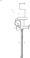
FIG. 1 is a side view of a photothermal treatment device according to an embodiment of the present invention. -
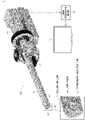
FIG. 2 is a perspective view of the photothermal treatment device according to the embodiment of the present invention. -
FIG. 3 is a front view of a probe tip of the photothermal treatment device. -
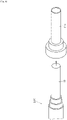
FIG. 4A is a side view showing a state in which a thermo-endoscope probe is inserted into a trocar. -
FIG. 4B is a conceptual view showing a state in which the trocar is inserted into an abdominal cavity of a rat and a lesion is irradiated with laser. -
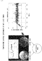
FIG. 5 is a view showing an image processing state of a thermal image. -
FIG. 6 is a view showing a temperature control experiment of a rat liver tumor. -
FIG. 7 is a view showing a depth of a tumor that can be treated when a target temperature during a photothermal treatment is changed. - Hereinafter, a photothermal treatment device according to an embodiment of the present invention and a usage method thereof are described with reference to the drawings. As shown in
FIGS. 1 and2 , aphotothermal treatment device 100 according to the embodiment of the present invention includes anelongated probe 10, anoperation part 20 disposed on a proximal end side of theprobe 10, and acommunication module 30 disposed between theprobe 10 and theoperation part 20. - The
probe 10 has a cylindrical shape with a diameter of about 14 mm and a length of about 200 mm. However, the diameter and the length can be changed as necessary, and for example, the diameter of theprobe 10 can be changed preferably in a range of 3 to 20 mm, and more preferably in a range of 5 to 16 mm. In the present embodiment, a diameter of about 14 mm is the most preferable size. The length of theprobe 10 can also be changed preferably in a range of 150 to 250 mm, more preferably in a range of 180 to 220 mm. In the present embodiment, a length of about 200 mm is the most preferable size. At a distal end of theprobe 10, an endoscopeobjective lens 40, anobjective lens 50 for infrared thermography, and a collimatinglens 60 for laser irradiation are disposed. - Infrared light emitted from an observation area in the body cavity is received by a thermopile element through the
objective lens 50 for infrared thermography and an infrared filter behind the objective lens, and is converted into electric energy. Then, the electric energy is sent to image processing means 70 by thecommunication module 30. - A
monitor 80 inFIG. 2 is configured to display an endoscopic image in the body cavity imaged by the endoscope objective lens and a charge-coupled device (CCD) element behind the endoscopeobjective lens 40 and a thermal image of the infrared thermography. The endoscopic image and the thermal image can be separately displayed on themonitor 80, but by switching a switch, the endoscopic image and the thermal image can be synthesized by the image processing means 70 and displayed on themonitor 80 in a superimposed state. In the synthesis, superimposition display is realized by performing adjustment by software so that the endoscopic image and the thermal image substantially overlap with each other in consideration of deviation of the optical axes of the endoscopic image and the thermal image. - A
laser fiber 90 constituting heating light irradiation means together with the collimatinglens 60 is connected to the rear side of the collimatinglens 60. Then, laser light for photothermal treatment can be emitted to a lesion tissue through thelaser fiber 90 and thecollimating lens 60. The laser light for photothermal treatment herein includes laser light having a wavelength within a range from a visible light range to an infrared range. - Next, a method of applying the
photothermal treatment device 100 to a rat liver tumor is described. First, as shown inFIG. 4A , theprobe 10 of thephotothermal treatment device 100 is inserted into atrocar 110. Next, as shown inFIG. 4B , thetrocar 110 is inserted into the abdominal cavity of the rat, and the target lesion tissue in which the light absorbing agent is accumulated is irradiated with the laser light. At the same time, the maximum temperature is acquired from the thermal image of the infrared thermography, and the laser output is subjected to Proportional-Integral-Differential (PID) control so that the target temperature of 50°C is achieved by the control personal computer (PC). The target temperature should be appropriately changed according to the type and the size of the target lesion tissue. In the present embodiment, the target temperature is set to 50°C, but the laser output can be PID-controlled preferably in a range of 43°C to 100°C, more preferably in a range of 43°C to 90°C, and most preferably at 80°C. - In the image processing means 70 in the control PC, as shown in
FIG. 5 (b) , 9 × 9 pixels around the maximum temperature pixel are extracted from the thermal image inFIG. 5 (a) , and are approximated to a two-dimensional Gaussian function. Then, as shown inFIG. 5 (c) , the maximum temperature of the Gaussian distribution is regarded as the tumor temperature, and the PID control is performed on the basis of the tumor temperature. Alternatively, the PID control may be performed based on the average temperature in a predetermined region (for example, in the 9 × 9 pixel region) of the thermal image. -
FIG. 6 shows a change in the tumor temperature when the rat liver tumor is actually subjected to photothermal treatment, and it has been confirmed that the tumor temperature can be controlled within a range of ±5°C of the target temperature of 50°C. Because the doctor in the operating room can easily confirm a state in which the thermal image is superimposed and displayed on the endoscopic image or a state in which both images are separately displayed on themonitor 80, the doctor can easily recognize a subtle temperature difference and can quickly and accurately position the laser light by accurate probe operation with respect to the target tissue in the body cavity in which the photosensitizer is excited. -
FIG. 7 shows the depth of tumor that can be treated (treatment depth) when the target temperature is changed (45°C to 100°C) at the time of actual photothermal treatment for the rat liver tumor. When the tumor temperature is changed from 45°C to 80°C, the treatment depth increases in proportion to the target temperature, the treatment depth becomes maximum (about 9 mm) at the target temperature of 80°C, and the treatment depth decreases at a higher target temperature. The doctor in the operating room can perform efficient tumor treatment while avoiding damage of normal tissue, by measuring the size of the tumor in advance by ultrasonic imaging or the like and changing the target temperature according to the depth of the tumor. - Even after the laser light is positioned, it has been conventionally necessary for the doctor to continue to appropriately control the output and the irradiation time of the laser corresponding to the thermal image that changes from moment to moment with the progress of laser irradiation. However, according to the photothermal treatment device of the present embodiment, the temperature change of the lesion tissue in the laser irradiation region can be easily grasped, and the output and the irradiation time of the laser can be automatically controlled by the control PC.
- Therefore, the lesion tissue can be easily and appropriately heated at a predetermined temperature for a predetermined time. Thereby, the lesion tissue can be reliably necrotized by the photothermal treatment, and the destruction of the surrounding healthy tissue can be minimized.
- Although the embodiment of the present invention has been described above, the present invention is not limited to the above embodiment, and various modifications can be made. For example, the thermographic imaging means may be equipped with a zoom lens to enlarge and display the thermal image of the lesion tissue. In addition, by coaxially arranging the optical axis of the endoscopic imaging means and the optical axis of the thermographic imaging means in the probe of the thermo-endoscope, the positional deviation of the image in the superimposed display of the endoscopic image and the thermal image can be minimized and the deviation of the display range in a case where both images are separately displayed can be minimized, and the image processing means can be simplified.
-
- 10
- Probe
- 20
- Operation part
- 30
- Communication module
- 40
- Endoscope objective lens
- 50
- Thermography objective lens
- 60
- Collimating lens
- 70
- Image processing means
- 80
- Monitor
- 90
- Laser fiber
- 100
- Photothermal treatment device
- 110
- Trocar
Claims (8)
- A photothermal treatment device comprising a thermo-endoscope, comprising:irradiation means that irradiates a lesion tissue in a body cavity with heating light;a thermo-endoscope including endoscopic imaging means that images an inside of the body cavity, and thermographic imaging means that images a surface temperature of a tissue in the body cavity as a thermal image; andcontrol means that controls an output and an irradiation time of the irradiation means based on temperature information of the thermal image obtained from the thermographic imaging means.
- The photothermal treatment device according to claim 1, further comprising image processing means that displays an endoscopic image obtained from the endoscopic imaging means and the thermal image obtained from the thermographic imaging means in a superimposed manner on one display means.
- The photothermal treatment device according to claim 1 or 2, wherein the control means controls the output and the irradiation time of the irradiation means based on an average temperature in a predetermined region of the thermal image.
- The photothermal treatment device according to claim 1 or 2, wherein the control means approximately derives a two-dimensional Gaussian function from a temperature distribution in a predetermined region of the thermal image, and controls the laser output and the irradiation time of the irradiation means based on a maximum temperature of a Gaussian distribution obtained from the Gaussian function.
- The photothermal treatment device according to claim 3 or 4, wherein the control means controls the output by Proportional-Integral-Differential (PID) control.
- The photothermal treatment device according to any one of claims 1 to 5, wherein, in a probe of the thermo-endoscope, the endoscopic imaging means has an optical axis that is coaxially disposed with an optical axis of the thermographic imaging means.
- The photothermal treatment device according to any one of claims 1 to 6, wherein the irradiation means emits, as heating light, laser light having a wavelength within a range from a visible light range to an infrared range.
- The photothermal treatment device according to any one of claims 1 to 7, wherein the thermographic imaging means is infrared thermography.
Applications Claiming Priority (2)
| Application Number | Priority Date | Filing Date | Title |
|---|---|---|---|
| JP2020014371 | 2020-01-31 | ||
| PCT/JP2021/001527 WO2021153319A1 (en) | 2020-01-31 | 2021-01-18 | Light/heat treatment device having thermo-endoscope |
Publications (2)
| Publication Number | Publication Date |
|---|---|
| EP4098320A1 true EP4098320A1 (en) | 2022-12-07 |
| EP4098320A4 EP4098320A4 (en) | 2024-02-14 |
Family
ID=77079712
Family Applications (1)
| Application Number | Title | Priority Date | Filing Date |
|---|---|---|---|
| EP21748450.0A Withdrawn EP4098320A4 (en) | 2020-01-31 | 2021-01-18 | Light/heat treatment device having thermo-endoscope |
Country Status (5)
| Country | Link |
|---|---|
| US (1) | US20230043393A1 (en) |
| EP (1) | EP4098320A4 (en) |
| JP (1) | JPWO2021153319A1 (en) |
| KR (1) | KR20220152995A (en) |
| WO (1) | WO2021153319A1 (en) |
Families Citing this family (1)
| Publication number | Priority date | Publication date | Assignee | Title |
|---|---|---|---|---|
| CN116269171B (en) * | 2023-03-23 | 2025-11-04 | 钜嘉联合科技股份有限公司 | Thermal imaging endoscope system, its abnormal area display method and display device, and thermal image processing computer |
Family Cites Families (14)
| Publication number | Priority date | Publication date | Assignee | Title |
|---|---|---|---|---|
| JPH02203871A (en) * | 1989-01-31 | 1990-08-13 | Asahi Optical Co Ltd | Temperature measuring endoscope for laser treatment |
| US6669685B1 (en) * | 1997-11-06 | 2003-12-30 | Biolase Technology, Inc. | Tissue remover and method |
| US7179222B2 (en) * | 1996-11-20 | 2007-02-20 | Olympus Corporation | Fluorescent endoscope system enabling simultaneous achievement of normal light observation based on reflected light and fluorescence observation based on light with wavelengths in infrared spectrum |
| JP2000079089A (en) | 1998-09-07 | 2000-03-21 | Olympus Optical Co Ltd | Thermograph for endoscope |
| JP2003010110A (en) * | 2001-07-04 | 2003-01-14 | Olympus Optical Co Ltd | Imaging device |
| US20180279864A1 (en) * | 2002-03-12 | 2018-10-04 | Beth Israel Deaconess Medical Center | Protoporphyrin ix (ppix) imaging |
| JP3930359B2 (en) * | 2002-03-29 | 2007-06-13 | オリンパス株式会社 | Sentinel lymph node detection apparatus and detection method |
| IL154101A0 (en) * | 2003-01-23 | 2003-07-31 | Univ Ramot | Minimally invasive controlled surgical system with feedback |
| WO2015061588A1 (en) * | 2013-10-23 | 2015-04-30 | The Trustees Of Dartmouth College | Surgical vision augmentation system |
| US10420608B2 (en) * | 2014-05-20 | 2019-09-24 | Verily Life Sciences Llc | System for laser ablation surgery |
| JP6451532B2 (en) * | 2015-07-13 | 2019-01-16 | 新日鐵住金株式会社 | Maintenance evaluation method for glass fiber reinforced plastic equipment |
| WO2018002925A1 (en) * | 2016-06-27 | 2018-01-04 | Ramot At Tel-Aviv University Ltd. | Monitoring tissue treatment using thermography |
| KR101852439B1 (en) * | 2016-09-01 | 2018-04-27 | 광주과학기술원 | Endoscope Device for Detecting Disease based on Thermal Images |
| JP2020510820A (en) * | 2017-03-16 | 2020-04-09 | トリナミクス ゲゼルシャフト ミット ベシュレンクテル ハフツング | Detector for optically detecting at least one object |
-
2021
- 2021-01-18 US US17/790,326 patent/US20230043393A1/en not_active Abandoned
- 2021-01-18 JP JP2021574649A patent/JPWO2021153319A1/ja not_active Withdrawn
- 2021-01-18 KR KR1020227024800A patent/KR20220152995A/en not_active Withdrawn
- 2021-01-18 WO PCT/JP2021/001527 patent/WO2021153319A1/en not_active Ceased
- 2021-01-18 EP EP21748450.0A patent/EP4098320A4/en not_active Withdrawn
Also Published As
| Publication number | Publication date |
|---|---|
| JPWO2021153319A1 (en) | 2021-08-05 |
| KR20220152995A (en) | 2022-11-17 |
| WO2021153319A1 (en) | 2021-08-05 |
| EP4098320A4 (en) | 2024-02-14 |
| US20230043393A1 (en) | 2023-02-09 |
Similar Documents
| Publication | Publication Date | Title |
|---|---|---|
| JP6126534B2 (en) | Ablation and temperature measurement equipment | |
| US6093148A (en) | Ultrasonic wave diagnosis apparatus | |
| CN104066368B (en) | Systems and methods for visualizing ablated tissue | |
| US5492126A (en) | Probe for medical imaging and therapy using ultrasound | |
| US20060052661A1 (en) | Minimally invasive control surgical system with feedback | |
| JP2005500108A (en) | Apparatus and method for thermal excision of biological tissue | |
| US7056318B2 (en) | Temperature controlled heating device and method to heat a selected area of a biological body | |
| KR20130109792A (en) | Robot system and control method thereof for surgery | |
| EP1269931A1 (en) | Medical energy irradiation apparatus | |
| EP3254730B1 (en) | Imaging dot matrix laser treatment instrument | |
| Su et al. | Automatic laser ablation control algorithm for an novel endoscopic laser ablation end effector for precision neurosurgery | |
| WO2020154596A1 (en) | Single use devices with integrated vision capabilities | |
| CA3245101A1 (en) | Efficient biological tissue treatment systems and methods | |
| WO2012147733A1 (en) | Treatment support system and medical image processing device | |
| JP2021506365A (en) | Robot optical navigation surgical system | |
| US20230043393A1 (en) | Light/heat treatment device having thermo-endoscope | |
| Su et al. | Micro laser ablation system integrated with image sensor for minimally invasive surgery | |
| US20240382255A1 (en) | Techniques For Delivering Laser Energy To A Target Site Of An Organism | |
| JP7505120B2 (en) | Phototherapy device, phototherapy device operation method, and phototherapy program | |
| JP2005287832A (en) | Heat treatment apparatus | |
| Campbell et al. | Thermal imaging in surgery | |
| Omori et al. | Mid-Infrared Robotic Laser Surgery System in Neurosurgery | |
| König | Minimally invasive medicine | |
| Bronzino | Thermal Imaging in Surgery | |
| Thomas | Paul Campbell |
Legal Events
| Date | Code | Title | Description |
|---|---|---|---|
| STAA | Information on the status of an ep patent application or granted ep patent |
Free format text: STATUS: THE INTERNATIONAL PUBLICATION HAS BEEN MADE |
|
| PUAI | Public reference made under article 153(3) epc to a published international application that has entered the european phase |
Free format text: ORIGINAL CODE: 0009012 |
|
| STAA | Information on the status of an ep patent application or granted ep patent |
Free format text: STATUS: REQUEST FOR EXAMINATION WAS MADE |
|
| 17P | Request for examination filed |
Effective date: 20220818 |
|
| AK | Designated contracting states |
Kind code of ref document: A1 Designated state(s): AL AT BE BG CH CY CZ DE DK EE ES FI FR GB GR HR HU IE IS IT LI LT LU LV MC MK MT NL NO PL PT RO RS SE SI SK SM TR |
|
| DAV | Request for validation of the european patent (deleted) | ||
| DAX | Request for extension of the european patent (deleted) | ||
| STAA | Information on the status of an ep patent application or granted ep patent |
Free format text: STATUS: THE APPLICATION HAS BEEN WITHDRAWN |
|
| A4 | Supplementary search report drawn up and despatched |
Effective date: 20240111 |
|
| RIC1 | Information provided on ipc code assigned before grant |
Ipc: A61B 1/045 20060101ALI20240105BHEP Ipc: A61B 1/00 20060101ALI20240105BHEP Ipc: A61N 5/067 20060101AFI20240105BHEP |
|
| 18W | Application withdrawn |
Effective date: 20240129 |