EP4087860B1 - Compound and method for treatment of alzheimer's disease - Google Patents
Compound and method for treatment of alzheimer's disease Download PDFInfo
- Publication number
- EP4087860B1 EP4087860B1 EP21700377.1A EP21700377A EP4087860B1 EP 4087860 B1 EP4087860 B1 EP 4087860B1 EP 21700377 A EP21700377 A EP 21700377A EP 4087860 B1 EP4087860 B1 EP 4087860B1
- Authority
- EP
- European Patent Office
- Prior art keywords
- brichos
- bri2
- bri2 brichos
- seq
- amino acid
- Prior art date
- Legal status (The legal status is an assumption and is not a legal conclusion. Google has not performed a legal analysis and makes no representation as to the accuracy of the status listed.)
- Active
Links
Images







Classifications
-
- C—CHEMISTRY; METALLURGY
- C07—ORGANIC CHEMISTRY
- C07K—PEPTIDES
- C07K14/00—Peptides having more than 20 amino acids; Gastrins; Somatostatins; Melanotropins; Derivatives thereof
- C07K14/435—Peptides having more than 20 amino acids; Gastrins; Somatostatins; Melanotropins; Derivatives thereof from animals; from humans
- C07K14/46—Peptides having more than 20 amino acids; Gastrins; Somatostatins; Melanotropins; Derivatives thereof from animals; from humans from vertebrates
- C07K14/47—Peptides having more than 20 amino acids; Gastrins; Somatostatins; Melanotropins; Derivatives thereof from animals; from humans from vertebrates from mammals
-
- C—CHEMISTRY; METALLURGY
- C07—ORGANIC CHEMISTRY
- C07K—PEPTIDES
- C07K14/00—Peptides having more than 20 amino acids; Gastrins; Somatostatins; Melanotropins; Derivatives thereof
- C07K14/435—Peptides having more than 20 amino acids; Gastrins; Somatostatins; Melanotropins; Derivatives thereof from animals; from humans
-
- A—HUMAN NECESSITIES
- A61—MEDICAL OR VETERINARY SCIENCE; HYGIENE
- A61P—SPECIFIC THERAPEUTIC ACTIVITY OF CHEMICAL COMPOUNDS OR MEDICINAL PREPARATIONS
- A61P25/00—Drugs for disorders of the nervous system
- A61P25/28—Drugs for disorders of the nervous system for treating neurodegenerative disorders of the central nervous system, e.g. nootropic agents, cognition enhancers, drugs for treating Alzheimer's disease or other forms of dementia
-
- A—HUMAN NECESSITIES
- A61—MEDICAL OR VETERINARY SCIENCE; HYGIENE
- A61K—PREPARATIONS FOR MEDICAL, DENTAL OR TOILETRY PURPOSES
- A61K38/00—Medicinal preparations containing peptides
Definitions
- the present invention pertains to the field of medicine. More specifically, this invention relates to medicaments for treatment and medical treatment of Alzheimer's disease in a mammal such as man.
- Alzheimer's disease is one of the most common causes of dementia in man. It is a chronic and fatal disease associated with neural cell degeneration in the brain of the affected individual, characterized by the presence of amyloid plaques consisting of extracellular deposits of amyloid ⁇ -peptide (A ⁇ -peptide).
- a ⁇ PP A ⁇ precursor protein
- a ⁇ 42 Proteolytic processing of the the A ⁇ precursor protein (A ⁇ PP) generates A ⁇ peptides of different lengths, whereof A ⁇ 42 is the most aggregation prone and toxic.
- the kinetics of A ⁇ 42 aggregation follows recently defined nucleation-dependent microscopic events.
- a ⁇ 42 monomers associate and form a nucleus, from which a fibril can start to elongate.
- monomers attach to the surface of a fibril, which catalyzes the formation of a new nucleus, leading to exponential fibril growth.
- This monomer dependent secondary nucleation autocatalytic pathway is the predominant source of toxic A ⁇ 42 species.
- Key challenges in finding a treatment of AD is to specifically reduce A ⁇ 42 neurotoxicity, rather than focusing on overall aggregation and plaque formation.
- chaperones play an important role by aiding the correct folding of proteins in the complex intracellular milieu.
- a number of molecular chaperones, such as heat-shock proteins (Hsp) are known to be important in the folding process and have been extensively studied. Some of these chaperones are apparently able to interact with and have an impact on the amyloid fibril formation of certain polypeptides.
- the BRICHOS domain has been found in several human precursor proteins, initially in Bri 2, Cho ndromodulin-1 and pro s urfactant protein C (proSP-C). Sanchez-Pulido et al., Trends Biochem Sci 27(7): 329-332,2002 provides a review of BRICHOS domains.
- the BRICHOS domain has been suggested to prevent amyloid formation of aggregation prone regions (clients) of the respective proprotein during biosynthesis.
- Recombinant human (rh) BRICHOS domains from proSP-C and Bri2 are efficient inhibitors also of amyloid formation of non-client proteins such as medin, islet amyloid polypeptide, A ⁇ 40 and A ⁇ 42.
- WO 2011/162655 discloses that the BRICHOS domain of Bri2 decreases amyloid fibril formation and aggregation of A ⁇ peptide and ABri/ADan peptides.
- Bri2 is produced in the central nervous system (CNS), with expression in neurons of the hippocampus and cortex in humans, and colocalizes with senile plaques in AD patients.
- Known sequences of Bri2 include sequences derived from Erpetoichtys calabaricus (NCBI accession no. XP_028655526) and Acipenser ruthenus (UniProt/TrEMBL accession no. A0A444U9X2). Under physiological conditions, the BRICHOS domain is released by proteolysis from the Bri2 precursor protein.
- Recombinant human (rh) Bri2 BRICHOS is efficient in inhibiting A ⁇ 42 fibril formation in vitro and in alleviating the related neurotoxicity to hippocampal slice preparations and in Drosophila models.
- Rh proSP-C BRICHOS specifically impedes the secondary nucleation step in A ⁇ 42 fibril formation.
- Rh Bri2 BRICHOS modulates both elongation and secondary nucleation events, but different assembly states of Bri2 BRICHOS affect A ⁇ fibril formation in different ways.
- Bri2 BRICHOS monomers are most potent in preventing A ⁇ 42 induced disruption of neuronal network activity, while dimers most efficiently suppress A ⁇ 42 overall fibril formation and oligomers inhibit non-fibrillar protein aggregation ( Chen et al., Nat.
- the Bri2 BRICHOS monomers are not long-term stable and form high molecular weight oligomers in a concentration-dependent manner in phosphate buffer or in mouse serum in vitro, which is accompanied by reduced potency against A ⁇ 42 fibril formation. Conversion of Bri2 BRICHOS monomers to high molecular weight oligomers may be relevant for AD, as increased amounts of different Bri2 forms were found in AD brain compared with healthy controls. These observations imply that modulating the distribution of Bri2 BRICHOS assembly states so that the amount of monomers is increased is a concept to combat A ⁇ 42 neurotoxicity, see e.g. WO 2012/138284 .
- amyloid plaques consisting of extracellular deposits in the brain of a mammal of proteins that are prone to fibrillate.
- the present invention is generally based on the insight that monomers of chaperone proteins which have a high identity to the BRICHOS domains of Bri2 from human are useful for medical treatment Alzheimer's disease.
- the present disclosure provides according to different aspects a new isolated protein; a pharmaceutical composition comprising the protein; use of the protein as a medicament use in treatment of Alzheimer's Disease; and a method of treating Alzheimer's Disease comprising administration of the protein.
- Bri2 (SEQ ID NO: 1), also referred to as integral membrane protein 2B (ITM2B), contains an evolutionary conserved Brichos domain spanning residues 137-231 ( Fig 1 , SEQ ID NO: 7-12).
- the Brichos domain of Bri2 may alternatively be considered as spanning residues 113-231 (SEQ ID NO: 3).
- the present invention is generally based on the insight that monomers of chaperone proteins which have a high identity to the BRICHOS domains of Bri2 from human are useful for medical treatment of Alzheimer's disease.
- a single point mutant of Bri2 BRICHOS that stabilizes the monomeric state. It is stable during long-term incubation in mouse serum and blood-brain barrier (BBB) permeable.
- BBB blood-brain barrier
- This mutant monomer is potent in preventing A ⁇ 42 neurotoxicity, specifically suppresses secondary nucleation during fibril formation and, importantly, significantly potentiates wild-type (wt) protein against A ⁇ 42 neurotoxicity.
- Systemic administration of the single point mutant of Bri2 BRICHOS shows an improvement in the memory response and behaviour of AD mice. It also shows reduction in astrocyte mediated neuroinflammation.
- an isolated protein selected from the group consisting of proteins comprising a moiety of 90-200 amino acid residues having at least 70% identity to one of the Brichos domains of Bri2 from human having SEQ ID NO: 7 or SEQ ID NO: 3, wherein the amino acid residue corresponding to position 221 (Arg) in SEQ ID NO: 1 is not Arg, Lys or His.
- the Arg residue in position 221 of SEQ ID NO: 1 corresponds to the Arg residue in position 109 of SEQ ID NO: 3, and to the Arg residue in position 85 of SEQ ID NO: 7.
- % identity is calculated as follows.
- the query sequence is aligned to the target sequence using the CLUSTAL W algorithm ( Thompson, J.D., Higgins, D.G. and Gibson, T.J., Nucleic Acids Research, 22: 4673-4680 (1994 )).
- a comparison is made over the window corresponding to the shortest of the aligned sequences.
- the amino acid residues at each position are compared, and the percentage of positions in the query sequence that have identical correspondences in the target sequence is reported as % identity.
- % similarity is calculated as described for "% identity", with the exception that the hydrophobic residues Ala, Val, Phe, Pro, Leu, Ile, Trp, Met and Cys are similar; the basic residues Lys, Arg and His are similar; the acidic residues Glu and Asp are similar; and the hydrophilic, uncharged residues Gln, Asn, Ser, Thr and Tyr are similar.
- the remaining natural amino acid Gly is not similar to any other amino acid in this context.
- the isolated protein sequence may be 70% similar to another protein sequence; or it may be 70% identical to another sequence; or it may be 70% identical and furthermore 90% similar to another sequence.
- the amino acid sequence having at least the given identity to the Brichos domain of Bri2 from human consists of more than or equal to 70, such as more than or equal to 80, such as more than or equal to 90 amino acid residues.
- a preferable size range is 70-140 amino acid residues, such as 80-140 amino acid residues, e.g. 90-140 amino acid residues.
- the Brichos domains of Bri2 from human SEQ ID NO: 7
- chimpanzee SEQ ID NO: 8
- bovine SEQ ID NO: 9
- pig SEQ ID NO: 10
- mouse SEQ ID NO: 11
- rat SEQ ID NO: 12
- the isolated protein is selected from the group consisting of proteins comprising an amino acid sequence having at least 80%, preferably at least 90%, such as at least 95%, identity to any one of the Brichos domains of Bri2 from human, including SEQ ID NO: 7 and SEQ ID NO: 3.
- the isolated protein contains all amino acid residues that are conserved in the Brichos domains of Bri2 from human (SEQ ID NO: 7), chimpanzee (SEQ ID NO: 8), bovine (SEQ ID NO: 9), pig (SEQ ID NO: 10), mouse (SEQ ID NO: 11) and rat (SEQ ID NO: 12), i.e.
- the isolated protein is selected from the group consisting of proteins comprising any one of the Brichos domains of Bri2 from human including SEQ ID NO: 7 and SEQ ID NO: 3, i.e. it contains one of these Brichos domains, but wherein the amino acid residue corresponding to position 221 (Arg) in SEQ ID NO: 1 is not Arg, Lys or His.
- the isolated protein is selected from the group consisting of proteins having at least 70%, such as at least 80%, preferably at least 90%, such as at least 95%, or even 100% identity to Bri2 from human (SEQ ID NO: 1), but wherein the amino acid residue corresponding to position 221 (Arg) in SEQ ID NO: 1 is not Arg, Lys or His.
- the isolated protein is SEQ ID NO: 1, but wherein the amino acid residue corresponding to position 221 (Arg) in SEQ ID NO: 1 is not Arg, Lys or His.
- the isolated protein is selected from the group consisting of any one of the Brichos domains of Bri2 from human including SEQ ID NO: 7 and SEQ ID NO: 3, i.e. it contains one of these Brichos domains, but wherein the amino acid residue corresponding to position 221 (Arg) in SEQ ID NO: 1 is not Arg, Lys or His.
- amino acid residue corresponding to position 221 in SEQ ID NO: 1 is preferably selected from the group consisting of Glu, Asp, Gln and Asn, more preferably from the group consisting of Glu and Asp.
- amino acid residue corresponding to position 221 in SEQ ID NO: 1 is preferably Glu.
- amino acid residue corresponding to position 221 in SEQ ID NO: 1 may also be Asp.
- the moiety has the amino acid sequence of SEQ ID NO: 4 or the amino acid sequence of SEQ ID NO: 13.
- Examples of specific isolated proteins include proteins which have the amino acid sequence of SEQ ID NO: 4 or the amino acid sequence of SEQ ID NO: 13.
- a preferred group of isolated proteins disclosed herein is the monomer fraction thereof.
- One preferred method of providing a monomer fraction is separation by Size Exclusion Chromatography.
- the isolated protein is preferably not comprising an amino acid sequence having at least 70% identity to residues 1-89 of Bri2 from human (SEQ ID NO: 5). In certain embodiments, the isolated protein is not comprising an amino acid sequence having at least 50% identity to residues 1-89 of Bri2 from human (SEQ ID NO: 5). This implies that the isolated protein contains a core amino acid sequence which displays a high similarity or identity to the Brichos domain of Bri2 and optionally one or more other amino acid sequences, which other amino acid sequences may not display a high similarity or identity to residues 1-89 of Bri2 from human (SEQ ID NO: 5).
- the isolated protein is not comprising an amino acid sequence that consists of more than or equal to 10 amino acid residues having at least the given identity to residues 1-89 of Bri2 from human (SEQ ID NO: 5).
- the isolated protein is preferably not comprising an amino acid sequence having at least 70% identity to residues 244-266 of Bri2 from human, i.e. human Bri23 (SEQ ID NO: 6). In certain embodiments, the isolated protein is not comprising an amino acid sequence having at least 50% identity to residues human Bri23 (SEQ ID NO: 6). As set out above, this implies that the isolated protein contains a core amino acid sequence which displays a high similarity or identity to the Brichos domain of Bri2 and optionally one or more other amino acid sequences, which other amino acid sequences may not display a high similarity or identity to human Bri23 (SEQ ID NO: 6).
- the isolated protein is not comprising an amino acid sequence that consists of more than or equal to 10 amino acid residues having at least the given identity to human Bri23 (SEQ ID NO: 6).
- Proteins comprising a core amino acid sequence having high identity with the Bri2 BRICHOS sequence as set out above may further comprise additional amino acid sequences which do not interfere with the function of the core amino acid sequence, i.e. interaction with A ⁇ peptides.
- the additional amino acid sequences may be connected to the N-terminal of the core amino acid sequence, to the C-terminal of the core amino acid sequence, or both. It may also be connected via amino acid side chains, e.g. via a disulphide bond.
- the additional amino acid sequences may be essentially non-functional or may provide additional functionality to the resulting protein, e.g. solubility, stability or a desired affinity. Both the core amino acid sequence and any additional amino acid sequences may be chemically modified, including post-translational chemical modifications.
- the isolated protein consists of less than or equal to 500, such as less than or equal to 300, such as less than or equal to 250, such as less than or equal to 200, such as less than or equal to 150 or even 100 amino acid residues.
- the isolated protein consists of more than or equal to 90, such as more than or equal to 100 amino acid residues.
- a preferable size range is 90-300 amino acid residues, such as 90-200 or 90-140 amino acid residues.
- Means of counteracting proteotoxicity include chaperone-mediated prevention of amyloid formation, disaggregation of pre-existing aggregates, and aggregate sequestration.
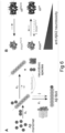
- Fig. 6 provides a model for potentiation of chaperone activity against A ⁇ 42 neurotoxicity by shifting the Bri2 BRICHOS assembly state:
- the Bri2 BRICHOS R221E monomer predominantly attenuates the secondary nucleation rate, while the dimer substantially affects both the elongation and secondary nucleation rate ( Figs. 3 and 6A ). This causes a reduction of the generated number of nucleation units in the presence of Bri2 BRICHOS R221E monomers, but not dimers ( Fig. 4A ).
- the molecular size of the Bri2 BRICHOS R221E dimer matches well the surface of the cross-sectional area of recently published A ⁇ 42 fibril structures, providing a possible explanation why the Bri2 BRICHOS dimer, besides the secondary nucleation rate k 2 , also efficiently attenuates the elongation rate k + .
- the different specificity of the Bri2 BRICHOS dimer compared to the monomer also support that secondary nucleation and elongation events occur at distinct sites on A ⁇ 42 aggregates. While structural details about the soluble neurotoxic A ⁇ 42 species are still missing, a ⁇ -structure state of toxic A ⁇ 42 species/oligomers has been reported. We observed that the molecular size of the Bri2 BRICHOS monomer fits well a single layer of ⁇ -structured A ⁇ 42 molecules, which may build up neurotoxic A ⁇ 42 species ( Fig. 6A ).
- the ability to efficiently reduce the generation of new nucleation units together with the well-matched molecular size for interactions with putative neurotoxic A ⁇ 42 species potentially makes the Bri2 BRICHOS monomer most efficient in preventing A ⁇ 42-induced neurotoxicity to hippocampal ⁇ oscillations ( Fig. 4B ).
- AD aging For AD aging is the main risk factor, but detailed underlying mechanisms are missing. Chaperone network decline during aging is considered to affect many aging-associated diseases. In aging organisms, the balance between misfolded proteins and functional chaperones is disturbed. Increased amounts of different Bri2 forms were observed in AD brains compared to the healthy controls. Improved chaperone activity could be achieved by increasing the local concentration, but since chaperones are precisely balanced, overproduction of certain chaperone may result in disease. Hence, the concept to augment the capacity of certain chaperone networks holds potential for preventing and treating pathologies associated with proteome deterioration.
- nucleic acid comprising a sequence encoding the isolated protein disclosed herein.
- exemplary nucleic acids are SEQ ID NO: 16 encoding an NT*-Bri2 BRICHOS mutant and the partial sequences of SEQ ID NO: 16 encoding the Bri2 BRICHOS mutants having SEQ ID NO: 4 and SEQ ID NO: 13.
- a pharmaceutical composition comprising a therapeutically effective amount of the isolated protein disclosed herein or the nucleic acid disclosed herein.
- the pharmaceutical composition also contains a suitable pharmaceutical carrier therefor.
- the pharmaceutical composition is useful as a medicament.
- the pharmaceutical composition is useful in treatment of Alzheimer's disease in a mammal, including man.
- a preferred group of isolated proteins which is useful in the pharmaceutical composition is the monomer fraction thereof.
- One preferred method of providing a monomer fraction is separation by Size Exclusion Chromatography.
- the isolated protein can be incorporated into pharmaceutical compositions.
- Such compositions typically include the isolated protein and a suitable pharmaceutically acceptable carrier.
- a suitable pharmaceutical carrier includes solvents, dispersion media, coatings, isotonic and absorption delaying agents, and the like, that are compatible with pharmaceutical administration.
- Supplementary active compounds can also be incorporated into the compositions.
- a pharmaceutical composition is formulated to be compatible with its intended route of administration.
- routes of administration include parenteral (e.g. intravenous, intradermal, subcutaneous, intrathecal, and intracerebral ventricular (e.g. using an Omaya reservoir-shunt with in-line filter that is surgically placed into the cisternal space) administration.
- parenteral delivery systems for a composition include slow-dissolving polymer particles, implantable infusion systems, and liposomes.
- Solutions or suspensions used for parenteral application can include the following components: a sterile diluent such as water for injection, saline solution, fixed oils, polyethylene glycols, glycerine, propylene glycol or other synthetic solvents; antibacterial agents such as benzyl alcohol or methyl parabens; antioxidants such as ascorbic acid or sodium bisulfite; chelating agents such as ethylenediaminetetraacetic acid; buffers such as acetates, citrates or phosphates and agents for the adjustment of tonicity such as sodium chloride or dextrose. pH can be adjusted with acids or bases, such as hydrochloric acid or sodium hydroxide.
- the parenteral preparation can be enclosed in ampoules, disposable syringes or multiple dose vials made of glass or plastic.
- Treatment of Alzheimer's disease may also be effected by direct delivery of the isolated protein to the central nervous system, preferentially to the brain.
- compositions suitable for injectable use include sterile aqueous solutions (where water soluble) or dispersions and sterile powders for the extemporaneous preparation of sterile injectable solutions or dispersion.
- suitable carriers include physiological saline, bacteriostatic water, Cremophor EL TM or phosphate buffered saline (PBS).
- the composition must be sterile and should be fluid to the extent that easy syringability exists. It should be stable under the conditions of manufacture and storage and must be preserved against the contaminating action of microorganisms such as bacteria and fungi.
- the carrier can be a solvent or dispersion medium containing, for example, water, ethanol, polyol (for example, glycerol, propylene glycol, and liquid polyetheylene glycol, and the like), and suitable mixtures thereof.
- the proper fluidity can be maintained, for example, by the use of a coating on particles of the isolated protein (e.g. lecithin), by the maintenance of the required particle size in the case of dispersion and by the use of surfactants.
- Prevention of the action of microorganisms can be achieved by various antibacterial and antifungal agents, for example, parabens, chlorobutanol, phenol, ascorbic acid, thimerosal, and the like.
- isotonic agents in the composition.
- examples of such agents include sugars, polyalcohols such as mannitol and sorbitol, and sodium chloride.
- Prolonged absorption of the injectable compositions can be brought about by including in the composition an agent which delays absorption, for example, aluminum monostearate or gelatin.
- Sterile injectable solutions can be prepared by incorporating the isolated protein in the required amount in an appropriate solvent with one or a combination of ingredients enumerated above, as required, followed by filtered sterilization.
- dispersions are prepared by incorporating the isolated protein into a sterile vehicle which contains a basic dispersion medium and the required other ingredients from those enumerated above.
- the preferred methods of preparation are vacuum drying and freeze-drying, which yield a powder of the isolated protein plus any additional desired ingredient from a previously sterile-filtered solution thereof.
- the isolated protein is prepared with a carrier that will protect the compound against rapid elimination from the body, such as a controlled release formulation, including implants and microencapsulated delivery systems.
- a carrier that will protect the compound against rapid elimination from the body
- a controlled release formulation including implants and microencapsulated delivery systems.
- Biodegradable, biocompatible polymers can be used, such as ethylene vinyl acetate, polyanhydrides, polyglycolic acid, collagen, polyorthoesters, and polylactic acid. Methods for preparation of such formulations will be apparent to those skilled in the art.
- Liposomal suspensions (including liposomes targeted to cells specifically affected by Alzheimer's disease with monoclonal antibodies) can also be used as pharmaceutically acceptable carriers. These can be prepared according to methods known to those skilled in the art.
- Dosage unit form refers to physically discrete units suited as unitary dosages for the subject to be treated, each unit containing a predetermined quantity of the isolated protein calculated to produce the desired therapeutic effect in association with the required pharmaceutical carrier.
- Toxicity and therapeutic effects of the isolated proteins can be determined by standard pharmaceutical procedures in cell cultures or experimental animals, e.g. for determining the LD 50 (the dose lethal to 50% of the population) and the ED 50 (the dose therapeutically effective in 50% of the population).
- Suitable animal models can be used such as those described for amyloidoses in Sturchler-Pierrat et al, Rev Neurosci, 10: 15-24, 1999 ; Seabrook et al, Neuropharmacol 38: 1-17, 1999 ; DeArmond et al, Brain Pathology 5: 77-89, 1995 ; Telling, Neuropathol Appl Neurobiol 26: 209-220, 2000 ; and Price et al, Science 282: 1079-1083, 1998 .
- the dose ratio between toxic and therapeutic effects is the therapeutic index and it can be expressed as the ratio LD 50 /ED 50 .
- Compounds that exhibit high therapeutic indices are preferred. While compounds that exhibit toxic side effects may be used, care should be taken to design a delivery system that targets such compounds to the site of affected tissue in order to minimize potential damage to unaffected cells and thereby reduce side effects.
- the dosage of a compound lies preferably within a range of circulating concentrations that include the ED 50 with little or no toxicity.
- the dosage may vary within this range depending upon the dosage form employed and the route of administration utilized.
- the therapeutically effective dose can be estimated initially from cell culture assays in which, e.g. the rate of fibril formation or the rate of cell death is observed.
- a dose may be formulated in animal models to achieve a circulating plasma concentration range that includes the IC 50 (i. e., the concentration of the test compound which achieves a half-maximal inhibition of symptoms) as determined in cell culture. Such information can be used to more accurately determine useful doses in humans. Levels in plasma may be measured, for example, by high performance liquid chromatography.
- a therapeutically effective amount of the isolated protein ranges from about 0.1 to 100 mg/kg body weight, more preferably about 1 to 100 mg/kg body weight, and even more preferably about 1 to 50 mg/kg body weight.
- the compound can be administered over an extended period of time to the subject, e. g., over the subject's lifetime.
- a dosage of 1 mg/kg to 100 mg/kg is usually appropriate, such as is the case for antibodies designated to act in the brain.
- the compound can be administered for a limited time period.
- the compound can also be administered chronically.
- certain factors may influence the dosage and timing required to effectively treat a subject, including but not limited to the severity of the disease or disorder, previous treatments, the general health and/or age of the subject, and other diseases present.
- treatment of a subject with a therapeutically effective amount of a compound can include a single treatment or, preferably, can include a series of treatments.
- Recombinant isolated proteins for administration to mice expressing the human APP or to humans can be prepared in several ways.
- the recombinant proteins can be purified as described herein.
- the isolated protein When the isolated protein is to be administered to an animal (e.g. a human) to treat Alzheimer's disease, a physician, veterinarian, or researcher may, for example, prescribe a relatively low dose at first, subsequently increasing the dose until an appropriate response is obtained.
- an animal e.g. a human
- a physician, veterinarian, or researcher may, for example, prescribe a relatively low dose at first, subsequently increasing the dose until an appropriate response is obtained.
- the specific dose level for any particular animal subject will depend upon a variety of factors including the activity of the specific compound employed, the age, body weight, general health, gender, and diet of the subject, the time of administration, the route of administration, the rate of excretion, any drug combination, and the degree of expression or activity to be modulated.
- compositions can be included in a container, pack, or dispenser together with instructions for administration.
- the instructions can include directions to use the composition to treat an individual having or at risk for Alzheimer's disease.
- a method of treating Alzheimer's disease, in a mammal, including man, in need thereof comprising administration to said mammal of a therapeutically effective amount of the isolated protein or the pharmaceutical composition.
- a preferred group of isolated proteins which is useful in the method of treatment is the monomer fraction thereof.
- One preferred method of providing a monomer fraction is separation by Size Exclusion Chromatography.
- the treatment may be a preventive treatment. In other specific embodiments, the treatment may be a palliative treatment. In certain specific embodiments, the treatment may be a curative treatment.
- treatment is defined as the application or administration of the isolated protein to a patient, or application or administration of the isolated protein to an isolated tissue or cell line from a patient, who has Alzheimer's disease, a symptom of disease or a predisposition toward disease, with the purpose to cure, heal, alleviate, relieve, alter, remedy, ameliorate, improve or affect the disease, the symptoms of disease or the predisposition toward disease.
- a method for preventing a disease or condition i.e. decreasing the risk of contracting, or decreasing the rate at which symptoms appear that are associated with a disease or condition
- a disease or condition i.e. decreasing the risk of contracting, or decreasing the rate at which symptoms appear that are associated with a disease or condition
- fibril formation caused by A ⁇ peptide by administering to the subject the isolated protein that reduces aggregation of the polypeptide.
- Subjects at risk for Alzheimer's disease can be identified by, for example, any or a combination of appropriate diagnostic or prognostic assays known in the art.
- Administration of a prophylactic agent can occur prior to the manifestation of symptoms characteristic of the disease, such that the disease is prevented or, alternatively, delayed in its progression.
- the isolated protein can be administered to a patient at therapeutically effective doses to prevent, treat or ameliorate disorders involving fibril formation associated with Alzheimer's disease.
- a therapeutically effective dose refers to that amount of the compound sufficient to result in amelioration of symptoms of the disorders. Toxicity and therapeutic efficacy of such compounds can be determined by standard pharmaceutical procedures as described above.
- the protein can be administrated by gene therapy, such as by using expression vectors, plasmids or viruses to transfect cells in the neural system, preferably brain, such that the isolated protein is expressed by these cells in the central neural system. This is useful for the treatment of Alzheimer's disease.
- a construct having SEQ ID NO: 16 and coding for a His 6 -NT*-thrombin cleavage site-Bri2 BRICHOS R221E was transfected into SHuffle T7 competent E . coli cells. The cells were incubated at 30°C in LB medium containing 15 ⁇ g mL-1 kanamycin, until an OD600 nm around 0.9, when the temperature was lowered to 20°C. Isopropyl ⁇ -D-1-thiogalactopyranoside (IPTG) was added to 0.5 mM and the cells were incubated overnight.
- IPTG Isopropyl ⁇ -D-1-thiogalactopyranoside
- the cells were then harvested by 7,000 ⁇ g centrifugation at 4°C, after which the cell pellets were resuspended in 20 mM Tris pH 8.0, and sonicated for 5 min on ice (2 s on, 2 s off, 65% power).
- the lysate was centrifuged (24,000 ⁇ g) at 4°C for 30 min and the target protein was purified with a Ni-NTA column.
- the fusion proteins were cleaved with thrombin (1:1000 enzyme to substrate, w/w) at 4°C overnight and loaded over a second Ni-NTA column.
- the resulting Bri2 BRICHOS R221E protein had the amino acid sequence of SEQ ID NO: 4.
- an AU1 tag (DTYRYI) or an mCherry domain was fused to the C-terminal of rh wt Bri2 BRICHOS by PCR amplification.
- the construct coding for His6-NT*-thrombin cleavage site-Bri2 BRICHOS wt-AU1 or His6-NT*-thrombin cleavage site-Bri2 BRICHOS wt-m Cherry were expressed and purified with the same protocol as the one for Bri2 BRICHOS R221E.
- the rh wt Bri2 BRICHOS-AU1 or rh wt Bri2 BRICHOS-mCherry oligomers were isolated by Superdex 200 PG column (GE Healthcare, UK).
- rh Bri2 BRICHOS R221E Prior to ESI-MS analysis, rh Bri2 BRICHOS R221E (SEQ ID NO: 4) monomers isolated by SEC were buffer exchanged into 200 mM ammonium acetate pH 7.5 using BioSpin microcentrifuge columns (BioRad, US). The final concentration of monomeric subunit was 20 ⁇ M. Spectra were recorded on a Waters Synapt G1 mass spectrometer (Waters, Milford, MA) modified for high mass analysis. Samples were introduced into the mass spectrometer using in-house produced gold-coated borosilicate capillaries. Instrument settings were: capillary voltage 1.5 V, sample cone voltage 30 V, extraction cone voltage 4 V, collision trap voltage 50 V, and transfer voltage 10 V. The source pressure was 7 mbar, trap gas was N 2 with a flow rate of 8 mL h -1 . Data analysis was performed using Waters MassLynx 4.1 software.
- CD spectroscopy bis-ANS fluorescence and CS thermal aggregation CD spectra were recorded from 260 to 190 nm at 25°C in 1 mm path length quartz cuvettes in an Aviv 410 Spectrometer (Lakewood, NJ, USA) with protein concentration of 12 ⁇ M.
- the wavelength step was 0.5 nm, averaging time 0.3 s, time constant 100 ms, and bandwidth 1 nm.
- the spectra shown are averages of three consecutive scans.
- CS from porcine heart was diluted in 40 mM HEPES/KOH pH 7.5 to 600 nM and then equilibrated at 45°C with and without different concentrations of various rh Bri2 BRICHOS R221E species.
- the aggregation kinetics were measured in triplicate using a microplate reader (FLUOStar Galaxy from BMG Labtech, Offenberg, Germany) by reading the apparent increase in absorbance at 360 nm during incubation at 45°C under quiescent conditions.
- Rh Bri2 BRICHOS R221E (SEQ ID NO: 4) oligomers, dimers and monomers at 20 ⁇ M concentrations (referred to the monomeric subunits) were incubated in ThT assay buffer (20 mM sodium phosphate, pH 8.0 with 0.2 mM EDTA and 0.02% NaN 3 ) at 37°C. Samples were taken out after 0, 1, 4 and 24 h, and analysed for assembly states by SDS-PAGE under reducing and non-reducing conditions. Ten ⁇ M rh Bri2 BRICHOS R221E oligomers and monomers were incubated in ThT assay buffer at 37°C overnight and analyzed by native PAGE.
- a ⁇ 42 Recombinant Met-A ⁇ (1-42), here referred to as A ⁇ 42, was produced in BL21*(DE3) pLysS E. coli (B strain) cells and purified by ion exchange.
- the purified A ⁇ 42 proteins were lyophilized overnight and re-dissolved in 7 M Gdn-HCl and then injected into a Superdex 75 column (GE Healthcare, UK) for monomer isolation in 20 mM sodium phosphate pH 8.0 with 0.2 mM EDTA and 0.02% NaN3.
- the A ⁇ 42 concentration was calculated from the absorbances at 280 and 300 nm using an extinction coefficient of 1,424 M-1cm-1 for (A280-A300).
- Purified A ⁇ 42 monomers were aliquoted in low-binding Eppendorf tubes (Axygene).
- the fluorescence was recorded using a 440 nm excitation filter and a 480 nm emission filter (FLUOStar Galaxy from BMG Labtech, Offenberg, Germany).
- the aggregation kinetics of A ⁇ 42 at different concentrations in the presence of a constant concentration (0.9 ⁇ M) of rh Bri2 BRICHOS R221E monomers and dimers were measured in the same manner.
- 3 ⁇ M A ⁇ 42 monomer was incubated at 37°C for about 20 h, and the generated fibrils were then sonicated in a water bath for 3 min.
- the elongation rate constant k + (see below) in the presence of rh Bri2 BRICHOS R221E monomers or dimers was calculated from the highly seeded experiments.
- the initial slope of the concave aggregation traces was determined by a linear fit of the first 25-30 min traces. Aggregation traces were normalized and averaged using 3-4 replicates for all the experiments, and data defining one dataset was recorded from the same plate.
- Rh Bri2 BRICHOS R221E (SEQ ID NO: 4) monomer (5 ⁇ M), rh wt Bri2 BRICHOS oligomer (5 ⁇ M) and the mixtures (rh Bri2 BRICHOS R221E monomer and rh wt Bri2 BRICHOS oligomer, 5 ⁇ M:5 ⁇ M or 9 ⁇ M:4.5 ⁇ M) were incubated at 37°C overnight. Then the activities (at equal total rh Bri2 BRICHOS concentration) were tested against A ⁇ 42 fibril formation by ThT assay, and for prevention of A ⁇ 42 neurotoxicity by ⁇ -oscillation measurements in hippocampal slices (see below). The samples were also analysed by native PAGE and western blotting using rabbit anti AU1 antibody (1 :1000, Abcam, UK).
- Eq. (1) the ⁇ 1/2 and r max were evaluated for aggregation traces with and without rh Bri2 BRICHOS R221E species.
- the microscopic rate constants k n , k+, and k 2 are the primary nucleation, elongation, and secondary nucleation rate constants, respectively, and the parameters n C and n 2 are the reaction orders for primary and secondary nucleation, respectively.
- the number of nucleation units was calculated by integrating the nucleation rate r n (t) over the reaction.
- the grids were then incubated with goat anti-Bri2 BRICHOS antibody (1:200 dilution) overnight at 4°C, and washed 3 ⁇ 10 min with TBS. Finally the grids were incubated with anti-goat IgG secondary antibody (1:40 dilution) coupled to 10 nm gold particles (BBI Solutions, UK, EM.RAG10) for 2 h at room temperature, and washed 5 ⁇ 10 min with 1 ⁇ TBS. Excess solution was removed from the grid surface with the edge of a Kleenex paper towel. For staining, 2 ⁇ L of 2.5% uranyl acetate was added to each grid (kept about 20 s), and excess solution was removed. The grids were dried for about 20 sec, and analysed by transmission electron microscopy (TEM, Jeol JEM2100F at 200 kV).
- TEM transmission electron microscopy
- the brain was dissected out and placed in ice-cold ACSF (artificial cerebrospinal fluid) modified for dissection.
- This solution contained 80 mM NaCl, 24 mM NaHCOs, 25 mM glucose, 1.25 mM NaH 2 PO 4 , 1 mM ascorbic acid, 3 mM NaPyruvate, 2.5 mM KCl, 4 mM MgCl 2 , 0.5 mM CaCl 2 and 75 mM sucrose.
- Horizontal sections (350 ⁇ m thick) of the ventral hippocampi of both hemispheres were prepared with a Leica VT1200S vibratome (Microsystems, Sweden).
- sections were transferred to a submerged incubation chamber containing standard ACSF: 124 mM NaCl, 30 mM NaHCOs, 10 mM glucose, 1.25 mM NaH 2 PO 4 , 3.5 mM KCl, 1.5 mM MgCl 2 and 1.5 mM CaCl 2 .
- the chamber was held at 34°C for at least 20 min after dissection. It was subsequently allowed to cool to ambient room temperature ( ⁇ 22°C) for a minimum of 40 min.
- Proteins (A ⁇ 42, rh Bri2 BRICHOS R221E (SEQ ID NO: 4) species and combinations thereof) were added to the incubation solution for 15 min before transferring slices to the interface-style recording chamber for extracellular recordings. While incubating, slices were continuously supplied with carbogen gas (5% CO2, 95% O 2 ) bubbled into the ACSF.
- carbogen gas 5% CO2, 95% O 2
- LFP Local field potentials
- the interface chamber recording solution contained 124 mM NaCl, 30 mM NaHCO 3 , 10 mM glucose, 1.25 mM NaH 2 PO 4 , 3.5 mM KCl, 1.5 mM MgCl 2 and 1.5 mM CaCl 2 .
- Interface chamber LFP recordings were performed with a 4-channel amplifier/signal conditioner M102 amplifier (Electronics lab, Faculty of Mathematics and Natural Sciences, University of Cologne, Cologne, Germany). The signals were sampled at 10 kHz, conditioned using a Hum Bug 50 Hz noise eliminator (LFP signals only; Quest Scientific, North Vancouver, BC, Canada), software low-pass filtered at 1 kHz, digitized and stored using a Digidata 1322A and Clampex 9.6 software (Molecular Devices, CA, USA).
- a 4-channel amplifier/signal conditioner M102 amplifier Electronics lab, Faculty of Mathematics and Natural Sciences, University of Cologne, Cologne, Germany. The signals were sampled at 10 kHz, conditioned using a Hum Bug 50 Hz noise eliminator (LFP signals only; Quest Scientific, North Vancouver, BC, Canada), software low-pass filtered at 1 kHz, digitized and stored using a Digidata 1322A and Clampex 9.6 software (Molecular Devices, CA, USA).
- Power spectral density plots (from 60 s long LFP recordings) were calculated in averaged Fourier-segments of 8,192 points using Axograph X (Kagi, Berkeley, CA, USA). Oscillation power was calculated by integrating the power spectral density between 20 and 80 Hz. Data is reported as means ⁇ standard errors of the means. For statistical analysis the Student's t-test (unpaired) was used. Significance levels are * p ⁇ 0.05; ** p ⁇ 0.01; ***p ⁇ 0.001. All experiments were performed with parallel controls from the same animal/preparation.
- Lyophilized 15N-A ⁇ 40 was dissolved in 10 mM NaOH to a concentration of 2 mg mL-1 and sonicated for 2 min in a water-ice bath. The solution was diluted to reach a final A ⁇ concentration of 75 ⁇ M in 16 mM sodium-phosphate buffer, pH 7.4, 0.02% NaN 3 , 0.2 mM EDTA and 5% D 2 O. 1 H- 15 N HSQC spectra were recorded at 8°C on a 500 MHz Bruker spectrometer, equipped with a cryogenic probe, using 2,048x128 complex points and 32 scans.
- mice Three months old C57BL/6NTac (Taconic, Denmark) male mice were kept under controlled humidity and temperature on a 12-hour light-dark cycle, group-housed (seven per cage) with food and water available ad libitum. All animal experiments were approved by the ethical committees of Södra Sweden's Djurförsöksetiska Nämnd (dnr S 6-15) and Linköping's etiska nämnd (ID855). Mice received a single i.v.
- mice were placed in a single cage under a heat lamp for 5 minutes, in order to dilate the tail veins.
- the mice were anesthetized with isofluran and intracardially perfused with 40 mL of saline (0.9% NaCl) 1, 2, or 6 h after the injections. Brains were quickly removed and snap-frozen in dry ice and stored at -80°C until analysis.
- Blood samples were collected from the tail vein at 5, 30 60 120 and 360 min after rh Bri2 BRICHOS R221E monomers were injected.
- the lateral tail vein was punctured using a 27-gauge needle and 50-100 ⁇ L of blood was collected at each time point.
- Blood samples were coagulated, centrifuged at 4°C for 10 min at 3000 rpm, and serum was collected and stored at -20°C. Before the analysis the samples were diluted 1:5 in 1 ⁇ PBS.
- Mouse tissues were homogenized in 50 mM Tris/HCl (pH 7.4), 150 mM NaCl, 1.0% (v/v) Triton X-100, 0.1% (w/v) SDS and 10 mM EDTA supplemented with protease inhibitors. Brain samples were centrifuged at 4°C for 30 min at 14000 rpm, then the supernatant was collected and stored at -20°C. The protein concentrations were determined by the Bradford method. Serum and brain samples were prepared in denaturing buffer containing 2% SDS, 0.03 M Tris, 5% 2-mercaptoethanol, 10% glycerol, bromophenol blue, and heated 10 min at 96°C.
- sample volume for serum and brain homogenates was normalized so that 100 ⁇ g total proteins were loaded per well.
- Samples were separated on 10% SDS-PAGE gels under reducing conditions and blotted onto nitrocellulose membranes (GE Healthcare). After blotting, the membranes were blocked in 5% milk/PBS for 1 h, followed by incubation with a goat anti-Bri2 BRICHOS antibody (1:300) in 5% milk, 0.1% Tween/PBS overnight at 4°C. The membranes were washed three times with 0.1% Tween/PBS and incubated with a secondary anti-goat antibody (1:10000) for 1 h at RT. After washing, images were acquired using a fluorescent imaging system (Li-Cor, Odysey CLx).
- Serums half-lives were calculated after densitometric analysis of the rh Bri2 BRICHOS R221E monomeric band intensities with ImageJ. The concentrations were expressed as relative intensities and normalized for each curve to the sample intensity at 5 min. The apparent half-life was obtained using GraphPad Prism by a non-linear one phase decay analysis.
- APP NLF mice express APP at physiological levels and contains Swedish (KM670/671NL) and Beyreuther/Iberian (1716F) mutations ( Saito et al., Nature Neuroscience 17(5): 661-663 (2014 )).
- the model is preferable as it overproduces A ⁇ 42 without overexpressing APP.
- mice were anesthetized using 2-4% isoflurane and injected with PBS or rh Bri2 BRICHOS R221E with slow infusion. Two mice from each group died after receiving 14th-16th injections.
- the Y-maze apparatus made of gray plastic, consisted of three compartments (36 ⁇ 15 cm) that extended from a center platform (15 ⁇ 15 ⁇ 15 cm). Each mouse was placed in one arm facing the center of the maze and then allowed to explore freely for 5 min. The apparatus was cleaned with 70% ethanol to remove any odor cues between each session. The spontaneous behavior of the mice were manually determined by dividing the number of alterations/(total entries-1).
- Elevated plus maze Elevated plus maze .
- the apparatus consisted of two opposing open arms (30 ⁇ 5 cm) and two opposing closed arms (30 ⁇ 5 cm, surrounded by 15 cm high, transparent walls) that extended from a center platform (5 ⁇ 5 cm).
- the maze was elevated 40 cm above the floor in a room with diffuse light. Mice were placed individually in the center of the apparatus facing an open arm and allowed to freely explore the apparatus for 5 min.
- the apparatus was cleaned with 70% ethanol to remove any odor cues between each session. Time spent in open arms (s) and numbers of entries into an open arm were measured by the video tracking EthoVision ® XT software, Noldus (Wageningen, The Netherlands).
- the open-field arena was a square plastic box (35 ⁇ 35) with 20 cm walls. Each mouse was gently placed in a corner facing the opaque wall and were allowed to freely explore for 5 min for habituation. After a gap of 2 h, mice were placed in same open box with two similar sample objects (Lego) placed diagonally near the corners of the box and allowed to explore them freely. Next day, one of the objects was replaced with a novel object (egg timer) and mice were allowed to explore the two objects for 5 min. The behavior of each mouse was video-monitored and analyzed using EthoVision ® XT software, Noldus (Wageningen, The Netherlands). The arena was cleaned with 70% alcohol after each session.
- the habituation phase was used to analyze the activity of the mice using the activity tracking feature of the Ethovision software.
- the exploration behavior of mice was analyzed from time the spent for the similar objects during the familiarization phase.
- the discrimination index was calculated by dividing the time spent exploring the new object over total time spent exploring both objects.
- each mouse was trained for 4 trials per day, for four consecutive days with an inter-trial interval of 30 min. Mice were released facing the wall of the pool from one of following starting points: North, East, South, or West and allowed to search the platform for 60 s. During each day the starting position remained constant. If a mouse could not find the platform after 60s, it was gently guided towards the platform and allowed to stay for 15s.
- reference memory was assessed by removing the platform on the fifth day, 24 h after the last acquisition trial, using same release point for all the mice.
- a video camera was placed above the center of the pool and connected to a EthoVision ® XT video-tracking system, Noldus (Wageningen, The Netherlands). The latency to find the hidden platform was used as a measure of learning.
- the total time spent in the target quadrant was employed as a measure of retention of acquired spatial memory.
- mice were trained and tested in conditioning chambers (17 ⁇ 10 ⁇ 10 cm) with a stainless-steel grid floor (Kinder Scientific, USA) surrounded by a sound-attenuating white chest. On the conditioning day, each mouse was individually placed in the conditioning chamber and allowed to explore freely for 2 min, followed by 30s sound (65 dB) and mild foot shock (0.5 mA, 2 s). One more session of sound and footshock were presented with an inter-stimulus interval of 1 min, and the mice were returned to their home cage 30 s after the last footshock. The chambers were cleaned with 70% ethanol to remove any odor cues between each session.
- mice were placed in the same chamber and allowed to explore freely for 3 min without footshock administration for context based freezing.
- mice were placed in a black square chamber placed inside the sound-attenuating white chest and allowed to explore freely for 2 min, followed by 2 min sound during which the freezing response was assessed for cue-based fear conditioning.
- freezing time was calculated using Motor Monitor software (Kinder Scientific, USA).
- Activity corrected freezing was calculated by subtracting the baseline (initial 2min period of conditioning day for context fear conditioning and initial 2 min period of 3rd day for cued fear conditioning) from the total freezing time.
- Mouse cortex and hippocampus were divided in TRIS soluble (TS) fraction and guanidine-hydrochloride soluble (GS).
- a ⁇ X-40 and A ⁇ X-42 were quantified using an A ⁇ ELISA kit (IBL, Japan) according to the manufacturer's instructions.
- Sections were deparaffinized by washing in Xylene and in decreasing (99-70%) concentrations of ethanol, followed by staining with 1% filtered Thioflavin-S prepared in distilled water for 1 h in the dark at room temperature. The sections were washed in 70% and 95% of ethanol followed by washing with distilled water. The sections were then incubated with Hoechst nuclear stain, followed by washing with PBS-T thrice and covered with PermaFluor water soluble mounting media.
- the sections (4 sections/mouse) were then visualized with Nikon Eclipse E800 confocal microscope and imaged (Nikon DS-Qi2 camera) in 2x magnification. Plaques were counted (by a blinded observer) from the total surface of both cortex and hippocampus region.
- mice tissue sections on glass slides were deparaffinized by washing in Xylene and in decreasing (99-70%) concentrations of ethanol.
- slides were pressure boiled in citrate buffer solution (0.1 M citric acid and 0.1 M sodium citrate) at 110°C for 5 min and then washed with tap water followed by PBS-Tween 0.05% for 5 minutes each. Sections were then incubated with TNB blocking buffer (0.1M Tris-HCl pH 7.5, 0.15 M NaCl and 0.5% Blocking Reagent; PerkinElmer, USA) or NGS (normal goat serum, Vector Laboratories, USA) for 30 min at room temperature.
- TNB blocking buffer 0.1M Tris-HCl pH 7.5, 0.15 M NaCl and 0.5% Blocking Reagent; PerkinElmer, USA
- NGS normal goat serum, Vector Laboratories, USA
- the brain sections were then incubated with anti-A ⁇ (82E1; IBL, USA) 1:2000 in TNB buffer, anti-GFAP (glial fibrillary acidic protein; Agilant technologies, USA) 1:500 in TNB buffer or anti-lba1 (Fujifilm Wako, Japan) 1:250 in NGS at 4°C, overnight. Thereafter, sections were incubated with biotinylated anti-mouse or anti-rabbit antibodies (Vector Laboratories; UK) 1:200 in TNB buffer or NGS for 2 hours at room temperature and then incubated with HRP-Conjugated Streptavidin (PerkinElmer; USA) 1:100 in TNB buffer or NGS for 30 min. For signal amplification, samples were incubated for 10 min in tyramide (PerkinElmer; USA) 1:50 in Amplification Reagent.
- biotinylated anti-mouse or anti-rabbit antibodies Vector Laboratories; UK
- samples were incubated for 15 min with slow agitation with Hoechst solution, 1:3000 in PBS-T followed by mounting with PermaFluor Aqueous Mounting Medium (ThermoScientific, USA) and dried overnight. Between each incubation step, samples were washed 3x in PBS-T for 5 min with slow agitation. The sections (4 per mouse) were then visualized with Nikon Eclipse E800 confocal microscope and imaged with Nikon DS-Qi2 camera in 2x and 10 ⁇ magnifications for further analysis on ImageJ software (National Institutes of Health, MD).
- Cortex and hippocampus tissue were homogenized in RIPA buffer (Thermo Fisher Scientific, USA) with complete protease inhibitor mixture (Sigma-Aldrich, USA). The supernatants obtained were subjected to protein concentration determination using the Bradford protein assay (Bio-Rad, USA). 50 ug protein was then loaded onto 4-20% precast gels (Bio-Rad, USA) and transferred to 0.2 ⁇ m nitrocellulose or PVDF membranes (Amersham TM Hybond ⁇ , GE healthcare, Germany).
- Membranes were blocked with 5% skim milk in TBS-T (0.05% Tween-20) and probed with anti-GFAP (Agilant technologies, USA) 1:3000, anti-AIF-1/lba1 (Novus biologicals, USA) 1:1000, or Anti-A ⁇ 1-16 (6E10; Biolegend, USA) 1:1000 and kept overnight at 4°C and then incubated with anti-mouse or anti-rabbit Cy3 1:2500 (GE Healthcare, UK), anti-mouse IgG HRP-conjugated 1:5000 (GE Healthcare, UK) or fluorescently labelled anti-rabbit secondary antibody 1:10000 (Li-COR Biosciences, USA). Membranes were washed with TBS-T thrice after each incubation step.
- cortex tissue was homogenized in 4x Pipes buffer (Sigma Aldrich, USA) followed by ultra-centrifugation at 70000 rpm for 30 min. The pellet obtained was resuspended in Pipes buffer and measured for protein concentration using Bradford protein assay. 2.5 ug/ul protein was incubated at 37°C for 30 min followed by chloroform/methanol, 2:1 (v/v), extraction with gentle mixing for 30 min. To the white interface obtained after centrifugation (15000 rpm, 15 min) chloroform/methanol, 2:1 (v/v), was added with gentle mixing for 1h followed by centrifugation (15000 rpm, 15 min).
- the pellet was dried using speed vac for 2 min and resuspended in sample buffer (SDS sample buffer containing 9M urea) and kept overnight at room temperature. 40 ug protein was then loaded onto 8-16% precast gels (Bio-Rad, USA), transferred to 0.2 ⁇ m nitrocellulose membrane and probed with anti-APP C-terminal antibody (Sigma-Aldrich, USA) 1:5000 for detection of CTFa/ ⁇ fragments of APP. Anti- ⁇ -actin (Sigma-Aldrich, USA) 1:3500 or anti-GAPDH (Sigma-Aldrich, USA) 1:5000 were used as loading controls. Protein bands were visualized using Amersham imager 600 (GE Healthcare) or Odyssey CLx (Li-COR Biosciences, USA) and band intensity was measured with Image J software and normalized with loading control.
- the number of oligomers generated in vitro was estimated from the amount of GS-soluble A ⁇ 42 in cortex brain homogenates of the rh Bri2 BRICHOS R221E treated group, assuming a brain density of 1.04 g/ml. Further the ratio of BRICHOS:A ⁇ 42 in the brain was estimated based on the extent of BBB passage of wildtype rh Bri2 BRICHOS after 2 h (400 ⁇ 150 nM), giving a ratio for BRICHOS:A ⁇ 42 in the AppNL-F mice of 0.14 ⁇ 0.06.
- a ⁇ 42 levels in human AD patients were estimated from published values for grey and white matter, using a weighted average of 55.4% grey and 44.6% white matter. Applying the same calculations and assumptions, including that BRICHOS has the same BBB permeability in humans as in mice, as described above for the reduction of new nucleation units, the in vivo effect for human AD A ⁇ 42 levels can be estimated, including the published standard deviation values of the A ⁇ 42 levels for the error estimation.
- the crystal structure of rh proSP-C BRICHOS shows a homotrimer in which residues from helix 2 point into a pocket of the neighbouring subunit.
- Arg221 is surface-exposed in helix 2 and can point into the pocket of a neighbouring subunit. It is believed that the Arg221 Glu mutation introduces opposite surface electrostatic potential, destabilizes the oligomer and generates a stable subunit monomer.
- Rh Bri2 BRICHOS R221E was produced in fusion with a solubility tag, NT*, that was recently developed based on the N-terminal domain of spider silk proteins.
- Purified NT*-Bri2 BRICHOS R221E was separated into oligomers, dimers and monomers by size exclusion chromatography (SEC).
- SEC size exclusion chromatography
- the mutant forms to a large extent monomer, suggesting that Arg221 indeed contributes to Bri2 BRICHOS oligomerization.
- isolated rh Bri2 BRICHOS R221E oligomers are partially linked by intersubunit disulfide bonds, and the dimers are disulfide dependent.
- Electrospray ionization mass spectrometry confirmed the quaternary structure of rh Bri2 BRICHOS R221E monomers isolated by SEC.
- the mass determined by ESI-MS, 14050.2 Da, is in perfect agreement with the calculated mass, 14050.1 Da, of a monomer in which the two conserved Cys form an intramolecular disulfide bond.
- Circular dichroism (CD) spectroscopy showed that the overall secondary structure of monomeric rh Bri2 BRICHOS R221E is similar to wt monomers.
- rh Bri2 BRICHOS R221E The abilities of different assembly states of rh Bri2 BRICHOS R221E to prevent non-fibrillar protein aggregation were assessed against thermo-denatured citrate synthase (CS).
- CS thermo-denatured citrate synthase
- the dimers and monomers were comparatively inefficient in suppressing CS aggregation, while the oligomers efficiently reduce aggregation of CS, like the case for wt species.
- concentration of different rh Bri2 BRICHOS R221E species is referred to its monomeric subunits.
- Isolated rh wt Bri2 BRICHOS monomers form dimers upon increasing concentration as seen by SEC, while rh Bri2 BRICHOS R221E monomers did not show altered migration on SEC at increasing concentration.
- incubation of rh Bri2 BRICHOS R221E monomers in 20 mM NaPi pH 8.0 at 37°C overnight followed by native PAGE showed that they maintain a monomeric state, which is not the case for the wt protein.
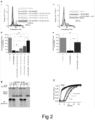
- Fig 2A shows example traces and example power spectra (i and ii) under control conditions (gray), after 15 min incubation with 50 nM A ⁇ 42, 50 nM A ⁇ 42 + 25 nM pre-incubated rh Bri2 BRICHOS R221E monomer, 50 nM A ⁇ 42 + 25 nM pre-incubated rh wt Bri2 BRICHOS oligomer, 50 nM A ⁇ 42 + 50 nM pre-incubated rh wt Bri2 BRICHOS oligomer, 50 nM A ⁇ 42 + 50 nM pre-incubated rh Bri2 BRICHOS R221E oligomer, and 50 nM A ⁇ 42 + a pre-incubated mixture of 25 nM rh wt Bri2 BRICHOS oligomer and 25 nM rh Bri2 BRICHOS R221E monomer.
- Rh Bri2 BRICHOS R221E monomer and rh wt Bri2 BRICHOS oligomers containing an AU1 tag for immunodetection 5:5 ⁇ M and 9:4.5 ⁇ M
- the dashed boxes indicate migration of wt Bri2 BRICHOS monomers.
- Native PAGE and western blot analysis showed that coincubation of rh wt Bri2 BRICHOS oligomers with rh Bri2 BRICHOS R221E monomer lead to the release of smaller species, especially monomers ( Fig. 2B ).
- Fig. 2C shows ThT kinetic analysis of 3 ⁇ M A ⁇ 42 alone, in the presence of 3 ⁇ M rh wt Bri2 BRICHOS oligomer, 3 ⁇ M rh Bri2 BRICHOS R221E monomer, or a pre-incubated mixture of 1.5 ⁇ M wt oligomer and 1.5 ⁇ M R221E monomer.
- Fig 3 illustrates the effect of rh Bri2 BRICHOS R221E monomers and dimers on the microscopic events of A ⁇ 42 fibril formation:
- the rh Bri2 BRICHOS R221E monomer showed a dose dependent progressive reduction of A ⁇ 42 fibril formation at substoichiometric concentrations, and the aggregation kinetics follows a typical sigmoidal behaviour ( Fig. 3A ).
- the fibrillization half-time, ⁇ 1 / 2 increases with increasing rh Bri2 BRICHOS R221E monomer concentration, while the maximum rate of aggregation, rmax, shows a mono-exponential decline ( Fig. 3A,C ).
- the rh Bri2 BRICHOS R221E dimers show similar effects as the monomers, but are more efficient in supressing the overall rate of A ⁇ 42 fibril formation ( Fig. 3B,C ).
- the final ThT fluorescence intensity which is an indicator of the mass of mature fibrils, shows a linear increase against A ⁇ 42 concentration, and does not change significantly in the presence of different concentrations of rh Bri2 BRICHOS R221E species.
- a ⁇ 42 fibrillization kinetics are described by a set of microscopic rate constants, i.e. for primary ( k n ) and secondary (monomer-dependent k 2 and monomer-independent k - ) nucleation as well as elongation ( k + ).
- rate constants i.e. for primary ( k n ) and secondary (monomer-dependent k 2 and monomer-independent k - ) nucleation as well as elongation ( k + ).
- Perturbations of the individual microscopic rate constants k + , k n and k 2 are most relevant since their relative contributions determine the number of newly formed nucleation units, which might be linked to the generation of neurotoxic A ⁇ 42 oligomeric species.
- the primary nucleation rate k n or elongation k + as the sole fitting parameter gave rise to insufficient fits for both rh Bri2 BRICHOS species, which is further confirmed by NMR data showing that neither monomers nor dimers of rh Bri2 BRICHOS R221E interact with A ⁇ 40 monomers.
- k 2 as the sole free fitting parameter the kinetics were adequately described for rh Bri2 BRICHOS R221E monomers, but not for the dimers.
- the rh Bri2 R221E monomer mainly inhibits the secondary nucleation during A ⁇ 42 fibril formation.
- the dimers inhibit both secondary nucleation ( k 2 ) and fibril-end elongation ( k + ).
- Relative inhibition of A ⁇ 42 secondary nucleation and elongation may be linked to reduction of formation of toxic low-molecular weight species, as the number of new nucleation units is decreased by inhibiting secondary nucleation but increased by inhibiting elongation.
- the results shown in Fig 3F hence could have important implications for the expected amounts of toxic A ⁇ 42 species formed in the presence of rh Bri2 BRICHOS R221E dimers and monomers, respectively.
- the amounts of new A ⁇ 42 nucleation units were first calculated from the combined rate constants, determined from the global fit of different A ⁇ 42 concentrations in the presence and absence of constant concentration of rh Bri2 BRICHOS R221E, and the A ⁇ 42 fibril elongation rate at 30% molar equivalent of different rh Bri2 BRICHOS R221E species determined from the seeded aggregation experiments ( Fig. 3D-F ).
- Fig 4 illustrates the effect of rh Bri2 BRICHOS R221E monomers and dimers on A ⁇ 42 oligomer formation and neurotoxicity:
- Fig 5 illustrates the effects on A ⁇ 42 toxicity in mouse hippocampal slices of different rh Bri2 BRICHOS R221E species.
- the numbers under the histograms denote the number of biological replicates, and the data are reported as means ⁇ standard errors of the means. *** p ⁇ 0.001; ns, no significant difference. Noteworthily, increasing the concentration of the rh Bri2 BRICHOS dimer from 50 nM to 100 nM resulted in a significant prevention of A ⁇ 42-induced toxicity ( Fig. 5 ).
- Fig. 7 illustrates that rh Bri2 BRICHOS R221E forms stable monomers that pass the BBB:
- Rh BRICHOS R221E monomers at a dose of 20 mg/kg were injected intravenously in C57BL/6NTac mice while control mice received PBS only, and half-life in serum and BBB permeability were evaluated from western blots of serum and brain homogenates, respectively.
- the serum half-life of rh Bri2 BRICHOS R221E monomers is 53 ⁇ 19 min ( Fig. 7B ), which is longer than the half-life of rh wt Bri2 BRICHOS monomers, dimers or oligomers (around 30-40 min).
- Rh Bri2 BRICHOS R221E was detected in brain homogenates by western blotting two hours after injection ( Fig. 7C ), which indicates that rh Bri2 BRICHOS R221E permeates the BBB, like the wt protein.
- Fig. 9 illustrates that rh Bri2 BRICHOS R221E administered mice shows a trend towards reduced A ⁇ 42 levels in guanidine hydrochloride (A) soluble fraction of the cortex region, whereas no effect was seen in the A ⁇ 42 levels in the TRIS soluble fraction (B).
- Rh Bri2 BRICHOS R221E administered mice also showed reduced expression of GFAP (C), an astroglial cell marker implicated in inflammation, suggesting that rh Bri2 BRICHOS R221E parenteral treatment in aged AD mice lowers neuroinflammation.
- C astroglial cell marker implicated in inflammation
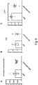
- Rh Bri2 BRICHOS R221E improved the physical activity of mice compared to PBS injected control mice, and their physical activity levels approached the values for age-matched wt mice ( Fig. 8A-B ).
- Fig. 8 illustrates that rh Bri2 BRICHOS R221E improves the physical activity and exploration with no change in anxiety levels in AppNL-F mice. Histograms represent mean activity ( Fig 8A ) and velocity ( Fig 8B ). Data are shown as mean ⁇ SEM for PBS and rh Bri2 BRICHOS R221E treated AppNL-F mice and for age-matched wt controls. Unpaired parametric two-tailed t-test was used to calculate the p-values for all analyses. A 4.99 value in PBS group was removed as an outlier for panel (B).
- FC Fear conditioning
- App NL-G-F knock-in mice which carry the Arctic mutation in addition to the Swedish and the Iberian mutations, show a stronger and earlier AD-like pathology, including behavior deficits, compared to App NL-F knock-in mice.
- Tris or guanidine hydrochloride-soluble A ⁇ 40 and A ⁇ 42 levels in hippocampus and cortex were analyzed by ELISA, which revealed no change in A ⁇ 40 or A ⁇ 42 levels in either brain region between PBS and rh Bri2 BRICHOS R221E treated App NL-F mice.
- staining of brain tissue with Thioflavin S dye for amyloid or immunostaining using A ⁇ antibody revealed that rh Bri2 BRICHOS R221E treated mice have a lower Thioflavin S positive plaque count in cortex ( Fig. 11A ) and show a lower total A ⁇ positive plaque load in cortex ( Fig. 11B ) compared to PBS controls.
- Data are represented as mean ⁇ SEM. Unpaired parametric two-tailed t-test was used to calculate the p-values.
- GFAP glial fibrillary acidic protein
- Brain tissue homogenates from cortex and hippocampus were analyzed to evaluate Iba1 levels in rh Bri2 BRICHOS R221E and PBS administered App NL-F mice.
- a ⁇ immunostaining amyloid plaque count
- Thioflavin S staining amyloid plaque count
- GFAP immunostaining astrogliosis
- Iba1 immunostaining microgliosis
- colocalization A ⁇ and GFAP immunostaining
- the area of A ⁇ positive plaques tends to correlate positively with GFAP and Iba1 staining, as well as with A ⁇ and GFAP colocalization, in particular in cortex (based on data presented in Fig. 11B , Fig 12B-C and Fig 13 ).
- the number of Thioflavin S positive plaques generally correlate negatively with GFAP and Iba1 staining, as well as with A ⁇ and GFAP colocalization in PBS treated mice (based on data presented in Fig. 11A , Fig 12B-C and Fig 13 ).
- a ⁇ plaque load and Thioflavin S plaque counts tend to be inversely correlated with each other in the PBS treated mice (based on data presented in Fig. 11A-B ).
- rh Bri2 BRICHOS R221E treated mice show no correlation between A ⁇ plaque load and Thioflavin S plaque counts (based on data presented in Fig. 11A-B ). Moreover, the trend of negative correlation between Thioflavin S plaque count and GFAP and Iba1 staining seen in PBS mice is not seen in rh Bri2 BRICHOS R221E treated mice (based on data presented in Fig. 11A and Fig. 12B ; Fig. 11A and Fig.
- Rh Bri2 BRICHOS has shown a unique ability to attenuate generation of new nucleation units by monomer dependent secondary nucleation events, which potentially can convert to neurotoxic A ⁇ 42 oligomers during in vitro fibril formation. Further, rh Bri2 BRICHOS blocks A ⁇ 42 induced reduction of y oscillations in hippocampal slices ex vivo and has emerged as a model compound in molecular studies of A ⁇ 42 fibrillation.
- the predicted reduction of oligomer generation in vitro and lowering of A ⁇ 42 mediated neurotoxicity ex vivo that correspond to the estimated in vivo rh Bri2 BRICHOS R221E/A ⁇ 42 ratio agree well with the reductions of A ⁇ positive and Thioflavin S positive plaques, GFAP staining, Iba1 levels in cortex by western blots, as well as increased physical activity observed in App NL-F mice after rh Bri2 BRICHOS R221E.
Landscapes
- Health & Medical Sciences (AREA)
- Chemical & Material Sciences (AREA)
- Life Sciences & Earth Sciences (AREA)
- Organic Chemistry (AREA)
- Medicinal Chemistry (AREA)
- General Health & Medical Sciences (AREA)
- Engineering & Computer Science (AREA)
- Bioinformatics & Cheminformatics (AREA)
- Biomedical Technology (AREA)
- Neurology (AREA)
- Neurosurgery (AREA)
- Proteomics, Peptides & Aminoacids (AREA)
- Biophysics (AREA)
- Genetics & Genomics (AREA)
- Biochemistry (AREA)
- Molecular Biology (AREA)
- Gastroenterology & Hepatology (AREA)
- Toxicology (AREA)
- Zoology (AREA)
- Psychiatry (AREA)
- Veterinary Medicine (AREA)
- Public Health (AREA)
- Animal Behavior & Ethology (AREA)
- Pharmacology & Pharmacy (AREA)
- Nuclear Medicine, Radiotherapy & Molecular Imaging (AREA)
- General Chemical & Material Sciences (AREA)
- Chemical Kinetics & Catalysis (AREA)
- Hospice & Palliative Care (AREA)
- Medicines That Contain Protein Lipid Enzymes And Other Medicines (AREA)
- Peptides Or Proteins (AREA)
- Pharmaceuticals Containing Other Organic And Inorganic Compounds (AREA)
Applications Claiming Priority (2)
| Application Number | Priority Date | Filing Date | Title |
|---|---|---|---|
| EP20150738 | 2020-01-08 | ||
| PCT/EP2021/050168 WO2021140140A1 (en) | 2020-01-08 | 2021-01-07 | Compound and method for treatment of alzheimer's disease |
Publications (3)
| Publication Number | Publication Date |
|---|---|
| EP4087860A1 EP4087860A1 (en) | 2022-11-16 |
| EP4087860C0 EP4087860C0 (en) | 2023-11-22 |
| EP4087860B1 true EP4087860B1 (en) | 2023-11-22 |
Family
ID=69156193
Family Applications (1)
| Application Number | Title | Priority Date | Filing Date |
|---|---|---|---|
| EP21700377.1A Active EP4087860B1 (en) | 2020-01-08 | 2021-01-07 | Compound and method for treatment of alzheimer's disease |
Country Status (5)
| Country | Link |
|---|---|
| US (1) | US20230002458A1 (https=) |
| EP (1) | EP4087860B1 (https=) |
| JP (1) | JP7820821B2 (https=) |
| CN (1) | CN115943153A (https=) |
| WO (1) | WO2021140140A1 (https=) |
Families Citing this family (2)
| Publication number | Priority date | Publication date | Assignee | Title |
|---|---|---|---|---|
| WO2024079221A1 (en) | 2022-10-12 | 2024-04-18 | Alphabeta Ab | The bri2 brichos domain for delivery of proteins into cns neurons |
| WO2025078660A1 (en) | 2023-10-11 | 2025-04-17 | Alphabeta Ab | The bri2 brichos domain for treatment of parkinson´s disease |
Family Cites Families (4)
| Publication number | Priority date | Publication date | Assignee | Title |
|---|---|---|---|---|
| WO2000058353A1 (en) * | 1998-12-10 | 2000-10-05 | Shanghai Second Medical University | Npcahh01: human transmembrane protein e3-16 gene |
| WO2006138355A2 (en) * | 2005-06-14 | 2006-12-28 | Albert Einstein College Of Medicine Of Yeshiva University | Effect of bri proteins on abeta production |
| AU2010356144B2 (en) | 2010-06-24 | 2015-06-04 | Alphabeta Ab | Compound and method for treatment of Alzheimer's disease and familial dementia |
| WO2012138284A1 (en) | 2011-04-05 | 2012-10-11 | Alphabeta Ab | Amyloidosis target useful in methods of treatment and for screening of compounds |
-
2021
- 2021-01-07 US US17/784,591 patent/US20230002458A1/en active Pending
- 2021-01-07 JP JP2022541987A patent/JP7820821B2/ja active Active
- 2021-01-07 EP EP21700377.1A patent/EP4087860B1/en active Active
- 2021-01-07 WO PCT/EP2021/050168 patent/WO2021140140A1/en not_active Ceased
- 2021-01-07 CN CN202180008401.XA patent/CN115943153A/zh active Pending
Also Published As
| Publication number | Publication date |
|---|---|
| EP4087860C0 (en) | 2023-11-22 |
| CN115943153A (zh) | 2023-04-07 |
| JP7820821B2 (ja) | 2026-02-26 |
| WO2021140140A1 (en) | 2021-07-15 |
| JP2023509187A (ja) | 2023-03-07 |
| US20230002458A1 (en) | 2023-01-05 |
| EP4087860A1 (en) | 2022-11-16 |
Similar Documents
| Publication | Publication Date | Title |
|---|---|---|
| JP7379615B2 (ja) | 補体活性のモジュレーター | |
| KR20200095485A (ko) | 보체 활성의 조절인자 | |
| CN101611152A (zh) | 用于调控set的方法及其用途 | |
| M Fayaz et al. | CypD: the key to the death door | |
| BRPI0708349A2 (pt) | composição farmacéutica e uso de peptìdeos | |
| JP5893614B2 (ja) | アルツハイマー病および家族性認知症の治療のための化合物および方法 | |
| EP4087860B1 (en) | Compound and method for treatment of alzheimer's disease | |
| Lin et al. | Ozone inhibits APP/Aβ production and improves cognition in an APP/PS1 transgenic mouse model | |
| Kiris et al. | Evaluation of the therapeutic effect of lycoramine on Alzheimer’s disease in mouse model | |
| Liu et al. | Role of mitochondrial quality control in neurodegenerative disease progression | |
| US20210038589A1 (en) | Uses, compositions and methods | |
| JP2019510088A (ja) | Parisのファルネシル化によってパーキンソン病を予防または治療する方法 | |
| EP3622958A1 (en) | Use of potassium ion channel inhibitor for treatment of depression and pharmaceutical composition | |
| US12083086B2 (en) | Compositions and methods for treating Alzheimer's disease | |
| CN112153897B (zh) | 用于治疗神经病学和其他病症的组合物和方法 | |
| Cadile | Molecular determinants of diaphragm muscle impairment in a mouse model of Spinal Muscular Atrophy and a possible nutraceutical intervention | |
| Borbély | Development of Non-Transgenic Animal Models of Alzheimer’s Disease and Investigation of Drug Candidates for its Treatment | |
| US20240226062A1 (en) | Methods and compositions for the treatment of disorders characterized by a kidins220 dysfunction in a subject | |
| Ospina | Small Heat Shock Protein 22 Preserves Neuronal Function in a Murine Model of Tauopathy | |
| Greco | Deficiency in CCR5, a Mediator of Neuroinflammation, Normalizes TIMP Levels, Working Memory, and Gamma Oscillation Power in APOE4 Targeted Replacement Mice | |
| Urbinati | Targeting brain mitochondrial bioenergetics as an attractive therapeutic avenue for Rett syndrome: evidence from female mouse models | |
| Balke | Regulation des Autophagieproteins ULK1 im MPTP-Mausmodell des Morbus Parkinson | |
| Catumbela | In vivo Examination of Peripheral Drivers of Alzheimer’s Disease | |
| Habib | Therapeutic Modulation of Alzheimer's Disease with Biological (HUCBS) and Pharmacological (LISPRO) Approaches | |
| Antoniel | The unique histidine of F-ATP synthase subunit OSCP mediates regulation of the permeability transition by matrix pH |
Legal Events
| Date | Code | Title | Description |
|---|---|---|---|
| STAA | Information on the status of an ep patent application or granted ep patent |
Free format text: STATUS: UNKNOWN |
|
| STAA | Information on the status of an ep patent application or granted ep patent |
Free format text: STATUS: THE INTERNATIONAL PUBLICATION HAS BEEN MADE |
|
| PUAI | Public reference made under article 153(3) epc to a published international application that has entered the european phase |
Free format text: ORIGINAL CODE: 0009012 |
|
| STAA | Information on the status of an ep patent application or granted ep patent |
Free format text: STATUS: REQUEST FOR EXAMINATION WAS MADE |
|
| 17P | Request for examination filed |
Effective date: 20220808 |
|
| AK | Designated contracting states |
Kind code of ref document: A1 Designated state(s): AL AT BE BG CH CY CZ DE DK EE ES FI FR GB GR HR HU IE IS IT LI LT LU LV MC MK MT NL NO PL PT RO RS SE SI SK SM TR |
|
| DAV | Request for validation of the european patent (deleted) | ||
| DAX | Request for extension of the european patent (deleted) | ||
| GRAP | Despatch of communication of intention to grant a patent |
Free format text: ORIGINAL CODE: EPIDOSNIGR1 |
|
| STAA | Information on the status of an ep patent application or granted ep patent |
Free format text: STATUS: GRANT OF PATENT IS INTENDED |
|
| INTG | Intention to grant announced |
Effective date: 20230626 |
|
| GRAS | Grant fee paid |
Free format text: ORIGINAL CODE: EPIDOSNIGR3 |
|
| GRAA | (expected) grant |
Free format text: ORIGINAL CODE: 0009210 |
|
| STAA | Information on the status of an ep patent application or granted ep patent |
Free format text: STATUS: THE PATENT HAS BEEN GRANTED |
|
| AK | Designated contracting states |
Kind code of ref document: B1 Designated state(s): AL AT BE BG CH CY CZ DE DK EE ES FI FR GB GR HR HU IE IS IT LI LT LU LV MC MK MT NL NO PL PT RO RS SE SI SK SM TR |
|
| REG | Reference to a national code |
Ref country code: GB Ref legal event code: FG4D |
|
| REG | Reference to a national code |
Ref country code: CH Ref legal event code: EP |
|
| REG | Reference to a national code |
Ref country code: DE Ref legal event code: R096 Ref document number: 602021007054 Country of ref document: DE |
|
| REG | Reference to a national code |
Ref country code: IE Ref legal event code: FG4D |
|
| U01 | Request for unitary effect filed |
Effective date: 20231222 |
|
| U07 | Unitary effect registered |
Designated state(s): AT BE BG DE DK EE FI FR IT LT LU LV MT NL PT SE SI Effective date: 20240108 |
|
| U20 | Renewal fee for the european patent with unitary effect paid |
Year of fee payment: 4 Effective date: 20240108 |
|
| PG25 | Lapsed in a contracting state [announced via postgrant information from national office to epo] |
Ref country code: GR Free format text: LAPSE BECAUSE OF FAILURE TO SUBMIT A TRANSLATION OF THE DESCRIPTION OR TO PAY THE FEE WITHIN THE PRESCRIBED TIME-LIMIT Effective date: 20240223 |
|
| PG25 | Lapsed in a contracting state [announced via postgrant information from national office to epo] |
Ref country code: IS Free format text: LAPSE BECAUSE OF FAILURE TO SUBMIT A TRANSLATION OF THE DESCRIPTION OR TO PAY THE FEE WITHIN THE PRESCRIBED TIME-LIMIT Effective date: 20240322 |
|
| PG25 | Lapsed in a contracting state [announced via postgrant information from national office to epo] |
Ref country code: ES Free format text: LAPSE BECAUSE OF FAILURE TO SUBMIT A TRANSLATION OF THE DESCRIPTION OR TO PAY THE FEE WITHIN THE PRESCRIBED TIME-LIMIT Effective date: 20231122 |
|
| PG25 | Lapsed in a contracting state [announced via postgrant information from national office to epo] |
Ref country code: IS Free format text: LAPSE BECAUSE OF FAILURE TO SUBMIT A TRANSLATION OF THE DESCRIPTION OR TO PAY THE FEE WITHIN THE PRESCRIBED TIME-LIMIT Effective date: 20240322 Ref country code: GR Free format text: LAPSE BECAUSE OF FAILURE TO SUBMIT A TRANSLATION OF THE DESCRIPTION OR TO PAY THE FEE WITHIN THE PRESCRIBED TIME-LIMIT Effective date: 20240223 Ref country code: ES Free format text: LAPSE BECAUSE OF FAILURE TO SUBMIT A TRANSLATION OF THE DESCRIPTION OR TO PAY THE FEE WITHIN THE PRESCRIBED TIME-LIMIT Effective date: 20231122 |
|
| PG25 | Lapsed in a contracting state [announced via postgrant information from national office to epo] |
Ref country code: RS Free format text: LAPSE BECAUSE OF FAILURE TO SUBMIT A TRANSLATION OF THE DESCRIPTION OR TO PAY THE FEE WITHIN THE PRESCRIBED TIME-LIMIT Effective date: 20231122 Ref country code: PL Free format text: LAPSE BECAUSE OF FAILURE TO SUBMIT A TRANSLATION OF THE DESCRIPTION OR TO PAY THE FEE WITHIN THE PRESCRIBED TIME-LIMIT Effective date: 20231122 Ref country code: NO Free format text: LAPSE BECAUSE OF FAILURE TO SUBMIT A TRANSLATION OF THE DESCRIPTION OR TO PAY THE FEE WITHIN THE PRESCRIBED TIME-LIMIT Effective date: 20240222 Ref country code: HR Free format text: LAPSE BECAUSE OF FAILURE TO SUBMIT A TRANSLATION OF THE DESCRIPTION OR TO PAY THE FEE WITHIN THE PRESCRIBED TIME-LIMIT Effective date: 20231122 |
|
| PG25 | Lapsed in a contracting state [announced via postgrant information from national office to epo] |
Ref country code: CZ Free format text: LAPSE BECAUSE OF FAILURE TO SUBMIT A TRANSLATION OF THE DESCRIPTION OR TO PAY THE FEE WITHIN THE PRESCRIBED TIME-LIMIT Effective date: 20231122 |
|
| PG25 | Lapsed in a contracting state [announced via postgrant information from national office to epo] |
Ref country code: SK Free format text: LAPSE BECAUSE OF FAILURE TO SUBMIT A TRANSLATION OF THE DESCRIPTION OR TO PAY THE FEE WITHIN THE PRESCRIBED TIME-LIMIT Effective date: 20231122 |
|
| PG25 | Lapsed in a contracting state [announced via postgrant information from national office to epo] |
Ref country code: SM Free format text: LAPSE BECAUSE OF FAILURE TO SUBMIT A TRANSLATION OF THE DESCRIPTION OR TO PAY THE FEE WITHIN THE PRESCRIBED TIME-LIMIT Effective date: 20231122 Ref country code: SK Free format text: LAPSE BECAUSE OF FAILURE TO SUBMIT A TRANSLATION OF THE DESCRIPTION OR TO PAY THE FEE WITHIN THE PRESCRIBED TIME-LIMIT Effective date: 20231122 Ref country code: RO Free format text: LAPSE BECAUSE OF FAILURE TO SUBMIT A TRANSLATION OF THE DESCRIPTION OR TO PAY THE FEE WITHIN THE PRESCRIBED TIME-LIMIT Effective date: 20231122 Ref country code: CZ Free format text: LAPSE BECAUSE OF FAILURE TO SUBMIT A TRANSLATION OF THE DESCRIPTION OR TO PAY THE FEE WITHIN THE PRESCRIBED TIME-LIMIT Effective date: 20231122 |
|
| REG | Reference to a national code |
Ref country code: DE Ref legal event code: R097 Ref document number: 602021007054 Country of ref document: DE |
|
| PG25 | Lapsed in a contracting state [announced via postgrant information from national office to epo] |
Ref country code: MC Free format text: LAPSE BECAUSE OF FAILURE TO SUBMIT A TRANSLATION OF THE DESCRIPTION OR TO PAY THE FEE WITHIN THE PRESCRIBED TIME-LIMIT Effective date: 20231122 |
|
| PG25 | Lapsed in a contracting state [announced via postgrant information from national office to epo] |
Ref country code: MC Free format text: LAPSE BECAUSE OF FAILURE TO SUBMIT A TRANSLATION OF THE DESCRIPTION OR TO PAY THE FEE WITHIN THE PRESCRIBED TIME-LIMIT Effective date: 20231122 |
|
| REG | Reference to a national code |
Ref country code: CH Ref legal event code: PL |
|
| PLBE | No opposition filed within time limit |
Free format text: ORIGINAL CODE: 0009261 |
|
| STAA | Information on the status of an ep patent application or granted ep patent |
Free format text: STATUS: NO OPPOSITION FILED WITHIN TIME LIMIT |
|
| PG25 | Lapsed in a contracting state [announced via postgrant information from national office to epo] |
Ref country code: CH Free format text: LAPSE BECAUSE OF NON-PAYMENT OF DUE FEES Effective date: 20240131 |
|
| 26N | No opposition filed |
Effective date: 20240823 |
|
| PG25 | Lapsed in a contracting state [announced via postgrant information from national office to epo] |
Ref country code: CH Free format text: LAPSE BECAUSE OF NON-PAYMENT OF DUE FEES Effective date: 20240131 |
|
| PG25 | Lapsed in a contracting state [announced via postgrant information from national office to epo] |
Ref country code: IE Free format text: LAPSE BECAUSE OF NON-PAYMENT OF DUE FEES Effective date: 20240107 |
|
| PG25 | Lapsed in a contracting state [announced via postgrant information from national office to epo] |
Ref country code: IE Free format text: LAPSE BECAUSE OF NON-PAYMENT OF DUE FEES Effective date: 20240107 |
|
| U20 | Renewal fee for the european patent with unitary effect paid |
Year of fee payment: 5 Effective date: 20250115 |
|
| PGFP | Annual fee paid to national office [announced via postgrant information from national office to epo] |
Ref country code: GB Payment date: 20250115 Year of fee payment: 5 |
|
| PG25 | Lapsed in a contracting state [announced via postgrant information from national office to epo] |
Ref country code: CY Free format text: LAPSE BECAUSE OF FAILURE TO SUBMIT A TRANSLATION OF THE DESCRIPTION OR TO PAY THE FEE WITHIN THE PRESCRIBED TIME-LIMIT; INVALID AB INITIO Effective date: 20210107 |
|
| PG25 | Lapsed in a contracting state [announced via postgrant information from national office to epo] |
Ref country code: HU Free format text: LAPSE BECAUSE OF FAILURE TO SUBMIT A TRANSLATION OF THE DESCRIPTION OR TO PAY THE FEE WITHIN THE PRESCRIBED TIME-LIMIT; INVALID AB INITIO Effective date: 20210107 |
|
| PG25 | Lapsed in a contracting state [announced via postgrant information from national office to epo] |
Ref country code: TR Free format text: LAPSE BECAUSE OF FAILURE TO SUBMIT A TRANSLATION OF THE DESCRIPTION OR TO PAY THE FEE WITHIN THE PRESCRIBED TIME-LIMIT Effective date: 20231122 |