EP4067925B1 - Magnetresonanztomografievorrichtung mit einheit zur asymmetrischen felderzeugung - Google Patents
Magnetresonanztomografievorrichtung mit einheit zur asymmetrischen felderzeugung Download PDFInfo
- Publication number
- EP4067925B1 EP4067925B1 EP21165607.9A EP21165607A EP4067925B1 EP 4067925 B1 EP4067925 B1 EP 4067925B1 EP 21165607 A EP21165607 A EP 21165607A EP 4067925 B1 EP4067925 B1 EP 4067925B1
- Authority
- EP
- European Patent Office
- Prior art keywords
- magnet
- magnetic resonance
- imaging volume
- imaging device
- resonance imaging
- Prior art date
- Legal status (The legal status is an assumption and is not a legal conclusion. Google has not performed a legal analysis and makes no representation as to the accuracy of the status listed.)
- Active
Links
Images


Classifications
-
- A—HUMAN NECESSITIES
- A61—MEDICAL OR VETERINARY SCIENCE; HYGIENE
- A61B—DIAGNOSIS; SURGERY; IDENTIFICATION
- A61B5/00—Measuring for diagnostic purposes; Identification of persons
- A61B5/05—Detecting, measuring or recording for diagnosis by means of electric currents or magnetic fields; Measuring using microwaves or radio waves
- A61B5/055—Detecting, measuring or recording for diagnosis by means of electric currents or magnetic fields; Measuring using microwaves or radio waves involving electronic [EMR] or nuclear [NMR] magnetic resonance, e.g. magnetic resonance imaging
-
- G—PHYSICS
- G01—MEASURING; TESTING
- G01R—MEASURING ELECTRIC VARIABLES; MEASURING MAGNETIC VARIABLES
- G01R33/00—Arrangements or instruments for measuring magnetic variables
- G01R33/20—Arrangements or instruments for measuring magnetic variables involving magnetic resonance
- G01R33/28—Details of apparatus provided for in groups G01R33/44 - G01R33/64
- G01R33/38—Systems for generation, homogenisation or stabilisation of the main or gradient magnetic field
- G01R33/3806—Open magnet assemblies for improved access to the sample, e.g. C-type or U-type magnets
-
- G—PHYSICS
- G01—MEASURING; TESTING
- G01R—MEASURING ELECTRIC VARIABLES; MEASURING MAGNETIC VARIABLES
- G01R33/00—Arrangements or instruments for measuring magnetic variables
- G01R33/20—Arrangements or instruments for measuring magnetic variables involving magnetic resonance
- G01R33/28—Details of apparatus provided for in groups G01R33/44 - G01R33/64
- G01R33/32—Excitation or detection systems, e.g. using radio frequency signals
- G01R33/34—Constructional details, e.g. resonators, specially adapted to MR
- G01R33/34015—Temperature-controlled RF coils
- G01R33/34023—Superconducting RF coils
-
- G—PHYSICS
- G01—MEASURING; TESTING
- G01R—MEASURING ELECTRIC VARIABLES; MEASURING MAGNETIC VARIABLES
- G01R33/00—Arrangements or instruments for measuring magnetic variables
- G01R33/20—Arrangements or instruments for measuring magnetic variables involving magnetic resonance
- G01R33/28—Details of apparatus provided for in groups G01R33/44 - G01R33/64
- G01R33/38—Systems for generation, homogenisation or stabilisation of the main or gradient magnetic field
- G01R33/3808—Magnet assemblies for single-sided MR wherein the magnet assembly is located on one side of a subject only; Magnet assemblies for inside-out MR, e.g. for MR in a borehole or in a blood vessel, or magnet assemblies for fringe-field MR
-
- G—PHYSICS
- G01—MEASURING; TESTING
- G01R—MEASURING ELECTRIC VARIABLES; MEASURING MAGNETIC VARIABLES
- G01R33/00—Arrangements or instruments for measuring magnetic variables
- G01R33/20—Arrangements or instruments for measuring magnetic variables involving magnetic resonance
- G01R33/28—Details of apparatus provided for in groups G01R33/44 - G01R33/64
- G01R33/38—Systems for generation, homogenisation or stabilisation of the main or gradient magnetic field
- G01R33/381—Systems for generation, homogenisation or stabilisation of the main or gradient magnetic field using electromagnets
- G01R33/3815—Systems for generation, homogenisation or stabilisation of the main or gradient magnetic field using electromagnets with superconducting coils, e.g. power supply therefor
-
- G—PHYSICS
- G01—MEASURING; TESTING
- G01R—MEASURING ELECTRIC VARIABLES; MEASURING MAGNETIC VARIABLES
- G01R33/00—Arrangements or instruments for measuring magnetic variables
- G01R33/20—Arrangements or instruments for measuring magnetic variables involving magnetic resonance
- G01R33/28—Details of apparatus provided for in groups G01R33/44 - G01R33/64
- G01R33/38—Systems for generation, homogenisation or stabilisation of the main or gradient magnetic field
- G01R33/383—Systems for generation, homogenisation or stabilisation of the main or gradient magnetic field using permanent magnets
-
- G—PHYSICS
- G01—MEASURING; TESTING
- G01R—MEASURING ELECTRIC VARIABLES; MEASURING MAGNETIC VARIABLES
- G01R33/00—Arrangements or instruments for measuring magnetic variables
- G01R33/20—Arrangements or instruments for measuring magnetic variables involving magnetic resonance
- G01R33/28—Details of apparatus provided for in groups G01R33/44 - G01R33/64
- G01R33/38—Systems for generation, homogenisation or stabilisation of the main or gradient magnetic field
- G01R33/385—Systems for generation, homogenisation or stabilisation of the main or gradient magnetic field using gradient magnetic field coils
-
- G—PHYSICS
- G01—MEASURING; TESTING
- G01R—MEASURING ELECTRIC VARIABLES; MEASURING MAGNETIC VARIABLES
- G01R33/00—Arrangements or instruments for measuring magnetic variables
- G01R33/20—Arrangements or instruments for measuring magnetic variables involving magnetic resonance
- G01R33/28—Details of apparatus provided for in groups G01R33/44 - G01R33/64
- G01R33/42—Screening
- G01R33/421—Screening of main or gradient magnetic field
- G01R33/4215—Screening of main or gradient magnetic field of the gradient magnetic field, e.g. using passive or active shielding of the gradient magnetic field
-
- G—PHYSICS
- G01—MEASURING; TESTING
- G01R—MEASURING ELECTRIC VARIABLES; MEASURING MAGNETIC VARIABLES
- G01R33/00—Arrangements or instruments for measuring magnetic variables
- G01R33/20—Arrangements or instruments for measuring magnetic variables involving magnetic resonance
- G01R33/44—Arrangements or instruments for measuring magnetic variables involving magnetic resonance using nuclear magnetic resonance [NMR]
- G01R33/48—NMR imaging systems
- G01R33/483—NMR imaging systems with selection of signals or spectra from particular regions of the volume, e.g. in vivo spectroscopy
- G01R33/4833—NMR imaging systems with selection of signals or spectra from particular regions of the volume, e.g. in vivo spectroscopy using spatially selective excitation of the volume of interest, e.g. selecting non-orthogonal or inclined slices
- G01R33/4835—NMR imaging systems with selection of signals or spectra from particular regions of the volume, e.g. in vivo spectroscopy using spatially selective excitation of the volume of interest, e.g. selecting non-orthogonal or inclined slices of multiple slices
-
- G—PHYSICS
- G01—MEASURING; TESTING
- G01R—MEASURING ELECTRIC VARIABLES; MEASURING MAGNETIC VARIABLES
- G01R33/00—Arrangements or instruments for measuring magnetic variables
- G01R33/20—Arrangements or instruments for measuring magnetic variables involving magnetic resonance
- G01R33/44—Arrangements or instruments for measuring magnetic variables involving magnetic resonance using nuclear magnetic resonance [NMR]
- G01R33/48—NMR imaging systems
- G01R33/54—Signal processing systems, e.g. using pulse sequences ; Generation or control of pulse sequences; Operator console
- G01R33/56—Image enhancement or correction, e.g. subtraction or averaging techniques, e.g. improvement of signal-to-noise ratio and resolution
Definitions
- the invention relates to a magnetic resonance imaging device comprising a field generation unit for generating at least one magnetic gradient field, wherein the field generation unit comprises a first magnet and a second magnet confining an imaging volume of the magnetic resonance imaging device in two spatial directions and wherein the first magnet and the second magnet are arranged asymmetrically with respect to the imaging volume.
- the invention further relates to a method for acquiring an image of a diagnostically relevant body region of a patient with a magnetic resonance imaging device according to the invention.
- Magnetic resonance tomography represents a prominent imaging method for acquiring images of an interior of the examination object.
- the examination object is usually positioned in a strong and homogeneous static magnetic field (B0 field) of a magnetic resonance imaging device.
- the static magnetic field may comprise magnetic field strengths of 0.2 Tesla to 7 Tesla, thus aligning nuclear spins inside the examination object along the static magnetic field.
- radiofrequency excitation pulses are emitted into the examination subject. Each radiofrequency excitation pulse causes a magnetization of nuclear spins within the examination object to deviate from the static magnetic field by an amount which is known as the flip angle.
- a radiofrequency excitation pulse may comprise an alternating (electro-)magnetic field with a frequency which corresponds to the Larmor frequency at the respective static magnetic field strength.
- Excited nuclear spins may exhibit a rotating and decaying magnetization (nuclear magnetic resonance), which can be detected using dedicated radiofrequency antennas.
- rapidly switched magnetic gradient fields are superimposed on the static magnetic field.
- the received nuclear magnetic resonances are typically digitized and stored as complex values in a k-space matrix.
- This k-space matrix can be used as a basis for a reconstruction of magnetic resonance images and for determining spectroscopic data.
- a magnetic resonance image is typically reconstructed by means of a multi-dimensional Fourier transformation of the k-space matrix.
- the patient is typically enclosed in a bore or between a pair of magnets of the magnetic resonance imaging device. Due to spatial restrictions within the bore or a space between the pair of magnets, magnetic resonance imaging provides limited use for surgical procedures or biopsies. As the patient needs to be positioned inside the magnetic resonance imaging device, a size of the bore as well as other dimensions of the magnetic resonance imaging device are dictated by the size of the human body. From a cost and/or space utilization perspective, this may be unsatisfactory, especially if the examination is restricted to a body region of the patient, which is significantly smaller than an imaging volume provided by the magnetic resonance imaging device. Furthermore, patients with a claustrophobic condition or children may not tolerate elongated examination times usually associated with a magnetic resonance measurement.
- WO 2007/004109 A1 discloses an MRI apparatus including a main magnet system having a first main magnet portion and a second main magnet portion disposed away from the first main magnet portion along a Z-axis.
- JP 2003 102699 A pertains to an open MRI apparatus comprising a first magnet and a second magnet which are separated from each other and arranged so that the surface of the first magnet generating the static magnetic field is inclined with respect to the surface of the second magnet.
- US 5 717 333 A describes a magnet arrangement for a diagnostic magnetic resonance apparatus with a first unit and a second unit arranged at a distance from the first unit, the operating in combination to generate a magnetic field arranged between the first unit and the second unit.
- WO 99/56293 A2 discloses an open unipolar magnetic structure for generating a highly uniform magnetic field using permanently magnetized blocks of magnetic material as the predominant generator of the uniform magnetic field in a desired region of interest.
- EP 2 745130 A1 describes a hybrid magnet for an MRI machine, comprising a superconducting coil and a pole piece assembly associated therewith, the pole piece assembly comprising a ferromagnetic member having a primary face for facing a subject to be imaged.
- the inventive magnetic resonance imaging device comprises a field generation unit, wherein the field generation unit comprises a first magnet and a second magnet confining an imaging volume of the magnetic resonance imaging device from two spatial directions and wherein the field generation unit is configured to provide a static magnetic field within an imaging volume.
- the first magnet may represent a main magnet providing the magnetic field within the imaging volume.
- a static magnetic field may be a B0-field of the magnetic resonance imaging device comprising either an essentially homogeneous magnet field or a predetermined magnetic gradient field within the imaging volume.
- the second magnet may be smaller than the first magnet. It is conceivable that the second magnet is primarily configured to improve a characteristic of the magnetic field provided by the first magnet, such as a magnetic field strength, a magnetic field direction and/or a magnetic field homogeneity, within the imaging volume. It is also conceivable, that the second magnet enhances an efficiency of the field generation unit in comparison to a unilateral magnet design.
- the first magnet and the second magnet may be carried by a supporting structure. The supporting structure may be mechanically connected to the first magnet and the second magnet to maintain a predetermined relative position of the first magnet and the second magnet.
- the first magnet and the second magnet are positioned along a common axis.
- the first magnet and/or the second magnet may be rotationally symmetric and the common axis may be an axis of rotational symmetry of the first magnet and the second magnet.
- the imaging volume of the field generation unit may be positioned along the common axis between the first magnet and the second magnet.
- the first magnet and the second magnet may also be positioned in such a way, that an axis of rotational symmetry of the first magnet is angled with respect to an axis of rotational symmetry of the second magnet.
- a trajectory defined by a centroid of a projection of the first magnet in the direction of the imaging volume corresponds to an axis of rotational symmetry of the first magnet.
- a trajectory defined by a centroid of a projection of the second magnet in the direction of the imaging volume may correspond to an axis of rotational symmetry of the second magnet.
- the first magnet and/or the second magnet are not required to comprise a rotational symmetry.
- a magnetic field axis of the first magnet corresponds to or is angled with respect to a magnetic field axis of the second magnet.
- an axis through the first magnet and/or the second magnet and a center of the imaging volume is characterized by a trajectory of a centroid of a projection of the first magnet and/or the second magnet in the direction of the center of the imaging volume.
- the first magnet and the second magnet confine the imaging volume in two spatial directions.
- the first magnet may confine the imaging volume in a first spatial direction and the second magnet may confine the imaging volume in a second spatial direction which is different from the first spatial direction.
- the first spatial direction and the second spatial direction may be oriented in parallel.
- the first spatial direction and the second spatial direction may be oriented in opposite directions at an angle of 180°.
- the two spatial directions may also be oriented at an angle below 180° or above 180°.
- An imaging volume may be characterized by a predefined magnetic field direction and/or magnetic field strength.
- the imaging volume may comprise a volume with a substantially uniform magnetic field direction and/or a uniform magnetic field strength.
- Such a volume may be an isocenter of the magnetic resonance imaging device.
- the imaging volume comprises a predefined magnetic gradient field.
- Such a magnetic gradient field may be used for spatial encoding of magnetic resonance signals acquired from an examination object positioned within the imaging volume.
- the first magnet and the second magnet are arranged asymmetrically with respect to the imaging volume.
- the first magnet and the second magnet may be arranged asymmetrically with respect to any plane oriented orthogonally along a trajectory of a projection of the first magnet and/or the second magnet in a direction of the imaging volume.
- the term projection may be interpreted as an imaginary image of the first magnet and/or the second magnet on a two-dimensional plane.
- An asymmetric arrangement of the first magnet and the second magnet may also signify that a size, a shape, an amount of magnetic material, a magnetic field strength and/or an arbitrary other property of the first magnet differs from the second magnet. It is also conceivable, that an absolute distance between the first magnet and the imaging volume is different from an absolute distance between the second magnet and the imaging volume.
- the first magnet and the second magnet may comprise different shapes and/or geometries.
- the first magnet and the second magnet are arranged in such a way to provide an access to the imaging volume along at least two perpendicular spatial directions, wherein a direction of access to the imaging volume is angled with respect to a main magnetic field direction of the magnetic field in the imaging volume.
- the first magnet and the second magnet may be carried by a supporting structure and the imaging volume may be positioned in a free volume or gap between the first magnet and the second magnet. It is conceivable, that an access to the imaging volume is provided along any unobstructed line connecting an arbitrarily chosen position in an examination room with the center of the imaging volume.
- the examination room may be a room wherein the magnet resonance imaging device is positioned and/or wherein a magnetic resonance measurement may be performed.
- a first direction of access to the imaging volume may be oriented essentially perpendicular to the axis of rotational symmetry of the first magnet and/or the second magnet.
- a second direction of access may be oriented essentially perpendicular to the first direction of access and the axis of rotational symmetry of the first magnet and/or the second magnet.
- the first direction of access and/or the second direction of access may also comprise an arbitrary angle with respect to the axis of rotational symmetry of the first magnet and/or the second magnet.
- the imaging volume may be accessed via a plurality of directions of access.
- the imaging volume is accessible via at least two essentially perpendicular spatial directions.
- the imaging volume may also be accessible via three essentially perpendicular spatial directions.
- the three perpendicular spatial directions may correspond to the axes of a cartesian coordinate system, the origin of the cartesian coordinate system being positioned in the center of the imaging volume.
- the magnetic resonance imaging device includes a radiofrequency system comprising at least one radiofrequency antenna for transmitting and/or receiving radiofrequency radiation.
- the at least one radiofrequency antenna may be carried by the first magnet, the second magnet and/or the supporting structure.
- the radiofrequency antenna may be positioned on or recessed into a pole face of the first magnet and/or the second magnet.
- the at least one radiofrequency antenna may also be configured as a local coil.
- Such a local coil may be attached to the examination object, e. g. a human or an animal.
- the radiofrequency antenna may be placed in different positions relative to the magnetic resonance imaging device.
- an accessibility and/or an openness of the magnetic resonance imaging device can be enhanced in comparison to conventional magnetic resonance imaging devices.
- a space for movement of an extremity of a patient and/or a medical instrument can advantageously be increased.
- a suitability of the magnetic resonance imaging device for surgical and/or therapeutical interventions can be increased.
- an obstruction of a view of the patient by a bore or a field generation unit of the magnetic resonance imaging device can significantly be reduced in comparison to conventional magnetic resonance imaging devices.
- abortions of a magnetic resonance measurements of claustrophobic patients may be reduced and an eye-contact between parents and children can be maintained during an imaging examination.
- the inventive magnetic resonance imaging device may easily be combined with other imaging modalities, such as a flatbed or C-arm X-ray scanner, an ultrasound scanner, as well as optical imaging devices.
- imaging modalities such as a flatbed or C-arm X-ray scanner, an ultrasound scanner, as well as optical imaging devices.
- other imaging modalities may favorably provide high resolution navigator data which may be used to improve an efficiency of a magnetic resonance imaging examination as well as a quality of acquired magnetic resonance images.
- an inventive asymmetric arrangement of the field generation unit may also be less sensitive to dynamic field changes.
- a lighter construction of the supporting structure may be used decreasing costs and further enhancing accessibility and/or openness of the magnetic resonance imaging device.
- the first magnet comprises a superconducting magnet and the second magnet may comprise a permanent magnet or an electromagnet.
- a permanent magnet may consist of any suitable magnetic material such as AlNiCo (aluminum-nickel-cobalt), NeFeB (neodymium-iron-boron) or SmCo (samarium-cobalt) alloys.
- the permanent magnet may comprise any desired shape.
- the permanent magnet comprises a bar shape.
- a bar shape may include a cuboid bar shape, a cylindrical bar shape or a bar shape with a polygonal cross-section, such as a prism. Bar-shaped permanent magnets provide a low-cost solution for generating a magnetic field within the imaging volume.
- the permanent magnet may be composed of smaller, stacked permanent magnets. The use of a permanent magnet may favorably avoid costs and space required for cooling equipment usually associated with superconducting magnets and electromagnets.
- An electromagnet may be a non-superconducting magnet.
- an electromagnet may comprise an electrical conductor wound around a magnetic core made of, for example, a ferromagnetic or ferrimagnetic material.
- the magnetic core of the electromagnet may comprise a cylindrical shape, a cuboid shape, a prism shape or any other desirable shape.
- the magnetic field strength can be favorably increased in comparison to a permanent magnet of comparable size. Higher magnetic field strengths can advantageously enhance a quality and/or a signal-to-noise ratio of a magnetic resonance image acquired via the magnetic resonance imaging device.
- a superconducting magnet may comprise coils and/or tubular magnet segments of superconducting wire.
- the superconducting wire may be connected to a cryostat to keep a temperature of the superconducting wire below a predefined value.
- liquid helium may be used as a coolant, ensuring a temperature of the superconducting wire below 4 K.
- the coils of superconducting wire may be arranged in a variety of shapes, such as a solenoid, as a substantially planar loop or a tubular magnet segment.
- the first magnet is larger than the second magnet and comprises a superconducting magnet.
- the second magnet may comprise a permanent magnet as a low-cost solution for better control of a characteristic of the magnetic field provided by the first magnet.
- the first magnet comprises a plurality of magnet segments, wherein each magnet segment comprises a magnetic field axis and wherein the magnetic field axes of the plurality of magnet segments are oriented in parallel to a magnetic field axis of the first magnet.
- the magnet segments may be positioned separately from one another or adjacent to one another.
- a magnetic field axis may correspond to an axis passing through a south pole and a north pole of the first magnet.
- the north pole may relate to a point on a surface of the first magnet, where a direction of the magnetic field is oriented away from the first magnet in an essentially linear fashion.
- the south pole may relate to a point on a surface of the first magnet, where a direction of the magnetic field is oriented towards the first magnet in an essentially linear fashion.
- the magnetic field axis of the first magnet corresponds to an axis of rotational symmetry of the first magnet.
- the surface of the first magnet may be a pole face of the first magnet.
- the magnetic field axes of the plurality of magnet segments substantially correspond to the magnetic field axis of the first magnet.
- the first magnet comprises a superconducting magnet with a plurality of tubular magnet segments, wherein each tubular magnet segment comprises a wire coiled in such a way to provide a tubular shape.
- the plurality of tubular magnet segments comprises a circular axial cross-section.
- the cross-section of the plurality of tubular magnet segments may also be elliptic, oval or polygonal.
- the first magnet may comprise a discrete rotational symmetry of a higher order of n > 1. This may signify, that a rotation of the first magnet about the axis of rotational symmetry by an angle of 360°/n does not change the first magnet.
- a tubular magnet segment may represent a substantially planar or flat loop or coil of superconducting wire.
- the tubular magnet segment may comprise one or several windings of superconducting wire.
- a substantially planar or flat loop or coil of wire may be characterized in that a diameter or a circumference of the tubular magnet segment significantly exceeds its width along the axis of rotational symmetry of the first magnet.
- the tubular magnet segment comprises a plurality of windings, thus comprising a more three-dimensional shape extending along the axis of rotational symmetry of the first magnet.
- each tubular magnet segment may be positioned along the magnetic field axis and/or the axis of rotational symmetry of the first magnet.
- the plurality of tubular magnet segments may be positioned in uniform or arbitrary intervals along the axis of rotational symmetry of the first magnet. It is also conceivable that two or more tubular magnet segments with different diameters are concentrically arranged at essentially the same position on the axis of rotational symmetry of the first magnet.
- the plurality of tubular magnet segments is electrically connected via dedicated bridges or wires.
- Such bridges or wires may comprise a highly conductive metal, such as gold, copper or aluminum, but also a superconducting material.
- the second magnet comprises a plurality of magnet segments, wherein each magnet segment comprises a magnetic field axis and wherein the magnetic field axes of the plurality of magnet segments are oriented in parallel to a magnetic field axis of the second magnet.
- a property of the magnetic field within the imaging volume may advantageously be adapted to a specific imaging application.
- a plurality of magnet segments may be arranged in such a way, that the position, the magnetic field strength and/or the magnetic field homogeneity of the magnetic field between the first magnet and the second magnet is optimized for imaging of an eye region or of a dental region of a patient.
- Each magnet segment of the first magnet comprises a tubular shape, wherein a diameter of at least a first magnet segment of the plurality of magnet segments differs from a diameter of at least a second magnet segment of the plurality of magnet segments. It is also conceivable, that a diameter of at least a third, at least a fourth or at least a fifth magnet segment differs from a diameter of the first magnet segment, the second magnet segment and/or other magnet segments. In one embodiment, each magnet segment of the plurality of magnet segment has a different diameter. However, it is also conceivable, that a number of two, three, four or more magnet segments of the plurality of magnet segments have the same diameter.
- the magnetic field axis of each magnet segment of the plurality of magnet segments is positioned along the magnetic field axis of the first magnet in such a way, that an overall shape of the first magnet corresponds to a cone, a frustum, or a sequence thereof.
- the magnetic field axis of each magnet segment may correspond to an axis of rotational symmetry of each magnet segment.
- the overall shape of the first magnet may be characterized by a shape of a virtual envelope or a virtual envelope curve circumferentially enclosing the plurality of magnet segments of the first magnet.
- the first magnet comprises a superconducting magnet with a plurality of tubular magnet segments.
- Each tubular magnet segment may comprises a wire coiled in such a way to provide a tubular shape.
- a number of tubular magnet segments of the plurality of tubular magnet segments are arranged along the axis of rotational symmetry of the first magnet in such a way, that their diameters are increasing or decreasing in one direction, thus providing an overall shape of a cone or a frustum.
- a number of tubular magnet segments arranged with decreasing diameters in one direction may be followed by a number of tubular magnet segments with increasing diameters in said direction, thus providing an overall shape of a disk or an hourglass.
- arbitrary shapes of the first magnet may be provided by correct sequencing of tubular magnet segments with different or uniform diameters.
- a position and/or a shape of the imaging volume may favorably be adapted to match a target anatomy, such as a prostate or a heart of a patient.
- a dedicated magnetic resonance imaging device may be provided, which enables a tailored coverage of the target anatomy while enhancing accessibility and/or openness in comparison to conventional magnetic resonance imaging devices.
- a size and/or a system cost of such a dedicated magnetic resonance imaging device may be reduced in comparison to conventional devices.
- the second magnet may be at least one of
- a low temperature superconducting wire may comprise superconducting properties at a temperature of about 4 K
- high temperature superconducting materials may comprise superconducting properties at temperatures in the range of 30 to 90 K.
- high temperature superconductors or materials with high temperature superconducting properties are barium copper oxides (e.g. YBCO, ReBCO), calcium copper oxides (e.g. BSCCO) as well as doped fullerides (e.g. Cs 2 RbC 60 ), magnesium diboride and the like.
- one or more tubular magnet segments of a superconducting magnet may be arranged along an axis of rotational symmetry of the second magnet in such a way, that the one or more tubular magnet segments at least partially encompass a permanent magnet, an electromagnet or an induced magnet along at least a section of the axis of rotational symmetry.
- the first magnet comprises a superconducting magnet circumferentially encompassing a permanent magnet along at least a section of the axis of rotational symmetry of the first magnet.
- a permanent magnet, an electromagnet, an induced magnet or a superconducting magnet act as a stray field containment unit configured to align or modify a magnetic stray field of the field generation unit in a predefined manner.
- An induced magnet may be an iron pole, which is magnetized in the presence of another magnet.
- characteristics of the magnetic field may favorably be optimized for a specific imaging application.
- An optimized magnetic field may decrease costs of the magnetic resonance imaging device, but also improve an image quality, an image acquisition duration and/or a patient experience.
- the first magnet comprises a superconducting magnet and the second magnet comprises a superconducting magnet, wherein the magnetic resonance imaging device comprises one of:
- a cryostat may comprise a coolant thermally coupled with the first magnet and/or the second magnet.
- the first magnet and the second magnet may comprise a high temperature superconducting material and/or a low temperature superconducting material which may be cooled at respective temperatures via the cryostat.
- the cryostat may be any kind of container, which is configured to store or preserve the coolant at a superconducting temperature of the first magnet and/or the second magnet.
- the cryostat may comprise a thermal insulation configured to reduce input of heat energy from components of the magnetic resonance imaging device and/or the environment of the magnetic resonance imaging device.
- the cryostat contains a fluid with a low boiling point, such as argon, nitrogen, neon, helium and the like.
- the cryostat further comprises a pulse tube refrigerator, a Gifford-McMahon refrigerator, a Sterling cryocooler, a Joule-Thomson cooler and the like, which is configured to maintain a predetermined temperature of the coolant in the cryostat.
- first magnet and the second magnet are connected to a combined cryostat.
- sections of the combined cryostat may encompass at least a part of the first magnet and at least a part of the second magnet.
- the supporting structure carrying the first magnet and the second magnet may also comprise fluid channels configured to guide the coolant and enable a heat exchange between the coolant and superconducting wires of the first magnet and/or the second magnet.
- the coolant may be transported through the fluid channels via a compressor or similar a device.
- the first magnet may be connected to a first cryostat and the second magnet may be connected to a second cryostat.
- the first magnet comprises a low temperature superconducting magnet
- the second magnet comprises a high temperature superconducting magnet
- the coolant may also be passed through fluid channels of the first magnet and subsequently through fluid channels of the second magnet.
- a temperature level of the coolant may be increased from the superconducting temperature of the low temperature superconducting magnet before passing through the high temperature superconducting magnet with a higher superconducting temperature.
- a combined cryostat may be sufficient to adequately cool both the first magnet and the second magnet.
- spatial requirements for cooling equipment and/or energy costs associated with cooling of the magnetic resonance imaging device may favorably be reduced in comparison to conventional magnetic resonance imaging devices.
- the inventive magnetic resonance imaging device includes a supporting structure configured for providing structural support to the field generation unit, wherein the supporting structure comprises a positioning unit configured to adjust a position and/or an orientation of the field generation unit in at least one spatial direction.
- the positioning unit may be configured to position the magnetic resonance device along one axis or along two perpendicular axes of a cartesian coordinate system.
- the positioning unit is configured to position the magnetic resonance imaging device along three perpendicular axes of a cartesian coordinate system.
- the positioning unit may comprise a rail system and/or a telescope system configured to transport the magnetic resonance imaging device along predetermined trajectories.
- the positioning unit may further comprise a hinge and/or a movable joint configured for adjusting an orientation of the magnetic resonance imaging device.
- the magnetic resonance imaging device may be positioned and/or oriented relative to a diagnostically relevant body region of a patient via the positioning unit.
- the patient may rest in a predefined posture, such as a lying position, a sitting position or a standing position, while the magnetic resonance imaging device is positioned and/or oriented relative to the patient.
- An adjustment of the position and/or orientation of the magnetic resonance imaging device may involve adjusting a position of the imaging volume to cover at least a part of the diagnostically relevant body region.
- the patient may also be placed on a patient positioning device configured for positioning the patient in a desired orientation to the magnetic resonance imaging device.
- the magnetic resonance imaging device is configured as a mobile unit, wherein the supporting structure and/or the positioning unit are configured to enable an adjustment of a position and/or an orientation of the magnetic resonance imaging device and wherein the supporting structure and/or the positioning unit are detached from an examination room wherein the magnetic resonance imaging device is positioned.
- a mobile unit may comprise wheels and/or other means enabling an adjustment of a position and/or an orientation of the magnetic resonance imaging device. It is conceivable, that the wheels are mechanically connected to the supporting structure and/or represent at least a part of the positioning unit configured to adjust a position and/or an orientation of the field generation unit in at least one spatial direction.
- An examination room may be a dedicated space in a hospital or any sort of hall or room configured for performing a magnetic resonance measurement.
- the imaging volume of the magnetic resonance imaging device may favorably be adapted to a position of a body region of an immobile or limited movable patient.
- a process of matching the imaging volume with the diagnostically relevant body region of the patient is advantageously facilitated by the asymmetric arrangement of the field generation unit providing more space for maneuvering the patient and/or the magnetic resonance imaging device.
- the first magnet and/or the second magnet comprise a pole face directed towards the imaging volume, wherein the pole face directed towards the imaging volume of the first magnet and/or the second magnet comprises a non-planar surface.
- a pole face may be characterized by a terminating surface of the first magnet and/or the second magnet. It is conceivable, that a magnetic field strength at the pole face of the first magnet and/or second magnet is particularly high.
- a pole face may also represent a side of the first magnet and/or second magnet, which is directed towards or away from the imaging volume.
- a pole face may be a terminating surface of a north pole and/or a south pole.
- the pole face may relate to a surface of an imaginary membrane, an imaginary envelope or an imaginary envelope curve circumferentially enclosing the magnet.
- a tubular magnet segment of a superconducting magnet may comprise a planar, circular pole face at each end of its tubular shape.
- an imaginary envelope curve circumferentially enclosing the plurality of tubular magnet segments may comprise a pole face with a curved surface.
- a curvature or shape of the pole face may relate to a sequence of tubular magnet segments as well as their individual diameters.
- a non-planar surface of the pole face may be characterized by a curved, an arched an indented and/or a bulged surface of the pole face.
- the pole face directed towards the imaging volume of the first magnet and/or the second magnet comprises the shape of a cone, a frustum or a hemisphere.
- the pole face directed towards the imaging volume of the first magnet and/or the pole face directed towards the imaging volume of the second magnet comprises a bulge and/or an indentation with an irregular or complex three-dimensional shape. It is conceivable that the non-planar surface of the pole face is shaped in such a way, that an accessibility to the imaging volume is enhanced in comparison to a planar pole face.
- the non-planar surface of the pole face directed towards the imaging volume of the first magnet and/or the second magnet comprises a cut-out and/or an indentation configured to accommodate a body part of a patient positioned in a gap between the first magnet and the second magnet.
- the accessibility to the imaging volume between the first magnet and the second magnet may favorably be enhanced.
- the magnetic resonance imaging device may be dedicated dental scanner.
- the non-planar surface may be shaped as a cone which favorably accommodates for a shoulder of the patient, when a head of the patient is positioned between the first magnet and the second magnet.
- a distance between the first magnet and the second magnet may favorably be decreased with minor impairment of the patient.
- a magnetic field strength and/or a magnetic field homogeneity in the imaging volume may favorably be increased.
- the inventive magnetic resonance imaging device further comprises a stray field containment unit configured to align a magnetic stray field of the field generation unit in such a way, that a magnetic field strength and/or a magnetic field homogeneity in a center of the imaging volume is increased, wherein the stray field containment unit comprises at least one of
- ferromagnetic materials are iron, cobalt, nickel, but also lanthanoids, gadolinium and the like. Ferromagnetic materials may be deployed as a solid material or as a fluid.
- the backing plate and/or the yoke may be made of iron or an iron alloy.
- the yoke may represent the supporting structure or at least a part of the supporting structure.
- the yoke may provide structural support to the field generation unit and maintain a predefined distance between the first magnet and the second magnet.
- the yoke may be configured to counteract attractive magnetic forces between the first magnet and the second magnet. It is conceivable, that the yoke is attached to the pole faces of the first magnet and the second magnet, which are directed away from the imaging volume.
- the yoke may comprise a substantially C-shaped or U-shaped arm or pillar to avoid traversing or limiting a free volume in proximity to the imaging volume.
- the arm or pillar of the yoke may comprise one or more separate elements evenly distributed around the first magnet and the second magnet. However, the one or more separate elements of the arm or pillar may also be distributed on one side of the field generation unit.
- the backing plate is attached to a side or pole face of the first magnet and/or the second magnet which is directed away from the imaging volume.
- the backing plate is centered along the axis of rotational symmetry of the first magnet and/or the second magnet.
- the backing plate may also be centered along the magnet field axis of the first magnet and/or the second magnet. It is conceivable, that an axial cross-section of the backing plate matches an axial cross section of the first magnet and/or the second magnet.
- the baking plate and/or the yoke may also comprise a ferromagnetic fluid, such as ferromagnetic nanoparticles in a solution, a suspension or an emulsion. In this case, the ferromagnetic fluid may be enclosed in a dedicated container or channel within the backing plate and/or the yoke.
- the shield coil may comprise a resistive wire comprising a highly conductive metal, such as gold, copper or aluminum.
- the shield coil may also comprise a superconducting material, such as a high temperature superconductor or a low temperature superconductor.
- the shield coil may be carried by the first magnet and/or the second magnet.
- the shield coil may be mechanically attached to the first magnet and/or the second magnet.
- a center of the essentially circular shield coil is positioned along the axis of rotational symmetry of the first magnet and/or the second magnet.
- the first magnet comprises a first shield coil and the second magnet comprises a second shield coil.
- the first magnet comprises an iron backing plate, whereas the second magnet comprises a shield coil.
- the first magnet and the second magnet are carried by an iron yoke.
- the first magnet and or the second magnet may still comprise a shield coil for a better alignment of the magnetic stray field.
- further combinations and/or arrangements of the first magnet, the second magnet and the stray field containment unit are conceivable.
- an alignment of the magnetic stray field can advantageously be achieved without limiting or compromising the accessibility of the imaging volume.
- characteristics of the magnetic field within the imaging volume may favorably be improved.
- a yoke connecting the first magnet and the second magnet may advantageously be used for magnetic field flux optimization but also provide mechanical support, vibration damping, a thermal bridge and/or a connection to a cryostat.
- the field generation unit comprises a gradient field system with at least one gradient coil for generating at least one magnetic gradient field, wherein the first magnet and/or the second magnet comprise a pole face directed towards the imaging volume and wherein the at least one gradient coil of the gradient system is
- the at least one gradient coil is positioned on the pole face directed towards the imaging volume of the first magnet, thus covering at least a part of said pole face.
- a surface of the at least one gradient coil may essentially match a non-planar surface of the magnetic pole face directed towards the imaging volume.
- the pole face directed towards the imaging volume of the first magnet comprises an indentation or a cut-out, wherein the at least one gradient coil is positioned.
- the at least one gradient coil may be at least partially recessed into said pole face of the first magnet.
- the second magnet may comprise a second gradient coil.
- the second gradient coil may cover the pole face directed towards the imaging volume of the second magnet. It is also conceivable, that the second gradient coil is at least partially recessed into said pole face of the second magnet and/or matches a non-planar surface of said pole face of the second magnet.
- the magnetic resonance imaging device may further comprise a third gradient coil.
- the third gradient coil may be carried by the first magnet and/or the second magnet. However, the third gradient coil may also be carried by the supporting structure.
- the third gradient coil may be TRASE (transmit array spatial encoding) coil of a radiofrequency system. Such a TRASE coil may provide a radiofrequency excitation pulse phase gradient instead of a static gradient field provided by the first magnet and the second magnet.
- the TRASE coil may be carried, for example, by the first magnet, the second magnet and/or the supporting structure.
- a third gradient coil is omitted.
- spatial encoding in one spatial direction may be provided via a predefined magnetic field gradient generated by the first magnet and the second magnet.
- a space efficient arrangement of the gradient system can favorably be provided.
- the first magnet and the second magnet are arranged in such a way, that an angle defining the access to the imaging volume exceeds 60°, 75°, 90° or 105°, wherein said angle is enclosed by the center of the imaging volume, the first magnet and the second magnet.
- the angle defining the access to the imaging volume may be constituted by a first tangent to the first magnet through the center of the imaging volume and a second tangent to the second magnet through the center of the imaging volume excluding an axis of rotational symmetry of the first magnet and the second magnet.
- the first tangent and the second tangent may lie in a common plane oriented in parallel to the axis of rotational symmetry of the first magnet and/or the second magnet.
- the angle defining the access to the imaging volume exceeds 75°, 90° or 105°.
- the angle may be increased by reducing a dimension of the first magnet and/or the second magnet. It is also conceivable, that increasing the angle may be achieved by providing a first magnet and/or a second magnet with a non-planar pole face as described above.
- the inventive magnetic resonance imaging device may comprise an increased angle enclosed by the center of the imaging volume, the first magnet and the second magnet in comparison to conventional C-type magnetic resonance imaging devices, thus improving an accessibility of the imaging volume.
- a magnetic resonance imaging examination of an extremity of the patient may require a main body of the patient to be placed outside the imaging volume.
- the first magnet and the second magnet taking a comfortable posture during the magnetic resonance imaging examination can be facilitated.
- entering the space between the first magnet and the second magnet can advantageously be facilitated, especially for patients with movement impairment or pain.
- the dimension of the imaging volume and/or the distance between the imaging volume and the first magnet may be defined by a position of a diagnostically relevant body region within the patient.
- the first distance may have to exceed a depth of the diagnostically relevant body region within the body of the patient.
- a distance between the first magnet and the second magnet may have to exceed a dimension of the body part which needs to be placed between the first magnet and the second magnet.
- a distance between the imaging volume and the first magnet may be reduced in comparison to a conventional C-type magnetic resonance imaging device.
- body regions positioned eccentrically with respect to a main axis of the body of the patient, such as the eyes and/or the teeth may favorably be imaged using a more compact asymmetric field generation unit.
- system costs may advantageously be reduced in comparison to conventional magnetic resonance imaging devices.
- the imaging volume comprises the shape of an ellipsoid, a disk, a star, a polyhedron, a torus or a combination thereof.
- the shape of the imaging volume may be adjusted by modifying a dimension and/or a shape of the first magnet and/or the second magnet, as well as a relative position between the first magnet and the second magnet.
- the first magnet comprises a superconducting magnet with a cone-shaped pole face directed towards the imaging volume.
- a length and/or inclination of the cone-shaped pole face may be adjusted via an arrangement and/or diameter of a plurality of tubular magnet segments of the superconducting magnet.
- the shape of the imaging volume may also be adjusted via a shape and/or position of the second magnet as well as a magnetic field containment unit.
- the imaging volume comprises a U-shape, a C-shape or the shape of a half-torus.
- An imaging volume with such shape may favorably be matched with a dental region, such as a jawbone or a dental arch, of the patient.
- the imaging volume comprises an ellipsoidal shape or an ovoid shape to enclose both eyes of a patient.
- the shape of the imaging volume is matched with a shape of a target anatomy of the dedicated magnetic resonance imaging device.
- an acquisition of magnetic resonance image data can favorably be limited to the target anatomy, thus increasing efficiency an imaging examination.
- a size of the magnetic resonance imaging device can advantageously be decreased in comparison to conventional magnetic resonance imaging devices.
- the first magnet comprises a hole which is directed along an axis of rotational symmetry of the first magnet providing an unobstructed view from a free volume between the first magnet and the second magnet through the first magnet.
- the first magnet includes an output unit, wherein the output unit is positioned on the pole face directed towards the imaging volume of the first magnet and wherein the output unit comprises a display oriented in the direction of the imaging volume in such a way, that at least one eye of a patient is aligned with the display of the output unit, when the patient is appropriately positioned for an imaging examination within a free volume between the first magnet and the second magnet.
- the display is configured to provide visual content to the patient during an imaging examination.
- Visual content may comprise video and/or image material as well as associated audio which may be output via dedicated speakers of the output unit or the examination room.
- a free view through the first magnet is simulated via the output unit and a camera positioned on a backside of the first magnet facing away from the imaging volume.
- an output of the display of the output unit may comprise a video signal captured by the camera positioned on the backside of the first magnet.
- the patient may be given the impression of being able to look through the first magnet.
- other suitable visual content may be provided to entertain, inform and/or relax the patient.
- the output unit and/or the first magnet may comprise a mechanical frame configured to arrest the head of the patient in a predefined position and/or orientation. It is also conceivable, that the output unit is at least partially recessed into the pole face directed towards the imaging volume of the first magnet.
- a patient positioned between the first magnet and the second magnet may favorably maintain visual contact with family members and/or members of the medical personnel, which are positioned behind the first magnet. Furthermore, the patient may be distracted from the imaging examination by providing entertaining or informational content via the output unit, thus reducing a risk of an abortion of the imaging examination, particularly in case of claustrophobic patients and/or children.
- Aligning at least a part of the diagnostically relevant body region of the patient with the imaging volume may comprise adjusting a position and/or an orientation of the magnetic resonance imaging device and/or a patient positioning device in such a way, that at least a part of the diagnostically relevant body region is positioned within (or covered by) the imaging volume between the first magnet and the second magnet.
- the part of the diagnostically relevant body region may be positioned closer to the first magnet than to the second magnet.
- a diagnostically relevant body region of the patient may be a jaw region, an eye region, a prostate, a heart and the like. However, other body regions are also conceivable.
- Performing the first magnetic resonance measurement comprises acquiring first magnetic resonance image data from the part of the diagnostically relevant body region covered by the imaging volume.
- Adjusting a relative position and/or orientation of the magnetic resonance imaging device and the patient may comprise rotating the magnetic resonance imaging device around the patient via the positioning unit. It is also conceivable, that the patient is rotated via the patient positioning device.
- the patient positioning device comprises a chair for the patient to sit in or a standing aid for the patient to lean upon in an essentially upright position.
- the patient positioning device may comprise adjustment means for adjusting an orientation and/or a spatial position of the patient relative to the magnetic resonance imaging device.
- Rotating the magnetic resonance imaging device and/or the patient positioning device may cause the imaging volume to move along a substantially circular or curved trajectory relative to the diagnostically relevant body region, thus covering at least a further part of the diagnostically relevant body region.
- the rotation of the magnetic resonance imaging device and/or the patient may involve a discontinuous or a quasi-continuous motion.
- Performing the second magnetic resonance measurement comprises acquiring second magnetic resonance image data from the further part of the diagnostically relevant body region.
- the imaging volume of the magnetic resonance imaging device may already cover the further part of the diagnostically relevant body region or move along the substantially circular or curved trajectory when the second magnetic resonance measurement is performed. It is conceivable, that at least a third, at least a fourth, at least a fifth or even further magnetic resonance measurements are performed in an analogous fashion to acquire magnetic resonance image data from a plurality of parts of the diagnostically relevant body region of the patient.
- the image of the diagnostically relevant body region is reconstructed in dependence of the first magnetic resonance image data and the second magnetic resonance image data.
- the image of the diagnostically relevant body region is reconstructed in dependence of magnetic resonance image data acquired from the plurality of parts of the diagnostically relevant body region of the patient.
- reconstructing the image of the diagnostically relevant body region may comprise reconstructing a first image from the first magnetic resonance image data and reconstructing a second image from the second magnetic resonance image data.
- the image of the diagnostically relevant body region may be created by stitching together the first image and the second image.
- first magnetic resonance image data and the second magnetic resonance image data are merged in an image space (k-space) before reconstructing the image of the diagnostically relevant body region from the image space.
- image space k-space
- other methods for reconstruction of the image of the diagnostically relevant body region in dependence of the first magnetic resonance imaging data and the second magnetic resonance image data may be used.
- Outputting the image of the diagnostically relevant body region of the patient may comprise saving the image of the diagnostically relevant body region on a storage unit of the magnetic resonance imaging device, a medical information system and/or a cloud.
- the image of the diagnostically relevant body region is output via an output unit of the magnetic resonance imaging device.
- magnetic resonance image data may favorably be acquired from a volume exceeding the imaging volume of the inventive magnetic resonance imaging device.
- Fig. 1 depicts a schematic representation of a magnetic resonance imaging device 10 configured to perform a magnetic resonance imaging examination of a jaw region and/or an eye region of a patient 15.
- An application of the magnetic resonance imaging device 10 for imaging of the jaw region and/or the eye region of the patient 15 is to be understood as an example.
- the magnetic resonance imaging device 10 may also be configured to perform cardiac imaging, mammography imaging, neurological imaging, urological imaging, orthopedics imaging, prostate imaging or imaging of other body regions of the patient 15.
- the field generation unit 12 of the magnetic resonance imaging device 10 may be positioned and/or oriented relative to a diagnostically relevant body region of the patient 15 via the positioning unit 29.
- the magnetic resonance imaging device 10 comprises a field generation unit 12 with a first magnet 13 and a second magnet 14.
- the first magnet 13 and the second magnet 14 are carried by a supporting structure 11 which maintains a predefined distance between the first magnet 13 and the second magnet 14.
- the supporting structure 11 may also be implemented as an iron yoke 18.
- a free volume between the first magnet 13 and the second magnet 14 represents an image acquisition region 17 configured to receive an examination object 15, e. g. a body region of the patient 15.
- the image acquisition region 17 is confined by the field generation unit 12 in two spatial directions.
- the patient 15 may be positioned within the image acquisition region 17 by means of a patient positioning device 16.
- the magnetic resonance imaging device 10 may also comprise a positioning unit 29 for adjusting a position and/or an orientation of the field generation unit 12 with respect to the patient 15.
- the positioning unit 29 may comprise a swivel joint configured to rotate the field generation unit 12 along a rotation direction WX and/or a rotation direction WY.
- a position of the field generation unit 12 along a Y-direction and/or a Z-direction may be adjusted via a suitable telescope system and/or rail system mechanically connected to the supporting structure 11.
- the positioning unit 29 may further be configured to position the field generation unit 12 along an X direction and/or rotate the field generation unit 12 in a WZ direction (not shown).
- the first magnet 13 is configured to generate a magnetic field in the image acquisition region 17, whereas the second magnet 14 may primarily be configured to enhance a characteristic of the magnetic field provided by the first magnet 13.
- the field generation unit 12 further comprises a gradient field system 27 with at least one gradient coil 28 for generating magnetic gradient fields used for spatial encoding of magnetic resonance signals acquired during a magnetic resonance imaging examination.
- the field generation unit 12 comprises a radiofrequency system with at least one radiofrequency antenna (not shown) configured to emit a radiofrequency excitation pulse in the image acquisition region 17.
- the at least one radiofrequency antenna may also be configured to receive magnetic resonance signals from the image acquisition region 17, in particular the imaging volume 30.
- the at least one radiofrequency antenna may also be configured as a local coil.
- the magnetic resonance imaging device 10 comprises a control unit 20.
- the control unit 20 is configured to control the magnetic resonance imaging device 10 to perform an imaging examination.
- the control unit 20 may comprise a signal connection with a gradient control unit 21 and a radiofrequency antenna control unit 22. It is also conceivable, that the gradient control unit 21 and the radiofrequency antenna control unit 22 are integrated within the control unit 20.
- the control unit 20 may comprise a processing unit 24 configured to coordinate an acquisition of magnetic resonance image data and/or a reconstruction of magnetic resonance image data acquired from the imaging volume 30. It is conceivable, that the processing unit 24 is also configured to evaluate and process data, such as magnetic resonance signals and/or magnetic resonance image data.
- the control unit 20 may comprise a controller, a microcontroller, an analog circuit, a logic unit and the like.
- the processing unit 24 may comprise a processor, such as a CPU, a GPU and the like. It is also conceivable, that the control unit 20 and/or the processing unit 24 comprise a memory and/or an internal storage, such as a RAM, a ROM, a PROM, an EPROM, an EEPROM, a flash memory, as well as an HDD, an SSD and the like.
- Control information such as imaging parameters and/or magnetic resonance image data
- the output unit 25 may comprise at least one monitor configured to display control information and/or images acquired via the magnetic resonance imaging device 10 to an operator of the magnetic resonance imaging device 10.
- the magnetic resonance imaging device 10 may further comprise an input unit 26 configured to receive information and/or parameters input by the operator during an imaging examination.
- the illustrated magnetic resonance imaging device 10 may of course include further components that magnetic resonance imaging devices 10 usually comprise.
- the general mode of operation of a magnetic resonance imaging device 10 is well-known to the skilled person. Thus, a further description of the general components or a sequencing of an imaging examination is not deemed necessary.
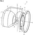
- Fig. 2 shows a sectional view of an example outside the scope of the present invention of a magnetic resonance imaging device 10.
- an angle 33 defining an access to the imaging volume 30 is enclosed by the center of the imaging volume 31, the first magnet 13 and the second magnet 14.
- the angle 33 is constituted by a first tangent 41 to the first magnet 13 through the center of the imaging volume 31 and a second tangent 42 to the second magnet 14 through the center of the imaging volume 31.
- the angle 33 excludes an axis 32 defined by a trajectory of a centroid of a projection of the first magnet 13 in the direction of the imaging volume 30.
- the axis 32 may correspond to the axis of rotational symmetry of the magnetic resonance imaging device 10.
- the gradient coil 28b of the second magnet 14 is positioned on the pole face directed towards the imaging volume 30 of the second magnet 14 in such a way, that it protrudes from the pole face.
- the gradient coil 28a of the first magnet 13 is recessed into the pole face directed towards the imaging volume 30 of the first magnet 13.
- the magnetic resonance imaging device may comprise at least a third gradient coil 28, which is not depicted in Fig. 2 .
- the frustum-shaped section of the second magnet 14 may be a tapered end-piece of the permanent magnet constituting the second magnet 14.
- the frustum-shaped section is constituted by an induced magnet, preferably iron, which is connected or mounted to the second magnet 14.
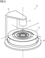
- Fig. 5 shows another example outside the scope of the present invention of a magnetic resonance imaging device 10.
- the second magnet 14 is an iron pole connected to an iron yoke 18. Due to the positioning of the iron pole relative to the magnetic field of the first magnet 13, the iron pole acts as an induced magnet.
- the iron yoke 18 is connected to a backing plate 36 covering the pole face of the first magnet 14 which is directed away from the imaging volume 30. It is conceivable, that the iron pole increases the magnetic field strength and/or the magnetic field homogeneity in the center of the imaging volume 31 in comparison to a permanent magnet with similar dimensions. Using a small iron pole may further increase the accessibility to the imaging volume 30.
- An upper body of the patient 15 is positioned between the first magnet 13 and the second magnet 14 in such a way, that a position of the jaw region of the patient 15 coincides with a position of the imaging volume 30.
- the pole face directed towards the imaging volume 30 of the second magnet 14 may accommodate for the back or the shoulders of the patient 15 during an imaging examination, thus increasing patient comfort as well as the accessibility of the imaging volume 30.
- the angle 33 defining the accessibility of the imaging volume 30 is increased by a tapering of the pole face directed towards the imaging volume 30 of the second magnet 14.
- the superconducting magnets of the first magnet 13 and the second magnet 14 both comprise a plurality of tubular magnet segments 34a and 34b, which are oriented parallelly along the axis of rotational symmetry 32.
- the diameter of the tubular magnet segments 34b of the second magnet 14 decreases in a direction of the imaging volume 30, thus providing a cone-shaped pole face.
- the tubular magnet segments 34a of the first magnet 13 are arranged in such a way, that the diameter of a first group of tubular magnet segments decreases in the direction facing away from the imaging volume 30.
- the first group of tubular magnet segments is sequenced by a second group of tubular magnet segments with a larger diameter in a further distance to the imaging volume 30.
- the overall shape of the superconducting magnet may thus correspond to a frustum sequenced by a short cylindrical section.
- the pole face directed towards the imaging volume 30 of the first magnet 13 comprises a planar surface.
- This arrangement of the first magnet 13 and the second magnet 14 may be particularly suitable for accommodating the patient 15 in a standing or upright position.
- Fig. 8 shows a further example outside the scope of the present invention of a magnetic resonance imaging device 10, comprising a permanent magnet as a second magnet 14 and a superconducting magnet as a first magnet 13.
- the first magnet 13 comprises coils of superconducting wires, wherein the lighter colored sections of the first magnet 13 may correspond to reversed coils in comparison to the darker colored sections.
- the imaging volume 30 may be positioned in proximity to the first magnet 13.
- the distance between the pole face directed towards the imaging volume 30 and the center of the imaging volume 31 may substantially correspond to a distance between a tip of a nose and a center of an eye of the patient 15.
- the imaging volume 30 may be spherical, as shown in Fig. 8 , or oval. It is also conceivable that the imaging volume 30 comprises the shape of an ellipsoid, a disk, a star, a polyhedron, a torus or a combination thereof.
- the axis of rotational symmetry of the second magnet 14 may coincide with the axis of rotational symmetry 32 of the first magnet 13. However, it is also conceivable that the axis of rotational symmetry of the second magnet 14 is shifted with respect to the axis of rotational symmetry of the first magnet 13 in a parallel manner. The axis of rotational symmetry of the second magnet 14 may be angled with respect to the axis of rotational symmetry 32 of the first magnet 13.
- At least a part of the diagnostically relevant body region of the patient 15 is aligned with the imaging volume 30.
- Aligning the diagnostically relevant body region of the patient 15 with the imaging volume 30 may comprise adjusting a position and/or an orientation of the patient 15 via a patient positioning device 16 as shown in Fig. 1 or Fig. 3 .
- Positioning and/or orienting the patient 15 may be performed automatically, semi-automatically or manually.
- aligning the diagnostically relevant body region of the patient 15 with the imaging volume 30 comprises adjusting a position and/or an orientation of the magnetic resonance imaging device 10 via the positioning unit 29 and/or the supporting structure 11. It is also conceivable, that a first aligning (e. g.
- a coarse aligning) of the diagnostically relevant body region of the patient 15 and the imaging volume 30 is accomplished via the patient positioning device 16 and a second aligning (e.g. a fine tuning) of the diagnostically relevant body region of the patient 15 and the imaging volume 30 is accomplished is accomplished via the positioning unit of the magnetic resonance imaging device 10 or vice versa.
- Aligning the imaging volume 30 with the diagnostically relevant body region may comprise adjusting a position of the imaging volume 30 to match a position of at least a part of the diagnostically relevant body region of the patient 15.
- the diagnostically relevant body region of the patient 15 is larger than the imaging volume 30 of the magnetic resonance imaging device 10.
- the imaging volume 30 of the magnetic resonance imaging device 10.
- some diagnostically relevant body regions of the patient 15 may be assessed by placing the patient in a gap between the first magnet 13 and the second magnet 14 either facing the first magnet 13 or the second magnet 14.
- a first magnetic resonance measurement is performed to acquire first magnetic resonance image data from the diagnostically relevant body region.
- the magnetic resonance measurement may comprise an arbitrary imaging sequence, such as a UTE (ultra-short echo time), a ZTE (zero echo time), a TSE (turbo spin echo), a GRE (gradient echo) sequence or any other known imaging sequence appropriate for performing an imaging examination of the diagnostically relevant body region of the patient 15.
- the first magnetic resonance image data may be limited to the part of the diagnostically relevant body region of the patient 15 covered by the imaging volume 30 of the magnetic resonance imaging device 10.
- the step S3 comprises adjusting a relative position and/or orientation of the magnetic resonance imaging device 10 and the patient 15 via rotation of the magnetic resonance imaging device 10 and/or a patient positioning device 16.
- a position and/or orientation of the magnetic resonance imaging device 10 and/or the patient positioning device 16 may be adjusted to align a further part of the diagnostically relevant body region with the imaging volume 30.
- the first magnet 13 and the second magnet 14 are rotated around the patient 15 in such a way, that the imaging volume 30 covers a different part of the diagnostically relevant body region of the patient 15.
- the further part of the diagnostically relevant body region may exclude parts of the diagnostically relevant body region from which the first magnetic resonance image data have been ac-quired.
- the further part of the diagnostically relevant body region may also include at least a section of the diagnostically relevant body region from which the first magnetic resonance image data have been acquired.
- a second magnetic resonance measurement is performed to acquire second magnetic resonance image data from the diagnostically relevant body region.
- the second magnetic resonance measurement comprises a same type of imaging sequence used for the first magnetic resonance measurement.
- the second magnetic resonance image data may be limited to the further part of the diagnostically relevant body region covered by the adjusted imaging volume 30 according to step S3.
- the second magnetic resonance image data may also comprise sections of the diagnostically relevant body region, which are already comprised within the first magnetic resonance image data.
- the steps S3 and S4 are repeated several times, each time aligning a further part of the diagnostically relevant body region with the imaging volume 30 by adjusting a position and/or an orientation of the magnetic resonance imaging device 10 and/or the patient positioning device 16.
- the magnetic resonance imaging device 10 is rotated with respect to the patient 15 in such way, that the center of the imaging volume 31 is positioned along a substantially circular or curved trajectory.
- the relative positions of the imaging volume 30 and the diagnostically relevant body region are selected in such a way, that a continuous imaging region covering the diagnostically relevant body region is obtained.
- the imaging region may follow the shape of a U or a C and consist of a plurality of imaging volumes positioned along a contour of the U or C.
- the image of the diagnostically relevant body region of the patient 15 is output.
- Outputting may comprise saving the image of the diagnostically relevant body region on a storage unit of the magnetic resonance imaging device 10, a medical information system and/or a cloud.
- the image of the diagnostically relevant body region may also be output to the operator of the magnetic resonance imaging device 10 via the output unit 25 and/or an output unit positioned on the pole face of the first magnet 13 or second magnet 14 facing the patient 15.
Landscapes
- Physics & Mathematics (AREA)
- General Physics & Mathematics (AREA)
- Condensed Matter Physics & Semiconductors (AREA)
- Health & Medical Sciences (AREA)
- Life Sciences & Earth Sciences (AREA)
- High Energy & Nuclear Physics (AREA)
- Nuclear Medicine, Radiotherapy & Molecular Imaging (AREA)
- General Health & Medical Sciences (AREA)
- Radiology & Medical Imaging (AREA)
- Engineering & Computer Science (AREA)
- Electromagnetism (AREA)
- Spectroscopy & Molecular Physics (AREA)
- Optics & Photonics (AREA)
- Epidemiology (AREA)
- Signal Processing (AREA)
- Pathology (AREA)
- Public Health (AREA)
- Veterinary Medicine (AREA)
- Animal Behavior & Ethology (AREA)
- Surgery (AREA)
- Molecular Biology (AREA)
- Medical Informatics (AREA)
- Heart & Thoracic Surgery (AREA)
- Biomedical Technology (AREA)
- Biophysics (AREA)
- General Life Sciences & Earth Sciences (AREA)
- Vascular Medicine (AREA)
- Magnetic Resonance Imaging Apparatus (AREA)
Claims (12)
- Magnetresonanztomografievorrichtung (10), die eine Felderzeugungseinheit (12) umfasst, wobei die Felderzeugungseinheit (12) einen ersten Magnet (13) und einen zweiten Magnet (14) umfasst, die ein Bildgebungsvolumen (30) der Magnetresonanztomografievorrichtung (10) in zwei räumlichen Richtungen begrenzen und wobei die Felderzeugungseinheit (12) dazu ausgelegt ist, ein statisches Magnetfeld innerhalb des Bildgebungsvolumens (30) bereitzustellen, wobei der erste Magnet (13) und der zweite Magnet (14) asymmetrisch in Bezug auf das Bildgebungsvolumen (30) angeordnet sind und wobei der erste Magnet (13) und der zweite Magnet (14) in einer solchen Weise angeordnet sind, dass sie entlang von mindestens zwei senkrechten räumlichen Richtung einen Zugang zu dem Bildgebungsvolumen (30) bereitstellen, wobei eine Zugangsrichtung zu dem Bildgebungsvolumen (30) in Bezug auf eine Hauptrichtung des Magnetfelds in dem Bildgebungsvolumen (30) abgewinkelt ist, wobei der erste Magnet (13) einen supraleitenden Magnet mit einer Vielzahl von Magnetsegmenten umfasst, wobei jedes Magnetsegment eine Magnetfeldachse umfasst und wobei die Magnetfeldachsen der Vielzahl von Magnetsegmenten parallel zu einer Magnetfeldachse des ersten Magnets (13) ausgerichtet sind,
wobei jedes Magnetsegment eine Röhrenform umfasst, wobei• ein Durchmesser von mindestens einem Magnetsegment aus der Vielzahl von Magnetsegmenten von einem Durchmesser von mindestens einem zweiten Magnetsegment aus der Vielzahl von Magnetsegmenten abweicht und• dadurch gekennzeichnet, dass die Magnetfeldachse von jedem Magnetsegment aus der Vielzahl von Magnetsegmenten entlang der Magnetfeldachse des ersten Magnets (13) in einer solchen Weise positioniert ist, dass eine Gesamtform des ersten Magnets (13) einem Kegel, einem Kegelstumpf oder einer Sequenz davon entspricht. - Magnetresonanztomografievorrichtung (10) nach Anspruch 1, wobei der erste Magnet (13) einen supraleitenden Magnet umfasst und der zweite Magnet(14) einen Permanentmagnet oder einen Elektromagnet umfasst.
- Magnetresonanztomografievorrichtung (10) nach Anspruch 1, wobei der erste Magnet (13) einen supraleitenden Magnet umfasst und der zweite Magnet(14) einen supraleitenden Magnet umfasst, wobei die Magnetresonanztomografievorrichtung (10) einen umfasst von:• einem kombinierten Kryostat, der mit dem ersten Magnet (13) und dem zweiten Magnet (14) verbunden ist, oder• einem ersten Kryostat, der mit dem ersten Magnet (13) verbunden ist, und einem zweiten Kryostat, der mit dem zweiten Magnet (14) verbunden ist.
- Magnetresonanztomografievorrichtung (10) nach einem der vorhergehenden Ansprüche, die ferner eine Stützstruktur (11) beinhaltet, die dazu ausgelegt ist, um eine strukturelle Stütze für die Felderzeugungseinheit (12) bereitzustellen, wobei die Stützstruktur (11) eine Positionierungseinheit (29) umfasst, die dazu ausgelegt ist, um eine Position und/oder eine Ausrichtung der Felderzeugungseinheit (12) in mindestens einer räumlichen Richtung einzustellen.
- Magnetresonanztomografievorrichtung (10) nach einem der vorhergehenden Ansprüche, wobei der erste Magnet (13) und/oder der zweite Magnet (14) eine Polfläche umfassen, die in Richtung auf das Bildgebungsvolumen (30) ausgerichtet ist, wobei die Polfläche, die in Richtung auf das Bildgebungsvolumen (30) des ersten Magnets (13) und/oder des zweiten Magnets (14) ausgerichtet ist, eine nicht-ebene Oberfläche umfasst.
- Magnetresonanztomografievorrichtung (10) nach Anspruch 5, wobei die Polfläche, die in Richtung auf das Bildgebungsvolumen (30) des ersten Magnets (13) und/oder des zweiten Magnets (14) ausgerichtet ist, die Form eines Kegels, eines Kegelstumpfs oder einer Halbkugel umfasst.
- Magnetresonanztomografievorrichtung (10) nach einem der vorhergehenden Ansprüche, die ferner eine Streufeldeindämmungseinheit umfasst, die dazu ausgelegt ist, ein magnetisches Streufeld der Felderzeugungseinheit (12) in einer solchen Weise auszurichten, dass eine Magnetfeldstärke und/oder eine Magnetfeldhomogenität in einem Zentrum des Bildgebungsvolumens (31) vergrößert wird, wobei die Streufeldeindämmungseinheit mindestens eine umfasst von• einer Rückseitenplatte (36), die an dem ersten Magnet (13) und/oder dem zweiten Magnet (14) befestigt ist, wobei die Rückseitenplatte (36) ein ferromagnetisches Material umfasst und wobei die Rückseitenplatte (36) an einer Polfläche des ersten Magnets (13) und/oder des zweiten Magnets (14) befestigt ist, die von dem Bildgebungsvolumen (30) weggerichtet ist,• einem Bügel (18), der an dem ersten Magnet (13) und dem zweiten Magnet (14) befestigt ist, wobei der Bügel (18) ein ferromagnetisches Material umfasst,• einer Abschirmungsspule (35), die an dem ersten Magnet (13) und/oder dem zweiten Magnet (14) befestigt ist, wobei die Abschirmungsspule (35) eine Kreisform umfasst und wobei ein Zentrum der Abschirmungsspule (35) entlang einer Bewegungsbahn positioniert ist, die durch einen Flächenschwerpunkt einer Projektion des ersten Magnets (13) und/oder des zweiten Magnets (14) in der Richtung des Zentrums des Bildgebungsvolumens (31) definiert wird.
- Magnetresonanztomografievorrichtung (10) nach einem der vorhergehenden Ansprüche, wobei die Felderzeugungseinheit (12) ein Gradientenfeldsystem (27) mit mindestens einer Gradientenspule (28) zum Erzeugen mindestens eines magnetischen Gradientenfeldes umfasst, wobei der erste Magnet (13) und/oder der zweite Magnet (14) eine Polfläche umfassen, die in Richtung auf das Bildgebungsvolumen (30) ausgerichtet ist, und wobei die mindestens eine Gradientenspule (28) des Gradientenfeldsystems (27)• in der Nähe der Polfläche positioniert ist, die in Richtung auf das Bildgebungsvolumen (30) des ersten Magnets (13) und/oder des zweiten Magnets (14) ausgerichtet ist, und/oder• mindestens teilweise in die Polfläche vertieft ist, die in Richtung auf das Bildgebungsvolumen (30) des ersten Magnets (13) und/oder des zweiten Magnets (14) ausgerichtet ist.
- Magnetresonanztomografievorrichtung (10) nach einem der vorhergehenden Ansprüche, wobei der erste Magnet (13) und der zweite Magnet (14) in einer solchen Weise angeordnet sind, dass ein Winkel, der den Zugang zu dem Bildgebungsvolumen (30) definiert, 60°, 75°, 90° oder 105° überschreitet, wobei der Winkel durch ein Zentrum des Bildgebungsvolumens (31), den ersten Magnet (13) und den zweiten Magnet (14) eingeschlossen ist.
- Magnetresonanztomografievorrichtung (10) nach Anspruch 9, wobei ein zweiter Abstand zwischen dem Zentrum des Bildgebungsvolumens (31) und einem nächsten Punkt auf einer Polfläche, die in Richtung auf das Bildgebungsvolumen (30) des zweiten Magnets (14) ausgerichtet ist, einen ersten Abstand zwischen dem Zentrum des Bildgebungsvolumens (31) und einem nächsten Punkt auf einer Polfläche, die in Richtung auf das Bildgebungsvolumen (30) des ersten Magnets (13) ausgerichtet ist, überschreitet, und wobei ein Verhältnis des zweiten Abstands und des ersten Abstands in einem Bereich zwischen 1,25 und 8 liegt.
- Magnetresonanztomografievorrichtung (10) nach einem der vorhergehenden Ansprüche, wobei das Bildgebungsvolumen (30) die Form eines Ellipsoids, einer Scheibe, eines Sterns, eines Polyeders, eines Torus oder einer Kombination davon umfasst.
- Verfahren zum Erfassen eines Bildes eines diagnostisch relevanten Körperbereichs eines Patienten (15) mit einer Magnetresonanztomografievorrichtung (10) nach einem der vorhergehenden Ansprüche, die folgenden Schritte umfassend:• Abgleichen (S1) mindestens eines Teils des diagnostisch relevanten Körperbereichs des Patienten (15) mit dem Bildgebungsvolumen (30),• Durchführen (S2) einer ersten Magnetresonanzmessung, um erste Magnetresonanzbilddaten von dem diagnostisch relevanten Körperbereich zu erfassen,• Einstellen (S3) einer relativen Position und/oder Ausrichtung der Magnetresonanztomografievorrichtung (10) und des Patienten (15) durch eine Drehung der Magnetresonanztomografievorrichtung und/oder einer Patientenpositionierungsvorrichtung (16),• Durchführen (S4) einer zweiten Magnetresonanzmessung, um zweite Magnetresonanzbilddaten von dem diagnostisch relevanten Körperbereich zu erfassen,• Wiederherstellen (S5) des Bildes des diagnostisch relevanten Körperbereichs des Patienten (15) in Abhängigkeit von den ersten Magnetresonanzbilddaten und den zweiten Magnetresonanzbilddaten,• Ausgeben (S6) des Bildes des diagnostisch relevanten Körperbereichs des Patienten (15).
Priority Applications (6)
| Application Number | Priority Date | Filing Date | Title |
|---|---|---|---|
| EP21165607.9A EP4067925B1 (de) | 2021-03-29 | 2021-03-29 | Magnetresonanztomografievorrichtung mit einheit zur asymmetrischen felderzeugung |
| EP21182676.3A EP4067926B1 (de) | 2021-03-29 | 2021-06-30 | Magnetresonanztomografievorrichtung mit einer gekrümmten einheit zur erzeugung eines magnetischen feldes |
| CN202210301485.2A CN115128531B (zh) | 2021-03-29 | 2022-03-25 | 具有弯曲场产生单元的磁共振成像设备 |
| US17/705,839 US11899084B2 (en) | 2021-03-29 | 2022-03-28 | Magnetic resonance imaging device with asymmetric field generation unit |
| US17/705,871 US11921176B2 (en) | 2021-03-29 | 2022-03-28 | Magnetic resonance imaging device with a curved field generation unit |
| CN202210318638.4A CN115137344B (zh) | 2021-03-29 | 2022-03-29 | 具有不对称的场生成单元的磁共振成像设备 |
Applications Claiming Priority (1)
| Application Number | Priority Date | Filing Date | Title |
|---|---|---|---|
| EP21165607.9A EP4067925B1 (de) | 2021-03-29 | 2021-03-29 | Magnetresonanztomografievorrichtung mit einheit zur asymmetrischen felderzeugung |
Publications (2)
| Publication Number | Publication Date |
|---|---|
| EP4067925A1 EP4067925A1 (de) | 2022-10-05 |
| EP4067925B1 true EP4067925B1 (de) | 2025-05-07 |
Family
ID=75277874
Family Applications (2)
| Application Number | Title | Priority Date | Filing Date |
|---|---|---|---|
| EP21165607.9A Active EP4067925B1 (de) | 2021-03-29 | 2021-03-29 | Magnetresonanztomografievorrichtung mit einheit zur asymmetrischen felderzeugung |
| EP21182676.3A Active EP4067926B1 (de) | 2021-03-29 | 2021-06-30 | Magnetresonanztomografievorrichtung mit einer gekrümmten einheit zur erzeugung eines magnetischen feldes |
Family Applications After (1)
| Application Number | Title | Priority Date | Filing Date |
|---|---|---|---|
| EP21182676.3A Active EP4067926B1 (de) | 2021-03-29 | 2021-06-30 | Magnetresonanztomografievorrichtung mit einer gekrümmten einheit zur erzeugung eines magnetischen feldes |
Country Status (3)
| Country | Link |
|---|---|
| US (2) | US11899084B2 (de) |
| EP (2) | EP4067925B1 (de) |
| CN (2) | CN115128531B (de) |
Families Citing this family (3)
| Publication number | Priority date | Publication date | Assignee | Title |
|---|---|---|---|---|
| EP4067925B1 (de) * | 2021-03-29 | 2025-05-07 | Siemens Healthineers AG | Magnetresonanztomografievorrichtung mit einheit zur asymmetrischen felderzeugung |
| EP4113146B1 (de) | 2021-06-30 | 2025-05-07 | Siemens Healthineers AG | Magnetresonanzbildgebungsvorrichtung mit einer konkav geformten felderzeugungseinheit |
| WO2025196150A1 (en) * | 2024-03-19 | 2025-09-25 | Deepspin Gmbh | Mri patient bed |
Citations (1)
| Publication number | Priority date | Publication date | Assignee | Title |
|---|---|---|---|---|
| EP2745130B1 (de) * | 2011-08-15 | 2018-07-04 | Emscan Limited | Hybridmagnet für die bildgebende magnetische resonanz |
Family Cites Families (23)
| Publication number | Priority date | Publication date | Assignee | Title |
|---|---|---|---|---|
| JPS63241905A (ja) | 1987-03-27 | 1988-10-07 | Sumitomo Special Metals Co Ltd | 磁界発生装置 |
| US5307039A (en) * | 1992-09-08 | 1994-04-26 | General Electric Company | Frustoconical magnet for magnetic resonance imaging |
| US5304930A (en) * | 1993-02-01 | 1994-04-19 | Panacea Medical Laboratories | Remotely positioned MRI system |
| EP0766094B1 (de) * | 1995-09-28 | 2002-05-29 | Siemens Aktiengesellschaft | Magnetanordnung für ein diagnostisches Magnetresonanzgerät |
| US6265959B1 (en) * | 1998-04-29 | 2001-07-24 | New York University | Open unipolar magnetic structure |
| AU2001287741A1 (en) * | 2000-09-26 | 2002-04-08 | Koninklijke Philips Electronics N.V. | Vertical field type mri apparatus with a conical cavity situated in the main magnet |
| EP1342099A1 (de) * | 2000-09-26 | 2003-09-10 | Koninklijke Philips Electronics N.V. | Gerät der bildgebenden magnetischen resonanz mit vertikalem feld und mit einer im hauptmagneten angeordneten konischen gradientenspule |
| JP2003102699A (ja) * | 2001-09-28 | 2003-04-08 | Hitachi Medical Corp | 開放型磁気共鳴イメージング装置 |
| US6707363B1 (en) * | 2003-01-06 | 2004-03-16 | Brk Wireless Company, Inc. | NMR head imaging system |
| DE102004005278B4 (de) * | 2004-02-03 | 2008-09-11 | Siemens Ag | Verfahren zur Herstellung transversaler nichtzylindrischer Gradientenspulen mit zumindest einem divergenten Abschnitt |
| US7109708B2 (en) * | 2004-08-19 | 2006-09-19 | General Electric Company | Systems, methods and apparatus of a magnetic resonance imaging magnet to produce an asymmetrical stray field |
| US7498810B2 (en) * | 2004-09-11 | 2009-03-03 | General Electric Company | Systems, methods and apparatus for specialized magnetic resonance imaging with dual-access conical bore |
| CN100415162C (zh) * | 2005-03-09 | 2008-09-03 | Ge医疗系统环球技术有限公司 | 磁体系统和磁共振成像系统 |
| WO2007004109A1 (en) * | 2005-06-30 | 2007-01-11 | Koninklijke Philips Electronics, N.V. | Asymmetric magnet for mri |
| US7737693B2 (en) * | 2006-03-04 | 2010-06-15 | Tursiop Technologies Llc | Signal acquisition and processing method and apparatus for magnetic resonance imaging |
| DE102013214880A1 (de) * | 2013-07-30 | 2015-02-05 | Siemens Aktiengesellschaft | Magnetresonanzgerät |
| WO2016180786A1 (en) * | 2015-05-12 | 2016-11-17 | Koninklijke Philips N.V. | Magnetic resonance examination system with field probes |
| CN205514601U (zh) * | 2016-02-03 | 2016-08-31 | 汪红志 | 一种半开放式核磁共振成像系统 |
| CN109313244A (zh) * | 2016-06-16 | 2019-02-05 | 皇家飞利浦有限公司 | 具有集成的调制器和开关单元的磁场梯度线圈组件 |
| US10627464B2 (en) * | 2016-11-22 | 2020-04-21 | Hyperfine Research, Inc. | Low-field magnetic resonance imaging methods and apparatus |
| EP3899565A2 (de) * | 2018-12-19 | 2021-10-27 | Hyperfine, Inc. | System und verfahren zur erdung von patienten während der magnetresonanzbildgebung |
| US11675034B2 (en) * | 2020-05-04 | 2023-06-13 | Siemens Healthcare Gmbh | Magnetic resonance scanner and magnetic resonance imaging system |
| EP4067925B1 (de) * | 2021-03-29 | 2025-05-07 | Siemens Healthineers AG | Magnetresonanztomografievorrichtung mit einheit zur asymmetrischen felderzeugung |
-
2021
- 2021-03-29 EP EP21165607.9A patent/EP4067925B1/de active Active
- 2021-06-30 EP EP21182676.3A patent/EP4067926B1/de active Active
-
2022
- 2022-03-25 CN CN202210301485.2A patent/CN115128531B/zh active Active
- 2022-03-28 US US17/705,839 patent/US11899084B2/en active Active
- 2022-03-28 US US17/705,871 patent/US11921176B2/en active Active
- 2022-03-29 CN CN202210318638.4A patent/CN115137344B/zh active Active
Patent Citations (1)
| Publication number | Priority date | Publication date | Assignee | Title |
|---|---|---|---|---|
| EP2745130B1 (de) * | 2011-08-15 | 2018-07-04 | Emscan Limited | Hybridmagnet für die bildgebende magnetische resonanz |
Also Published As
| Publication number | Publication date |
|---|---|
| CN115128531A (zh) | 2022-09-30 |
| US20220308139A1 (en) | 2022-09-29 |
| CN115137344B (zh) | 2025-09-30 |
| US20220308142A1 (en) | 2022-09-29 |
| EP4067926B1 (de) | 2025-08-27 |
| US11921176B2 (en) | 2024-03-05 |
| EP4067925A1 (de) | 2022-10-05 |
| EP4067926A1 (de) | 2022-10-05 |
| CN115137344A (zh) | 2022-10-04 |
| US11899084B2 (en) | 2024-02-13 |
| CN115128531B (zh) | 2025-08-22 |
Similar Documents
| Publication | Publication Date | Title |
|---|---|---|
| US11899084B2 (en) | Magnetic resonance imaging device with asymmetric field generation unit | |
| JP3556948B2 (ja) | 磁気共鳴イメージング用の円錐台形磁石 | |
| JPH0217038A (ja) | 磁気共鳴イメージング装置 | |
| US6507192B1 (en) | Nuclear magnetic resonance apparatus and methods of use and facilities for incorporating the same | |
| EP1267714B1 (de) | Vorrichtung und verfahren für die bildgebung mit magnetischer resonanz | |
| US9714992B2 (en) | Versatile superconducting magnet for extremities magnetic resonance imaging | |
| JPH07213506A (ja) | 核スピン共鳴トモグラフィ装置 | |
| US8947090B2 (en) | Electromagnet assembly | |
| US7680525B1 (en) | Method for lateral motion magnetic resonance imaging | |
| CN111913142A (zh) | 基本场磁体装置、磁共振断层造影系统和测量方法 | |
| JPH09192117A (ja) | 磁気共鳴イメージング装置 | |
| US5431164A (en) | Therapy tomograph | |
| US11852703B2 (en) | Magnetic resonance imaging device with a concave-shaped field generation unit | |
| JPH10108848A (ja) | 核磁気共鳴による浅い体積部の検査装置 | |
| JP4045769B2 (ja) | 磁場発生装置及びこれを用いるmri装置 | |
| JP2002531196A (ja) | 磁気共鳴撮像システム | |
| EP2972443B1 (de) | Flexibel verwendbarer supraleitender magnet für die magnetresonanzbildgebung von extremitäten | |
| US20250362361A1 (en) | Dedicated Magnetic Resonance Device | |
| Wald et al. | Portable brain scanner technology for use in emergency medicine | |
| US12298369B2 (en) | Dedicated magnetic resonance device | |
| US6100690A (en) | Radio frequency coil for magnetic resonance imaging apparatus | |
| US20240077557A1 (en) | Superconducting magnet and mri apparatus | |
| US20250271518A1 (en) | System and method for hybrid magnet design for magnetic resonance imaging systems | |
| WO2025184246A1 (en) | Short-bore mri system with non-spherical field of view |
Legal Events
| Date | Code | Title | Description |
|---|---|---|---|
| PUAI | Public reference made under article 153(3) epc to a published international application that has entered the european phase |
Free format text: ORIGINAL CODE: 0009012 |
|
| STAA | Information on the status of an ep patent application or granted ep patent |
Free format text: STATUS: THE APPLICATION HAS BEEN PUBLISHED |
|
| AK | Designated contracting states |
Kind code of ref document: A1 Designated state(s): AL AT BE BG CH CY CZ DE DK EE ES FI FR GB GR HR HU IE IS IT LI LT LU LV MC MK MT NL NO PL PT RO RS SE SI SK SM TR |
|
| STAA | Information on the status of an ep patent application or granted ep patent |
Free format text: STATUS: REQUEST FOR EXAMINATION WAS MADE |
|
| 17P | Request for examination filed |
Effective date: 20230403 |
|
| RBV | Designated contracting states (corrected) |
Designated state(s): AL AT BE BG CH CY CZ DE DK EE ES FI FR GB GR HR HU IE IS IT LI LT LU LV MC MK MT NL NO PL PT RO RS SE SI SK SM TR |
|
| RAP1 | Party data changed (applicant data changed or rights of an application transferred) |
Owner name: SIEMENS HEALTHINEERS AG |
|
| GRAP | Despatch of communication of intention to grant a patent |
Free format text: ORIGINAL CODE: EPIDOSNIGR1 |
|
| STAA | Information on the status of an ep patent application or granted ep patent |
Free format text: STATUS: GRANT OF PATENT IS INTENDED |
|
| RIC1 | Information provided on ipc code assigned before grant |
Ipc: G01R 33/3815 20060101ALI20241122BHEP Ipc: G01R 33/385 20060101ALI20241122BHEP Ipc: G01R 33/38 20060101AFI20241122BHEP |
|
| INTG | Intention to grant announced |
Effective date: 20241213 |
|
| GRAS | Grant fee paid |
Free format text: ORIGINAL CODE: EPIDOSNIGR3 |
|
| GRAA | (expected) grant |
Free format text: ORIGINAL CODE: 0009210 |
|
| STAA | Information on the status of an ep patent application or granted ep patent |
Free format text: STATUS: THE PATENT HAS BEEN GRANTED |
|
| AK | Designated contracting states |
Kind code of ref document: B1 Designated state(s): AL AT BE BG CH CY CZ DE DK EE ES FI FR GB GR HR HU IE IS IT LI LT LU LV MC MK MT NL NO PL PT RO RS SE SI SK SM TR |
|
| REG | Reference to a national code |
Ref country code: GB Ref legal event code: FG4D |
|
| REG | Reference to a national code |
Ref country code: DE Ref legal event code: R081 Ref document number: 602021030262 Country of ref document: DE Owner name: SIEMENS HEALTHINEERS AG, DE Free format text: FORMER OWNER: ANMELDERANGABEN UNKLAR / UNVOLLSTAENDIG, 80297 MUENCHEN, DE |
|
| REG | Reference to a national code |
Ref country code: CH Ref legal event code: EP |
|
| REG | Reference to a national code |
Ref country code: DE Ref legal event code: R096 Ref document number: 602021030262 Country of ref document: DE |
|
| REG | Reference to a national code |
Ref country code: IE Ref legal event code: FG4D |
|
| REG | Reference to a national code |
Ref country code: NL Ref legal event code: MP Effective date: 20250507 |
|
| PG25 | Lapsed in a contracting state [announced via postgrant information from national office to epo] |
Ref country code: FI Free format text: LAPSE BECAUSE OF FAILURE TO SUBMIT A TRANSLATION OF THE DESCRIPTION OR TO PAY THE FEE WITHIN THE PRESCRIBED TIME-LIMIT Effective date: 20250507 Ref country code: ES Free format text: LAPSE BECAUSE OF FAILURE TO SUBMIT A TRANSLATION OF THE DESCRIPTION OR TO PAY THE FEE WITHIN THE PRESCRIBED TIME-LIMIT Effective date: 20250507 Ref country code: PT Free format text: LAPSE BECAUSE OF FAILURE TO SUBMIT A TRANSLATION OF THE DESCRIPTION OR TO PAY THE FEE WITHIN THE PRESCRIBED TIME-LIMIT Effective date: 20250908 |
|
| REG | Reference to a national code |
Ref country code: LT Ref legal event code: MG9D |
|
| PG25 | Lapsed in a contracting state [announced via postgrant information from national office to epo] |
Ref country code: NO Free format text: LAPSE BECAUSE OF FAILURE TO SUBMIT A TRANSLATION OF THE DESCRIPTION OR TO PAY THE FEE WITHIN THE PRESCRIBED TIME-LIMIT Effective date: 20250807 Ref country code: GR Free format text: LAPSE BECAUSE OF FAILURE TO SUBMIT A TRANSLATION OF THE DESCRIPTION OR TO PAY THE FEE WITHIN THE PRESCRIBED TIME-LIMIT Effective date: 20250808 |
|
| PG25 | Lapsed in a contracting state [announced via postgrant information from national office to epo] |
Ref country code: PL Free format text: LAPSE BECAUSE OF FAILURE TO SUBMIT A TRANSLATION OF THE DESCRIPTION OR TO PAY THE FEE WITHIN THE PRESCRIBED TIME-LIMIT Effective date: 20250507 Ref country code: NL Free format text: LAPSE BECAUSE OF FAILURE TO SUBMIT A TRANSLATION OF THE DESCRIPTION OR TO PAY THE FEE WITHIN THE PRESCRIBED TIME-LIMIT Effective date: 20250507 |
|
| REG | Reference to a national code |
Ref country code: AT Ref legal event code: MK05 Ref document number: 1793024 Country of ref document: AT Kind code of ref document: T Effective date: 20250507 |
|
| PG25 | Lapsed in a contracting state [announced via postgrant information from national office to epo] |
Ref country code: BG Free format text: LAPSE BECAUSE OF FAILURE TO SUBMIT A TRANSLATION OF THE DESCRIPTION OR TO PAY THE FEE WITHIN THE PRESCRIBED TIME-LIMIT Effective date: 20250507 |
|
| PG25 | Lapsed in a contracting state [announced via postgrant information from national office to epo] |
Ref country code: HR Free format text: LAPSE BECAUSE OF FAILURE TO SUBMIT A TRANSLATION OF THE DESCRIPTION OR TO PAY THE FEE WITHIN THE PRESCRIBED TIME-LIMIT Effective date: 20250507 |
|
| PG25 | Lapsed in a contracting state [announced via postgrant information from national office to epo] |
Ref country code: AT Free format text: LAPSE BECAUSE OF FAILURE TO SUBMIT A TRANSLATION OF THE DESCRIPTION OR TO PAY THE FEE WITHIN THE PRESCRIBED TIME-LIMIT Effective date: 20250507 |
|
| PG25 | Lapsed in a contracting state [announced via postgrant information from national office to epo] |
Ref country code: RS Free format text: LAPSE BECAUSE OF FAILURE TO SUBMIT A TRANSLATION OF THE DESCRIPTION OR TO PAY THE FEE WITHIN THE PRESCRIBED TIME-LIMIT Effective date: 20250807 |
|
| PG25 | Lapsed in a contracting state [announced via postgrant information from national office to epo] |
Ref country code: IS Free format text: LAPSE BECAUSE OF FAILURE TO SUBMIT A TRANSLATION OF THE DESCRIPTION OR TO PAY THE FEE WITHIN THE PRESCRIBED TIME-LIMIT Effective date: 20250907 |
|
| PG25 | Lapsed in a contracting state [announced via postgrant information from national office to epo] |
Ref country code: LV Free format text: LAPSE BECAUSE OF FAILURE TO SUBMIT A TRANSLATION OF THE DESCRIPTION OR TO PAY THE FEE WITHIN THE PRESCRIBED TIME-LIMIT Effective date: 20250507 |