EP4059443B1 - Verfahren und vorrichtung zur superauflösenden rekonstruktion eines dreidimensionalen kontrastverstärkten ultraschallbildes - Google Patents
Verfahren und vorrichtung zur superauflösenden rekonstruktion eines dreidimensionalen kontrastverstärkten ultraschallbildes Download PDFInfo
- Publication number
- EP4059443B1 EP4059443B1 EP21809726.9A EP21809726A EP4059443B1 EP 4059443 B1 EP4059443 B1 EP 4059443B1 EP 21809726 A EP21809726 A EP 21809726A EP 4059443 B1 EP4059443 B1 EP 4059443B1
- Authority
- EP
- European Patent Office
- Prior art keywords
- dimensional local
- dimensional
- local image
- image sequence
- super
- Prior art date
- Legal status (The legal status is an assumption and is not a legal conclusion. Google has not performed a legal analysis and makes no representation as to the accuracy of the status listed.)
- Active
Links
Images


Classifications
-
- A—HUMAN NECESSITIES
- A61—MEDICAL OR VETERINARY SCIENCE; HYGIENE
- A61B—DIAGNOSIS; SURGERY; IDENTIFICATION
- A61B8/00—Diagnosis using ultrasonic, sonic or infrasonic waves
- A61B8/08—Clinical applications
- A61B8/0891—Clinical applications for diagnosis of blood vessels
-
- G—PHYSICS
- G06—COMPUTING OR CALCULATING; COUNTING
- G06T—IMAGE DATA PROCESSING OR GENERATION, IN GENERAL
- G06T3/00—Geometric image transformations in the plane of the image
- G06T3/40—Scaling of whole images or parts thereof, e.g. expanding or contracting
- G06T3/4053—Scaling of whole images or parts thereof, e.g. expanding or contracting based on super-resolution, i.e. the output image resolution being higher than the sensor resolution
-
- A—HUMAN NECESSITIES
- A61—MEDICAL OR VETERINARY SCIENCE; HYGIENE
- A61B—DIAGNOSIS; SURGERY; IDENTIFICATION
- A61B8/00—Diagnosis using ultrasonic, sonic or infrasonic waves
- A61B8/52—Devices using data or image processing specially adapted for diagnosis using ultrasonic, sonic or infrasonic waves
- A61B8/5207—Devices using data or image processing specially adapted for diagnosis using ultrasonic, sonic or infrasonic waves involving processing of raw data to produce diagnostic data, e.g. for generating an image
-
- A—HUMAN NECESSITIES
- A61—MEDICAL OR VETERINARY SCIENCE; HYGIENE
- A61B—DIAGNOSIS; SURGERY; IDENTIFICATION
- A61B8/00—Diagnosis using ultrasonic, sonic or infrasonic waves
- A61B8/48—Diagnostic techniques
- A61B8/481—Diagnostic techniques involving the use of contrast agents, e.g. microbubbles introduced into the bloodstream
-
- A—HUMAN NECESSITIES
- A61—MEDICAL OR VETERINARY SCIENCE; HYGIENE
- A61B—DIAGNOSIS; SURGERY; IDENTIFICATION
- A61B8/00—Diagnosis using ultrasonic, sonic or infrasonic waves
- A61B8/48—Diagnostic techniques
- A61B8/483—Diagnostic techniques involving the acquisition of a 3D volume of data
-
- A—HUMAN NECESSITIES
- A61—MEDICAL OR VETERINARY SCIENCE; HYGIENE
- A61B—DIAGNOSIS; SURGERY; IDENTIFICATION
- A61B8/00—Diagnosis using ultrasonic, sonic or infrasonic waves
- A61B8/52—Devices using data or image processing specially adapted for diagnosis using ultrasonic, sonic or infrasonic waves
- A61B8/5215—Devices using data or image processing specially adapted for diagnosis using ultrasonic, sonic or infrasonic waves involving processing of medical diagnostic data
- A61B8/5223—Devices using data or image processing specially adapted for diagnosis using ultrasonic, sonic or infrasonic waves involving processing of medical diagnostic data for extracting a diagnostic or physiological parameter from medical diagnostic data
-
- A—HUMAN NECESSITIES
- A61—MEDICAL OR VETERINARY SCIENCE; HYGIENE
- A61B—DIAGNOSIS; SURGERY; IDENTIFICATION
- A61B8/00—Diagnosis using ultrasonic, sonic or infrasonic waves
- A61B8/52—Devices using data or image processing specially adapted for diagnosis using ultrasonic, sonic or infrasonic waves
- A61B8/5215—Devices using data or image processing specially adapted for diagnosis using ultrasonic, sonic or infrasonic waves involving processing of medical diagnostic data
- A61B8/5238—Devices using data or image processing specially adapted for diagnosis using ultrasonic, sonic or infrasonic waves involving processing of medical diagnostic data for combining image data of patient, e.g. merging several images from different acquisition modes into one image
-
- G—PHYSICS
- G06—COMPUTING OR CALCULATING; COUNTING
- G06T—IMAGE DATA PROCESSING OR GENERATION, IN GENERAL
- G06T5/00—Image enhancement or restoration
- G06T5/20—Image enhancement or restoration using local operators
- G06T5/30—Erosion or dilatation, e.g. thinning
-
- G—PHYSICS
- G06—COMPUTING OR CALCULATING; COUNTING
- G06T—IMAGE DATA PROCESSING OR GENERATION, IN GENERAL
- G06T5/00—Image enhancement or restoration
- G06T5/70—Denoising; Smoothing
-
- G—PHYSICS
- G06—COMPUTING OR CALCULATING; COUNTING
- G06T—IMAGE DATA PROCESSING OR GENERATION, IN GENERAL
- G06T7/00—Image analysis
- G06T7/10—Segmentation; Edge detection
- G06T7/136—Segmentation; Edge detection involving thresholding
-
- G—PHYSICS
- G06—COMPUTING OR CALCULATING; COUNTING
- G06T—IMAGE DATA PROCESSING OR GENERATION, IN GENERAL
- G06T7/00—Image analysis
- G06T7/30—Determination of transform parameters for the alignment of images, i.e. image registration
-
- G—PHYSICS
- G06—COMPUTING OR CALCULATING; COUNTING
- G06T—IMAGE DATA PROCESSING OR GENERATION, IN GENERAL
- G06T7/00—Image analysis
- G06T7/50—Depth or shape recovery
-
- G—PHYSICS
- G06—COMPUTING OR CALCULATING; COUNTING
- G06T—IMAGE DATA PROCESSING OR GENERATION, IN GENERAL
- G06T7/00—Image analysis
- G06T7/60—Analysis of geometric attributes
- G06T7/68—Analysis of geometric attributes of symmetry
-
- G—PHYSICS
- G06—COMPUTING OR CALCULATING; COUNTING
- G06T—IMAGE DATA PROCESSING OR GENERATION, IN GENERAL
- G06T2207/00—Indexing scheme for image analysis or image enhancement
- G06T2207/10—Image acquisition modality
- G06T2207/10016—Video; Image sequence
-
- G—PHYSICS
- G06—COMPUTING OR CALCULATING; COUNTING
- G06T—IMAGE DATA PROCESSING OR GENERATION, IN GENERAL
- G06T2207/00—Indexing scheme for image analysis or image enhancement
- G06T2207/10—Image acquisition modality
- G06T2207/10132—Ultrasound image
- G06T2207/10136—3D ultrasound image
-
- G—PHYSICS
- G06—COMPUTING OR CALCULATING; COUNTING
- G06T—IMAGE DATA PROCESSING OR GENERATION, IN GENERAL
- G06T2207/00—Indexing scheme for image analysis or image enhancement
- G06T2207/30—Subject of image; Context of image processing
- G06T2207/30004—Biomedical image processing
Definitions
- the present application relates to the field of image processing technologies, in particular to a super-resolution reconstruction method and an apparatus for three-dimensional contrast-enhanced ultrasound images, a non-transitory computer readable storage medium and an electronic device.
- Ultrasound Localization Microscopy mainly achieves a purpose of microvascular imaging by locating isolated microbubble contrast agents in microvessels, and importance of it is self-evident. Compared with the prior art, ULM improves a spatial resolution by about ten times, and realizes three-dimensional ultrasound super-resolution imaging of complicated microvasculature in three-dimensional space.
- WO 2018/222724A1 discloses systems and methods for super-resolution ultrasound imaging of microvessels in a subject.
- Ultrasound data are acquired from a region-of-interest in a subject who has been administered a microbubble contrast agent.
- the ultrasound data are acquired while the microbubbles are moving through, or otherwise present in, the region-of-interest.
- the region of interest may include, for instance, microvessels or other microvasculature in the subject.
- the prior three-dimensional ultrasound super-resolution imaging methods based on ULM not only require a long-time acquisition of three-dimensional contrast-enhanced ultrasound images, but also are susceptible to noise interference, which in turn leads to a decrease in localization precision of microbubbles, so it is difficult to achieve the theoretical spatial resolution of ULM with low signal-to-noise ratio contrast-enhanced ultrasound images collected in a short period of time in clinic.
- an embodiment of the present application provides a super-resolution reconstruction method for three-dimensional contrast-enhanced ultrasound images, applied to a first three-dimensional local image sequence including microbubbles.
- the super-resolution reconstruction method for three-dimensional contrast-enhanced ultrasound images includes: performing a first thinning operation on the first three-dimensional local image sequence to generate a second three-dimensional local image sequence corresponding to the first three-dimensional local image sequence, where the first thinning operation is used to enhance motion trajectories of microbubbles;
- the determining distance information between a microbubble area in the first three-dimensional local image and a background area corresponding to the microbubble area includes: performing binarization processing on the first three-dimensional local images based on the microbubble area and the background area to generate a binarized image; and determining the distance information between the microbubble area and the background area based on the binarized image.
- the multiple second three-dimensional local images included in the second three-dimensional local image sequence are obtained by performing the first thinning operation on the multiple first three-dimensional local images in the first three-dimensional local image sequence frame by frame.
- a pixel value of a pixel coordinate corresponding to the microbubble area is set to 1
- a pixel value of a pixel coordinate corresponding to the background area is set to 0.
- the microbubble area includes a plurality of pixel blocks.
- the determining distance information between a microbubble area in the first three-dimensional local image and a background area corresponding to the microbubble area includes: determining a shortest distance from each pixel block in the plurality of pixel blocks to the background area respectively; and determining the distance information based on the shortest distances respectively corresponding to the plurality of pixel blocks.
- the degree of radial symmetry estimation operation is implemented based on a degree of radial symmetry calculation method in super-resolution radial fluctuations.
- a number of sampling points is 12 and a sampling radius is 1 in the degree of radial symmetry estimation operation.
- the method before the performing a first thinning operation on the first three-dimensional local image sequence to generate a second three-dimensional local image sequence corresponding to the first three-dimensional local image sequence, the method further includes: performing a registration operation on the first three-dimensional local image sequence.
- the registration operation includes a rigid registration operation and/or a flexible registration operation.
- the performing a registration operation on the first three-dimensional local image sequence includes: performing, by using three-dimensional Morphon multi-scale registration method, the registration operation on the first three-dimensional local image sequence.
- the performing an image reconstruction operation based on the first three-dimensional local image sequence subjected to the first thinning operation and the second thinning operation, so as to generate a three-dimensional super-resolution image includes: performing an accumulation operation on the third three-dimensional local images of the third three-dimensional local image sequence based on image sequence information corresponding to the first three-dimensional local image sequence, so as to generate the three-dimensional super-resolution image, where the accumulation operation refers to accumulating each frame of the third three-dimensional local image sequence on the same location to form the three-dimensional super-resolution image.
- an embodiment of the present application provides a computer readable storage medium according to claim 8.
- an embodiment of the present application provides an electronic device according to claim 9.
- the super-resolution reconstruction method for three-dimensional contrast-enhanced ultrasound images by means of performing thinning operations (for example, respectively performing a first thinning operation and a second thinning operation) on the first three-dimensional local image sequence, motion trajectories of microbubbles are highlighted, thereby improving a signal-to-noise ratio of an image.
- thinning operations for example, respectively performing a first thinning operation and a second thinning operation
- embodiments of the present application may greatly improve a reconstruction efficiency and reconstruction precision of the three-dimensional super-resolution image reconstruction operation.
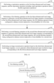
- FIG. 1 is a schematic diagram of a scene applicable for an embodiment of the present application.
- a scene applicable for an embodiment of the present application includes a server 1 and an ultrasound imaging device 2. There is a communication connection relationship between the server 1 and the ultrasound imaging device 2.
- the ultrasound imaging device 2 is configured to acquire a first three-dimensional local image sequence including microbubbles.
- the server 1 is configured to perform at least one thinning operation on the first three-dimensional local image sequence, the thinning operation being configured to enhance motion trajectories of microbubbles; and perform an image reconstruction operation based on the first three-dimensional local image sequence subjected to the at least one thinning operation, so as to generate a three-dimensional super-resolution image. That is, the scene realizes a super-resolution reconstruction method for three-dimensional contrast-enhanced ultrasound images.
- FIG. 2 is a schematic diagram of another scene applicable for an embodiment of the present application.
- the scene includes an image processing device 3, the image processing device 3 including an image acquisition module 301 and a calculation module 302. There is a communication connection between the image acquisition module 301 and the calculation module 302.
- the image acquisition module 301 in the image processing device 3 may be configured to perform functions of the ultrasound imaging device 2 in the scene shown in FIG. 1 .
- the calculation module 302 in the image processing device 3 may be configured to perform functions of the server 1 in the scene shown in FIG. 1 . Embodiments of the present application will not be repeated here.
- the above scene shown in FIG. 2 realizes the super-resolution reconstruction method for three-dimensional contrast-enhanced ultrasound images using the image processing device 3 with no need to perform data transmission operations using related devices such as servers, the above scene shown in FIG. 2 is capable of ensuring the reconstruction efficiency of super-resolution imaging for three-dimensional contrast-enhanced ultrasound images.
- FIG. 3 is a schematic flowchart of a super-resolution reconstruction method for three-dimensional contrast-enhanced ultrasound images according to an exemplary embodiment of the present application. As shown in FIG. 3 , the super-resolution reconstruction method for three-dimensional contrast-enhanced ultrasound images according to embodiments of the present application include the following steps.
- Step 10 performing a first thinning operation on the first three-dimensional local image sequence to generate a second three-dimensional local image sequence corresponding to the first three-dimensional local image sequence, the first thinning operation being used to enhance motion trajectories of microbubbles for a first time.
- the first three-dimensional local image sequence mentioned in Step 10 is a three-dimensional local image sequence including microbubbles acquired by an ultrasound imaging device. That is, the first three-dimensional local image sequence includes a plurality of first three-dimensional local images, and the first three-dimensional local image includes microbubble areas.
- a three-dimensional super-resolution image including an overall structure of blood vessels (such as blood vessel structure in tumor) may be reconstructed. That is, in the embodiment of the present application, the meaning of "local" is that each first three-dimensional local image represents a local image content of the three-dimensional super-resolution image. After corresponding processing of all the first three-dimensional local images, the three-dimensional super-resolution image including the overall structure may be generated.
- the first thinning operation is configured to enhance motion trajectories of microbubbles, for example, enhancing central (central axis) regions of the microbubbles.
- the first thinning operation mentioned in the embodiments of the present application may suppress noise in a background area.
- a plurality of frames of second three-dimensional local images included in the second three-dimensional local image sequence are obtained by performing the first thinning operation on a plurality of frames of the first three-dimensional local images in the first three-dimensional local image sequence frame by frame.
- j 1,2 , ⁇ N
- j 1,2 , ⁇ N .
- Step 20 performing a second thinning operation on the second three-dimensional local image sequence to generate a third three-dimensional local image sequence corresponding to the second three-dimensional local image sequence, the second thinning operation being configured to enhance motion trajectories of the microbubbles for a second time.
- the second thinning operation is configured to enhance the motion trajectories of the microbubbles based on a non-localization-based manner. For example, instead of locating the microbubble areas in images, the motion trajectories of the microbubbles are enhanced by performing a degree of radial symmetry estimation operation on pixels in the second three-dimensional local images of the second three-dimensional local image sequence.
- the embodiments of the present application do not limit the specific implementation of the second thinning operation mentioned in Step 20, as long as the second thinning operation can enhance the motion trajectories of the microbubbles.
- Step 30 performing an image reconstruction operation based on a third three-dimensional local image sequence, so as to generate a three-dimensional super-resolution image corresponding to the third three-dimensional local image sequence.
- the performing an image reconstruction operation based on a third three-dimensional local image sequence, so as to generate a three-dimensional super-resolution image corresponding to the third three-dimensional local image sequence includes: performing an accumulation operation on the third three-dimensional local images in the third three-dimensional local image sequence based on image sequence information corresponding to the third three-dimensional local image sequence, so as to generate the three-dimensional super-resolution image.
- the first thinning operation is performed on the first three-dimensional local image sequence to generate the second three-dimensional local image sequence corresponding to the first three-dimensional local image sequence.
- the second thinning operation is performed on the second three-dimensional local image sequence to generate the third three-dimensional local image sequence corresponding to the second three-dimensional local image sequence.
- the image reconstruction operation is performed based on the third three-dimensional local image sequence, so as to generate the three-dimensional super-resolution image corresponding to the third three-dimensional local image sequence.
- the super-resolution reconstruction method for the three-dimensional contrast-enhanced ultrasound images by means of respectively performing the first thinning operation and the second thinning operation on the first three-dimensional local image sequence, motion trajectories of microbubbles are highlighted, thereby improving a signal-to-noise ratio of an image.
- embodiments of the present application may greatly improve efficiency and precision of the three-dimensional super-resolution image reconstruction operation.
- the first thinning operation and the second thinning operation mentioned in the embodiment shown in FIG. 3 are not limited to coexist.
- the super-resolution reconstruction method for three-dimensional contrast-enhanced ultrasound images includes: performing at least one thinning operation on the first three-dimensional local image sequence, the thinning operation being configured to enhance motion trajectories of microbubbles; and performing the image reconstruction operation based on the first three-dimensional local image sequence subjected to the at least one thinning operation, so as to generate the three-dimensional super-resolution image.
- FIG. 4 is a schematic flowchart for performing a first thinning operation on a first three-dimensional local image sequence to generate a second three-dimensional local image sequence corresponding to the first three-dimensional local image sequence according to an exemplary embodiment of the present application.
- the embodiment of the present application is extended based on the embodiment of the present application shown in FIG. 3 . Differences between the embodiment of the present application and the embodiment shown in FIG. 3 are emphatically described below, and similarities may not be described repeatedly.
- the step performing a first thinning operation on the first three-dimensional local image sequence to generate a second three-dimensional local image sequence corresponding to the first three-dimensional local image sequence includes the following steps.
- Step 11 determining, for each frame of first three-dimensional local images in the first three-dimensional local image sequence, distance information between a microbubble area in the first three-dimensional local image and a background area corresponding to the microbubble area.
- the microbubble area refers to an image area corresponding to microbubbles in the first three-dimensional local image
- the background area refers to a background image area that does not include microbubbles in the first three-dimensional local image.
- the distance information is distance information of a three-dimensional space.
- the microbubble area corresponds to a plurality of pixel coordinates arranged in a three-dimensional space.
- FIG. 5 is a schematic flowchart for determining distance information between a microbubble area in the first three-dimensional local image and a background area corresponding to the microbubble area according to an exemplary embodiment of the present application.
- the microbubble area includes a plurality of pixel blocks.
- the determining distance information between a microbubble area in the first three-dimensional local image and a background area corresponding to the microbubble area includes the following steps.
- Step 111 determining a shortest distance from each pixel block in the plurality of pixel blocks to the background area respectively.
- Step 112 determining the distance information based on the shortest distances respectively corresponding to the plurality of pixel blocks.
- the shortest distance mentioned in Step 111 is Euclidean distance or Manhattan distance, preferably Euclidean distance.
- the pixel blocks mentioned above are individual pixel units.
- pixel coordinates corresponding to the pixel blocks are pixel coordinates corresponding to the pixel units.
- the pixel blocks mentioned above are pixel blocks formed by a plurality of adjacent pixel units.
- pixel coordinates corresponding to the pixel blocks are pixel coordinates at center points of the plurality of pixel units.
- Step 12 performing a first weighting operation on the first three-dimensional local images based on the distance information to generate first weighted images.
- a pixel grayscale value and a shortest distance corresponding to each pixel coordinate are respectively determined. Then, the shortest distance is multiplied by the pixel grayscale value to determine a new pixel grayscale value corresponding to the pixel coordinate, and then the first weighted image corresponding to the first three-dimensional local image is finally determined.
- the shortest distance between a central region of the microbubble and a reference region is larger than that of the marginal region of the microbubble. Therefore, after performing the first weighting operation on the first three-dimensional local images based on the distance information, the central region (i.e., a central axis region along microbubble trajectory) of the microbubbles of the first three-dimensional local image may be effectively highlighted (i.e., enhanced), and the noise in the background area is removed, so as to achieve a purpose of enhancing the motion trajectories of the microbubbles.
- Step 13 generating the second three-dimensional local image sequence based on the first weighted images respectively corresponding to the first three-dimensional local images in the first three-dimensional local image sequence.
- the super-resolution reconstruction method for three-dimensional contrast-enhanced ultrasound images effectively highlights (i.e., enhances) the central region of the microbubbles in the first three-dimensional local image sequence, removes the noise in the background area, and improves a signal-to-noise ratio of the image, and then highlights (i.e., enhances) three-dimensional motion trajectories of microbubbles along the vessel in the moving directions of microbubbles, thereby improving efficiency and precision of three-dimensional super-resolution reconstruction.
- the method before the performing a first weighting operation on the first three-dimensional local images based on the distance information to generate first weighted images, the method further includes: performing binarization processing on the first three-dimensional local images based on the microbubble area and the background area to generate binarized images.
- the performing a first weighting operation on the first three-dimensional local images based on the distance information to generate first weighted images includes: performing the first weighting operation on the binarized images based on the distance information corresponding to the first three-dimensional local images to generate the first weighted images.
- Binarizing the first three-dimensional local images based on the embodiment of the present application may simplify the calculation procedure of the step of generating the first weighted images corresponding to the first three-dimensional local images, speed up the calculation process, and further improve the reconstruction efficiency.
- FIG. 6, FIG. 7a and FIG. 7b take a two-dimensional image including a rectangle (analogously as a microbubble) as an example to show.
- the two-dimensional image is used here for convenience and clarity only.
- FIG. 6 is a schematic diagram of a process of generating first weighted images according to an exemplary embodiment of the present application.
- an image on the left side of FIG. 6 is a pixel distribution diagram of a two-dimensional image.
- the pixel values of pixel coordinates corresponding to the rectangular area is set to 1, and the pixel values of the pixel coordinates corresponding to the background area is set to 0.
- An image on the right side of FIG. 6 is a pixel distribution diagram of the first weighted image obtained by performing distance weighting (i.e., the first weighting operation) on the image on the left side of FIG. 6 .
- the shortest distance is determined by using the "chessboard distance".
- a weighted value i.e., a new pixel value
- a weighted value i.e., a new pixel value
- FIG. 7a and FIG. 7b are schematic diagrams of weighting effect of a first weighting operation according to an exemplary embodiment of the present application. Specifically, FIG. 7a shows imaging effect corresponding to the image on the left side of FIG. 6. FIG. 7b shows imaging effect corresponding to the image on the right side of FIG. 6 . As shown in FIG. 7a and FIG. 7b , it can be clearly seen that after a first decoupling operation, the central region of the rectangle in the two-dimensional image is enhanced (i.e., highlighted), and the marginal region is weakened.
- the central regions of the microbubbles in the first three-dimensional local image may be enhanced (i.e., may be highlighted), and the marginal regions may be weakened.
- a large amount of noise such as background noise
- the motion trajectories of the microbubbles is enhanced.
- FIG. 8 is a schematic flowchart for performing a second thinning operation on a second three-dimensional local image sequence to generate a third three-dimensional local image sequence corresponding to the second three-dimensional local image sequence according to an exemplary embodiment of the present application.
- the embodiment of the present application is extended based on the embodiment of the present application shown in FIG. 3 . Differences between the embodiment of the present application and the embodiment shown in FIG. 3 are emphatically described below, and similarities may not be described repeatedly.
- the performing a second thinning operation on the second three-dimensional local image sequence to generate a third three-dimensional local image sequence corresponding to the second three-dimensional local image sequence includes the following steps.
- Step 21 performing, for each second three-dimensional local image in the second three-dimensional local image sequence, a degree of radial symmetry estimation operation on each pixel unit in the second three-dimensional local images, so as to determine a weight value corresponding to the pixel unit.
- the degree of radial symmetry estimation operation mentioned in Step 21 is implemented based on a degree of radial symmetry calculation method in super-resolution radial fluctuations ( Zhang, J. J Ultrasound Med. 2020 ).
- a number of sampling points is 12 and a sampling radius is 1 in the degree of radial symmetry operation.
- An inventor of the present application found that after setting the number of sampling points to 12 and the sampling radius to 1 in the degree of radial symmetry estimation operation, precision of determined three-dimensional motion trajectories of the microbubbles are significantly improved.
- the pixel unit mentioned in Step 21 is a pixel point in the second three-dimensional local image.
- Step 22 performing second weighting operation on the second three-dimensional local images based on the weight values respectively corresponding to the pixel units in the second three-dimensional local images to generate second weighted images.
- Step 23 generating a third three-dimensional local image sequence based on the second weighted images respectively corresponding to the second three-dimensional local images in the second three-dimensional local image sequence.
- the super-resolution reconstruction method for three-dimensional contrast-enhanced ultrasound images according to an embodiment of the present application, for each second three-dimensional local image in the second three-dimensional local image sequence, by performing the degree of radial symmetry estimation operation on each pixel unit in the second three-dimensional local images, determine the weight values corresponding to the pixel units.
- the second weighted images are generated by performing the second weighting operation on the second three-dimensional local images based on the weight values respectively corresponding to the pixel units in the second three-dimensional local images.
- the second thinning operation is performed on the second three-dimensional local image sequence through the method of generating the third three-dimensional local image sequence based on the second weighted images respectively corresponding to the second three-dimensional local images in the second three-dimensional local image sequence, to generate the third three-dimensional local image sequence corresponding to the second three-dimensional local image sequence.
- the motion trajectories of the microbubbles in the second three-dimensional local images may be further enhanced, the embodiments of the present application further improve the efficiency and precision of the super-resolution reconstruction.
- FIG. 9a to FIG. 9c are schematic diagrams for intuitive explanation of a degree of radial symmetry estimation operation.
- FIG. 9a is a schematic diagram of a Gaussian distribution of pixels corresponding to a second three-dimensional local image in a second three-dimensional local image sequence including microbubbles.
- the point spread function of a microbubble may be viewed as a Gaussian distribution.
- the height of each pixel column represents a pixel value, and a higher position corresponds to a larger pixel value.
- the microbubbles in the image show grayscale features that the middle part is brighter and the closer to the edge, the darker of the grayscale value.
- a gradient field image shown in FIG. 9b may be obtained. Based on the gradient field image shown in FIG. 9b , it can be found that if all the gradient vectors (black arrows shown in FIG. 9b ) are extended, they will intersect at a center point of the microbubble, that is, the gradient at the center is convergent. This also means that if a straight line is drawn arbitrarily through the center point, the gradient vectors on left and right of the straight line are completely symmetrical, and the further away from the center point, the weaker the symmetry. Therefore, if the degree of radial symmetry of each pixel point in FIG. 9a is estimated, a degree of radial symmetry diagram shown in FIG. 9c will be obtained.
- the third three-dimensional local image in which the central region of the microbubble area is enhanced and the marginal region of the microbubble area is weakened (suppressed) is obtained.
- the third three-dimensional local images correspond to the second three-dimensional local images.
- FIG. 10a and FIG. 10b are schematic diagrams of weighting effect after a degree of radial symmetry estimation operation is performed on the microbubbles.
- the microbubbles are moving, the microbubbles are elongated along a flowing direction as shown in FIG. 10a .
- a second weighted image shown in FIG. 10b may be obtained.
- FIG. 10b the motion trajectories of the microbubbles are clearly preserved.
- the gradient may be calculated based on the following equations (1) and (2).
- G x x y ⁇ I x y ⁇ x
- G y x y ⁇ I x y ⁇ y
- N is an arbitrarily defined variable used to determine a number of gradient samples.
- N is set to 12
- ( x i ′ , y i ′ ) is one of the sample points.
- gradient vectors G xi and G yi at ( x i ′ , y i ′ ) are obtained from original image gradient vectors G x and G y .
- the gradient line at ( x i ′ , y i ′ ) is determined by a gradient line formula at sample point ( x i ′ , y i ′ ) given in the following formula (3).
- 0 x ⁇ x i ′ G yi ⁇ y ⁇ y i ′ G xi
- the estimated degree of radial symmetry may be obtained based on the following formula (7).
- FIG. 11 is a schematic flowchart of a super-resolution reconstruction method for a three-dimensional contrast-enhanced ultrasound images according to another exemplary embodiment of the present application.
- the embodiment of the present application is extended based on the embodiment of the present application shown in FIG. 3 . Differences between the embodiment of the present application and the embodiment shown in FIG. 3 are emphatically described below, and similarities may not be described repeatedly.
- Step 40 performing a registration operation on the first three-dimensional local image sequence, the registration operation including rigid registration operations and/or flexible registration operations.
- a three-dimensional Morphon multi-scale registration method is used to perform the registration operation on the first three-dimensional local image sequence.
- a decomposition scale is 3 layers.
- a deformation field is processed by Gaussian kernel smoothing, and a size of Gaussian kernel is 10 pixels.
- the registration operation on the first three-dimensional local image sequence is performed.
- the first thinning operation on the first three-dimensional local image sequence is performed to generate the second three-dimensional local image sequence corresponding to the first three-dimensional local image sequence.
- the second thinning operation on the second three-dimensional local image sequence is performed to generate the third three-dimensional local image sequence corresponding to the second three-dimensional local image sequence.
- the image reconstruction operation is performed based on the third three-dimensional local image sequence, so as to generate the three-dimensional super-resolution image corresponding to the third three-dimensional local image sequence.
- the super-resolution reconstruction method for three-dimensional contrast-enhanced ultrasound images by performing the registration operation on the first three-dimensional local image sequence before performing the first thinning operation and the second thinning operation on the three-dimensional local image sequence, effectively suppresses the influence of tissue motion (such as motion caused by breath) on the accuracy of microbubble localization, thereby further improving the accuracy of microbubble localization, and further improving the precision of super-resolution reconstruction.
- FIG. 12 is a schematic structural diagram of a super-resolution reconstruction apparatus for three-dimensional contrast-enhanced ultrasound images according to an exemplary embodiment of the present application.
- the super-resolution reconstruction apparatus for the three-dimensional contrast-enhanced ultrasound images provided by an embodiment of the present application includes the following modules.
- a first thinning module 100 is configured to perform a first thinning operation on a first three-dimensional local image sequence to generate a second three-dimensional local image sequence corresponding to the first three-dimensional local image sequence.
- the first thinning operation is configured to enhance motion trajectories of microbubbles for a first time.
- a second thinning module 200 is configured to perform a second thinning operation on the second three-dimensional local image sequence to generate a third three-dimensional local image sequence corresponding to the second three-dimensional local image sequence.
- the second thinning operation is configured to enhance motion trajectories of microbubbles for a second time.
- a reconstruction module 300 is configured to perform an image reconstruction operation based on the third three-dimensional local image sequence, so as to generate a three-dimensional super-resolution image corresponding to the third three-dimensional local image sequence.
- a super-resolution reconstruction apparatus for three-dimensional contrast-enhanced ultrasound images includes: a thinning module and a reconstruction module signally connected to the thinning module.
- the thinning module is configured to perform at least one thinning operation on the first three-dimensional local image sequence.
- the thinning operation is configured to enhance motion trajectories of microbubbles.
- the reconstruction module is configured to perform an image reconstruction operation based on the first three-dimensional local image sequence subjected to the at least one thinning operation, so as to generate the three-dimensional super-resolution image.
- FIG. 13 is a schematic structural diagram of a super-resolution reconstruction apparatus for three-dimensional contrast-enhanced ultrasound images according to another exemplary embodiment of the present application.
- the embodiment of the present application is extended based on the embodiment of the present application shown in FIG. 12 . Differences between the embodiment of the present application and the embodiment shown in FIG. 12 are emphatically described below, and similarities may not be described repeatedly.
- a first thinning module 100 includes the following units.
- a distance information determination unit 110 is configured to determine, for each frame of first three-dimensional local images in the first three-dimensional local image sequence, distance information between a microbubble area in the first three-dimensional local image and a background area corresponding to the microbubble area.
- a first generation unit 120 is configured to perform a first weighting operation on the first three-dimensional local images based on the distance information to generate first weighted images
- a second generation unit 130 is configured to generate a second three-dimensional local image sequence based on the first weighted images respectively corresponding to the first three-dimensional local images in the first three-dimensional local image sequence.
- the distance information determination unit 110 is further configured to: determine a shortest distance from each pixel block in the plurality of pixel blocks to the background area respectively, and determine the distance information based on the shortest distances respectively corresponding to the plurality of pixel blocks.
- FIG. 14 is a schematic structural diagram of a super-resolution reconstruction apparatus for three-dimensional contrast-enhanced ultrasound images according to another exemplary embodiment of the present application.
- the embodiment of the present application is extended based on the embodiment of the present application shown in FIG. 12 . Differences between the embodiment of the present application and the embodiment shown in FIG. 12 are emphatically described below, and similarities may not be described repeatedly.
- a second thinning module 200 includes the following units.
- a weight value determination unit 210 is configured to perform, for each frame of second three-dimensional local images in the second three-dimensional local image sequence, a degree of radial symmetry estimation operation on each pixel unit in the second three-dimensional local images, so as to determine a weight value corresponding to the pixel unit.
- a third generation unit 220 is configured to perform a second weighting operation on the second three-dimensional local images based on the weight values respectively corresponding to the pixel units in the second three-dimensional local images to generate second weighted images.
- a fourth generation unit 230 is configured to generate a third three-dimensional local image sequence based on the second weighted images respectively corresponding to the second three-dimensional local images in the second three-dimensional local image sequence.
- FIG. 15 is a schematic structural diagram of a super-resolution reconstruction apparatus for three-dimensional contrast-enhanced ultrasound images according to another exemplary embodiment of the present application.
- the embodiment of the present application is extended based on the embodiment of the present application shown in FIG. 12 . Differences between the embodiment of the present application and the embodiment shown in FIG. 12 are emphatically described below, and similarities may not be described repeatedly.
- the super-resolution reconstruction apparatus for the three-dimensional contrast-enhanced ultrasound images further includes the following module.
- a registration module 400 is configured to perform a registration operation on a first three-dimensional local image sequence.
- the registration operation includes rigid registration operations and/or flexible registration operations.
- Operations and functions of the first thinning module 100, the second thinning module 200, the reconstruction module 300, the registration module 400, and the distance information determination unit 110, the first generation unit 120, the second generation unit 130 included in the first thinning module 100, and the weight value determination unit 210, the third generation unit 220, the fourth generation unit 230 included in the second thinning module 200 in the super-resolution reconstruction apparatus for the three-dimensional contrast-enhanced ultrasound images provided in FIG. 12 to FIG. 15 may refer to the super-resolution reconstruction method for three-dimensional contrast-enhanced ultrasound images provided in FIG. 3 to FIG. 11 . In order to avoid repetitions, details are not repeated here.
- FIG. 16 is a schematic structural diagram of the electronic device according to an exemplary embodiment of the present application.
- the electronic device 50 includes one or more processors 501 and a memory 502.
- the processor 501 may be a Central Processing Unit (CPU) or another form of processing unit with data processing capability and/or instruction execution capability, and may control another component in the electronic device to perform an expected function.
- CPU Central Processing Unit
- the processor 501 may be a Central Processing Unit (CPU) or another form of processing unit with data processing capability and/or instruction execution capability, and may control another component in the electronic device to perform an expected function.
- the memory 502 may include one or more computer program products, which may include various forms of computer-readable storage media, such as a volatile memory and/or non-volatile memory.
- the volatile memory may include, for example, a Random Access Memory (RAM) and/or a cache (cache).
- the non-volatile memory may include, for example, a Read-Only Memory (ROM), a hard disk, and a flash memory.
- the compute-readable storage medium may store one or more computer program instructions, and the processor 501 may run the super-resolution reconstruction method for three-dimensional contrast-enhanced ultrasound images and/or other expected functions of the embodiments in the present application described above.
- the compute-readable storage medium may further store information, such as the second three-dimensional local image sequence, or the like.
- the electronic device 50 may further include an input apparatus 503 and an output apparatus 504, and these components are interconnected by using a bus system and/or another form of connection mechanism (not shown).
- the input apparatus 503 may also include, for example, a keyboard, a mouse, and so on.
- the output apparatus 504 may output various information including the determined three-dimensional contrast-enhanced ultrasound images to the external.
- the output device 504 may include, for example, a display, a communication network and a remote output device connected to it, and so on.
- the electronic device 50 may further include any other suitable component depending on specific application cases.
- an embodiment of the present application may also be a computer program product that includes computer program instructions.
- the processor is enabled to perform the steps of the super-resolution reconstruction method for three-dimensional contrast-enhanced ultrasound images according to the embodiments of the present application described in the "Exemplary Methods" part of this specification.
- the computer program product may write program code for performing the operations of the embodiments of the present application in any combination of one or more programming languages, and the programming languages include object-oriented programming languages such as Java and C++, and further include general procedural programming languages such as "C" or similar programming languages.
- the program code may be executed entirely on a user computing device, partly on a user device, as a stand-alone software package, partly on a user computing device while partly on a remote computing device, or entirely on a remote computing device or a server.
- an embodiment of the present application may also be a computer-readable storage medium, where the computer-readable storage medium stores computer program instructions.
- the processor is enabled to perform the steps of the super-resolution reconstruction method for three-dimensional contrast-enhanced ultrasound images according to the embodiments of the present application described in the "Exemplary Methods" part of this specification.
- the computer-readable storage medium may use any combination of one or more readable media.
- the readable medium may be a readable signal medium or a readable storage medium.
- the readable storage medium may include, for example, but not limited to, an electronic, magnetic, optical, electromagnetic, infrared, or semiconductor system, apparatus, or means, or any combination of the above.
- the readable storage medium include: an electrical connection having one or more wires, a portable computer disk, a hard disk, a Random Access Memory (RAM), a Read-Only Memory (ROM), an Erasable Programmable Read-Only Memory (EPROM or a flash memory), an optical fiber, a portable Compact Disk Read-Only Memory (CD-ROM), an optical storage means, a magnetic storage means, or any suitable combination of the above.
- RAM Random Access Memory
- ROM Read-Only Memory
- EPROM or a flash memory Erasable Programmable Read-Only Memory
- CD-ROM Compact Disk Read-Only Memory
- CD-ROM Compact Disk Read-Only Memory
Landscapes
- Health & Medical Sciences (AREA)
- Life Sciences & Earth Sciences (AREA)
- Engineering & Computer Science (AREA)
- Physics & Mathematics (AREA)
- Medical Informatics (AREA)
- Animal Behavior & Ethology (AREA)
- Biophysics (AREA)
- Nuclear Medicine, Radiotherapy & Molecular Imaging (AREA)
- Pathology (AREA)
- Radiology & Medical Imaging (AREA)
- Biomedical Technology (AREA)
- Heart & Thoracic Surgery (AREA)
- Veterinary Medicine (AREA)
- Molecular Biology (AREA)
- Surgery (AREA)
- Public Health (AREA)
- General Health & Medical Sciences (AREA)
- General Physics & Mathematics (AREA)
- Theoretical Computer Science (AREA)
- Computer Vision & Pattern Recognition (AREA)
- Hematology (AREA)
- Vascular Medicine (AREA)
- Geometry (AREA)
- Physiology (AREA)
- Ultra Sonic Daignosis Equipment (AREA)
- Image Processing (AREA)
Claims (9)
- Ein Superauflösungsrekonstruktionsverfahren für dreidimensionale, kontrastverstärkte Ultraschallbilder, das auf eine erste dreidimensionale, lokale Bildsequenz, die Mikroblasen beinhaltet, angewendet wird, beinhaltend:Durchführen (10) eines ersten Verdünnungsvorgangs an der ersten dreidimensionalen, lokalen Bildsequenz, um eine zweite dreidimensionale, lokale Bildsequenz, die mit der ersten dreidimensionalen, lokalen Bildsequenz korrespondiert, zu erzeugen, wobei der erste Verdünnungsvorgang verwendet wird, um Bewegungstrajektorien von Mikroblasen zu verstärken;Durchführen (20) eines zweiten Verdünnungsvorgangs an der zweiten dreidimensionalen, lokalen Bildsequenz, um eine dritte dreidimensionale, lokale Bildsequenz, die mit der zweiten dreidimensionalen, lokalen Bildsequenz korrespondiert, zu erzeugen, wobei der zweite Verdünnungsvorgang verwendet wird, um Bewegungstrajektorien von Mikroblasen ein zweites Mal zu verstärken; undDurchführen eines Bildrekonstruktionsvorgangs basierend auf der dritten dreidimensionalen, lokalen Bildsequenz, um ein dreidimensionales Superauflösungsbild zu erzeugen;wobei das Durchführen (10) eines ersten Verdünnungsvorgangs an der ersten dreidimensionalen, lokalen Bildsequenz, um eine zweite dreidimensionale, lokale Bildsequenz, die mit der ersten dreidimensionalen, lokalen Bildsequenz korrespondiert, zu erzeugen, Folgendes beinhaltet:Bestimmen (11), für jedes Einzelbild des ersten dreidimensionalen, lokalen Bildes in der ersten dreidimensionalen, lokalen Bildsequenz, von Abstandsinformationen zwischen einem Mikroblasenbereich in dem ersten dreidimensionalen, lokalen Bild und einem Hintergrundbereich, der mit dem Mikroblasenbereich korrespondiert;Durchführen (12) eines ersten Gewichtungsvorgangs an dem ersten dreidimensionalen, lokalen Bild basierend auf den Abstandsinformationen, um ein erstes gewichtetes Bild zu erzeugen; undErzeugen (13) der zweiten dreidimensionalen, lokalen Bildsequenz basierend auf den ersten gewichteten Bildern, die jeweils mit den ersten dreidimensionalen, lokalen Bildern in der ersten dreidimensionalen, lokalen Bildsequenz korrespondieren;wobei das Durchführen (20) eines zweiten Verdünnungsvorgangs an der zweiten dreidimensionalen, lokalen Bildsequenz, um eine dritte dreidimensionale, lokale Bildsequenz, die mit der zweiten dreidimensionalen, lokalen Bildsequenz korrespondiert, zu erzeugen, Folgendes beinhaltet:Durchführen (21), für jedes Einzelbild des zweiten dreidimensionalen, lokalen Bildes in der zweiten dreidimensionalen, lokalen Bildsequenz, eines Grades eines Radialsymmetrieschätzungsvorgangs an jeder Pixeleinheit in den zweiten dreidimensionalen, lokalen Bildern, um einen Gewichtswert, der mit der Pixeleinheit korrespondiert, zu bestimmen;Durchführen (22) eines zweiten Gewichtungsvorgangs an den zweiten dreidimensionalen, lokalen Bildern basierend auf den Gewichtswerten, die jeweils mit den Pixeleinheiten in den zweiten dreidimensionalen, lokalen Bildern korrespondieren, um ein zweites gewichtetes Bild zu erzeugen; undErzeugen (23) der dritten dreidimensionalen, lokalen Bildsequenz basierend auf den zweiten gewichteten Bildern, die jeweils mit den zweiten dreidimensionalen, lokalen Bildern in der zweiten dreidimensionalen, lokalen Bildsequenz korrespondieren.
- Superauflösungsrekonstruktionsverfahren für dreidimensionale, kontrastverstärkte Ultraschallbilder gemäß Anspruch 1, wobei das Bestimmen (11) von Abstandsinformationen zwischen einem Mikroblasenbereich in dem ersten dreidimensionalen, lokalen Bild und einem Hintergrundbereich, der mit dem Mikroblasenbereich korrespondiert, Folgendes beinhaltet:Durchführen einer Binarisierungsverarbeitung an den ersten dreidimensionalen, lokalen Bildern basierend auf dem Mikroblasenbereich und dem Hintergrundbereich, um ein binarisiertes Bild zu erzeugen; undBestimmen der Abstandsinformationen zwischen dem Mikroblasenbereich und dem Hintergrundbereich basierend auf dem binarisierten Bild.
- Superauflösungsrekonstruktionsverfahren für dreidimensionale, kontrastverstärkte Ultraschallbilder gemäß Anspruch 1 oder 2, wobei mehrere Einzelbilder der zweiten dreidimensionalen, lokalen Bilder, die in der zweiten dreidimensionalen, lokalen Bildsequenz beinhaltet sind, durch das einzelbildweise Durchführen des ersten Verdünnungsvorgangs an mehreren Einzelbildern der ersten dreidimensionalen, lokalen Bilder in der ersten dreidimensionalen, lokalen Bildsequenz erhalten werden.
- Superauflösungsrekonstruktionsverfahren für dreidimensionale, kontrastverstärkte Ultraschallbilder gemäß einem der Ansprüche 1 bis 3, wobei ein Pixelwert einer Pixelkoordinate, die mit dem Mikroblasenbereich korrespondiert, auf 1 gesetzt wird und ein Pixelwert einer Pixelkoordinate, die mit dem Hintergrundbereich korrespondiert, auf 0 gesetzt wird.
- Superauflösungsrekonstruktionsverfahren für dreidimensionale, kontrastverstärkte Ultraschallbilder gemäß Anspruch 4, wobei der Mikroblasenbereich eine Vielzahl von Pixelblöcken beinhaltet und das Bestimmen (11) von Abstandsinformationen zwischen einem Mikroblasenbereich in dem ersten dreidimensionalen, lokalen Bild und einem Hintergrundbereich, der mit dem Mikroblasenbereich korrespondiert, Folgendes beinhaltet:Bestimmen (111) eines kürzesten Abstands jeweils von jedem Pixelblock in der Vielzahl von Pixelblöcken zu dem Hintergrundbereich; undBestimmen (112) der Abstandsinformationen basierend auf den kürzesten Abständen, die jeweils mit der Vielzahl von Pixelblöcken korrespondieren.
- Superauflösungsrekonstruktionsverfahren für dreidimensionale, kontrastverstärkte Ultraschallbilder gemäß Anspruch 1, wobei der Grad des Radialsymmetrieschätzungsvorgangs basierend auf einem Grad eines Radialsymmetrieberechnungsverfahrens in Superauflösungsradialfluktuationen implementiert wird.
- Superauflösungsrekonstruktionsverfahren für dreidimensionale, kontrastverstärkte Ultraschallbilder gemäß einem der Ansprüche 1 bis 6, wobei das Verfahren vor dem Durchführen (10) eines ersten Verdünnungsvorgangs an der ersten dreidimensionalen, lokalen Bildsequenz, um eine zweite dreidimensionale, lokale Bildsequenz, die mit der ersten dreidimensionalen, lokalen Bildsequenz korrespondiert, zu erzeugen, ferner Folgendes beinhaltet:
Durchführen (40) eines Registrierungsvorgangs an der ersten dreidimensionalen, lokalen Bildsequenz, wobei der Registrierungsvorgang starre Registrierungsvorgänge und/oder elastische Registrierungsvorgänge beinhaltet. - Ein nichttransitorisches, computerlesbares Speichermedium, das ein Computerprogramm zum Ausführen des Superauflösungsrekonstruktionsverfahrens für dreidimensionale, kontrastverstärkte Ultraschallbilder gemäß einem der Ansprüche 1 bis 7 speichert.
- Eine elektronische Vorrichtung (50), beinhaltend:einen Prozessor (501); undeinen Speicher (502) zum Speichern ausführbarer Anweisungen des Prozessors;wobei der Prozessor konfiguriert ist, um das Superauflösungsrekonstruktionsverfahren für ein dreidimensionales, kontrastverstärktes Ultraschallbild gemäß einem der Ansprüche 1 bis 7 auszuführen.
Applications Claiming Priority (2)
| Application Number | Priority Date | Filing Date | Title |
|---|---|---|---|
| CN202010419606.4A CN111588409B (zh) | 2020-05-18 | 2020-05-18 | 三维超声造影图像的超分辨重建方法及装置 |
| PCT/CN2021/093469 WO2021233188A1 (zh) | 2020-05-18 | 2021-05-13 | 三维超声造影图像的超分辨重建方法及装置 |
Publications (3)
| Publication Number | Publication Date |
|---|---|
| EP4059443A1 EP4059443A1 (de) | 2022-09-21 |
| EP4059443A4 EP4059443A4 (de) | 2023-02-01 |
| EP4059443B1 true EP4059443B1 (de) | 2023-11-08 |
Family
ID=72185721
Family Applications (1)
| Application Number | Title | Priority Date | Filing Date |
|---|---|---|---|
| EP21809726.9A Active EP4059443B1 (de) | 2020-05-18 | 2021-05-13 | Verfahren und vorrichtung zur superauflösenden rekonstruktion eines dreidimensionalen kontrastverstärkten ultraschallbildes |
Country Status (4)
| Country | Link |
|---|---|
| US (1) | US12229916B2 (de) |
| EP (1) | EP4059443B1 (de) |
| CN (1) | CN111588409B (de) |
| WO (1) | WO2021233188A1 (de) |
Families Citing this family (4)
| Publication number | Priority date | Publication date | Assignee | Title |
|---|---|---|---|---|
| CN111588409B (zh) | 2020-05-18 | 2023-04-07 | 南京超维景生物科技有限公司 | 三维超声造影图像的超分辨重建方法及装置 |
| CN116115268B (zh) * | 2023-02-14 | 2025-09-16 | 西安交通大学 | 一种新型的快速超声超分辨成像方法、系统、设备及介质 |
| CN116763356B (zh) * | 2023-06-16 | 2025-12-02 | 西安交通大学 | 一种高时空分辨超声平面波造影增强微血管成像方法及系统 |
| CN119693423B (zh) * | 2024-12-02 | 2025-11-14 | 深圳大学 | 一种超声微血管成像中的运动校正与配准方法、设备、介质及产品 |
Family Cites Families (14)
| Publication number | Priority date | Publication date | Assignee | Title |
|---|---|---|---|---|
| US20150196279A1 (en) * | 2011-10-18 | 2015-07-16 | Riverside Research Institute | Synthetic-focusing strategies for real-time annular-array imaging |
| EP3286727B1 (de) * | 2014-11-07 | 2019-07-03 | Antaros Medical AB | Ganzkörperbildregistrierungsverfahren und verfahren zur analyse von bildern davon |
| EP4349263B1 (de) * | 2017-05-31 | 2026-03-04 | Mayo Foundation for Medical Education and Research | Verfahren zur superauflösenden ultraschalldarstellung von mikrogefässen |
| CN107361791B (zh) * | 2017-07-21 | 2020-10-09 | 北京大学 | 一种快速超分辨血流成像方法 |
| CN107753062B (zh) * | 2017-11-27 | 2021-03-16 | 西安交通大学 | 基于马尔科夫链蒙特卡洛多目标追踪的经颅超声脑血管造影超分辨率成像方法 |
| CN108324324A (zh) * | 2018-03-12 | 2018-07-27 | 西安交通大学 | 一种超声低频经颅容积超分辨率三维造影成像方法及系统 |
| CN108836392B (zh) * | 2018-03-30 | 2021-06-22 | 中国科学院深圳先进技术研究院 | 基于超声rf信号的超声成像方法、装置、设备及存储介质 |
| US11748849B2 (en) * | 2018-10-19 | 2023-09-05 | Mayo Foundation For Medical Education And Research | Systems and methods for Kalman filter-based microvessel inpainting for super-resolution imaging |
| EP3905960B1 (de) * | 2019-01-03 | 2024-09-25 | Koninklijke Philips N.V. | Systeme und verfahren zur kontrastverstärkten bildgebung |
| US11083435B2 (en) * | 2019-01-17 | 2021-08-10 | Bk Medical Aps | Super resolution ultrasound imaging |
| CN110772285B (zh) * | 2019-10-31 | 2022-05-17 | 南京景瑞康分子医药科技有限公司 | 一种超声超分辨成像方法 |
| CN111588409B (zh) * | 2020-05-18 | 2023-04-07 | 南京超维景生物科技有限公司 | 三维超声造影图像的超分辨重建方法及装置 |
| CN111588410B (zh) * | 2020-05-18 | 2023-05-16 | 南京超维景生物科技有限公司 | 基于超声造影图像的图像处理方法及装置 |
| CN112435305A (zh) * | 2020-07-09 | 2021-03-02 | 上海大学 | 一种基于深度学习的超高分辨超声成像方法 |
-
2020
- 2020-05-18 CN CN202010419606.4A patent/CN111588409B/zh active Active
-
2021
- 2021-05-13 WO PCT/CN2021/093469 patent/WO2021233188A1/zh not_active Ceased
- 2021-05-13 EP EP21809726.9A patent/EP4059443B1/de active Active
-
2022
- 2022-06-10 US US17/838,059 patent/US12229916B2/en active Active
Also Published As
| Publication number | Publication date |
|---|---|
| WO2021233188A1 (zh) | 2021-11-25 |
| EP4059443A1 (de) | 2022-09-21 |
| US20220309613A1 (en) | 2022-09-29 |
| EP4059443A4 (de) | 2023-02-01 |
| CN111588409A (zh) | 2020-08-28 |
| US12229916B2 (en) | 2025-02-18 |
| CN111588409B (zh) | 2023-04-07 |
Similar Documents
| Publication | Publication Date | Title |
|---|---|---|
| EP4059443B1 (de) | Verfahren und vorrichtung zur superauflösenden rekonstruktion eines dreidimensionalen kontrastverstärkten ultraschallbildes | |
| Liu et al. | Deep learning for ultrasound localization microscopy | |
| CN111598965B (zh) | 超声造影图像的超分辨重建预处理方法和超分辨重建方法 | |
| US12190473B2 (en) | Super-resolution flow field reconstruction method and device thereof | |
| JP5977214B2 (ja) | 画像処理方法および装置並びにプログラム | |
| CN107133946B (zh) | 医学图像处理方法、装置及设备 | |
| EP4059442B1 (de) | Bildverarbeitungsverfahren und -gerät basierend auf einem ultraschallkontrastbild | |
| US12346999B2 (en) | Data processing method, apparatus, device and storage medium | |
| US20110262015A1 (en) | Image processing apparatus, image processing method, and storage medium | |
| US20140212013A1 (en) | Method and Apparatus for Generating a Derived Image Using Images of Different Types | |
| EP2867853B1 (de) | Bildqualitätsgesteuerte nichtstarre bildaufzeichnung | |
| CN112150571A (zh) | 一种图像运动伪影消除方法、装置、设备及存储介质 | |
| US20200178845A1 (en) | Systems and methods for pulmonary ventilation from image processing | |
| JP7548296B2 (ja) | 管状フィーチャのセグメント化 | |
| Zhao et al. | Comparison of the existing tool localisation methods on two‐dimensional ultrasound images and their tracking results | |
| Wen et al. | GPU-accelerated kernel regression reconstruction for freehand 3D ultrasound imaging | |
| Amir-Khalili et al. | Real-time extraction of local phase features from volumetric medical image data | |
| US8831309B2 (en) | Filter approach to catheter electrode detection and tracking | |
| CN121414606B (zh) | 一种基于熵引导的实时激光散斑成像方法、装置及系统 | |
| EP4497392A1 (de) | Erzeugung eines oder mehrerer bewegungsindikatoren | |
| Chiu et al. | Interpretable deep learning analysis on correction of motion blur X-ray images to ensure the efficiency and reliability of clinical decisions | |
| Madhavan et al. | Improved Edge Detection for Brain Tumor Using Multi-threading | |
| Zontak et al. | Speeding up 3D speckle tracking using PatchMatch | |
| Mori et al. | Robust adversarial learning model to segment non-speckle regions in blood flow echo | |
| CN107067448A (zh) | 一种图像处理方法、装置及设备 |
Legal Events
| Date | Code | Title | Description |
|---|---|---|---|
| STAA | Information on the status of an ep patent application or granted ep patent |
Free format text: STATUS: THE INTERNATIONAL PUBLICATION HAS BEEN MADE |
|
| PUAI | Public reference made under article 153(3) epc to a published international application that has entered the european phase |
Free format text: ORIGINAL CODE: 0009012 |
|
| STAA | Information on the status of an ep patent application or granted ep patent |
Free format text: STATUS: REQUEST FOR EXAMINATION WAS MADE |
|
| 17P | Request for examination filed |
Effective date: 20220617 |
|
| AK | Designated contracting states |
Kind code of ref document: A1 Designated state(s): AL AT BE BG CH CY CZ DE DK EE ES FI FR GB GR HR HU IE IS IT LI LT LU LV MC MK MT NL NO PL PT RO RS SE SI SK SM TR |
|
| A4 | Supplementary search report drawn up and despatched |
Effective date: 20230105 |
|
| RIC1 | Information provided on ipc code assigned before grant |
Ipc: A61B 8/08 20060101AFI20221226BHEP |
|
| GRAP | Despatch of communication of intention to grant a patent |
Free format text: ORIGINAL CODE: EPIDOSNIGR1 |
|
| STAA | Information on the status of an ep patent application or granted ep patent |
Free format text: STATUS: GRANT OF PATENT IS INTENDED |
|
| DAV | Request for validation of the european patent (deleted) | ||
| DAX | Request for extension of the european patent (deleted) | ||
| INTG | Intention to grant announced |
Effective date: 20230724 |
|
| GRAS | Grant fee paid |
Free format text: ORIGINAL CODE: EPIDOSNIGR3 |
|
| GRAA | (expected) grant |
Free format text: ORIGINAL CODE: 0009210 |
|
| STAA | Information on the status of an ep patent application or granted ep patent |
Free format text: STATUS: THE PATENT HAS BEEN GRANTED |
|
| AK | Designated contracting states |
Kind code of ref document: B1 Designated state(s): AL AT BE BG CH CY CZ DE DK EE ES FI FR GB GR HR HU IE IS IT LI LT LU LV MC MK MT NL NO PL PT RO RS SE SI SK SM TR |
|
| REG | Reference to a national code |
Ref country code: GB Ref legal event code: FG4D |
|
| REG | Reference to a national code |
Ref country code: CH Ref legal event code: EP |
|
| REG | Reference to a national code |
Ref country code: DE Ref legal event code: R096 Ref document number: 602021006703 Country of ref document: DE |
|
| REG | Reference to a national code |
Ref country code: IE Ref legal event code: FG4D |
|
| REG | Reference to a national code |
Ref country code: LT Ref legal event code: MG9D |
|
| REG | Reference to a national code |
Ref country code: NL Ref legal event code: MP Effective date: 20231108 |
|
| PG25 | Lapsed in a contracting state [announced via postgrant information from national office to epo] |
Ref country code: GR Free format text: LAPSE BECAUSE OF FAILURE TO SUBMIT A TRANSLATION OF THE DESCRIPTION OR TO PAY THE FEE WITHIN THE PRESCRIBED TIME-LIMIT Effective date: 20240209 |
|
| PG25 | Lapsed in a contracting state [announced via postgrant information from national office to epo] |
Ref country code: IS Free format text: LAPSE BECAUSE OF FAILURE TO SUBMIT A TRANSLATION OF THE DESCRIPTION OR TO PAY THE FEE WITHIN THE PRESCRIBED TIME-LIMIT Effective date: 20240308 |
|
| PG25 | Lapsed in a contracting state [announced via postgrant information from national office to epo] |
Ref country code: LT Free format text: LAPSE BECAUSE OF FAILURE TO SUBMIT A TRANSLATION OF THE DESCRIPTION OR TO PAY THE FEE WITHIN THE PRESCRIBED TIME-LIMIT Effective date: 20231108 |
|
| REG | Reference to a national code |
Ref country code: AT Ref legal event code: MK05 Ref document number: 1628909 Country of ref document: AT Kind code of ref document: T Effective date: 20231108 |
|
| PG25 | Lapsed in a contracting state [announced via postgrant information from national office to epo] |
Ref country code: NL Free format text: LAPSE BECAUSE OF FAILURE TO SUBMIT A TRANSLATION OF THE DESCRIPTION OR TO PAY THE FEE WITHIN THE PRESCRIBED TIME-LIMIT Effective date: 20231108 |
|
| PG25 | Lapsed in a contracting state [announced via postgrant information from national office to epo] |
Ref country code: AT Free format text: LAPSE BECAUSE OF FAILURE TO SUBMIT A TRANSLATION OF THE DESCRIPTION OR TO PAY THE FEE WITHIN THE PRESCRIBED TIME-LIMIT Effective date: 20231108 |
|
| PG25 | Lapsed in a contracting state [announced via postgrant information from national office to epo] |
Ref country code: ES Free format text: LAPSE BECAUSE OF FAILURE TO SUBMIT A TRANSLATION OF THE DESCRIPTION OR TO PAY THE FEE WITHIN THE PRESCRIBED TIME-LIMIT Effective date: 20231108 |
|
| PG25 | Lapsed in a contracting state [announced via postgrant information from national office to epo] |
Ref country code: NL Free format text: LAPSE BECAUSE OF FAILURE TO SUBMIT A TRANSLATION OF THE DESCRIPTION OR TO PAY THE FEE WITHIN THE PRESCRIBED TIME-LIMIT Effective date: 20231108 Ref country code: LT Free format text: LAPSE BECAUSE OF FAILURE TO SUBMIT A TRANSLATION OF THE DESCRIPTION OR TO PAY THE FEE WITHIN THE PRESCRIBED TIME-LIMIT Effective date: 20231108 Ref country code: IS Free format text: LAPSE BECAUSE OF FAILURE TO SUBMIT A TRANSLATION OF THE DESCRIPTION OR TO PAY THE FEE WITHIN THE PRESCRIBED TIME-LIMIT Effective date: 20240308 Ref country code: GR Free format text: LAPSE BECAUSE OF FAILURE TO SUBMIT A TRANSLATION OF THE DESCRIPTION OR TO PAY THE FEE WITHIN THE PRESCRIBED TIME-LIMIT Effective date: 20240209 Ref country code: ES Free format text: LAPSE BECAUSE OF FAILURE TO SUBMIT A TRANSLATION OF THE DESCRIPTION OR TO PAY THE FEE WITHIN THE PRESCRIBED TIME-LIMIT Effective date: 20231108 Ref country code: BG Free format text: LAPSE BECAUSE OF FAILURE TO SUBMIT A TRANSLATION OF THE DESCRIPTION OR TO PAY THE FEE WITHIN THE PRESCRIBED TIME-LIMIT Effective date: 20240208 Ref country code: AT Free format text: LAPSE BECAUSE OF FAILURE TO SUBMIT A TRANSLATION OF THE DESCRIPTION OR TO PAY THE FEE WITHIN THE PRESCRIBED TIME-LIMIT Effective date: 20231108 Ref country code: PT Free format text: LAPSE BECAUSE OF FAILURE TO SUBMIT A TRANSLATION OF THE DESCRIPTION OR TO PAY THE FEE WITHIN THE PRESCRIBED TIME-LIMIT Effective date: 20240308 |
|
| PG25 | Lapsed in a contracting state [announced via postgrant information from national office to epo] |
Ref country code: SE Free format text: LAPSE BECAUSE OF FAILURE TO SUBMIT A TRANSLATION OF THE DESCRIPTION OR TO PAY THE FEE WITHIN THE PRESCRIBED TIME-LIMIT Effective date: 20231108 Ref country code: RS Free format text: LAPSE BECAUSE OF FAILURE TO SUBMIT A TRANSLATION OF THE DESCRIPTION OR TO PAY THE FEE WITHIN THE PRESCRIBED TIME-LIMIT Effective date: 20231108 Ref country code: PL Free format text: LAPSE BECAUSE OF FAILURE TO SUBMIT A TRANSLATION OF THE DESCRIPTION OR TO PAY THE FEE WITHIN THE PRESCRIBED TIME-LIMIT Effective date: 20231108 Ref country code: NO Free format text: LAPSE BECAUSE OF FAILURE TO SUBMIT A TRANSLATION OF THE DESCRIPTION OR TO PAY THE FEE WITHIN THE PRESCRIBED TIME-LIMIT Effective date: 20240208 Ref country code: LV Free format text: LAPSE BECAUSE OF FAILURE TO SUBMIT A TRANSLATION OF THE DESCRIPTION OR TO PAY THE FEE WITHIN THE PRESCRIBED TIME-LIMIT Effective date: 20231108 Ref country code: HR Free format text: LAPSE BECAUSE OF FAILURE TO SUBMIT A TRANSLATION OF THE DESCRIPTION OR TO PAY THE FEE WITHIN THE PRESCRIBED TIME-LIMIT Effective date: 20231108 |
|
| PG25 | Lapsed in a contracting state [announced via postgrant information from national office to epo] |
Ref country code: DK Free format text: LAPSE BECAUSE OF FAILURE TO SUBMIT A TRANSLATION OF THE DESCRIPTION OR TO PAY THE FEE WITHIN THE PRESCRIBED TIME-LIMIT Effective date: 20231108 |
|
| PG25 | Lapsed in a contracting state [announced via postgrant information from national office to epo] |
Ref country code: CZ Free format text: LAPSE BECAUSE OF FAILURE TO SUBMIT A TRANSLATION OF THE DESCRIPTION OR TO PAY THE FEE WITHIN THE PRESCRIBED TIME-LIMIT Effective date: 20231108 |
|
| PG25 | Lapsed in a contracting state [announced via postgrant information from national office to epo] |
Ref country code: SK Free format text: LAPSE BECAUSE OF FAILURE TO SUBMIT A TRANSLATION OF THE DESCRIPTION OR TO PAY THE FEE WITHIN THE PRESCRIBED TIME-LIMIT Effective date: 20231108 |
|
| PG25 | Lapsed in a contracting state [announced via postgrant information from national office to epo] |
Ref country code: SM Free format text: LAPSE BECAUSE OF FAILURE TO SUBMIT A TRANSLATION OF THE DESCRIPTION OR TO PAY THE FEE WITHIN THE PRESCRIBED TIME-LIMIT Effective date: 20231108 Ref country code: SK Free format text: LAPSE BECAUSE OF FAILURE TO SUBMIT A TRANSLATION OF THE DESCRIPTION OR TO PAY THE FEE WITHIN THE PRESCRIBED TIME-LIMIT Effective date: 20231108 Ref country code: RO Free format text: LAPSE BECAUSE OF FAILURE TO SUBMIT A TRANSLATION OF THE DESCRIPTION OR TO PAY THE FEE WITHIN THE PRESCRIBED TIME-LIMIT Effective date: 20231108 Ref country code: IT Free format text: LAPSE BECAUSE OF FAILURE TO SUBMIT A TRANSLATION OF THE DESCRIPTION OR TO PAY THE FEE WITHIN THE PRESCRIBED TIME-LIMIT Effective date: 20231108 Ref country code: EE Free format text: LAPSE BECAUSE OF FAILURE TO SUBMIT A TRANSLATION OF THE DESCRIPTION OR TO PAY THE FEE WITHIN THE PRESCRIBED TIME-LIMIT Effective date: 20231108 Ref country code: DK Free format text: LAPSE BECAUSE OF FAILURE TO SUBMIT A TRANSLATION OF THE DESCRIPTION OR TO PAY THE FEE WITHIN THE PRESCRIBED TIME-LIMIT Effective date: 20231108 Ref country code: CZ Free format text: LAPSE BECAUSE OF FAILURE TO SUBMIT A TRANSLATION OF THE DESCRIPTION OR TO PAY THE FEE WITHIN THE PRESCRIBED TIME-LIMIT Effective date: 20231108 |
|
| REG | Reference to a national code |
Ref country code: DE Ref legal event code: R097 Ref document number: 602021006703 Country of ref document: DE |
|
| REG | Reference to a national code |
Ref country code: GB Ref legal event code: 732E Free format text: REGISTERED BETWEEN 20240725 AND 20240731 |
|
| REG | Reference to a national code |
Ref country code: DE Ref legal event code: R081 Ref document number: 602021006703 Country of ref document: DE Owner name: NANJING LEAPSONICS TECHNOLOGY CO., LTD., NANJI, CN Free format text: FORMER OWNER: NANJING TRANSCEND VIVOSCOPE BIO-TECHNOLOGY CO., LTD, NANJING, JIANGSU, CN |
|
| PLBE | No opposition filed within time limit |
Free format text: ORIGINAL CODE: 0009261 |
|
| STAA | Information on the status of an ep patent application or granted ep patent |
Free format text: STATUS: NO OPPOSITION FILED WITHIN TIME LIMIT |
|
| 26N | No opposition filed |
Effective date: 20240809 |
|
| PG25 | Lapsed in a contracting state [announced via postgrant information from national office to epo] |
Ref country code: SI Free format text: LAPSE BECAUSE OF FAILURE TO SUBMIT A TRANSLATION OF THE DESCRIPTION OR TO PAY THE FEE WITHIN THE PRESCRIBED TIME-LIMIT Effective date: 20231108 |
|
| PG25 | Lapsed in a contracting state [announced via postgrant information from national office to epo] |
Ref country code: SI Free format text: LAPSE BECAUSE OF FAILURE TO SUBMIT A TRANSLATION OF THE DESCRIPTION OR TO PAY THE FEE WITHIN THE PRESCRIBED TIME-LIMIT Effective date: 20231108 |
|
| REG | Reference to a national code |
Ref country code: CH Ref legal event code: PL |
|
| PG25 | Lapsed in a contracting state [announced via postgrant information from national office to epo] |
Ref country code: MC Free format text: LAPSE BECAUSE OF FAILURE TO SUBMIT A TRANSLATION OF THE DESCRIPTION OR TO PAY THE FEE WITHIN THE PRESCRIBED TIME-LIMIT Effective date: 20231108 |
|
| PG25 | Lapsed in a contracting state [announced via postgrant information from national office to epo] |
Ref country code: LU Free format text: LAPSE BECAUSE OF NON-PAYMENT OF DUE FEES Effective date: 20240513 |
|
| PG25 | Lapsed in a contracting state [announced via postgrant information from national office to epo] |
Ref country code: MC Free format text: LAPSE BECAUSE OF FAILURE TO SUBMIT A TRANSLATION OF THE DESCRIPTION OR TO PAY THE FEE WITHIN THE PRESCRIBED TIME-LIMIT Effective date: 20231108 Ref country code: LU Free format text: LAPSE BECAUSE OF NON-PAYMENT OF DUE FEES Effective date: 20240513 Ref country code: CH Free format text: LAPSE BECAUSE OF NON-PAYMENT OF DUE FEES Effective date: 20240531 |
|
| REG | Reference to a national code |
Ref country code: BE Ref legal event code: MM Effective date: 20240531 |
|
| PG25 | Lapsed in a contracting state [announced via postgrant information from national office to epo] |
Ref country code: IE Free format text: LAPSE BECAUSE OF NON-PAYMENT OF DUE FEES Effective date: 20240513 |
|
| PG25 | Lapsed in a contracting state [announced via postgrant information from national office to epo] |
Ref country code: BE Free format text: LAPSE BECAUSE OF NON-PAYMENT OF DUE FEES Effective date: 20240531 |
|
| PGFP | Annual fee paid to national office [announced via postgrant information from national office to epo] |
Ref country code: DE Payment date: 20250513 Year of fee payment: 5 |
|
| PGFP | Annual fee paid to national office [announced via postgrant information from national office to epo] |
Ref country code: GB Payment date: 20250521 Year of fee payment: 5 |
|
| PGFP | Annual fee paid to national office [announced via postgrant information from national office to epo] |
Ref country code: FR Payment date: 20250530 Year of fee payment: 5 |
|
| PG25 | Lapsed in a contracting state [announced via postgrant information from national office to epo] |
Ref country code: CY Free format text: LAPSE BECAUSE OF FAILURE TO SUBMIT A TRANSLATION OF THE DESCRIPTION OR TO PAY THE FEE WITHIN THE PRESCRIBED TIME-LIMIT; INVALID AB INITIO Effective date: 20210513 |
|
| PG25 | Lapsed in a contracting state [announced via postgrant information from national office to epo] |
Ref country code: HU Free format text: LAPSE BECAUSE OF FAILURE TO SUBMIT A TRANSLATION OF THE DESCRIPTION OR TO PAY THE FEE WITHIN THE PRESCRIBED TIME-LIMIT; INVALID AB INITIO Effective date: 20210513 |
|
| PG25 | Lapsed in a contracting state [announced via postgrant information from national office to epo] |
Ref country code: FI Free format text: LAPSE BECAUSE OF FAILURE TO SUBMIT A TRANSLATION OF THE DESCRIPTION OR TO PAY THE FEE WITHIN THE PRESCRIBED TIME-LIMIT Effective date: 20231108 |