EP4059442B1 - Bildverarbeitungsverfahren und -gerät basierend auf einem ultraschallkontrastbild - Google Patents
Bildverarbeitungsverfahren und -gerät basierend auf einem ultraschallkontrastbild Download PDFInfo
- Publication number
- EP4059442B1 EP4059442B1 EP21808103.2A EP21808103A EP4059442B1 EP 4059442 B1 EP4059442 B1 EP 4059442B1 EP 21808103 A EP21808103 A EP 21808103A EP 4059442 B1 EP4059442 B1 EP 4059442B1
- Authority
- EP
- European Patent Office
- Prior art keywords
- contrast
- enhanced ultrasound
- ultrasound image
- pixel
- image sequence
- Prior art date
- Legal status (The legal status is an assumption and is not a legal conclusion. Google has not performed a legal analysis and makes no representation as to the accuracy of the status listed.)
- Active
Links
Images


Classifications
-
- A—HUMAN NECESSITIES
- A61—MEDICAL OR VETERINARY SCIENCE; HYGIENE
- A61B—DIAGNOSIS; SURGERY; IDENTIFICATION
- A61B8/00—Diagnosis using ultrasonic, sonic or infrasonic waves
- A61B8/08—Clinical applications
- A61B8/0891—Clinical applications for diagnosis of blood vessels
-
- A—HUMAN NECESSITIES
- A61—MEDICAL OR VETERINARY SCIENCE; HYGIENE
- A61B—DIAGNOSIS; SURGERY; IDENTIFICATION
- A61B8/00—Diagnosis using ultrasonic, sonic or infrasonic waves
- A61B8/48—Diagnostic techniques
- A61B8/481—Diagnostic techniques involving the use of contrast agents, e.g. microbubbles introduced into the bloodstream
-
- A—HUMAN NECESSITIES
- A61—MEDICAL OR VETERINARY SCIENCE; HYGIENE
- A61B—DIAGNOSIS; SURGERY; IDENTIFICATION
- A61B8/00—Diagnosis using ultrasonic, sonic or infrasonic waves
- A61B8/48—Diagnostic techniques
- A61B8/483—Diagnostic techniques involving the acquisition of a 3D volume of data
-
- A—HUMAN NECESSITIES
- A61—MEDICAL OR VETERINARY SCIENCE; HYGIENE
- A61B—DIAGNOSIS; SURGERY; IDENTIFICATION
- A61B8/00—Diagnosis using ultrasonic, sonic or infrasonic waves
- A61B8/52—Devices using data or image processing specially adapted for diagnosis using ultrasonic, sonic or infrasonic waves
- A61B8/5207—Devices using data or image processing specially adapted for diagnosis using ultrasonic, sonic or infrasonic waves involving processing of raw data to produce diagnostic data, e.g. for generating an image
-
- A—HUMAN NECESSITIES
- A61—MEDICAL OR VETERINARY SCIENCE; HYGIENE
- A61B—DIAGNOSIS; SURGERY; IDENTIFICATION
- A61B8/00—Diagnosis using ultrasonic, sonic or infrasonic waves
- A61B8/52—Devices using data or image processing specially adapted for diagnosis using ultrasonic, sonic or infrasonic waves
- A61B8/5215—Devices using data or image processing specially adapted for diagnosis using ultrasonic, sonic or infrasonic waves involving processing of medical diagnostic data
- A61B8/5223—Devices using data or image processing specially adapted for diagnosis using ultrasonic, sonic or infrasonic waves involving processing of medical diagnostic data for extracting a diagnostic or physiological parameter from medical diagnostic data
-
- A—HUMAN NECESSITIES
- A61—MEDICAL OR VETERINARY SCIENCE; HYGIENE
- A61B—DIAGNOSIS; SURGERY; IDENTIFICATION
- A61B8/00—Diagnosis using ultrasonic, sonic or infrasonic waves
- A61B8/52—Devices using data or image processing specially adapted for diagnosis using ultrasonic, sonic or infrasonic waves
- A61B8/5215—Devices using data or image processing specially adapted for diagnosis using ultrasonic, sonic or infrasonic waves involving processing of medical diagnostic data
- A61B8/5238—Devices using data or image processing specially adapted for diagnosis using ultrasonic, sonic or infrasonic waves involving processing of medical diagnostic data for combining image data of patient, e.g. merging several images from different acquisition modes into one image
-
- G—PHYSICS
- G06—COMPUTING OR CALCULATING; COUNTING
- G06T—IMAGE DATA PROCESSING OR GENERATION, IN GENERAL
- G06T2207/00—Indexing scheme for image analysis or image enhancement
- G06T2207/10—Image acquisition modality
- G06T2207/10132—Ultrasound image
- G06T2207/10136—3D ultrasound image
Definitions
- the present application relates to the field of image processing technologies, in particular to an image processing method and an apparatus based on contrast-enhanced ultrasound images, a non-transitory computer readable storage medium and an electronic device.
- Ultrasound Localization Microscopy mainly achieves a purpose of microvascular imaging by localizing isolated microbubble signals. Therefore, an influence of acoustic diffraction limit may be overcome by using ULM, and importance of ULM is self-evident.
- microbubbles may affect the accuracy of microbubble localization process, and microbubbles in deeper tissues are susceptible to interference from tissue motion (such as soft tissue motion due to cardiac beating). Therefore, in the prior art, the precision of microbubble localization is poor under clinical settings.
- WO 2018/222724A1 discloses systems and methods for super-resolution ultrasound imaging of microvessels in a subject are described.
- Ultrasound data are acquired from a region-of-interest in a subject who has been administered a microbubble contrast agent.
- the ultrasound data are acquired while the microbubbles are moving through, or otherwise preent in, the region-of-interest.
- the region-of-interest may include, tracking, and accumulating the microbubbles in the ultrasound data, super-resolution images of the microvessels can be generated.
- XP 11676446A discloses a method of super-resolution ultrasound microvessel imaging with spatiotemporal non-local means filtering and bipartite craph-based microbubble tracking, addressing the potential noise issue in in vivo human super-resolution imaging with ultrafast plane-wave imaging.
- the rich spatiotemporal information provided by ultrafast imaging presents features that allow microbubble signals to be separated from background noise.
- the high-frame-rate recording of microbubble data enables the implementation of robust tracking algorithms commonly used in particle tracking velocimetry.
- NLM nonlocal means
- an image processing method and an apparatus based on contrast-enhanced ultrasound images, a non-transitory computer readable storage medium and an electronic device are provided according to embodiments of the present application.
- the image processing method based on contrast-enhanced ultrasound images by performing a first decoupling operation and a second decoupling operation on a first contrast-enhanced ultrasound image sequence respectively, not only improves a signal-to-noise ratio of a contrast-enhanced ultrasound image sequence, but also improves a degree of spatial-temporal sparsity (including the temporal sparsity and the spatial sparsity) of the microbubbles, thereby breaking limitation of a microbubble concentration in existing microbubble localization methods, and further providing a prerequisite for improving accuracy of locating the microbubbles.
- the embodiments of the present application effectively solve problems of the microbubble concentration and background noise in a clinical application scene, thereby providing favorable conditions for improving speed and accuracy of subsequent super-resolution image reconstruction.
- FIG. 1 is a schematic diagram of a scene applicable to an embodiment of the present application.
- a scene applicable to an embodiment of the present application includes a server 1 and an ultrasound imaging device 2. There is a communication connection relationship between the server 1 and the ultrasound imaging device 2.
- the ultrasound imaging device 2 is configured to acquire a first contrast-enhanced ultrasound image sequence including microbubbles.
- the server 1 is configured to perform a first decoupling operation on the first contrast-enhanced ultrasound image sequence to generate a second contrast-enhanced ultrasound image sequence corresponding to the first contrast-enhanced ultrasound image sequence, the first decoupling operation being used to improve spatial sparsity of the microbubbles; and perform a second decoupling operation on the second contrast-enhanced ultrasound image sequence to generate a third contrast-enhanced ultrasound image sequence corresponding to the second contrast-enhanced ultrasound image sequence, the second decoupling operation being used to improve temporal sparsity of the microbubbles. That is, the scene realizes an image processing method based on contrast-enhanced ultrasound images.
- the server 1 Since the above scene shown in FIG. 1 realizes, by the server 1, the image processing method based on the contrast-enhanced ultrasound images, not only adaptability of the scene is improved, but also calculation procedure of the ultrasound imaging device 2 is effectively reduced.
- the image acquisition module 301 in the image processing device 3 may be configured to perform functions of the ultrasound imaging device 2 in the scene shown in FIG. 1 .
- the calculation module 302 in the image processing device 3 may be configured to perform functions of the server 1 in the scene shown in FIG. 1 . Repetition is not included here in the embodiments of the present application.
- the above scene shown in FIG. 2 realizes, by the image processing device 3, the image processing method based on contrast-enhanced ultrasound images, and there is no need to perform data transmission operations with related devices such as servers, the above scene shown in FIG. 2 may ensure real-time performance of the image processing method based on contrast-enhanced ultrasound images.
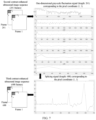
- FIG. 3 is a schematic flowchart of an image processing method based on contrast-enhanced ultrasound images according to an exemplary embodiment of the present application. As shown in FIG. 3 , the image processing method based on contrast-enhanced ultrasound images according to embodiments of the present application include the following steps.
- Step 10 performing a first decoupling operation on the first contrast-enhanced ultrasound image sequence to generate a second contrast-enhanced ultrasound image sequence corresponding to the first contrast-enhanced ultrasound image sequence, the first decoupling operation being used to improve spatial sparsity of microbubbles.
- the first decoupling operation is realized by highlighting a central region of the microbubbles along microbubble trajectories, and the first decoupling operation may suppress noise in a background region.
- the microbubble region refers to an image region corresponding to the microbubbles in the first contrast-enhanced ultrasound image.
- the background region refers to a background image region that does not include the microbubbles in the first contrast-enhanced ultrasound image.
- first decoupling operation to process the first contrast-enhanced ultrasound image sequence may not only effectively improve a signal-to-noise ratio of a contrast-enhanced ultrasound images, but also help to improve the spatial sparsity of the microbubbles (that is, a level of spatial sparsity), thereby providing a prerequisite for improving accuracy of microbubble localization process.
- the above-mentioned spatial sparsity refers to sparsity among a plurality of microbubbles included in same contrast-enhanced ultrasound images.
- a plurality of second contrast-enhanced ultrasound images included in the second contrast-enhanced ultrasound image sequence are obtained by performing the first decoupling operation on the plurality of first contrast-enhanced ultrasound images included in the first contrast-enhanced ultrasound image sequence one by one.
- j 1,2 , ⁇ N
- j 1,2 , ⁇ N .
- the embodiment of the present application does not limit a specific implementation manner of the second decoupling operation mentioned in Step 20, as long as the second decoupling operation can improve the temporal sparsity of the microbubbles, so as to provide a favorable condition for a subsequent microbubble locating operation and an ultrasound super-resolution reconstruction operation.
- the image processing method based on contrast-enhanced ultrasound images by performing the first decoupling operation and the second decoupling operation on the first contrast-enhanced ultrasound image sequence respectively, not only improves the signal-to-noise ratio of a contrast-enhanced ultrasound image sequence, but also improves spatial-temporal sparsity (including the temporal sparsity and the spatial sparsity) of the microbubbles, thereby breaking a limitation of low microbubble concentration in existing ultrasound localization microscopy methods, and further providing a prerequisite for improving accuracy of microbubble localization.
- the embodiments of the present application effectively solve problems of the high microbubble concentration and background noise in a clinical application, thereby providing favorable conditions for improving speed and accuracy of subsequent super-resolution image reconstruction.
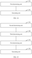
- the step of performing a second decoupling operation on the second contrast-enhanced ultrasound image sequence to generate a third contrast-enhanced ultrasound image sequence corresponding to the second contrast-enhanced ultrasound image sequence includes the following steps.
- the second contrast-enhanced ultrasound image sequence includes a plurality of second contrast-enhanced ultrasound images, and image sequence information based on time is included between the plurality of second contrast-enhanced ultrasound images. Image sizes of the plurality of second contrast-enhanced ultrasound images are the same.
- Step 22 generating the third contrast-enhanced ultrasound image sequence corresponding to the second contrast-enhanced ultrasound image sequence based on the one-dimensional grayscale fluctuation signal corresponding to each of the plurality of pixel coordinates.
- Step 222 performing, for each pixel coordinate in the plurality of pixel coordinates, a filtering operation on the first multi-scale decomposition signal set corresponding to the pixel coordinate to generate a second multi-scale decomposition signal set corresponding to the pixel coordinate, the filtering operation being used to remove a decomposition signal of a preset scale in the first multi-scale decomposition signal set.
- the image processing method By performing a multi-scale decomposition operation on the one-dimensional grayscale fluctuation signal corresponding to each of the plurality of pixel coordinates to generate a first multi-scale decomposition signal set corresponding to each of the plurality of pixel coordinates, then performing, for each pixel coordinate in the plurality of pixel coordinates, a filtering operation on the first multi-scale decomposition signal set corresponding to the pixel coordinate to generate a second multi-scale decomposition signal set corresponding to the pixel coordinate, then performing a splicing operation on the second multi-scale decomposition signal set corresponding to the pixel coordinate to generate a splicing signal corresponding to the pixel coordinate, and at last generating the third contrast-enhanced ultrasound image sequence corresponding to the second contrast-enhanced ultrasound image sequence based on the splicing signal corresponding to each of the plurality of pixel coordinates, the image processing method based on contrast-enhanced ultrasound images according to the embodiment of the present application achieves a purpose of
- a fourth step performing the operations of the first step to the third step for each pixel coordinate, thereby obtaining the splicing signal corresponding to each pixel coordinate.
- the length of the splicing signal corresponding to each pixel coordinate is 688.
- a third contrast-enhanced ultrasound image sequence including 688 images is generated based on the splicing signal corresponding to each pixel coordinate.
- the embodiments of the present application achieve the temporal sparsity of the microbubbles.
- the temporal sparsity together with the spatial sparsity mentioned above forms the spatial-temporal sparsity of the microbubbles.
- the microbubbles with spatial-temporal sparsity are helpful to improve the accuracy of microbubble localization.
- the image processing method before the step of determining a one-dimensional grayscale fluctuation signal corresponding to each of a plurality of pixel coordinates based on the plurality of pixel coordinates corresponding to the second contrast-enhanced ultrasound image sequence and a pixel value set corresponding to each of the plurality of pixel coordinates, the image processing method further includes the following steps.
- the space coordinate system is a three-dimensional space coordinate system including an X axis, a Y axis and a Z axis.
- the X axis and the Y axis are pixel coordinate axes, and the Z axis represents the image acquisition sequence or order.
- FIG. 10 is a schematic flowchart of determining distance information between a microbubble region in the contrast-enhanced ultrasound image and a background region corresponding to the microbubble region according to an exemplary embodiment of the present application.
- the microbubble region includes a plurality of pixel blocks, and determining distance information between a microbubble region in the contrast-enhanced ultrasound image and a background region corresponding to the microbubble region (Step 11), includes the following steps.
- Step 12 performing a pixel weighting operation on the contrast-enhanced ultrasound image based on the distance information corresponding to the contrast-enhanced ultrasound image to generate a weighted image corresponding to the contrast-enhanced ultrasound image.
- the calculation procedure of the step of generating the weighted image corresponding to the contrast-enhanced ultrasound image may be simplified, thereby improving a speed of image processing.
- a second determining unit 230 configured to determine a space coordinate system corresponding to the second contrast-enhanced ultrasound image sequence.
- the space coordinate system may represent image sequence information.
- the input apparatus 403 may also include, for example, a keyboard, a mouse, and so on.
- the output apparatus 404 may output various information such as the third contrast-enhanced ultrasound image sequence determined above to the outside.
- the output device 404 may include, for example, a display, a communication network and a remote output device connected to it, and so on.
- an embodiment of the present application may also be a computer-readable storage medium, where the computer-readable storage medium stores computer program instructions.
- the processor is enabled to perform the steps of the image processing method based on contrast-enhanced ultrasound images according to the embodiments of the present application described in the "Exemplary Methods" part of this specification.
Landscapes
- Health & Medical Sciences (AREA)
- Life Sciences & Earth Sciences (AREA)
- Engineering & Computer Science (AREA)
- Biomedical Technology (AREA)
- Medical Informatics (AREA)
- Veterinary Medicine (AREA)
- Biophysics (AREA)
- Nuclear Medicine, Radiotherapy & Molecular Imaging (AREA)
- Pathology (AREA)
- Radiology & Medical Imaging (AREA)
- Public Health (AREA)
- Heart & Thoracic Surgery (AREA)
- Physics & Mathematics (AREA)
- Molecular Biology (AREA)
- Surgery (AREA)
- Animal Behavior & Ethology (AREA)
- General Health & Medical Sciences (AREA)
- Hematology (AREA)
- Vascular Medicine (AREA)
- Computer Vision & Pattern Recognition (AREA)
- Physiology (AREA)
- Ultra Sonic Daignosis Equipment (AREA)
- Image Processing (AREA)
Claims (7)
- Ein Bildverarbeitungsverfahren auf der Basis kontrastverstärkter Ultraschallbilder, das auf eine erste Sequenz kontrastverstärkter Ultraschallbilder, die Mikroblasen beinhaltet, angewendet wird, beinhaltend:Durchführen (10) eines ersten Entkopplungsvorgangs an der ersten Sequenz kontrastverstärkter Ultraschallbilder, um eine zweite Sequenz kontrastverstärkter Ultraschallbilder, die mit der ersten Sequenz kontrastverstärkter Ultraschallbilder korrespondiert, zu erzeugen, um eine geringe räumliche Dichte der Mikroblasen zu verbessern; undDurchführen (20) eines zweiten Entkopplungsvorgangs an der zweiten Sequenz kontrastverstärkter Ultraschallbilder, um eine dritte Sequenz kontrastverstärkter Ultraschallbilder, die mit der zweiten Sequenz kontrastverstärkter Ultraschallbilder korrespondiert, zu erzeugen, um eine geringe zeitliche Dichte der Mikroblasen zu verbessern;wobei das Durchführen (10) des ersten Entkopplungsvorgangs Folgendes beinhaltet:Bestimmen (11), für jedes Bild in der ersten Sequenz kontrastverstärkter Ultraschallbilder, von Abstandsinformationen zwischen einem Mikroblasenbereich und einem mit dem Mikroblasenbereich korrespondierenden Hintergrundbereich;Durchführen (12) eines Pixelgewichtungsvorgangs an dem kontrastverstärkten Ultraschallbild auf der Basis der Abstandsinformationen, um ein gewichtetes Bild zu erzeugen; undErzeugen (13) der zweiten Sequenz kontrastverstärkter Ultraschallbilder jeweils auf der Basis der gewichteten Bilder; und wobeidas Durchführen (20) des zweiten Entkopplungsvorgangs Folgendes beinhaltet:Bestimmen (21) eindimensionaler Grauwertfluktuationssignale, die mit einer Vielzahl von Pixelkoordinaten und Pixelwertmengen korrespondieren, die mit der zweiten Sequenz kontrastverstärkter Ultraschallbilder korrespondieren,wobei das Bestimmen (21) der eindimensionalen Grauwertfluktuationssignale Folgendes beinhaltet: Durchführen eines Splicings, für jede Pixelkoordinate, der Pixelwertmenge in das eindimensionale Grauwertfluktuationssignal auf der Basis einer Bildsequenzreihenfolge, die mit der Zeitdimension korrespondiert; undErzeugen (22) der dritten Sequenz kontrastverstärkter Ultraschallbilder auf der Basis der eindimensionalen Grauwertfluktuationssignale;wobei das Bestimmen (11) der Abstandsinformationen ferner Folgendes beinhaltet:Durchführen einer Binarisierungsverarbeitung an dem kontrastverstärkten Ultraschallbild auf der Basis des Mikroblasenbereichs und des Hintergrundbereichs, um ein binarisiertes Bild zu erzeugen, wobei der Mikroblasenbereich eine Vielzahl von Pixelblöcken beinhaltet;Bestimmen (111), für jeden Pixelblock der Vielzahl von Pixelblöcken, eines kürzesten Abstands von dem Pixelblock zu dem Hintergrundbereich; undBestimmen (112), auf der Basis kürzester Abstände, die jeweils mit der Vielzahl von Pixelblöcken und dem binarisierten Bild korrespondieren, der Abstandsinformationen zwischen dem Mikroblasenbereich und dem Hintergrundbereich;wobei das Durchführen (12) des Pixelgewichtungsvorgangs ferner Folgendes beinhaltet:Bestimmen, auf der Basis jeder mit dem Mikroblasenbereich korrespondierenden Pixelkoordinate, eines Pixelgrauwerts und eines kürzesten Abstands, die mit der Pixelkoordinate korrespondieren;Multiplizieren des Pixelgrauwerts und des kürzesten Abstands, die mit der Pixelkoordinate korrespondieren, um einen gewichteten Wert, der mit der Pixelkoordinate korrespondiert, zu bestimmen; undBestimmen des gewichteten Bilds, das mit dem kontrastverstärkten Ultraschallbild korrespondiert, auf der Basis gewichteter Werte, die jeweils mit allen mit dem Mikroblasenbereich korrespondierenden Pixelkoordinaten korrespondieren.
- Bildverarbeitungsverfahren auf der Basis kontrastverstärkter Ultraschallbilder gemäß Anspruch 1, wobei das Erzeugen (22) der dritten Sequenz kontrastverstärkter Ultraschallbilder Folgendes beinhaltet:Durchführen (221) eines Multiskalenzerlegungsvorgangs durch Nutzen einer mehrstufigen Wavelet-Zerlegung an den eindimensionalen Grauwertfluktuationssignalen, um erste Multiskalenzerlegungssignalmengen, die jeweils mit der Vielzahl von Pixelkoordinaten korrespondieren, zu erzeugen;Durchführen (222), für jede Pixelkoordinate in der Vielzahl von Pixelkoordinaten, eines Filtervorgangs an der ersten Multiskalenzerlegungssignalmenge, um eine zweite Multiskalenzerlegungssignalmenge, die mit der Pixelkoordinate korrespondiert, zu erzeugen, wobei der Filtervorgang genutzt wird, um ein Zerlegungssignal einer vorgegebenen Skala in der ersten Multiskalenzerlegungssignalmenge zu entfernen;Durchführen (223) eines Splicing-Vorgangs an der zweiten Multiskalenzerlegungssignalmenge, um ein mit der Pixelkoordinate korrespondierendes Splicing-Signal zu erzeugen,wobei das Durchführen (223) des Splicing-Vorgangs an der zweiten Multiskalenzerlegungssignalmenge Folgendes beinhaltet:sequenzielles Durchführen, für die zweite Multiskalenzerlegungssignalmenge, des Splicing-Vorgangs an mit Zerlegungsskalen korrespondierenden Zerlegungssignalen in einer aufsteigenden Reihenfolge von Zerlegungsstufen oder -skalen, um das Splicing-Signal zu erzeugen; undErzeugen (224) der dritten Sequenz kontrastverstärkter Ultraschallbilder auf der Basis der Splicing-Signale.
- Bildverarbeitungsverfahren auf der Basis kontrastverstärkter Ultraschallbilder gemäß Anspruch 2, wobei das Zerlegungssignal der vorgegebenen Skala ein Zerlegungssignal mit einer kleinsten Skala in der ersten Multiskalenzerlegungssignalmenge beinhaltet.
- Bildverarbeitungsverfahren auf der Basis kontrastverstärkter Ultraschallbilder gemäß Anspruch 3, wobei das Zerlegungssignal der vorgegebenen Skala ferner ein Zerlegungssignal mit einer zweiten kleinsten Skala in der ersten Multiskalenzerlegungssignalmenge beinhaltet.
- Bildverarbeitungsverfahren auf der Basis kontrastverstärkter Ultraschallbilder gemäß einem der Ansprüche 1 bis 4, wobei vor dem Bestimmen (21) der eindimensionalen Grauwertfluktuationssignale:Bestimmen (23) eines mit der zweiten Sequenz kontrastverstärkter Ultraschallbilder korrespondierenden Raumkoordinatensystems, wobei das Raumkoordinatensystem in der Lage ist, Bildsequenzinformationen darzustellen; undBestimmen (24) der Vielzahl von Pixelkoordinaten und Pixelwertmengen auf der Basis der zweiten Sequenz kontrastverstärkter Ultraschallbilder und des Raumkoordinatensystems.
- Ein nichttransitorisches computerlesbares Speichermedium, das ein Computerprogramm zum Ausführen des Bildverarbeitungsverfahrens auf der Basis kontrastverstärkter Ultraschallbilder gemäß einem der Ansprüche 1 bis 5 speichert.
- Eine elektronische Vorrichtung (40), beinhaltend:einen Prozessor (401); undeinen Speicher (402) zum Speichern von durch den Prozessor ausführbaren Anweisungen;wobei der Prozessor konfiguriert ist, um das Bildverarbeitungsverfahren auf der Basis kontrastverstärkter Ultraschallbilder gemäß einem der Ansprüche 1 bis 5 auszuführen.
Applications Claiming Priority (2)
| Application Number | Priority Date | Filing Date | Title |
|---|---|---|---|
| CN202010419611.5A CN111588410B (zh) | 2020-05-18 | 2020-05-18 | 基于超声造影图像的图像处理方法及装置 |
| PCT/CN2021/093474 WO2021233189A1 (zh) | 2020-05-18 | 2021-05-13 | 基于超声造影图像的图像处理方法及装置 |
Publications (4)
| Publication Number | Publication Date |
|---|---|
| EP4059442A1 EP4059442A1 (de) | 2022-09-21 |
| EP4059442A4 EP4059442A4 (de) | 2023-02-08 |
| EP4059442B1 true EP4059442B1 (de) | 2024-07-10 |
| EP4059442B8 EP4059442B8 (de) | 2024-08-21 |
Family
ID=72180140
Family Applications (1)
| Application Number | Title | Priority Date | Filing Date |
|---|---|---|---|
| EP21808103.2A Active EP4059442B8 (de) | 2020-05-18 | 2021-05-13 | Bildverarbeitungsverfahren und -gerät basierend auf einem ultraschallkontrastbild |
Country Status (4)
| Country | Link |
|---|---|
| US (1) | US12390196B2 (de) |
| EP (1) | EP4059442B8 (de) |
| CN (1) | CN111588410B (de) |
| WO (1) | WO2021233189A1 (de) |
Families Citing this family (6)
| Publication number | Priority date | Publication date | Assignee | Title |
|---|---|---|---|---|
| CN111588409B (zh) * | 2020-05-18 | 2023-04-07 | 南京超维景生物科技有限公司 | 三维超声造影图像的超分辨重建方法及装置 |
| CN111588410B (zh) * | 2020-05-18 | 2023-05-16 | 南京超维景生物科技有限公司 | 基于超声造影图像的图像处理方法及装置 |
| CN113768547B (zh) * | 2021-09-14 | 2024-03-22 | 南京超维景生物科技有限公司 | 冠状动脉成像方法及装置、存储介质及电子设备 |
| CN118415679B (zh) * | 2023-01-31 | 2025-11-28 | 青岛海信医疗设备股份有限公司 | 心脏血流动力学参数的确定方法及电子设备 |
| WO2025165808A1 (en) * | 2024-01-31 | 2025-08-07 | The Board Of Trustees Of The University Of Illinois | Elevational resolution-enhanced 3d ultrasound localization imaging with linear array transducers |
| CN119423825B (zh) * | 2024-03-04 | 2025-11-07 | 北京大学 | 一种基于相位涨落的高清超声血流成像方法 |
Family Cites Families (7)
| Publication number | Priority date | Publication date | Assignee | Title |
|---|---|---|---|---|
| US20150196279A1 (en) * | 2011-10-18 | 2015-07-16 | Riverside Research Institute | Synthetic-focusing strategies for real-time annular-array imaging |
| CN103006272B (zh) * | 2013-01-09 | 2014-12-03 | 东南大学 | 基于超声交织编程的速度分布测量方法 |
| EP4349263B1 (de) * | 2017-05-31 | 2026-03-04 | Mayo Foundation for Medical Education and Research | Verfahren zur superauflösenden ultraschalldarstellung von mikrogefässen |
| WO2019095376A1 (zh) * | 2017-11-20 | 2019-05-23 | 深圳迈瑞生物医疗电子股份有限公司 | 超声造影成像方法及超声成像系统 |
| CN108324324A (zh) * | 2018-03-12 | 2018-07-27 | 西安交通大学 | 一种超声低频经颅容积超分辨率三维造影成像方法及系统 |
| CN110772285B (zh) * | 2019-10-31 | 2022-05-17 | 南京景瑞康分子医药科技有限公司 | 一种超声超分辨成像方法 |
| CN111588410B (zh) * | 2020-05-18 | 2023-05-16 | 南京超维景生物科技有限公司 | 基于超声造影图像的图像处理方法及装置 |
-
2020
- 2020-05-18 CN CN202010419611.5A patent/CN111588410B/zh active Active
-
2021
- 2021-05-13 WO PCT/CN2021/093474 patent/WO2021233189A1/zh not_active Ceased
- 2021-05-13 EP EP21808103.2A patent/EP4059442B8/de active Active
-
2022
- 2022-06-10 US US17/838,106 patent/US12390196B2/en active Active
Also Published As
| Publication number | Publication date |
|---|---|
| EP4059442B8 (de) | 2024-08-21 |
| EP4059442A1 (de) | 2022-09-21 |
| WO2021233189A1 (zh) | 2021-11-25 |
| EP4059442A4 (de) | 2023-02-08 |
| US12390196B2 (en) | 2025-08-19 |
| CN111588410A (zh) | 2020-08-28 |
| US20220296216A1 (en) | 2022-09-22 |
| CN111588410B (zh) | 2023-05-16 |
Similar Documents
| Publication | Publication Date | Title |
|---|---|---|
| EP4059442B1 (de) | Bildverarbeitungsverfahren und -gerät basierend auf einem ultraschallkontrastbild | |
| US11798150B2 (en) | Super-resolution reconstruction preprocessing method and super-resolution reconstruction method for contrast-enhanced ultrasound images | |
| JP6367261B2 (ja) | 知識ベース超音波画像強調 | |
| US11589840B2 (en) | Methods for super-resolution ultrasound imaging of microvessels | |
| US12229916B2 (en) | Super-resolution reconstruction method and apparatus for three-dimensional contrast-enhanced ultrasound images | |
| KR20140089669A (ko) | 영상에서 스펙클을 제거하는 방법, 장치 및 시스템. | |
| JP2021186430A (ja) | 画像処理装置及び画像処理方法。 | |
| Shahadi et al. | Eulerian video magnification: a review | |
| CN118121233B (zh) | 基于奇异值分解与Frangi滤波的超声微血管成像方法与系统 | |
| Shi et al. | Ultrasound image denoising autoencoder model based on lightweight attention mechanism | |
| Jahren et al. | Reverberation suppression in echocardiography using a causal convolutional neural network | |
| Qiang et al. | An adaptive spatiotemporal filter for ultrasound localization microscopy based on density canopy clustering | |
| CN109363722B (zh) | 彩色血流成像中运动伪像的抑制方法及设备 | |
| WO2020118296A1 (en) | Systems and methods for quantifying vessel features in ultrasound doppler images | |
| Park et al. | Ultrasound-guided biopsy tracking using data-driven needle identification in application to kidney | |
| Xia et al. | Enhanced SVD filter based on singular vector subspace denoising improves ultrafast ultrasound microvascular imaging performance | |
| Piepenbrock et al. | Tissue motion estimation of contrast enhanced ultrasound images with a stable principal component pursuit | |
| Koutras et al. | Audio-visual temporal saliency modeling validated by fmri data | |
| Khvostikov et al. | Influence of ultrasound despeckling on the liver fibrosis classification | |
| CN113768547B (zh) | 冠状动脉成像方法及装置、存储介质及电子设备 | |
| Binh et al. | Improving the quality of medical images in shearlet domain | |
| Ouzir et al. | A Proximal Algorithm for Joint Blood Flow Computation and Tissue Motion Compensation in Doppler Ultrafast Ultrasound Imaging | |
| HK40109430A (en) | Methods for super-resolution ultrasound imaging of microvessels | |
| Jin et al. | Detection of Substances in the Left Atrial Appendage by Spatiotemporal Motion Analysis Based on 4D-CT | |
| Bharti et al. | Cascaded Filter Analysis on Ultrasound Images of Fetus. |
Legal Events
| Date | Code | Title | Description |
|---|---|---|---|
| STAA | Information on the status of an ep patent application or granted ep patent |
Free format text: STATUS: THE INTERNATIONAL PUBLICATION HAS BEEN MADE |
|
| PUAI | Public reference made under article 153(3) epc to a published international application that has entered the european phase |
Free format text: ORIGINAL CODE: 0009012 |
|
| STAA | Information on the status of an ep patent application or granted ep patent |
Free format text: STATUS: REQUEST FOR EXAMINATION WAS MADE |
|
| 17P | Request for examination filed |
Effective date: 20220617 |
|
| AK | Designated contracting states |
Kind code of ref document: A1 Designated state(s): AL AT BE BG CH CY CZ DE DK EE ES FI FR GB GR HR HU IE IS IT LI LT LU LV MC MK MT NL NO PL PT RO RS SE SI SK SM TR |
|
| A4 | Supplementary search report drawn up and despatched |
Effective date: 20230111 |
|
| RIC1 | Information provided on ipc code assigned before grant |
Ipc: A61B 8/08 19850101AFI20230104BHEP |
|
| STAA | Information on the status of an ep patent application or granted ep patent |
Free format text: STATUS: EXAMINATION IS IN PROGRESS |
|
| DAV | Request for validation of the european patent (deleted) | ||
| DAX | Request for extension of the european patent (deleted) | ||
| 17Q | First examination report despatched |
Effective date: 20230814 |
|
| GRAP | Despatch of communication of intention to grant a patent |
Free format text: ORIGINAL CODE: EPIDOSNIGR1 |
|
| STAA | Information on the status of an ep patent application or granted ep patent |
Free format text: STATUS: GRANT OF PATENT IS INTENDED |
|
| INTG | Intention to grant announced |
Effective date: 20240130 |
|
| GRAS | Grant fee paid |
Free format text: ORIGINAL CODE: EPIDOSNIGR3 |
|
| GRAA | (expected) grant |
Free format text: ORIGINAL CODE: 0009210 |
|
| STAA | Information on the status of an ep patent application or granted ep patent |
Free format text: STATUS: THE PATENT HAS BEEN GRANTED |
|
| REG | Reference to a national code |
Ref country code: DE Ref legal event code: R081 Ref document number: 602021015610 Country of ref document: DE Owner name: NANJING LEAPSONICS TECHNOLOGY CO., LTD., NANJI, CN Free format text: FORMER OWNER: NANJING TRANSCEND VIVOSCOPE BIO-TECHNOLOGY CO., LTD, NANJING, JIANGSU, CN |
|
| AK | Designated contracting states |
Kind code of ref document: B1 Designated state(s): AL AT BE BG CH CY CZ DE DK EE ES FI FR GB GR HR HU IE IS IT LI LT LU LV MC MK MT NL NO PL PT RO RS SE SI SK SM TR |
|
| REG | Reference to a national code |
Ref country code: CH Ref legal event code: EP |
|
| RAP2 | Party data changed (patent owner data changed or rights of a patent transferred) |
Owner name: NANJING LEAPSONICS TECHNOLOGY CO., LTD. |
|
| REG | Reference to a national code |
Ref country code: CH Ref legal event code: PK Free format text: BERICHTIGUNG B8 |
|
| REG | Reference to a national code |
Ref country code: DE Ref legal event code: R096 Ref document number: 602021015610 Country of ref document: DE |
|
| REG | Reference to a national code |
Ref country code: LT Ref legal event code: MG9D |
|
| REG | Reference to a national code |
Ref country code: NL Ref legal event code: MP Effective date: 20240710 |
|
| PG25 | Lapsed in a contracting state [announced via postgrant information from national office to epo] |
Ref country code: PT Free format text: LAPSE BECAUSE OF FAILURE TO SUBMIT A TRANSLATION OF THE DESCRIPTION OR TO PAY THE FEE WITHIN THE PRESCRIBED TIME-LIMIT Effective date: 20241111 |
|
| REG | Reference to a national code |
Ref country code: AT Ref legal event code: MK05 Ref document number: 1701301 Country of ref document: AT Kind code of ref document: T Effective date: 20240710 |
|
| PG25 | Lapsed in a contracting state [announced via postgrant information from national office to epo] |
Ref country code: NL Free format text: LAPSE BECAUSE OF FAILURE TO SUBMIT A TRANSLATION OF THE DESCRIPTION OR TO PAY THE FEE WITHIN THE PRESCRIBED TIME-LIMIT Effective date: 20240710 |
|
| PG25 | Lapsed in a contracting state [announced via postgrant information from national office to epo] |
Ref country code: PT Free format text: LAPSE BECAUSE OF FAILURE TO SUBMIT A TRANSLATION OF THE DESCRIPTION OR TO PAY THE FEE WITHIN THE PRESCRIBED TIME-LIMIT Effective date: 20241111 Ref country code: NL Free format text: LAPSE BECAUSE OF FAILURE TO SUBMIT A TRANSLATION OF THE DESCRIPTION OR TO PAY THE FEE WITHIN THE PRESCRIBED TIME-LIMIT Effective date: 20240710 |
|
| PG25 | Lapsed in a contracting state [announced via postgrant information from national office to epo] |
Ref country code: NO Free format text: LAPSE BECAUSE OF FAILURE TO SUBMIT A TRANSLATION OF THE DESCRIPTION OR TO PAY THE FEE WITHIN THE PRESCRIBED TIME-LIMIT Effective date: 20241010 |
|
| PG25 | Lapsed in a contracting state [announced via postgrant information from national office to epo] |
Ref country code: GR Free format text: LAPSE BECAUSE OF FAILURE TO SUBMIT A TRANSLATION OF THE DESCRIPTION OR TO PAY THE FEE WITHIN THE PRESCRIBED TIME-LIMIT Effective date: 20241011 Ref country code: FI Free format text: LAPSE BECAUSE OF FAILURE TO SUBMIT A TRANSLATION OF THE DESCRIPTION OR TO PAY THE FEE WITHIN THE PRESCRIBED TIME-LIMIT Effective date: 20240710 Ref country code: PL Free format text: LAPSE BECAUSE OF FAILURE TO SUBMIT A TRANSLATION OF THE DESCRIPTION OR TO PAY THE FEE WITHIN THE PRESCRIBED TIME-LIMIT Effective date: 20240710 |
|
| PG25 | Lapsed in a contracting state [announced via postgrant information from national office to epo] |
Ref country code: BG Free format text: LAPSE BECAUSE OF FAILURE TO SUBMIT A TRANSLATION OF THE DESCRIPTION OR TO PAY THE FEE WITHIN THE PRESCRIBED TIME-LIMIT Effective date: 20240710 |
|
| PG25 | Lapsed in a contracting state [announced via postgrant information from national office to epo] |
Ref country code: LV Free format text: LAPSE BECAUSE OF FAILURE TO SUBMIT A TRANSLATION OF THE DESCRIPTION OR TO PAY THE FEE WITHIN THE PRESCRIBED TIME-LIMIT Effective date: 20240710 |
|
| PG25 | Lapsed in a contracting state [announced via postgrant information from national office to epo] |
Ref country code: AT Free format text: LAPSE BECAUSE OF FAILURE TO SUBMIT A TRANSLATION OF THE DESCRIPTION OR TO PAY THE FEE WITHIN THE PRESCRIBED TIME-LIMIT Effective date: 20240710 Ref country code: IS Free format text: LAPSE BECAUSE OF FAILURE TO SUBMIT A TRANSLATION OF THE DESCRIPTION OR TO PAY THE FEE WITHIN THE PRESCRIBED TIME-LIMIT Effective date: 20241110 |
|
| PG25 | Lapsed in a contracting state [announced via postgrant information from national office to epo] |
Ref country code: HR Free format text: LAPSE BECAUSE OF FAILURE TO SUBMIT A TRANSLATION OF THE DESCRIPTION OR TO PAY THE FEE WITHIN THE PRESCRIBED TIME-LIMIT Effective date: 20240710 |
|
| PG25 | Lapsed in a contracting state [announced via postgrant information from national office to epo] |
Ref country code: ES Free format text: LAPSE BECAUSE OF FAILURE TO SUBMIT A TRANSLATION OF THE DESCRIPTION OR TO PAY THE FEE WITHIN THE PRESCRIBED TIME-LIMIT Effective date: 20240710 Ref country code: RS Free format text: LAPSE BECAUSE OF FAILURE TO SUBMIT A TRANSLATION OF THE DESCRIPTION OR TO PAY THE FEE WITHIN THE PRESCRIBED TIME-LIMIT Effective date: 20241010 |
|
| PG25 | Lapsed in a contracting state [announced via postgrant information from national office to epo] |
Ref country code: RS Free format text: LAPSE BECAUSE OF FAILURE TO SUBMIT A TRANSLATION OF THE DESCRIPTION OR TO PAY THE FEE WITHIN THE PRESCRIBED TIME-LIMIT Effective date: 20241010 Ref country code: PL Free format text: LAPSE BECAUSE OF FAILURE TO SUBMIT A TRANSLATION OF THE DESCRIPTION OR TO PAY THE FEE WITHIN THE PRESCRIBED TIME-LIMIT Effective date: 20240710 Ref country code: NO Free format text: LAPSE BECAUSE OF FAILURE TO SUBMIT A TRANSLATION OF THE DESCRIPTION OR TO PAY THE FEE WITHIN THE PRESCRIBED TIME-LIMIT Effective date: 20241010 Ref country code: LV Free format text: LAPSE BECAUSE OF FAILURE TO SUBMIT A TRANSLATION OF THE DESCRIPTION OR TO PAY THE FEE WITHIN THE PRESCRIBED TIME-LIMIT Effective date: 20240710 Ref country code: IS Free format text: LAPSE BECAUSE OF FAILURE TO SUBMIT A TRANSLATION OF THE DESCRIPTION OR TO PAY THE FEE WITHIN THE PRESCRIBED TIME-LIMIT Effective date: 20241110 Ref country code: HR Free format text: LAPSE BECAUSE OF FAILURE TO SUBMIT A TRANSLATION OF THE DESCRIPTION OR TO PAY THE FEE WITHIN THE PRESCRIBED TIME-LIMIT Effective date: 20240710 Ref country code: GR Free format text: LAPSE BECAUSE OF FAILURE TO SUBMIT A TRANSLATION OF THE DESCRIPTION OR TO PAY THE FEE WITHIN THE PRESCRIBED TIME-LIMIT Effective date: 20241011 Ref country code: FI Free format text: LAPSE BECAUSE OF FAILURE TO SUBMIT A TRANSLATION OF THE DESCRIPTION OR TO PAY THE FEE WITHIN THE PRESCRIBED TIME-LIMIT Effective date: 20240710 Ref country code: ES Free format text: LAPSE BECAUSE OF FAILURE TO SUBMIT A TRANSLATION OF THE DESCRIPTION OR TO PAY THE FEE WITHIN THE PRESCRIBED TIME-LIMIT Effective date: 20240710 Ref country code: BG Free format text: LAPSE BECAUSE OF FAILURE TO SUBMIT A TRANSLATION OF THE DESCRIPTION OR TO PAY THE FEE WITHIN THE PRESCRIBED TIME-LIMIT Effective date: 20240710 Ref country code: AT Free format text: LAPSE BECAUSE OF FAILURE TO SUBMIT A TRANSLATION OF THE DESCRIPTION OR TO PAY THE FEE WITHIN THE PRESCRIBED TIME-LIMIT Effective date: 20240710 |
|
| REG | Reference to a national code |
Ref country code: DE Ref legal event code: R097 Ref document number: 602021015610 Country of ref document: DE |
|
| PG25 | Lapsed in a contracting state [announced via postgrant information from national office to epo] |
Ref country code: SM Free format text: LAPSE BECAUSE OF FAILURE TO SUBMIT A TRANSLATION OF THE DESCRIPTION OR TO PAY THE FEE WITHIN THE PRESCRIBED TIME-LIMIT Effective date: 20240710 Ref country code: DK Free format text: LAPSE BECAUSE OF FAILURE TO SUBMIT A TRANSLATION OF THE DESCRIPTION OR TO PAY THE FEE WITHIN THE PRESCRIBED TIME-LIMIT Effective date: 20240710 Ref country code: RO Free format text: LAPSE BECAUSE OF FAILURE TO SUBMIT A TRANSLATION OF THE DESCRIPTION OR TO PAY THE FEE WITHIN THE PRESCRIBED TIME-LIMIT Effective date: 20240710 |
|
| PG25 | Lapsed in a contracting state [announced via postgrant information from national office to epo] |
Ref country code: CZ Free format text: LAPSE BECAUSE OF FAILURE TO SUBMIT A TRANSLATION OF THE DESCRIPTION OR TO PAY THE FEE WITHIN THE PRESCRIBED TIME-LIMIT Effective date: 20240710 |
|
| PG25 | Lapsed in a contracting state [announced via postgrant information from national office to epo] |
Ref country code: IT Free format text: LAPSE BECAUSE OF FAILURE TO SUBMIT A TRANSLATION OF THE DESCRIPTION OR TO PAY THE FEE WITHIN THE PRESCRIBED TIME-LIMIT Effective date: 20240710 Ref country code: SK Free format text: LAPSE BECAUSE OF FAILURE TO SUBMIT A TRANSLATION OF THE DESCRIPTION OR TO PAY THE FEE WITHIN THE PRESCRIBED TIME-LIMIT Effective date: 20240710 |
|
| PLBE | No opposition filed within time limit |
Free format text: ORIGINAL CODE: 0009261 |
|
| STAA | Information on the status of an ep patent application or granted ep patent |
Free format text: STATUS: NO OPPOSITION FILED WITHIN TIME LIMIT |
|
| 26N | No opposition filed |
Effective date: 20250411 |
|
| PGFP | Annual fee paid to national office [announced via postgrant information from national office to epo] |
Ref country code: DE Payment date: 20250513 Year of fee payment: 5 |
|
| PGFP | Annual fee paid to national office [announced via postgrant information from national office to epo] |
Ref country code: GB Payment date: 20250521 Year of fee payment: 5 |
|
| PGFP | Annual fee paid to national office [announced via postgrant information from national office to epo] |
Ref country code: FR Payment date: 20250530 Year of fee payment: 5 |
|
| PG25 | Lapsed in a contracting state [announced via postgrant information from national office to epo] |
Ref country code: SE Free format text: LAPSE BECAUSE OF FAILURE TO SUBMIT A TRANSLATION OF THE DESCRIPTION OR TO PAY THE FEE WITHIN THE PRESCRIBED TIME-LIMIT Effective date: 20240710 |
|
| REG | Reference to a national code |
Ref country code: CH Ref legal event code: H13 Free format text: ST27 STATUS EVENT CODE: U-0-0-H10-H13 (AS PROVIDED BY THE NATIONAL OFFICE) Effective date: 20251223 |
|
| PG25 | Lapsed in a contracting state [announced via postgrant information from national office to epo] |
Ref country code: LU Free format text: LAPSE BECAUSE OF NON-PAYMENT OF DUE FEES Effective date: 20250513 |
|
| PG25 | Lapsed in a contracting state [announced via postgrant information from national office to epo] |
Ref country code: CH Free format text: LAPSE BECAUSE OF NON-PAYMENT OF DUE FEES Effective date: 20250531 |
|
| REG | Reference to a national code |
Ref country code: BE Ref legal event code: MM Effective date: 20250531 |
|
| PG25 | Lapsed in a contracting state [announced via postgrant information from national office to epo] |
Ref country code: MC Free format text: LAPSE BECAUSE OF FAILURE TO SUBMIT A TRANSLATION OF THE DESCRIPTION OR TO PAY THE FEE WITHIN THE PRESCRIBED TIME-LIMIT Effective date: 20240710 |
|
| PG25 | Lapsed in a contracting state [announced via postgrant information from national office to epo] |
Ref country code: IE Free format text: LAPSE BECAUSE OF NON-PAYMENT OF DUE FEES Effective date: 20250513 |
|
| PG25 | Lapsed in a contracting state [announced via postgrant information from national office to epo] |
Ref country code: BE Free format text: LAPSE BECAUSE OF NON-PAYMENT OF DUE FEES Effective date: 20250531 |