EP4010498B1 - Verfahren zur diagnose von brustkrebs - Google Patents
Verfahren zur diagnose von brustkrebs Download PDFInfo
- Publication number
- EP4010498B1 EP4010498B1 EP20750680.9A EP20750680A EP4010498B1 EP 4010498 B1 EP4010498 B1 EP 4010498B1 EP 20750680 A EP20750680 A EP 20750680A EP 4010498 B1 EP4010498 B1 EP 4010498B1
- Authority
- EP
- European Patent Office
- Prior art keywords
- mir
- breast cancer
- membrane
- sample
- urine
- Prior art date
- Legal status (The legal status is an assumption and is not a legal conclusion. Google has not performed a legal analysis and makes no representation as to the accuracy of the status listed.)
- Active
Links
Images


Classifications
-
- C—CHEMISTRY; METALLURGY
- C12—BIOCHEMISTRY; BEER; SPIRITS; WINE; VINEGAR; MICROBIOLOGY; ENZYMOLOGY; MUTATION OR GENETIC ENGINEERING
- C12Q—MEASURING OR TESTING PROCESSES INVOLVING ENZYMES, NUCLEIC ACIDS OR MICROORGANISMS; COMPOSITIONS OR TEST PAPERS THEREFOR; PROCESSES OF PREPARING SUCH COMPOSITIONS; CONDITION-RESPONSIVE CONTROL IN MICROBIOLOGICAL OR ENZYMOLOGICAL PROCESSES
- C12Q1/00—Measuring or testing processes involving enzymes, nucleic acids or microorganisms; Compositions therefor; Processes of preparing such compositions
- C12Q1/68—Measuring or testing processes involving enzymes, nucleic acids or microorganisms; Compositions therefor; Processes of preparing such compositions involving nucleic acids
- C12Q1/6876—Nucleic acid products used in the analysis of nucleic acids, e.g. primers or probes
- C12Q1/6883—Nucleic acid products used in the analysis of nucleic acids, e.g. primers or probes for diseases caused by alterations of genetic material
- C12Q1/6886—Nucleic acid products used in the analysis of nucleic acids, e.g. primers or probes for diseases caused by alterations of genetic material for cancer
-
- C—CHEMISTRY; METALLURGY
- C12—BIOCHEMISTRY; BEER; SPIRITS; WINE; VINEGAR; MICROBIOLOGY; ENZYMOLOGY; MUTATION OR GENETIC ENGINEERING
- C12N—MICROORGANISMS OR ENZYMES; COMPOSITIONS THEREOF; PROPAGATING, PRESERVING, OR MAINTAINING MICROORGANISMS; MUTATION OR GENETIC ENGINEERING; CULTURE MEDIA
- C12N15/00—Mutation or genetic engineering; DNA or RNA concerning genetic engineering, vectors, e.g. plasmids, or their isolation, preparation or purification; Use of hosts therefor
- C12N15/09—Recombinant DNA-technology
- C12N15/10—Processes for the isolation, preparation or purification of DNA or RNA
- C12N15/1003—Extracting or separating nucleic acids from biological samples, e.g. pure separation or isolation methods; Conditions, buffers or apparatuses therefor
- C12N15/1017—Extracting or separating nucleic acids from biological samples, e.g. pure separation or isolation methods; Conditions, buffers or apparatuses therefor by filtration, e.g. using filters, frits, membranes
-
- C—CHEMISTRY; METALLURGY
- C12—BIOCHEMISTRY; BEER; SPIRITS; WINE; VINEGAR; MICROBIOLOGY; ENZYMOLOGY; MUTATION OR GENETIC ENGINEERING
- C12Q—MEASURING OR TESTING PROCESSES INVOLVING ENZYMES, NUCLEIC ACIDS OR MICROORGANISMS; COMPOSITIONS OR TEST PAPERS THEREFOR; PROCESSES OF PREPARING SUCH COMPOSITIONS; CONDITION-RESPONSIVE CONTROL IN MICROBIOLOGICAL OR ENZYMOLOGICAL PROCESSES
- C12Q1/00—Measuring or testing processes involving enzymes, nucleic acids or microorganisms; Compositions therefor; Processes of preparing such compositions
- C12Q1/68—Measuring or testing processes involving enzymes, nucleic acids or microorganisms; Compositions therefor; Processes of preparing such compositions involving nucleic acids
- C12Q1/6806—Preparing nucleic acids for analysis, e.g. for polymerase chain reaction [PCR] assay
-
- G—PHYSICS
- G01—MEASURING; TESTING
- G01N—INVESTIGATING OR ANALYSING MATERIALS BY DETERMINING THEIR CHEMICAL OR PHYSICAL PROPERTIES
- G01N33/00—Investigating or analysing materials by specific methods not covered by groups G01N1/00 - G01N31/00
- G01N33/48—Biological material, e.g. blood, urine; Haemocytometers
- G01N33/50—Chemical analysis of biological material, e.g. blood, urine; Testing involving biospecific ligand binding methods; Immunological testing
- G01N33/5005—Chemical analysis of biological material, e.g. blood, urine; Testing involving biospecific ligand binding methods; Immunological testing involving human or animal cells
- G01N33/5008—Chemical analysis of biological material, e.g. blood, urine; Testing involving biospecific ligand binding methods; Immunological testing involving human or animal cells for testing or evaluating the effect of chemical or biological compounds, e.g. drugs, cosmetics
- G01N33/5076—Chemical analysis of biological material, e.g. blood, urine; Testing involving biospecific ligand binding methods; Immunological testing involving human or animal cells for testing or evaluating the effect of chemical or biological compounds, e.g. drugs, cosmetics involving cell organelles, e.g. Golgi complex, endoplasmic reticulum
-
- G—PHYSICS
- G01—MEASURING; TESTING
- G01N—INVESTIGATING OR ANALYSING MATERIALS BY DETERMINING THEIR CHEMICAL OR PHYSICAL PROPERTIES
- G01N33/00—Investigating or analysing materials by specific methods not covered by groups G01N1/00 - G01N31/00
- G01N33/48—Biological material, e.g. blood, urine; Haemocytometers
- G01N33/50—Chemical analysis of biological material, e.g. blood, urine; Testing involving biospecific ligand binding methods; Immunological testing
- G01N33/53—Immunoassay; Biospecific binding assay; Materials therefor
- G01N33/574—Immunoassay; Biospecific binding assay; Materials therefor for cancer
- G01N33/57407—Specifically defined cancers
- G01N33/57415—Specifically defined cancers of breast
-
- C—CHEMISTRY; METALLURGY
- C12—BIOCHEMISTRY; BEER; SPIRITS; WINE; VINEGAR; MICROBIOLOGY; ENZYMOLOGY; MUTATION OR GENETIC ENGINEERING
- C12Q—MEASURING OR TESTING PROCESSES INVOLVING ENZYMES, NUCLEIC ACIDS OR MICROORGANISMS; COMPOSITIONS OR TEST PAPERS THEREFOR; PROCESSES OF PREPARING SUCH COMPOSITIONS; CONDITION-RESPONSIVE CONTROL IN MICROBIOLOGICAL OR ENZYMOLOGICAL PROCESSES
- C12Q2600/00—Oligonucleotides characterized by their use
- C12Q2600/178—Oligonucleotides characterized by their use miRNA, siRNA or ncRNA
Definitions
- BC breast cancer
- BC-associated mortality rates could be reduced in general due to improved screening and treatment options, heterogeneous quality pattern in different countries sustain a crucial weak point in BC management [2, 7].
- Routine BC detection methods including palpation, mammography and ultrasound are characterized by various limitations, e.g. moderate sensitivity and specificity rates especially at denser mammary tissue, decreased patient compliance [8, 9].
- moderate sensitivity and specificity rates especially at denser mammary tissue, decreased patient compliance [8, 9].
- Liquid biopsy-based disease biomarkers offer a range of promising prospects as important prognostic, diagnostic and theranostic tools [10].
- Various biomarker types are available in body liquids via minimal- or non-invasive sampling procedures [10].
- circulating microRNA (miRNA / miR) molecules qualify as robust and reliable matrices in regard to detectability and specificity in disease diagnosis and monitoring [10, 11].
- urinary miRs were demonstrated [12].
- the applicability of urinary miRs with diagnostic features was proven also in other tumor types [10, 13-15].
- expression analysis of these small nucleic acids provides distinct characteristic pattern to distinguish cancer patients from healthy controls [12, 14, 16].
- let-7-family members of miRNAs could be linked to breast carcinogenesis [17-22].
- Distinct let-7 miRNAs bear the potential to be employed as prospective molecular markers in BC diagnostics and as therapeutic targets [22-25].
- miR-423 The functional background of miR-423 and the crucial impact of altered SNPs (single nucleotide polymorphisms) in the miR-423 gene on BC biology became apparent in recent investigations [50, 51]. Another functional aspect is provided by the combined expression levels of miR-423 and miR-4417, which could differentiate 70.1 % of hereditary and non-hereditary BCs [52].
- a serum-based BC biomarker study could identify a 3-miR signature comprising the expression levels of miR-424, miR-29c, and miR-199a with the highest diagnostic accuracy for distinguishing breast cancer patients from healthy controls [55]. Oncogenic effects could be linked to miR-660 expression, with triggering functions in BC proliferation, migration and anti-apoptotic activities [56].
- a BC tissue-derived next generation sequencing analysis extracted miR-660 as one promising biomarker candidate with significant prognostic features in overall survival (OS) and recurrence-free survival (RFS) in BC patients [57].
- EP 3070178 A1 describes studies on urine samples of breast cancer patients. It was found that the urinary miRNAs miR-21, miR-125b, miR-375 and miR-451 displayed significantly decreased expression levels in breast cancer patients compared to healthy controls. In addition, the urinary miRNA miR-155 showed an increased expression level in breast cancer patients. EP 3070178 A1 suggests that in particular determination of the expression level of the four miRNAs miR-21, miR-125b, miR-155 and miR-451 in urine samples allows for a discrimination between healthy individuals and BC patients.
- WO 2019/132688 A1 discloses a method for isolating extracellular vesicles from a sample of a biological fluid of a subject, comprising passing the (pre-filtrated) sample through a (second) membrane filter that does not bind biological polymers and has average pore sizes in the range of 100-150 nm. The retentate material comprising exosomes is collected and lysed for further analysis.
- the inventors investigated urine samples of breast cancer patients and surprisingly found that certain miRNA types and combinations thereof are useful in diagnosing breast cancer.
- a panel of miRNA types comprising at least miR-424, miR-660, and let7-i, was found to be a highly specific combinatory biomarker tool in discrimination of breast cancer patients vs. healthy controls based on urine specimen.
- the inventors further developed a highly efficient method for isolating exosomes from urine and other body fluids, which is particularly useful in the method for diagnosing BC described herein.
- the present invention encompasses methods of diagnosing whether a subject has, or is at risk for developing, breast cancer, comprising determining the level of at least one miR gene product in a urine sample from the subject and comparing the level of the miR gene product in the urine sample to the level of the respective miR gene product in a control sample.
- a "miR gene product,” “microRNA,” “miR,” or “miRNA” refers to the unprocessed (e.g., precursor) or processed (e.g., mature) RNA transcript from a miR gene. As the miR gene products are not translated into protein, the term “miR gene products” does not include proteins.
- the unprocessed miR gene transcript is also called a “miR precursor” or “miR prec” and typically comprises an RNA transcript of about 70-100 nucleotides in length.
- the miR precursor can be processed by digestion with an RNAse (for example, Dicer, Argonaut, or RNAse III (e.g., E.
- This active 19-25 nucleotide RNA molecule is also called the "processed" miR gene transcript or "mature" miRNA.
- the active 19-25 nucleotide RNA molecule can be obtained from the miR precursor through natural processing routes (e.g., using intact cells or cell lysates) or by synthetic processing routes (e.g., using isolated processing enzymes, such as isolated Dicer, Argonaut, or RNAse III). It is understood that the active 19-25 nucleotide RNA molecule can also be produced directly by biological or chemical synthesis, without having been processed from the miR precursor.
- microRNA When a microRNA is referred to herein by name, the name corresponds to both the precursor and mature forms, unless otherwise indicated.
- Table 1 depicts the nucleotide sequences of preferred mature human microRNAs used in the present invention.
- Table 1 Name of microRNA Mature sequence SEQ ID NO: of mature sequence miR-423 UGAGGGGCAGAGAGCGAGACUUU 10 miR-424 CAGCAGCAAUUCAUGUUUUGAA 11 miR-660 UACCCAUUGCAUAUCGGAGUUG 12 let7-i UGAGGUAGUAGUUUGUGCUGUU 17 miR-125b UCCCUGAGACCCUAACUUGUGA 7 let7-d AGAGGUAGUAGGUUGCAUAGUU 14 miR-17 CAAAGUGCUUACAGUGCAGGUAG 4 let7-f UGAGGUAGUAGAUUGUAUAGUU 16 miR194 UGUAACAGCAACUCCAUGUGGA 8 miR222 AGCUACAUCUGGCUACUGGGU 9
- a "subject" can be any mammal that has, or is suspected of having, breast cancer.
- the subject is a human who has, or is suspected of having, breast cancer.
- the subject is a female human, e.g. a woman at an age of 30 to 70 years, 35 to 69 years, 40 to 67 years, or 45 to 65 years.
- urine sample refers to a sample which comprises or consists of urine, or which is derived from urine.
- native urine is typically processed prior to analysis. The processing may include (but is not limited to) RNA isolation and/or purification, centrifugation, nucleic acid precipitation, dissolution of precipitated nucleic acid, concentration, dilution, filtration and combinations thereof.
- Such processed samples are encompassed by the term "urine sample”.
- the urine sample to be analysed is obtainable or obtained by enriching or isolating extracellular vesicles from urine.
- the urine sample is obtainable or obtained by enriching or isolating exosomes from urine.
- the urine sample to be analysed is obtainable or obtained by a method for isolating exosomes described hereinbelow.
- extracellular vesicles refers to membrane-contained vesicles released by cells.
- EVs can be broadly classified into 3 main classes: (a) Microvesicles (or microparticles or ectosomes) that are produced by outward budding and fission of the plasma membrane; (b) Exosomes that are formed within the endosomal network and released upon fusion of multi-vesicular bodies with the plasma membrane; and (c) Apoptotic bodies which are released as blebs of cells undergoing apoptosis.
- the level of at least one miR gene product is determined in a urine sample obtained from the subject.
- a corresponding control sample which is also a urine sample, can be obtained from a healthy human individual or population of healthy individuals.
- the healthy "control" individual preferably has the same gender as the subject, and optionally a similar age (i.e. +/- 10 years the age of the subject). Most preferably, the "control" individual is a healthy woman at an age of 30 to 70 years, 35 to 69 years, 40 to 67 years, or 45 to 65 years.
- the control urine sample is then processed along with the urine sample from the subject, so that the levels of miR gene product in the subject's urine sample can be compared to the respective miR gene product levels in the control sample.
- a reference miR expression standard for the urine sample can also be used as a control.
- an alteration e.g., an increase or decrease
- the level of the at least one miR gene product in the test sample is greater than the level of the respective miR gene product in the control sample (i.e., expression of the miR gene product is "up-regulated”).
- expression of a miR gene product is "up-regulated” when the amount of miR gene product in a urine sample from a subject is greater than the amount of the same gene product in a control sample.
- the level of the at least one miR gene product in the urine sample is less than the level of the respective miR gene product in the control sample (i.e., expression of the miR gene product is "down-regulated”).
- expression of a miR gene is “down-regulated” when the amount of miR gene product produced from that gene in a urine sample from a subject is less than the amount produced from the same gene in a control sample.
- the relative miR gene expression in the control and normal samples can be determined with respect to one or more RNA expression standards.
- the standards can comprise, for example, the urinary miR gene expression level in healthy subject, or the average level of urinary miR gene expression previously obtained for a population of healthy human controls. "Healthy" means that the subject or human controls do not have breast cancer or another type of cancer.
- the method comprises determining the level of a miR-424 gene product, a miR-660 gene product and a let7-i gene product, wherein a decrease in the levels of the let7-i and miR-660 gene products in the urine sample, relative to the respective levels of the respective miR gene products in a control sample, and an increase in the level of the miR-424 gene product in the urine sample, relative to the level of the miR-424 gene product in the control sample, is indicative of the subject having breast cancer.
- the level of a miR gene product in a urine sample can be measured using any technique that is suitable for detecting RNA expression levels in a urine sample.
- Nucleic acids can used be as probes or primers for embodiments involving nucleic acid hybridization. As such, they may be used to assess miRNA expression.
- the nucleotide sequences of the invention may be used for their ability to selectively form duplex molecules with complementary stretches of DNAs and/or RNAs or to provide primers for amplification of DNA or RNA from samples.
- RNA extraction complementary DNA synthesis (reverse transcription) and PCR amplification is monitored by addition of "spike-in" control RNA specimen of defined concentration to the RNA lysis buffer as recommended by Marabita [4].
- the detailed procedure is decribed herein in section 1.6 of Example 1.
- each miR gene product is normalized by converting it into normalized relative quantities (NRQ) for the respective miR gene product.
- NRQ normalized relative quantities
- the NRQ is used as the level of miR gene product.
- RNA to cDNA can be used to determine the relative concentrations of specific miRNA species. By determining that the concentration of a specific mRNA species varies, it is shown that the gene encoding the specific mRNA species is differentially expressed.
- Another method for amplification is ligase chain reaction ("LCR").
- LCR ligase chain reaction
- U.S. Pat. No. 4,883,750 describes a method similar to LCR for binding probe pairs to a target sequence.
- a method based on PCR TM and oligonucleotide ligase assay (OLA), disclosed in U.S. Pat. No. 5,912,148 may also be used.
- an oligolibrary in microchip format (i.e., a microarray), may be constructed containing a set of oligonucleotide probes that are specific for a set of miR genes.
- a microarray the expression level of multiple microRNAs in a urine sample can be determined by reverse transcribing the RNAs to generate a set of target oligodeoxynucleotides, and hybridizing them to probe the oligonucleotides on the microarray to generate a hybridization, or expression, profile.
- the hybridization profile of the test sample can then be compared to that of a control sample to determine which microRNAs have an altered expression level in breast cancer cells.
- probe oligonucleotide or “probe oligodeoxynucleotide” refers to an oligonucleotide that is capable of hybridizing to a target oligonucleotide.
- Target oligonucleotide or “target oligodeoxynucleotide” refers to a molecule to be detected (e.g., via hybridization).
- miR-specific probe oligonucleotide or "probe oligonucleotide specific for a miR” is meant a probe oligonucleotide that has a sequence selected to hybridize to a specific miR gene product, or to a reverse transcript of the specific miR gene product.
- the method of the invention may further comprise the step of comparing the level of the miR gene product in the urine sample to a control level of the miR gene product (e.g. the level in a control sample), and diagnosing whether the subject has breast cancer, wherein a decrease or increase of miR gene product in the urine sample, relative to the control level of miR gene product is indicative of the subject having breast cancer.
- a control level of the miR gene product e.g. the level in a control sample
- Breast cancer is a cancer that starts in the breast, usually in the inner lining of the milk ducts or lobules. There are different types of breast cancer, with different stages (spread), aggressiveness, and genetic makeup. Breast cancer subtypes may be categorized on an immunohistochemical basis.
- the breast cancer to be diagnosed, detected, monitored or screened in accordance with this invention may be invasive ductal carcinoma, ductal carcinoma in situ, or invasive lobular carcinoma. Alternatively, BC may be classified on the basis of receptor status.
- the breast cancer to be diagnosed, detected, monitored or screened in accordance with this invention may therefore be:
- the level of a miR gene product in the urine sample, relative to the level of the respective miR gene product in a control sample is typically increased by at least 10% or at least 25% or at least 50% or at least 100%, to be indicative of breast cancer.
- the level of a miR gene product in the urine sample, relative to the level of the respective miR gene product in a control sample is usually decreased by at least 10% or at least 25% or at least 50%, to be indicative of breast cancer.
- the level of miR-660 gene product, and/or let7-i gene product in the urine sample from the subject, relative to the control level of the corresponding miR gene product may be decreased by at least 25% to be indicative of breast cancer, and/or the level of miR-424 gene product, in the urine sample from the subject, relative to the control level of miR-424 gene product, may be increased by at least 50% to be indicative of breast cancer.
- the level of miR-660 gene product, and/or let7-i gene product in the urine sample from the subject, relative to the control level of the corrsponding miR gene product may be decreased by at least 50% to be indicative of breast cancer, and/or the level of miR-424 gene product, in the urine sample from the subject, relative to the control level of miR-424 gene product, may be increased by at least 100% to be indicative of breast cancer.
- the level of miR-660 gene product, and/or let7-i gene product in the urine sample from the subject, relative to the control level of the corrsponding miR gene product may be decreased by at least 75% to be indicative of breast cancer
- the level of miR-424 gene product, in the urine sample from the subject, relative to the control level of miR-424 gene product may be increased by at least 200% to be indicative of breast cancer.
- the subject may be diagnosed as having breast cancer if
- the subject is diagnosed as having breast cancer if
- the present invention is a method for enriching or isolating extracellular vesicles, in particular exosomes, from a urine sample.
- the method comprises filtrating the body fluid sample through a filter membrane which has protein binding properties.
- the filter membrane is preferably a polyamide membrane, e.g. a Nylon membrane.
- the pore size is typically from about 0.1 ⁇ m to about 1 ⁇ m, preferably from about 0.15 ⁇ m to about 500 ⁇ m, more preferably from about 0.18 ⁇ m to about 0.25 ⁇ m, e.g. about 0.22 ⁇ m.
- the method further comprises, prior to the filtration through the protein-binding membrane, a pre-filtration through a membrane which does not have protein-binding properties.
- the pre-filtration may serve to separate larger EVs, such as microvesicles, from exosomes which have a smaller size.
- the pore size of the pre-filtration membrane is typically from about 0.18 ⁇ m to about 0.25 ⁇ m, preferably from about 0.19 ⁇ m to about 0.21 ⁇ m, e.g. about 0.2 ⁇ m.
- the pre-filtration membrane may be made of a cellulosic material such as cellulose acetate.
- the body fluid sample may be centrifuged to remove cell debris.
- the method of the invention further comprises recovering the exosomes from the membrane. This is typically achieved by contacting the membrane with a suitable solution such as a lysis buffer.
- a suitable solution such as a lysis buffer.
- Suitable buffers may include a buffer substance to maintain the pH of the lysis buffer in a range from about 6.5 to 8, or 7 to 7.5, e.g. about 7.2.
- the lysis buffer may contain a surfactant or detergent. Suitable surfactancts include sodium lauryl sulfate and Triton X100.
- the method for enriching or isolating exosomes or extracellular vesicles can be used in the method of diagnosing cancer described herein, e.g. for providing the urine sample.
- the case-control cohort comprised of 69 untreated patients, newly diagnosed with primary BC in the adjuvant setting and 40 healthy female controls at the Department of Obstetrics and Gynecology, University Medical Center Freiburg. Control's health status was confirmed by medical examination performed by an experienced clinical physician including breast and regional lymph nodes to exclude potential BC and any history of other (malignant) disease or current inflammation. Furthermore healthy controls underwent breast and axillary ultrasound and mammography. By staging procedures according to the current national guidelines, all BC patients were tested to preclude cases of advanced stages with distant metastasis.
- Table 2 Cohort characteristics of breast cancer (BC) patients and healthy controls BC patients healthy controls p value N 69 40 median age, y 58 (30-79) 45 (20-72) 0. Histology invasive ductal 49 invasive lobular 5 Tumor stage pT1a 2 pT1b 5 pT1c 39 pT2 17 pT3 2 Nodal status pN0 53 pN1 11 pN2 1 Grading G1 10 G2 43 G3 8 Hormone receptor status ER positive 53 PR positive 50 HER2neu status positive 4
- Cryopreserved native urine specimen were thawed at room temperature and centrifuged (4000rpm; 15min; 4°C) to remove potential cell debris residues or other solid precipitates from pure urine supernatant.
- RNA isolation was performed utilizing the Norgen Total RNA Purification Kit (#17200, NORGEN Biotek Corp., Thorold, Canada). According to manufacturer's protocol [58] (Appendix B: 'Total RNA Purification from Plasma or Serum') 600 ⁇ l lysis buffer were extruded through filter membrane. Purified RNA was eluted in 35 ⁇ l elution buffer. Further processing of RNA specimen was defined to a standard volume of 2.5 ⁇ l.
- Reverse transcription (RT) of miRNAs was realized by utilizing 2.5 ⁇ l of RNA sample, 5 ⁇ l of 5x RT-Buffer, 0.1mM of ATP, 0.5 ⁇ M RT-primer, 0.1mM of each deoxynucleotide (dATP, dCTP, dGTP, dTTP), 25 units of Maxima Reverse Transcriptase (Thermo) and 1 unit of poly(A) polymerase (New England Biolabs GmbH, Frankfurt, Germany) in a final volume of 25 ⁇ l. Reaction incubation was set to 30min at 42°C followed by enzyme inactivation at 85°C for 10min.
- RT Primer design for RT reaction was geared to 'miRprimer' software tool by Busk [59].
- RT Primer information is listed in Table 3.
- cDNA samples were diluted in H2O to a final volume of 200 ⁇ l and stored at -20°C until further processing.
- Table 3 Information on primers utilized in expression analysis of miRNA types.
- PCR reaction set up: 1 ⁇ l cDNA, in-house qPCR buffer (containing TRIS pH8.1, dATP, dCTP, dGTP, dTTP, magnesium, potassium ammonium, SYBRGreen (Jena Bioscience, Jena, Germany), enhancers) 0.25U HotStart Taq Polymerase (Jena Bioscience) in a total volume of 10 ⁇ l.
- qPCR primers were designed via 'miRprimer' software [59] and are listed in Table 3.
- PCR program comprised of initial denaturation (95°C; 2min); 40 cycles: denaturation (95°C; 5sec) / annealing/extension (60°C; 30sec); melting curve.
- RNA lysis buffer As recommended by Marabita et al. [60].
- 5fM two exogenous synthetic RNA types (Caenorhabditis elegans: cel-miR-39 and Arabidopsis thaliana: ath-miR-159; Biomers.net GmbH, Ulm, Germany; sequences in Table 3) were added to RNA lysis buffer (Norgen).
- NF Normalization factor
- Biomarker assessment focused on the extraction of the diagnostic value of all thirteen analyzed miRNA types and combinations of them in regard to urine-based breast cancer detection. To this aim we used several statistical approaches, described as follows.
- the complete panel of all thirteen circulating miRNA specimen could be reliably detected in all urine specimen of the test cohort.
- Sample-dependent variations in expression levels of miRNAs due to variable urine parameters e.g. dilution, concentration of nucleic acids
- Sample-dependent variations in expression levels of miRNAs due to variable urine parameters e.g. dilution, concentration of nucleic acids
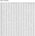
- miR-424 (94.81 %; up) 2. miR-660 (64.23%; down) 3. miR-423 (58.96%; down) 4. let7-i (58.96%; down) 5. let7-f (20.32%; down) 6. miR-17 (12.46%; up) 7. miR-222 (10.79%, up) 8. miR-194 (9.44%; up) 9. miR-125b (5.82%; up) 10. let7-d (3.57%; up)
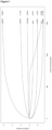
- miRNA types After 1500 boosting steps, seven miRNA types had been selected (including those obtained by the earlier approaches). These were miR-424 (up), miR-423 (down), miR-660 (down), let-7i (down), miR-194 (up), let7-a (down), miR-125b (up), see Figure 3 .
- the first model characterized by the lowest AIC (AIC 15.0) occurred only here, not in the previous approaches.
- the second model contains the four miRNA types (miR-424 + miR-423 + miR-660 + let7-i ) with the highest inclusion frequency in approaches (1) and (2). The second model occurred also in approach (3).
- a panel of four miRNA types comprising miR-424 , miR-423 , miR-660 , and let7-i could be elected as highly specific combinatory biomarker tool in discrimination of breast cancer patients vs. healthy controls based on urine specimen.
- AIC Akaike information criterion AUC area under the curve miRNA / miR micro ribonucleic acid NF normalization factor NGS next generation sequencing NRQ normalized relative quantities PET positron-emission tomography qPCR quantitative polymerase chain reaction ROC receiver operating characteristic SNP single nucleotide polymorphism
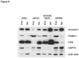
- Human body fluids including serum, urine, ascites, and amniotic liquor served as matrices to test the filter-based microvesicle isolation method.
- Liquid biopsy specimen were obtained according to the WMA declaration of Helsinki [ 30] .
- body fluid specimen were collected and stored in sterile sampling tubes at -20°C until further processing. Additional pre-treatment steps and detailed information are described for each sample matrix in the following.
- Serum Blood samples were collected by using S-Monovette Serum Gel (#04.1925, Sarstedt AG, Nuembrecht, Germany) and allowed to clot at room temperature for 20min followed by centrifugation at 2500g for 10min at 20°C to obtain pure serum. Serum samples were transferred to a fresh 15 ml poly propylene (pp) tube (#62.554.502; Sarstedt AG) and stored at -20°C until further processing.
- pp poly propylene
- Urine Patients provided urine specimen collected in a sterile 70ml screw-on cap urine sampling tube (#75.9922.745, Sarsted AG). Urine samples were transferred and aliquoted to 10ml Urine Monovette (#10.252; Sarsted AG) and stored at -20°C until further processing.
- Fresh frozen liquid biopsy samples were thawed at room temperature and centrifuged at 4000g for 15min to remove sediment and cellular debris. Following centrifugation, supernatant was transferred in a new 15ml pp tube (Sarstedt AG) for further processing.
- specimen matrices serum, ascites and amniotic liquor were diluted with sterile 1x Dulbecco's phosphate-buffered saline (DPBS, #14190185; ThermoFisher Scientific, Düsseldorf, Germany) in ratio 2:3 (2ml sample:3ml DPBS) to facilitate the filtration process.
- DPBS sterile 1x Dulbecco's phosphate-buffered saline
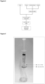
- Pre-treated sample liquids were transferred individually to a 20ml syringe (#4617207V; BRAUN, Melsungen, Germany) with a screwed on 0.2 ⁇ m cellulose acetate (CA) syringe filter (#514-0061; VWR International GmbH, Darmstadt, Germany) followed by a second screwed on 0.22 ⁇ m nylon syringe filter (#02542904; Perkin Elmer, Waltham, USA).
- CA cellulose acetate
- 0.22 ⁇ m nylon syringe filter #02542904; Perkin Elmer, Waltham, USA.
- the cascaded filters were washed with 5ml 1x DPBS (Thermo) using a fresh 5ml syringe (#4617053V; BRAUN, Melsungen, Germany). Washing guarantees the removal of sample residues falling below preset filter limitation that possibly interfere with downstream applications. Rinsed filters were then separated.
- the CA filter retained all particles >200nm, including microvesicles within the respective molecular size limit. Due to the non-protein binding characteristics of the upstream CA filter, all microvesicles ⁇ 200nm flow through and subsequently bound on the protein binding nylon membrane of the second filter.
- Elution of the two different filter-bound microvesicle fractions was carried out by extruding 500 ⁇ l protein lysis buffer (100mM HEPES pH 7.2, 0.1% SDS, 0.1% TRITON X100) through each of the used separate filters with a new 2ml syringe.
- 500 ⁇ l protein lysis buffer 100mM HEPES pH 7.2, 0.1% SDS, 0.1% TRITON X100
- Serum, ascites and amniotic liquor specimen were diluted with DPBS as fore cited and a final volume of 5ml was filtrated to harvest the two different size fractions of microvesicles.
- Protein concentrations of specimen isolates were quantified utilizing BCA method (#23227; ThermoFisher Scientific, Düsseldorf, Germany). Protein extracts obtained via the novel isolation method were analyzed by Western blot technique. 10 ⁇ g of total protein were separated by SDS-PAGE and transferred to an IMMOBILON-P membrane (#IPVH00010; Merck, Darmstadt, Germany) by using a Mini PROTEAN tetra cell (BioRad, Ober, Germany).
- ECL reaction was carried out according to Haan & Behrmann [ 31 ] .
- X-Ray films (Super RX-N, Fujifilm Europe, Düsseldorf, Germany) were processed utilizing the AGFA CP1000 X-Ray film processor.
Landscapes
- Health & Medical Sciences (AREA)
- Chemical & Material Sciences (AREA)
- Life Sciences & Earth Sciences (AREA)
- Engineering & Computer Science (AREA)
- Immunology (AREA)
- Organic Chemistry (AREA)
- Biomedical Technology (AREA)
- Molecular Biology (AREA)
- Analytical Chemistry (AREA)
- Biotechnology (AREA)
- Wood Science & Technology (AREA)
- Genetics & Genomics (AREA)
- Zoology (AREA)
- Biochemistry (AREA)
- Proteomics, Peptides & Aminoacids (AREA)
- General Health & Medical Sciences (AREA)
- Physics & Mathematics (AREA)
- Microbiology (AREA)
- Bioinformatics & Cheminformatics (AREA)
- General Engineering & Computer Science (AREA)
- Pathology (AREA)
- Urology & Nephrology (AREA)
- Hematology (AREA)
- Biophysics (AREA)
- Cell Biology (AREA)
- Food Science & Technology (AREA)
- Oncology (AREA)
- Hospice & Palliative Care (AREA)
- General Physics & Mathematics (AREA)
- Medicinal Chemistry (AREA)
- Tropical Medicine & Parasitology (AREA)
- Chemical Kinetics & Catalysis (AREA)
- Crystallography & Structural Chemistry (AREA)
- Plant Pathology (AREA)
- Toxicology (AREA)
- Measuring Or Testing Involving Enzymes Or Micro-Organisms (AREA)
- Investigating Or Analysing Biological Materials (AREA)
Claims (15)
- Verfahren zum Anreichern oder Isolieren extrazellulärer Vesikel aus einer Körperflüssigkeitsprobe, z. B. einer Urinprobe, umfassend (i) gegebenenfalls Filtrieren der Körperflüssigkeitsprobe durch eine erste Membran, wobei die erste Membran eine Porengröße von 180 nm bis 250 nm hat und keine Proteine bindet; (ii) Filtrieren der Körperflüssigkeit oder, wenn Schritt (i) ausgeführt wird, des Filtrats, das aus Schritt (i) erhalten wurde, durch eine zweite Membran, wobei die zweite Membran eine Porengröße von 180 nm bis 250 nm hat und eine proteinbindende Membran ist; und (iii) Gewinnen der extrazellulären Vesikel aus der zweiten Membran.
- Verfahren nach Anspruch 1, wobei die Vesikel aus der Gruppe ausgewählt sind, die aus Mikrovesikeln und Exosomen besteht.
- Verfahren nach Anspruch 1 oder 2, wobei die Körperflüssigkeit Blut, Plasma, Urin, Aszitesflüssigkeit, Speichel oder Fruchtwasser ist.
- Verfahren nach Anspruch 1 oder 2, wobei die Körperflüssigkeit Urin ist.
- Verfahren nach einem der Ansprüche 1 bis 4, wobei die erste Membran Celluloseacetat umfasst oder im Wesentlichen daraus besteht.
- Verfahren nach einem der Ansprüche 1 bis 5, wobei die zweite Membran ein Polyamid umfasst oder im Wesentlichen daraus besteht.
- Verfahren nach einem der Ansprüche 1 bis 6, wobei die Porengröße der ersten Membran etwa 0,20 µm beträgt.
- Verfahren nach einem der Ansprüche 1 bis 7, wobei die Porengröße der zweiten Membran etwa 0,22 µm beträgt.
- Verfahren nach einem der Ansprüche 1 bis 8, wobei die Körperflüssigkeit oder die Körperflüssigkeitsprobe vor Schritt (ii) oder, wenn Schritt (i) ausgeführt wird, vor Schritt (i) einem Zentrifugationsschritt unterzogen wird.
- Verfahren nach Anspruch 9, wobei der Zentrifugationsschritt das Zentrifugieren der Körperflüssigkeit oder der Körperflüssigkeitsprobe bei 3.000g bis 5.000g für 5 bis 30 Minuten umfasst.
- Verfahren nach einem der vorstehenden Ansprüche, wobei das Gewinnen durch Kontaktieren der zweiten Membran mit einem Lysepuffer bewirkt wird.
- Verfahren nach Anspruch 11, wobei der Lysepuffer mindestens ein oberflächenaktives Mittel umfasst.
- Verfahren nach Anspruch 12, wobei das mindestens eine oberflächenaktive Mittel Natriumlaurylsulfat (SDS) und/oder Polyethylenglycol-p-(1,1,3,3-tetramethylbutyl)-phenylether (Triton X100) ist.
- Verfahren nach einem der vorstehenden Ansprüche, umfassend das Abtrennen von Mikrovesikeln aus Exosomen, vorzugsweise mittels Ausführung von Schritt (i).
- Verfahren zur Diagnose, ob ein Individuum Krebs hat, umfassend dass Bestimmen des Spiegels an miR-424, miR-660 und let7-i in einer Urinprobe, wobei einer Veränderung des Spiegels an miR-424, miR-660 und let7-i in der Urinprobe, bezogen auf den Spiegel der jeweiligen miR-Genprodukte in einer Kontrollprobe, angibt, dass das Individuum Krebs hat, wobei die Urinprobe durch Anreichern oder Isolieren extrazellulärer Vesikel oder Exosome mittels eines Verfahrens, wie in einem der Ansprüche 1 bis 14 definiert, bereitgestellt wird.
Applications Claiming Priority (2)
| Application Number | Priority Date | Filing Date | Title |
|---|---|---|---|
| EP19190989.4A EP3772543B1 (de) | 2019-08-09 | 2019-08-09 | Verfahren zur diagnose von brustkrebs |
| PCT/EP2020/072248 WO2021028339A1 (en) | 2019-08-09 | 2020-08-07 | Method for diagnosing breast cancer |
Publications (3)
| Publication Number | Publication Date |
|---|---|
| EP4010498A1 EP4010498A1 (de) | 2022-06-15 |
| EP4010498C0 EP4010498C0 (de) | 2023-09-27 |
| EP4010498B1 true EP4010498B1 (de) | 2023-09-27 |
Family
ID=67587618
Family Applications (2)
| Application Number | Title | Priority Date | Filing Date |
|---|---|---|---|
| EP19190989.4A Active EP3772543B1 (de) | 2019-08-09 | 2019-08-09 | Verfahren zur diagnose von brustkrebs |
| EP20750680.9A Active EP4010498B1 (de) | 2019-08-09 | 2020-08-07 | Verfahren zur diagnose von brustkrebs |
Family Applications Before (1)
| Application Number | Title | Priority Date | Filing Date |
|---|---|---|---|
| EP19190989.4A Active EP3772543B1 (de) | 2019-08-09 | 2019-08-09 | Verfahren zur diagnose von brustkrebs |
Country Status (6)
| Country | Link |
|---|---|
| US (1) | US20220325355A1 (de) |
| EP (2) | EP3772543B1 (de) |
| KR (1) | KR20220042223A (de) |
| AU (1) | AU2020328835A1 (de) |
| ES (2) | ES2960366T3 (de) |
| WO (1) | WO2021028339A1 (de) |
Families Citing this family (1)
| Publication number | Priority date | Publication date | Assignee | Title |
|---|---|---|---|---|
| US20240240256A1 (en) * | 2023-01-12 | 2024-07-18 | The General Hospital Corporation | MOLECULAR ANALYSIS OF EXTRACELLULAR VESICLES (EVs) FOR THE PREDICTION AND MONITORING OF DRUG RESISTANCE IN CANCER |
Family Cites Families (26)
| Publication number | Priority date | Publication date | Assignee | Title |
|---|---|---|---|---|
| US4883750A (en) | 1984-12-13 | 1989-11-28 | Applied Biosystems, Inc. | Detection of specific sequences in nucleic acids |
| AU622104B2 (en) | 1987-03-11 | 1992-04-02 | Sangtec Molecular Diagnostics Ab | Method of assaying of nucleic acids, a reagent combination and kit therefore |
| US5858652A (en) | 1988-08-30 | 1999-01-12 | Abbott Laboratories | Detection and amplification of target nucleic acid sequences |
| US5846709A (en) | 1993-06-15 | 1998-12-08 | Imclone Systems Incorporated | Chemical process for amplifying and detecting nucleic acid sequences |
| FR2708288B1 (fr) | 1993-07-26 | 1995-09-01 | Bio Merieux | Procédé d'amplification d'acides nucléiques par transcription utilisant le déplacement, réactifs et nécessaire pour la mise en Óoeuvre de ce procédé. |
| US5928905A (en) | 1995-04-18 | 1999-07-27 | Glaxo Group Limited | End-complementary polymerase reaction |
| NZ287118A (en) | 1994-05-28 | 1999-01-28 | Tepnel Medical Ltd | Method of detecting and amplifying oligonucleotides |
| US5942391A (en) | 1994-06-22 | 1999-08-24 | Mount Sinai School Of Medicine | Nucleic acid amplification method: ramification-extension amplification method (RAM) |
| CA2195562A1 (en) | 1994-08-19 | 1996-02-29 | Pe Corporation (Ny) | Coupled amplification and ligation method |
| US5935825A (en) | 1994-11-18 | 1999-08-10 | Shimadzu Corporation | Process and reagent for amplifying nucleic acid sequences |
| US5843650A (en) | 1995-05-01 | 1998-12-01 | Segev; David | Nucleic acid detection and amplification by chemical linkage of oligonucleotides |
| US5612473A (en) | 1996-01-16 | 1997-03-18 | Gull Laboratories | Methods, kits and solutions for preparing sample material for nucleic acid amplification |
| US5928906A (en) | 1996-05-09 | 1999-07-27 | Sequenom, Inc. | Process for direct sequencing during template amplification |
| US5939291A (en) | 1996-06-14 | 1999-08-17 | Sarnoff Corporation | Microfluidic method for nucleic acid amplification |
| US5849546A (en) | 1996-09-13 | 1998-12-15 | Epicentre Technologies Corporation | Methods for using mutant RNA polymerases with reduced discrimination between non-canonical and canonical nucleoside triphosphates |
| US5849497A (en) | 1997-04-03 | 1998-12-15 | The Research Foundation Of State University Of New York | Specific inhibition of the polymerase chain reaction using a non-extendable oligonucleotide blocker |
| US5866366A (en) | 1997-07-01 | 1999-02-02 | Smithkline Beecham Corporation | gidB |
| US5916776A (en) | 1997-08-27 | 1999-06-29 | Sarnoff Corporation | Amplification method for a polynucleotide |
| US5932451A (en) | 1997-11-19 | 1999-08-03 | Incyte Pharmaceuticals, Inc. | Method for unbiased mRNA amplification |
| US20050239102A1 (en) * | 2003-10-31 | 2005-10-27 | Verdine Gregory L | Nucleic acid binding oligonucleotides |
| EP1966390A1 (de) * | 2005-12-29 | 2008-09-10 | Exiqon A/S | Nachweis der gewebeherkunft von krebs |
| FR2957821B1 (fr) | 2010-03-24 | 2014-08-29 | Inst Francais Du Petrole | Nouvelle zone de regeneration du catalyseur divisee en secteurs pour unites catalytiques regeneratives |
| US9719129B2 (en) * | 2011-06-10 | 2017-08-01 | Hitachi Chemical Co., Ltd. | Methods for isolating vesicles from biological fluids |
| US12188092B2 (en) * | 2015-03-09 | 2025-01-07 | Agency For Science, Technology And Research | Method of determining the risk of developing breast cancer by detecting the expression levels of microRNAs (miRNAs) |
| EP3070178B1 (de) | 2015-03-20 | 2020-02-12 | Albert-Ludwigs-Universität Freiburg | Verfahren zur diagnose von brustkrebs |
| RU2745613C1 (ru) * | 2017-12-26 | 2021-03-29 | Общество с ограниченной ответственностью "Простагност" (ООО "Простагност") | Способ и устройство для выделения внеклеточных везикул из биологических жидкостей с помощью каскадной ультрафильтрации |
-
2019
- 2019-08-09 ES ES19190989T patent/ES2960366T3/es active Active
- 2019-08-09 EP EP19190989.4A patent/EP3772543B1/de active Active
-
2020
- 2020-08-07 US US17/634,124 patent/US20220325355A1/en active Pending
- 2020-08-07 EP EP20750680.9A patent/EP4010498B1/de active Active
- 2020-08-07 WO PCT/EP2020/072248 patent/WO2021028339A1/en not_active Ceased
- 2020-08-07 KR KR1020227007637A patent/KR20220042223A/ko active Pending
- 2020-08-07 AU AU2020328835A patent/AU2020328835A1/en active Pending
- 2020-08-07 ES ES20750680T patent/ES2961524T3/es active Active
Also Published As
| Publication number | Publication date |
|---|---|
| EP3772543A1 (de) | 2021-02-10 |
| EP3772543B1 (de) | 2023-09-27 |
| EP3772543C0 (de) | 2023-09-27 |
| US20220325355A1 (en) | 2022-10-13 |
| EP4010498A1 (de) | 2022-06-15 |
| ES2960366T3 (es) | 2024-03-04 |
| KR20220042223A (ko) | 2022-04-04 |
| WO2021028339A1 (en) | 2021-02-18 |
| EP4010498C0 (de) | 2023-09-27 |
| ES2961524T3 (es) | 2024-03-12 |
| AU2020328835A1 (en) | 2022-03-03 |
Similar Documents
| Publication | Publication Date | Title |
|---|---|---|
| Kong et al. | Detection of differentially expressed microRNAs in serum of pancreatic ductal adenocarcinoma patients: miR-196a could be a potential marker for poor prognosis | |
| AU2008262252A1 (en) | Methods for determining hepatocellular carcinoma subtype and detecting hepatic cancer stem cells | |
| EP3269823B1 (de) | Verfahren zur unterstützung der erkennung von alzheimer erkrankung oder leichter kognitiver beeinträchtigung | |
| Nashtahosseini et al. | Circulating status of microRNAs 660‐5p and 210‐3p in breast cancer patients | |
| CN104450901A (zh) | 快速诊断川崎病的核酸标记物及其试剂盒 | |
| EP3115467A1 (de) | Verfahren zur unterstützung des nachweises von bauchspeicheldrüsenkrebs | |
| CN102876676A (zh) | 一种与胰腺癌相关的血清/血浆miRNA标志物及其应用 | |
| CN107523647A (zh) | 检测早期食管癌预后情况的LncRNA组合及含有该组合的试剂盒 | |
| KR102096498B1 (ko) | 대장암 진단 또는 재발 예측을 위한 마이크로RNA-4732-5p 및 이의 용도 | |
| US20200188356A1 (en) | Novel Circular RNA Biomarkers for Heart Failure | |
| EP3265580B1 (de) | Signatur der gesundheit | |
| CN108588226A (zh) | 检测乳腺癌脑转移的miRNA组合及含有该组合的试剂盒 | |
| EP3372696B1 (de) | Verfahren und kits zur beurteilung des risikos der entwicklung oder zur diagnose von endometriumkrebs | |
| CN108950003B (zh) | 一种用于诊断乳腺癌的miRNA标志物及其miRNA的应用 | |
| EP4010498B1 (de) | Verfahren zur diagnose von brustkrebs | |
| Wickramasinghe et al. | Quantitative real-time PCR as a novel detection method for micro-RNAs expressed by cervical cancer tissue: a review | |
| US20150247202A1 (en) | Microrna based method for diagnosis of colorectal tumors and of metastasis | |
| CN104131113A (zh) | 一种miRNA检测试剂盒及其应用 | |
| KR102096499B1 (ko) | 대장암 진단 또는 재발 예측을 위한 마이크로rna-3960 및 이의 용도 | |
| CN106701761A (zh) | 长链非编码rna nr‑027469.1及制剂或诊断剂或药物或试剂盒和应用 | |
| WO2014093504A1 (en) | Microrna biomarkers for graft versus host disease | |
| HK40042429A (en) | Method for diagnosing breast cancer | |
| Nateghi et al. | Circulating miR-193b-3p and miR-376a-3p involved in Iranian patients with multiple sclerosis | |
| US20150087549A1 (en) | Methods of using tissue biomarkers for indication of progression from barretts esophagus to esophageal adenocarcinoma | |
| CN108796081A (zh) | miR-494-3p的应用 |
Legal Events
| Date | Code | Title | Description |
|---|---|---|---|
| STAA | Information on the status of an ep patent application or granted ep patent |
Free format text: STATUS: UNKNOWN |
|
| STAA | Information on the status of an ep patent application or granted ep patent |
Free format text: STATUS: THE INTERNATIONAL PUBLICATION HAS BEEN MADE |
|
| PUAI | Public reference made under article 153(3) epc to a published international application that has entered the european phase |
Free format text: ORIGINAL CODE: 0009012 |
|
| STAA | Information on the status of an ep patent application or granted ep patent |
Free format text: STATUS: REQUEST FOR EXAMINATION WAS MADE |
|
| 17P | Request for examination filed |
Effective date: 20220209 |
|
| AK | Designated contracting states |
Kind code of ref document: A1 Designated state(s): AL AT BE BG CH CY CZ DE DK EE ES FI FR GB GR HR HU IE IS IT LI LT LU LV MC MK MT NL NO PL PT RO RS SE SI SK SM TR |
|
| DAV | Request for validation of the european patent (deleted) | ||
| DAX | Request for extension of the european patent (deleted) | ||
| GRAP | Despatch of communication of intention to grant a patent |
Free format text: ORIGINAL CODE: EPIDOSNIGR1 |
|
| STAA | Information on the status of an ep patent application or granted ep patent |
Free format text: STATUS: GRANT OF PATENT IS INTENDED |
|
| INTG | Intention to grant announced |
Effective date: 20230321 |
|
| GRAS | Grant fee paid |
Free format text: ORIGINAL CODE: EPIDOSNIGR3 |
|
| GRAA | (expected) grant |
Free format text: ORIGINAL CODE: 0009210 |
|
| STAA | Information on the status of an ep patent application or granted ep patent |
Free format text: STATUS: THE PATENT HAS BEEN GRANTED |
|
| AK | Designated contracting states |
Kind code of ref document: B1 Designated state(s): AL AT BE BG CH CY CZ DE DK EE ES FI FR GB GR HR HU IE IS IT LI LT LU LV MC MK MT NL NO PL PT RO RS SE SI SK SM TR |
|
| REG | Reference to a national code |
Ref country code: GB Ref legal event code: FG4D |
|
| REG | Reference to a national code |
Ref country code: CH Ref legal event code: EP |
|
| REG | Reference to a national code |
Ref country code: DE Ref legal event code: R096 Ref document number: 602020018321 Country of ref document: DE |
|
| REG | Reference to a national code |
Ref country code: IE Ref legal event code: FG4D |
|
| U01 | Request for unitary effect filed |
Effective date: 20231023 |
|
| U07 | Unitary effect registered |
Designated state(s): AT BE BG DE DK EE FI FR IT LT LU LV MT NL PT SE SI Effective date: 20231030 |
|
| PG25 | Lapsed in a contracting state [announced via postgrant information from national office to epo] |
Ref country code: GR Free format text: LAPSE BECAUSE OF FAILURE TO SUBMIT A TRANSLATION OF THE DESCRIPTION OR TO PAY THE FEE WITHIN THE PRESCRIBED TIME-LIMIT Effective date: 20231228 |
|
| U1N | Appointed representative for the unitary patent procedure changed after the registration of the unitary effect |
Representative=s name: KRAUS & WEISERT PATENTANWAELTE PARTGMBB; DE |
|
| PG25 | Lapsed in a contracting state [announced via postgrant information from national office to epo] |
Ref country code: RS Free format text: LAPSE BECAUSE OF FAILURE TO SUBMIT A TRANSLATION OF THE DESCRIPTION OR TO PAY THE FEE WITHIN THE PRESCRIBED TIME-LIMIT Effective date: 20230927 Ref country code: NO Free format text: LAPSE BECAUSE OF FAILURE TO SUBMIT A TRANSLATION OF THE DESCRIPTION OR TO PAY THE FEE WITHIN THE PRESCRIBED TIME-LIMIT Effective date: 20231227 Ref country code: HR Free format text: LAPSE BECAUSE OF FAILURE TO SUBMIT A TRANSLATION OF THE DESCRIPTION OR TO PAY THE FEE WITHIN THE PRESCRIBED TIME-LIMIT Effective date: 20230927 Ref country code: GR Free format text: LAPSE BECAUSE OF FAILURE TO SUBMIT A TRANSLATION OF THE DESCRIPTION OR TO PAY THE FEE WITHIN THE PRESCRIBED TIME-LIMIT Effective date: 20231228 |
|
| REG | Reference to a national code |
Ref country code: ES Ref legal event code: FG2A Ref document number: 2961524 Country of ref document: ES Kind code of ref document: T3 Effective date: 20240312 |
|
| PG25 | Lapsed in a contracting state [announced via postgrant information from national office to epo] |
Ref country code: IS Free format text: LAPSE BECAUSE OF FAILURE TO SUBMIT A TRANSLATION OF THE DESCRIPTION OR TO PAY THE FEE WITHIN THE PRESCRIBED TIME-LIMIT Effective date: 20240127 |
|
| PG25 | Lapsed in a contracting state [announced via postgrant information from national office to epo] |
Ref country code: SM Free format text: LAPSE BECAUSE OF FAILURE TO SUBMIT A TRANSLATION OF THE DESCRIPTION OR TO PAY THE FEE WITHIN THE PRESCRIBED TIME-LIMIT Effective date: 20230927 Ref country code: RO Free format text: LAPSE BECAUSE OF FAILURE TO SUBMIT A TRANSLATION OF THE DESCRIPTION OR TO PAY THE FEE WITHIN THE PRESCRIBED TIME-LIMIT Effective date: 20230927 Ref country code: IS Free format text: LAPSE BECAUSE OF FAILURE TO SUBMIT A TRANSLATION OF THE DESCRIPTION OR TO PAY THE FEE WITHIN THE PRESCRIBED TIME-LIMIT Effective date: 20240127 Ref country code: CZ Free format text: LAPSE BECAUSE OF FAILURE TO SUBMIT A TRANSLATION OF THE DESCRIPTION OR TO PAY THE FEE WITHIN THE PRESCRIBED TIME-LIMIT Effective date: 20230927 Ref country code: SK Free format text: LAPSE BECAUSE OF FAILURE TO SUBMIT A TRANSLATION OF THE DESCRIPTION OR TO PAY THE FEE WITHIN THE PRESCRIBED TIME-LIMIT Effective date: 20230927 |
|
| PG25 | Lapsed in a contracting state [announced via postgrant information from national office to epo] |
Ref country code: PL Free format text: LAPSE BECAUSE OF FAILURE TO SUBMIT A TRANSLATION OF THE DESCRIPTION OR TO PAY THE FEE WITHIN THE PRESCRIBED TIME-LIMIT Effective date: 20230927 |
|
| REG | Reference to a national code |
Ref country code: DE Ref legal event code: R097 Ref document number: 602020018321 Country of ref document: DE |
|
| PLBE | No opposition filed within time limit |
Free format text: ORIGINAL CODE: 0009261 |
|
| STAA | Information on the status of an ep patent application or granted ep patent |
Free format text: STATUS: NO OPPOSITION FILED WITHIN TIME LIMIT |
|
| 26N | No opposition filed |
Effective date: 20240628 |
|
| U20 | Renewal fee for the european patent with unitary effect paid |
Year of fee payment: 5 Effective date: 20240814 |
|
| PGFP | Annual fee paid to national office [announced via postgrant information from national office to epo] |
Ref country code: GB Payment date: 20240822 Year of fee payment: 5 |
|
| PGFP | Annual fee paid to national office [announced via postgrant information from national office to epo] |
Ref country code: CH Payment date: 20240901 Year of fee payment: 5 Ref country code: ES Payment date: 20240918 Year of fee payment: 5 |
|
| PG25 | Lapsed in a contracting state [announced via postgrant information from national office to epo] |
Ref country code: MC Free format text: LAPSE BECAUSE OF FAILURE TO SUBMIT A TRANSLATION OF THE DESCRIPTION OR TO PAY THE FEE WITHIN THE PRESCRIBED TIME-LIMIT Effective date: 20230927 |
|
| PG25 | Lapsed in a contracting state [announced via postgrant information from national office to epo] |
Ref country code: IE Free format text: LAPSE BECAUSE OF NON-PAYMENT OF DUE FEES Effective date: 20240807 |