EP3949896B1 - Detecting and monitoring development of a dental condition - Google Patents
Detecting and monitoring development of a dental condition Download PDFInfo
- Publication number
- EP3949896B1 EP3949896B1 EP21175612.7A EP21175612A EP3949896B1 EP 3949896 B1 EP3949896 B1 EP 3949896B1 EP 21175612 A EP21175612 A EP 21175612A EP 3949896 B1 EP3949896 B1 EP 3949896B1
- Authority
- EP
- European Patent Office
- Prior art keywords
- tooth
- digital
- teeth
- representations
- models
- Prior art date
- Legal status (The legal status is an assumption and is not a legal conclusion. Google has not performed a legal analysis and makes no representation as to the accuracy of the status listed.)
- Active
Links
Images


Classifications
-
- A—HUMAN NECESSITIES
- A61—MEDICAL OR VETERINARY SCIENCE; HYGIENE
- A61C—DENTISTRY; APPARATUS OR METHODS FOR ORAL OR DENTAL HYGIENE
- A61C19/00—Dental auxiliary appliances
- A61C19/04—Measuring instruments specially adapted for dentistry
- A61C19/05—Measuring instruments specially adapted for dentistry for determining occlusion
-
- A—HUMAN NECESSITIES
- A61—MEDICAL OR VETERINARY SCIENCE; HYGIENE
- A61B—DIAGNOSIS; SURGERY; IDENTIFICATION
- A61B5/00—Measuring for diagnostic purposes; Identification of persons
- A61B5/0059—Measuring for diagnostic purposes; Identification of persons using light, e.g. diagnosis by transillumination, diascopy, fluorescence
- A61B5/0082—Measuring for diagnostic purposes; Identification of persons using light, e.g. diagnosis by transillumination, diascopy, fluorescence adapted for particular medical purposes
- A61B5/0088—Measuring for diagnostic purposes; Identification of persons using light, e.g. diagnosis by transillumination, diascopy, fluorescence adapted for particular medical purposes for oral or dental tissue
-
- A—HUMAN NECESSITIES
- A61—MEDICAL OR VETERINARY SCIENCE; HYGIENE
- A61B—DIAGNOSIS; SURGERY; IDENTIFICATION
- A61B5/00—Measuring for diagnostic purposes; Identification of persons
- A61B5/103—Measuring devices for testing the shape, pattern, colour, size or movement of the body or parts thereof, for diagnostic purposes
- A61B5/11—Measuring movement of the entire body or parts thereof, e.g. head or hand tremor or mobility of a limb
- A61B5/1111—Detecting tooth mobility
-
- A—HUMAN NECESSITIES
- A61—MEDICAL OR VETERINARY SCIENCE; HYGIENE
- A61B—DIAGNOSIS; SURGERY; IDENTIFICATION
- A61B5/00—Measuring for diagnostic purposes; Identification of persons
- A61B5/45—For evaluating or diagnosing the musculoskeletal system or teeth
- A61B5/4538—Evaluating a particular part of the muscoloskeletal system or a particular medical condition
- A61B5/4542—Evaluating the mouth, e.g. the jaw
- A61B5/4547—Evaluating teeth
-
- A—HUMAN NECESSITIES
- A61—MEDICAL OR VETERINARY SCIENCE; HYGIENE
- A61B—DIAGNOSIS; SURGERY; IDENTIFICATION
- A61B5/00—Measuring for diagnostic purposes; Identification of persons
- A61B5/45—For evaluating or diagnosing the musculoskeletal system or teeth
- A61B5/4538—Evaluating a particular part of the muscoloskeletal system or a particular medical condition
- A61B5/4542—Evaluating the mouth, e.g. the jaw
- A61B5/4552—Evaluating soft tissue within the mouth, e.g. gums or tongue
-
- A—HUMAN NECESSITIES
- A61—MEDICAL OR VETERINARY SCIENCE; HYGIENE
- A61B—DIAGNOSIS; SURGERY; IDENTIFICATION
- A61B5/00—Measuring for diagnostic purposes; Identification of persons
- A61B5/45—For evaluating or diagnosing the musculoskeletal system or teeth
- A61B5/4538—Evaluating a particular part of the muscoloskeletal system or a particular medical condition
- A61B5/4542—Evaluating the mouth, e.g. the jaw
- A61B5/4557—Evaluating bruxism
-
- A—HUMAN NECESSITIES
- A61—MEDICAL OR VETERINARY SCIENCE; HYGIENE
- A61C—DENTISTRY; APPARATUS OR METHODS FOR ORAL OR DENTAL HYGIENE
- A61C19/00—Dental auxiliary appliances
- A61C19/04—Measuring instruments specially adapted for dentistry
-
- A—HUMAN NECESSITIES
- A61—MEDICAL OR VETERINARY SCIENCE; HYGIENE
- A61C—DENTISTRY; APPARATUS OR METHODS FOR ORAL OR DENTAL HYGIENE
- A61C9/00—Impression cups, i.e. impression trays; Impression methods
- A61C9/004—Means or methods for taking digitized impressions
- A61C9/0046—Data acquisition means or methods
- A61C9/0053—Optical means or methods, e.g. scanning the teeth by a laser or light beam
-
- A—HUMAN NECESSITIES
- A61—MEDICAL OR VETERINARY SCIENCE; HYGIENE
- A61C—DENTISTRY; APPARATUS OR METHODS FOR ORAL OR DENTAL HYGIENE
- A61C2204/00—Features not otherwise provided for
- A61C2204/007—Features not otherwise provided for using wear indicators
Definitions
- This disclosure generally relates to methods, systems, computer program products for detecting and monitoring development of a dental condition.
- the disclosure relates to detecting and monitoring such a development by comparing digital 3D representations of the patient's set of teeth recorded at different points in time.
- US2016004811 discloses a method for detecting tooth wear using intra-oral 3D scans.
- the invention concerns a computer program product as defined in claims 1-10 and a system as defined in the claims 11 and 12.
- Selecting corresponding regions on the locally aligned 3D tooth models provides the advantage that a true geometrical correspondence can be established. Determining tooth movement, between different points in time based on the distance between anatomical corresponding regions on the tooth surface provides a more accurate measure than prior art methods which measure distance between closest parts of the tooth surfaces which not necessarily relate to the anatomically identical parts.
- the anatomical correct distance and movement can e.g. be determined from a transformation matrix which locally aligns the first and second 3D tooth models.
- the detection of the development of e.g. a tooth movement by comparing two digital 3D representations acquired at different points in time can be extended to comparing several digital 3D representations and to monitor the development of the patient's teeth over time.
- the monitoring may involve several other digital 3D representations recorded in between, before and/or after the first and second digital 3D representations such that a series of digital 3D representations is recorded.
- the comparison can then e.g. be between two subsequently acquired digital 3D representations or between the latest acquired digital 3D representation and the first digital 3D representation acquired for the patient.
- the development since the last visit at the clinic can be detected.
- an overall development since the beginning of the monitoring is detected.
- the user interface configured for implementing the disclosed method provides that the operator can decide which of several obtained digital 3D representations should be compared
- the user interface may be configured to visualize the development by aligning all the digital 3D representations and controlling the transparency of the different digital 3D representations based on a timeline indicator.
- the transparency of a given digital 3D representation then increases when the timeline indicator is positioned away from the corresponding point in time. In that case, preferably only the closest one or two digital 3D representations can be seen for any given indicator position on the timeline.
- the digital 3D representations may be obtained using different scanning techniques know to the skilled person, such as an intra oral scanner configured for recording the topography of the patient's set of teeth, i.e. the shape of the gingiva and/or the shape of the individual teeth and their relative arrangement in the mouth.
- an intra oral scanner configured for recording the topography of the patient's set of teeth, i.e. the shape of the gingiva and/or the shape of the individual teeth and their relative arrangement in the mouth.
- the digital 3D representations further comprise texture data, such as color and/or shade data. This may e.g. be the case when the teeth were scanned using an intra oral scanner capable of recording tooth colors. This provides that changes in the color and/or shade of the teeth can be detected and monitored. Accordingly in some embodiments the detecting comprises determining a color value for at least a region of interest in the first and second digital 3D representation and determining the change in the color value between the first and second digital 3D representation.
- texture data such as color and/or shade data.
- a patient's set of teeth may refer to the patient's gingiva and/or some or all of the teeth.
- the method comprises segmentation of the first and second digital 3D representations.
- the segmentation identifies the parts of the first and second digital 3D representations which correspond to the different teeth.
- the segmentation provides that the identified teeth and the gingiva can be separated and treated as independent 3D models of the individual teeth and the gingiva.
- the method comprises a globally aligning the first and second digital 3D representations. This is advantageous e.g. when the method is for monitoring changes in the position of the individual teeth, where a direct comparison between the teeth parts of the globally aligned digital 3D representations can be used to detect any tooth movement over time.
- the alignment provides that the spatial correspondence between the digital 3D representations is determined.
- the global alignment is based on parts of the digital 3D representations corresponding to parts of the set of teeth which most likely have not changed/moved during the time elapsed between the first and second digital 3D representations where recorded.
- the rugae in the patient's upper jaw can be used in the global alignment of the digital 3D representations as well as teeth which are not expected to move, such as the patient's molar teeth during an orthodontic treatment correcting the position of the anterior teeth only.
- the global alignment can e.g. be based on 3 points defined on corresponding parts of the digital 3D representations, on operator selected areas of the digital 3D representations, or on the teeth of one or more of the quadrants in the patient's set of teeth. When a single tooth is moved e.g. during an orthodontic treatment the global alignment may also be based on the neighboring teeth.
- the alignment may comprise aligning each digital 3D representation with the previous digital 3D representation in the series, i.e. the closest earlier digital 3D representation. In some cases it may however also be advantageous to allow the operator to decide which of the previous digital 3D representations a given digital 3D representation should be aligned with.
- the method comprises locally aligning segmented teeth of the first and second digital 3D representations.
- the local alignment may be realized by aligning corresponding digital 3D models of the teeth extracted from the digital 3D representations, such as e.g. aligning digital 3D models of a canine tooth extracted from the digital 3D representations.
- the 3D models of the segmented teeth of the first digital 3D representation are thus aligned with the corresponding 3D models of the teeth in the second digital 3D representation on a tooth to tooth basis.
- the alignment is local on the scale of the individual teeth rather than on the global scale of the entire set of teeth.
- There transformations used for aligning different segmented teeth may thus be different in contrast to a global alignment where the same transformation is applied to all the teeth and the gingiva.
- the local alignment of the 3D models of the segmented teeth provides the advantage that corresponding teeth of the digital 3D representations can be aligned accurately regardless of any relative movement of the teeth in between the recording of the digital 3D representations. A true anatomical correspondence between teeth of the digital 3D representations can thus be obtained.
- selecting one or more corresponding regions on the locally aligned first and second 3D tooth models comprises selecting the entire surface of the first and/or second 3D tooth model.
- the movement of the tooth is then determined for the entire tooth surface providing a robust determining of the moved distance and that e.g. a distance map can be device from the globally aligned first and second 3D tooth models or from the globally aligned first and second digital 3D representations.
- the distance map expressing the variation in the distance over the entire tooth surface
- a change in a parameter relating to the dental condition is detected based on the comparison.
- the parameter can relate to different indications for the patient's set of size and/or shape of one or more teeth.
- the parameter or changes in the parameter over time can provide information relating to the development of one or more dental conditions for the set of teeth.
- the detected change in the parameter then visualizes and/or quantifies the development of the dental condition between the first and second points in time, i.e. during the elapsed between the recording of the first and second digital 3D representations.
- a reduction in the tooth size can be caused by Bruxism induced tooth wear, such that by following changes in the tooth size the damages caused by the Bruxism are monitored.
- the digital 3D representations of the patient's set of teeth includes data relating to the gingiva different parameters expressing a condition of the gingiva can be determined and changes in the parameters can be detected and monitored by the comparison of the digital 3D representations.
- the non-claimed method comprises:
- the geometrical correspondence and/or the transformation between the 3D tooth models can e.g. be expressed in terms of a transformation matrix for locally aligning the segmented teeth.
- the local transformation may be expressed as a transformation matrix which brings the 3D tooth models into the same coordinate system, such as a transformation matrix which brings the first 3D tooth model into the coordinate system of the second 3D tooth model or vice versa.
- the digital 3D representations also are arranged according to the local transformation the tooth portions of the first and second digital 3D representations are aligned with the same precision as the 3D tooth models. This provides the advantage that a change in the position or shape of the gingival boundary easy and precisely can be inferred by comparing the aligned digital 3D representations.
- only sections or regions of interest of the digital 3D representations are aligned according to the transformation.
- the sections including data for at least part of the gingival boundary and the detection of the gingival recession is then at least partly based on these sections or regions of interest.
- Using only a section of the digital 3D representations has the advantage that less computer power is required for the different operations.
- the detecting is based on comparing the locally aligned segmented teeth of the first and second digital 3D representations, i.e. the aligned 3D teeth models obtained by the segmentation of the teeth of the first and second digital 3D representations are compared to determine whether the size or shape of at least one tooth has changed. Comparing the individually aligned 3D teeth models has the advantage that any tooth movement between the recording of the first and second digital 3D representations will not interfere with the measurement of the change in tooth size and/or shape.
- the claimed method comprises correlating a detected change in the tooth size with a threshold value relating to an expected depth of the patient's enamel.
- a threshold value relating to an expected depth of the patient's enamel.
- an alarm may be given warning the operator that the patient is close to missing enamel in the region of the tooth where the threshold value is exceeded.
- a detected change in the tooth shape at the occlusal surface may also be correlated with a threshold value and an alarm given when e.g. the structure on the occlusal surface has diminished significantly.
- the digital 3D representations comprise data expressing the color and/or shade of the teeth
- changes in the color and/or shade of the teeth can be detected and monitored over time.
- the parameter relates to color and/or shade of at least one tooth and the development of the dental condition is detected based on a change in the tooth color and/or shade.
- a monitoring of changes in the tooth color or tooth shade over time can then be provided by comparing digital 3D representations recorded over time.
- the detecting comprises selecting a region of interest and recording a change in color and/or shade value between the first and second digital 3D representation for the region of interest. This is e.g. advantageous when the dentist has identified a region of interest on the patient's teeth where he wants to evaluate and/or monitor changes in tooth color or shade.
- the detected change in the color or shade value is correlated with an expected change in the color or shade in response to the development of caries. The color of a region where caries is developing is known to change to a whiter shade before becoming more brownish. This provides the advantage that the dentist can easily evaluate or monitor whether caries is developing in that particular region.
- the detecting comprises determining the area of a region of a tooth having a color or shade different from the remaining parts of the tooth and recording a in the area from the first to the second digital 3D representation.
- the detected change in the area is correlated with an expected change in response to the development of caries, i.e. an expected increase in the area over time as the caries region grows.
- an alarm signal may be prompted to the operator when the change reaches a threshold value.
- a threshold value e.g. be when the while spot begins to change color to brown.
- the detecting comprises determining a color of shade difference map for first and second digital 3D representations by comparing the shade data or color data of the two digital 3D representations. This provides that any changes in the color over the different parts of the teeth easily can be visualized e.g. by displaying the difference map on a screen.
- the non-claimed method is used for monitoring overall changes in the color of a patient's teeth over time, such as for monitoring the darkening of the teeth after a bleaching.
- the method may also be used for detecting the result of a bleaching treatment by detecting how many steps down the shade ladder the bleaching treatment has resulted in.
- the dental condition parameter relates to the position and/or area of one or more antagonist contact points over the tooth surfaces.
- the distribution and size of the contact points may change over time e.g. because of Bruxism induced wear of the occlusal surfaces or in response to a gradual change in the patient's jaw movement in a bite.
- the derived change relates to changes in the position or area of one or more of the antagonist contact points.
- This derived change in the area can then be correlated with a minimum area, while the displacement of a contact point on the occlusal surface can be correlated with a maximum displacement.
- threshold i.e. the minimum area or the maximum displacement
- the non-claimed method comprises generating a graph showing the derived change in the parameter over time.
- the graph may e.g. represent the change in the tooth size in response to Bruxism, the changes in the position of the gingival boundary in response to gingival retraction, change in color/shade values of the teeth or gingiva, etc. over time.
- the invention relates to a computer program product comprising program code means for causing a data processing system to perform the method according to any of the embodiments when said program code means are executed on the data processing system.
- Disclosed is a non-claimed non-transitory computer readable medium encoded with a computer program product providing a graphical user interface for deriving, evaluating, monitoring or visualizing a change in a condition of a patient's set of teeth by a method comprising:
- the system comprises a data processing unit and a non-transitory computer readable medium encoded with a computer program product providing a digital tool for deriving, evaluating, monitoring or visualizing the change
- the system is configured for loading a first digital 3D representation of the teeth recorded at a first point in time and a second digital 3D representation of the teeth recorded at a second point in time into the computer readable medium
- the computer program product is configured for comparing at least parts of the first and second digital 3D representations and detecting based on the comparison a change in a parameter relating to the dental condition when program code of the computer program product are executed on the data processing unit.
- Fig. 1 shows a general high-level workflow overview of features useful for supporting the understanding of the method for detecting development of a dental condition for a patient's set of teeth between a first and a second point in time.
- the workflow 100 includes steps 102, 103 for obtaining a first and a second digital 3D representation of the patient's set of teeth.
- the digital 3D representations can be recorded using an intra oral scanner, such as the TRIOS 3 intra oral scanner by 3shape A/S which can record both the topography and color of the patient's set of teeth.
- the recorded digital 3D representations then expresses both the geometry and colors of the scanned teeth at the first and second points in time. Color calibrating the scanner regularly or just prior to the scanning provides that the measured colors are true and that the color recorded at one visit at the dentist can be compared with the colors measured at another visit.
- step 104 the first and second digital 3D representations are globally aligned using e.g. a 3-point alignment where 3 corresponding regions are marked on the first and second digital 3D representations.
- the aligned digital 3D representations can then be visualized in the same user interface and comparisons between shapes and sizes of teeth can be made straightforward.
- the global alignment of the digital 3D representations can be performed using a computer implemented algorithm, such sa an Iterative Closest Point (ICP) algorithm, employed to minimize the difference between digital 3D representations.
- ICP Iterative Closest Point
- step 105 the aligned first and second digital 3D representations are compared e.g. by calculating a difference map showing the distance between the digital 3D representations at the different parts of the teeth of teeth.
- a difference map can e.g. be used for monitoring tooth movement during an orthodontic treatment.
- a change in a parameter relating to the dental condition can be detected in step 106 and the change in the parameter can be correlated with a development of a dental condition in step 107.
- the global alignment and comparison of the digital 3D representations provide that a change in the tooth color to a more brownish color in a region of the teeth can be detected and the region can be visualized to the operator.
- the change in the color can be measured using color values of e.g. the RGB system and can be correlated with knowledge of the usual changes in tooth colors during development of caries.
- Fig. 2 shows a digital 3D representation of a set of teeth and segmentation of the digital 3D representation to create a 3D model of a tooth.
- the digital 3D representation 230 has topography data for four anterior teeth 2311, 2312, 2313, 2314 and for a portion of the corresponding gingiva with the gingival boundary 232 as indicated in Fig. 2A .
- the segmentation of the digital 3D representation provides a 3D tooth model 233 which has the shape of the corresponding tooth part of the digital 3D representation 2312 and is bounded by the gingival boundary 232.
- the 3D tooth model 233 is still arranged along with the other parts of the digital 3D representation according to the arrangement of the tooth in the digital 3D representation.
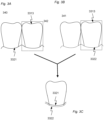
- Fig. 3 illustrates a non-claimed embodiment for detecting gingival retraction at the patient's left central incisor 3313.
- This tooth is segmented in both the first 340 and second 341 digital 3D representation of the set of teeth showing the two central incisors in the lower jaw and the gingival boundary 332 as seen inn Figs. 3A and 3B .
- the change in the position of the gingival boundary is so small that when the two digital 3D representations are seen separately the change is hardly visible.
- a section having topography data relating to both the tooth surface and the gingiva is selected and the two sections are locally aligned based on the tooth topography data. The local alignment can be performed using iterative closest point algorithm.
- Fig. 4A shows cross sections of a first 3D tooth model 451 and a second 3D tooth model 451 segmented from a first and a second digital 3D representation, respectively.
- the first digital 3D representation can for example represent the patient's teeth at the onset of an orthodontic treatment and the second digital 3D representation at some point during the treatment.
- a transformation T which aligns the first 3D tooth model with the second 3D tooth model is determined and applied to the first 3D tooth model to provide that the two tooth models are locally aligned as illustrated in Fig. 4B .
- Fig. 4C three portions 4551, 4561 and 4571 are selected on the first 3D tooth model 451.
- first and second 3D tooth models are locally aligned the anatomically corresponding portions 4552, 4562 and 4572 can readily and precisely be identified on the second 3D tooth model 451.
- first and second digital 3D representations are global aligned based e.g. on the other teeth of the same quadrant or same arch. The anatomical correct distance between the marked regions can then be determined and based on these distances a measure for the movement of the tooth between the first and second point in time can be derived.
- the portions on the first 3D tooth model can be selected by an operator or by the computer program product when this is configured for detecting appropriate portions, such as characteristic portions on the cusp.
- the selected portion can also be the entire tooth surface such that a distance map is derived showing the movement for the entire surface.
- Fig. 5 shows a schematic of a system.
- the system 570 has a computer device 571 with a data processing unit in the form of a microprocessor 572 and a non-transitory computer readable medium 573 encoded with a computer program product providing a digital tool for determining the movement of teeth e.g. during an orthodontic treatment.
- the system further has a visual display unit 576, a computer keyboard 574 and a computer mouse 575 for entering data and activating virtual buttons of a user interface visualized on the visual display unit 576.
- the visual display unit 576 can e.g. be a computer screen.
- the computer device 571 is capable of storing obtained digital 3D representations of the patient's teeth in the computer readable medium 573 and loading these into the microprocessor 572 for processing.
- the digital 3D representations can be obtained from a 3D color scanner 577, such as the 3Shape TRIOS 3 intra-oral scanner, which is capable of recording a digital 3D representation containing both geometrical data and color data for the teeth.
- the digital 3D representation can also include diagnostic data, such fluorescence data obtained using an intra-oral scanner.
- the computer readable medium 573 can further store computer implemented algorithms for segmenting a digital 3D representation to create digital 3D models of the individual teeth and for selecting regions on the surface for a local alignment.
- digital 3D models for the same tooth is created from different digital 3D representations, such as digital 3D representations recorded at different points in time
- the digital 3D models can be locally aligned using e.g. Iterative Closest Point algorithms (ICP) for minimizing the distance between the surfaces of the digital 3D representations.
- ICP Iterative Closest Point algorithms
- the digital 3D representations of the patient's entire set of teeth or sections thereof can be globally aligned also using such ICP algorithms.
- the tooth movement When the tooth movement has been determined it can be visualized to the operator in the visual display unit 576 e.g. as a distance map or using a cross sectional view of the 3D tooth models or the digital 3D representations.
- the digital 3D models of the individual teeth can be stored on the computer readable medium and be re-used at the next visit for the identification of individual teeth in a digital 3D representation recorded at the next visit.
- a claim may refer to any of the preceding claims, and "any” is understood to mean “any one or more” of the preceding claims.
- the features of the non-claimed and claimed parts of the method described above and in the following are implemented in software and carried out on a data processing system or other processing means caused by the execution of computer-executable instructions.
- the instructions may be program code means loaded in a memory, such as a RAM, from a storage medium or from another computer via a computer network.
- the described features may be implemented by hardwired circuitry instead of software or in combination with software.
Landscapes
- Health & Medical Sciences (AREA)
- Life Sciences & Earth Sciences (AREA)
- Dentistry (AREA)
- Oral & Maxillofacial Surgery (AREA)
- Animal Behavior & Ethology (AREA)
- General Health & Medical Sciences (AREA)
- Public Health (AREA)
- Veterinary Medicine (AREA)
- Biomedical Technology (AREA)
- Engineering & Computer Science (AREA)
- Biophysics (AREA)
- Epidemiology (AREA)
- Physics & Mathematics (AREA)
- Molecular Biology (AREA)
- Surgery (AREA)
- Medical Informatics (AREA)
- Heart & Thoracic Surgery (AREA)
- Pathology (AREA)
- Physical Education & Sports Medicine (AREA)
- Orthopedic Medicine & Surgery (AREA)
- Rheumatology (AREA)
- Optics & Photonics (AREA)
- Physiology (AREA)
- Audiology, Speech & Language Pathology (AREA)
- Dental Tools And Instruments Or Auxiliary Dental Instruments (AREA)
- Measuring And Recording Apparatus For Diagnosis (AREA)
Priority Applications (1)
| Application Number | Priority Date | Filing Date | Title |
|---|---|---|---|
| EP24195002.1A EP4437943B1 (en) | 2016-02-24 | 2017-02-24 | Detecting and monitoring development of a dental condition |
Applications Claiming Priority (3)
| Application Number | Priority Date | Filing Date | Title |
|---|---|---|---|
| DKPA201670103 | 2016-02-24 | ||
| PCT/EP2017/054296 WO2017144647A1 (en) | 2016-02-24 | 2017-02-24 | Detecting and monitoring development of a dental condition |
| EP17707016.6A EP3419554B1 (en) | 2016-02-24 | 2017-02-24 | Detecting and monitoring development of a dental condition |
Related Parent Applications (1)
| Application Number | Title | Priority Date | Filing Date |
|---|---|---|---|
| EP17707016.6A Division EP3419554B1 (en) | 2016-02-24 | 2017-02-24 | Detecting and monitoring development of a dental condition |
Related Child Applications (2)
| Application Number | Title | Priority Date | Filing Date |
|---|---|---|---|
| EP24195002.1A Division EP4437943B1 (en) | 2016-02-24 | 2017-02-24 | Detecting and monitoring development of a dental condition |
| EP24195002.1A Division-Into EP4437943B1 (en) | 2016-02-24 | 2017-02-24 | Detecting and monitoring development of a dental condition |
Publications (2)
| Publication Number | Publication Date |
|---|---|
| EP3949896A1 EP3949896A1 (en) | 2022-02-09 |
| EP3949896B1 true EP3949896B1 (en) | 2024-09-25 |
Family
ID=58159081
Family Applications (3)
| Application Number | Title | Priority Date | Filing Date |
|---|---|---|---|
| EP21175612.7A Active EP3949896B1 (en) | 2016-02-24 | 2017-02-24 | Detecting and monitoring development of a dental condition |
| EP24195002.1A Active EP4437943B1 (en) | 2016-02-24 | 2017-02-24 | Detecting and monitoring development of a dental condition |
| EP17707016.6A Active EP3419554B1 (en) | 2016-02-24 | 2017-02-24 | Detecting and monitoring development of a dental condition |
Family Applications After (2)
| Application Number | Title | Priority Date | Filing Date |
|---|---|---|---|
| EP24195002.1A Active EP4437943B1 (en) | 2016-02-24 | 2017-02-24 | Detecting and monitoring development of a dental condition |
| EP17707016.6A Active EP3419554B1 (en) | 2016-02-24 | 2017-02-24 | Detecting and monitoring development of a dental condition |
Country Status (8)
| Country | Link |
|---|---|
| US (3) | US10835361B2 (enExample) |
| EP (3) | EP3949896B1 (enExample) |
| JP (1) | JP2019506243A (enExample) |
| KR (1) | KR102844394B1 (enExample) |
| CN (1) | CN109069237B (enExample) |
| BR (1) | BR112018017121A2 (enExample) |
| ES (1) | ES2884167T3 (enExample) |
| WO (1) | WO2017144647A1 (enExample) |
Families Citing this family (23)
| Publication number | Priority date | Publication date | Assignee | Title |
|---|---|---|---|---|
| CA2763826C (en) | 2009-06-17 | 2020-04-07 | 3Shape A/S | Focus scanning apparatus |
| WO2015118120A1 (en) | 2014-02-07 | 2015-08-13 | 3Shape A/S | Detecting tooth shade |
| EP3465495A1 (en) * | 2016-05-30 | 2019-04-10 | 3Shape A/S | Predicting the development of a dental condition |
| US10136972B2 (en) * | 2016-06-30 | 2018-11-27 | Align Technology, Inc. | Historical scan reference for intraoral scans |
| US20200022790A1 (en) * | 2016-10-10 | 2020-01-23 | 3Shape A/S | Common Placement Support for Artificial Teeth |
| US10052180B1 (en) | 2016-12-29 | 2018-08-21 | David D. Richter | Virtual dental articulator and system |
| US10499793B2 (en) | 2017-02-17 | 2019-12-10 | Align Technology, Inc. | Longitudinal analysis and visualization under limited accuracy system |
| WO2020182880A1 (en) | 2019-03-11 | 2020-09-17 | 3Shape A/S | System and method for generating digital three-dimensional dental models |
| EP3709305A1 (en) | 2019-03-11 | 2020-09-16 | 3Shape A/S | Method for graphically presenting a plurality of scans |
| CN113412413B (zh) * | 2019-04-25 | 2024-11-01 | 普罗费塞公司 | 用于对振动进行成像和感测的系统和方法 |
| US10849723B1 (en) | 2019-05-07 | 2020-12-01 | Sdc U.S. Smilepay Spv | Scanning device |
| DE102019112800A1 (de) * | 2019-05-15 | 2020-11-19 | K Line Europe Gmbh | Verfahren und Vorrichtung zur Überwachung einer orthodontischen Maßnahme |
| US11622843B2 (en) | 2019-06-25 | 2023-04-11 | James R. Glidewell Dental Ceramics, Inc. | Processing digital dental impression |
| CN113491532B (zh) * | 2020-03-20 | 2024-03-22 | 苏州佳世达光电有限公司 | 口扫机系统及口扫机的操作方法 |
| KR102590775B1 (ko) * | 2021-03-11 | 2023-10-19 | 주식회사 메디트 | 데이터 처리 방법 |
| US12329597B2 (en) * | 2021-04-09 | 2025-06-17 | Align Technology, Inc. | Capturing true bite and occlusion contacts |
| EP4080454B1 (en) * | 2021-04-23 | 2024-08-07 | Ivoclar Vivadent AG | Method for exporting a three-dimensional esthetic dental design model from an augmented reality application to a computer-aided design application |
| US12387338B2 (en) * | 2021-06-15 | 2025-08-12 | Imagoworks Inc. | Method for automated tooth segmentation of three dimensional scan data using deep learning and computer readable medium having program for performing the method |
| EP4120182B1 (de) * | 2021-07-12 | 2025-08-27 | DENTSPLY SIRONA Inc. | Selbstkalibrierendes dentales dvt unterstützt durch maschinelles lernen |
| US20240362751A1 (en) * | 2021-07-15 | 2024-10-31 | Sony Semiconductor Solutions Corporation | Image processing device, image processing method and light scanner system |
| KR102702505B1 (ko) * | 2021-08-13 | 2024-09-04 | 오스템임플란트 주식회사 | 치과 교정치료 계획 수립 방법 및 그 장치 |
| CN116777818B (zh) * | 2022-03-11 | 2024-05-24 | 广州星际悦动股份有限公司 | 口腔清洁方案确定方法、装置、电子设备及存储介质 |
| US20240398520A1 (en) * | 2023-05-31 | 2024-12-05 | Perceptive Technologies, Inc. | Automated laser metrology for dental surgery |
Family Cites Families (73)
| Publication number | Priority date | Publication date | Assignee | Title |
|---|---|---|---|---|
| DE2514930A1 (de) | 1975-04-05 | 1976-10-14 | Opto Produkte Ag | Verfahren zur optischen ermittlung und zum vergleich von formen und lagen von objekten |
| FR2639211A1 (fr) | 1988-11-18 | 1990-05-25 | Hennson Int | Procede de correlation des saisies tridimensionnelles d'organes humains et dispositif pour sa mise en oeuvre |
| IL120867A0 (en) | 1997-05-20 | 1997-09-30 | Cadent Ltd | Computer user interface for orthodontic use |
| US5879158A (en) | 1997-05-20 | 1999-03-09 | Doyle; Walter A. | Orthodontic bracketing system and method therefor |
| AU744385B2 (en) | 1997-06-20 | 2002-02-21 | Align Technology, Inc. | Method and system for incrementally moving teeth |
| US6450807B1 (en) | 1997-06-20 | 2002-09-17 | Align Technology, Inc. | System and method for positioning teeth |
| US5975893A (en) | 1997-06-20 | 1999-11-02 | Align Technology, Inc. | Method and system for incrementally moving teeth |
| US6409504B1 (en) | 1997-06-20 | 2002-06-25 | Align Technology, Inc. | Manipulating a digital dentition model to form models of individual dentition components |
| EP1119309B1 (en) | 1998-10-08 | 2016-06-01 | Align Technology, Inc. | Computer automated development of an orthodontic treatment plan and appliance |
| US6514074B1 (en) | 1999-05-14 | 2003-02-04 | Align Technology, Inc. | Digitally modeling the deformation of gingival |
| US6406292B1 (en) | 1999-05-13 | 2002-06-18 | Align Technology, Inc. | System for determining final position of teeth |
| US6512994B1 (en) | 1999-11-30 | 2003-01-28 | Orametrix, Inc. | Method and apparatus for producing a three-dimensional digital model of an orthodontic patient |
| US6602070B2 (en) | 1999-05-13 | 2003-08-05 | Align Technology, Inc. | Systems and methods for dental treatment planning |
| EP1191896B1 (en) | 1999-05-13 | 2008-06-25 | Align Technology, Inc. | System for determining final position of teeth |
| US6318994B1 (en) | 1999-05-13 | 2001-11-20 | Align Technology, Inc | Tooth path treatment plan |
| US9833167B2 (en) | 1999-05-18 | 2017-12-05 | Mediguide Ltd. | Method and system for superimposing virtual anatomical landmarks on an image |
| US6819318B1 (en) | 1999-07-23 | 2004-11-16 | Z. Jason Geng | Method and apparatus for modeling via a three-dimensional image mosaic system |
| US7160110B2 (en) | 1999-11-30 | 2007-01-09 | Orametrix, Inc. | Three-dimensional occlusal and interproximal contact detection and display using virtual tooth models |
| US7296996B2 (en) | 1999-11-30 | 2007-11-20 | Orametrix, Inc. | Virtual bracket placement and evaluation |
| US6632089B2 (en) | 1999-11-30 | 2003-10-14 | Orametrix, Inc. | Orthodontic treatment planning with user-specified simulation of tooth movement |
| US6648640B2 (en) | 1999-11-30 | 2003-11-18 | Ora Metrix, Inc. | Interactive orthodontic care system based on intra-oral scanning of teeth |
| US7013191B2 (en) | 1999-11-30 | 2006-03-14 | Orametrix, Inc. | Interactive orthodontic care system based on intra-oral scanning of teeth |
| US6350120B1 (en) | 1999-11-30 | 2002-02-26 | Orametrix, Inc. | Method and apparatus for designing an orthodontic apparatus to provide tooth movement |
| US7373286B2 (en) | 2000-02-17 | 2008-05-13 | Align Technology, Inc. | Efficient data representation of teeth model |
| US6463344B1 (en) | 2000-02-17 | 2002-10-08 | Align Technology, Inc. | Efficient data representation of teeth model |
| US6711432B1 (en) | 2000-10-23 | 2004-03-23 | Carnegie Mellon University | Computer-aided orthopedic surgery |
| US6971873B2 (en) | 2000-04-19 | 2005-12-06 | Orametrix, Inc. | Virtual bracket library and uses thereof in orthodontic treatment planning |
| AU2001255638A1 (en) | 2000-04-25 | 2001-11-07 | Align Technology, Inc. | Systems and methods for dental treatment planning |
| US6621491B1 (en) | 2000-04-27 | 2003-09-16 | Align Technology, Inc. | Systems and methods for integrating 3D diagnostic data |
| US6947038B1 (en) | 2000-04-27 | 2005-09-20 | Align Technology, Inc. | Systems and methods for generating an appliance with tie points |
| US7027642B2 (en) | 2000-04-28 | 2006-04-11 | Orametrix, Inc. | Methods for registration of three-dimensional frames to create three-dimensional virtual models of objects |
| US7736147B2 (en) | 2000-10-30 | 2010-06-15 | Align Technology, Inc. | Systems and methods for bite-setting teeth models |
| US20020064759A1 (en) | 2000-11-30 | 2002-05-30 | Durbin Duane Milford | Method and system for viewing, altering and archiving digital models of dental structures and computer integrated manufacturing of physical models of dental structures |
| US20150305830A1 (en) | 2001-04-13 | 2015-10-29 | Orametrix, Inc. | Tooth positioning appliance and uses thereof |
| US8021147B2 (en) | 2001-04-13 | 2011-09-20 | Orametrix, Inc. | Method and system for comprehensive evaluation of orthodontic care using unified workstation |
| US7717708B2 (en) | 2001-04-13 | 2010-05-18 | Orametrix, Inc. | Method and system for integrated orthodontic treatment planning using unified workstation |
| US7156655B2 (en) | 2001-04-13 | 2007-01-02 | Orametrix, Inc. | Method and system for comprehensive evaluation of orthodontic treatment using unified workstation |
| US7343305B2 (en) | 2001-05-03 | 2008-03-11 | University Of Florida Research Foundation, Inc. | Method and system for recording carious lesions |
| US6739870B2 (en) | 2001-09-26 | 2004-05-25 | 3M Innovative Properties Company | Use of finite element analysis for orthodontic mechanics and appliance selection |
| US7292716B2 (en) | 2001-10-31 | 2007-11-06 | Imagnosis Inc. | Medical simulation apparatus and method for controlling 3-dimensional image display in the medical simulation apparatus |
| US7347686B2 (en) | 2002-01-22 | 2008-03-25 | Geodigm Corporation | Method and apparatus using a scanned image for marking bracket locations |
| US7155373B2 (en) | 2002-02-22 | 2006-12-26 | 3M Innovative Properties Company | Selection of orthodontic brackets |
| US6979196B2 (en) | 2002-06-21 | 2005-12-27 | Align Technology, Inc. | Systems and methods for automated bite-setting of tooth models |
| US7077647B2 (en) | 2002-08-22 | 2006-07-18 | Align Technology, Inc. | Systems and methods for treatment analysis by teeth matching |
| US7156661B2 (en) * | 2002-08-22 | 2007-01-02 | Align Technology, Inc. | Systems and methods for treatment analysis by teeth matching |
| US20040122306A1 (en) * | 2002-12-20 | 2004-06-24 | Eastman Kodak Company | Intra-oral imaging system and method for dimensional measurement below the gumline |
| US7955076B2 (en) * | 2003-04-03 | 2011-06-07 | Kao Corporation | Carious tooth detection device |
| JP4798712B2 (ja) * | 2004-09-24 | 2011-10-19 | 株式会社日立メディコ | 医用画像表示装置及び方法並びにプログラム |
| US7623942B2 (en) * | 2005-01-06 | 2009-11-24 | Touchstone C Alex | Method and apparatus for selecting non-opacious dental materials |
| KR100854634B1 (ko) | 2006-11-29 | 2008-08-27 | 강릉대학교산학협력단 | 3차원 역공학 기술을 이용한 치아 이동 자동측정 방법 |
| EP4066777B1 (en) * | 2007-10-12 | 2024-06-05 | Align Technology, Inc. | Prosthodontic and orthodontic apparatus and methods |
| US10307221B2 (en) * | 2007-12-21 | 2019-06-04 | 3M Innovative Properties Company | Orthodontic treatment monitoring based on reduced images |
| US20100324875A1 (en) | 2008-02-12 | 2010-12-23 | Kalili Thomas K | Process for orthodontic, implant and dental prosthetic fabrication using 3d geometric mesh teeth manipulation process |
| US8768036B2 (en) | 2008-05-23 | 2014-07-01 | Eyeic, Inc. | System and method for detecting and tracking change in dental X-rays and dental images |
| US20120249551A1 (en) * | 2009-06-11 | 2012-10-04 | Synarc Inc. | alignment of shapes of body parts from images |
| US20110110575A1 (en) * | 2009-11-11 | 2011-05-12 | Thiagarajar College Of Engineering | Dental caries detector |
| US8244028B2 (en) | 2010-04-30 | 2012-08-14 | Align Technology, Inc. | Virtual cephalometric imaging |
| US8520918B2 (en) * | 2010-05-02 | 2013-08-27 | Kelce S Wilson | Medical diagnostic image change highlighter |
| CN102985894B (zh) | 2010-07-15 | 2017-02-08 | 惠普发展公司,有限责任合伙企业 | 第一响应和第二响应 |
| WO2012083960A1 (en) | 2010-12-22 | 2012-06-28 | 3Shape A/S | System and method for scanning objects being modified |
| US8416984B2 (en) | 2011-01-20 | 2013-04-09 | Carestream Health, Inc. | Automatic tooth charting using digital images |
| GB201115265D0 (en) * | 2011-09-05 | 2011-10-19 | Materialise Dental Nv | A method and system for 3d root canal treatment planning |
| US10617489B2 (en) | 2012-12-19 | 2020-04-14 | Align Technology, Inc. | Creating a digital dental model of a patient's teeth using interproximal information |
| CN104955418B (zh) * | 2012-12-24 | 2017-10-13 | 牙医技术G·P·L·有限公司 | 用在龈下测量的装置及方法 |
| US9158889B2 (en) | 2013-04-26 | 2015-10-13 | Oral4D Systems Ltd. | Electronic dental charting |
| US9510757B2 (en) * | 2014-05-07 | 2016-12-06 | Align Technology, Inc. | Identification of areas of interest during intraoral scans |
| US9626462B2 (en) * | 2014-07-01 | 2017-04-18 | 3M Innovative Properties Company | Detecting tooth wear using intra-oral 3D scans |
| US11147652B2 (en) * | 2014-11-13 | 2021-10-19 | Align Technology, Inc. | Method for tracking, predicting, and proactively correcting malocclusion and related issues |
| US10074178B2 (en) * | 2015-01-30 | 2018-09-11 | Dental Imaging Technologies Corporation | Intra-oral image acquisition alignment |
| US9770217B2 (en) * | 2015-01-30 | 2017-09-26 | Dental Imaging Technologies Corporation | Dental variation tracking and prediction |
| US9547903B2 (en) * | 2015-04-16 | 2017-01-17 | Carestream Health, Inc. | Method for quantifying caries |
| KR102492183B1 (ko) * | 2016-01-19 | 2023-01-26 | 삼성전자주식회사 | 사용자단말기 및 그 제어방법 |
| US10410430B2 (en) * | 2016-02-12 | 2019-09-10 | 3M Innovative Properties Company | Synchronization and animation of views showing digital 3D models of teeth |
-
2017
- 2017-02-24 JP JP2018544318A patent/JP2019506243A/ja active Pending
- 2017-02-24 ES ES17707016T patent/ES2884167T3/es active Active
- 2017-02-24 BR BR112018017121A patent/BR112018017121A2/pt not_active Application Discontinuation
- 2017-02-24 KR KR1020187027945A patent/KR102844394B1/ko active Active
- 2017-02-24 EP EP21175612.7A patent/EP3949896B1/en active Active
- 2017-02-24 US US16/079,256 patent/US10835361B2/en active Active
- 2017-02-24 CN CN201780025362.8A patent/CN109069237B/zh active Active
- 2017-02-24 WO PCT/EP2017/054296 patent/WO2017144647A1/en not_active Ceased
- 2017-02-24 EP EP24195002.1A patent/EP4437943B1/en active Active
- 2017-02-24 EP EP17707016.6A patent/EP3419554B1/en active Active
-
2020
- 2020-10-08 US US17/066,068 patent/US20210045859A1/en not_active Abandoned
-
2022
- 2022-06-01 US US17/829,580 patent/US20220287811A1/en active Pending
Also Published As
| Publication number | Publication date |
|---|---|
| KR102844394B1 (ko) | 2025-08-07 |
| US20220287811A1 (en) | 2022-09-15 |
| WO2017144647A1 (en) | 2017-08-31 |
| KR20180119630A (ko) | 2018-11-02 |
| JP2019506243A (ja) | 2019-03-07 |
| CN109069237A (zh) | 2018-12-21 |
| EP3949896A1 (en) | 2022-02-09 |
| ES2884167T3 (es) | 2021-12-10 |
| US10835361B2 (en) | 2020-11-17 |
| US20210045859A1 (en) | 2021-02-18 |
| EP3419554B1 (en) | 2021-05-26 |
| EP4437943A2 (en) | 2024-10-02 |
| CN109069237B (zh) | 2021-12-28 |
| EP4437943B1 (en) | 2026-04-15 |
| US20190060042A1 (en) | 2019-02-28 |
| EP4437943A3 (en) | 2025-04-16 |
| EP3419554A1 (en) | 2019-01-02 |
| BR112018017121A2 (pt) | 2018-12-26 |
Similar Documents
| Publication | Publication Date | Title |
|---|---|---|
| EP3949896B1 (en) | Detecting and monitoring development of a dental condition | |
| US11931128B2 (en) | Deriving tooth condition information for populating digital dental charts | |
| US11707347B2 (en) | Detecting tooth shade | |
| US11918438B2 (en) | Predicting the development of a dental condition | |
| KR102932237B1 (ko) | 디지털 3 차원 치과적 모델들을 생성하기 위한 시스템 및 방법 |
Legal Events
| Date | Code | Title | Description |
|---|---|---|---|
| PUAI | Public reference made under article 153(3) epc to a published international application that has entered the european phase |
Free format text: ORIGINAL CODE: 0009012 |
|
| STAA | Information on the status of an ep patent application or granted ep patent |
Free format text: STATUS: THE APPLICATION HAS BEEN PUBLISHED |
|
| AC | Divisional application: reference to earlier application |
Ref document number: 3419554 Country of ref document: EP Kind code of ref document: P |
|
| AK | Designated contracting states |
Kind code of ref document: A1 Designated state(s): AL AT BE BG CH CY CZ DE DK EE ES FI FR GB GR HR HU IE IS IT LI LT LU LV MC MK MT NL NO PL PT RO RS SE SI SK SM TR |
|
| STAA | Information on the status of an ep patent application or granted ep patent |
Free format text: STATUS: REQUEST FOR EXAMINATION WAS MADE |
|
| 17P | Request for examination filed |
Effective date: 20220804 |
|
| RBV | Designated contracting states (corrected) |
Designated state(s): AL AT BE BG CH CY CZ DE DK EE ES FI FR GB GR HR HU IE IS IT LI LT LU LV MC MK MT NL NO PL PT RO RS SE SI SK SM TR |
|
| STAA | Information on the status of an ep patent application or granted ep patent |
Free format text: STATUS: EXAMINATION IS IN PROGRESS |
|
| 17Q | First examination report despatched |
Effective date: 20230307 |
|
| GRAP | Despatch of communication of intention to grant a patent |
Free format text: ORIGINAL CODE: EPIDOSNIGR1 |
|
| STAA | Information on the status of an ep patent application or granted ep patent |
Free format text: STATUS: GRANT OF PATENT IS INTENDED |
|
| INTG | Intention to grant announced |
Effective date: 20231120 |
|
| GRAJ | Information related to disapproval of communication of intention to grant by the applicant or resumption of examination proceedings by the epo deleted |
Free format text: ORIGINAL CODE: EPIDOSDIGR1 |
|
| STAA | Information on the status of an ep patent application or granted ep patent |
Free format text: STATUS: EXAMINATION IS IN PROGRESS |
|
| GRAP | Despatch of communication of intention to grant a patent |
Free format text: ORIGINAL CODE: EPIDOSNIGR1 |
|
| STAA | Information on the status of an ep patent application or granted ep patent |
Free format text: STATUS: GRANT OF PATENT IS INTENDED |
|
| INTC | Intention to grant announced (deleted) | ||
| INTG | Intention to grant announced |
Effective date: 20240417 |
|
| GRAS | Grant fee paid |
Free format text: ORIGINAL CODE: EPIDOSNIGR3 |
|
| GRAA | (expected) grant |
Free format text: ORIGINAL CODE: 0009210 |
|
| STAA | Information on the status of an ep patent application or granted ep patent |
Free format text: STATUS: THE PATENT HAS BEEN GRANTED |
|
| AC | Divisional application: reference to earlier application |
Ref document number: 3419554 Country of ref document: EP Kind code of ref document: P |
|
| AK | Designated contracting states |
Kind code of ref document: B1 Designated state(s): AL AT BE BG CH CY CZ DE DK EE ES FI FR GB GR HR HU IE IS IT LI LT LU LV MC MK MT NL NO PL PT RO RS SE SI SK SM TR |
|
| P01 | Opt-out of the competence of the unified patent court (upc) registered |
Free format text: CASE NUMBER: APP_47242/2024 Effective date: 20240815 |
|
| REG | Reference to a national code |
Ref country code: GB Ref legal event code: FG4D |
|
| REG | Reference to a national code |
Ref country code: CH Ref legal event code: EP |
|
| REG | Reference to a national code |
Ref country code: DE Ref legal event code: R096 Ref document number: 602017085164 Country of ref document: DE |
|
| REG | Reference to a national code |
Ref country code: IE Ref legal event code: FG4D |
|
| REG | Reference to a national code |
Ref country code: LT Ref legal event code: MG9D |
|
| PG25 | Lapsed in a contracting state [announced via postgrant information from national office to epo] |
Ref country code: NO Free format text: LAPSE BECAUSE OF FAILURE TO SUBMIT A TRANSLATION OF THE DESCRIPTION OR TO PAY THE FEE WITHIN THE PRESCRIBED TIME-LIMIT Effective date: 20241225 |
|
| PG25 | Lapsed in a contracting state [announced via postgrant information from national office to epo] |
Ref country code: GR Free format text: LAPSE BECAUSE OF FAILURE TO SUBMIT A TRANSLATION OF THE DESCRIPTION OR TO PAY THE FEE WITHIN THE PRESCRIBED TIME-LIMIT Effective date: 20241226 Ref country code: FI Free format text: LAPSE BECAUSE OF FAILURE TO SUBMIT A TRANSLATION OF THE DESCRIPTION OR TO PAY THE FEE WITHIN THE PRESCRIBED TIME-LIMIT Effective date: 20240925 |
|
| PG25 | Lapsed in a contracting state [announced via postgrant information from national office to epo] |
Ref country code: BG Free format text: LAPSE BECAUSE OF FAILURE TO SUBMIT A TRANSLATION OF THE DESCRIPTION OR TO PAY THE FEE WITHIN THE PRESCRIBED TIME-LIMIT Effective date: 20240925 |
|
| PG25 | Lapsed in a contracting state [announced via postgrant information from national office to epo] |
Ref country code: LV Free format text: LAPSE BECAUSE OF FAILURE TO SUBMIT A TRANSLATION OF THE DESCRIPTION OR TO PAY THE FEE WITHIN THE PRESCRIBED TIME-LIMIT Effective date: 20240925 |
|
| PG25 | Lapsed in a contracting state [announced via postgrant information from national office to epo] |
Ref country code: RS Free format text: LAPSE BECAUSE OF FAILURE TO SUBMIT A TRANSLATION OF THE DESCRIPTION OR TO PAY THE FEE WITHIN THE PRESCRIBED TIME-LIMIT Effective date: 20241225 |
|
| REG | Reference to a national code |
Ref country code: NL Ref legal event code: MP Effective date: 20240925 |
|
| PG25 | Lapsed in a contracting state [announced via postgrant information from national office to epo] |
Ref country code: RS Free format text: LAPSE BECAUSE OF FAILURE TO SUBMIT A TRANSLATION OF THE DESCRIPTION OR TO PAY THE FEE WITHIN THE PRESCRIBED TIME-LIMIT Effective date: 20241225 Ref country code: NO Free format text: LAPSE BECAUSE OF FAILURE TO SUBMIT A TRANSLATION OF THE DESCRIPTION OR TO PAY THE FEE WITHIN THE PRESCRIBED TIME-LIMIT Effective date: 20241225 Ref country code: LV Free format text: LAPSE BECAUSE OF FAILURE TO SUBMIT A TRANSLATION OF THE DESCRIPTION OR TO PAY THE FEE WITHIN THE PRESCRIBED TIME-LIMIT Effective date: 20240925 Ref country code: GR Free format text: LAPSE BECAUSE OF FAILURE TO SUBMIT A TRANSLATION OF THE DESCRIPTION OR TO PAY THE FEE WITHIN THE PRESCRIBED TIME-LIMIT Effective date: 20241226 Ref country code: FI Free format text: LAPSE BECAUSE OF FAILURE TO SUBMIT A TRANSLATION OF THE DESCRIPTION OR TO PAY THE FEE WITHIN THE PRESCRIBED TIME-LIMIT Effective date: 20240925 Ref country code: BG Free format text: LAPSE BECAUSE OF FAILURE TO SUBMIT A TRANSLATION OF THE DESCRIPTION OR TO PAY THE FEE WITHIN THE PRESCRIBED TIME-LIMIT Effective date: 20240925 |
|
| REG | Reference to a national code |
Ref country code: AT Ref legal event code: MK05 Ref document number: 1726039 Country of ref document: AT Kind code of ref document: T Effective date: 20240925 |
|
| PG25 | Lapsed in a contracting state [announced via postgrant information from national office to epo] |
Ref country code: NL Free format text: LAPSE BECAUSE OF FAILURE TO SUBMIT A TRANSLATION OF THE DESCRIPTION OR TO PAY THE FEE WITHIN THE PRESCRIBED TIME-LIMIT Effective date: 20240925 |
|
| PG25 | Lapsed in a contracting state [announced via postgrant information from national office to epo] |
Ref country code: PT Free format text: LAPSE BECAUSE OF FAILURE TO SUBMIT A TRANSLATION OF THE DESCRIPTION OR TO PAY THE FEE WITHIN THE PRESCRIBED TIME-LIMIT Effective date: 20250127 Ref country code: IS Free format text: LAPSE BECAUSE OF FAILURE TO SUBMIT A TRANSLATION OF THE DESCRIPTION OR TO PAY THE FEE WITHIN THE PRESCRIBED TIME-LIMIT Effective date: 20250125 |
|
| PG25 | Lapsed in a contracting state [announced via postgrant information from national office to epo] |
Ref country code: RO Free format text: LAPSE BECAUSE OF FAILURE TO SUBMIT A TRANSLATION OF THE DESCRIPTION OR TO PAY THE FEE WITHIN THE PRESCRIBED TIME-LIMIT Effective date: 20240925 Ref country code: SM Free format text: LAPSE BECAUSE OF FAILURE TO SUBMIT A TRANSLATION OF THE DESCRIPTION OR TO PAY THE FEE WITHIN THE PRESCRIBED TIME-LIMIT Effective date: 20240925 |
|
| PG25 | Lapsed in a contracting state [announced via postgrant information from national office to epo] |
Ref country code: ES Free format text: LAPSE BECAUSE OF FAILURE TO SUBMIT A TRANSLATION OF THE DESCRIPTION OR TO PAY THE FEE WITHIN THE PRESCRIBED TIME-LIMIT Effective date: 20240925 |
|
| PG25 | Lapsed in a contracting state [announced via postgrant information from national office to epo] |
Ref country code: AT Free format text: LAPSE BECAUSE OF FAILURE TO SUBMIT A TRANSLATION OF THE DESCRIPTION OR TO PAY THE FEE WITHIN THE PRESCRIBED TIME-LIMIT Effective date: 20240925 Ref country code: EE Free format text: LAPSE BECAUSE OF FAILURE TO SUBMIT A TRANSLATION OF THE DESCRIPTION OR TO PAY THE FEE WITHIN THE PRESCRIBED TIME-LIMIT Effective date: 20240925 |
|
| PG25 | Lapsed in a contracting state [announced via postgrant information from national office to epo] |
Ref country code: CZ Free format text: LAPSE BECAUSE OF FAILURE TO SUBMIT A TRANSLATION OF THE DESCRIPTION OR TO PAY THE FEE WITHIN THE PRESCRIBED TIME-LIMIT Effective date: 20240925 Ref country code: PL Free format text: LAPSE BECAUSE OF FAILURE TO SUBMIT A TRANSLATION OF THE DESCRIPTION OR TO PAY THE FEE WITHIN THE PRESCRIBED TIME-LIMIT Effective date: 20240925 |
|
| PG25 | Lapsed in a contracting state [announced via postgrant information from national office to epo] |
Ref country code: IT Free format text: LAPSE BECAUSE OF FAILURE TO SUBMIT A TRANSLATION OF THE DESCRIPTION OR TO PAY THE FEE WITHIN THE PRESCRIBED TIME-LIMIT Effective date: 20240925 Ref country code: SK Free format text: LAPSE BECAUSE OF FAILURE TO SUBMIT A TRANSLATION OF THE DESCRIPTION OR TO PAY THE FEE WITHIN THE PRESCRIBED TIME-LIMIT Effective date: 20240925 |
|
| REG | Reference to a national code |
Ref country code: DE Ref legal event code: R097 Ref document number: 602017085164 Country of ref document: DE |
|
| PG25 | Lapsed in a contracting state [announced via postgrant information from national office to epo] |
Ref country code: DK Free format text: LAPSE BECAUSE OF FAILURE TO SUBMIT A TRANSLATION OF THE DESCRIPTION OR TO PAY THE FEE WITHIN THE PRESCRIBED TIME-LIMIT Effective date: 20240925 |
|
| PLBE | No opposition filed within time limit |
Free format text: ORIGINAL CODE: 0009261 |
|
| STAA | Information on the status of an ep patent application or granted ep patent |
Free format text: STATUS: NO OPPOSITION FILED WITHIN TIME LIMIT |
|
| 26N | No opposition filed |
Effective date: 20250626 |
|
| PG25 | Lapsed in a contracting state [announced via postgrant information from national office to epo] |
Ref country code: SE Free format text: LAPSE BECAUSE OF FAILURE TO SUBMIT A TRANSLATION OF THE DESCRIPTION OR TO PAY THE FEE WITHIN THE PRESCRIBED TIME-LIMIT Effective date: 20240925 |
|
| PG25 | Lapsed in a contracting state [announced via postgrant information from national office to epo] |
Ref country code: MC Free format text: LAPSE BECAUSE OF FAILURE TO SUBMIT A TRANSLATION OF THE DESCRIPTION OR TO PAY THE FEE WITHIN THE PRESCRIBED TIME-LIMIT Effective date: 20240925 |
|
| REG | Reference to a national code |
Ref country code: CH Ref legal event code: PL |
|
| PG25 | Lapsed in a contracting state [announced via postgrant information from national office to epo] |
Ref country code: LU Free format text: LAPSE BECAUSE OF NON-PAYMENT OF DUE FEES Effective date: 20250224 |
|
| PG25 | Lapsed in a contracting state [announced via postgrant information from national office to epo] |
Ref country code: CH Free format text: LAPSE BECAUSE OF NON-PAYMENT OF DUE FEES Effective date: 20250228 |
|
| REG | Reference to a national code |
Ref country code: BE Ref legal event code: MM Effective date: 20250228 |
|
| PG25 | Lapsed in a contracting state [announced via postgrant information from national office to epo] |
Ref country code: HR Free format text: LAPSE BECAUSE OF FAILURE TO SUBMIT A TRANSLATION OF THE DESCRIPTION OR TO PAY THE FEE WITHIN THE PRESCRIBED TIME-LIMIT Effective date: 20240925 |
|
| PG25 | Lapsed in a contracting state [announced via postgrant information from national office to epo] |
Ref country code: BE Free format text: LAPSE BECAUSE OF NON-PAYMENT OF DUE FEES Effective date: 20250228 |
|
| PG25 | Lapsed in a contracting state [announced via postgrant information from national office to epo] |
Ref country code: IE Free format text: LAPSE BECAUSE OF NON-PAYMENT OF DUE FEES Effective date: 20250224 |
|
| PGFP | Annual fee paid to national office [announced via postgrant information from national office to epo] |
Ref country code: GB Payment date: 20260220 Year of fee payment: 10 |
|
| PGFP | Annual fee paid to national office [announced via postgrant information from national office to epo] |
Ref country code: DE Payment date: 20260218 Year of fee payment: 10 |
|
| PGFP | Annual fee paid to national office [announced via postgrant information from national office to epo] |
Ref country code: FR Payment date: 20260219 Year of fee payment: 10 |